1
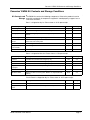
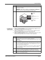
EMSA "Gel Shift" Kit
User Manual
AY1XXX
P/N 13009 Rev. C 090508
DRAFT September 5, 2008 11:40 am
EMSATtile.fm
Panomics, Inc.
EMSA "Gel Shift" Kit User Manual
Copyright
© Copyright 2008, Panomics, Inc. All rights reserved.
Trademarks
Procarta is a registered trademark of Panomics, Inc.
Mini-PROTEAN and Mini Trans-Blot are registered trademarks of Bio-Rad Laboratories, Inc, FluorChem is registered trademark of Alpha Innotech,
Hyperfilm and ECL are trademarks of GE Healthcare Companies, Biodyne is a registered trademark of Pall Corporation, Whatman is a registered
trademark of Whatman International LTD.
Citing Panomics in Publications
When describing a procedure for publication using this product, we would appreciate it if you would refer to it as the EMSA Kit from Panomics. If a
paper cites our EMSA product and is published in a research journal, the lead author(s) may receive a travel stipend for use at a technology
conference or tradeshow by sending a copy of the paper to our technical support group at [email protected] or via fax at (510) 818-2610.
Disclaimer
Panomics, Inc. reserves the right to change its products and services at any time to incorporate technological developments. This manual is subject
to change without notice.
Although this manual has been prepared with every precaution to ensure accuracy, Panomics, Inc. assumes no liability for any errors or omissions, nor
for any damages resulting from the application or use of this information.
DRAFT September 5, 2008 11:40 am
EMSATtile.fm
About the User Manual
About the User Manual
Who Should Read Anyone that has purchased an EMSA “Gel Shift” Kit from Panomics to measure the
this Manual binding activity of a specific TF from nuclear extracts.
This manual provides the following:
♦
Kit contents and storage conditions
♦
Assay procedures
♦
Troubleshooting
Safety Warnings CAUTION All chemicals should be considered potentially hazardous. We recommend that
and Precautions this product and its components be handled by those trained in laboratory techniques and be
used according to the principles of good laboratory practice.
Note This product is intended for research use only. It is not for diagnosis of disease in
humans or animals.
For More For information about the products mentioned in this manual, visit our website at
Information www.panomics.com.
EMSA "Gel Shift" User Manual
Page 3
About the EMSA “Gel Shift” Kit
About the EMSA “Gel Shift” Kit
Introduction to Panomics’ Electrophoretic-Mobility Shift Assay (EMSA) Kits are useful tools for
Panomics’ EMSA identifying proteins that interact with DNA. This rapid technique is based on the
Kits separation of free DNA from protein/DNA complexes due to the differences in their
electrophoretic mobility in native (non-denaturing) polyacrylamide gels. When a
protein binds specifically to a labeled dsDNA sequence, it migrates slower than
non-bound dsDNA in a polyacrylamide gel, thus resulting in discrete bands
corresponding to the individual protein/DNA complex. A typical EMSA experiment is
performed by incubating a biotin-labeled transcription factor (TF) Probe with treated
and untreated nuclear extracts. The protein/DNA complexes are separated on a
non-denaturing polyacrylamide gel. The gel is transferred to a nylon membrane and
detected using strepatvidin-HRP and a chemiluminescent substrate. The shifted
bands corresponding to the protein/DNA complexes are visualized relative to the
unbound dsDNA. The bands are visualized after exposure to film or
chemiluminescent-imaging system (Figure 1).
For more specific binding, you may wish to add unlabeled specific dsDNA probe
(cold probe; provided) to the protein/DNA reaction mixture which competes with the
labeled dsDNA probe (biotin-labeled probe) for binding to the protein. This causes
the labeled probe to migrate to the bottom of the gel and reduces the intensity of the
shifted band.
Panomics’ EMSA Kits are suitable for validation of results obtained using Procarta®
Transcription Factor Assay, Protein/DNA Arrays (Cat. #s MA1010 to MA1015) or to
validate binding activity of a specific transcription factor (protein) to DNA. Enough
reagents are provided to perform 25 binding reactions. Panomics' TF EMSA Kits
measure the activity of specific transcription factors in nuclear extracts. The assay is
highly specific, precise, and requires 4-8 µg of protein/well. Panomics currently offers
more than 400 EMSA assays. For a complete list of available EMSA Kits, visit
www.panomics.com.
1 2 3 4
Figure 1: Example EMSA image. Lane 1: Labeled EMSA Probe only with no sample,
Lane 2: Labeled EMSA Probe with untreated sample. Lane 3: Labeled EMSA Probe
with treated sample. Lane 4: Treated sample with cold and labeled EMSA probes.
Page 4
EMSA "Gel Shift" User Manual
Panomics’ EMSA Kit Contents and Storage Conditions
Panomics’ EMSA Kit Contents and Storage Conditions
Kit Contents and The EMSA Kit contains the following components. Refer to the product insert for
Storage quantities and details of components supplied. If stored properly, reagents have a
shelf-life of 6 months.
Box 1 is shipped on dry ice. Please store at -20°C upon receipt
Box 1 Components
Description
Volume
Storage
5X Binding Buffer
Poly d(I-C)
Aqueous buffered solution for TF binding
50 µL
–20°C
Used in TF binding reactions to act as competitor to TF Probe
25 µL
–20°C
Loading Dye
Dye for loading samples and monitoring electrophoresis and
transfer
60 µL
–20°C
2X Blocking Buffer
Aqueous buffer for blocking during detection
50 mL
–20°C
Control Nuclear
Extract
Nuclear Extract prepared from HeLa cell line for use with control
probe and cold control probe
5 µL
–20°C
Control Probe
Control reagent
10 µL
–20°C
Cold (unlabeled)
Control Probe
Control reagent
10 µL
–20°C
Distilled H20
Distilled, purified water
500 µL
–20°C
Box 2 is shipped on blue ice. Please store at -4°C upon receipt.
Box 2 Components
Description
Volume
Storage
Solution I
Chemiluminescent detection reagent (1 mL)
1 mL
4°C
Solution II
Chemiluminescent detection reagent (1 mL)
1 mL
4°C
Solution III
Chemiluminescent detection reagent (8 mL)
8 mL
4°C
Streptavdin-HRP
Conjugate
Used for detection of biotinylaled oligo on membrane (100 µL)
100 µL
4°C
10X Detection Buffer
Aqueous buffer for detection step (10 mL)
10 mL
4°C
10X Wash Buffer
Aqueous buffer for washing after addition of streptavidin-HRP
(30 mL)
30 mL
4°C
Plastic Pouch is shipped on dry ice. Please store at -20°C upon receipt.
Plastic Pouch
Description
Volume
Storage
TF Probe
TF-specific Probe, biotinylated
1 vial
–20°C
Cold (unlabeled) TF
Probe
TF-specific Probe for use in competition assay
1 vial
–20°C
EMSA "Gel Shift" User Manual
Page 5
Panomics’ EMSA Kit Contents and Storage Conditions
Required Equipment and Materials Not Provided
Reagents
Item
Source
30% Acrylamide/Bis Solution
Bio-Rad P/N 161-0156
Glycerol
Sigma P/N G5516
Ammonium Persulfate (APS)
Bio-Rad P/N 161-0700
TEMED
Bio-Rad P/N 161-0800
10X TBE Stock Solution (1.0 M Tris, 0.9M Boric Acid 0.01
M EDTA)
Invitrogen P/N 15581-044
Nuclear Extraction Kit
Panomics P/N AY2002
DC Protein Assay Kit, Protein Determination Kit
Bio-Rad P/N 500-0122
Item
Source
Electrophoresis Unit
Mini-PROTEAN® II (P/N
165-2944 from Bio-Rad)
Electroblotting Device
Mini Trans-Blot® (P/N
170-3930 from Bio-Rad)
Dry Oven
Major Lab Supplier
UV Cross Linker (optional)
Stratalinker® (P/N 400071
from Stratagene)
Chemiluminescent Imaging System
(optional if using X-Ray film)
FluorChem® From Alpha
Innotech
Rotating shaker
Major Lab Supplier
Thermal cycler
Major Lab Supplier
Item
Source
Blotting Paper
Whatmann® 3MM Paper
X-Ray Film
(optional if using Chemiluminescent Imaging system)
Hyperfilm™ ECL™ (P/N
RPN3114K from GE
Healthcare)
Positively Charged Nylon Membrane, 0.45 µm
Biodyne® B, (P/N 60201
from Pall)
Equipment
Materials
Page 6
EMSA "Gel Shift" User Manual
Guidelines for Assay Design and Analysis
Guidelines for Assay Design and Analysis
Preparing Samples The protein concentration of the nuclear extract inputs should be at least 1 µg/µL. If
the samples can not reach this minimum concentration, you may need to prepare
new nuclear extracts. You can either increase the number of cells used during the
process or use less Buffer B when the pellet is resuspended during the final
incubation step of the nuclear extraction process.
General Guidelines ♦
Read this user manual and all product inserts before performing the assay.
♦
Store all reagents at the recommended temperatures.
♦
Use Panomics’ Nuclear Extraction Kit for best results.
Assay Procedures
Before You Start ♦
♦
Thaw Positive Control and sample nuclear extracts on ice.
Thaw TF-Specific Probe and TF-Specific Cold Probe on ice.
Preparing Nuclear We recommend using a commercially available kit, such as Panomics’ Nuclear
Extracts Extraction Kit (P/N AY2002). Please note that the provided control nuclear extract is a
positive control only when used with the supplied control probe.
♦
We recommend a minimum of 4 µg of nuclear extract per lane and loading
multiple wells with varying concentrations to determine the optimal nuclear
extract concentration.
♦
You will need adjust the amount of nuclease free water accordingly so that the
total volume of nuclear extract and nuclease free water is no more than 6 µL.
♦
When measuring protein concentration, we only recommend using the DC
Protein assay from Bio-Rad (Lowry assay) when used in conjunction with our
Nuclear Extraction Kit.
Forming TF-DNA
Complexes Forming TF-DNA complexes:
Step
Action
1
Prepare nuclear extract
2
For each nuclear extract sample, combine the following components into a sterile
0.5-mL microcentrifuge tube (PCR tube).
• 2.0 µL Nuclear Extract (at a concentration of 2 µg/µL)
• 1.0 µL Poly d(I-C) (1 µg/µL)
• 2.0 µL of 5X Binding Buffer
• 4.0 µL nuclease-free water
Mix above reagents and incubate at RT for 5 minutes
3
EMSA "Gel Shift" User Manual
Add 1.0 µL of TF Probe and proceed to step 6. Total volume should be 10 µL.
Page 7
Assay Procedures
Forming TF-DNA complexes: (continued)
Step
4
Action
(OPTIONAL) If a competition assay is desired (see Figure1, Lane 4), follow steps 4
and 5.
• 2.0 µL Nuclear Extract (at a concentration of 2 µg/µL)
• 1.0 µL Poly d(I-C) (1µg/µL)
• 2.0 µL of 5X Binding Buffer
• 2.0 µL nuclease-free water
Mix and incubate 5 min at room temp
5
Add 2.0 µL Cold TF Probe to the above mixture and incubate for 5 minutes at RT.
Then add 1.0 µL of labeled TF Probe and proceed to Step 6.
6
Incubate samples at 15°C for 30 min in a thermal cycler
Gel Preparation Non-Denaturing Gel
Step
1
Action
Prepare and cast a 6.0% non denaturing polyacrylamide gel. Be sure to dilute
solutions/buffers, as described in the Appendix. Mix the following components into
a sterile 50-mL centrifuge tube (add in order listed):
• 1 mL of 10X TBE
• 4 mL of 30% Acrylamide/Bis
• 625 µL of 80% Glycerol
• 14.375 mL of deionized, sterile water
• 300 µL of 10% APS
• 20 µL TEMED
Total volume is 20 mL
2
Pre-chill 0.5X TBE to 4°C before running your gel.
3
Run gel in chilled 0.5X TBE for 10 min at 120V before loading samples into gel.
Prior to loading your samples, flush the wells with a transfer pipet.
4
Mix samples with 1 µL of Loading Dye provided and load 10 µL of sample to each
lane.
5
Run the gel at 4°C (in an ice bath or refrigerator) at 120V until the dye reaches 1
inch from the bottom of the gel (Approx. time: 50-55 min).
Transfer
Step
1
Action
Presoak Pall Biodyne B nylon membrane in 0.5X TBE. Prepare four sheets of
gel-sized Whatman 3MM paper (8 x 10 cm). Presoak two sheets of Whatman 3MM
paper in 0.5X TBE.
IMPORTANT Please ensure that only Pall Biodyne B nylon membrane is used.
Any other membrane may cause high backgrounds
2
Page 8
After electrophoresis, carefully remove one glass from the gel. Cover with one
sheet of dry Whatman 3MM paper and the gel will stick to the paper. Gently lift the
Whatman paper and gel away from the glass plate and note the orientation of the
gel. Add an additional Whatman paper and soak in 0.5X TBE. Sandwich the gel
with the pre-soaked Biodyne B membrane and two sheets of presoaked Whatman
paper then add the fiber pads on both ends. See illustration below
EMSA "Gel Shift" User Manual
Assay Procedures
Step
3
Action
Place the sandwich in an electoblotting device and fill tank with 0.5X TBE. Transfer
for 30 minutes at 300mA.
IMPORTANT Ensure that the sandwich is oriented properly. The Biodyne B
membrane should be closest to the positive pole (red) and the polyacrylamide gel
should be closest to the negative pole (black). See Figure 2 below.
Figure 2
Red
Fiber pad
Whatman 3MM paper- 2 sheets
Membrane
Gel
Whatman 3MM paper-2 sheets
Fiber pad
Black
Immobilization ♦
and Detection
100 mL or 1X Blocking Buffer: To 50 mL of deionized water, add 50 mL of 2X
Blocking Buffer provided. Mix well and store at 4°C
♦
300mL of 1X Wash Buffer: To 270 mL of deionized water, add 30 mL of 10X
Wash Buffer provided. Mix Well and store at room temperature
♦
100mL of 1X Detection Buffer: To 90 mL of deionized water, add 10 mL of 10X
Detection Buffer (provided). Mix well and store at room temperature.
Step
1
Action
After transfer, remove the membrane from the sandwich of Whatman paper and gel
and place between two fresh sheets of Whatman 3MM paper.
IMPORTANT If transfer was successful, the loading dye should be slightly visible
on the membrane.
2
The oligos on the membrane can be fixed by baking the membrane for 1 hour in a
dry oven at 80°C. Alternatively, the oligos can be fixed using a UV crosslinker for 3
min. If UV crosslinking, ensure that the side of the membrane that was closest to
the gel is exposed to the UV light source.
Note At this point in the experiment, you can choose to continue or stop at this
point. The membrane is stable for several months if stored between Whatman
paper and in the dark.
3
Transfer the membrane to a new container containing 20 mL of 1X Blocking Buffer.
If more than 1 membrane was prepared, each membrane will need its own
container. Lids from a 200 µL pipette tip box can be used for 8 x 10 cm blots.
4
Block the membrane by incubating at room temperature with the 1X Blocking
Buffer for 15 min with gentle shaking.
EMSA "Gel Shift" User Manual
Page 9
Assay Procedures
Step
Action
5
Remove 1 mL of the 1X Blocking Buffer from the blot container and place into a
clean microcentrifuge tube. Add 20 µL of Streptavidin-HRP to the tube and vortex
for 10 seconds.
6
Transfer the diluted Streptavidin-HRP mixture to the container with the blot and
continue shaking at room temperature for another 15 min.
IMPORTANT When adding the mixture to the container, avoid pouring the
contents of the microcentrifuge tube directly onto the membrane
7
Decant the diluted Steptavidin-HRP solution. Wash each membrane for 8 minutes,
3 times at room temperature with 20 mL of 1X Wash Buffer.
8
Decant any excess wash buffer and then add 20 mL of 1X Detection Buffer to each
membrane and incubate at room temperature for 5 min.
9
Take a plastic sheet protector and cut the sheet protector so that the membrane
fits between the inside of the pocket of the two sheets. Alternatively two pieces of
transparency film can be cut and the membrane sandwiched between each piece.
10
Prepare 2 mL of working Substrate Solution for each membrane by mixing (in
order): 200 µL Solution I with 200 µL Solution II, briefly vortex, then add 1.6 mL of
Solution III and mix.
11
On a flat and even surface, remove the top plastic sheet of the “sandwiched”
membrane and pipette 2 mL of the mixed working Substrate Solution onto each
membrane. Replace the top plastic sheet and ensure that the substrate solution is
evenly distributed over the membrane with no air bubbles. Incubate at room
temperature for 5 min.
IMPORTANT Remove excess substrate by gently applying pressure over the top
sheet and using a paper towel to wipe up any excess fluid.
12
Expose the membranes using either Hyperfilm ECL (2-10 minutes) or a
chemiluminescent imaging system (12-15 min), such as the FluorChem Imager
from Alpha Innotech Corp.
IMPORTANT Several different exposure times to film or the imaging system may
be needed for an optimal EMSA image.
Page 10
EMSA "Gel Shift" User Manual
Troubleshooting
Troubleshooting
Possible Problems
and
Recommended
Solutions
Observation
Possible Cause
Recommended Action
Weak or no signal on free
probe
Insufficient Transfer
Follow instructions in the
user manual for performing
the assay.
Exposure Time to Short
Increase time of exposure
Membrane is too dry
Keep membrane moist
during detection
Samples overdeveloped
Shorten the development
time.
Non-specific protein bound
to membrane
Increase blocking or
washing times.
Wrong type of membrane
used
Only use Biodyne B, a
positively charged nylon
membrane.
Not enough protein used
The amount of extract will
vary according to the
extract preparation, DNA
binding affinity of the
protein, and quality of the
extract. We recommend
using a minimum of 4 µg of
nuclear extract protein
should give a sufficient band
shift result
Insufficient protein
Confirm the concentration
of the protein assay using a
Lowry assay. If needed,
increase the amount of
protein in the nuclear
extract. We recommend
preparing nuclear extracts
with Panomics’ Nuclear
Extraction Kit (P/N AY2002).
Target protein not activated
(induced)
Review induction
procedures. You may need
to change cell lines, inducer,
or induction conditions.
There is no clear band in the
lane
Disassociation of protein
and probe has occurred
during running of the gel
Keep all buffers and other
components chilled while
running the gel to minimize
disassociation
There are multiple bands in
the lane
Some non-specific binding
of probes can occur
Determine specific bands by
performing competition
reactions with supplied
unlabeled probes.
High background
No Shift Observed
EMSA "Gel Shift" User Manual
Page 11
Contacting Panomics
Contacting Panomics
For ordering information or technical support, contact the appropriate resource
provided below according to your geographical location.
Location
U.S. Corporate Headquarters
Address
Panomics, Inc.
6519 Dumbarton Circle
Fremont CA 94555 USA
Telephone
1.510.818.2600
FAX
1.510.818.2610
Email
[email protected]
Technical Support
[email protected] or
1.877.726.6642 option 3
Ordering Information
[email protected]
Location
European Headquarters
Address
Panomics Srl
Via Sardegna 1
20060 Vignate
Milano Italy
Telephone
+39.02.95.360.250
FAX
+39.02.95.360.992
Email
[email protected]
Technical Support
[email protected]
Ordering Information
[email protected]
Location
Asia Pacific Headquarters
Address
Panomics, Inc.
16F Gemdale Plaza
Tower A, No. 91
Jianguo Road
Beijing 100022 P.R. China
Telephone
+86.10.59208157
FAX
+86.10.59208111
Email
[email protected]
Technical Support
[email protected]
Ordering Information
[email protected]
For an updated list of FAQs and product support literature, visit our
website at www.panomics.com.
Page 12
EMSA "Gel Shift" User Manual