1
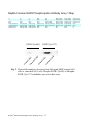
Li StarFish S.r.l.
Via Cavour, 35 - 20063 Cernusco S/N (MI), Italy
Tel. +39-02-92150794 - Fax. +39-02-92157285
[email protected] -www.listarfish.it
RayBio® Human EGFR Phosphorylation Antibody Array 1
User Manual
For Simultaneously Detecting the Relative Levels of Phosphorylation
of EGF Receptors at 17 Different Phosphorylation Sites.
(Cat# RAY-AAH-PER-1-2; RAY-AAH-PER-1-4; RAY-AAH-PER-1-8)
Please read manual carefully before starting experiment
We Provide You with Excellent
Protein Array Systems and Service
RayBio® Human EGFR Phosphorylation Antibody Array 1
Protocol
TABLE OF CONTENTS
I.
Introduction……..……………………………...
How It Works………………..…………………
II. Materials Provided……………………………..
III. Additional Materials Required…………………
IV. Reagent Preparation……………………………
V. Overview and General Considerations…………
a. Preparation of Samples………………………
b. Handling Array Membrane………………….
c. Incubation……………………………………
VI. Protocol…………………………………………
a. Blocking and Incubation…………………….
b. Detection…………………………………… .
VII. Interpretation of Results………………………..
VIII.Troubleshooting Guide…………………………
IX. Reference List………………………………….
RayBio® Human EGFR Phosphorylation Antibody Array 1 1
2
3
4
5
5
7
6
7
7
8
8
10
11
14
15
I. Introduction
The EGFR family of membrane receptors consists of four different
proteins called EGFR/ErbB1/HER1, ErbB2/HER2, ErbB3/HER3, and
ErbB4/HER4. Under normal physiological conditions, the ErbB receptors
play crucial roles in propagating signals regulating cell proliferation,
differentiation, motility, and apoptosis. EGF receptor family shows clear
differences between individual receptors, and also a large overlap. ErbB1 is
the family member with most interaction partners and the highest percentage
of tyrosine residues with more than one binding partner. ErbB3 is
characterized by a large number of binding sites for phosphatidylinositol-3kinase (PI3K), while ErbB2 has only few interaction partners with Shc as the
most frequent one. ErbB1 and ErbB4 have a variety of phosphotyrosines that
bind Grb2, or Grb2 and Shc. The ErbB1 and ErbB4 have a greater diversity
of interaction partners than ErbB2 and ErbB3. ErbB1 and ErbB2 are often
over-expressed or amplified in cancers, making them important targets for
drugs currently in use or under development.
With the RayBio® Human EGFR Phosphorylation Antibody Array 1,
researchers can now simultaneously detect the relative level of
phosphorylation of 17 different specific sites for Human EGFR family in cell
lysate. By monitoring the changes in protein phosphorylation in your
experimental model system, you can verify pathway activation in your cell
lines without spending excess time and effort in performing an analysis of
immunoprecipitation and/or Western Blot.
By using RayBio® Human EGFR Phosphorylation antibody Array 1,
treated or untreated cell lysate is added into antibody array membranes. The
antibody array membranes are washed and cocktail of biotin-congujated antiEGFR is used to detect phosphorylated EGFR on activated receptors. After
incubation with HRP-streptavidin, the signals are visualized by
chemiluminescence.
RayBio® Human EGFR Phosphorylation Antibody Array 1 2
Here’s how it works
+
Sample
Antibody array
chips
Incubation of Sample with
arrayed antibody chips
2 hrs
Cocktail of biotinconjugated anti-EGFR
2 hrs
Incubation with cocktail
of biotin-conjugated antiEGFR
Labeled Streptavidin
Incubation with labeled
Streptavidin
Data analysis
and graph
RayBio® Human EGFR Phosphorylation Antibody Array 1 3
2 hrs
II. Materials Provided
Upon receipt, the kit should be stored at –20 °C. After initial use 2X
Cell Lysis Buffer, Blocking Buffer, 20X Wash Buffer I, 20X Wash
Buffer II, Cocktail of Biotin-Conjugated Anti-EGFR and HRPConjugated Streptavidin should be stored at 4 °C to avoid repeated
freeze-thaw cycles. Array membrane, Protease Inhibitor Cocktail
and Phosphatase Inhibitor Cocktail should be kept at –20 ° C.
Please use within 6 months from the date of shipment.
• RayBio® Human EGFR Phosphorylation Antibody Array 1
membrane (2, 4, or 8 membranes)
• 2X Cell Lysis Buffer (5 ml)
• Protease Inhibitor Cocktail (1 tube for 2-4 membranes, and 2
for 8 membranes)
• 100X Phosphatase Inhibitor Cocktail Set I Concentrate (1 tube
for 2–4 membranes, and 2 for 8 membranes)
• Phosphatase Inhibitor Cocktail Set II (1 tube for 2–4
membranes, and 2 for 8 membranes)
• Blocking Buffer (25 ml for less 4 membranes and 50 ml for 8
membranes)
• 20X Wash Buffer I (30 ml)
• 20X Wash Buffer II (30 ml)
• Cocktail of Biotin-Conjugated Anti-EGFR (1 tube for 2
membranes, 2 for 4 membranes, and 4 for 8 membranes)
• 1,000X HRP-Conjugated Streptavidin (18 µl).
• Detection Buffer C (1.5 ml for 2~4 membranes, 2.5 ml for 8
membranes)
• Detection Buffer D (1.5 ml for 2~4 membranes, 2.5 ml for 8
membranes)
• Eight-Well Tray (1 each)
RayBio® Human EGFR Phosphorylation Antibody Array 1 4
• Plastic sheets
III. Additional Materials Required
•
•
•
•
Small plastic boxes or containers
Shaker
Plastic sheet protector or Saran Wrap
Kodak X-Omat™ AR film (REF 165 1454) and film processor
or Chemiluminescence imaging system
IV. Reagent Preparation
1. Protease Inhibitor Cocktail: Briefly spin down the Protease
Inhibitor Cocktail tube before use. Add 60 µl of 1X Lysis Buffer
into the vial to prepare a 100X Protease Inhibitor Cocktail
Concentrate.
2. Phosphatase Inhibitor Cocktail Set II: Briefly spin down the
Phosphatase Inhibitor Cocktail Set II tube before use. Add 180 µl
of 1X Lysis Buffer into each vial to prepare 25X Phosphatase
Inhibitor Cocktail Set II Concentrate. Dissolve the powder
thoroughly by a gentle mix.
3. 2X Cell Lysis Buffer: Cell lysis buffer should be diluted 2-fold
with deionized or distilled water before use. Add 20 µl of
prepared 100X Protease Inhibitor Cocktail Concentrate and 20 µl
of 100X Phosphatase Inhibitor Cocktail Set I Concentrate (bring
Set I concentrate tube to room temperature to thaw the
solution before use) , and 80 µl Phosphatase Inhibitor Cocktail
Set II into 1.9 ml 1X Lysis Buffer before use. Mix well.
RayBio® Human EGFR Phosphorylation Antibody Array 1 5
4. 20X Washing Buffer I or II: If the Wash Buffer Concentrate
(20X) contains visible crystals, warm to room temperature and
mix gently until dissolved. Dilute 25 ml of Wash Buffer
Concentrate into deionized or distilled water to yield 500 ml of
1X Wash Buffer.
5. Cocktail of Biotin-Conjugated Anti-EGFR: Briefly spin the
Detection Antibody tube before use. Add 100 µl of Blocking
Buffer to the tube. Mix gently and transfer all mixture to a tube
containing 2.1 ml of Blocking Buffer to prepare 1X Cocktail of
Biotin-Conjugated Anti-EGFR.
6. 1000X HRP-Conjugated Streptavidin: briefly spin down the HRPStreptavidin Concentrate and pipette up and down to mix gently
before use. E.g. add 5 µl of HRP-Conjugated Streptavidin
concentrate into a tube with 5 ml Blocking Buffer. Mix gently to
prepare 1X HRP-Conjugated Streptavidin (don’t store the
diluted Streptavidin for next day use).
Note: mix tube containing 1,000X HRP-Conjugated Streptavidin
well before use since precipitation may form during storage.
V. Overview and General Considerations
A. Preparation of Samples
The cell lysate can be prepared as follows.
For attached cells, remove supernatant from cell culture, wash cells
twice with cold 1X PBS (for suspension cells, pellet the cells by
spinning down the cells at 1500 rpm for 10 min) making sure to
RayBio® Human EGFR Phosphorylation Antibody Array 1 6
remove any remaining PBS before adding Lysis Buffer. Solubilize
the cells at 2x10⁷ cells/ml in 1X Lysis Buffer containing Protease
Inhibitor Cocktail and Phosphatase Inhibitor Cocktail Set I and Set
II. Pipette up and down to resuspend cells and rock the lysates
gently at 2–8 °C for 30 minutes. Transfer extracts to microfuge
tubes and centrifuge at 14,000 x g for 10 min.
It is recommended that sample protein concentrations be determined
using a total protein assay. For incubation with the EGFR
Phosphorylation Antibody Array 1, use at a protein concentration of
50-1000 µg/ml for cell lysates.
Lysates should be used immediately or aliquot and stored at -70 °C.
Thawed lysates should be kept on ice prior to use.
If you experience high background, you may further dilute your
samples.
B. Handling Array Membranes
• Always use forceps to handle membranes, and grip the
membranes by the edges only.
• Never allow array membranes to dry during experiments.
• Avoid touch Array membrane by hand, tips or any sharp tools.
C. Incubation
• Completely cover membranes with sample or buffer during
incubation, and cover eight-well tray with lid to avoid drying.
• Avoid foaming during incubation steps.
• Perform all incubation and wash steps under gentle rotation.
• Several incubation steps such as step 4 (sample incubation), or
step 8 (biotin-Ab incubation) or step 10 (HRP-streptavidin
incubation) may be done at 4 °C for overnight.
RayBio® Human EGFR Phosphorylation Antibody Array 1 7
VI. Protocol
A. Blocking and Incubation
1. Place each membrane into the provided 8-well tray (top left
corner marked with “-”).
Note: The printed side should be facing upward.
2. Add 1 ml Blocking Buffer and incubate at room temperature with
gentle shaking for 1 hour to block membranes.
3. Decant Blocking Buffer from each container. Add 1.2 ml of
sample into each array membrane, and cover with the lid.
Incubate at room temperature for 2 hours. Dilute sample using
Blocking Buffer.
Note: 1). We recommended using 1.0 ml of 50-1000 µg/ml
concentration of cell lysates (as starting point, we
recommended to use a concentration of 200 µg/ml of cell
lysate. Dilute the cell lysates at least 5 folds with
Blocking Buffer.
Note: 2). The amount of sample used depends on the abundance
of protein. More of the sample can be used if signals are
too weak. If signals are too strong, the sample can be
diluted further.
Note: 3). Incubation may be done at room temperature for 2
hours. Over night at 4°C
RayBio® Human EGFR Phosphorylation Antibody Array 1 8
4. Decant the samples from each container, and wash 3 times with
2 ml of 1X Wash Buffer I at room temperature with shaking. 3
min per wash.
5. Carefully remove each array membrane and place all of
membranes into a plastic container with a minimum of 20 ml of
1X Wash Buffer I. Rinse the 8-Well Multi-dish with deionized
or distilled water and dry thoroughly. Wash array membranes
with 1X Wash Buffer with shaking. Repeat 2 times for a total of
3 washes. 5 min per wash.
6. Wash 3 times with a minimum of 20 ml of 1X Wash Buffer II at
room temperature with shaking. 5 min per wash.
7. Carefully remove each array membrane from the container,
return it to the 8-well tray.
8. Add 1 ml of diluted Cocktail of Biotin-Conjugated Anti-EGFR to
each membrane. Incubate at room temperature with gentle
shaking for 2 hours.
Note: Incubation may be done at 40C for overnight.
9. Wash as directed in steps 5, 6 and 7.
10. Add 1.5 ml of 1X HRP-conjugated streptavidin to each
membrane.
Note: Mix tube containing 1X HRP-Conjugated Streptavidin
well before use since precipitation may form during storage.
11. Incubate at room temperature for 2 hours.
RayBio® Human EGFR Phosphorylation Antibody Array 1 9
Note: incubation may be done at 40C for overnight.
12. Wash as directed in steps 5 and 6.
B. Detection
* Do not let the membrane dry out during detection. The
detection process must be completed within 40 minutes
without stopping.
1. Proceed with detection reaction.
Add 250 µl of Detection Buffer C and 250 µl of Detection
Buffer D for one membrane; mix both solutions; Drain off
excess wash buffer by holding the membrane vertically with
forceps. Place membrane protein side up (“-” mark is on the
protein side top left corner) on a clean plastic sheet (provided in
the kit). Pipette the mixed Detection Buffer on to the membrane
and incubate at room temperature with gentle shaking for 2
minutes. Ensure that the detection mixture is completely and
evenly covering the membrane without any air bubbles.
2. Drain off excess detection reagent by holding the membrane
vertically with forceps and touching the edge against a tissue.
Gently place the membrane, protein side up, on a piece of
plastic sheet (“-” mark is on the protein side top left corner).
Cover the array with another piece of plastic sheet. Gently
smooth out any air bubbles. Avoid using pressure on the
membrane.
3. Detect signal directly from membrane using chemiluminescene
RayBio® Human EGFR Phosphorylation Antibody Array 1 10
imaging system or expose to x-ray film (we recommend to use
Kodak X-Omat™ AR film) detect signal using film developer.
Expose the membranes for 40 Seconds. Then re-expose the
film according to the intensity of signals. If the signals are too
strong (background too high), reduce exposure time (eg, 5–30
seconds). If the signals are too weak, increase exposure time
(eg, 5–20 min or overnight). Or re-incubate membranes
overnight with 1X HRP-conjugated streptavidin, and repeat
detection on the second day.
4. Save membranes at –20 °C to –80 °C for future reference.
VII. Interpretation of Results:
The following figure shows RayBio® Human EGFR
Phosphorylation Antibody Array 1 membranes probed with different
cell lines. The signals were detected by using a chemiluminescene
imaging device. Membranes also can be exposed to Kodak XOmat™ film at room temperature. A biotinylated protein provides
positive signals, which can be used to identify the orientation and to
normalize the results from different wells being compared.
One important parameter is the background signal. To obtain the
best results, we suggest that several exposures be attempted. We
also strongly recommend using a negative control in which the
sample is replaced with an appropriate mock buffer according to the
array protocol, particularly during your first experiment.
By comparing the signal intensities, relative expression levels of
target proteins can be made. The intensities of signals can be
quantified by densitometry. Positive control can be used to
normalize the results from different membranes being compared.
RayBio® Human EGFR Phosphorylation Antibody Array 1 11
Antibody affinity to its target varies significantly between
antibodies. The intensity detected on the array with each antibody
depends on this affinity; therefore, signal intensity comparison can
be performed only within the same antibody/antigen system and not
between different antibodies.
EGFR (Tyr845)
EGFR (Tyr845)
EGFR
(Tyr1173)
EGF treated A431 cells
(Cell lysate: 200 µg/ml)
EGFR
(Tyr1173)
Untreated A431 cells
(Cell lysate: 200 µg/ml)
Fig. 1. Human epidermoid carcinoma cell line, A431 cells that were 8090% confluent were serum starved overnight, then exposed to 100 ng/ml
EGF for 20 minutes at 37 °C. Control cells were serum starved without the
subsequent stimulation with EGF. Cell lysates were prepared following the
"Preparation of Sample" portion of our protocol V.
To use the
RayBio®Phosphorylation Antibody Array 1, treated or untreated cell lysate
was added into antibody array membrane. The antibody array membranes
were washed and cocktail of biotinylated anti-EGFR was used to detect
phosphorylated proteins on activated receptors. After incubation with HRPConjugated Streptavidin, the signals were visualized by chemiluminescence.
RayBio® Human EGFR Phosphorylation Antibody Array 1 12
RayBio® Human EGFR Phosphorylation Antibody Array 1 Map
P1
P1
Blank
Blank
EGFR (Ser1046/1047)
EGFR (Ser1046/1047)
ErbB2 (Ser1113)
ErbB2 (Ser1113)
P2
P2
Blank
Blank
EGFR (Ser1070)
EGFR (Ser1070)
ErbB3 (Tyr1289)
ErbB3 (Tyr1289)
P3
P3
Blank
Blank
ErbB2 (Tyr877)
ErbB2 (Tyr877)
ErbB4 (Tyr1284)
ErbB4 (Tyr1284)
Blank
Blank
Blank
Blank
ErbB2 (Tyr1112)
ErbB2 (Tyr1112)
Blank
Blank
EGFR (Tyr845)
EGFR (Tyr845)
EGFR (Tyr1086)
EGFR (Tyr1086)
ErbB2 (Tyr1248)
ErbB2 (Tyr1248)
Neg
Neg
EGFR (Tyr992)
EGFR (Tyr992)
EGFR(Tyr1148)
EGFR(Tyr1148)
ErbB2 (Thr686)
ErbB2 (Thr686)
Blank
Blank
EGFR (Tyr1045)
EGFR (Tyr1045)
EGFR (Tyr1173)
EGFR (Tyr1173)
Blank
Blank
P4
P4
EGFR (Tyr1173)
Tr
ea
te
d
A4
U
31
nt
re
at
ed
A
43
1
Tr
ea
te
d
A
43
1
U
nt
re
at
ed
A
43
1
EGFR (Tyr845)
Neg
Neg
EGFR (Tyr1068)
EGFR (Tyr1068)
ErbB2 (Tyr 1221/1222)
ErbB2 (Tyr 1221/1222)
Blank
Blank
Fig. 2. Western blot analysis of extracts from 100 ng/ml hEGF treated A431
cells or untreated A431 cells. Phospho-EGFR (Tyr845) or PhosphoEGFR (Tyr1173) antibodies was used in this assay.
RayBio® Human EGFR Phosphorylation Antibody Array 1 13
VIII. Troubleshooting Guide
Problem
Cause
Weak signal or no 1. Taking too much time
signal
for Detection.
2. Film developer does
not work properly.
3. Did not mix HRPstreptavidin well before
use.
Recommendation
1. The whole detection process must be
completed in 30 min.
2. Fix film developer.
3. Mix tube containing HRP-Conjugate
Streptavidin well before use since
precipitates may form during storage.
4. Sample is too dilute.
4. Increase sample concentration
5. Other.
1.Reduce blocking concentration by diluting in
1X Wash Buffer II.
2. Slightly increase HRP concentrations.
3. Slightly increase biotinylate-antibody
concentrations.
4. Expose film for overnight to detect weak
signal.
Uneven signal
1. Bubbles formed
during incubation.
2. Membranes were not
completely covered by
solution.
High background 1. Exposure to x-ray file
is too long.
2. Membranes were
allowed to dry out during
experiment.
3. Sample is too
concentrated.
1. Remove bubbles during incubation.
2. Completely cover membranes with solution.
1. Decrease exposure time.
2. Completely cover membranes with solution
during experiment.
3. Use more diluted sample.
RayBio® Human EGFR Phosphorylation Antibody Array 1 14
IX. Reference List
1. Holbro T, Hynes NE (2004). ErbB receptors: directing key signaling
networks throughout life. Annu Rev Pharmacol Toxicol 44: 195–217
2. Marmor MD, Skaria KB, Yarden Y (2004). Signal transduction and
oncogenesis by ErbB/HER receptors. Int J Radiat Oncol Biol Phys 58:
903–913
3. Huang RP, Huang R, Fan Y, and Lin Y (2001). A novel method
for high- throughput protein profiling from conditioned media
and patient’s sera. Ana. Biochem. 294(1):55-62.
4. Huang R, Lin Y, Wang CC, J et al (2002).Connexin suppresses
human glioblastoma cell growth by down-regulation of monocyte
chemotactic protein 1, as discovered using protein array
technology. Cancer Res. 62:2806-2812.
RayBio® Human EGFR Phosphorylation Antibody Array 1 15