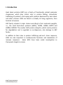
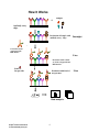
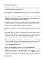
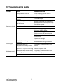
1
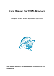
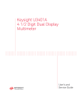
RayBio® C-Series
Human Heat Shock Protein Antibody Array
User Manual
Cat# AAH-HSP-1-2
AAH-HSP-1-4
AAH-HSP-1-8
For Simultaneous Detection of the
Relative Levels of 9 Heat shock protein Markers
Version 1.0
August 31, 2013
Please read manual carefully before starting experiment
RayBiotech, Inc.
We Provide You with Excellent
Protein Array Systems and Service
Tel (Toll Free): 1-888-494-8555 or 770-729-2992; Fax: 1-888-547-0580 Website:
www.raybiotech.com; Email: [email protected]
RayBiotech, Inc.
RayBio® Human Heat Shock Protein Antibody Array Kit
TABLE OF CONTENTS
I.
Introduction……..………………………………
2
II.
Principle of the Assay……………………… …
3
III. Kit Components & Storage…………………
5
IV. Reagent Preparation…………………………
6
V.
Overview and General Considerations… 7
a. Preparation of Samples…………………..
7
b. Handling Array Membranes……………… 8
c. Incubation………………………………………
8
VI. Protocol………………………………………………
8
a. Blocking and Incubation…………………
8
b. Detection………………………………………
10
VII. Interpretation of Results…………………….
11
VIII. Array Map…………………………………………… 12
IX. Troubleshooting Guide………………………
®
RayBio Human Heat Shock
Protein Antibody Array Kit
1
14
I. Introduction
Heat shock proteins (HSP) are a family of functionally related molecular
chaperones, which play critical roles in protein folding, intracellular
trafficking of proteins, and coping with proteins denatured by heat shock
and other stresses. HSPs are found in virtually all living organisms, from
bacteria to humans.
HSP family includes 5 major classes according to their molecular weights,
i.e., the small heat-shock proteins (sHSPs), HSP33, HSP60, HSP70 and
HSP90/ HSP100. The smaller 8-kD protein ubiquitin, which marks proteins
for degradation and is regarded as co-chaperone, also belongs to HSP
family.
In addition to their roles in protein trafficking and and stress response,
HSPs are also important in cardiovascular functions and modulation of
immune responses. Some HSPs have been under investigation as
therapeutic targets in cancer.
®
RayBio Human Heat Shock
Protein Antibody Array Kit
2
II. Principle of the Assay
With the RayBio® Heat Shock Protein Antibody Array kit, researchers can
now simultaneously detect the relative level of 9 HSP related proteins in cell
lysates. By monitoring the changes in protein levels in different
experimental model systems, researchers can study pathway activation
without
spending
excess
time
and
effort
in
performing
immunoprecipitations and/or Western Blotting.
Each array membrane is pre-printed with capture antibodies; treated or
untreated cell lysate is then added to each membrane. After extensive
washing, the membranes are incubated with a cocktail of biotin-conjugated
anti-apoptotic protein antibodies. After incubation with HRP-Streptavidin,
the signals are visualized by chemiluminescence.
®
RayBio Human Heat Shock
Protein Antibody Array Kit
3
How It Works
+
Sample
Antibody array
chips
Incubation of Sample with
antibody array chips
Overnight
Cocktail of biotinconjugated antiHSP marker
2 hrs
Incubation
with cocktail
of biotin-conjugated antiHSP marker
LabeledStreptavidin
Incubation with labeled
Streptavidin
Data analysis
®
RayBio Human Heat Shock
Protein Antibody Array Kit
4
2 hrs
III. Kit Components & Storage
Store entire kit at ≤ -20 °C immediately upon arrival. Kit must used within the 6 month
expiration date.
ITEM
COMPONENT
2 SAMPLE KIT
4 SAMPLE KIT
1
2
8 SAMPLE KIT
Human HSP Array C1*
2 membranes
4 membranes
8 membranes
Blocking Buffer
1 vial (25 ml)
1 vial (25 ml)
2 vials (25 ml/ea)
Detection Antibody
3
1 vial
2 vials
4 vials
Cocktail
1,000X HRP-Streptavidin
4
1 vial (50 µl)
Concentrate
20X Wash Buffer I
5
1 vial (10 ml)
1 vial (10 ml)
1 vial (20 ml)
Concentrate
20X Wash Buffer II
6
1 vial (10 ml)
1 vial (10 ml)
1 vial (20 ml)
Concentrate
2X Cell Lysis Buffer
7
1 vial (5 ml)
Concentrate
8
Detection Buffer C
1 vial (1.5 ml)
1 vial (1.5 ml)
1 vial (2.5 ml)
9
Detection Buffer D
1 vial (1.5 ml)
1 vial (1.5 ml)
1 vial (2.5 ml)
Protease Inhibitor
1 vial
1 vial
2 vials
Cocktail
8-Well Incubation Tray
10
1 tray
w/ Lid
11 Plastic Sheets
4 sheets
12 Array Map Template
1 template
13 User Manual
1 booklet
* Each plastic package contains 2 or 4 membranes.
**For up to 3 months (unless stated otherwise) or until expiration date.
STORAGE
TEMPERATURE
AFTER THAWING**
≤ -20 °C
2-8 °C
(up to 3 days after dilution)
2-8 °C
≤ -20 °C
Room Temperature
Additional Materials Required
•
•
•
•
•
•
Pipettors, pipet tips and other common lab consumables
Orbital shaker or oscillating rocker
Tissue Paper, Blotting Paper or Chromatography Paper
Adhesive Tape or Plastic Wrap
Distilled or De-ionized Water
Chemiluminescent blot documentation system (CCD Camera, X-Ray Film and a
suitable film processor, Gel documentation system, or other chemiluminescent
detection system capable of imaging a Western blot)
®
RayBio Human Heat Shock
Protein Antibody Array Kit
5
IV. Reagent Preparation
1. 1X Cell Lysis Buffer: 2x Cell lysis buffer should be diluted 2-fold with
deionized or distilled water before use.
Note: Proceed to 2. before completing the rest of the 2X Cell Lysis Buffer
preperation.
Add 20 µl of prepared 100X Protease Inhibitor Cocktail Concentrate
(bring the tube to room temperature to thaw the solution before use)
into 1.98 ml 1X Lysis Buffer before use. Mix well.
2. Protease Inhibitor Cocktail: Briefly spin down the Protease Inhibitor
Cocktail tube before use. Add 60 µl of 1X Lysis Buffer into the vial to
prepare a 100X Protease Inhibitor Cocktail Concentrate.
Note: Be sure to complete 2X Cell Lysis Buffer preparation before proceeding
to 3.
3. 20X Wash Buffer I or II: If the Wash Buffer Concentrate (20X) contains
visible crystals, warm to room temperature and mix gently until
dissolved. Dilute 20 ml of Wash Buffer Concentrate into 380 ml of
deionized or distilled water to yield 400 ml of 1X Wash Buffer.
4. Detection Antibody Cocktail: Briefly spin the vial before use. Add 150 µl
of Blocking Buffer to the tube. Mix gently and transfer the entire mixture
to a tube containing 1.80 ml of Blocking Buffer to prepare 1X Cocktail of
Biotin-Conjugated Antibody Mix.
5. 1000X HRP-Conjugated Streptavidin: briefly spin down the HRP
Streptavidin Concentrate to remove any liquid from the vial cap and
pipette up and down to mix gently before use. Prepare 1X HRPConjugated Streptavidin. Example: add 5 µl of HRP-Conjugated
Streptavidin concentrate into a tube with 4.995 ml Blocking Buffer and
®
RayBio Human Heat Shock
Protein Antibody Array Kit
6
mix gently. Use immediately, do not store the 1X Streptavidin for next
day use.
Note: mix tube containing 1,000X HRP-Conjugated Streptavidin
well before use since precipitation may form during storage.
V. Overview and General Considerations
A. Preparation of Samples
Remove supernatant from cell culture for attached cells, wash cells twice
with cold 1X PBS (for suspension cells, pellet the cells by spinning down the
cells at 1500 rpm for 10 min) making sure to remove any remaining PBS
before adding Lysis Buffer. Solubilize the cells at 2x10⁷ cells/ml in 1X Lysis
Buffer containing Protease Inhibitor Cocktail (prepared in step 1 of Reagent
Preparation). Pipette up and down to resuspend cells and rock the lysates
gently at 2–8 °C for 30 minutes. Transfer extracts to microfuge tubes and
centrifuge at 14,000 x g for 10 min.
It is recommended that sample protein concentrations be determined using
a total protein assay such as the BCA Assay. Before applying to the
membranes, all lysates should be diluted at least 5-fold with 1X Blocking
Buffer. For the RayBio® Human Heat Shock Protein Antibody Array Kit,
200 µg/ml to 600 µg/ml of total protein from cell lysates (after dilution
with Blocking Buffer) should be used for incubation. Lysates should be used
immediately or aliquotted and stored at -80 °C. Thawed lysates should be
kept on ice prior to use.
Note: If you experience high background, you may further dilute your
samples.
®
RayBio Human Heat Shock
Protein Antibody Array Kit
7
B. Handling Array Membranes
• Always use forceps to handle membranes, and grip the membranes by
the edges only. Flat-tipped forceps are best for handling membranes.
• Never allow array membranes to dry during experiments.
• Avoid touching array membrane by hand, tips or any sharp tools.
C. Incubation
• Completely cover membranes with sample or buffer during
incubation, and cover eight-well tray with lid to avoid drying.
• Avoid bubbles and foaming during incubation steps.
• Perform all incubation and wash steps under gentle shaking (1-2
cycles/sec).
• The following incubations may be done overnight at 4°C: all wash
steps, Step 3 (sample incubation), Step 7 (biotin-Ab incubation) and
Step 10 (HRP-streptavidin incubation).
VI. Protocol
A. Blocking and Incubation
1. Place each membrane into the provided 8-well tray (top left corner
marked with “-”).
Note: The printed side should be facing upward.
2. Add 1 ml Blocking Buffer and incubate at room temperature with gentle
shaking for 30 minutes to block membranes.
3. Decant Blocking Buffer from each well. Add 1 ml diluted sample onto
each array membrane, and cover with the lid. Incubate at 2 – 8 0C
®
RayBio Human Heat Shock
Protein Antibody Array Kit
8
overnight on a rocker or shaker (low speed 1-2 cycles/sec). Dilute
sample using Blocking Buffer.
Note:
1) Dilute cell lysates at least 5-fold with Blocking Buffer to avoid
high background.
Note: 2) Optimal sample dilution factors should be determined
empirically. More sample can be used if signals are too weak. If
signals are too strong, the sample can be diluted further.
Note: 3) Incubation may be done at room temperature for 4 hours, but
this may cause lower signals.
4. Decant the samples from each well, and wash 3 times with 1.5 ml of 1X
Wash Buffer I at room temperature with shaking, 5 min per wash.
5. Wash 2 times with 1.5 ml of 1X Wash Buffer II at room temperature
with shaking, 5 min per wash.
6. Carefully remove wash buffer from each well containing array
membranes.
7. Add 1 ml of diluted Cocktail of Biotin-Conjugated Antibody Mix to each
membrane. Incubate at 2 – 8 0C with gentle shaking overnight.
Note: Incubation may be done at room temperature for 2 hours.
8. Wash as directed in steps 4, 5 and 6.
9. Add 1.5 ml of 1X HRP-conjugated Streptavidin to each membrane.
10. Incubate HRP-streptavidin at room temperature for 1.5 hours.
Note: incubation may be done overnight at 40C.
®
RayBio Human Heat Shock
Protein Antibody Array Kit
9
11. Wash as directed in steps 5 and 6.
B. Detection
NOTE: Do not let the membrane dry out during detection. The detection
process must be completed within 40 minutes without stopping.
1. Proceed with detection reaction.
Add 250 µl of Detection Buffer C and 250 µl of Detection Buffer D for
one membrane; mix both solutions; Drain off excess wash buffer by
holding the membrane vertically with forceps. Place membrane
protein side up (“-” mark is on the protein side top left corner) on a
clean plastic sheet (provided in the kit). Pipette the mixed Detection
Buffer on to the membrane and incubate at room temperature with
gentle shaking for 2 minutes. Ensure that the detection mixture is
completely and evenly covering the membrane without any air
bubbles.
2. Drain off excess detection reagent by holding the membrane vertically
with forceps and touching the edge against a tissue. Gently place the
membrane, protein side up, on a piece of plastic sheet (“-” mark is on
the protein side top left corner). Cover the array with another piece of
plastic sheet. Gently smooth out any air bubbles. Avoid using pressure
on the membrane.
3. Detect signal directly from membrane using chemiluminescence
imaging system or expose to x-ray film (we recommend Kodak XOmat™ AR film) to detect signal using film developer. Expose the
membranes for 40 Seconds. Then re-expose the film according to the
intensity of signals. If the signals are too strong (background too high),
reduce exposure time (e.g., 5–30 seconds). If the signals are too weak,
increase exposure time (eg, 2–20 min or overnight). Or re-incubate
®
RayBio Human Heat Shock
Protein Antibody Array Kit
10
membranes overnight with 1X HRP-conjugated Streptavidin, and
repeat detection on the second day.
4. Save membranes at –20 °C to –80 °C for future reference.
VII. Interpretation of Results
Membrane signals are normally detected by a chemiluminescene imaging
device. Membranes also can be exposed to Kodak X-Omat™ film at room
temperature. A biotinylated protein provides positive signals, which can be
used to identify the orientation and to normalize the results from different
wells being compared.
By comparing the signal intensities, relative expression levels of target
proteins can be made. The intensities of signals can be quantified by
densitometry. The positive controls can be used to normalize the results
from different membranes being compared.
Antibody affinity to its target varies significantly between antibodies. The
intensity detected on the array with each antibody depends on this affinity;
therefore, signal intensity comparison can be performed only within the
same antibody/antigen system and not between different antibodies.
One important parameter is the baseline signal response. To obtain the best
results, we suggest that several exposures be attempted. We also strongly
recommend using a negative control in which the sample is replaced with
an appropriate mock buffer according to the array protocol, particularly
during your first experiment.
®
RayBio Human Heat Shock
Protein Antibody Array Kit
11
VIII. Human Heat Shock Protein Antibody Array Map
POSA
NEG
HSP90
GRP75
HSP27 HSP32
UBIQ
HSP40 HSP60 HSP70
HSP10
NEG
NEG
POSA
X. Representative Data using Human Heat Shock Protein Antibody
Array
®
RayBio Human Heat Shock
Protein Antibody Array Kit
12
IX. Troubleshooting Guide
Problem
Weak or no signal
Cause
Recommendation
Taking too much time for
detection
Entire detection process must be
completed in 30 minutes
Film developer does not work
properly
Fix film developer
Did not mix HRP-streptavidin
well before use
Mix tube containing HRP-streptavidin
well before use since precipitates may
form during storage.
Sample is too dilute
Increase sample concentration
Reduce blocking concentration by
diluting in 1X Wash Buffer II
Slightly increase HRP concentration
Other
Slightly increase biotin-antibody
concentration
Expose film overnight
Uneven signal
Bubbles formed during
incubation
Remove bubbles during incubation
Membranes were not
completely covered by solution
Completely cover membranes with
solution
Exposure to x-ray film is too long Decrease exposure time
High background
Membranes were allowed to dry Completely cover membranes with
out during experiment
solution during experiment
Sample is too concentrated
®
RayBio Human Heat Shock
Protein Antibody Array Kit
Increase sample dilution
13
RayBiotech, Inc., the protein array pioneer company, strives to
research and develop new products to meet demands of the
biomedical community. RayBio® patent-pending technology allows
detection of over 1000 cytokines, chemokines and other proteins in a
single experiment. Our format is simple, sensitive, reliable and cost
effective. Products include: Cytokine Arrays, Chemokine Arrays,
ELISA kits, EIA kits, Phosphotyrosine kits, Recombinant Proteins,
Antibodies, and custom services.
This product is for research use only
©2009 RayBiotech, Inc.
®
RayBio Human Heat Shock
Protein Antibody Array Kit
14