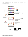
1
RayBio® Human Apoptosis Antibody Array Kit User Manual Cat# AAH-APO-1-2 AAH-APO-1-4 AAH-APO-1-8 For Simultaneous Detection of the Relative Levels of 43 Apoptosis-related Markers Please read manual carefully before starting experiment RayBiotech, Inc. We Provide You with Excellent Protein Array Systems and Service Tel :( Toll Free) 1-888-494-8555 or 770-729-2992; Fax: 1-888-547-0580 Website: www.raybiotech.com; Email: [email protected] RayBiotech, Inc. RayBio® Human Apoptosis Antibody Array Kit TABLE OF CONTENTS I. Introduction……..…………………………… II. Principle of the Assay……………………… III. Materials…………………………………….. a. Material Provided………………………… b. Additional Materials Required………….. IV. Reagent Preparation……………………….. V. Overview and General Considerations…… a. Preparation of Samples………………….. b. Handling Array Membrane………………. c. Incubation…………………………………. VI. Protocol………………………………………. a. Blocking and Incubation…………………. b. Detection………………………………….. VII. Interpretation of Results……………………. VIII. Array Map……………………………........... IX. Troubleshooting Guide…………………… X. Reference List……………………………… ® RayBio Human Apoptosis Antibody Array Kit 1 2 4 6 6 6 7 8 8 8 8 9 9 10 12 14 15 16 I. Introduction Apoptosis is the process of programmed cell death that involves a series of biochemical events leading to characteristic cell morphology and death. These events include blebbing, changes to the cell membrane such as loss of membrane asymmetry and attachment, cell shrinkage, nuclear fragmentation, chromatin condensation, and chromosomal DNA fragmentation. Studies on apoptosis have increased substantially since the early 1990s. In addition to its importance as a biological phenomenon such as cell termination, homeostasis, development and lymphocyte interactions, deregulation of apoptosis has been implicated in many diseases. Excessive apoptosis causes hypotrophy, such as in ischemic damage, whereas insufficient apoptosis results in uncontrolled cell proliferation, such as HIV progression and cancer development. Apoptosis is mediated by a diverse range of cell signals, both extracellular and intracellular. Extracellular signals may include toxins, hormones, growth factors, nitric oxide or cytokines. Intracellular apoptotic signaling may be induced in response to stress via, heat, radiation, nutrient deprivation, viral infection, hypoxia and increased intracellular calcium concentration or the binding of nuclear receptors by glucocorticoids. These signals may positively or negatively induce apoptosis. Two apoptotic signal transduction pathways in mammals have been reported: the TNF-induced model and the Fas-Fas ligand-mediated model. TNF is the major extrinsic mediator of apoptosis. Most cells in the human body have two receptors for TNF: TNF-R1 and TNF-R2. The binding of TNF to TNF-R1 has been shown to initiate the pathway that leads to caspase activation via the intermediate membrane proteins TNF receptor-associated death domain (TRADD) and Fasassociated death domain protein (FADD). Binding of this receptor can also indirectly lead to the activation of transcription factors involved in ® RayBio Human Apoptosis Antibody Array Kit 2 cell survival and inflammatory responses. The Fas receptor (also known as Apo-1 or CD95) binds the Fas ligand. The interaction between Fas and FasL results in the formation of the death-inducing signaling complex (DISC), which contains the FADD, caspase-8 and caspase-10. Following TNF-R1 and Fas activation in mammalian cells a balance between pro-apoptotic (BAX, BID, BAK, or BAD) and antiapoptotic (Bcl-Xl and Bcl-2) members of the Bcl-2 family is established. This balance is the proportion of pro-apoptotic homodimers that form in the outer-membrane of mitochondrion. The pro-apoptotic homodimers are required to make the mitochondrial membrane permeable for the release of caspase activators such as cytochrome c and SMAC. Control of pro-apoptotic proteins under normal cell conditions of non-apoptotic cells is incompletely understood. Mitochondria are an important site for apoptosis. Mitochondrial proteins known as SMACs (second mitochondria-derived activator of caspases) are released into the cytosol following an increase in permeability. SMAC binds to inhibitor of apoptosis proteins (IAPs) and deactivates them, preventing the IAPs from arresting the apoptotic process and therefore allowing apoptosis to proceed. IAP also normally suppresses the activity of caspases (cysteine aspartic acid proteases), which carry out the degradation of the cell. Therefore, the degradative activity of caspases seems to be indirectly regulated by mitochondrial permeability. Cytochrome c is also released from mitochondria due to formation of a channel, MAC, in the outer mitochondrial membrane, and serves a regulatory function as it precedes morphological change associated with apoptosis. Once cytochrome c is released it binds with Apaf-1 and ATP, which then bind to pro-caspase-9 to create a protein complex known as an apoptosome. The apoptosome cleaves the pro-caspase to its active form of caspase-9, which in turn activates the effector caspase-3. The tumor-suppressor protein p53 also plays critical role in apoptosis. p53 accumulates in response to DNA damage via interferon-alpha and interferon-beta pathways, which induce transcription of the p53 ® RayBio Human Apoptosis Antibody Array Kit 3 gene and result in the increase of p53 protein level and enhancement of cancer cell apoptosis. p53 prevents the cell from replicating by stopping the cell cycle at G1, or interphase, to give the cell time to repair, however it will induce apoptosis if damage is extensive and repair efforts fail. Any disruption to the regulation of the p53 or interferon genes will result in impaired apoptosis and the possible formation of tumors. A recent report has shown the involvement of IGFBPs (insulin-like growth factor-binding protein) in apoptosis. IGFBP1 protein localizes to mitochondria where it binds to the BAK and hinders BAK activation and apoptosis induction. When IGFBP1 is in a complex with BAK, formation of a proapoptotic p53/BAK complex and apoptosis induction is impaired, both in cultured cells and in liver. In contrast, livers of IGFBP1-deficient mice exhibit spontaneous apoptosis that is accompanied by p53 mitochondrial accumulation and BAK oligomerization. These results identify IGFBP1 as a negative regulator of the BAK-dependent pathway of apoptosis, whose expression integrates the transcriptional and mitochondrial functions of the p53 tumor suppressor protein. II. Principle of the Assay With RayBio® Human Apoptosis Antibody Array kit, researchers can now simultaneously detect the relative level of 43 apoptosis related proteins in cell lysates. By monitoring the changes in protein levels in different experimental model systems, researchers can study pathway activation without spending excess time and effort in performing immunoprecipitations and/or Western Blotting. With RayBio Human Apoptosis Antibody Array kit, each of the capture antibodies is printed on the membranes, and then treated or untreated cell lysate is added into antibody array membranes. After extensive washing, the membranes are incubated with a cocktail of biotin-conjugated anti-apoptotic protein antibodies. After incubation ® RayBio Human Apoptosis Antibody Array Kit 4 with HRP-Streptavidin, chemiluminescence. the signals are visualized How It Works + Sample Antibody array chips Incubation of Sample with arrayed antibody chips Overnight Cocktail of biotinconjugated antiapoptosis marker Overnight Incubation with cocktail of- biotin-conjugated antiapoptosis marker LabeledStreptavidin Incubation with labeled Streptavidin Data analysis ® RayBio Human Apoptosis Antibody Array Kit 5 2 hrs by IIIa. Materials Provided Upon receipt, the kit should be stored at –20 °C. After initial use 2X Cell Lysis Buffer, Blocking Buffer, 20X Wash Buffer I, 20X Wash Buffer II, Cocktail of Biotin-Conjugated antibody mix and HRPConjugated Streptavidin should be stored at 4°C to avoid repeated freeze-thaw cycles. Array membrane, Protease Inhibitor Cocktail should be kept at –20 ° C. Please use the entire kit within 6 months from the date of shipment. 1. 2. 3. 4. 5. 6. 7. 8. 9. 10. 11. 12. 13. RayBio® Human Apoptosis Antibody Array 1 membranes (2, 4, or 8 membranes) 2X Cell Lysis Buffer (5 ml) Protease Inhibitor Cocktail (1 tube for 2-4 membranes, and 2 for 8 membranes) 1X Blocking Buffer (25 ml for 2-4 membranes and 50 ml for 8 membranes) 20X Wash Buffer I (10 ml for 2-4 membranes and 20ml for 8 membranes) 20X Wash Buffer II (10 ml for 2-4 membranes and 20ml for 8 membranes) Cocktail of Biotin-Conjugated Antibody Mix (1 vial for 2 membranes, 2 for 4 membranes, and 4 for 8 membranes) 1,000X HRP-Conjugated Streptavidin (I vial) Detection Buffer C (1.5 ml for 2-4 membranes, 2.5 ml for 8 membranes) Detection Buffer D (1.5 ml for 2-4 membranes, 2.5 ml for 8 membranes) Eight-Well Tray (1 each) Plastic sheets Manual ® RayBio Human Apoptosis Antibody Array Kit 6 IIIb. Additional Materials Required • • • • Small plastic boxes or containers Shaker Plastic sheet protector or Saran Wrap Kodak X-Omat™ AR film (REF 165 1454) and film processor or Chemiluminescence imaging system IV. Reagent Preparation 1. Protease Inhibitor Cocktail: Briefly spin down the Protease Inhibitor Cocktail tube before use. Add 60 µl of 1X Lysis Buffer into the vial to prepare a 100X Protease Inhibitor Cocktail Concentrate. 2. 2X Cell Lysis Buffer: Cell lysis buffer should be diluted 2-fold with deionized or distilled water before use. Add 20 µl of prepared 100X Protease Inhibitor Cocktail Concentrate (bring the tube to room temperature to thaw the solution before use) into 1.98 ml 1X Lysis Buffer before use. Mix well. 3. 20X Wash Buffer I or II: If the Wash Buffer Concentrate (20X) contains visible crystals, warm to room temperature and mix gently until dissolved. Dilute 20 ml of Wash Buffer Concentrate into deionized or distilled water to yield 400 ml of 1X Wash Buffer. 4. Cocktail of Antibody Mix: Briefly spin the Biotin-Conjugated Antibody tube before use. Add 150 µl of Blocking Buffer to the tube. Mix gently and transfer all mixture to a tube containing 1.80 ml of Blocking Buffer to prepare 1X Cocktail of Biotin-Conjugated Antibody Mix. 5. 1000X HRP-Conjugated Streptavidin: briefly spin down the HRP Streptavidin Concentrate and pipette up and down to mix gently before use. Prepare 1X HRP-Conjugated Streptavidin. Example: ® RayBio Human Apoptosis Antibody Array Kit 7 add 5 µl of HRP-Conjugated Streptavidin concentrate into a tube with 5 ml Blocking Buffer and mix gently. Do not store the 1X Streptavidin for next day use. Note: mix tube containing 1,000X HRP-Conjugated Streptavidin well before use since precipitation may form during storage. V. Overview and General Considerations A. Preparation of Samples Remove supernatant from cell culture for attached cells, wash cells twice with cold 1X PBS (for suspension cells, pellet the cells by spinning down the cells at 1500 rpm for 10 min) making sure to remove any remaining PBS before adding Lysis Buffer. Solubilize the cells at 2x10⁷ cells/ml in 1X Lysis Buffer containing Protease Inhibitor Cocktail (prepared in step 2 of Reagent Preparation). Pipette up and down to resuspend cells and rock the lysates gently at 2–8 °C for 30 minutes. Transfer extracts to microfuge tubes and centrifuge at 14,000 x g for 10 min. It is recommended that sample protein concentrations be determined using a total protein assay such as Bradford Assay. Before applying to the membranes, all lysates should be diluted at least 5-fold with 1X Blocking Buffer. For the RayBio® Human Apoptosis Antibody Array Kit, 200 µg/ml to 600 µg/ml of total protein from cell lysates (after dilution with Blocking Buffer) should be used for incubation. Lysates should be used immediately or aliquotted and stored at -70 °C. Thawed lysates should be kept on ice prior to use. If you experience high background, you may further dilute your samples. B. Handling Array Membranes ® RayBio Human Apoptosis Antibody Array Kit 8 • Always use forceps to handle membranes, and grip the membranes by the edges only. Flat-tipped forceps are best for handling membranes. • Never allow array membranes to dry during experiments. • Avoid touching array membrane by hand, tips or any sharp tools. C. Incubation • Completely cover membranes with sample or buffer during incubation, and cover eight-well tray with lid to avoid drying. • Avoid foaming during incubation steps. • Perform all incubation and wash steps under gentle shaking (1-2 cycles/sec). • All wash steps and several incubation steps such as step 3 (sample incubation), or step 7 (biotin-Ab incubation) or step 10 (HRP-streptavidin incubation) may be done overnight at 4 °C. VI. Protocol A. Blocking and Incubation 1. Place each membrane into the provided 8-well tray (top left corner marked with “-”). Note: The printed side should be facing upward. 2. Add 1 ml Blocking Buffer and incubate at room temperature with gentle shaking for 30 minutes to block membranes. 3. Decant Blocking Buffer from each well. Add 1 ml diluted sample onto each array membrane, and cover with the lid. Incubate at 2 – 8 0C overnight on a rocker or shaker (low speed 1-2 cycles/sec). Dilute sample using Blocking Buffer. ® RayBio Human Apoptosis Antibody Array Kit 9 Note: 1). Dilute cell lysates at least 5 folds with Blocking Buffer to avoid high background. Note: 2). Optimal sample dilution factors should be determined empirically. More of the sample can be used if signals are too weak. If signals are too strong, the sample can be diluted further. Note: 3). Incubation may be done at room temperature for 4 hours, but at the cost of impaired sensitivity of the signals. 4. Decant the samples from each well, and wash 3 times with 1.5 ml of 1X Wash Buffer I at room temperature with shaking, 5 min per wash. 5. Wash 2 times with 1.5 ml of 1X Wash Buffer II at room temperature with shaking, 5 min per wash. 6. Carefully remove wash buffer from each well containing array membranes. 7. Add 1 ml of diluted Cocktail of Biotin-Conjugated Antibody Mix to each membrane. Incubate at 2 – 8 0C with gentle shaking overnight. Note: Incubation may be done at room temperature for 2 hours. 8. Wash as directed in steps 4, 5 and 6. 9. Add 1.5 ml of 1X HRP-conjugated Streptavidin to each membrane. Note: Mix tube containing 1X HRP-Conjugated Streptavidin well before use since precipitation may form during storage. 10. Incubate at room temperature for 1.5 hours. ® RayBio Human Apoptosis Antibody Array Kit 10 Note: incubation may be done overnight at 40C. 12. Wash as directed in steps 5 and 6. B. Detection * Do not let the membrane dry out during detection. The detection process must be completed within 40 minutes without stopping. 1. Proceed with detection reaction. Add 250 µl of Detection Buffer C and 250 µl of Detection Buffer D for one membrane; mix both solutions; Drain off excess wash buffer by holding the membrane vertically with forceps. Place membrane protein side up (“-” mark is on the protein side top left corner) on a clean plastic sheet (provided in the kit). Pipette the mixed Detection Buffer on to the membrane and incubate at room temperature with gentle shaking for 2 minutes. Ensure that the detection mixture is completely and evenly covering the membrane without any air bubbles. 2. Drain off excess detection reagent by holding the membrane vertically with forceps and touching the edge against a tissue. Gently place the membrane, protein side up, on a piece of plastic sheet (“-” mark is on the protein side top left corner). Cover the array with another piece of plastic sheet. Gently smooth out any air bubbles. Avoid using pressure on the membrane. 3. Detect signal directly from membrane using chemiluminescence imaging system or expose to x-ray film (we recommend to use Kodak X-Omat™ AR film) detect signal using film developer. Expose the membranes for 40 Seconds. Then re-expose the film according to the intensity of signals. If the signals are too strong (background too high), reduce exposure time (e.g., 5–30 seconds). If the signals are too weak, increase exposure time ® RayBio Human Apoptosis Antibody Array Kit 11 (eg, 2–20 min or overnight). Or re-incubate membranes overnight with 1X HRP-conjugated Streptavidin, and repeat detection on the second day. 4. Save membranes at –20 °C to –80 °C for future reference. VII. Interpretation of Results The following figures show RayBio® Human Apoptosis Antibody Array membranes probed with different cell lines. The signals were detected by a chemiluminescene imaging device. Membranes also can be exposed to Kodak X-Omat™ film at room temperature. A biotinylated protein provides positive signals, which can be used to identify the orientation and to normalize the results from different wells being compared. By comparing the signal intensities, relative expression levels of target proteins can be made. The intensities of signals can be quantified by densitometry. Positive control can be used to normalize the results from different membranes being compared. Antibody affinity to its target varies significantly between antibodies. The intensity detected on the array with each antibody depends on this affinity; therefore, signal intensity comparison can be performed only within the same antibody/antigen system and not between different antibodies. One important parameter is the baseline signal response. To obtain the best results, we suggest that several exposures be attempted. We also strongly recommend using a negative control in which the sample is replaced with an appropriate mock buffer according to the array protocol, particularly during your first experiment. ® RayBio Human Apoptosis Antibody Array Kit 12 1. Apoptotic protein profiling in selected cancer cell lines Hela BT549 MD231 Bad bax bcl-2 Bcl-w, bid, bim, cas3, cas8 cIAP2, cytoC Livin XIAP a HTRA Smac, Survivin Fig 1. Apoptotic protein profiling in HeLa cell lines 400 µg of cell lysates from human MD231, BT549 and HeLa cell line were incubated overnight with RayBio Human Apoptosis Antibody Array membrane. Control membrane was incubated with blocking buffer. The antibody array membranes were then washed and cocktail of biotinylated antibody mix was used to detect apoptosis-related proteins. After incubation with HRP-Conjugated Streptavidin, the signals were visualized after 5 minute exposure by chemiluminescene. ® RayBio Human Apoptosis Antibody Array Kit 13 2. Apoptotic protein profiling in induced Jurkat cell lines A Bad bax bcl-2 A. Untreated Jurkat cells B. Induced Jurkat cells Bcl-w, bid, bim, cas3, cas8 cIAP2, cytoC Livin XIAP B HTRA In bold font: Increase after induction In underline font: decrease after induction In regular font: no change after induction Smac, Survivin Fig 2. Apoptotic protein profiling in Induced and uninduced Jurkat cell lines Jurkat cells were treated with apoptosis inducer set for 7 hours (10mM Actinomycin D, 2mM Camptothecin, 100mM Cycloheximide, 10mM Dexamethasone, and 100mM Etopiside). 500 µg of cell lysate from both induced and uninduced Jurkat cells were incubated overnight with RayBio Human Apoptosis Antibody Array membrane. Control membrane was incubated with blocking buffer. The antibody array membranes were then washed and cocktail of biotinylated antibody mix was used to detect apoptosis-related proteins. After incubation with HRP-Conjugated Streptavidin, the signals were visualized after 5 minute exposure by chemiluminescene. ® RayBio Human Apoptosis Antibody Array Kit 14 3. Apoptotic protein profiling in induced MCF-7 cell lines A Bad bax bcl-2 • • Bcl-w, bid, bim, cas3, cas8 cIAP2, cytoC Livin XIAP B HTRA Untreated MCF-7 cells Induced MCF-7cells In bold font: Increase after induction In underline font: decrease after induction In regular font: no change after induction Smac, Survivin Fig 3. Apoptotic protein profiling in Induced and uninduced MCF-7 cell lines MCF-7 cells were treated with apoptosis inducer set for 7 hours (10mM Actinomycin D, 2mM Camptothecin, 100mM Cycloheximide, 10mM Dexamethasone, and 100mM Etopiside). 500 µg of cell lysate from both induced and uninduced MCF-7 cells were incubated overnight with RayBio Human Apoptosis Antibody Array membrane. Control membrane was incubated with blocking buffer. The antibody array membranes were then washed and cocktail of biotinylated antibody mix was used to detect apoptosis-related proteins. After incubation with HRP-Conjugated Streptavidin, the signals were visualized after 5 minute exposure by chemiluminescence. ® RayBio Human Apoptosis Antibody Array Kit 15 VIII. Human Apoptosis Antibody Array Map 1 A B B D E Fas G H I J K L M N Pos Pos Neg Neg Blank Blank bad bax bcl-2 bcl-w BID BIM Caspase3 caspase8 2 Pos Pos Neg Neg Blank Blank bad bax bcl-2 bcl-w BID BIM Caspase3 caspase8 3 CD40 CD40L cIAP-2 cytoC DR6 Fas FasL blank HSP27 HSP60 HSP70 HTRA IGF-I IGF-II 4 CD40 CD40L cIAP-2 cytoC DR6 Fas FasL blank HSP27 HSP60 HSP70 HTRA IGF-I IGF-II 5 IGFBP-1 IGFBP-2 IGFBP-3 IGFBP-4 IGFBP-5 IGFBP-6 IGF-1sR livin p21 p27 p53 SMAC Survivin sTNF-R1 6 IGFBP-1 IGFBP-2 IGFBP-3 IGFBP-4 IGFBP-5 IGFBP-6 IGF-1sR livin p21 p27 p53 SMAC Survivin sTNF-R1 7 sTNF-R2 TNF-alpha TNF-beta TRAILR-1 TRAILR-2 TRAILR-3 TRAILR-4 XIAP Blank Blank Neg Neg Neg Pos 8 sTNF-R2 TNF-alpha TNF-beta TRAILR-1 TRAILR-2 TRAILR-3 TRAILR-4 XIAP Blank Blank Neg Neg Neg Pos ® RayBio Human Apoptosis Antibody Array Kit 16 IX. Troubleshooting Guide Problem Cause Weak signal or no 1. Taking too much time signal for Detection. 2. Film developer does not work properly. 3. Did not mix HRPstreptavidin well before use. Recommendation 1. The whole detection process must be completed in 30 min. 2. Fix film developer. 3. Mix tube containing HRP-Conjugate Streptavidin well before use since precipitates may form during storage. 4. Sample is too dilute. 4. Increase sample concentration 5. Other. 1.Reduce blocking concentration by diluting in 1X Wash Buffer II. 2. Slightly increase HRP concentrations. 3. Slightly increase biotinylate-antibody concentrations. 4. Expose film for overnight to detect weak signal. Uneven signal 1. Bubbles formed during incubation. 2. Membranes were not completely covered by solution. High background 1. Exposure to x-ray file is too long. 2. Membranes were allowed to dry out during experiment. 3. Sample is too concentrated. ® RayBio Human Apoptosis Antibody Array Kit 1. Remove bubbles during incubation. 2. Completely cover membranes with solution. 1. Decrease exposure time. 2. Completely cover membranes with solution during experiment. 3. Use more diluted sample. 17 X. Reference List 1. Zaparta JM, Krajewska M, Krajewski S, Huang RP, Takayama S, Wang HG, Adamson E, and Reed JR (1998). Expression of multiple apoptosis-regulatory genes in human breast cancer cell lines and primary tumors. Breast Can Res &Treat 47: 129-140. 2. Huang RP, Huang R, Fan Y, and Lin Y (2001). A novel method for high- throughput protein profiling from conditioned media and patient’s sera. Ana. Biochem. 294(1):55-62. RayBiotech, Inc., the protein array pioneer company, strives to research and develop new products to meet demands of the biomedical community. RayBio® patent-pending technology allows detection of over 500 cytokines, chemokines and other proteins in a single experiment. Our format is simple, sensitive, reliable and cost effective. Products include: Cytokine Arrays, Chemokine Arrays, ELISA kits, EIA kits, Phosphotyrosine kits, Recombinant Proteins, Antibodies, and custom services. ® RayBio Human Apoptosis Antibody Array Kit 18 This product is for research use only ©2009 RayBiotech, Inc. ® RayBio Human Apoptosis Antibody Array Kit 19