1
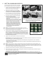
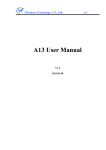
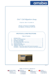
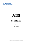
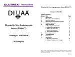
Cell Invasion Assay Kit
Product No.: CIA101 & CIA200
96-well, 3-D Assay for Investigating
Cell Invasion of Adherent Cell Lines
Protocol & Instructions
Patent Pending
Platypus Technologies, LLC
5520 Nobel Drive, Suite 100
Madison, WI 53711
Toll Free: (866) 296-4455
Phone: (608) 237-1270
Fax: (608) 237-1271
www.pLatypustech.com
Bringing Science to the SurfaceTM
RM0027.02
Oris™ CELL INVASION ASSAY
I.
INTRODUCTION
The Oris™ Cell Invasion Assay is a reproducible, sensitive, and flexible assay that can be used to monitor cell invasion.
Formatted for a 96-well plate, the assay utilizes Oris™ Cell Seeding Stoppers (made from a medical-grade silicone) to
restrict cell seeding to the outer annular regions of the wells. Removal of the stoppers reveals a 2 mm diameter
unseeded region in the center of each well, i.e., the invasion zone, into which the seeded cells may then invade. The
Oris™ Detection Mask is applied to the plate bottom and restricts visualization to the invasion zone, thus allowing only
invading cells to be detected (see Figure 1). The Oris™ Cell Invasion Assay is designed to be used with any
commercially available stain or labeling technique and the readout can be performed by microscopic examination or by
using a plate reader. The Oris™ Cell Invasion Assay kit has been uniquely designed to detect cellular invasion in vitro
within a 3-dimensional extracellular matrix comprised of a basement membrane extract (BME) purified from Murine
Engelbreth-Holm-Swarm (EHS) tumor. The Oris™ Cell Invasion Assay system has been designed for use with
adherent cell cultures. Performance of the Oris™ Cell Invasion Assay was optimized using the invasive HT-1080
fibrosarcoma and the non-invasive 3T3-Swiss albino fibroblast cell lines.
Using the Oris™ Cell Invasion Assay offers the following benefits:
• Membrane-free Cell Invasion- no cumbersome
•
cell culture inserts or Transwell® membrane
devices to limit cellular movement; there is no
•
membrane that restricts the ability to image cells.
• Preserve Cell Morphology- the Oris™ Cell
•
Invasion Assay provides a more native 3-D
environment since cells are ensconced in an
extracellular matrix.
Rehydrate
BME Coating
& Populate
Wells with
Stoppers
Apply Mask
(shown in black)
and Add Cells to
BME-coated Wells
Incubate to
Allow Cell
Attachment in
Outer Region
of Wells
Remove
Stoppers &
Apply BME
Overlay
Real-time Monitoring- Invasion-induced changes
in cell structure can be monitored in real-time with a
microscope or digital imaging system.
Versatile- analyze cells using multiple fluorescent
probes, labels or colorimetric stains in a single well.
Flexible- design kinetic or endpoint assays without
the use of special instrumentation using a
fluorescence plate reader.
Incubate to
Allow Cells to
Invade Into the
Detection Zone
of Wells
Figure 1. Schematic of Oris™ Cell Invasion Assay
II.
Seeded Cells that
HAVE NOT Invaded
into the Detection
Zone are Blocked
from View
Observe
Invaded Cells
Using
Microscope or
Plate Reader
ORISTM PLATE DIMENSIONS (per well)
Diameter of Well
6.5 mm
Diameter of Stopper Space (Detection Zone)
2 mm
Suggested Media Volume per Well (populated with Stoppers)
100 µl
Effective Area of Outer Annular Region (seeding region) per Well
30.03 mm2
Effective Area of Central Detection Zone per Well
3.14 mm2
Important: Read Instructions Before Performing any OrisTM Assay.
Platypus Technologies®
Bringing Science to the SurfaceTM
RM0027.02
pg. 2
III.
MATERIALS PROVIDED
Product No.: CIA101
Component
Oris™ BME Coated 96-well Plate
Oris™ Cell Seeding Stoppers
Oris™ Detection Mask
Oris™ Stopper Tool
Oris™ Basement Membrane Extract (BME)
Quantity
1
96
1
1
5 mL
Storage
Refrigerate at 4°C
Room Temperature
Room Temperature
Room Temperature
< -20°C*
Product No.: CIAU200
Component
Oris™ BME Coated 96-well Plates
Oris™ Cell Seeding Stoppers
Oris™ Detection Mask
Oris™ Stopper Tool
Oris™ Basement Membrane Extract (BME)
Quantity
2
2 x 96
2
2
2 x 5 mL
Storage
Refrigerate at 4°C
Room Temperature
Room Temperature
Room Temperature
< -20°C*
* Oris™ BME reagent can be stored at -20°C in a manual defrost freezer if kit will be used within 3 months of receipt. For
long-term storage, store Oris™ BME reagent at -80°C until the expiration date of the kit.
IV.
MATERIALS REQUIRED
•
•
•
•
•
•
•
•
•
V.
Biological Cells
Cell Culture Growth Medium with Fetal Bovine Serum
Sterile PBS
Serum-Free Cell Culture Medium
Sterile Pipette Tips and Pipette or Multi-Channel Pipette
Trypsin or Non-Enzymatic Cell Removal Reagent or Scraper
Inverted Microscope (optional)
Fluorescence Microplate Reader (optional)
Cell Labeling Fluorescent Agent (eg., CellTracker™ Green, Calcein AM) - required if performing assay readout via plate
reader.
PRECAUTIONS AND RECOMMENDATIONS
• For Research Use Only. Not for use in diagnostic procedures.
• The kit should not be used beyond the expiration date on the kit label.
• Recommendations for Oris™ BME:
- Thaw on ice (2-8°C) overnight.
- The inherent variability of invasiveness between different cell lines may make it necessary to dilute the Oris™ BME prior
to application. Less invasive cell lines may require a more permissive barrier. Do not dilute Oris™ BME lower than
9 mg/mL as this will inhibit gel formation.
- Aliquot and freeze any remaining Oris™ BME. Avoid repeated freeze-thaw cycles.
• It should be appreciated that there will be lot-to-lot differences in the performance of biologically derived materials, such as
BME and FBS that may impact the extent of invasion for a given cell line, as well as in the behavior of different cell lines with
respect to invasive capabilities. To minimize the effects of biological variation in cell invasion and improve reproducibility,
we recommend, 1) conducting invasion assays with cells lines within a narrow passage number [10-12 passages], and 2)
using an invasion inhibitor (i.e., Latrunculin) with an established dose response curve. Controlling these two parameters will
help establish a reference point when comparing data from different experiments.
Platypus Technologies®
Bringing Science to the SurfaceTM
RM0027.02
pg. 3
V.
ORISTM CELL INVASION ASSAY PROTOCOL
The following steps should be performed in a biological
hood using aseptic technique to prevent contamination.
1.
If desired, cells can be starved by incubating for 18 24 hours in serum-free medium prior to assay (0.5%
fetal bovine serum may be used if needed).
2.
Pipette 100 µl of PBS into each well of the BMEcoated plate and let the plate sit at room temperature
(or 37°C) for 1 hour to reconstitute the BME layer.
3.
Remove the PBS from each well and incubate plate
(37°C) for 20 - 30 minutes to allow the plate to dry.
4.
Under sterile conditions, populate the 96-well plate
with Oris™ Cell Seeding Stoppers:
• Vertically position the tip ends of two, 4-stopper
strips into one full column of 8 wells at a time
(Figure 2A).
• Gently press down on the strip backbone to partially
insert the stoppers halfway into the well (Figure 2B).
• When both stopper strips have been partially
inserted in 1 column, ensure that the position of the
stoppers is vertical with respect to the well wall,
making any necessary adjustments (Figure 2C).
• Using the Oris™ Stopper Tool, firmly press down on
the strip backbone to fully insert the stoppers into
each well (Figure 2D and 2E). Repeat for all
remaining columns.
A)
B)
C)
D)
E)
Figure 2. Stopper Insertion Process. A) Placement of Stoppers into
Wells, B) Close-up of Stoppers Partially Inserted into Wells,
C) Proper Placement of Stoppers, D) Pressing of Stoppers
into Wells, and E) Fully Inserted Stoppers
A
B
C
NOTE: It is extremely important to ensure that the stoppers are inserted
perpendicular to the well bottom and fully engaged with the bottom of the well.
Failure to do so will increase the CV of your data set.
5.
6.
Visually inspect the underside of the populated 96-well plate to ensure that the
bottom of the Oris™ Cell Seeding Stoppers are firmly sealed against the well
bottom. To inspect the stoppers, turn the plate over and examine the stoppers
for sealing (see Figure 3). If incomplete sealing is observed, return the plate to
the upright position and use a sterile instrument to gently push the stopper back
into the well until sealing is observed.
Figure 3. Stoppers that are A) Partially
Sealed, B) Unsealed, & C)
Completely Sealed
Apply the Oris™ Detection Mask to the bottom of the 96-well plate.
First Time Users: In order to prevent splashing of well contents, familiarize yourself with the attachment and removal of the
Detection Mask before any liquids are placed in the wells.
• Orient the chamfered corners of the mask with those of the 96-well plate, ensuring that the A1 corner of the mask is
aligned with the A1 well of the plate.
• Align the holes in the attachment lugs with the bosses on the bottom of the 96-well plate and gently press the mask onto
the bottom of the plate.
NOTE: It may be necessary to wash the mask with ethanol to remove dust and debris since the mask is not sterile. The
mask may be applied at any point during the assay. For kinetic assays, it is often most convenient to apply the mask at the
beginning of the assay before any liquids are placed in the well. For endpoint assays, using fixed and stained cells, it is often
most convenient to apply the mask just before reading assay results.
7.
If performing a kinetic analysis of Cell Invasion, pre-stain the cells with a fluorescent stain now.
8.
Collect cells and prepare a suspension that is 10-fold greater in density than the optimal seeding concentration using cell
culture growth medium containing 10% FBS.
First Time Users: The optimum seeding density of cells must be determined as an integral part of the design of the cell
invasion assay. Please see Appendix I for a discussion of this process.
Platypus Technologies®
Bringing Science to the SurfaceTM
RM0027.02
pg. 4
9.
Pipette 100 µl of suspended cells into each test well through either of the side ports of the Cell Seeding Stopper.
NOTE: For best results, add or extract media by placing the pipette tip along the wall of the well. Care should be taken not
to disturb the Cell Seeding Stopper or the BME layer when introducing the pipette tip into the well. A gel loading tip may be
useful.
10. IMPORTANT: Lightly tap the plate on your work surface to evenly distribute well contents (extreme tapping may result in
splashing of well contents and lead to contamination).
11. Incubate the seeded plate containing the Oris™ Cell Seeding Stoppers in a humidified chamber (37°C, 5% CO2) for at least
4 hours (cell line dependent) to permit cell attachment.
A)
B)
C)
12. Remove plate from incubator.
13. Using the Oris™ Stopper Tool, remove all stoppers (see Figure
4).
• Secure the 96-well plate by holding it firmly against the deck of
your work space. Slide the tines of the stopper tool under the
backbone of the stopper strip, keeping the underside of the tool
flush with the top surface of the plate.
• Lift the stopper tool vertically to gently remove the stopper.
D)
E)
NOTE: DO NOT use the stopper tool as a lever to pry the
stoppers from the well (see Figure 6E), as doing so may cause
displacement of seeded cells.
14. Remove media with a pipette and gently wash wells with 100 µl
PBS (or media) to remove any unattached cells. Do not aspirate
using an in-house vacuum.
15. Add 40 µl of the thawed Oris™ BME to each well (supplements,
such as FBS or growth factors, may be mixed with BME, if
desired).
Figure 4. Removal of Stoppers. Panels A, B, and C) Position
the Tines of the Stopper Tool between the Stopper
Tips, D) Lift Vertically, and E) Do NOT Pry
Stoppers
NOTE: Oris™ BME gels in 5-10 min above 15°C, therefore, you must keep the thawed Oris™ BME on ice until ready to use.
In addition, the use of chilled pipette tips/reservoirs might be beneficial. Since different cell lines and different treatments
can result in a wide range of invasive behavior, the BME overlay may be optimized to fit each experiment by diluting this
material to permit more invasion. However, do NOT dilute the Oris™ BME below a concentration of 9 mg/mL.
16. Incubate plate in a humidified chamber (37°C, 5% CO2) for 30 - 60 minutes to permit polymerization of Oris™ BME.
17. For reference purposes, designate several ‘reference’ wells to which a staining agent will be added (t=0 pre-invasion
controls). After staining of the reference wells, capture well data via microscope, plate reader or digital imaging system.
18. Add 100 µl of serum-free cell culture medium on top of the BME overlay.
Optional: Invasion inhibitors or stimulants may be added to the media.
19. Incubate plate in a humidified chamber (37°C, 5% CO2) to permit cell invasion (length of incubation is cell line dependent).
The Oris™ BME will remain gelled for up to 14 days. Refresh media or supplements, every 48 - 72 hours, as needed, for
the duration of the invasion experiment.
20. If performing an endpoint analysis of cell invasion, apply stain and read results (see Appendix I).
NOTE: Oris™ Cell Seeding Stoppers are for single use only; Platypus can not guarantee the integrity or performance of the
stopper material after a second sterilization procedure.
Platypus Technologies®
Bringing Science to the SurfaceTM
RM0027.02
pg. 5
VI.
DATA ACQUISITION
The readout of the Oris™ Cell Invasion Assay can be conducted at any time, allowing the user to perform a kinetic assay or an
endpoint assay. The Oris™ Cell Invasion Assay is designed to be used with any commercially available stain or labeling
technique. The readout can be performed by microscopic examination or by plate reader.
Microscopic Analysis
• Cell counting or image capture / analysis (using software, such as Image J freeware, available from NIH)
Plate Reader Analysis
• Setup on individual plate readers varies according to make and model. Consult your user manual for proper operation.
• The plate reader MUST be set to use the bottom probe read.
Sample Data Obtained via Microscopic Examination and Plate Reader are shown in Figure 5.
5
• Wells were seeded with 50,000 HT-1080 cells (i.e., 100 µl of 5x10 cells/mL) that had been serum starved for 18 hours and the plate was
TM
then incubated for 4 hours. The stoppers were removed from the wells and the Oris BME (with 10% FBS or without FBS) was overlayed
on the cells. The plate was incubated in a humidified chamber for 48 hours to permit cell invasion. Cells were labeled with Calcein AM and
images were captured using a Zeiss Axiovert microscope (5X magnification). Fluorescence in the analytic zone was quantified by using a
plate reader. Each column represents the mean +/- SD of at least 4 wells. A non-invasive cell line, 3T3-Swiss albino, served as the
negative control. The images below, captured without a detection mask in place, illustrate representative data from pre-invasion (t=0 hrs)
and post-invasion (t=48 hrs) wells. The graph depicts the average RFU’s in the invasion zones for each condition, confirming the invasion
augmenting effect of FBS on serum starved HT-1080 cells and the lack of invasion by non-invasive 3T3-Swiss albino cells.
• As observed in Figure 5, HT-1080 cells formed invadopodial structures projecting into the central analytic zone (panel 1b) as compared to a
time zero control (panel 1a).
• Invasion by serum-starved HT-1080 cells was augmented by including 10% v/v fetal bovine serum (FBS) in the BME overlay (panel 1c).
• In contrast, as observed in images 2b and 2c, the non-invasive 3T3-Swiss albino cells did not form any invadopodial structures and showed
a nominal amount of FBS-independent migration into the central detection zone as compared to a time zero control (image 2a).
Behavior of Invasive HT-1080 Cells in Oris
1a
1b
TM
Cell Invasion Assay
1c
Measurement of Cells in the OrisTM Detection Zone Using a Plate Reader
t=0
t=48h (no FBS)
t=48h (10% FBS)
Behavior of Non-Invasive 3T3-Swiss albino Cells in Oris
Invasion Assay
2a
2b
TM
Cell
2c
Invasive HT-1080 Cells
t=0
t=48h (no FBS)
Non-Invasive 3T3-Swiss albino Cells
t=48h (10% FBS)
Figure 5. Cell Invasion Data obtained via Microscopic Examination and Plate Reader Analysis
Platypus Technologies®
Bringing Science to the SurfaceTM
RM0027.02
pg. 6
Immunostaining Analysis
• As determined by immunostaining (shown in Figure 6), the structures projecting into the BME formed by the invasive HT-1080 cells
exhibited classic hallmarks of invadopodia, namely colocalization of F-actin with cortactin (panel a), and the expression of proteases such
as MMP-9 and cathepsin B (panels b and c). These invadopodia could also be observed in 3-dimensions at different focal points along the
z-axis. This is in contrast to any migratory activity observed by non-invasive cells that occurs only within the 2-dimensional space of the x
and y axes (data not shown).
TM
• Cells from the Oris Cell Invasion Assay were fixed, permeabilized, and pre-treated by sequential incubations in 3.7% formaldehyde, PBS,
TM
0.5% Triton X-100, PBS, Image-iT FX signal enhancer, and PBS. Immunostaining was performed by incubating with primary antibodies
(2 hours, 37°C) and Alexa Fluor® 488 conjugated secondary antibodies at 1:200 dilution (1 hour, 37°C) followed by an Alexa Fluor® 555
phalloidin counterstain (1 hour). Images were collected using a Nikon TE300 inverted microscope equipped with a Photometrics Coolsnap
TM
fx CCD camera, deconvolved using Slidebook v4.2 (Intelligent Imaging Innovations) and processed using Adobe® Photoshop® CS2
(Adobe Systems).
Anti-Cortactin
a
F-actin is present in all eukaryotic cells, it is a major component of the
cytoskeleton and functions to define and maintain the cell shape. Cortactin is a
cellular protein that is present in and promotes the formation of lamellipodia and
invadopodia. These structures propel cells over surfaces as they move toward a
target. Results depicted above demonstrate distinct areas of Cortactin and F-actin
colocalization characteristic of invadopodia (Ref. 1 & 2).
Anti-Cathepsin B
Cathepsin B is a lysosomal protease that facilitates cell invasion by degrading the
surrounding extracellular matrix. Results depicted above demonstrate distinct
areas of lysosomal and peripheral cellular expression of Cathepsin B
characteristic of protease expression observed in cancer cell invasion (Ref. 3).
b
Anti-MMP-9
c
MMP-9 is a protease that is involved in matrix degradation in cancer cell invasion.
Results depicted above demonstrate evidence of MMP-9 expression and secretion
into surrounding areas of ECM, which are established characteristics of proteases
observed at invadopodia sites (Ref. 1& 2).
Figure 6. Confirmation of Cell Invasion using Immunostains
References:
1. Weaver, AM; Clin Exp Metastasis 2006. "Invadopodia: specialized cell structures for cancer invasion." 23:97-105.
2. Furmaniak-Kazmierczak et al; Circulation Research 2007. "Formation of extracellular matrix-digesting invadopodia by primary aortic
smooth muscle cells." 100:1328-1336.
3. Hulkower, KI et al; Eur. J. Biochem 2000. "Fluorescent microplate assay for cancer cell-associated cathepsin B." 267:4165-4170.
Oris™ is a trademark of Platypus Technologies, LLC.
CellTracker™ Green is a trademark of Invitrogen Corporation.
Transwell® is a registered trademark of Corning, Inc.
Platypus Technologies®
Bringing Science to the SurfaceTM
RM0027.02
pg. 7
VII. ORDERING INFORMATION
Product No.
CIA101
CIA200
Product Description
Oris™ Cell Invasion Assay, 1-pack:
Oris™ BME-coated 96-well plate, 1
OrisTM Basement Membrane Extract, 5 mL
Oris™ Cell Seeding Stoppers, 96
Oris™ Detection Mask, 1
Oris™ Stopper Tool, 1
Oris™ Cell Invasion Assay, 2-pack:
Oris™ BME-coated 96-well plates, 2
OrisTM Basement Membrane Extract, 2 x 5 mL
Oris™ Cell Seeding Stoppers, 2 x 96
Oris™ Detection Mask, 2
Oris™ Stopper Tool, 2
Package Size
1-pack
2-pack
To place an order, visit the Platypus Technologies website at: www.platypustech.com/order_main.html.
For technical assistance, contact Technical Support at (866) 296-4455 or [email protected].
VIII.
TERMS & CONDITIONS
Certain uses of these products may be covered by U.S. Pat. No. 6,284,197, No. 7018838, No. 10/597,118, No. 11/342,413, and No. 60/836,109,
licensed to PLATYPUS. Certain applications of PLATYPUS products may require licenses from other parties. Determining the existence and
scope of such third party intellectual property is the responsibility of the PURCHASER. Purchase of the product provides the PURCHASER with
a limited non-transferable license under any PLATYPUS patents or patent applications to use the product for internal research unless there is a
written limitation to this license in the product literature. PURCHASER is responsible for carefully reviewing the product literature and respecting
any limitations to this license, e.g. limitations for commercial use or research by for-profit institutions. These products may not be resold,
modified for resale, used to manufacture commercial products, or used to develop commercial products without the express written approval of
PLATYPUS.
These products are intended for research or laboratory use only and are not to be used for any other purposes, including, but not limited to,
unauthorized commercial purposes, in vitro diagnostic purposes, ex vivo or in vivo therapeutic purposes, investigational use, in foods, drugs,
devices or cosmetics of any kind, or for consumption by or use in connection with or administration or application to humans or animals.
PLATYPUS warrants that its products shall conform substantially to the description of such goods as provided in product catalogues and
literature accompanying the goods until their respective expiration dates or, if no expiration date is provided, for 6 months from the date of receipt
of such goods. PLATYPUS will replace, free of charge, any product that does not conform to the specifications. This warranty limits
PLATYPUS's liability only to the replacement of the nonconforming product.
THIS WARRANTY IS EXCLUSIVE AND PLATYPUS MAKES NO OTHER WARRANTY, EXPRESS OR IMPLIED, INCLUDING WITHOUT
LIMITATION, ANY IMPLIED WARRANTY OF MERCHANTABILITY OR FITNESS FOR A PARTICULAR PURPOSE. The stated express
warranties, and the remedy provided for breach thereof, are in lieu of all other liability or obligations of PLATYPUS for any damages whatsoever
arising out of or in connection with the delivery, use, misuse, performance, or the inability to use any of its products. IN NO EVENT SHALL
PLATYPUS BE LIABLE UNDER ANY LEGAL THEORY (INCLUDING BUT NOT LIMITED TO CONTRACT, NEGLIGENCE, STRICT LIABILITY
IN TORT, OR WARRANTY OF ANY KIND) FOR ANY INDIRECT, SPECIAL, INCIDENTAL, CONSEQUENTIAL, OR EXEMPLARY DAMAGES
(INCLUDING BUT NOT LIMITED TO LOST PROFITS) EVEN IF PLATYPUS HAD NOTICE OF THE POSSIBILITY OF SUCH DAMAGES.
Without limiting the effect of the preceding sentence, PLATYPUS's maximum liability, if any, shall not exceed the purchase price paid by
PURCHASER for the product.
This warranty shall not be effective if PLATYPUS determines, in its sole discretion that PURCHASER has altered or misused the goods or has
failed to use or store them in accordance with instructions furnished by PLATYPUS. PLATYPUS’s sole and exclusive liability and
PURCHASER’s exclusive remedy with respect to goods proved to PLATYPUS’s satisfaction (applying analytical methods reasonably selected by
PLATYPUS) to be defective or nonconforming shall be the replacement of such goods free of charge, upon the return of such goods in
accordance with our instructions, although at its discretion, PLATYPUS may provide a credit or refund. If PLATYPUS manufactures custom
goods for PURCHASER based on instructions, specifications, or other directions provided by PURCHASER, PLATYPUS shall not be liable for
the lack of sufficiency, fitness for purpose or quality of the goods to the extent attributable to such instructions, specifications, or other directions.
PLATYPUS shall not be liable for any loss, damage or penalty as a result of any delay in or failure to manufacture, deliver or otherwise perform
hereunder due to any cause beyond PLATYPUS’s reasonable control.
PLATYPUS shall not be liable for injury or damages resulting from the use or misuse of any of its products.
Platypus Technologies®
Bringing Science to the SurfaceTM
RM0027.02
pg. 8
APPENDIX I: Determining Optimal Cell Seeding Concentration
This appendix is intended to assist in determining the cell seeding density needed to achieve confluency of your cell
line when using the Oris™ Cell Invasion Assay. To that end, several dilutions of cell suspensions will be investigated.
NOTE: The Oris™ Detection Mask MUST be removed from the 96-well plate prior to the start of the following steps:
1. Collect cells by trypsinization or by non-enzymatic means (such as mechanical scraping) and calculate total
number of cells.
2. Pellet cells by centrifugation and resuspend to a final concentration of 1,000,000 cells/mL in culture media.
3. Seed a 100 µl portion of cells, at 2-fold serial dilutions in the 96-well plate starting at 100,000 cells/well (a
suggested starting amount), as shown below. Keep in mind that the cell seeding area of the well with the stopper
in place is ~ 0.3 cm2 and based on the typical seeding density of your cells, you can infer the appropriate cell
number for your first serial dilution.
Column
Cells / well
Number of wells
1
100,000
6
2
50,000
6
3
25,000
6
4. Incubate the plate in a humidified chamber (37°C, 5% CO2) for 16 hours (or ample time to allow for one doubling)
with cell seeding stoppers in place.
5. Following cell attachment, remove the Oris™ Cell Seeding Stoppers from each well (see Figure 6) and gently
wash the wells with PBS to remove non-adhered cells.
• Secure the 96-well plate by holding it firmly against the deck of your work space. Slide the tines of the stopper tool under
the backbone of the stopper strip, keeping the underside of the tool flush with the top surface of the plate.
• Lift the stopper tool vertically to gently remove the stopper. Do not use the tool as a lever to pry the stoppers from the
well as doing so may cause displacement of the seeded cells.
6. Visually inspect the cells by microscope to determine the cell seeding concentration that yields a confluent layer.
NOTE: If you plan to obtain the results of the Oris™ Cell Invasion Assay via colorimetric or microscopic analysis, you
have successfully determined the optimal cell seeding concentration for your cell line. Proceed to Step 2 of the Cell
Invasion Assay Protocol. If you plan to obtain the results of the Oris™ Cell Invasion Assay via a fluorescence plate
reader, proceed with the following steps to optimize your plate reader settings.
7. The Oris™ Cell Invasion Assay has been designed to work with all types of fluorescence stains and staining
techniques. The precise method for staining cells with fluorescence stains varies according to the nature of the
individual stain. Please consult the manufacturer of your fluorescence stain for specific considerations.
First Time Users: For a guide to using Calcein AM, see below:
a.
b.
c.
d.
e.
f.
g.
Carefully remove liquid from wells, being very careful not to disturb the BME overlay.
Wash wells with 100 µl of PBS (containing both Calcium and Magnesium).
Add 100 µl of Calcein AM to each well at an appropriate concentration [for a fully-seeded 96-well plate, combine
5 µl of reconstituted Calcein AM (1mg/mL in dry DMSO) with 10 mL of phenol red-free and serum-free media or
1x PBS].
Incubate plate at 37°C for 30 - 60 minutes.
Remove plate from incubator and remove staining solution. Do not aspirate using an in-house vacuum.
Wash wells with PBS, removing PBS at last step.
Attach mask and read in plate reader at 485/528, sensitivity 55 nm.
8. Apply the Oris™ Detection Mask to the plate.
9. Using the bottom probe of a fluorescence plate reader, obtain the total output from each well (adjust the gain
settings to achieve optimal dynamic range). To determine optimal dynamic range, consider the following factors:
a)
b)
The gain setting that permits detection of the lowest concentration of cells.
The gain setting that permits discrimination between cell numbers at higher densities.
NOTE: When using a plate reader to analyze the Oris™ Cell Invasion Assay, it is important to stain cells using a
fluorescence reagent that uniformly stains cells.
Once you have determined the optimal cell seeding concentration for your cell line, proceed to Step 2 of the Cell
Invasion Assay Protocol.
Platypus Technologies®
Bringing Science to the SurfaceTM
RM0027.02
pg. 9