1
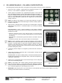
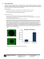
Bringing Science to the SurfaceTM OrisTM Cell Migration Assay – Collagen I Coated Product No.: CMACC1.101 & CMACC5.101 96-well, 2-D Assay for Investigating Cell Migration of Adherent Cell Lines on Collagen I PROTOCOL & INSTRUCTIONS Table of Contents I. II. III. IV. V. VI. VII. VIII. INTRODUCTION .......................................................................................... 2 ORISTM PLATE DIMENSIONS ..................................................................... 3 MATERIALS PROVIDED ............................................................................. 3 MATERIALS REQUIRED ............................................................................. 3 CELL MIGRATION ASSAY – COLLAGEN I COATED PROTOCOL ........... 4 DATA ACQUISITION.................................................................................... 6 ORDERING INFORMATION ........................................................................ 7 TERMS & CONDITIONS .............................................................................. 7 Appendix I: Determining Optimal Cell Seeding Concentration..................... 8 Appendix II: Determining Optimal Fluorescence Plate Reader Settings ..... 8 Platypus Technologies, LLC 5520 Nobel Drive, Suite 100, Madison, WI 53711 Toll Free: 866.3296.4455 Phone: 608.237.1270 Fax: 608.237.1271 www.platypustech.com SP0061.04 Oris™ CELL MIGRATION ASSAY – COLLAGEN I COATED I. INTRODUCTION The Oris™ Cell Migration Assay – Collagen I Coated is a reproducible, sensitive, and flexible assay that can be used to monitor cell migration. Formatted for a 96-well plate, the assay utilizes Oris™ Cell Seeding Stoppers made from a medicalgrade silicone to restrict cell seeding to the outer annular regions of the wells. Removal of the stoppers reveals a 2mm diameter unseeded region in the center of each well, i.e., the detection zone, into which the seeded cells may then migrate. The Oris™ Detection Mask is applied to the plate bottom and restricts visualization to the detection zones, allowing only cells that have migrated to be detected (see Figure 1). The Oris™ Cell Migration Assay – Collagen I Coated is designed to be used with any commercially available stain or labeling technique. Readout can be performed by microscopy or use of a microplate reader. The Oris™ Cell Migration Assay – Collagen I Coated system has been designed for use with adherent cell cultures. This assay has been successfully used with HT-1080, T47D, MCF10A, HeLa, HUVEC, and MDA-MB-231 cell lines. Using the Oris™ Cell Migration Assay – Collagen I Coated offers the following features & benefits: Membrane-free Migration - perform studies Versatile - analyze data using multiple probes in a single without manipulating transmembrane inserts. well by using a microscope, digital imager, or fluorescence microplate reader. Reproducible Results obtain well-to-well CV's < 12% Flexible - perform kinetic or endpoint cell migration due to the unique design. assays without the use of special instrumentation. Preserves Cell Morphology - monitor changes in cell Specific – measure cell migration directly on an extracellular structure in real-time. matrix coated surface. Seed & Adhere Cells onto Oris™ Collagen I Coated Plate Remove Stoppers to Create Detection Zone Allow Cells to Migrate into Detection Zone Analyze Cells in Detection Zone: Microplate Reader Analysis - Detection Mask Attached; Image Analysis - No Mask Required Figure 1. Schematic of Oris™ Cell Migration Assay – Collagen I Coated Platypus Technologies, LLC SP0061.04 5520 Nobel Drive, Suite 100 Madison WI 53711 USA www.platypustech.com Toll Free: 866.296.4455 Phone: 608.237.1270 Fax: 608.237.1271 pg. 2 II. ORISTM PLATE DIMENSIONS (per well) Diameter of Well 6.5 mm Diameter of Stopper Space (Detection Zone) 2 mm Suggested Media Volume per Well (populated with Stoppers) 100 µL Effective Area of Outer Annular Region (seeding region) per Well 30.03 mm2 Effective Area of Central Detection Zone per Well 3.14 mm2 Plate Height 14.9 mm Plate Height with Lid (with Oris TM Cell Seeding Stoppers) 17.9 mm Offset of Wells (A-1 location, X) 14.4 mm Offset of Wells (A-1 location, Y) 11.2 mm Distance between Wells 9 mm (on center) Well Depth 12.2 mm Thickness of Well Bottom 0.25 mm Well Coating Material Collagen I, rat-tail Storage Conditions Refrigerate (4°C) Important: Read Instructions Before Performing any OrisTM Assay. III. MATERIALS PROVIDED Product No.: CMACC1.101 Oris™ Collagen I Coated, 96-well (black, clear bottom) Plate with Oris™ Cell Seeding Stoppers, 1 Oris™ Detection Mask, 1 Oris™ Stopper Tool, 1 IV. Product No.: CMACC5.101 Oris™ Collagen I Coated, 96-well (black, clear bottom) Plates with Oris™ Cell Seeding Stoppers, 5 Oris™ Detection Mask, 1 Oris™ Stopper Tool, 1 MATERIALS REQUIRED Biological Cells Sterile PBS (containing both Calcium and Magnesium) Complete Cell Culture Growth Medium (containing serum) Sterile Pipette Tips/Pipette or Multi-Channel Pipette Trypsin or Cell Scraper Inverted Microscope (optional) Fluorescence Microplate Reader (optional) Cell Culture Labeling Medium (phenol red-free/serum-free media) Cell Labeling Fluorescent Agent (eg., CellTracker™ Green, Calcein AM) - required if performing assay readout via microplate reader. Oris™ is a trademark of Platypus Technologies, LLC. CellTracker™ Green is a trademark of Invitrogen Corporation. Platypus Technologies, LLC SP0061.04 5520 Nobel Drive, Suite 100 Madison WI 53711 USA www.platypustech.com Toll Free: 866.296.4455 Phone: 608.237.1270 Fax: 608.237.1271 pg. 3 V. CELL MIGRATION ASSAY – COLLAGEN I COATED PROTOCOL The following steps should be performed in a biological hood using aseptic technique to prevent contamination. 1. Remove the Oris™ Collagen I Coated Plate from refrigeration and place on lab bench for ~1 hour to allow it to equilibrate to room temperature. 2. Visually inspect the underside of the populated 96-well plate to ensure that the Oris™ Cell Seeding Stoppers are firmly sealed against the bottom of the plate. To inspect the stoppers, turn the plate over and examine the stoppers for sealing (see Figure 2). If incomplete sealing is observed, return the plate to the upright position and use a sterile instrument to gently push the stopper back into the well until sealing is observed. NOTE: The sealing of the stoppers can be most easily observed if the plate is tipped at an angle and viewed under indirect light to reveal the “bullseye” pattern at the bottom of each well. 3. Apply the Oris™ Detection Mask to the bottom of the 96-well plate if microplate reader data is being collected. The Detection Mask is not necessary if collecting imaging data. First Time Users: In order to prevent splashing of well contents, familiarize yourself with the attachment and removal of the Detection Mask before any liquids are placed in the wells. A B C Figure 2. Stoppers that are A) Partially Sealed B) Unsealed C) Completely Sealed Aperture Orientation A-1 Corner Chamfer Attachment Lugs Orient the chamfered corners of the mask with those of the 96-well plate, ensuring that the A1 corner of the mask is aligned with the A1 well of the plate (see Figure 3). Align the holes in the attachment lugs with the bosses on the bottom of the 96well plate. Gently press the mask until it is flush with the bottom of the 96-well plate. NOTE: It may be necessary to wash the mask with ethanol to remove dust and debris since the mask is not sterile. The mask may be applied at any point during the assay. For kinetic assays, it is often most convenient to apply the mask at the beginning of the assay before any liquids are placed in the well. For endpoint assays, using fixed and stained cells, it is often most convenient to apply the mask just before reading assay results. 4. If performing a kinetic analysis of cell migration, pre-label cells with a fluorescent stain now. 5. Collect cells and prepare a suspension that is 10-fold greater in density than the optimal seeding concentration. Figure 3. Features of Detection Mask First Time Users: The optimum seeding density of cells must be determined as an integral part of the design of the cell migration assay. Please refer to Appendix I for a discussion of this process. 6. Pipette 100 µL of suspended cells into each test well through one of the side ports of the Oris™ Cell Seeding Stopper. NOTE: For best results, add or extract media by placing the pipette tip along the wall of the well (see Figure 4). Care should be taken not to disturb the Collagen I Coating or the Oris™ Cell Seeding Stopper when introducing the pipette tip into the well. A slender/elongated tip or a gel loading tip may be useful. 7. Figure 4. Media is Added with Single or Multi-Channel Pipette IMPORTANT: Lightly tap the plate on your work surface to evenly distribute well contents (extreme tapping may result in splashing of well contents and lead to contamination). Platypus Technologies, LLC SP0061.04 5520 Nobel Drive, Suite 100 Madison WI 53711 USA www.platypustech.com Toll Free: 866.296.4455 Phone: 608.237.1270 Fax: 608.237.1271 pg. 4 V. CELL MIGRATION ASSAY – COLLAGEN I COATED PROTOCOL, continued 8. Incubate the seeded plate containing the Oris™ Cell Seeding Stoppers in a humidified chamber (37°C, 5% CO2) for 4 to 18 hours (cell line dependent) to permit cell attachment. 9. Remove plate from incubator. 10. Designate several ‘reference’ wells in which the stoppers will remain in place until results are read (t=0 pre-migration controls). 11. Using the Oris™ Stopper Tool, remove all other stoppers (see Figure 5). NOTE: It may be necessary to wash the Oris™ Stopper Tool with 70% ethanol as the Stopper Tool is not sterile. Secure the 96-well plate by holding it firmly against the deck of your work space. Slide the tines of the Oris™ Stopper Tool under the backbone of the stopper strip, keeping the underside of the Oris™ Stopper Tool flush with the top surface of the plate. Lift the Oris™ Stopper Tool vertically to gently remove the stoppers. NOTE: DO NOT use the Oris™ Stopper Tool as a lever to pry the stoppers from the well (see Figure 5E), as doing so may cause displacement of seeded cells and may distort the detection zone area. A) B) D) C) E) 12. Remove media with a pipette and gently wash wells with 100 µL of sterile PBS (or media) to remove any unattached cells. Do not aspirate using an in-house vacuum. 13. Add 100 µL of fresh culture media to each well. 14. Incubate plate in a humidified chamber (37°C, 5% CO2) to permit cell migration. Cells may be examined microscopically throughout the incubation period to monitor progression of migration. Migration time will vary depending upon cell type, experimental design, and plate coating. Figure 5. Removal of Stoppers. Panels A, B, and C) Position the Tines of the Stopper Tool between the Stopper Tips, D) Lift Vertically, and E) Do NOT Pry Stoppers 15. If performing an endpoint analysis of cell migration, stain cells with a fluorescent stain after sufficient migration has occurred. Refer to Section VI and Appendix II for further information on data acquisition and fluorescence staining technique. NOTE: Oris™ Cell Seeding Stoppers are for single use only; Platypus cannot guarantee the integrity of the stopper material after a second sterilization procedure. Platypus Technologies, LLC SP0061.04 5520 Nobel Drive, Suite 100 Madison WI 53711 USA www.platypustech.com Toll Free: 866.296.4455 Phone: 608.237.1270 Fax: 608.237.1271 pg. 5 VI. DATA ACQUISITION The readout of the Oris™ Cell Migration Assay – Collagen I Coated can be conducted at any time, allowing the user to perform a kinetic assay or an endpoint assay. The Oris™ Cell Migration Assay – Collagen I Coated is designed to be used with any commercially available stain or labeling technique. The readout can be performed by using a microscope, a microplate reader, or a High Content Screening or High Content Imaging Analysis platform. Microscopy Analysis Cell counting or image capture / analysis software, such as NIH ImageJ freeware, can be used. Note: Microscopy observations are possible using phase contrast or bright field microscopy. No need to attach the Oris™ Detection Mask to the Oris™ plate. Microplate Reader Analysis Attach the Oris™ Detection Mask to the bottom of the Oris™ plate (refer to Section V, step 3). Optimal settings will vary according to the microplate reader make and model. Consult Appendix II and the equipment user manual for your particular instrument. The microplate reader MUST be set to read from the bottom of the plate. Sample data using a fluorescent stain and microplate reader analysis are shown in Figure 6. Collagen I coated wells 5 populated with Oris™ Cell Seeding Stoppers were seeded with 25,000 MDA-MB-231 cells/well (i.e., 100 µL of 2.5x10 cells/mL) and the plate was incubated for 6 hours. The stoppers were then removed from test wells, but remained in place in the pre-migration reference wells. The seeded plate was incubated in a humidified chamber for 24 hours. After 24 hours, stoppers were removed from reference wells and all wells were fluorescently stained with Calcein AM for quantification using a microplate reader. The images below (6A), captured without a detection mask in place, illustrate representative data from pre-migration (t=0 hrs) and post-migration (t = 24 hrs) wells (dashed outlines depict size/location of detection mask). The graph (6B) depicts the average fluorescence signal +/- SD in the detection zones for each condition (n= at least 8 wells/condition). Platypus Technologies, LLC SP0061.04 5520 Nobel Drive, Suite 100 Madison WI 53711 USA www.platypustech.com Toll Free: 866.296.4455 Phone: 608.237.1270 Fax: 608.237.1271 pg. 6 VII. ORDERING INFORMATION Product Name Oris™ Pro Cell Migration Assays Oris™ Pro 384 Cell Migration Assays Coating Size Tissue Culture Treated 1-pack (PROCMA1) 5-pack (PROCMA5) Collagen I Coated 1-pack (PROCMACC1) 5-pack (PROCMACC5) Tissue Culture Treated 5-pack (PRO384CMA5) Collagen I Coated 5-pack (PRO384CMACC5) Tissue Culture Treated 1-pack (CMA1.101) 5-pack (CMA5.101) Collagen I Coated 1-pack (CMACC1.101) 5-pack (CMACC5.101) Biocompatible Gel Biocompatible Gel Oris™ Cell Migration Assays Oris™ Cell Migration Assembly Kits Detection Zone Format Oris™ Cell Seeding Stoppers (pre-populated) Fibronectin Coated 1-pack (CMAFN1.101) 5-pack (CMAFN5.101) TriCoated 1-pack (CMATR1.101) 5-pack (CMATR5.101) Universal (Tissue Culture Treated) 1-pack (CMAU101) 5-pack (CMAU505) FLEX (Tissue Culture Treated) 4-pack (CMAUFL4) Collagen I (low overlay conc.) 1-pack (PROIA1) 3-pack (PROIA3) Biocompatible Gel Collagen I (high overlay conc.) 1-pack (PROIAPLUS1) 3-pack (PROIAPLUS3) Biocompatible Gel Oris™ Pro 96-well Invasion Assays Oris™ Cell Seeding Stoppers (not pre-populated) For a complete list of assays, visit Platypus Technologies at www.platypustech.com/order_main.html. For technical assistance, contact Technical Support at (866) 296-4455 or [email protected]. VIII. TERMS & CONDITIONS Certain uses of these products may be covered by U.S. Pat. No. 7,842,499 issued to or patents applied for by PLATYPUS. Certain applications of PLATYPUS products may require licenses from other parties. Determining the existence and scope of such third party intellectual property is the responsibility of the PURCHASER. Purchase of the product provides the PURCHASER with a limited non-transferable license under any PLATYPUS patents or patent applications to use the product for internal research unless there is a written limitation to this license in the product literature. PURCHASER is responsible for carefully reviewing the product literature and respecting any limitations to this license, e.g. limitations for commercial use or research by for-profit institutions. These products may not be resold, modified for resale, used to manufacture commercial products, or used to develop commercial products without the express written approval of PLATYPUS. These products are intended for research or laboratory use only and are not to be used for any other purposes, including, but not limited to, unauthorized commercial purposes, in vitro diagnostic purposes, ex vivo or in vivo therapeutic purposes, investigational use, in foods, drugs, devices or cosmetics of any kind, or for consumption by or use in connection with or administration or application to humans or animals. PLATYPUS warrants that its products shall conform substantially to the description of such goods as provided in product catalogues and literature accompanying the goods until their respective expiration dates or, if no expiration date is provided, for 6 months from the date of receipt of such goods. PLATYPUS will replace, free of charge, any product that does not conform to the specifications. This warranty limits PLATYPUS's liability only to the replacement of the nonconforming product. THIS WARRANTY IS EXCLUSIVE AND PLATYPUS MAKES NO OTHER WARRANTY, EXPRESS OR IMPLIED, INCLUDING WITHOUT LIMITATION, ANY IMPLIED WARRANTY OF MERCHANTABILITY OR FITNESS FOR A PARTICULAR PURPOSE. The stated express warranties, and the remedy provided for breach thereof, are in lieu of all other liability or obligations of PLATYPUS for any damages whatsoever arising out of or in connection with the delivery, use, misuse, performance, or the inability to use any of its products. IN NO EVENT SHALL PLATYPUS BE LIABLE UNDER ANY LEGAL THEORY (INCLUDING BUT NOT LIMITED TO CONTRACT, NEGLIGENCE, STRICT LIABILITY IN TORT, OR WARRANTY OF ANY KIND) FOR ANY INDIRECT, SPECIAL, INCIDENTAL, CONSEQUENTIAL, OR EXEMPLARY DAMAGES (INCLUDING BUT NOT LIMITED TO LOST PROFITS) EVEN IF PLATYPUS HAD NOTICE OF THE POSSIBILITY OF SUCH DAMAGES. Without limiting the effect of the preceding sentence, PLATYPUS's maximum liability, if any, shall not exceed the purchase price paid by PURCHASER for the product. This warranty shall not be effective if PLATYPUS determines, in its sole discretion that PURCHASER has altered or misused the goods or has failed to use or store them in accordance with instructions furnished by PLATYPUS. PLATYPUS’s sole and exclusive liability and PURCHASER’s exclusive remedy with respect to goods proved to PLATYPUS’s satisfaction (applying analytical methods reasonably selected by PLATYPUS) to be defective or nonconforming shall be the replacement of such goods free of charge, upon the return of such goods in accordance with our instructions, although at its discretion, PLATYPUS may provide a credit or refund. If PLATYPUS manufactures custom goods for PURCHASER based on instructions, specifications, or other directions provided by PURCHASER, PLATYPUS shall not be liable for the lack of sufficiency, fitness for purpose or quality of the goods to the extent attributable to such instructions, specifications, or other directions. PLATYPUS shall not be liable for any loss, damage or penalty as a result of any delay in or failure to manufacture, deliver or otherwise perform hereunder due to any cause beyond PLATYPUS’s reasonable control. PLATYPUS shall not be liable for injury or damages resulting from the use or misuse of any of its products. Platypus Technologies, LLC SP0061.04 5520 Nobel Drive, Suite 100 Madison WI 53711 USA www.platypustech.com Toll Free: 866.296.4455 Phone: 608.237.1270 Fax: 608.237.1271 pg. 7 APPENDIX I: Determining Optimal Cell Seeding Concentration This procedure is intended to assist in determining the cell seeding density needed to achieve confluency of your cell line when using the Oris™ Cell Migration Assay – Collagen I Coated. The intended goal is to achieve 90-95% confluency of the monolayer surrounding the Oris™ Cell Seeding Stoppers without overgrowth. 1. 2. 3. 4. A suggested starting point is to evaluate three serial dilutions at the cell densities shown below. The cell seeding area of the 2 well with the stopper in place is ~ 0.3 cm . Based on the typical seeding density of your particular cell culture, you can infer a different cell number for your first serial dilution and adjust the numbers below accordingly. Prepare a log-phase culture of the cell line to be tested. Collect cells and determine the total number of cells present. Pellet cells by centrifugation. Prepare three cell suspensions in culture media at final concentrations of 1.0 x 10⁶, 0.5 x 10⁶ 6 and 0.25 x 10 cells/mL. Dispense 100 µL of cell suspension per well into the 96-well plate to result in the following plate layout: Column 1 2 3 Cells / well 100,000 50,000 25,000 Number of wells 8 8 8 Incubate the plate in a humidified chamber (37°C, 5% CO2) for 4 - 18 hours (cell line dependent) with cell seeding stoppers in place to allow the cells to firmly attach to the well surface. 6. Following cell attachment, remove the Oris™ Cell Seeding Stoppers from each well (see Figure 5) and gently wash the wells with PBS to remove non-attached cells. Secure the 96-well plate by holding it firmly against the deck of your work space. Slide the tines of the Oris™ Stopper Tool under the backbone of the stopper strip, keeping the underside of the Stopper Tool flush with the top surface of the plate. Lift the Oris™ Stopper Tool vertically to gently remove the stopper. Do not use the Oris™ Stopper Tool as a lever to pry the stoppers from the well as doing so may cause displacement of the seeded cells. 7. Without a Detection Mask in place, use a microscope to visually inspect each well to determine the minimum cell seeding concentration that yields a confluent monolayer at the perimeter of the detection zone. At this point, if you plan to obtain the results of the Oris™ Cell Migration Assay – Collagen I Coated via colorimetric or microscopy analysis, you have successfully determined the optimal cell seeding concentration to be used in Step 5 of the Cell Migration Assay – Collagen I Coated Protocol. 5. APPENDIX II: Determining Optimal Fluorescence Microplate Reader Settings This procedure is intended to assist in optimizing your instrument settings when using a fluorescence microplate reader to capture data from the Oris™ Cell Migration Assay – Collagen I Coated. 1. 2. Using the optimal cell seeding concentration determined in Appendix I, perform a cell migration assay per Section V, Cell Migration Assay – Collagen I Coated Protocol using culture conditions expected to result in robust cell migration. Be sure to include equal numbers of pre-migration reference wells (stoppers left in place until staining) and post-migration test wells (stoppers removed after cell attachment period). A minimum of 8 wells per condition are recommended. Perform the desired fluorescent staining technique. The Oris™ Cell Migration Assay – Collagen I Coated has been designed to work with all types of fluorescent stains and staining techniques. The precise method for staining cells with fluorescent stains varies according to the nature of the individual stain. It is important to stain cells using a fluorescent reagent that uniformly stains cells. Probes affected by experimental conditions will increase variability of results and reduce correlation between fluorescence signal and cell migration. Please consult the manufacturer of your fluorescent stain for specific considerations. The following is an example Fluorescent Staining Protocol for using Calcein AM: a. b. c. d. e. f. To stain one fully-seeded 96-well plate, combine 5 μL of Calcein AM (1 mg/mL in dry DMSO) with 10 mL of phenol redfree and serum-free media or 1x PBS (containing both Ca++ and Mg++). Protect diluted Calcein AM solution from light until ready to use in step d. Carefully remove culture medium from wells. Wash wells with 100 μL of PBS (containing containing both Ca++ and Mg++). Add 100 μL of diluted Calcein AM solution to each well. Incubate plate at 37°C for 30 - 60 minutes. Attach mask and read promptly with microplate reader using appropriate filter set and sensitivity/gain settings (for a BioTek Synergy™ HT microplate reader, use 485/528 nm excitation/emission filters, sensitivity 55 nm). 3. If not already in place, apply the Oris™ Detection Mask to the plate. Using the bottom probe of a fluorescence microplate reader, obtain the fluorescence reading from each well. To achieve the optimal dynamic range, adjust the instrument settings (e.g., gain) to result in the greatest difference in fluorescence signal between pre-migration and post-migration wells. Refer to the instrument manual for your microplate reader for further guidance on instrument settings. You have now successfully determined the optimal cell seeding concentration (to be used in Step 5 of the Cell Migration Assay – Collagen I Coated Protocol) and microplate reader settings for analysis of cell migration using a fluorescence microplate reader. Platypus Technologies, LLC SP0061.04 5520 Nobel Drive, Suite 100 Madison WI 53711 USA www.platypustech.com Toll Free: 866.296.4455 Phone: 608.237.1270 Fax: 608.237.1271 pg. 8