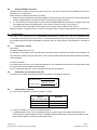
1
Mycoplasma pn. polyvalent ELISA (recombinant) with polyvalent IgA+M conjugate IgG, IgA+M Test Kit Order No.: EC214.00 (IgG, IgA+M Test Kit) Color Coding: dark blue FOR IN VITRO DIAGNOSIS ONLY Sekisui Virotech GmbH Löwenplatz 5 65428 Rüsselsheim - Germany Tel.: +49-6142-6909-0 Fax: +49-6142-966613 http://www.sekisuivirotech.com Druckdatum 04.02.2014 REV 2 / My coplasma pn. poly v alent ELISA IgG, IgA+M GB Contents 1. Intended Use ......................................................................................................................... 3 2. Diagnostic Relevance ............................................................................................................ 3 3. Test Principle......................................................................................................................... 3 4. Package Contents ................................................................................................................. 3 4.1 IgG, IgA+M Testkit .......................................................................................................................................................... 3 5. Storage and Shelf-Life of the test kit and the ready to use reagents ..................................... 4 6. Precautions and Warnings .................................................................................................... 4 7. Material required but not supplied......................................................................................... 4 8. Test Procedure ...................................................................................................................... 5 8.1 8.2 8.3 8.4 9. Examination Material....................................................................................................................................................... 5 Preparation of Reagents ................................................................................................................................................. 5 Virotech ELISA Test Procedure....................................................................................................................................... 5 Usage of ELISA processors ............................................................................................................................................ 6 Test Evaluation ...................................................................................................................... 6 9.1 9.2 9.3 9.4 9.5 Test function control........................................................................................................................................................ 6 Calculation of the Vir otech Units (VE) ............................................................................................................................. 6 Interpretation of Results .................................................................................................................................................. 6 Interpretation Scheme ..................................................................................................................................................... 7 Limits of the Test............................................................................................................................................................. 7 10. Performance Data .................................................................................................................. 8 10.1 10.2 10.3 10.4 10.5 Analytic al Sensitivity and Specif icity ................................................................................................................................ 8 Diagnostic Sensitivity ...................................................................................................................................................... 8 Prevalence (expected values) ......................................................................................................................................... 8 Intra-Assay Coefficient of Variation (Repeatability) ......................................................................................................... 8 Inter-Assay Coefficient of Variation (Reproducibility)....................................................................................................... 9 11. Literature ............................................................................................................................... 9 12. Test Procedure Schemata ................................................................................................... 10 Seite 2 v on 10 My coplasma pn. polyv alent ELISA IgG, IgA+M GB REV 2 Druckdatum 04.02.2014 1. Intended Use Mycoplasma pn. polyvalent ELISA is used for the semiquantitative and qualitative detection of IgG and the combined detection of IgA and IgM antibodies in human serum. The detection of IgG antibodies is adjusted so that fresh infections are mainly detected. To enhance the efficiency of the diagnostic testing, IgA and IgM antibodies are determined together w ith polyvalent IgA+IgM mixed conjugate. 2. Diagnostic Relevance The bacteria Mycoplasma pneumoniae, w hich is lacking cell w all components, is the cause of atypical pneumonia and tracheobronchitis of humans and affects mostly children, young adults and immunodeficient people (1,2,3,4). So called adhesins (6), enable the bacteria to attach to the epithelial cells, against w hich the host develops antibodies. Studies made by Foy show , that in the USA 15 to 20% of all pneumonia cases are caused by Mycoplasma pneumoniae (8). The ELISA detects Mycoplasma-antibodies w ith a defined antigen fraction of the strain M129, w hich is defined via monospecific sera. It includes membrane proteins, cytoskeleton proteins and recombinant proteins. The incubation time during an infection w ith Mycoplasma pneumoniae is 10 – 21 days: Specific IgM-antibodies occur 6-10 days after infection. Basically, about 80% of the patients younger than 20 years develop IgM-antibodies and 40% of the patients that are older than 20 years. This means a specific IgM-response can be missing especially in older patients. IgM-antibodies may be detected, referring to literature, still at least one year after beginning of the symptoms. Specific IgG-antibodies appear 9-14 days after infection. They may persist up to 4 years. Specific IgA-antibodies appear one w eek after start of the infection and decrease about 5 w eeks after start of the infection again. As a rule, the IgA-titer exceeds the IgM-titer. Considering the fact that IgM-antibodies persist very long in some persons and are missing in others completely, it is important to detct beside the IgM- also the specific IgG- and IgA-titer. Re-infections often take place w ithout any production of IgM-antibodies but under significant increase of IgG- and IgA-antibody titers. Tw o patient sera, taken at an interval of 5-10 days allow a proper statement concerning the rise of the antibody titer (5). It is important to consider that a first attack of Mycoplsma pneumoniae does not leave a sufficient protection against a new colonization. For diagnosis it is necessary in any case to consider the clinical picture in addition to the serological results. My coplasma infections are generally treated successfully w ith antibiotics like Tetracycline and Macrolide. The treatment w ith non-suitable, w .g. cell-w all-specific antibiotics (penicillin) leads to a serological advantage for Mycoplasma against all Penicillin-sensitive microorganisms. 3. Test Principle The antibody searched for in the human serum forms an immune complex w ith the antigen coated on the microtiter -plate. Unbound immunoglobulins are removed by w ashing processes. The enzyme conjugate attaches to this complex. Unbound immunoglobulins are again removed by w ashing processes. After adding the substrate solution (TMB), a blue dye is produced by the bound enzyme (peroxidase). The color changes to yellow w hen the stopping solution is added. 4. Package Contents 4.1 1. 2. 3. 4. 5. 6. 7. 8. 9. 10. IgG, IgA+M Testkit 1 Microtiter-Plate consisting of 96 with antigen coated, breakable single w ells, lyophilised PBS-Dilution Buffer, (blue, ready to use), 2x50m l, pH 7,2, w ith preservative and Tw een 20 PBS-Washing Solution, (20x concentrated) 50m l, pH 7,2, w ith preservative and Tw een 20 IgG negative Control, 1300µl, human serum w ith protein-stabilizer and preservative, ready to use IgG cut-off Control, 1300µl, human serum w ith protein-stabilizer and preservative, ready to use IgG positive Control, 1300µl, human serum w ith protein-stabilizer and preservative, ready to use IgA+M negative Control, 1300µl, human serum w ith protein-stabilizer and preservative, ready to use IgA+M cut-off Control, 1300µl, human serum w ith protein-stabilizer and preservative, ready to use IgA+M positive Control, 1300µl, human serum w ith protein-stabilizer and preservative, ready to use IgG-Conjugate (anti-human), 11ml, (sheep or goat)-horseradish-peroxidase-conjugate with protein-stabilizer and preservative in Tris-Buffer, ready to use Seite 3 v on 10 My coplasma pn. polyv alent ELISA IgG, IgA+M GB REV 2 Druckdatum 04.02.2014 11. 12. 13. 5. IgA+M conjugate (anti-human IgA+IgM m ixture), 11m l, (sheep or goat)-horseradish peroxidase conjugate with protein stabilizers and preservatives in Tris buffer, ready-to-use Tetramethylbenzidine substrate solution (3,3’,5,5’-TMB), 11m l, ready to use Citrate-Stopping Solution, 6m l, contains an acid mixture Storage and Shelf-Life of the test kit and the ready to use reagents Store the testkit at 2-8°C. The shelf life of all components is show n on each respective label; for the kit shelf life please see Quality Control Certificate. 1. 2. 3. Microtiter strips/single w ells are to be resealed in package after taking out single w ells and stored w ith desiccant at 2-8°C. Reagents should immediately be returned to storage at 2-8°C after usage. The ready to use conjugate and the TMB-substrate solution are sensitive to light and have to be stored in dark. Should there be a color reaction of the substrate dilution due to incidence of light, it is not useable anymore. Take out only the amount of ready to use conjugate or TMB needed for the test insertion. Additional conjugate or TMB taken out may not be returned but must be dismissed. Material Controls Status Diluted Undiluted After Opening Microtitreplate After Opening Rheumatoid factor Absorbent Conjugate Tetramethylbenzidine Stop Solution Undiluted, After Opening Diluted After Opening After Opening After Opening After Opening Final Dilution (ready-to-use) Test Samples Washing Solution 6. Storage +2 to +8°C +2 to +8°C +2 to +8°C +2 to +8° (storage in the provided bag w ith desiccant bag) +2 to +8°C +2 to +8°C +2 to +8°C (protect from light) +2 to +8°C (protect from light) +2 to +8°C +2 to +8°C +2 to +25°C Shelf life max. 6h 1 w eek 3 months 3 months 3 months 1 w eek 3 months 3 months 3 months 3 months 4 w eeks Precautions and Warnings 1. 2. 3. 7. Only sera w hich have been tested and found to be negative for HIV -1 antibodies, HIV-2 antibodies, HCV antibodies and Hepatitis-B surface-antigen are used as control sera. Nevertheless, samples, diluted samples controls, conjugate and microtiter strips should be treated as potentially infectious material. Please handle products in accordance with laboratory directions. Those components that contain preservatives, the Citrate Stopping Solution and the TMB have an irritating effec t to skin, eyes and mucous. If body parts are contacted, immediately w ash them under flow ing water and possibly consult a doctor. The disposal of the used materials has to be done according to the country -specific guidelines. Material required but not supplied 1. 2. 3. 4. 5. 6. 7. 8. 9. 10. 11. Aqua dist./demin. Eight-channel pipette 50µl, 100µl Micropipettes: 10µl, 100µl, 1000µl Test tubes Paper tow els or absorbent paper Cover for ELISA-plates Disposal box for infectious material ELISA handw asher or automated EIA plate w ashing device ELISA plate spectrophotometer, w avelength = 450nm, reference length = 620nm (Reference Wavelength 620-690nm) Incubator RF-Sorbotech for Virotech IgA+M ELISA w ith anti-human IgA+IgM mixed conjugate (see too Preparation of Reagents) Seite 4 v on 10 My coplasma pn. polyv alent ELISA IgG, IgA+M GB REV 2 Druckdatum 04.02.2014 8. Test Procedure Working exactly referring to the Sekisui Virotech user manual is the prerequisite for obtaining correct results. 8.1 Examination Material Either serum or plasma can be used as test material, even if only serum is mentioned in the instructions. Any type of anticoagulant can be used for plasma. Alw ays prepare patient-dilution freshly. For a longer storage the sera must be frozen. Repeated defrosting shall be avoided. 1. Only fresh non-inactivated sera should be used. 2. Hyperlipaemic, haemolytic, microbially contaminated and turbid sera should not to be used (false positive/negative results). 8.2 Preparation of Reagents The Sekisui Virotech System Diagnostica offers a high degree of flexibility regarding the possibility to use the dilution buffer, w ashing solution, TMB, citrate stopping solution as w ell as the conjugate for all parameters and for all different lots. The ready to use controls (positive control, negative control, cut-off control) are param eter specific and only to use w ith the plate lot indicated in the Quality Control Certificate. 1. 2. 3. 4. 5. 8.3 Set incubator to 37°C and check proper temperature setting before start of incubation. Bring all reagents to room temperature before opening package of microtiter strips. Shake all liquid components w ell before use. Make up the w ashing solution concentrate to 1 L w ith distilled or demineralised w ater. If crystals have formed in the concentrate, please bring the concentrate to room temperature before use and shake w ell before use. High IgG titers or rheumatoid factors can interfere with the spec ific detection of IgM antibodies and lead to false positive or false negative results. For the correct detection of combined IgA and IgM with IgA+IgM mixed conjugate, it is therefore necessary to pretreat the sera with RF-SorboTech (VIROTECH Adsorption Material). Pre-adsorption is not used for the IgA+M controls. Virotech ELISA Test Procedure 1. 2. 3. 4. 5. 6. 7. 8. 9. 10. For each test mixture, pipette 100µl of the ready-to-use dilution buffer (blank), together with the negative, cut-off and positive IgG-, IgA+M controls, as w ell as the diluted patient sera. We propose a double insertion (blank, controls and patient sera); for cut-off control a double insertion is absolutely necessary. Working dilution of patient sera: 1+100; e.g. 10µl serum + 1ml dilution buffer. After pipetting start incubation for 30 min. at 37°C (w ith cover). End incubation period by w ashing microtiter strips 4 times w ith 350 – 400µl w ashing solution per w ell. Do not leave any w ashing solution in the w ells. Remove residues on a cellulose pad. Pipette 100µl of ready to use conjugate into each w ell. Incubation of conjugates: 30 min. at 37°C (w ith cover). Stop conjugate incubation by w ashing 4 times (pls. refer to point 3 above). Pipette 100µl of ready to use TMB into each w ell. Incubation of substrate solution: 30 min. at 37°C (w ith cover, keep in dark). Stopping of substrate reaction: pipette 50µl of citrate stop solution into each w ell. Shake plate carefully and thoroughly until liquid is completely mixed and a homogeneous yellow color is visible. Measure extinction (OD) at 450/620nm (Reference Wavelength 620-690nm). Set your photometer in such a w ay that the blank value is deducted from all other extinctions. Extinctions should be measured w ithin 1 hour after adding the stopping solution! Pls. refer to last page for Test Procedure Schemata Seite 5 v on 10 My coplasma pn. polyv alent ELISA IgG, IgA+M GB REV 2 Druckdatum 04.02.2014 8.4 Usage of ELISA processors All Sekisui Virotech ELISAs can be used on ELISA processors. The user is bound to proceed a validation of the devices (processors) on a regular basis. Sekisui Virotech recommends the follow ing procedure: 1. Sekisui Virotech recommends to proceed the validation of device referring to the instructions of the device manufacturer during the implementation of the ELISA processor respectively after bigger reparations. 2. It is recommended to check the ELISA-processor w ith the Validationkit (EC250.00) afterw ards. A regular check using the Validation kit shall be proceeded minimum once a quarter to test the accuracy of the processor. 3. The release criteria of the Quality Control Certificate of the product must be fulfilled for each test run. With this procedure, your ELISA processor w ill function properly and this w ill support quality assurance in your laboratory. 9. Test Evaluation The ready to use controls serve for a semiquantitative determination of specific IgG-, IgM- and IgA-antibodies. Their concentration can be expressed in Virotech units = VE. Fluctuations resulting from the test procedure can be balanced w ith this calculation method and a high reproducibility is achieved in this w ay. Use the means of the OD values for calculation of the VE. 9.1 Test function control a) OD-values The OD of the blank should be < 0.15. The OD-values of the negative controls should be low er than the OD-values mentioned in the Quality Control Certificate. The OD-values of the positive controls as w ell as of the cut-off controls should be above the OD-values mentioned in the Quality Control Certificate. b) Virotech Units (VE) The Virotech Units (VE) of the cut-off controls are defined as 10. The calculated VE of the positive controls should be w ithin the ranges mentioned in the Quality Control Certificate. If those requirements (OD-values, VE) are not fulfilled, the test has to be repeated. 9.2 Calculation of the Virotech Units (VE) The extinction of the blank value (450/620nm) has to be subtracted from all other extinctions. OD (positive control) x 10 OD (cut-off control) OD (patient serum) VE (patient serum) x 10 OD (cut-off control) VE (positive control) 9.3 Interpretation of Results a) In the IgA+M for all patients, in the IgG for patients > 14 years Result (IgG > 14 years, IgA+M) < 9.0 Evaluation 9.0 – 11.0 borderline > 11.0 positive negative b) In the IgG for children (0 -14 years), w hen IgA+M is positive For children betw een 0 and 14 years, the borderline range (cut-off) in the IgG can be displaced dow nwards, as the Virotech ELISA in the IgG is adjusted so that it predominantly detects acute infections. How ever, the condition for using this procedure is that the serum gives a positive IgA+M result. Seite 6 v on 10 My coplasma pn. polyv alent ELISA IgG, IgA+M GB REV 2 Druckdatum 04.02.2014 1. 2. 3. 9.4 Result (IgG 0 -14 years) Evaluation < 7.0 negative 7.0 – 8.0 borderline > 8.0 positive If the measured values are above the defined borderline range, they are considered to be positive. If an infection is to be reliably detected, the antibody content of tw o serum samples must be determined. One serum sample must be tested directly after the start of the infection. The second sample must be tested 5 -10 days later (convalescence sample). The antibody concentrations of the tw o samples must be tested in parallel in the same test batch. A correct diagnosis cannot be made on the basis of a single serum sample. As some individuals do not form IgM and, in general, all 3 antibody classes (IgG, IgM and IgA) are not tested, a high measure of diagnostic sensitivity can be achieved w ith the IgG and the IgA+M determinations together. If the measured values are below the defined borderline range, no measurable antigen specif ic antibodies are present in the sample. The samples are considered to be negative. Interpretation Scheme IgG IgA+M - - Interpretation - No increase in antibody titer to M. pneumoniae. No suspicion of M. pneumonia infection. If the clinical symptoms persist, repeat test later or consider differential diagnosis. - + - Increased antibody titer to M. pneumoniae in the IgA and/or IgM. Suspicion of early phase of acute M. pneumonia infection. Isolated false positive IgA or IgM results are alw ays possible in principle and may both play a role here. As confirmation, the IgG titer must be checked in 5-10 days. A check by Immunoblot (LINE) may also be recommended. + + - + - - 9.5 Increased antibody titer to M. pneumoniae. Suspicion of an acute infection w ith M. pneumoniae. Suspicion of an infection w ith M. pneumonia in the recent past. IgA+M have already dropped. Raised levels from a long past infection are also in principle possible. Limits of the Test 1. The interpretation of serological results shall alw ays include the clinical picture, epidemiological data and all further available laboratory results. 2. Even after taking the medical history, a clinical examination, standard laboratory tests and an X-ray, a Mycoplasma infection is difficult to distinguish from other infections of the upper and low er respiratory tract or other atypical pneumonia. If the case is uncertain or if the symptoms persist in spite of negative findings, w e recommend diagnosis by molecular biological procedures, to support the serology. 3. Cross-reactions w ith M. genitalium or M. hominis can not be excluded. Also EBV-positive sera can cross-react. Seite 7 v on 10 My coplasma pn. polyv alent ELISA IgG, IgA+M GB REV 2 Druckdatum 04.02.2014 10. Performance Data 10.1 Analytical Sensitivity and Specificity To determine the analytical sensitivity, 120 sera w ere tested in the IgG and IgA+M in comparison to Virotech Mycoplasma pneumoniae LINE. The serum panel w as made up as follow s: 70 clinically characterized sera from patients w ith established Mycoplasma pneumoniae-induced atypical pneumonia (provided by the CAPNETZ Foundation), 34 sera from children aged 1 to 14 years suspected of having a Mycoplasma pneumoniae infection and 16 sera from adult patients w ith suspected Mycoplasma pneumoniae infection. To determine the analytical specificity, 131 sera w ere tested in the IgG and IgA+M in comparison to the Virotech Mycoplasma pneumoniae LINE. The serum panel w as made up as follow s: 67 blood donor sera, 26 neonatal sera (patients aged 0 to 3 months) and 38 sera from patients w ith other respiratory diseases (21 B. pertussis positive sera and 17 Legionella pneumophila positive sera). Analytical Sensitivity - Reference: Mycoplasma pneumoniae LINE - Analytical Specificity - Reference: Mycoplasma pneumoniae LINE - 97% 94% 98% 98% IgG IgA+M 10.2 Diagnostic Sensitivity To determine the diagnostic sensitivty, 70 clinically characterized sera w ere tested from patients w ith established atypical pneumonia. These sera w ere taken from the stocks of the CAPNETZ Foundation. All patient samples had previously given a positive PCR result for Mycoplasma pneumoniae in the prior finding. The prior ELISA tests had been IgM-positive for 35 sera and negative for 35 sera (9). Although Mycoplasma pneumoniae-DNA can be detected in these patient samples, antibodies may not yet be detectable if the immune response is delayed. This explains the low sensitivity of the prior findings. IgG Diagnostic Sensitivity Prior finding of the CAPNETZ sera: PCR positive and 50% serologically positive 67% IgA+M 73% total: IgG and IgA+M 80% As a result of the combined evaluation of IgG and IgA+M, a markedly increased sensitivity of 80% w as attained for this critical serum panel. 10.3 Prevalence (expected values) 67 blood donor sera w ere tested in the IgG and IgA+M. IgG 10.4 IgA+M negative borderline 62 4 93% 6% 62 4 93% 6% positive 1 1% 1 1% Intra-Assay Coefficient of Variation (Repeatability) Strips of different plates of a batch w ere tested with two sera in a chessboard pattern. This gave a coefficient of variation of < 9% (n=2x48). Seite 8 v on 10 My coplasma pn. polyv alent ELISA IgG, IgA+M GB REV 2 Druckdatum 04.02.2014 10.5 Inter-Assay Coefficient of Variation (Reproducibility) 3 sera w ere tested in 10 independent test batches on 3 different test days. This gave a coefficient of variation of < 15%. 11. Literature 1. 2. 3. 4. 5. 6. 7. 8. 9. Clyde WAJ.: Clinical overview of typical Mycoplasma pneumoniae infections. J. Clin Infect. Dis. 1993, 17 (suppl. 1) 32-37 Hu, P.-C., Collier, A.M. and Baseman, J.B. (1977): Surface parasitism by Mycoplasma pneumoniae of respiratory epithelium. J. of Experimental med. 145, 1328-13343. Razin, S. (1992): Peculiar properties of mycoplasmas: the smallest s elf-replicating prokaryotes. FEMS Microbiol. Lett. 100, 423-432. Taylor-Robinson, D. (1996): Infections due to species of Mycoplasma and Ureaplasma: an update. Clin. Infect. Dis. 23, 671-684. Jacobs, E.: Mycoplasmen-Infektionen. mta. 1997, 12: 236-239 Jacobs, E.: Das Adhäsin von Mycoplasma pneumoniae: Seine Bedeutung als Virulenzfaktor in der Pathogenese und in der Diagnostik. Klin. Lab. 1994: 40: 228-229 Baum, H.V., Strubel, A., Nollert, J., Layh-Schmitt, G.:Tw o Cases of Fulminant Mycoplasma Pneumoniae Pneumonia w ithin 4 Month. Infection 28 2000 No.3. Foy, HM: Infections caused by Mycoplasma pneumoniae and possible carrier state in different populations of patients. J Clin Infect Dis 1993, 17(suppl. 1) 37-47. Baum, H. v. et.al.: Mycoplasma pneumoniae pneumonia revisited w ithin the German Competence Netw ork for Community-acquired pneumonia (CAPNETZ), BMC Infectious Diseases 2009, 9:62 Seite 9 v on 10 My coplasma pn. polyv alent ELISA IgG, IgA+M GB REV 2 Druckdatum 04.02.2014 12. Test Procedure Schemata Preparation of Patient Samples and Washing Solution ▼ Washing Solution: Fill up concentrate ▼ IgG-Samples – Dilution 1:101 e.g.: 10 µl serum/plasma + 1000 µl Dilution Buffer (Serum Dilution Buffer is ready to use) to 1 liter with aqua dest./demin. ▼ IgA+M-Samples – Dilution 1:100 Rheumafactor-absorption with RFSorboTech e.g.: 5 µl serum/plasma + 450 µl Dilution Buffer + 1 drop RF-SorboTech, incubate for 15 min. at room temperature. Test Procedure Samples Incubation 30 minutes at 37°C 100 µl Patient Samples blank value (Dilution Buffer) and controls 400 µl Washing Solution Wash 4times Remove Residues on a Cellulose Pad Conjugate Incubation 30 minutes at 37°C 100 µl Conjugate IgG, IgA+M 400 µl Washing Solution Wash 4times Remove Residues on a Cellulose Pad Substrate Incubation 30 minutes at 37°C Stopping 100 µl Substrate 50 µl Stop Solution shake carefully Measure Extinctions Seite 10 v on 10 My coplasma pn. polyv alent ELISA IgG, IgA+M GB Photometer at 450/620nm (Reference Wavelength 620690nm) REV 2 Druckdatum 04.02.2014