1
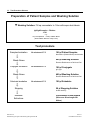
Helicobacter pylori ELISA IgG Testkit / IgA Testkit Order No.: EC143G00 (IgG Testkit) EC143A00 (IgA Testkit) Color Coding: IgG: blue metallic IgA: blue metallic/black FOR IN VITRO DIAGNOSIS ONLY Sekisui Virotech GmbH Löwenplatz 5 65428 Rüsselsheim / Germany Tel.: +49-6142-6909-0 Fax: +49-6142-966613 http://www.sekisuivirotech.com Druckdatum 03.02.2014 REV 14 / Helicobacter py lori IgG / Helicobacter py lori IgA ELISA GB Contents 1. Intended Use ......................................................................................................................... 3 2. Diagnostic Relevance ............................................................................................................ 3 3. Test Principle......................................................................................................................... 3 4. Package Contents ................................................................................................................. 4 4.1 4.2 IgG Testkit....................................................................................................................................................................... 4 IgA Testkit ....................................................................................................................................................................... 4 5. Storage and Shelflife of the Testkit and the ready to use reagents ....................................... 4 6. Precautions and Warnings .................................................................................................... 4 7. Material required but not supplied......................................................................................... 5 8. Test Procedure ...................................................................................................................... 5 8.1 8.2 8.3 8.4 9. Examination Material....................................................................................................................................................... 5 Preparation of Reagents ................................................................................................................................................. 5 Virotech ELISA Test Procedure....................................................................................................................................... 5 Usage of ELISA processors ............................................................................................................................................ 6 Test Evaluation ...................................................................................................................... 6 9.1 9.2 9.3 9.4 Test function control........................................................................................................................................................ 6 Calculation of the Vir otech Units (VE) ............................................................................................................................. 6 Interpretation Scheme IgG and IgA ................................................................................................................................. 6 Limits of the Test............................................................................................................................................................. 7 10. Performance Data .................................................................................................................. 7 10.1 10.2 10.3 10.4 Analytic sensitivity and specif icity.................................................................................................................................... 7 Infection (expected values) ............................................................................................................................................. 7 Intra-assay variation coefficient (repeatability) ................................................................................................................ 8 Inter-assay variation coefficient (reproducibility).............................................................................................................. 8 11. Literature ............................................................................................................................... 8 12. Test Procedure Scheme ........................................................................................................ 9 Seite 2 v on 9 Helicobacter py lori IgG / Helicobacter py lori IgA ELISA GB REV 14 Druckdatum 03.02.2014 1. Intended Use The ELISA testkit is intended for the qualitative and semiquantitative detection of Helicobacter pylori-specific IgG- and IgAantibodies in human serum. 2. Diagnostic Relevance Helicobacter (H.) pylori is a Gram-negative bacterium specialising in the gastric mucosa, w hich causes about 500,000 deaths w orldw ide due to gastric carcinoma (1, 6). The infection is usually acquired in childhood. Transmission is from person to person, and close contact w ithin the family and socioeconomic status play an important part. Accordingly, prevalence is much higher in developing countries than in industrial nations (7). In Germany the infection rate in adults is about 30% (5). H. pylori infection persists throughout life and causes chronic gastritis, w hich often remains w ithout clinical symptoms. How ever, in 20% of those affected, complications occur in the form of a gas tric ulcer, duodenal ulcer, gastric carcinoma or MALT lymphoma (mucosa-associated lymphatic tissue) (5). Healing of the ulcer disease and MALT lymphoma is possible w ith antibiotic therapy in the early stage (5). Early treatment can counteract the development of gastric carcinoma, i.e. diagnosis of H. pylori infection as soon as possible should be attempted. In patients w ho have not had previous treatment, the bacteria can be fully eliminated in 80%-96% of cases (2). Recurrent infection after successful H. pylori eradication is very low (approx. 1% per year) (7). If antibiotic therapy has already been used once, the success rates are low er on further treatment because of the increasing antibiotic resistance. For this reason, it is recommended to detect H. pylori from culture of gastric biopsies in patients w ho have previously been treated once and to carry out sensitivity testing (7,8). Invasive and non-invasive methods can be used to detect H. pylori infection. The invasive methods include the rapid urease test, histology, culture and PCR. In these methods, the pathogen is found in biopsies. The non-invasive tests include the urea breath test, the stool antigen test and detection of antibodies in the serum. All tests have advantages and disadvantages and are not absolutely accurate on their ow n. The method should therefore be selected according to the query (7). Serology is employed as an initial test in patients w ho have not been previously treated (2) in monitoring therapy long-term (1) in seroepidemiological investigations (3). Serology is indicated in cases w ith a reduced number of bacteria e.g. in: marked atrophy of the gastric mucosa gastric haemorrhage use of proton pump inhibitors. While all other methods can give false-negative results in these cases, detection of specific H. pylori antibodies w ith maintained sensitivity is possible (2). 3. Test Principle ELISA for detection of hum an serum IgG and IgA antibodies The antibody searched for in the human serum forms an immune complex w ith the antigen coated on the microtiter-plate. Unbound immunoglobulins are removed by w ashing processes. The enzyme conjugate attaches to this complex. Unbound conjugate is again removed by w ashing processes. After adding the substrate solution (TMB), a blue dye is produced by the bound enzyme (peroxidase). The color changes to yellow w hen the stopping solution is added. Im m unoblot procedure for the detection hum an serum IgG- and IgA-antibodies The Helicobacter pylori LINE from Virotech has the advantage that antibody production against specific virulence factors – e.g. CagA and VacA – of H. pylori can be detected. Seite 3 v on 9 Helicobacter py lori IgG / Helicobacter py lori IgA ELISA GB REV 14 Druckdatum 03.02.2014 4. Package Contents 4.1 IgG Testkit 1. 2. 3. 4. 5. 6. 7. 8. 9. 1 Microtiter-Plate consisting of 96 with antigen coated, breakable single w ells, lyophilised PBS-Dilution Buffer (blue, ready to use) 2x50m l, pH 7,2, w ith preservative and Tw een 20 PBS-Washing Solution (20x concentrated) 50m l, pH 7,2, w ith preservative and Tw een 20 IgG negative Control, 1300µl, human serum w ith protein-stabilizer and preservative, ready to use IgG cut-off Control, 1300µl, human serum w ith protein-stabilizer and preservative, ready to use IgG positive Control, 1300µl, human serum w ith protein-stabilizer and preservative, ready to use IgG-Conjugate (anti-human), 11ml, (sheep or goat)-horseradish-peroxidase-conjugate with protein-stabilizer and preservative in Tris-Buffer, ready to use Tetramethylbenzidine substrate solution (3,3’,5,5’-TMB), 11m l, ready to use Citrate-Stopping Solution, 6m l, contains an acid mixture 4.2 IgA Testkit 1. 2. 3. 4. 5. 6. 7. 8. 9. 5. 1 Microtiter-Plate consisting of 96 with antigen coated, breakable single w ells, lyophilised PBS-Dilution Buffer (blue, ready to use) 2x50m l, pH 7,2, w ith preservative and Tw een 20 PBS-Washing Solution (20x concentrated) 50m l, pH 7,2, w ith preservative and Tw een 20 IgA negative Control, 1300µl, human serum w ith protein-stabilizer and preservative, ready to use IgA cut-off Control, 1300µl, human serum w ith protein-stabilizer and preservative, ready to use IgA positive Control, 1300µl, human serum w ith protein-stabilizer and preservative, ready to use IgA-Conjugate 2 (anti-human), 11m l, (sheep or goat)-horseradish-peroxidase-conjugate with FCS and preservative in Tris-Buffer, ready to use Tetramethylbenzidine substrate solution (3,3’,5,5’-TMB), 11m l, ready to use Citrate-Stopping Solution, 6m l, contains an acid mixture Storage and Shelflife of the Testkit and the ready to use reagents Store the testkit at 2-8°C. The shelf life of all components is show n on each respective label; for the kit shelf life please see Quality Control Certificate. 1. Microtiter strips/single w ells are to be resealed in package after taking out single w ells and stored w ith desiccant at 2-8°C. Reagents should immediately be returned to storage at 2-8°C after usage. 2. The ready to use conjugate and the TMB-substrate solution are sensitive to light and have to be stored in dark. Should there be a color reaction of the substrate dilution due to incidence of light, it is not useable anymore. 3. Take out only the amount of ready to use conjugate or TMB needed for the test insertion. Additional conjugate or TMB taken out may not be returned but must be dismissed. Material Status Diluted Undiluted After Opening Test Samples Controls Microtitreplate After Opening Rheumatoid factor Absorbent Conjugate Tetramethylbenzidine Stop Solution Undiluted, After Opening Diluted After Opening After Opening After Opening After Opening Final Dilution (ready-to-use) Washing Solution 6. Storage +2 to +8°C +2 to +8°C +2 to +8°C +2 to +8° (storage in the provided bag w ith desiccant bag) +2 to +8°C +2 to +8°C +2 to +8°C (protect from light) +2 to +8°C (protect from light) +2 to +8°C +2 to +8°C +2 to +25°C Shelflife max. 6h 1 w eek 3 months 3 months 3 months 1 w eek 3 months 3 months 3 months 3 months 4 w eeks Precautions and Warnings 1. Only sera w hich have been tested and found to be negative for HIV -1 antibodies, HIV-2 antibodies, HCV antibodies and Hepatitis-B surface-antigen are used as control sera. Nevertheless, samples, diluted samples, controls, conjugates and Seite 4 v on 9 Helicobacter py lori IgG / Helicobacter py lori IgA ELISA GB REV 14 Druckdatum 03.02.2014 2. 3. 7. microtiter strips should be treated as potentially infectious material. Please handle products in accordance with laboratory directions. Those components that contain preservatives, the Citrate Stopping Solution and the TMB have an irritating effect to skin, eyes and mucous. If body parts are contacted, immediately w ash them under flow ing water and possibly consult a doctor. The disposal of the used materials has to be done according to the country -specific guidelines. Material required but not supplied 1. 2. 3. 4. 5. 6. 7. 8. 9. 10. 8. Aqua dest./demin. Eight-channel pipette 50µl, 100µl Micropipettes: 10µl, 100µl, 1000µl Test tubes Paper tow els or absorbent paper Cover for ELISA-plates Disposal box for infectious material ELISA handw asher or automated EIA plate w ashing device ELISA plate spectrophotometer, w avelength = 450nm, reference length = 620nm (Reference Wavelength 620-690nm) Incubator Test Procedure Working exactly referring to the Sekisui Virotech user manual is the prerequisite for obtaining correct results. 8.1 Examination Material Either serum or plasma can be used as test material, even if only serum is mentioned in the instructions. Any type of anticoagulant can be used for plasma. Alw ays prepare patient-dilution freshly. For a longer storage the sera must be frozen. Repeated defrosting should be avoided. 1. Only fresh non-inactivated sera should be used. 2. Hyperlipaemic, haemolytic, microbially contaminated and turbid sera should not to be used (false positive/negative results). 8.2 Preparation of Reagents The Sekisui Virotech System Diagnostica offers a high degree of flexibility regarding the possibility to use the dilution buffer, w ashing solution, TMB, citrate stopping solution as w ell as the conjugate for all parameters and for all different lots. The ready to use controls (positive control, negative control, cut-off control) are param eter specific and only to use w ith the plate lot indicated in the Quality Control Certificate. 1. Set incubator to 37°C and check proper temperature setting before start of incubation. 2. Bring all reagents to room temperature before opening package of microtiter strips. 3. Shake all liquid components w ell before use. 4. Make up the w ashing solution concentrate to 1 L w ith distilled or demineralised w ater. If crystals have formed in the concentrate, please bring the concentrate to room temperature before use and shake w ell before use. 8.3 Virotech ELISA Test Procedure 1. 2. 3. 4. 5. 6. For each test run, pipette 100µl each of ready to use dilution buffer (blank), IgG- and IgA-positive, negative and cut-off controls as w ell as diluted patient sera. We propose a double insertion (blank, controls and patient sera); for cut-off control a double insertion is absolutely necessary. Working dilution of patient sera: 1+100; e.g. 10µl serum + 1ml dilution buffer. After pipetting start incubation for 30 min. at 37°C (w ith cover). End incubation period by w ashing microtiter strips 4 times w ith 350 – 400µl w ashing solution per w ell. Do not leave any w ashing solution in the w ells. Remove residues on a cellulose pad. Pipette 100µl of ready to use conjugate into each w ell. Incubation of conjugates: 30 min. at 37°C (w ith cover). Stop conjugate incubation by w ashing 4 times (pls. refer to point 3 above). Seite 5 v on 9 Helicobacter py lori IgG / Helicobacter py lori IgA ELISA GB REV 14 Druckdatum 03.02.2014 7. 8. 9. 10. Pipette 100µl of ready to use TMB into each w ell. Incubation of substrate solution: 30 min. at 37°C (w ith cover, keep in dark). Stopping of substrate reaction: pipette 50µl of citrate stopping solution into each w ell. Shake plate carefully and thoroughly until liquid is completely mixed and a homogeneous yellow color is visible. Measure extinction (OD) at 450/620nm (Reference Wavelength 620-690nm). Set your photometer in such a w ay that the blank value is deducted from all other extinctions. Extinctions should be measured w ithin 1 hour after adding the stopping solution! Pls. refer to last page for Test Procedure Scheme 8.4 Usage of ELISA processors All Sekisui Virotech ELISAs can be used on ELISA processors. The user is bound to proceed a validation of the devices (processors) on a regular basis. Sekisui Virotech recommends the follow ing procedure: 1. Sekisui Virotech recommends to proceed the validation of device referring to the instructions of the device manufacturer during the implementation of the ELISA processor respectively after bigger reparations. 2. It is recommended to check the ELISA-processor w ith the Validationkit (EC250.00) afterw ards. A regular check using the Validationkit shall be proceeded minimum once a quarter to test the accuracy of the processor. 3. The release criteria of the Quality Control Certificate of the product must be fulfilled for each testrun. With this procedure, your ELISA processor w ill function properly and this w ill support quality assurance in your laboratory. 9. Test Evaluation The ready to use controls serve for a semiquantitative determination of specific IgG- and IgA-antibodies. Their concentration can be expressed in Virotech units = VE. Fluctuations resulting from the test procedure can be balanced w ith this calculation method and a high reproducibility is achieved in this w ay. Use the means of the OD values for calculation of the VE. 9.1 Test function control a) OD-values The OD of the blank should be < 0.15. The OD-values of the negative controls should be low er than the OD-values mentioned in the Quality Control Certificate. The OD-values of the positive controls as w ell as of the cut-off controls should be above the OD-values mentioned in the Quality Control Certificate. b) Virotech Units (VE) The Virotech Units (VE) of the cut-off controls are defined as 10 VE. The calculated VE of the positive controls should be w ithin the ranges mentioned in the Quality Control Certificate. If those requirements (OD-values, VE) are not fulfilled, the test has to be repeated. 9.2 Calculation of the Virotech Units (VE) The extinction of the blank value (450/620nm) has to be subtracted from all other extinctions. OD (positive control) x 10 OD (cut - off control) OD (patient serum) VE (patient serum) x 10 OD (cut - off control) VE (positive control) 9.3 Interpretation Scheme IgG and IgA Result (VE) < 9,0 9,0 - 11,0 > 11,0 Seite 6 v on 9 Helicobacter py lori IgG / Helicobacter py lori IgA ELISA GB Evaluation negative borderline positive REV 14 Druckdatum 03.02.2014 1. 2. 3. If the measured values are above the defined borderline range, they are considered to be positive. If the measured VE is w ithin the borderline range, no significant high antibody concentration is present, the samples are considered to be borderline. For the secure detection of an infection it is necessary to determine the antibody concentration of tw o serum samples. One sample shall be taken directly at the beginning of the infection and a second sample 5 – 10 days later (convalescent serum). The antibody concentration of both samples has to be tested in parallel, that means in one test run. A correct diagnosis based on the evaluation of a single serum sample is not possible. If the measured values are below the defined borderline range, no measurable antigen specific antibodies are present in the samples. The samples are considered to be negative. 9.4 Limits of the Test 5. 6. 7. The interpretation of serological results shall alw ays include the clinical picture, epidemiological data and all further available laboratory results. Both IgA and IgG Helicobacter pylori LINE results should be taken into consideration for the diagnosis of patient’s sera suspected to have a Helicobacter pylori infection. IgA-antibodies may persist 6 months to 3 years after successful treatment. Normally IgG-antibodies persist for many years. 10. Performance Data 10.1 Analytic sensitivity and specificity To determine the analytic result clinically defined sera (n=19), interlaboratory test sera (n=19), routine sera (n=40) and blood donor sera (n=80) w ere tested for IgG w ith a commercially available ELISA and the Virotech Helicobacter pylori LINE. Samples w ith discrepant results w ere additionally characterised using a commercially available immunoblot. Sera collective (n=158) Analytic result Helicobacter pylori ELISA IgG negative borderline positive negative 82 1 0 borderline positive 0 0 1 0 0 74 This results in an analytic sensitivity and specificity of >99.9% for IgG. The borderline results w ere not included in the analysis. To determine the analytic result clinically defined sera (n=20), interlaboratory test sera (n=19), routine sera (n=32) and blood donor sera (n=80) w ere tested for IgA w ith a commercially available ELISA and the Virotech Helicobacter pylori LINE. Samples w ith discrepant results w ere additionally characterised using a commercially available immunoblot . Sera collective (n=151) Analytic result Helicobacter pylori ELISA IgA negative negative 83 borderline 4 positive 9 borderline 5 0 7 positive 3 3 37 This results in an analytic sensitivity of 92.5% and specificity of 90.2% for IgAG. 10.2 Infection (expected values) To obtain the expected values (infection), 80 sera were tested for IgG and IgA. In a blood donor population, an infection rate of about 30% can be expected. About 90% show an IgA response . Seite 7 v on 9 Helicobacter py lori IgG / Helicobacter py lori IgA ELISA GB REV 14 Druckdatum 03.02.2014 IgG IgA n=80 No. % No. % Negative 58 72.5 60 75.0 Borderline 0 0 2 2,5 Positive 22 27.5 18 22.5 10.3 Intra-assay variation coefficient (repeatability) In one assay strips from different plates of one lot w ere tested w ith one serum. The variation coefficient obtained thus is < 9% for IgA and IgG. 10.4 Inter-assay variation coefficient (reproducibility) 3 sera w ere tested in 10 independent test batches in different laboratories and by different test persons . The variation coefficient obtained thus is < 15% for IgA and IgG. 11. Literature 1. 2. 3. 4. 5. 6. 7. Helicobacter pylori – Von der Grundlage zur Thearpie (1996) Herausgeber P. Malfertheiner, Thieme Verlag Homepage, Nationales Referenzzentrum für Helicobacter pylori; Institut für Medizinische Mikrobiologie und Hygiene der Universität Freiburg Zöller et al (1993) Nachw eis der Helicobacter pylori-Infektion: Rolle der Immundiagnostik. Klin. Lab. 39: 45-54 Brandis, Eggers (1994) Lehrbuch der Medizinischen Mikrobiologie, 7. Auflage, S.495 Kist M., Glocker E., Helicobacter-pylori-Infektionen: Studie ResiNet zur Resistenzentw icklung - aktuelle Ergebnisse, Epidemiologisches Bulletin, 2005, Nr. 24 Kist M., Glocker E., Suerbaum S., Pathogenese, Diagnostik und Therapie der Helicobacter-pylori-Infektion, Bundesgesundheitsblatt, 2005 Helicobacter-pylori und gastroduodenale Ulkuskrankheit, AWMF-Leitlinien-Register, Nr. 021/001, 2008 Seite 8 v on 9 Helicobacter py lori IgG / Helicobacter py lori IgA ELISA GB REV 14 Druckdatum 03.02.2014 12. Test Procedure Scheme Preparation of Patient Samples and Washing Solution ▼ Washing Solution: Fill up concentrate to 1 liter with aqua dest./demin. IgG/IgA-Samples – Dilution 1:101 e.g.: 10 µl serum/plasma + 1000 µl Dilution Buffer (Serum Dilution Buffer is ready to use) Test procedure Samples Incubation 30 minutes at 37°C 100 µl Patient Samples blank value (Dilution Buffer) and controls 400 µl Washing Solution Wash 4times Remove Residues on a Cellulose Pad Conjugate Incubation 30 minutes at 37°C 100 µl Conjugate IgG, IgA 400 µl Washing Solution Wash 4times Remove Residues on a Cellulose Pad Substrate Incubation 30 minutes at 37°C Stopping 100 µl Substrate 50 µl Stopping Solution shake carefully Measure Extinctions Seite 9 v on 9 Helicobacter py lori IgG / Helicobacter py lori IgA ELISA GB Photometer at 450/620nm (Reference Wavelength 620690nm) REV 14 Druckdatum 03.02.2014