1
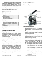
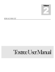
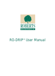
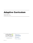
Kids Microscope
Instruction Manual for
MI-1100STD – Kids Microscope
MI-1100LED – Kids LED Cordless Microscope
Phone: 800.860.6272
Web: www.homesciencetools.com
Copyright 2005 by Home Training Tools, Ltd.
All rights reserved.
Welcome to an exciting world of discovery with
your new Kids Microscope! This manual will give you
a familiarity with the different features of your
microscope, how to use them, and how to preserve your
investment by proper maintenance and care.
There are two microscopes in the Kids Microscope
series. They share the same basic features and
functions, but you will find a discussion of the power
options for the MI-1100LED model on page 3.
Features & Definitions
Microscope Diagram
1. Eyepiece
Table of Contents
Table of Contents ................................................. 2 General Microscope Care .................................... 2 Unpacking ......................................................... 2 Cleaning............................................................ 2 Features & Definitions .......................................... 2 Microscope Diagram......................................... 2 Description of Components .............................. 2 Power Options for MI-1100LED ....................... 3 Operating Procedure ............................................ 3 Maintenance ......................................................... 4 Adjusting the Stage Stop .................................. 6 Changing the Tungsten Bulb in the MI-1100STD ...... 6 Changing the LED Bulb in the MI-LEDMIC ...... 6 Warranty ............................................................... 6 Troubleshooting ................................................... 7 Specifications ....................................................... 7 Ideas for Using Your Microscope ......................... 4 Prepared Slides .................................................... 5 General Microscope Care
Unpacking
Your Kids Microscope is shipped in a two-part
Styrofoam case. Keep this case for storage, transport,
and shipping. It is perfect packing material should you
ever need to send your microscope in for repairs
covered by the warranty.
When handling your microscope, always pick it up
by the arm. Avoid touching the lens surfaces on the
eyepiece or objective lens, as finger prints will decrease
image quality.
Cleaning
The best optical quality can be compromised by
dirty lenses. Using a dustcover and cleaning the lenses
regularly will greatly enhance your microscope use.
To clean lens surfaces, remove dust by using a soft
brush or a can of compressed air. Then moisten a piece
of lens paper (our item MI-PAPER) with some lens
cleaning solution (MI- LENSCLN). Gently clean the
eyepiece and objective lens exterior surface using a
circular motion. Repeat with a second paper moistened
with solution if necessary. Repeat once again with a
piece of dry lens paper until the lens is clean and dry. Do
not spray lens cleaner directly on the lens.
© Home Training Tools Ltd. 2005
Page 2 of 8
2. Arm
3. Nosepiece
6. Stage stop
4. Objective
lenses
7. Stage clips
5. Stage
8. Disc diaphragm
9. Focus knob
Light intensity
control MI-1100LED
10. Illuminator
Description of Components
1. Eyepiece: This is the part of the microscope that
you look through. It is inclined at a 45º angle for
comfortable viewing. It contains a lens that magnifies
10x.
2. Arm: The arm not only supports the head and
nosepiece, it is also the best “handle” for picking up
and moving the microscope.
3. Nosepiece: This is also called the “objective turret.”
It holds the objective lenses and rotates 360º. You
can change magnification by turning it until the lens
you want to use “clicks” into place.
4. Objective lenses: These are the lenses closest to
the specimen. The standard objectives are 4x, 10x,
and 40x, which multiply with the 10x eyepiece lens
to provide magnification levels of 40x, 100x, and
400x. The shortest lens has the lowest magnification
level, while the longest has the highest. The lenses
have the following features:
They are achromatic – they help prevent
color distortion.
They are parcentered – if you center your
slide using one objective, it will still be
centered when you move to another
objective.
They are parfocal – if you focus your
specimen using one objective, it will stay
coarsely focused when you move to another
objective. (You will still have to make minor
adjustments.)
Visit us at www.homesciencetools.com
The 40x objective is retractable – the tip
containing the lens is spring-loaded to
prevent damage to the objective or slide.
5. Stage: The stage is the platform that supports the
specimen slide below the objective lenses. It moves
up and down when you turn the focus knob, allowing
you to get just the right distance between the slide
and the lens.
6. Stage stop: This is a screw with a lock nut located
between the stage and the arm of the microscope. It
prevents the stage from coming too far up and
grinding against the objective lens. It is also called a
“safety rack stop,” and is pre-adjusted by the
manufacturer.
Instructions for readjusting it
manually are on page 4.
7. Stage clips: The stage clips hold microscope slides
in place. Pressing on the end closest to the arm of
the scope will lift up the other end, allowing you to
place your slide underneath.
8. Disc diaphragm: The diaphragm controls the
amount of light coming through the specimen in
order to provide optimum resolution for the objective
lens. The diaphragm on this microscope is a rotating
disc under the stage with holes that are numbered
by size; for example, a hole labeled 6 is 6mm in
diameter and a hole labeled 2 has a diameter of
2mm. Use the smaller holes for lower magnification
and the larger holes for higher magnification.
9. Focus knob: The focus knob is used to raise or
lower the stage until the image is in focus. The
focus mechanism uses a slip clutch to prevent
damage to the gears.
10. Illuminator:
The
illuminator
provides
light
underneath the stage. The MI-1100STD contains a
15-watt tungsten bulb. The MI-1100LED contains an
LED bulb and light intensity control knob located on
the base. This intensity control helps adjust
illumination contrast. Instructions for changing the
bulbs are on page 4.
Power Options for MI-1100LED
The LED Microscope comes with a built-in
rechargeable NiMH battery and charger. The fully
charged battery provides about 15 hours of totally
portable microscope use. The AC adapter is used to
recharge the battery. (The battery should be fully
charged before first use, or use the adapter.)
Red and green lights on the back of the microscope
indicate charging status. Please follow these charging
guidelines to maintain maximum battery life for your
microscope.
1. Turn off the illuminator and plug in the AC adapter.
2. A red light only indicates the battery is charging and
has less than 70% of full charge.
3. Both a red and green light indicates the battery is
charging and has 70-90% of charge.
© Home Training Tools Ltd. 2005
Page 3 of 8
4. A green light only indicates the battery is fully
charged and ready for use.
5. Typical charging time is 4-8 hours. Do not charge
the battery or leave the AC adapter plugged in for
more than 12 hours.
Operating Procedure
Now that you have an overview of what each
component of your microscope is for, you can follow this
step-by-step procedure to help you get started using it.
1. Set your microscope on a table or other flat surface
where you will have plenty of room to work. Plug the
microscope’s power cord into an outlet, making sure
that the excess cord is out of the way so no one can
trip over it. (The MI-1100LED also operates on
battery power.)
2. Flip the switch to turn on your microscope's light
source and then turn the disc diaphragm to the
largest hole, which allows the greatest amount of
light through. (You will adjust this again later for best
contrast.) The MI-1100LED also has a light intensity
control on the base: turn the intensity up fully.
3.
Rotate the nosepiece to the lowest-power (4x)
objective. You will hear a click when it is properly in
place. Always start with the lowest power: it is
easiest to scan a slide at a low setting, as you have
a larger field of view.
4. Turn the focus knob to move the stage down (away)
from the objective lens as far as possible.
5. Set a microscope slide (coverslip facing up) in place
under the stage clips. A prepared slide works best
when you do this for the first time. Move the slide
until the specimen is under the objective lens.
6. Adjust the focus knob until the specimen is in focus.
Slowly move the slide to center the specimen under
the lens, if necessary, by nudging it with your
fingers.
7. Adjust the diaphragm to get the best lighting. Start
with the most light and gradually lessen it until the
specimen image has clear, sharp contrast. On the
MI-1100LED you can also adjust the light intensity
control for contrast.
8. Scan the slide (right to left and top to bottom) at low
power to get an overview of the specimen (nudge
the slide very slowly with your fingers). Then center
the part of the specimen you want to view at higher
power.
9. Rotate the nosepiece to the 10x for 100x
magnification. Refocus and view the slide carefully.
Adjust the diaphragm again until the image has the
best contrast. Repeat with the 40x objective for 400x
magnification.
Visit us at www.homesciencetools.com
You have a microscope—now what? With the
following directions you can get started right away
making your own microscope slides!
Materials Needed:
- small cork
- plain glass microscope slide
- slide coverslip
- sharp knife or razor blade
- water
How to Make Simple Microscope Slides
How to make the microscope slide:
Learn more about using the Kids microscope by
making simple slides using common items from around
the house!
Carefully cut a very thin slice of cork using a razor blade
or sharp knife (the thinner
the slice, the easier it will
be to view with your
microscope). To make a
wet mount of the cork, put
one drop of water in the
center of a plain glass slide – the water droplet should
be larger than the slice of cork. Gently set the slice of
cork on top of the drop of water (tweezers might be
helpful for this). If you are not able to cut a thin enough
slice of the whole diameter of the cork, a smaller section
will work.
Ideas for Using Your Microscope
Materials Needed:
- clear Scotch tape
- a few granules of salt, sugar, ground coffee,
sand, or any other grainy material
Making Simple Slides
To make a slide, tear a 2½-3” long piece of Scotch
tape and set it sticky side up on the kitchen table or
other work area. Fold over about ½” of the tape on each
end to form finger holds on the sides of the slide. Next,
sprinkle a few grains of salt or sugar in the middle of the
sticky part of the slide.
You can repeat this with the other substances if you
like, just be sure to label each slide you make with an ink
pen or permanent marker so you will know what’s on the
slides!
You can make tape slides with many other
materials as well. Try hair (from pets and family
members), thread and fiber (from carpets or clothing), or
small dead insects such as gnats, ants, or fruit flies.
Label each slide and view them one at a time with your
microscope, experimenting with different magnification.
How to Make Your Own Prepared Slide
Learn how to make temporary mounts of specimens
and view them with your microscope. Below are a few
ideas for studying different types of cells found in items
that you probably already have around your house.
Cork Cells
In the late 1600s, a scientist named Robert Hooke
looked through his microscope at a thin slice of cork. He
noticed that the dead wood was made up of many tiny
compartments, and upon further observation Hooke
named these empty compartments cells. It was later
known that the cells in cork are only empty because the
living matter that once occupied them has died and left
behind tiny pockets of air. You can take a closer look at
the cells, also called lenticels, of a piece of cork by
following these instructions.
© Home Training Tools Ltd. 2005
Page 4 of 8
Take one coverslip and hold it at an angle to the
slide so that one edge
of it touches the water
droplet on the surface
of the slide.
Then being careful not to move the cork around,
lower the cover slip without trapping any air bubbles
beneath it. The water should form a seal around the
cork. Use the corner of a paper towel to blot up any
excess water at the edges of the coverslip. To keep the
slide from drying out, you can make a seal of petroleum
jelly around the cover slip with a toothpick. Begin with
the lowest-power objective to view your slide. Then
switch to a higher power objective to see more detail.
Use this same wet mount method for other specimens
such as cheek cells or leaf cells.
Record Your Observations
Our Microscope Observation worksheet (on the last
page) will help you keep track of what you see and
remember what you have learned. Blanks are provided
for recording general information about each slide (e.g.
wet mount stained with methylene blue). In addition,
there is space to write down your observations and
make sketches of what you see at each magnification
level.
Visit us at www.homesciencetools.com
Prepared Slides
The easiest way to build your microscope skills
(controlling focus, light contrast, and more) is to view
prepared slides. A slide starter set is included with your
microscope. Your starter slide set comes with five
prepared slides, described below. These descriptions
get you started working with each specimen, but you can
research more information on the internet.
Mouth Smear – Epithelial tissue is found many places in
the body – on the surface of the skin, the lining of the
mouth, stomach, and blood
nucleus
vessels, and in the glands.
cytoplasm
The mouth smear slide is
squamous epithelium taken
from the lining of the cheek.
Scan the slide on low power
(40x) and find a group of cells
that look one layer thick. You
should see very small dark 400x
spots in the middle of the cells – these are the nuclei,
which contain DNA and control cellular functions. As you
view the slide at higher power, you will see more detail.
Identify the nucleus, the cell membrane (the wall
between cells), and the cytoplasm (the fluid inside the
cell membrane).
Pollen – When a pollen grain begins to germinate, it
grows a long, thin tube called a pollen tube that
stretches down a flower’s style to the ovary. The two
sperm in the pollen grain 400x
sperm nucleus
travel through the pollen tube.
pollen
One of them then fertilizes the
egg in the ovary.
cell wall
As you look at the slide at
high power (400x), notice how
thick the walls of the pollen
grains are. Locate the pollen
tubes that have grown off
some of the grains. Look for dark spots in the tubes –
these are the sperm nuclei. If there is a large dark spot
in the pollen grain, this is the “generative nucleus” that
will split to produce the two sperm.
the cell gets energy from food) and the micronucleus
controls the cell’s reproduction. With some patience in
focusing you can see the cilia at 400x on some of the
specimens. If you look closely you may also see the oral
groove – it looks like a short depression coming in from
the edge of the cell.
Housefly Leg – Houseflies
have tiny hairs on their legs
and feet. They use these hairs
for feeling and tasting. Yes,
they really can taste with their
feet!
joint
hairs
As you look at the housefly
foot slide, you won’t get the 100x
whole specimen in focus at
one time. This is because it is a three-dimensional
specimen with different layers. Take a look at the foot at
low power. Notice that it has several different joints. At
higher powers you can see the individual hairs clearly.
Focus in to see where the hair joins the foot.
Frog Blood – Frog blood looks quite different from
human blood. Human blood cells don’t have nuclei, so
they
are
unable
to 400x
nucleus
reproduce themselves by
dividing their DNA. Instead,
they are made in our bone
marrow. Frog blood cells do
have nuclei and thus are
able to divide.
white blood cell
At lowest power hundreds of
tiny red blood cells will fill
your field of view. Even when magnified only 40x, a
miniscule purple dot is visible on each cell. This dot is
the nucleus. At higher power you may find a few white
blood cells – these are different shapes than the red
blood cells and appear to be mostly a purple nucleus
with only a thin light-colored layer outside. Red blood
cells carry oxygen throughout the body, while white
blood cells work as part of the immune system.
Paramecium – A paramecium is a single-celled
protozoan that moves using cilia, tiny hairs around its
cell wall that wave back and forth. It eats by sweeping
food down an oral groove lined with cilia into a gullet.
The gullet closes off when it is full and becomes a
floating storage unit, called
a food vacuole.
cilia
Take a good look at
different paramecia on your
slide. You can see a large,
dark macronucleus in each
paramecium, and in some
the
smaller
dark
micronucleus next to it. The
macronucleus controls the
metabolism of the cell (how
© Home Training Tools Ltd. 2005
oral groove
macronucleus
400x
Page 5 of 8
Visit us at www.homesciencetools.com
3. Carefully lay the microscope on its side.
Maintenance
Adjusting the Stage Stop
The stage stop is set at the factory to ensure that
the stage cannot come up far enough to hit the objective
lenses. Under normal circumstances you will not have to
adjust this. However, if you cannot focus a slide, follow
these steps:
1. Loosen the knurled locking
nut by turning it counterclockwise. (Use needlenose pliers for this.)
2. Loosen the stop screw.
on a standard slide until you
a sharp image.
Stop screw
Locking nut
Focus
obtain
4. Using a screwdriver, remove the
screw from the center of each rubber
foot.
5. Remove the bottom plate and gently
push the bulb in and turn it to release it from the
socket.
6. Replace with a new bulb, then put
the plate back in place and replace
the rubber feet.
Changing the LED Bulb in the MI-LEDMIC
1. Obtain the correct LED replacement bulb (our item
MI-BULB10). One is included with your microscope.
3. Tighten the stop screw by turning it clockwise until it
stops, then turn it back ½ turn.
2. Unplug the microscope from the power supply and
allow it to cool before replacing the bulb.
4. Re-tighten the locking nut.
3. Carefully lay the microscope on its side.
Changing the Tungsten Bulb in the MI-1100STD
4. Using a screwdriver, remove the screw from the
center of each rubber foot.
1. Obtain the correct 15-watt tungsten
replacement bulb (our item MIBULB2). One is included with your
microscope.
5. Remove the bottom plate and pull
the used LED bulb straight out
from its socket.
2. Unplug your microscope from the power supply and
allow it to cool before replacing the bulb.
6. Replace with a new LED bulb and
then screw the plate back on.
Warranty
Home Science Tools warrants this microscope to be free from defects in material and workmanship under normal use and
service for five years from the date of purchase. This warranty does not cover light bulbs, batteries, or damage due to
misuse, abuse, alterations, or accident. Warranty does not cover lenses that have become inoperable due to excessive
dirtiness as a result of misuse or lack of normal maintenance.
You will need to return your microscope freight prepaid for warranty service to Home Science Tools, or the repair facility
we designate. We will repair or replace your microscope at no charge and return it freight prepaid to you. Please call 1800-860-6272 to arrange warranty service before returning this instrument. Please note that warranties apply only to the
original purchaser and are not transferable.
© Home Training Tools Ltd. 2005
Page 6 of 8
Visit us at www.homesciencetools.com
Troubleshooting
If you are experiencing difficulty with your microscope, try these troubleshooting techniques:
Problem
Possible Reason and Solution
Light fails to
1. The batteries are dead (MI-1100LED). Use the AC adapter to recharge the batteries.
operate
2. The light intensity control is off (MI-1100LED). Turn up the light intensity.
3.
4.
The bulb is burned out. Replace the bulb. (See “Changing the Bulb,” p. 4.)
The incorrect bulb is installed. Replace with the correct bulb.
Light flickers
1. The bulb is not properly inserted into the socket. Properly insert the bulb.
2. The bulb is about to burn out. Replace the bulb.
No image
1. The nosepiece is not indexed properly. Move revolving nosepiece until the objective
lens clicks into position.
2. The light is too bright. Adjust the diaphragm.
Unable to
focus slide
1. The slide coverslip is too thick. Use 0.17 mm thick (No. 1) coverslip.
2. The slide is upside down. Place the slide on the stage with the coverslip facing up.
3. The stage stop is not set at the proper position. Adjust the stage stop. (See “Adjusting
the Stage Stop,” p. 4.)
Poor
resolution,
image not
sharp
Spots in field
1. The objective or eyepiece lenses are dirty. Clean the lenses. (See “Cleaning,” p. 2.)
2. There is too much light. Adjust the diaphragm.
Uneven
illumination of
field
1. The nosepiece is not indexed properly. Move revolving nosepiece until the objective
lens clicks into position.
2. The diaphragm is not properly indexed. Adjust the diaphragm to the proper level.
1. The specimen slide, objective, or eyepiece lens is dirty. Clean the slide or lenses.
(See “Cleaning,” p. 2.)
Specifications
Eyepiece
Widefield 10x eyepiece with fully coated optics. Inclined 45° head.
Nosepiece
3-hole, ball-bearing mounted with positive click stops.
Objectives
All objectives are achromatic, parfocalled, parcentered, and fully coated.
4x, 0.10 N.A., red ring, 3.6mm field of view, 40x magnification
10x, 0.25 N.A., yellow ring, 1.4mm field of view, 100x magnification
40x, 0.65 N.A., blue ring, 0.4mm field of view, 400x magnification, retractable
Focusing
Single intermediate focusing control with slip clutch. All metal rack-and-pinion focusing with
adjustable stage stop.
Stage
Acid and chemical resistant 95 x 95mm metal stage with stage clips (not designed for use
with a mechanical stage).
Condenser
Fixed 0.65 NA condenser.
Diaphragm
Calibrated 6-hole disc diaphragm.
Illuminator
15-watt tungsten illuminator with grounded 110-volt cord on model MI-1100STD, 20-watt
equivalent LED illuminator with AC Adapter or optional batteries on model MI-1100LED.
© Home Training Tools Ltd. 2005
Page 7 of 8
Visit us at www.homesciencetools.com
Date of slide:
Name of sample:
Collected from:
Stain:
Mount:
Lighting:
Observations
Sketches
40x magnification
100x magnification
400x magnification
© Home Training Tools Ltd. 2005
Other: _____________
Page 8 of 8
Visit us at www.homesciencetools.com