1
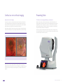
The First True Color Confocal Scanner on the Market
“White color and infrared confocal images:
the advantages of white color and confocality
together for better fundus images. The
infrared to see what our eye is not able to see.
What once was a dream... is now a reality!”
Prof. G. Staurenghi – Eye Clinic Director at University of Milan and Sacco Hospital
Company Profile
CenterVue designs and manufactures highly automated medical devices for the
diagnosis and management of ocular pathologies, including those that represent
the leading causes of blindness.
Our goal is to design Smartly Simple devices and to provide high-value services
that enable Eye Care Specialists to better preserve patients’ sight and quality of
vision, in particular by detecting preventable diseases; and in so doing dramatically
improve their quality of life.
CenterVue is headquartered in Padova, Italy, with the US branch in San Jose,
California. CenterVue is present in over 70 countries with its distribution network.
2
True Color Confocal Scanner
3
Confocal vs. non-confocal imaging
Presenting Eidon
Improvement in confocal images
The first true color scanning ophthalmoscope on the market
SLO systems are superior to conventional fundus cameras in many ways, as they exploit a
confocal imaging principle which limits the effect of backscattered light from deeper layers
and provides enhanced image quality. Another major advantage of SLO systems is that they
operate with much smaller pupils than conventional fundus cameras do.
EIDON is the first system to combine the advantages of SLO with the fidelity of true color
imaging, setting new performance standards in retinal imaging. EIDON provides unsurpassed
image quality, 60° field in a single exposure, a unique, live, confocal view of the retina, three
different imaging modalities and dilation-free operation, all integrated in a versatile system
that provides new opportunities in retinal diagnostics.
At the same time, though, SLO systems do not provide color images, as they typically employ
multiple, monochromatic, laser sources, resulting in black and white or pseudo-color images.
SLO, pseudo-color image
The device is operated via a tablet with a multi-touch, high resolution, color display; it works
with a dedicated software application and operates as a standalone unit. A joystick is provided
when manual operation of the device is desired.
EIDON, true color image
Differently from existing SLO systems, EIDON is a scanning ophthalmoscope that uses
WHITE light instead of monochromatic lasers, hence providing true color imaging and offering
major benefits in terms of fidelity to real retinal appearance, no distortion and dilation-free
operation.
4
True Color Confocal Scanner
5
Multiple Imaging Modalities
Theimportanceofwhite-lightconfocalimaging
Multiple confocal imaging modalities - true color, red-free, infrared - provide specific information concerning
different layers.
Thisparticulartechnologyfacilitatesdiagnosisandmonitoringofretinaldiseasessuchasdiabetic
retinopathy,age-relatedmaculardegenerationandglaucoma.EIDONimprovesretinaldiagnostics
capabilitiesinthatitoffers:
•
•
•
•
•
•
• Red-free is useful to enhance the visibility of the retinal vasculature and retinal nerve fiber layer
• Infrared (825 – 870 nm) provides information corresponding to deeper layers (choroid)
• True color is obtained using white illumination (440 - 650 nm)
Greatercontrastthanatraditional
funduscamera
Preservedimagequalityinpresenceofmedia opacitiessuchascataract
15micronsresolution
Nodilationdownto2.5mmpupil
Noopticdiscbleaching
Nosaturationoftheredchannellikein
traditionalfunduscameras
Whatyouseeiswhatitis!
Theuniquecombinationofconfocalimagingand
whitelightilluminationofferssuperiorimage
qualityandcolorfidelity.Usingwhitelight,the
retinaappearsasitlookswhendirectlyobserved,
astheentirevisiblespectrumispresentinthe
capturedimage.
Red
Orange
Yellow
Green
Blue
Indigo
Central field, color
Central field, red-free
Color mosaic, 110°
Nasal peripheral field, color 60°
Central field, infrared image
Optic disc detail
Violet
Widefieldofview
•
•
6
Widefieldopticsallowimagingthecentral
retinaaswellastheperiphery.
Infrared,liveviewingofawidefield(upto
110°)ispossibleusingtheprogrammable
internalfixationtarget.
•
•
Fixationtargetcanbedisplaceddirectlyon
thetouchscreentoframedifferentfields.
Anexternalfixationtargetisprovided,foruse
inthecounterlateraleye.
True Color Confocal Scanner
7
Comfortable for the patient and super friendly for the operator
LED flash technology guarantees maximum patient comfort as it uses a low power light source.
This in turn reduces pupil constriction and facilitates the test on non-cooperative subjects.
From fully automated to
fully manual mode and
everything in between
•
•
•
•
•
User friendly software interface
It takes no time to learn how to use
Fully automated
Compact device (no need for external computer)
Exam time is less than one minute per eye
(single field)
• High resolution Tablet
Intuitive commands provide
total flexibility during use,
ranging from fully automated
control to manual operation.
At any time it is possible to
stop the automatic alignment
and switch to manual mode
using the joystick, while autofocusing can be combined with
manual adjustment.
Technical specifications*
Class and type of applied part
1, B (according to EN 60601-1)
IP classification:
Manual mode allows:
Í
Í
Í
Í
Ergonomic and
motorized chin rest.
Improved cleaning
ability of the patient
rest cushions.
Touch screen interface
and high resolution,
2560x1600 pixel
display.
Override of the auto alignment function
Override of the auto-focusing function
Placement of the fixation target at any position
Override of the auto capture function
Automatic mode includes:
Í Auto alignment of the instrument to the patient’s pupil
Í Automatic focusing to correct for spherical
refraction (-12D + 15D)
Í Automatic exposure and capture of single or
multiple fields, in single or dual modality (color
and/or infrared)
Connectivity Anytime Everywhere
Digital joystick is used
for manual alignment
and focusing.
8
Connectors on the back
include 3 USB ports and
Ethernet.
EIDON offers embedded capabilities for Internet
and network connectivity, both wired and Wi-Fi, for
remote data viewing and secure data backup.
IPX0 (according to the degree of protection
provided by the enclosure with respect to harmful
penetration of particulate matter or water)
Image acquisition:
• Non-mydriatic (minimum pupil size 2,5 mm)
• Field of individual image: 60° (H) x 55° (V)
captured in a single exposure
• Sensor resolution: 14 Mpixel (4608 x 3288)
• Light source: infrared (825 - 870 nm)
and white LED (440 - 650 nm)
• Working distance: 28 mm
• Resolution: 60 pixel/deg
• Optical resolution on the retina: 15 microns
• Pixel pitch: 4.9 micron
Other features:
• Imaging modalities: color, IR, red-free
• Automatic operation: auto-alignment,
auto-focus, auto-exposure, auto-capture
• Auto-focusing adjustment range: -12D to +15D
• Dynamic, programmable internal fixation
target, in every position of the field
• Tablet operated, with 10.1” multi-touch,
color display
• Wi-Fi connectivity through tablet
• Ethernet connection through device
• Patient presence sensor
• Embedded hard disc (SSD, 240GB)
Dimensions:
• Unit Size: W 620 x H 590 x D 360 mm
• Unit weight: 25 kg
Power supply:
• 100-240 VAC, 50-60 Hz
• Power consumption: 80 W (see label)
Accessories:
•
•
•
•
•
•
•
•
•
External power supply
3D Joystick with holder
Tablet with holder and USB cable
User manual
Lens cap
Cleaning paper
Removable head-rest
External fixation
Eye occluder
* Specifications are subject to change
without notice for improvement.
True Color Confocal Scanner
9
” Eidon,
three best ways to obtain:
Infrared light images to detect what
is invisible to the human eye
Confocal aperture for getting sharp images
with better visualization of details
Confocal white light technology to obtain
real color images through a small pupil.”
Prof. G. Staurenghi - Eye Clinic Director
at University of Milan and Sacco Hospital
10
True Color Confocal Scanner
11
adimer.net
Centervue SpA
Via San Marco 9H
35129 Padova - Italy
Ph: +39 049 7396 147
Fax +39 049 7396 148
[email protected]
www.centervue.com
Centervue Inc.
92 Bonaventura Drive,
San Jose CA 95134 - USA
Ph: +1 408 988 8404
Fax: +1 408 716 3271
[email protected]
www.centervue.com
REV01-140830