1
pSecTag/FRT/V5-His TOPO® TA
Expression Kit
For five-minute cloning of Taq polymerase-amplified PCR
products into a vector for secreted expression in the FlpIn™ System
Catalog no. K6025-01
Version E
7 November 2010
25-0359
A Limited Label License covers this product (see Purchaser Notification). By use of
this product, you accept the terms and conditions of the Limited Label License.
User Manual
ii
Table of Contents
Table of Contents ......................................................................................................... iii
Important Information .................................................................................................. iv
Accessory Products..................................................................................................... vi
Introduction ................................................................................................................... 1
Overview................................................................................................................................................... 1
Methods ......................................................................................................................... 5
PCR Primer Design .................................................................................................................................. 5
Producing PCR Products.......................................................................................................................... 7
TOPO® Cloning Reaction and Transformation......................................................................................... 8
Optimizing the TOPO® Cloning Reaction ............................................................................................... 13
Transfection and Analysis ...................................................................................................................... 14
Purification .............................................................................................................................................. 18
Appendix...................................................................................................................... 19
Recipes................................................................................................................................................... 19
Purifying PCR Products.......................................................................................................................... 21
Addition of 3´ A-Overhangs Post-Amplification ...................................................................................... 23
pSecTag/FRT/V5-His TOPO® Control Reactions................................................................................... 24
pSecTag/FRT/V5-His-TOPO® Vector..................................................................................................... 27
pSecTag/FRT/V5-His/PSA Map ............................................................................................................. 29
Technical Service ................................................................................................................................... 30
Purchaser Notification ............................................................................................................................ 32
Product Specifications ............................................................................................................................ 35
References ............................................................................................................................................. 36
iii
Important Information
Shipping and
Storage
The pSecTag/FRT/V5-His TOPO® TA Expression Kit is shipped on dry ice. Each kit
contains a box with pSecTag/FRT/V5-His TOPO TA Cloning® reagents (Box 1) and a
box with One Shot® TOP10 competent cells (Box 2).
Store Box 1 at -20°C. Store Box 2 at -80°C.
®
TOPO TA Cloning® pSecTag/FRT/V5-His TOPO TA Cloning reagents (Box 1) are listed below. Note that
the user must supply Taq polymerase. Store Box 1 at -20°C.
Reagents
Item
Concentration
pSecTag/FRT/V5-His-TOPO vector, 10 ng/µl plasmid DNA in:
linearized
50% glycerol
®
Amount
20 µl
50 mM Tris-HCl, pH 7.4 (at 25°C)
1 mM EDTA
2 mM DTT
0.1% Triton X-100
100 µg/ml BSA
30 µM phenol red
10X PCR Buffer
100 mM Tris-HCl, pH 8.3 (at
42°C)
100 µl
500 mM KCl
25 mM MgCl2
0.01% gelatin
dNTP Mix
10 µl
12.5 mM dATP
12.5 mM dCTP
12.5 mM dGTP
12.5 mM dTTP
neutralized at pH 8.0 in water
Salt Solution
50 µl
1.2 M NaCl
0.06 M MgCl2
Sterile Water
--
1 ml
T7 Sequencing Primer
0.1 µg/µl in TE Buffer, pH 8
20 µl
BGH Reverse Sequencing Primer
0.1 µg/µl in TE Buffer, pH 8
20 µl
Expression Control Plasmid
0.5 µg/µl in TE buffer, pH 8
10 µl
Control PCR Primers
0.1 µg/µl each in TE Buffer, pH 8
10 µl
Control PCR Template
0.05 µg/µl in TE Buffer, pH 8
10 µl
(pSecTag/FRT/V5-His/PSA)
continued on next page
iv
Important Information, continued
Primer Sequences
The sequence of each primer is provided below:
Primer
One Shot® TOP10
Reagents
Sequence
pMoles Supplied
T7
5´-TAATACGACTCACTATAGGG-3´
328
BGH Reverse
5´-TAGAAGGCACAGTCGAGG-3´
358
The table below describes the items included in the One Shot® TOP10 Chemically
Competent E. coli kit. Transformation efficiency is at least 1 x 10⁹ cfu/µg DNA. Note that
One Shot® TOP10 cells may be ordered separately (Catalog no. C4040-03).
Store Box 2 at -80°C.
Item
Composition
SOC Medium
2% Tryptone
(may be stored at room
temperature or +4°C)
0.5% Yeast Extract
Amount
6 ml
10 mM NaCl
2.5 mM KCl
10 mM MgCl2
10 mM MgSO4
20 mM glucose
Genotype
TOP10 cells
--
21 x 50 µl
pUC19 Control DNA
10 pg/µl in 5 mM Tris-HCl, 0.5 mM
EDTA, pH 8
50 µl
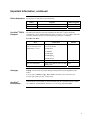
TOP10: Use this strain for general cloning of PCR products in pSecTag/FRT/V5-HisTOPO®.
F- mcrA ∆(mrr-hsdRMS-mcrBC) Φ80lacZ∆M15 ∆lacΧ74 recA1 araD139 ∆(araleu)7697 galU galK rpsL (StrR) endA1 nupG
One Shot®
Electrocomp™
TOP10 cells are also available as electrocompetent cells in a One Shot® format (Catalog
no. C4040-52). Transformation efficiency is 1 x 10⁹ cfu/µg supercoiled DNA.
v
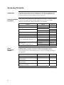
Accessory Products
Introduction
The products listed in this section are intended for use with the pSecTag/FRT/V5-His
TOPO® TA Expression Kit. For more information, refer to our World Wide Web site
(www.invitrogen.com) or call Technical Service (see page 30).
®
Products Available Some of the products included in the pSecTag/FRT/V5-His TOPO TA Expression Kit
™
System
are available
as
well
as
other
reagents
that
may
be
used
with
the
Flp-In
Separately
separately from Invitrogen. Ordering information is provided below.
Product
Amount
2 µg, lyophilized in TE
N560-02
Hygromycin
1g
R220-05
1g
R250-01
5g
R250-05
pFRT/lacZeo
20 µg, lyophilized in TE
V6015-20
pFRT/lacZeo2
20 µg, lyophilized in TE
V6022-20
pOG44
20 µg, lyophilized in TE
V6005-20
One Shot Kit
10 reactions
C4040-10
(TOP10 Chemically Competent Cells)
20 reactions
C4040-03
40 reactions
C4040-06
One Shot Kit
10 reactions
C4040-50
(TOP10 Electrocompetent Cells)
20 reactions
C4040-52
Zeocin
™
®
®
Flp-In™
Expression
Vectors
Catalog no.
T7 Promoter Primer
Additional Flp-In™ expression vectors are available from Invitrogen. For more
information about the features of each vector, refer to our World Wide Web site
(www.invitrogen.com) or call Technical Service (see page 30). Ordering information is
provided below.
Product
Amount
Catalog no.
20 µg, lyophilized in TE
V6010-20
pcDNA5/FRT/V5-His TOPO TA
Expression Kit
1 kit
K6020-01
pEF5/FRT/V5 Directional TOPO®
Expression Kit
1 kit
K6035-01
pEF5/FRT/V5-DEST
Gateway™ Vector Pack
6 µg
V6020-20
pcDNA5/FRT
®
continued on next page
vi
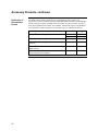
Accessory Products, continued
Flp-In™ Host Cell
Lines
For your convenience, Invitrogen has available several mammalian Flp-In™ host cell
lines that stably express the lacZ-Zeocin™ fusion gene from pFRT/lacZeo or
pFRT/lacZeo2. Each cell line contains a single integrated FRT site as confirmed by
Southern blot analysis. The cell lines should be maintained in medium containing
Zeocin™. For more information, see our World Wide Web site (www.invitrogen.com) or
call Technical Service (see page 30).
Cell Line
Catalog no.
3 x 10⁶ cells, frozen
R750-07
Flp-In™-CV-1
3 x 10⁶ cells, frozen
R752-07
™
Flp-In -CHO
™
Flp-In -BHK
™
Flp-In -3T3
™
Flp-In -Jurkat
Detection of
Recombinant
Proteins
Amount
Flp-In™-293
6
R758-07
6
R760-07
6
R761-07
6
R762-07
3 x 10 cells, frozen
3 x 10 cells, frozen
3 x 10 cells, frozen
3 x 10 cells, frozen
Expression of your recombinant fusion protein can be detected using an antibody to the
appropriate epitope. The table below describes the antibodies available for detection of
C-terminal fusion proteins expressed using pSecTag/FRT/V5-His-TOPO®. Horseradish
peroxidase (HRP) or alkaline phosphatase (AP)-conjugated antibodies allow one-step
detection using colorimetric or chemiluminescent detection methods.
Fifty microliters of each antibody is supplied which is sufficient for 25 Westerns.
Product
Anti-V5 Antibody
Anti-V5-HRP Antibody
Anti-V5-AP Antibody
Epitope
Catalog no.
Detects 14 amino acid epitope
derived from the P and V proteins
of the paramyxovirus, SV5
(Southern et al., 1991)
R960-25
R961-25
R962-25
GKPIPNPLLGLDST
Anti-His (C-term) Antibody
Anti-His(C-term)-HRP Antibody
Anti-His(C-term)-AP Antibody
Detects the C-terminal
polyhistidine (6xHis) tag (requires
the free carboxyl group for
detection (Lindner et al., 1997)
R930-25
R931-25
R932-25
HHHHHH-COOH
continued on next page
vii
Accessory Products, continued
Purification of
Recombinant
Protein
The metal binding domain encoded by the polyhistidine tag allows simple, easy
purification of your recombinant protein by Immobilized Metal Affinity Chromatography
(IMAC) using Invitrogen's ProBond™ Resin (see below). To purify proteins expressed
from pSecTag/FRT/V5-His-TOPO®, the ProBond™ Purification System or the ProBond™
resin in bulk are available separately. See the table below for ordering information.
Product
™
ProBond Metal-Binding Resin
Catalog no.
50 ml
R801-01
150 ml
R801-15
ProBond™ Purification System
6 purifications K850-01
ProBond™ Purification System with Anti-V5-HRP
Antibody
1 kit
K854-01
ProBond™ Purification System with Anti-His(C-term)- 1 kit
HRP Antibody
K853-01
Purification Columns
R640-50
(10 ml polypropylene columns)
viii
Quantity
50
Introduction
Overview
Introduction
The pSecTag/FRT/V5-His TOPO® TA Expression Kit combines the Flp-In™ System
with TOPO® Cloning technology to provide a highly efficient, rapid cloning strategy for
the direct insertion of Taq polymerase-amplified PCR products into a plasmid vector for
targeted and secreted expression of the gene of interest in mammalian cell lines. TOPO
Cloning® requires no ligase, post-PCR procedures, or PCR primers containing special,
additional sequences. For more information about TOPO® Cloning, see the next page.
pSecTag/FRT/V5His-TOPO® Vector
pSecTag/FRT/V5-His-TOPO® is a 5.2 kb expression vector designed to facilitate rapid
cloning and expression of PCR products using the Flp-In™ System (Catalog nos. K601001 and K6010-02) available from Invitrogen. When cotransfected with the pOG44 Flp
recombinase expression plasmid into a Flp-In™ mammalian host cell line, the
pSecTag/FRT/V5-His-TOPO® vector containing the PCR product of interest is integrated
in a Flp recombinase-dependent manner into the genome. The pSecTag/FRT/V5-HisTOPO® vector contains the following elements:
•
The human cytomegalovirus (CMV) immediate-early enhancer/promoter for highlevel constitutive expression of the gene of interest in a wide range of mammalian
cells (Andersson et al., 1989; Boshart et al., 1985; Nelson et al., 1987)
•
Murine Ig κ-chain leader sequence for directing secreted expression of the gene of
interest (Coloma et al., 1992)
•
TOPO® Cloning site for rapid and efficient cloning of Taq-amplified PCR products
(see the next page for more information)
•
C-terminal peptide containing the V5 epitope and a polyhistidine (6xHis) tag for
detection and purification of recombinant protein
•
FLP Recombination Target (FRT) site for Flp recombinase-mediated integration of
the vector into the Flp-In™ host cell line (see pages 2-3 for more information)
•
Hygromycin resistance gene for selection of stable cell lines (Gritz and Davies, 1983)
(see important note on page 3)
The control plasmid, pSecTag/FRT/V5-His/PSA, is included for use as a positive control
for transfection and secreted expression in the Flp-In™ host cell line of choice.
For more information about the Flp-In™ System, the pOG44 plasmid, and generation of
the Flp-In™ host cell line, refer to the Flp-In™ System manual. The Flp-In™ System
manual is supplied with the Flp-In™ Complete or Core Systems, but is also available for
downloading from our World Wide Web site (www.invitrogen.com) or by contacting
Technical Service (see page 30).
continued on next page
1
Overview, continued
How TOPO®
Cloning Works
The plasmid vector, pSecTag/FRT/V5-His-TOPO®, is supplied linearized with:
•
Single 3´ thymidine (T) overhangs for TA Cloning®
• Topoisomerase covalently bound to the vector (this is referred to as “activated” vector)
Taq polymerase has a nontemplate-dependent terminal transferase activity that adds a
single deoxyadenosine (A) to the 3´ ends of PCR products. The linearized vector supplied
in this kit has single, overhanging 3´ deoxythymidine (T) residues. This allows PCR inserts
to ligate efficiently with the vector.
Topoisomerase I from Vaccinia virus binds to duplex DNA at specific sites and cleaves the
phosphodiester backbone after 5′-CCCTT in one strand (Shuman, 1991). The energy from
the broken phosphodiester backbone is conserved by formation of a covalent bond between
the 3′ phosphate of the cleaved strand and a tyrosyl residue (Tyr-274) of topoisomerase I.
The phospho-tyrosyl bond between the DNA and enzyme can subsequently be attacked by
the 5′ hydroxyl of the original cleaved strand, reversing the reaction and releasing
topoisomerase (Shuman, 1994). TOPO® Cloning exploits this reaction to efficiently clone
PCR products (see below).
Topoisomerase
Tyr-274
O
CCCTT
GGGA
P
OH
A
PCR Product
HO
Tyr-274
O
A AGGG
TTCCC
P
Topoisomerase
FRT Site in the
pSecTag/FRT/V5His-TOPO® Vector
The pSecTag/FRT/V5-His-TOPO® vector contains a single FRT site immediately
upstream of the hygromycin resistance gene for Flp recombinase-mediated integration
and selection of the pSecTag/FRT/V5-His-TOPO® construct following cotransfection
of the vector (with pOG44) into a Flp-In™ mammalian host cell line. The FRT site
serves as both the recognition and cleavage site for the Flp recombinase and allows
recombination to occur immediately adjacent to the hygromycin resistance gene. The
flp recombinase is expressed from the pOG44 plasmid. For more information about the
FRT site and recombination, see the next page. For more information about pOG44,
refer to the pOG44 manual or the Flp-In™ System manual.
continued on next page
2
Overview, continued
Flp RecombinaseMediated DNA
Recombination
In the Flp-In™ System, integration of your pSecTag/FRT/V5-His-TOPO® expression
construct into the genome occurs via Flp recombinase-mediated intermolecular DNA
recombination. The hallmarks of Flp-mediated recombination are listed below.
•
Recombination occurs between specific FRT sites (see below) on the interacting
DNA molecules
•
Recombination is conservative and requires no DNA synthesis; the FRT sites are
preserved following recombination and there is minimal opportunity for
introduction of mutations at the recombination site
•
Strand exchange requires only the small 34 bp minimal FRT site (see below)
For more information about the Flp recombinase and conservative site-specific
recombination, refer to published reviews (Craig, 1988; Sauer, 1994).
FRT Site
The FRT site, originally isolated from Saccharomyces cerevisiae, serves as a binding site
for Flp recombinase and has been well-characterized (Gronostajski and Sadowski, 1985;
Jayaram, 1985; Sauer, 1994; Senecoff et al., 1985). The minimal FRT site consists of a
34 bp sequence containing two 13 bp imperfect inverted repeats separated by an 8 bp
spacer that includes an Xba I restriction site (see figure below). An additional 13 bp
repeat is found in most FRT sites, but is not required for cleavage (Andrews et al.,
1985). While Flp recombinase binds to all three of the 13 bp repeats, strand cleavage
actually occurs at the boundaries of the 8 bp spacer region (see figure below) (Andrews
et al., 1985; Senecoff et al., 1985).
Minimal FRT site
CS
GAAGTTCCTATTCCGAAGTTCCTATTCTCTAGAAAGTATAGGAAC TTC
Xba I
CS
CS = cleavage site
Important
The hygromycin resistance gene in pSecTag/FRT/V5-His-TOPO® lacks a promoter and
an ATG initiation codon; therefore, transfection of the pSecTag/FRT/V5-His-TOPO®
plasmid alone into mammalian host cells will not confer hygromycin resistance to the
cells. The SV40 promoter and ATG initiation codon required for expression of the
hygromycin resistance gene are integrated into the genome (in the Flp-In™ host cell
line) and are only brought into the correct proximity and frame with the hygromycin
resistance gene through Flp recombinase-mediated integration of pSecTag/FRT/V5His-TOPO® at the FRT site. For more information about the generation of the Flp-In™
host cell line and details of the Flp-In™ System, refer to the Flp-In™ System manual.
continued on next page
3
Overview, continued
Experimental
Outline
The table below describes the general steps needed to clone and express your gene of
interest. For more details, refer to the pages indicated.
Step
4
Action
Page
1
Design PCR primers to clone your gene of interest in frame with the
5-6
N-terminal Ig κ-chain secretion signal and the C-terminal peptide
containing the V5 epitope and the polyhistidine (6xHis) tag (if desired).
Consult the diagram on page 6 to help you design your PCR primers.
2
Produce your PCR product.
®
7
®
3
TOPO Clone your PCR product into pSecTag/FRT/V5-His-TOPO
and transform into One Shot® TOP10 E. coli. Select transformants on
LB plates containing 50-100 µg/ml ampicillin.
8-10
4
Analyze your transformants for the presence and orientation of insert
by restriction enzyme digestion.
11
5
Select a transformant with the correct restriction pattern and sequence it 11
to confirm that your gene is cloned in frame with the Ig κ-chain
secretion signal and the C-terminal peptide.
6
Cotransfect your pSecTag/FRT/V5-His-TOPO® construct and pOG44
into the Flp-In™ host cell line using your method of choice and select
for hygromycin resistant clones (see the Flp-In™ System manual for
more information).
14-15
7
Assay for expression of your protein of interest.
16-17
8
Purify your recombinant protein by chromatography on metal-chelating 18
resin (e.g. ProBond™).
Methods
PCR Primer Design
Introduction
It is important to properly design your PCR primers to ensure that you obtain the
recombinant protein you need for your studies. Use the information below and the
diagram on page 6 to design your PCR primers. Remember that your PCR product
will have single 3´ adenine overhangs.
General Molecular
Biology
Techniques
For help with E. coli transformations, restriction enzyme analysis, DNA sequencing, and
DNA biochemistry, refer to Molecular Cloning: A Laboratory Manual (Sambrook et al.,
1989) or Current Protocols in Molecular Biology (Ausubel et al., 1994).
Do not add 5´ phosphates to your primers for PCR. The PCR product synthesized will
not ligate into pSecTag/FRT/V5-His-TOPO®.
Fusion to the
Ig κ-Chain
Secretion Signal
In order to obtain proper secreted expression of your protein, you must design your 5′
PCR primer such that the PCR product will clone in frame with the initiation ATG of the
N-terminal Ig κ-chain secretion signal.
Fusion to the
C-terminal Peptide
If you wish to include the C-terminal peptide for detection with either the V5 or His
(C-term) antibodies or purification using the polyhistidine (6xHis) tag, you must design
your reverse PCR primer to remove the native stop codon and maintain the frame
through the DNA encoding the C-terminal peptide.
If you do not wish to include the C-terminal peptide, include the native stop codon in the
reverse PCR primer or design the primer to anneal downstream of the native stop codon.
Note: Cloning efficiencies may vary depending on the 5′ nucleotide sequence of your
primer (see page 26).
Use the diagram on the next page to design your PCR primers. Once you have designed
your PCR primers, proceed to page 7.
Signal Sequence
Processing
The Ig κ-chain secretion signal is processed from your recombinant protein by a signal
peptidase-directed cleavage after aspartic acid 21 in the signal sequence. For the location
of the signal cleavage site, refer to the diagram on the next page. Note that you will not
obtain native protein following cleavage of the signal sequence because of the
intervening sequences between the signal cleavage site and the beginning of your PCR
product (see the next page).
continued on next page
5
PCR Primer Design, continued
TOPO® Cloning Site
for
pSecTag/FRT/V5His-TOPO®
The diagram below is supplied to help you design appropriate PCR primers to correctly
clone and express your PCR product using pSecTag/FRT/V5-His-TOPO®. Restriction
sites are labeled to indicate the actual cleavage site. The vector is supplied linearized
between base pair 1012 and 1013. This is the TOPO® Cloning site. The complete
sequence of pSecTag/FRT/V5-His-TOPO® is available for downloading from our
World Wide Web site (www.invitrogen.com) or from Technical Service (page 30).
For a map and a description of the features of pSecTag/FRT/V5-His-TOPO®, see the
Appendix, pages 27-28.
CAAT
CMV promoter
721
AAAATCAACG GGACTTTCCA AAATGTCGTA ACAACTCCGC CCCATTGACG CAAATGGGCG
CMV forward priming site
781
TATA
3' end of CMV promoter
putative transcriptional start
GTAGGCGTGT ACGGTGGGAG GTCTATATAA GCAGAGCTCT CTGGCTAACT AGAGAACCCA
T7 promoter/priming site
841
Nhe I
CTGCTTACTG GCTTATCGAA ATTAATACGA CTCACTATAG GGAGACCCAA GCTGGCTAGC
Ig K-chain secretion signal
901
CACC ATG GAG ACA GAC ACA CTC CTG CTA TGG GTA CTG CTG CTC TGG GTT
Met Glu Thr Asp Thr Leu Leu Leu Trp Val Leu Leu Leu Trp Val
950
CCA GGT TCC ACT GGT GAC GCG GCC CAG CCG GCC AGG CGC GCG CGC CGT
Pro Gly Ser Thr Gly Asp Ala Ala Gln Pro Ala Arg Arg Ala Arg Arg
Signal Cleavage Site
995
1040
ACG AAG CTC GCC CTT
G CGG GA A
Thr Lys Leu Ala Leu
A AG GGC GAG CTT GGT ACC GAG CTC GGA
TTC CCG CTC
Lys Gly Glu Leu Gly Thr Glu Leu Gly
V5 epitope
Polyhistidine (6xHis) region
Pme I
CGT ACC GGT CAT CAT CAC CAT CAC CAT TGA GTT TAAACCCGCT GATCAGCCTC
Arg Thr Gly His His His His His His ***
BGH reverse priming site
1141
6
Bam H I
TCC GAA GGT AAG CCT ATC CCT AAC CCT CTC CTC GGT CTC GAT TCT ACG
Ser Glu Gly Lys Pro Ile Pro Asn Pro Leu Leu Gly Leu Asp Ser Thr
Age I
1088
PCR
Product
Asp718 I Kpn I
GACTGTGCCT TCTAGTTGCC AGCCATCTGT TGTTTGCCCC TCCCCCGTGC
Producing PCR Products
Introduction
Once you have decided on a PCR strategy and have synthesized the primers you are
ready to produce your PCR product.
Materials Supplied You will need the following reagents and equipment.
by the User
• Taq polymerase
Polymerase
Mixtures
•
Thermocycler
•
DNA template and primers for PCR product
If you wish to use a mixture containing Taq polymerase and a proofreading polymerase,
Taq must be used in excess of a 10:1 ratio to ensure the presence of 3´ A-overhangs on
the PCR product.
If you use polymerase mixtures that do not have enough Taq polymerase or a proofreading polymerase only, you can add 3′ A-overhangs using the method on page 23.
Producing PCR
Products
1.
Set up the following 50 µl PCR reaction. Use less DNA if you are using plasmid
DNA as a template and more DNA if you are using genomic DNA as a template.
Use the cycling parameters suitable for your primers and template. Be sure to
include a 7 to 30 minute extension at 72°C after the last cycle to ensure that all PCR
products are full length and 3´ adenylated.
DNA Template
10-100 ng
5 µl
10X PCR Buffer
0.5 µl
50 mM dNTPs
Primers (100-200 ng each)
Sterile water
add to a final volume of 49 µl
Taq Polymerase (1 unit/µl)
Total Volume
2.
1 µM each
1 µl
50 µl
Check the PCR product by agarose gel electrophoresis. You should see a single,
discrete band. If you do not see a single band, refer to the Note below.
If you do not obtain a single, discrete band from your PCR, you may gel-purify your
fragment before using the pSecTag/FRT/V5-His TOPO® TA Expression Kit (see page
21). Take special care to avoid sources of nuclease contamination and long exposure to
UV light. Alternatively, you may optimize your PCR to eliminate multiple bands and
smearing (Innis et al., 1990). The PCR Optimizer™ Kit (Catalog no. K1220-01) from
Invitrogen can help you optimize your PCR. Call Technical Service for more
information (page 30).
7
TOPO® Cloning Reaction and Transformation
Introduction
TOPO® Cloning technology allows you to produce your PCR products, ligate them into
pSecTag/FRT/V5-His-TOPO®, and transform the recombinant vector into TOP10 E. coli
in one day. It is important to have everything you need set up and ready to use to ensure
that you obtain the best possible results. If this is the first time you have TOPO® Cloned,
perform the control reactions on pages 24-25 in parallel with your samples. If you have
previously TOPO® Cloned, read the Note below.
Recent experiments at Invitrogen demonstrate that inclusion of salt (200 mM NaCl,
10 mM MgCl2) in the TOPO® Cloning reaction results in the following:
•
a 2- to 3-fold increase in the number of transformants.
•
allows for longer incubation times (up to 30 minutes). Longer incubation times can
result in an increase in the number of transformants obtained.
Including salt in the TOPO® Cloning reaction prevents topoisomerase I from rebinding
and potentially nicking the DNA after ligating the PCR product and dissociating from the
DNA. The result is more intact molecules leading to higher transformation efficiencies.
If you do not include salt in the TOPO® Cloning reaction, the number of transformants
obtained generally decreases as the incubation time increases beyond 5 minutes.
Important
Because of the above results, we recommend adding salt to the TOPO® Cloning reaction.
A stock salt solution is provided in the kit for this purpose. Note that the amount of salt
added to the TOPO® Cloning reaction varies depending on whether you plan to
transform chemically competent cells (provided) or electrocompetent cells (see
below). For this reason two different TOPO® Cloning reactions are provided to help you
obtain the best possible results. Read the following information carefully.
Chemically
Competent E. coli
For TOPO® Cloning and transformation into chemically competent E. coli, adding sodium
chloride and magnesium chloride to a final concentration of 200 mM NaCl, 10 mM
MgCl2 in the TOPO® Cloning reaction increases the number of colonies over time. A Salt
Solution (1.2 M NaCl; 0.06 M MgCl2) is provided to adjust the TOPO® Cloning reaction
to the recommended concentration of NaCl and MgCl2.
Electrocompetent
E. coli
For TOPO® Cloning and transformation of electrocompetent E. coli, salt must also be
included in the TOPO® Cloning reaction, but the amount of salt must be reduced to
50 mM NaCl, 2.5 mM MgCl2 to prevent arcing when electroporating. The Salt Solution
provided in the kit must be diluted 4-fold to prepare a 300 mM NaCl, 15 mM MgCl2
solution for convenient addition to the TOPO® Cloning reaction (see next page).
continued on next page
8
TOPO® Cloning Reaction and Transformation, continued
Materials Supplied In addition to general microbiological supplies (e.g. plates, spreaders), you will need the
following reagents and equipment.
by the User
•
42°C water bath (or electroporator with cuvettes, optional)
•
LB plates containing 50-100 µg/ml ampicillin (two for each transformation)
•
Reagents and equipment for agarose gel electrophoresis
•
37°C shaking and non-shaking incubator
There is no blue-white screening for the presence of inserts. Individual recombinant
plasmids need to be analyzed by restriction analysis or sequencing for the presence and
orientation of insert. Sequencing primers included in each kit can be used to sequence
across an insert in the multiple cloning site to confirm orientation and reading frame.
Preparation for
Transformation
Setting Up the
TOPO® Cloning
Reaction
For each transformation, you will need one vial of competent cells and two selective
plates.
•
Equilibrate a water bath to 42°C (for chemical transformation) or set up your
electroporator if you are using electrocompetent E. coli.
•
For electroporation, dilute a small portion of the Salt Solution 4-fold to prepare
Dilute Salt Solution (e.g. add 5 µl of the Salt Solution to 15 µl sterile water)
•
Warm the vial of SOC medium from Box 2 to room temperature.
•
Warm selective plates at 37°C for 30 minutes.
•
Thaw on ice 1 vial of One Shot® cells for each transformation.
The table below describes how to set up your TOPO® Cloning reaction (6 µl) for
eventual transformation into either chemically competent One Shot® TOP10 E. coli
(provided) or electrocompetent E. coli. Additional information on optimizing the TOPO®
Cloning reaction for your needs can be found on page 13.
Note: The red or yellow color of the TOPO® vector solution is normal and is used to
visualize the solution.
Reagent*
Chemically Competent E. coli
Electrocompetent E. coli
Fresh PCR product
0.5 to 4 µl
0.5 to 4 µl
Salt Solution
1 µl
--
Dilute Salt Solution
--
1 µl
Sterile Water
add to a final volume of 5 µl
add to a final volume of 5 µl
1 µl
1 µl
®
TOPO vector
*Store all reagents at -20°C when finished. Salt solutions and water can be stored at room temperature or +4°C.
continued on next page
9
TOPO® Cloning Reaction and Transformation, continued
Performing the
TOPO® Cloning
Reaction
One Shot®
Chemical
Transformation
1.
Mix reaction gently and incubate for 5 minutes at room temperature (22-23°C).
Note: For most applications, 5 minutes will yield plenty of colonies for analysis.
Depending on your needs, the length of the TOPO® Cloning reaction can be varied
from 30 seconds to 30 minutes. For routine subcloning of PCR products, 30 seconds
may be sufficient. For large PCR products (> 1 kb) or if you are TOPO® Cloning a
pool of PCR products, increasing the reaction time will yield more colonies.
2.
Place the reaction on ice and proceed to One Shot® Chemical Transformation
(next page) or Transformation by Electroporation (next page). Note: You may
store the TOPO® Cloning reaction at -20°C overnight.
1.
Add 2 µl of the TOPO® Cloning reaction from Step 2 previous page into a vial of
One Shot® TOP10 Chemically Competent E. coli and mix gently. Do not mix by
pipetting up and down.
2.
Incubate on ice for 5 to 30 minutes.
Note: Longer incubations on ice seem to have a minimal effect on transformation
efficiency. The length of the incubation is at the user’s discretion (see above).
Transformation by
Electroporation
3.
Heat-shock the cells for 30 seconds at 42°C without shaking.
4.
Immediately transfer the tubes to ice.
5.
Add 250 µl of room temperature SOC medium.
6.
Cap the tube tightly and shake the tube horizontally (200 rpm) at 37°C for 1 hour.
7.
Spread 25-200 µl from each transformation on a prewarmed selective plate and
incubate overnight at 37°C. We recommend that you plate two different volumes to
ensure that at least one plate will have well-spaced colonies.
8.
An efficient TOPO® Cloning reaction will produce hundreds of colonies. Pick
~10 colonies for analysis (see Analysis of Positive Clones, next page).
1.
Add 2 µl of the TOPO® Cloning reaction into a 0.1 cm cuvette containing 50 µl of
electrocompetent E. coli and mix gently. Do not mix by pipetting up and down.
Avoid formation of bubbles.
2.
Electroporate your samples using your own protocol and your electroporator.
Note: If you have problems with arcing, see next page.
3.
Immediately add 250 µl of room temperature SOC medium.
4.
Transfer the solution to a 15 ml snap-cap tube (e.g. Falcon) and shake for at least
1 hour at 37°C to allow expression of the antibiotic resistance gene.
5.
Spread 10-50 µl from each transformation on a prewarmed selective plate and
incubate overnight at 37°C. To ensure even spreading of small volumes, add 20 µl of
SOC. We recommend that you plate two different volumes to ensure that at least one
plate will have well-spaced colonies.
6.
An efficient TOPO® Cloning reaction will produce hundreds of colonies. Pick
~10 colonies for analysis (see Analysis of Positive Clones, next page).
continued on next page
10
TOPO® Cloning Reaction and Transformation, continued
Addition of the Dilute Salt Solution in the TOPO® Cloning Reaction brings the final
concentration of NaCl and MgCl2 in the TOPO® Cloning reaction to 50 mM and 2.5 mM,
respectively. To prevent arcing of your samples during electroporation, the volume of
cells should be between 50 and 80 µl (0.1 cm cuvettes) or 100 to 200 µl (0.2 cm cuvettes).
If you experience arcing during transformation, try one of the following suggestions:
Analysis of
Positive Clones
•
Reduce the voltage normally used to charge your electroporator by 10%
•
Reduce the pulse length by reducing the load resistance to 100 ohms
•
Ethanol-precipitate the TOPO® Cloning reaction and resuspend in water prior to
electroporation
1.
Culture 10 transformants overnight in 2-5 ml LB or SOB medium containing 50100 µg/ml ampicillin.
2.
Isolate plasmid DNA using your method of choice. If you need ultra-pure plasmid
DNA for automated or manual sequencing, we recommend the S.N.A.P.™ MiniPrep
Kit (Catalog no. K1900-01) or the S.N.A.P.™ MidiPrep Kit (Catalog no. K191001).
3.
Analyze the plasmids for the presence and orientation of insert by restriction
analysis. We recommend sequencing your constructs to confirm that your gene of
interest is cloned in frame with the C-terminal peptide. Sequencing primers are
included to help you sequence your insert (see page iv). Refer to the diagram on
page 6 for the sequence surrounding the TOPO® Cloning site.
If you need help with setting up restriction enzyme digests or DNA sequencing,
refer to general molecular biology texts (Ausubel et al., 1994; Sambrook et al.,
1989).
Alternative Method You may wish to use PCR to directly analyze positive transformants. You may use either
the forward or reverse sequencing primers included in the kit and a primer that
of Analysis
hybridizes within your insert. You will have to determine the amplification conditions.
If this is the first time you have used this technique, we recommend that you perform
restriction analysis in parallel to confirm that PCR gives you the correct result. Both false
positive and false negative results can be obtained because of mispriming or
contaminating template.
The following protocol is provided for your convenience. Other protocols are suitable.
1.
Prepare a PCR cocktail consisting of PCR buffer, dNTPs, primers, and Taq
polymerase. Use a 20 µl reaction volume. Multiply by the number of colonies to be
analyzed (e.g. 10).
2.
Pick 10 colonies and resuspend them individually in 20 µl of the PCR cocktail.
(Don't forget to make a patch plate to preserve the colonies for further analysis.)
3.
Incubate the reaction for 10 minutes at 94°C to lyse the cells and inactivate
nucleases.
4.
Amplify for 20 to 30 cycles using the appropriate conditions (see text above).
5.
For the final extension, incubate at 72°C for 10 minutes. Hold at +4°C.
6.
Visualize by agarose gel electrophoresis.
continued on next page
11
TOPO Cloning® Reaction and Transformation, continued
Important
Long-Term
Storage
12
If you have problems obtaining transformants or the correct insert, perform the control
reactions described on page 24-25. These reactions will help you troubleshoot your
experiment.
Once you have identified the correct clone, be sure to isolate a single colony and prepare
a glycerol stock for long term storage. We recommend that you also store the purified
plasmid DNA at -20°C.
1.
Streak the original colony on LB plates containing 50-100 µg/ml ampicillin.
2.
Isolate a single colony and inoculate into 1-2 ml of LB containing 50-100 µg/ml
ampicillin.
3.
Grow the culture to mid-log phase (OD600 = 0.5-0.7).
4.
Mix 0.85 ml of culture with 0.15 ml of sterile glycerol and transfer to a cryovial.
5.
Store at -80°C.
Optimizing the TOPO® Cloning Reaction
Introduction
The information below will help you optimize the TOPO® Cloning reaction for your
particular needs.
Faster Subcloning
The high efficiency of TOPO® Cloning technology allows you to streamline the cloning
process. If you routinely clone PCR products and wish to speed up the process,
consider the following:
•
Incubate the TOPO® Cloning reaction for only 30 seconds instead of 5 minutes.
You may not obtain the highest number of colonies, but with the high efficiency of
TOPO® Cloning, most of the transformants will contain your insert.
•
After adding 2 µl of the TOPO® Cloning reaction to chemically competent cells,
incubate on ice for only 5 minutes.
Increasing the incubation time to 30 minutes does not significantly improve
transformation efficiency.
More
Transformants
If you are TOPO® Cloning large PCR products, toxic genes, or cloning a pool of PCR
products, you may need more transformants to obtain the clones you want. To increase
the number of colonies:
•
Incubate the salt-supplemented TOPO® Cloning reaction for 20 to 30 minutes
instead of 5 minutes.
Increasing the incubation time of the salt-supplemented TOPO® Cloning reaction
allows more molecules to ligate, increasing the transformation efficiency. Addition
of salt appears to prevent topoisomerase from rebinding and nicking the DNA after
it has ligated the PCR product and dissociated from the DNA.
Cloning Dilute
PCR Products
To clone dilute PCR products, you may:
•
Increase the amount of the PCR product
•
Incubate the TOPO® Cloning reaction for 20 to 30 minutes
•
Concentrate the PCR product by precipitation
13
Transfection and Analysis
Introduction
Once you have TOPO® Cloned your gene of interest into pSecTag/FRT/V5-His-TOPO®
and have prepared purified plasmid DNA of your expression construct and pOG44, you
are ready to cotransfect the plasmids into your mammalian Flp-In™ host cell line to
generate your stable Flp-In™ expression cell line. We recommend that you include the
pSecTag/FRT/V5-His/PSA positive control vector (see the next page) and a mock
transfection (negative control) in your experiments to evaluate your results. General
information about transfection, selection, and expression analysis is provided in this
section. Specific guidelines and protocols for generating the Flp-In™ expression cell line
can be found in the Flp-In™ System manual.
MEND
ION
AT
RECOM
For detailed information about pOG44 and generation of the Flp-In™ host cell line, refer
to the Flp-In™ System manual. A separate manual for the pOG44 plasmid is also
available from our Web site (www.invitrogen.com) or by calling Technical Service (see
page 30).
Important
Several Flp-In™ host cell lines which stably express the lacZ-Zeocin™ fusion gene and
contain a single integrated FRT site are available from Invitrogen (see page vii for
ordering information). If you wish to express your gene of interest in 293, CV-1, CHO,
3T3, BHK, or Jurkat cells, you may want to use one of the Flp-In™ host cell lines to
establish your expression cell line. For more information, refer to our World Wide Web
site (www.invitrogen.com) or call Technical Service (see page 30).
We have observed down-regulation of the viral CMV promoter and subsequent loss of
gene expression when pcDNA5/FRT-based expression constructs are introduced into
3T3 or BHK cells. This behavior is not observed with pEF5/FRT-based expression
constructs. If you are generationg Flp-In™ expression cell lines using a 3T3 or BHK host
cell line, we recommend that you clone your gene of interest into a pEF5/FRT-based
expression plasmid (e.g. pEF5/FRT/V5-D-TOPO® or pEF5/FRT/V5-DEST). For more
information, refer to our Web site (www.invitrogen.com) or call Technical Service (see
page 30).
Plasmid
Preparation
Plasmid DNA for transfection into eukaryotic cells must be clean and free from phenol
and sodium chloride. Contaminants will kill the cells, and salt will interfere with lipid
complexing, decreasing transfection efficiency. We recommend isolating plasmid DNA
using the S.N.A.P.™ MiniPrep Kit (10-15 µg DNA, Catalog no. K1900-01), the S.N.A.P.™
MidiPrep Kit (10-200 µg DNA, Catalog no. K1910-01), or CsCl gradient centrifugation.
Methods of
Transfection
For established cell lines (e.g. HeLa, CHO), consult original references or the supplier of
your cell line for the optimal method of transfection. We recommend that you follow
exactly the protocol for your cell line. Pay particular attention to medium require-ments,
when to pass the cells, and at what dilution to split the cells. Further information is
provided in Current Protocols in Molecular Biology (Ausubel et al., 1994).
Methods for transfection include calcium phosphate (Chen and Okayama, 1987; Wigler
et al., 1977), lipid-mediated (Felgner et al., 1989; Felgner and Ringold, 1989) and
electroporation (Chu et al., 1987; Shigekawa and Dower, 1988). Invitrogen offers the
Calcium Phosphate Transfection Kit (Catalog no. K2780-01) and several lipid-based
reagents for mammalian cell transfection. For more information, refer to our World Wide
Web site (www.invitrogen.com) or call Technical Service (see page 30).
continued on next page
14
Transfection and Analysis, continued
Positive Control
pSecTag/FRT/V5-His/PSA is provided as a positive control vector for mammalian cell
transfection and expression (see page 29 for a map) and may be used to assay for
recombinant protein expression levels in your Flp-In™ host cell line. Cotransfection of
the positive control vector and pOG44 into your Flp-In™ host cell line allows you to
generate a stable cell line expressing prostate specific antigen (PSA) at the same
genomic locus as your gene of interest. If you have several different Flp-In™ host cell
lines, you may use the pSecTag/FRT/V5-His/PSA control vector to compare protein
expression levels between the various cell lines.
To propagate and maintain the plasmid:
1.
Resuspend the vector in 20 µl sterile water to prepare a 1 µg/µl stock solution. Use
the stock solution to transform a recA, endA E. coli strain like TOP10, DH5α,
JM109, or equivalent.
2.
Select transformants on LB agar plates containing 50-100 µg/ml ampicillin.
3.
Prepare a glycerol stock of a transformant containing plasmid for long-term storage.
Hygromycin B
The pSecTag/FRT/V5-His-TOPO® vector contains the hygromycin resistance gene
(Gritz and Davies, 1983) for selection of transfectants with the antibiotic, hygromycin B
(Palmer et al., 1987). When added to cultured mammalian cells, hygromycin B acts as an
aminocyclitol to inhibit protein synthesis. Hygromycin B liquid is supplied with the FlpIn™ Complete System and is also available separately from Invitrogen (Catalog no.
R220-05). For instructions to handle and store hygromycin B, see the Flp-In™ System
manual.
Determination of
Hygromycin
Sensitivity
Before generating a stable cell line expressing your protein of interest (Flp-In™ expression
cell line), we recommend that you generate a kill curve to determine the minimum
concentration of hygromycin required to kill your untransfected Flp-In™ host cell line.
Generally, concentrations between 10 and 400 µg/ml hygromycin are required for
selection of most mammalian cell lines. General guidelines for performing a kill curve are
provided in the Flp-In™ System manual.
Important
REMINDER: Remember that the hygromycin resistance gene in pSecTag/FRT/V5His-TOPO® lacks a promoter and an ATG initiation codon; therefore, transfection of
the pSecTag/FRT/V5-His-TOPO® plasmid alone into mammalian host cells will not
confer hygromycin resistance to the cells. The SV40 promoter and ATG initiation
codon required for expression of the hygromycin resistance gene are integrated into the
genome (in the Flp-In™ host cell line) and can only be brought into the correct
proximity and frame with the hygromycin resistance gene through Flp recombinasemediated integration of pSecTag/FRT/V5-His-TOPO® at the FRT site.
continued on next page
15
Transfection and Analysis, continued
Generation of FlpIn™ Expression
Cell Lines
Refer to the Flp-In™ System manual for detailed guidelines and instructions to
cotransfect your pSecTag/FRT/V5-His-TOPO® construct and pOG44 into the Flp-In™
host cell line to generate stable Flp-In™ expression cell lines. Once you have generated
your Flp-In™ expression cell line, see the next page for general guidelines to assay for
expression of your recombinant fusion protein.
Your gene of interest will be expressed from pSecTag/FRT/V5-His-TOPO® under the
control of the human CMV promoter. Once you have generated the Flp-In™ expression
cell line, note that your recombinant fusion protein will be constitutively expressed.
Detection of
Recombinant
Fusion Proteins
To detect expression of your recombinant fusion protein by Western blot analysis, you
may use Anti-V5 antibodies or Anti-His(C-term) antibodies available from Invitrogen (see
page vii for ordering information) or an antibody to your protein of interest. In addition,
the Positope™ Control Protein (Catalog no. R900-50) is available from Invitrogen for use
as a positive control for detection of fusion proteins containing a V5 epitope or a
polyhistidine (6xHis) tag. The ready-to-use WesternBreeze® Chromogenic Kits and
WesternBreeze® Chemiluminescent Kits are available from Invitrogen to facilitate
detection of antibodies by colorimetric or chemiluminescent methods. For more
information, refer to our World Wide Web site (www.invitrogen.com) or call Technical
Service (see page 30).
Detection of
Secreted Protein
from Medium
The medium in which your Flp-In™ expression cells are grown can be analyzed for
secreted, recombinant fusion protein by functional assay or Western blot analysis. If you
are also harvesting cells, see Preparation of Cell Lysates, next page. Before starting,
prepare 2X SDS-PAGE sample buffer. A recipe is provided on page 20 for your
convenience, but other recipes are suitable. If you are using pre-cast polyacrylamide gels
(see the next page), refer to the manufacturer’s instructions to prepare the appropriate
sample buffer.
1.
Prepare an SDS-PAGE gel that will resolve your expected recombinant protein.
2.
Harvest the medium from the cells.
Note: Depending on the sensitivity of your antibody, you may wish to concentrate the
media samples prior to Western blot analysis. You may use any method to
concentrate the media samples. We suggest using commercially available
ultrafiltration devices (e.g. Centricon) or a Speed-Vac.
3.
For each media sample, mix 20 µl of media with 20 µl of 2X SDS-PAGE sample
buffer.
4.
Boil the samples for 5 minutes. Centrifuge briefly.
5.
Load samples, electrophorese, blot, and probe with a suitable antibody (see above).
6.
Visualize proteins using your desired method.
The amino acids between the Ig κ-chain secretion signal and the TOPO® Cloning site
will add approximately 1.5 kDa to the size of your protein while the C-terminal tag
containing the V5 epitope and the polyhistidine (6xHis) tag will add approximately
3.6 kDa to the size of your protein.
continued on next page
16
Transfection and Analysis, continued
Preparation of Cell Before starting, prepare Cell Lysis Buffer. A recipe is provided on page 19 for your
convenience, but other recipes are suitable.
Lysates
1.
Prepare an SDS-PAGE gel that will resolve your expected recombinant protein
2.
Remove the medium from each plate and prepare samples as detailed on the previous
page.
3.
Wash cell monolayers (~5 x 10⁵ to 1 x 10⁶ cells) once with phosphate-buffered
saline (PBS, see the Appendix, page 20 for a recipe).
4.
Scrape cells into 1 ml PBS and pellet the cells at 1500 x g for 5 minutes.
5.
Resuspend in 50 µl Cell Lysis Buffer. Vortex.
6.
Incubate cell suspension at 37°C for 10 minutes to lyse the cells. Note: You may
prefer to lyse the cells at room temperature or on ice if degradation of your protein is
a potential problem.
7.
Centrifuge the cell lysate at 10,000 x g for 10 minutes at +4°C to pellet nuclei and
transfer the supernatant to a fresh tube. Assay the lysate for protein concentration.
Note: Do not use protein assays utilizing Coomassie Blue or other dyes. NP-40
interferes with the binding of the dye with the protein.
8.
Add SDS-PAGE sample buffer (see page 20 for a recipe) to a final concentration of
1X and boil the sample for 5 minutes.
9.
Load 20 µg of lysate onto an SDS-PAGE gel and electrophorese (see the next page).
Use the appropriate percentage of acrylamide to resolve your fusion protein.
Polyacrylamide
Gel
Electrophoresis
To facilitate separation and visualization of your recombinant fusion protein by
polyacrylamide gel electrophoresis, a wide range of pre-cast NuPAGE® and Novex®
Tris-Glycine polyacrylamide gels and electrophoresis apparatus are available from
Invitrogen. The NuPAGE® Gel System avoids the protein modifications associated with
Laemmli-type SDS-PAGE, ensuring optimal separation for protein analysis. In addition,
Invitrogen also carries a large selection of molecular weight protein standards and
staining kits. For more information about the appropriate gels, standards, and stains to
use to visualize your recombinant protein, refer to our World Wide Web site
(www.invitrogen.com) or call Technical Service (see page 30).
Assay for PSA
If you use pSecTag/FRT/V5-His/PSA as a positive control vector, you may assay for
PSA expression using your method of choice. We generally use the Total PSA Enzyme
Immunoassay Test Kit (American Laboratory Products, Catalog no. 025-BC-1019) to
assay for PSA expression.
Note that PSA is fused to the C-terminal peptide, so you can use Western blot analysis
and either the Anti-V5 antibody or the Anti-His(C-term) antibody to detect expression of
PSA. The PSA/V5-His protein fusion migrates around 31.5 kDa on an SDS-PAGE gel.
17
Purification
Introduction
Once you have generated your Flp-In™ expression cell line and have verified that your
recombinant fusion protein expresses, you may use metal-chelating resin such as
ProBond™ to purify the recombinant protein. General guidelines are provided below. For
more details about purification using ProBond™, refer to the ProBond™ Purification
System manual.
Purification of
Secreted
Recombinant
Protein
To purify secreted, recombinant protein from the medium, follow the manufacturer’s
instructions for the metal-chelating resin that you are using. Start with about 3 to 5 ml of
medium and load onto 1 to 2 ml of resin. Scale up or down depending on the level of
expression and the capacity of your resin.
Purification of
Recombinant
Protein from Cells
You may also purify recombinant protein from cell lysates. In this case, you will need
5 x 10⁶ to 1 x 10⁷ transfected cells for purification of your protein on a 2 ml ProBond™
column (or other metal-chelating column). If you are using ProBond™ to purify your
protein, refer to the protocol below to prepare cells for lysis. If you are using another
metal-chelating resin, refer to the manufacturer’s instructions to prepare your cells.
Preparation of
Cells for Lysis
Use the procedure below to prepare cells for lysis prior to purification of your protein on
ProBond™. You will need 5 x 10⁶ to 1 x 10⁷ stably transfected cells for purification of
your protein on a 2 ml ProBond™ column (see ProBond™ Purification System manual for
details).
Lysis of Cells
1.
Seed cells in either five T-75 flasks or 2 to 3 T-175 flasks.
2.
Grow the cells in selective medium until they are approximately 80-90% confluent.
3.
Harvest the cells by treating with trypsin-EDTA for 2 to 5 minutes or by scraping the
cells in PBS.
4.
Inactivate the trypsin by diluting with fresh medium (if necessary) and transfer the
cells to a sterile microcentrifuge tube.
5.
Centrifuge the cells at 1500 x g for 5 minutes. Resuspend the cell pellet in PBS.
6.
Centrifuge the cells at 1500 x g for 5 minutes. You may lyse the cells immediately or
freeze in liquid nitrogen and store at –70°C until needed.
If you are using ProBond™ resin, refer to the ProBond™ Purification System manual for
details about sample preparation for chromotography.
If you are using other metal-chelating resin, refer to the manufacturer’s instructions for
recommendations on sample preparation.
If you are not using metal-chelating resin to purify your secreted, recombinant protein, we
recommend that you culture the cells in serum-free medium or in reduced-serum medium to
avoid or lessen the amount of bovine serum albumin present in your sample. Whether you
are able to culture your cells in serum-free medium will depend on the nature of your cell
line and the availability of commercial serum-free media formulations for the particular cell
type. Refer to the supplier of your media or serum for more information.
18
Appendix
Recipes
LB (Luria-Bertani)
Medium and
Plates
Composition:
1.0% Tryptone
0.5% Yeast Extract
1.0% NaCl
pH 7.0
1.
For 1 liter, dissolve 10 g tryptone, 5 g yeast extract, and 10 g NaCl in 950 ml
deionized water.
2.
Adjust the pH of the solution to 7.0 with NaOH and bring the volume up to 1 liter.
3.
Autoclave on liquid cycle for 20 minutes. Allow solution to cool to ~55°C and add
50 µg/ml ampicillin if needed.
4.
Store at room temperature or at +4°C.
LB agar plates
X-Gal Stock
Solution
Cell Lysis Buffer
1.
Prepare LB medium as above, but add 15 g/L agar before autoclaving.
2.
Autoclave on liquid cycle for 20 minutes.
3.
After autoclaving, cool to ~55°C, add 50 µg/ml ampicillin and pour into 10 cm
plates.
4.
Let harden, then invert and store at +4°C, in the dark.
5.
To add X-gal to the plate, warm the plate to 37°C. Pipette 40 µl of the 40 mg/ml
X-gal stock solution (see below), spread evenly, and let dry 15 minutes. Protect
plates from light.
1.
To prepare a 40 mg/ml stock solution, dissolve 400 mg X-Gal in 10 ml dimethylformamide.
2.
Protect from light by storing in a brown bottle at -20°C.
50 mM Tris, pH 7.8
150 mM NaCl
1% Nonidet P-40
1.
This solution can be prepared from the following common stock solutions. For
100 ml, combine
1 M Tris base
5 ml
5 M NaCl
3 ml
Nonidet P-40
1 ml
2.
Bring the volume up to 90 ml with deionized water and adjust the pH to 7.8 with
HCl.
3.
Bring the volume up to 100 ml. Store at room temperature.
To prevent proteolysis, you may add 1 mM PMSF, 1 µM leupeptin, or 0.1 µM aprotinin
before use.
continued on next page
19
Recipes, continued
PhosphateBuffered Saline
(PBS)
2X SDS-PAGE
Sample Buffer
20
137 mM NaCl
2.7 mM KCl
10 mM Na2HPO4
1.8 mM KH2PO4
1.
Dissolve:
2.
Adjust pH to 7.4 with concentrated HCl.
3.
Bring the volume to 1 liter. You may wish to filter-sterilize or autoclave the
solution to increase shelf life.
1.
8 g NaCl
0.2 g KCl
1.44 g Na2HPO4
0.24 g KH2PO4
in 800 ml deionized water.
Combine the following reagents:
0.5 M Tris-HCl, pH 6.8
Glycerol (100%)
β-mercaptoethanol
Bromophenol Blue
SDS
2.
Bring the volume to 10 ml with sterile water.
3.
Aliquot and freeze at -20°C until needed.
2.5 ml
2.0 ml
0.4 ml
0.02 g
0.4 g
Purifying PCR Products
Introduction
Smearing, multiple banding, primer-dimer artifacts, or large PCR products (>3 kb) may
necessitate gel purification. If you intend to purify your PCR product, be extremely
careful to remove all sources of nuclease contamination. There are many protocols to
isolate DNA fragments or remove oligonucleotides. Refer to Current Protocols in
Molecular Biology, Unit 2.6 (Ausubel et al., 1994) for the most common protocols.
Three simple protocols are provided below.
Note that cloning efficiency may decrease with purification of the PCR product. You
may wish to optimize your PCR to produce a single band (see Producing PCR
Products, page 7).
Using the
S.N.A.P.™ Gel
Purification Kit
The S.N.A.P.™ Gel Purification Kit (Catalog no. K1999-25) allows you to rapidly purify
PCR products from regular agarose gels.
1.
Electrophorese amplification reaction on a 1 to 5% regular TAE agarose gel.
Note: Do not use TBE. Borate will interfere with the NaI step (Step 2.)
Quick S.N.A.P.™
Method
2.
Cut out the gel slice containing the PCR product and melt it at 65°C in 2 volumes of
6 M NaI.
3.
Add 1.5 volumes of Binding Buffer.
4.
Load solution (no more than 1 ml at a time) from Step 3 onto a S.N.A.P.™ column.
Centrifuge 1 minute at 3000 x g in a microcentrifuge and discard the supernatant.
5.
If you have solution remaining from Step 3, repeat Step 4.
6.
Add 900 µl of the Final Wash Buffer.
7.
Centrifuge 1 minute at full speed in a microcentrifuge and discard the flow-through.
8.
Repeat Step 7.
9.
Elute the purified PCR product in 40 µl of TE or sterile water. Use 4 µl for the
TOPO® Cloning reaction and proceed as described on page 9.
An even easier method is to simply cut out the gel slice containing your PCR product, place
it on top of the S.N.A.P.™ column bed, and centrifuge at full speed for 10 seconds. Use
1-2 µl of the flow-through in the TOPO® Cloning reaction (page 9). Be sure to make the gel
slice as small as possible for best results.
continued on next page
21
Purifying PCR Products, continued
Low-Melt Agarose
Method
If you prefer to use low-melt agarose, use the procedure below. Note that the gel
purification will result in a dilution of your PCR product and a potential loss of cloning
efficiency.
1.
22
Electrophorese as much as possible of your PCR reaction on a low-melt agarose gel
(0.8 to 1.2%) in TAE buffer.
2.
Visualize the band of interest and excise the band.
3.
Place the gel slice in a microcentrifuge tube and incubate the tube at 65°C until the
gel slice melts.
4.
Place the tube at 37°C to keep the agarose melted.
5.
Add 4 µl of the melted agarose containing your PCR product to the TOPO® Cloning
reaction as described on page 9.
6.
Incubate the TOPO® Cloning reaction at 37°C for 5 to 10 minutes. This is to keep
the agarose melted.
7.
Transform 2 to 4 µl directly into chemically competent One Shot® TOP10 cells using
the method on page 10.
Addition of 3´ A-Overhangs Post-Amplification
Introduction
Direct cloning of DNA amplified by Vent® or Pfu polymerases into TOPO® Cloning
vectors is often difficult because of very low cloning efficiencies. These low efficiencies
are caused by the 3´ to 5´ exonuclease activity, which removes the 3´ A-overhangs
necessary for TOPO® Cloning. Invitrogen has developed a simple method to clone these
blunt-ended fragments.
Before Starting
You will need the following items:
Procedure
•
Taq polymerase
•
A heat block equilibrated to 72°C
•
Phenol-chloroform (optional)
•
3 M sodium acetate (optional)
•
100% ethanol (optional)
•
80% ethanol (optional)
•
TE buffer (optional)
This is just one method for adding 3´ adenines. Other protocols may be suitable.
1.
After amplification with Vent® or Pfu polymerase, place vials on ice and add 0.7-1
unit of Taq polymerase per tube. Mix well. It is not necessary to change the buffer.
2.
Incubate at 72°C for 8-10 minutes (do not cycle).
3.
Place the vials on ice. The DNA amplification product is now ready for ligation into
pSecTag/FRT/V5-His-TOPO®.
Note: If you plan to store your sample(s) overnight before proceeding with TOPO®
Cloning, you may want to extract your sample(s) with phenol-chloroform to remove the
polymerases. After phenol-chloroform extraction, precipitate the DNA with ethanol and
resuspend the DNA in TE buffer to the starting volume of the amplification reaction.
You may also gel-purify your PCR product after amplification with Vent® or Pfu (see
page 21). After purification, add Taq polymerase buffer, dATP, and 0.5 unit of Taq
polymerase and incubate 10-15 minutes at 72°C. Use 4 µl in the TOPO® Cloning
reaction.
Vent® is a registered trademark of New England Biolabs.
23
pSecTag/FRT/V5-His TOPO® Control Reactions
Introduction
If you have trouble obtaining transformants or vector containing insert, perform the
following control reactions to help troubleshoot your experiment. Performing the control
reactions involves producing a control PCR product containing the lac promoter and the
LacZα fragment using the reagents included in the kit. Successful TOPO® Cloning of the
control PCR product in either direction will yield blue colonies on LB agar plates
containing antibiotic and X-gal.
Before Starting
Be sure to prepare LB plates containing 50-100 µg/ml ampicillin and X-gal (see page 19
for recipe) before performing the control reaction:
Producing Control
PCR Product
1.
To produce the 500 bp control PCR product containing the lac promoter and
LacZα, set up the following 50 µl PCR:
Control DNA Template (50 ng)
1 µl
10X PCR Buffer
5 µl
0.5 µl
50 mM dNTPs
Control PCR Primers (0.1 µg/µl each)
1 µl
41.5 µl
Sterile Water
Taq Polymerase (1 unit/µl)
1 µl
50 µl
Total Volume
2.
Overlay with 70 µl (1 drop) of mineral oil.
3.
Amplify using the following cycling parameters:
Step
4.
Time
Temperature
Initial Denaturation
2 minutes
94°C
Denaturation
1 minute
94°C
Annealing
1 minute
60°C
Extension
1 minute
72°C
Final Extension
7 minutes
72°C
Cycles
1X
25X
1X
Remove 10 µl from the reaction and analyze by agarose gel electrophoresis. A
discrete 500 bp band should be visible. Proceed to the Control TOPO® Cloning
Reactions, next page.
continued on next page
24
pSecTag/FRT/V5-His TOPO® Control Reactions, continued
Control TOPO®
Cloning Reactions
Using the control PCR product produced on the previous page and the TOPO® vector, set
up two 6 µl TOPO® Cloning reactions as described below.
1.
Set up control TOPO® Cloning reactions:
Reagent
"Vector Only"
"Vector + PCR Insert"
Sterile Water
4 µl
3 µl
Salt Solution or Dilute Salt Solution
1 µl
1 µl
Control PCR Product
--
1 µl
1 µl
1 µl
®
TOPO vector
Analysis of
Results
2.
Incubate at room temperature for 5 minutes and place on ice.
3.
Transform 2 µl of each reaction into separate vials of TOP10 One Shot® cells
(page 10).
4.
Spread 10-50 µl of each transformation mix onto LB plates containing 50-100 µg/ml
ampicillin and X-Gal (see page 19). Be sure to plate two different volumes to ensure
that at least one plate has well-spaced colonies. For plating small volumes, add 20 µl
of SOC to allow even spreading.
5.
Incubate overnight at 37°C.
Hundreds of colonies from the vector + PCR insert reaction should be produced. Greater
than 85% of these will be blue.
The “vector only” plate should yield very few colonies (<15% of the vector + PCR insert
plate) and these should be all white.
Transformation
Control
pUC19 plasmid is included to check the transformation efficiency of the One Shot®
competent cells. Transform one vial of One Shot® TOP10 cells with 10 pg of pUC19
using the protocol on page 10. Plate 10 µl of the transformation mixture plus 20 µl of
SOC to help ensure even spreading on LB plates containing 50 µg/ml ampicillin.
Transformation efficiency should be ~1 x 10⁹ cfu/µg DNA.
continued on next page
25
pSecTag/FRT/V5-His TOPO® Control Reactions, continued
Factors Affecting
Cloning Efficiency
Note that lower cloning efficiencies will result from the following variables. Most of
these are easily correctable, but if you are cloning large inserts, you may not obtain the
expected 85% (or more) cloning efficiency.
Variable
Solution
pH>9 in PCR amplification reaction
Check the pH of the PCR amplification
reaction and adjust with 1 M Tris-HCl, pH
8.
Incomplete extension during PCR
Be sure to include a final extension step of 7
to 30 minutes during PCR. Longer PCR
products will need a longer extension time.
Cloning large inserts (>3 kb)
Increase amount of insert. Or gel-purify as
described on page 21.
Excess (or overly dilute) PCR product
Reduce (or concentrate) the amount of PCR
product. Note that you may add up to 4 µl of
your PCR to the TOPO® Cloning reaction
(page 9).
Cloning blunt-ended fragments
Add 3´ A-overhangs by incubating with Taq
polymerase (page 23).
PCR cloning artifacts ("false positives")
TOPO® Cloning is very efficient for small
fragments (< 100 bp) present in certain PCR
reactions. Gel-purify your PCR product
(page 21) or optimize your PCR.
If your template DNA carries an ampicillin
marker, carryover into the TOPO® Cloning
reaction from the PCR may lead to false
positives. Linearize the template DNA prior
to PCR to eliminate carryover.
PCR product does not contain sufficient
3´ A-overhangs even though you used
Taq polymerase
26
Taq polymerase is less efficient at adding a
nontemplate 3´ A next to another A. Taq is
most efficient at adding a nontemplate 3´ A
next to a C. You may have to redesign your
primers so that they contain a 5´ G instead of
a 5´ T (Brownstein et al., 1996).
pSecTag/FRT/V5-His-TOPO® Vector
The figure below summarizes the features of the pSecTag/FRT/V5-His-TOPO® vector
(5185 bp). The vector is supplied linearized between nucleotides 1012 and 1013
(TOPO® Cloning site). For a more detailed explanation of each feature, see the next
page. The complete sequence of pSecTag/FRT/V5-His-TOPO® is available from
our Web site (www.invitrogen.com) or from Technical Service (see page 30).
Map
A
PCR Product
A
T
ATG IgK Leader
T
P
MV
BGH pA
V5 epitope
Age I
T7
Nhe I
P
Asp718 I
Kpn I
BamH I
TOPO
6xHis stop
TOPO
PC
T
FR
pA
p U C o ri
CMV promoter: bases 232-819
CMV forward priming site: bases 769-789
T7 promoter/priming site: bases 863-882
IgK secretion signal: bases 905-967
TOPO® Cloning site: bases 1012-1013
V5 epitope: bases 1046-1087
Polyhistidine (6xHis) region: bases 1097-1114
BGH reverse priming site: bases 1137-1154
BGH polyadenylation signal: bases 1143-1367
FRT site: bases 1651-1698
Hygromycin resistance gene (no ATG): bases 1706-2725
SV40 early polyadenylation signal: bases 2858-2988
pUC origin: bases 3371-4044 (complementary strand)
bla promoter: bases 5050-5148 (complementary strand)
Ampicillin (bla) resistance gene: bases 4189-5049 (complementary strand)
40
SV
n
Comments for pSecTag/FRT/V5-His-TOPO®
5185 nucleotides
Hygrom
yci
n
A m p i ci l li
pSecTag/FRT/
V5-His-TOPO®
5185 bp
continued on next page
27
pSecTag/FRT/V5-His-TOPO® Vector, continued
Features of
pSecTag/FRT/V5His-TOPO®
pSecTag/FRT/V5-His-TOPO® is a 5185 bp vector that expresses your gene of interest
under the control of the human CMV promoter. The table below describes the relevant
features of pSecTag/FRT/V5-His-TOPO®. All features have been functionally tested.
Feature
Benefit
Human cytomegalovirus (CMV)
immediate early promoter
Allows high-level expression of your gene of interest
(Andersson et al., 1989; Boshart et al., 1985; Nelson et al.,
1987)
CMV Forward priming site
Allows sequencing in the sense orientation
T7 promoter/priming site
Allows in vitro transcription in the sense orientation and
sequencing through the insert
Murine Ig κ-chain secretion signal
Directs secreted expression of the recombinant fusion
protein (Coloma et al., 1992)
TOPO® Cloning site
Allows insertion of your PCR product in frame with the Cterminal peptide containing the V5 epitope and
polyhistidine (6xHis) tag
V5 epitope
Allows detection of your recombinant protein with the
Anti-V5 Antibody (Catalog no. R960-25) or Anti V5-HRP
Antibody (Catalog no. R961-25) (Southern et al., 1991)
(Gly-Lys-Pro-Ile-Pro-Asn-Pro-LeuLeu-Gly-Leu-Asp-Ser-Thr)
Polyhistidine (6xHis) tag
Allows s purification of your recombinant protein on
metal-chelating resin such as ProBond™.
In addition, the C-terminal 6xHis tag is the epitope for the
Anti-His(C-term) Antibody (Catalog no. R930-25) and the
Anti-His(C-term)-HRP Antibody (Catalog no. R931-25)
(Lindner et al., 1997)
BGH Reverse priming site
Allows sequencing of the non-coding strand
Bovine growth hormone (BGH)
polyadenylation signal
Allows efficient transcription termination and polyadenylation of mRNA (Goodwin and Rottman, 1992)
Flp Recombination Target (FRT) site
Encodes a 34 bp (+14 bp of non-essential) sequence that
serves as the binding and cleavage site for Flp recombinase
(Gronostajski and Sadowski, 1985; Jayaram, 1985;
Senecoff et al., 1985)
Hygromycin resistance gene (no ATG) Allows selection of stable transfectants in mammalian cells
(Gritz and Davies, 1983) when brought in frame with a
promoter and an ATG initiation codon through Flp
recombinase-mediated recombination via the FRT site
28
SV40 early polyadenylation signal
Allows efficient transcription termination and
polyadenylation of mRNA
pUC origin
Allows high-copy number replication and growth in E.
coli
bla promoter
Allows expression of the ampicillin (bla) resistance gene
Ampicillin (bla) resistance gene
(β-lactamase)
Allows selection of transformants in E. coli
pSecTag/FRT/V5-His/PSA Map
The figure below summarizes the features of the pSecTag/FRT/V5-His/PSA vector. The
complete nucleotide sequence for pSecTag/FRT/V5-His/PSA is available for downloading from our World Wide Web site (www.invitrogen.com) or by contacting
Technical Service. See page 30 for more information.
T7
PSA
ATG IgK Leader
P
V
CM
V5 epitope
Age I
Map
Asp718 I
Kpn I
pSecTag/FRT/V5-His/PSA is a 5902 bp control vector expressing prostate specific
antigen (PSA). The PSA gene was amplified using PCR and TOPO® Cloned into
pSecTag/FRT/V5-His-TOPO®. PSA is expressed as a fusion to the V5 epitope and 6xHis
tag. The molecular weight of the fusion protein is approximately 31.5 kDa.
Nhe I
Description
6xHis stop
BGH pA
T
FR
pA
p U C o ri
CMV promoter: bases 232-819
CMV forward priming site: bases 769-789
T7 promoter/priming site: bases 863-882
IgK secretion signal: bases 905-967
PSA ORF: bases 1019-1729
V5 epitope: bases 1763-1804
Polyhistidine (6xHis) region: bases 1814-1831
BGH reverse priming site: bases 1854-1871
BGH polyadenylation signal: bases 1860-2084
FRT site: bases 2368-2415
Hygromycin resistance gene (no ATG): bases 2423-3442
SV40 early polyadenylation signal: bases 3575-3705
pUC origin: bases 4088-4761 (complementary strand)
bla promoter: bases 5767-5865 (complementary strand)
Ampicillin (bla) resistance gene: bases 4906-5766 (complementary strand)
40
SV
n
Comments for pSecTag/FRT/V5-His/PSA
5902 nucleotides
Hygrom
yci
n
A m p i ci l li
pSecTag/FRT/
V5-His/PSA
5902 bp
29
Technical Service
World Wide Web
Visit the Invitrogen Web Resource using your World Wide Web browser. At the site,
you can:
•
Get the scoop on our hot new products and special product offers
•
View and download vector maps and sequences
•
Download manuals in Adobe® Acrobat® (PDF) format
•
Explore our catalog with full color graphics
•
Obtain citations for Invitrogen products
•
Request catalog and product literature
Once connected to the Internet, launch your Web browser (Internet Explorer 5.0 or
newer or Netscape 4.0 or newer), then enter the following location (or URL):
http://www.invitrogen.com
...and the program will connect directly. Click on underlined text or outlined graphics to
explore. Don't forget to put a bookmark at our site for easy reference!
Contact Us
For more information or technical assistance, call, write, fax, or email. Additional
international offices are listed on our Web page (www.invitrogen.com).
Corporate Headquarters:
Invitrogen Corporation
1600 Faraday Avenue
Carlsbad, CA 92008 USA
Tel: 1 760 603 7200
Tel (Toll Free): 1 800 955 6288
Fax: 1 760 602 6500
E-mail:
[email protected]
MSDS Requests
Japanese Headquarters:
Invitrogen Japan K.K.
Nihonbashi Hama-Cho Park Bldg. 4F
2-35-4, Hama-Cho, Nihonbashi
Tel: 81 3 3663 7972
Fax: 81 3 3663 8242
E-mail: [email protected]
European Headquarters:
Invitrogen Ltd
Inchinnan Business Park
3 Fountain Drive
Paisley PA4 9RF, UK
Tel: +44 (0) 141 814 6100
Tech Fax: +44 (0) 141 814 6117
E-mail: [email protected]
To request an MSDS, visit our Web site at www.invitrogen.com. On the home page, go
to ‘Technical Resources’, select ‘MSDS’, and follow instructions on the page.
continued on next page
30
Technical Service, continued
Limited Warranty
Invitrogen is committed to providing our customers with high-quality goods and services.
Our goal is to ensure that every customer is 100% satisfied with our products and our
service. If you should have any questions or concerns about an Invitrogen product or
service, please contact our Technical Service Representatives.
Invitrogen warrants that all of its products will perform according to the specifications
stated on the certificate of analysis. The company will replace, free of charge, any
product that does not meet those specifications. This warranty limits Invitrogen
Corporation’s liability only to the cost of the product. No warranty is granted for
products beyond their listed expiration date. No warranty is applicable unless all product
components are stored in accordance with instructions. Invitrogen reserves the right to
select the method(s) used to analyze a product unless Invitrogen agrees to a specified
method in writing prior to acceptance of the order.
Invitrogen makes every effort to ensure the accuracy of its publications, but realizes that
the occasional typographical or other error is inevitable. Therefore Invitrogen makes no
warranty of any kind regarding the contents of any publications or documentation. If you
discover an error in any of our publications, please report it to our Technical Service
Representatives.
Invitrogen assumes no responsibility or liability for any special, incidental, indirect
or consequential loss or damage whatsoever. The above limited warranty is sole and
exclusive. No other warranty is made, whether expressed or implied, including any
warranty of merchantability or fitness for a particular purpose.
31
Purchaser Notification
Limited Use Label
License
No: 64 Flp-In™
System
Life Technologies Corporation (“Life Technologies”) has a license to sell the Flp-In™
System and its components (“System”) to scientists for research purposes only, under the
terms described below. Use of the System for any Commercial Purpose (as defined
below) requires the user to obtain commercial licenses as detailed below. Before using
the System, please read the terms and conditions set forth below. Your use of the System
shall constitute acknowledgment and acceptance of these terms and conditions. If you do
not wish to use the System pursuant to these terms and conditions, please contact Life
Technologies’ Technical Services within 10 days to return the unused and unopened
System for a full refund. Otherwise, please complete the User Registration Card and
return it to Life Technologies.
Life Technologies grants you a non-exclusive license to use the enclosed System for
research purposes only. The System is being transferred to you in furtherance of, and reliance on, such license. You may not use the System, or the materials contained therein,
for any Commercial Purpose without licenses for such purpose. Commercial Purpose
includes: any use of the System or Expression Products in a Commercial Product; any
use of the System or Expression Products in the manufacture of a Commercial Product;
any sale of the System or Expression Products; any use of the System or Expression
Products to facilitate or advance research or development of a Commercial Product; and
any use of the System or Expression Products to facilitate or advance any research or
development program the results of which will be applied to the development of a
Commercial Product. “Expression Products” means products expressed with the System,
or with the use of any vectors or host strains in the System. “Commercial Product”
means any product intended for sale or commercial use.
Access to the System must be limited solely to those officers, employees and students of
your entity who need access to perform the aforementioned research. Each such officer,
employee and student must be informed of these terms and conditions and agree, in
writing, to be bound by same. You may not distribute the System or the vectors or host
strains contained in it to others. You may not transfer modified, altered, or original
material from the System to a third party without written notification to, and written approval from Life Technologies. You may not assign, sub-license, rent, lease or otherwise
transfer any of the rights or obligations set forth herein, except as expressly permitted by
Life Technologies. This product is licensed under U.S. Patent Nos. 5,654,182 and
5,677,177 and is for research purposes only. Inquiries about licensing for commercial or
other uses should be directed to: The Salk Institute for Biological Studies, 10010 North
Torrey Pines Road, La Jolla, CA 92037, Attn.: Department of Intellectual Property and
Technology Transfer. Phone: 858-453-4100 ext 1703; Fax: 858-450-0509; Email:
[email protected] .
continued on next page
32
Purchaser Notification, continued
Limited Use Label
License
No: 5: Invitrogen
Technology
The purchase of this product conveys to the buyer the non-transferable right to use the
purchased amount of the product and components of the product in research conducted
by the buyer (whether the buyer is an academic or for-profit entity). The buyer cannot
sell or otherwise transfer (a) this product (b) its components or (c) materials made
using this product or its components to a third party or otherwise use this product or its
components or materials made using this product or its components for Commercial
Purposes. The buyer may transfer information or materials made through the use of this
product to a scientific collaborator, provided that such transfer is not for any
Commercial Purpose, and that such collaborator agrees in writing (a) not to transfer
such materials to any third party, and (b) to use such transferred materials and/or
information solely for research and not for Commercial Purposes. Commercial
Purposes means any activity by a party for consideration and may include, but is not
limited to: (1) use of the product or its components in manufacturing; (2) use of the
product or its components to provide a service, information, or data; (3) use of the
product or its components for therapeutic, diagnostic or prophylactic purposes; or (4)
resale of the product or its components, whether or not such product or its components
are resold for use in research. For products that are subject to multiple limited use label
licenses, the terms of the most restrictive limited use label license shall control. Life
Technologies Corporation will not assert a claim against the buyer of infringement of
patents owned or controlled by Life Technologies Corporation which cover this
product based upon the manufacture, use or sale of a therapeutic, clinical diagnostic,
vaccine or prophylactic product developed in research by the buyer in which this
product or its components was employed, provided that neither this product nor any of
its components was used in the manufacture of such product. If the purchaser is not
willing to accept the limitations of this limited use statement, Life Technologies is
willing to accept return of the product with a full refund. For information about
purchasing a license to use this product or the technology embedded in it for any use
other than for research use please contact Out Licensing, Life Technologies, 5791 Van
Allen Way, Carlsbad, California 92008; Phone (760) 603-7200 or e-mail: [email protected].
Limited Use Label
License
No: 22 Vectors
and Clones
Encoding
Histidine Hexamer
This product is licensed under U.S. Patent Nos. 5,284,933 and 5,310,663 and foreign
equivalents from Hoffmann-LaRoche, Inc., Nutley, NJ and/or Hoffmann-LaRoche
Ltd., Basel, Switzerland and is provided only for use in research. Information about
licenses for commercial use is available from QIAGEN GmbH, Max-Volmer-Str. 4,
D-40724 Hilden, Germany.
33
Product Specifications
Introduction
This section describes the criteria used to qualify the components in the pSecTag/FRT/V5His TOPO® TA Expression Kit.
Vectors
The pSecTag/FRT/V5-His supercoiled vector (parental vector of pSecTag/FRT/V5-HisTOPO®) and pSecTag/FRT/V5-His/PSA are qualified by restriction digest with specific
restriction enzymes as listed below. Please note that the pSecTag/FRT/V5-His plasmid is
qualified by restriction digest prior to adaptation with topoisomerase I, therefore,
restriction sites used to qualify the parental vector may no longer be present in the
topoisomerase I-adapted vector. Restriction digests must demonstrate the correct banding
pattern when electrophoresed on an agarose gel. The table below lists the restriction
enzymes and the expected fragments. The size of the parental pSecTag/FRT/V5-His
vector is 5167 bp.
Vector
Restriction Enzyme
Expected Fragments (bp)
pSecTag/FRT/V5-His
BamH I
EcoR I
Mlu I
Pvu II
5167
5167
839, 4328
1804, 3363
pSecTag/FRT/V5-His/PSA
BamH I
EcoR I
Mlu I
Pvu II
741, 5161
5902
1574, 4328
973, 1802, 3125
TOPO® Cloning
Efficiency
Once the pSecTag/FRT/V5-His vector has been adapted with topoisomerase I, it is lotqualified using the control reagents included in the kit. Under conditions described on
pages 24-25, a 500 bp control PCR product was TOPO® Cloned into pSecTag/FRT/V5His-TOPO® and subsequently transformed into the One Shot® competent E. coli
included with the kit. Each lot of vector should yield greater than 85% cloning
efficiency.
Primers
Both primers have been lot-qualified by DNA sequencing experiments using the
dideoxy chain termination technique.
One Shot®
Competent E. coli
All competent cells are qualified as follows:
• Cells are tested for transformation efficiency using the control plasmid included in
the kit. Transformed cultures are plated on LB plates containing 100 µg/ml
ampicillin and the transformation efficiency is calculated. Test transformations are
performed in duplicate. Transformation efficiency should be ~1 x 10⁹ cfu/µg DNA
for chemically competent cells and >1 x 10⁹ for electrocompetent cells.
• To verify the absence of phage contamination, 0.5-1 ml of competent cells are
added to LB top agar and poured onto LB plates. After overnight incubation, no
plaques should be detected.
• Untransformed cells are plated on LB plates 100 µg/ml ampicillin, 25 µg/ml
streptomycin, 50 µg/ml kanamycin, or 15 µg/ml chloramphenicol to verify the
absence of antibiotic-resistant contamination.
34
References
Andersson, S., Davis, D. L., Dahlbäck, H., Jörnvall, H., and Russell, D. W. (1989). Cloning, Structure, and
Expression of the Mitochondrial Cytochrome P-450 Sterol 26-Hydroxylase, a Bile Acid Biosynthetic Enzyme. J.
Biol. Chem. 264, 8222-8229.
Andrews, B. J., Proteau, G. A., Beatty, L. G., and Sadowski, P. D. (1985). The FLP Recombinase of the 2 Micron
Circle DNA of Yeast: Interaction with its Target Sequences. Cell 40, 795-803.
Ausubel, F. M., Brent, R., Kingston, R. E., Moore, D. D., Seidman, J. G., Smith, J. A., and Struhl, K. (1994).
Current Protocols in Molecular Biology (New York: Greene Publishing Associates and Wiley-Interscience).
Boshart, M., Weber, F., Jahn, G., Dorsch-Häsler, K., Fleckenstein, B., and Schaffner, W. (1985). A Very Strong
Enhancer is Located Upstream of an Immediate Early Gene of Human Cytomegalovirus. Cell 41, 521-530.
Brownstein, M. J., Carpten, J. D., and Smith, J. R. (1996). Modulation of Non-Templated Nucleotide Addition by
Taq DNA Polymerase: Primer Modifications that Facilitate Genotyping. BioTechniques 20, 1004-1010.
Chen, C., and Okayama, H. (1987). High-Efficiency Transformation of Mammalian Cells by Plasmid DNA. Molec.
Cell. Biol. 7, 2745-2752.
Chu, G., Hayakawa, H., and Berg, P. (1987). Electroporation for the Efficient Transfection of Mammalian Cells
with DNA. Nucleic Acids Res. 15, 1311-1326.
Coloma, M. J., Hastings, A., Wims, L. A., and Morrison, S. L. (1992). Novel Vectors for the Expression of
Antibody Molecules Using Variable Regions Generated by Polymerase Chain Reaction. J. Imm. Methods 152, 89104.
Craig, N. L. (1988). The Mechanism of Conservative Site-Specific Recombination. Ann. Rev. Genet. 22, 77-105.
Felgner, P. L., Holm, M., and Chan, H. (1989). Cationic Liposome Mediated Transfection. Proc. West. Pharmacol.
Soc. 32, 115-121.
Felgner, P. L. a., and Ringold, G. M. (1989). Cationic Liposome-Mediated Transfection. Nature 337, 387-388.
Goodwin, E. C., and Rottman, F. M. (1992). The 3´-Flanking Sequence of the Bovine Growth Hormone Gene
Contains Novel Elements Required for Efficient and Accurate Polyadenylation. J. Biol. Chem. 267, 16330-16334.
Gritz, L., and Davies, J. (1983). Plasmid-Encoded Hygromycin-B Resistance: The Sequence of Hygromycin-BPhosphotransferase Gene and its Expression in E. coli and S. Cerevisiae. Gene 25, 179-188.
Gronostajski, R. M., and Sadowski, P. D. (1985). Determination of DNA Sequences Essential for FLP-mediated
Recombination by a Novel Method. J. Biol. Chem. 260, 12320-12327.
Jayaram, M. (1985). Two-micrometer Circle Site-specific Recombination: The Minimal Substrate and the Possible
Role of Flanking Sequences. Proc. Natl. Acad. Sci. USA 82, 5875-5879.
Lindner, P., Bauer, K., Krebber, A., Nieba, L., Kremmer, E., Krebber, C., Honegger, A., Klinger, B., Mocikat, R.,
and Pluckthun, A. (1997). Specific Detection of His-tagged Proteins With Recombinant Anti-His Tag scFvPhosphatase or scFv-Phage Fusions. BioTechniques 22, 140-149.
Nelson, J. A., Reynolds-Kohler, C., and Smith, B. A. (1987). Negative and Positive Regulation by a Short Segment
in the 5´-Flanking Region of the Human Cytomegalovirus Major Immediate-Early Gene. Molec. Cell. Biol. 7, 41254129.
continued on next page
35
References, continued
Palmer, T. D., Hock, R. A., Osborne, W. R. A., and Miller, A. D. (1987). Efficient Retrovirus-Mediated Transfer
and Expression of a Human Adenosine Deaminase Gene in Diploid Skin Fibroblasts from an Adenosine-Deficient
Human. Proc. Natl. Acad. Sci. U.S.A. 84, 1055-1059.
Sambrook, J., Fritsch, E. F., and Maniatis, T. (1989). Molecular Cloning: A Laboratory Manual, Second Edition
(Plainview, New York: Cold Spring Harbor Laboratory Press).
Sauer, B. (1994). Site-Specific Recombination: Developments and Applications. Curr. Opin. Biotechnol. 5, 521527.
Senecoff, J. F., Bruckner, R. C., and Cox, M. M. (1985). The FLP Recombinase of the Yeast 2-micron Plasmid:
Characterization of its Recombination Site. Proc. Natl. Acad. Sci. USA 82, 7270-7274.
Shigekawa, K., and Dower, W. J. (1988). Electroporation of Eukaryotes and Prokaryotes: A General Approach to
the Introduction of Macromolecules into Cells. BioTechniques 6, 742-751.
Shuman, S. (1994). Novel Approach to Molecular Cloning and Polynucleotide Synthesis Using Vaccinia DNA
Topoisomerase. J. Biol. Chem. 269, 32678-32684.
Shuman, S. (1991). Recombination Mediated by Vaccinia Virus DNA Topoisomerase I in Escherichia coli is
Sequence Specific. Proc. Natl. Acad. Sci. USA 88, 10104-10108.
Southern, J. A., Young, D. F., Heaney, F., Baumgartner, W., and Randall, R. E. (1991). Identification of an Epitope
on the P and V Proteins of Simian Virus 5 That Distinguishes Between Two Isolates with Different Biological
Characteristics. J. Gen. Virol. 72, 1551-1557.
Wigler, M., Silverstein, S., Lee, L.-S., Pellicer, A., Cheng, Y.-C., and Axel, R. (1977). Transfer of Purified Herpes
Virus Thymidine Kinase Gene to Cultured Mouse Cells. Cell 11, 223-232.
©2000-2004, 2010 Invitrogen Corporation. All rights reserved.
For research use only. Not intended for any animal or human therapeutic or diagnostic use.
36
Corporate Headquarters
5791 Van Allen Way
Carlsbad, CA 92008
T: 1 760 603 7200
F: 1 760 602 6500
E: [email protected]
For country-specific contact information, visit our web site at www.invitrogen.com