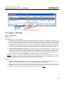
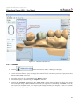
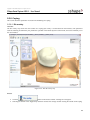
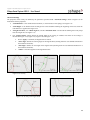
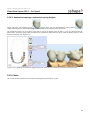
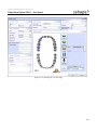
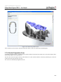
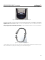
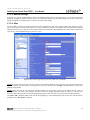
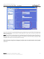
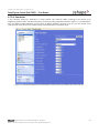
1
Dental System by 3 s h a p e User Manual – Version 2008-1 Copyright © 2000-2008 3Shape A/S. All rights reserved &RS\ULJKW6KDSH$6$OOULJKWVUHVHUYHG 6KDSH'HQWDO6\VWHP8VHU0DQXDO Introduction 3Shape Dental System is a unique combination of 3Shape’s expertise in 3D scanning and 3D CAD software for creating accurate, customized dental restorations. The manual describes 3Shape Dental System that includes the following products: DentalManager, DentalDesigner, ScanItRestoration, ScanItLibrary and Dental System Control Panel. For upgrade and configuration of 3Shape Dental System please refer to the “System Maintenance: how to upgrade and configure your system” chapter in this manual. 3Shape Dental System is completely open (output files are in a standard format) and offers the most advanced 3D technology packed into a very user-friendly interface. It allows scanning full casts, dies, implants or wax-ups automatically to easily design copings, full anatomical crowns, bridge frameworks or implant abutments from the scans. Customized abutments, bars and implant bridges are sold separately as an add-on module called “AbutmentDesigner”. 3Shape Dental System gives you the possibility to manufacture products on any suitable equipment and materials, as well as benefit from the increased number of outsourcing production centres. Please Note The purpose of the manual is to direct the user through the main functions and interfaces of the software. It is assumed the reader is familiar with the Windows operating environment. The Software described in the manual is covered by Terms and Conditions according to the End User License Agreement issued either by 3Shape A/S or by the Partner of 3Shape A/S by whom you have acquired this Partners particular version of the Software. This manual covers the 3Shape Dental System. Some functionalities may be included and some may not be included in your particular version. Thus there may be visualisations and functionality described in this manual which will not apply to your software version. The DentalManager, DentalDesigner, ScanItRestoration, ScanItLibrary, and Dental System Control Panel applications are generally all included in the software suite, however AbutmentDesigner and DentalManager Manufacturing and Inbox addon modules may not be included and may require additional license fee if applicable. If you have questions regarding the content of your particular software version please contact your software provider. 6KDSH$6+ROPHQV.DQDO&RSHQKDJHQ.'HQPDUN 3KRQH)D[(PDLOVXSSRUW#VKDSHFRP &RS\ULJKW6KDSH$6$OOULJKWVUHVHUYHG 6KDSH'HQWDO6\VWHP8VHU0DQXDO Table of contents 1. DENTALMANAGER..........................................................................................................................................................8 1.1 FOREWORD .................................................................................................................................................................8 1.2 GETTING STARTED WITH DENTALMANAGER ............................................................................................................. 9 1.2.1 DentalManager shortcut keys ..........................................................................................................................11 1.2.2 Create an Order ................................................................................................................................................13 1.2.2.1 Process .........................................................................................................................................................14 1.2.2.2 Defining an Order ........................................................................................................................................ 15 1.2.2.3 Modify an Existing Order ............................................................................................................................25 1.2.2.4 Delete Order .................................................................................................................................................26 1.2.3 Scan Order .......................................................................................................................................................26 1.2.3.1 Import Scan .................................................................................................................................................. 28 1.2.4 Model Order.....................................................................................................................................................29 1.2.5 Manufacturing ..................................................................................................................................................31 1.2.6 Closing .............................................................................................................................................................33 1.2.7 Features ............................................................................................................................................................33 1.2.7.1 Check Out Order ..........................................................................................................................................34 1.2.7.2 Search ..........................................................................................................................................................35 1.2.7.3 Select Orders with Common Values ............................................................................................................36 1.2.7.4 Creating output for local production using generic machines: the Generate CAM Output function ........... 37 1.2.7.5 Export orders................................................................................................................................................38 1.2.7.6 Print Contents ..............................................................................................................................................40 1.2.7.7 Print Labels ..................................................................................................................................................41 1.2.7.8 Reset ............................................................................................................................................................41 1.2.8 Help Menu .......................................................................................................................................................42 1.2.8.1 License Information .....................................................................................................................................43 1.2.8.2 Reporting Bugs ............................................................................................................................................44 1.3 INBOX MODULE ........................................................................................................................................................44 1.4 PRINTING AND READING LABELS ............................................................................................................................. 47 1.4.1 Label Printing in DentalManager .....................................................................................................................47 1.4.2 Barcode Reading ..............................................................................................................................................48 2 DENTALDESIGNER ....................................................................................................................................................49 2.1 FOREWORD ...............................................................................................................................................................49 2.2 GLOSSARY ................................................................................................................................................................50 2.3 THE DENTALDESIGNER¥ USER INTERFACE .............................................................................................................. 51 2.3.1 Main toolbar.....................................................................................................................................................52 2.3.2 Using Mouse and Keyboard.............................................................................................................................53 2.3.3 Overview toolbar ............................................................................................................................................. 56 2.3.4 Process toolbar .................................................................................................................................................57 2.3.4.1 Overview......................................................................................................................................................57 2.3.4.2 Composition.................................................................................................................................................57 2.3.4.3 Functioning ..................................................................................................................................................57 2.3.5 View toolbar .................................................................................................................................................... 58 2.3.5.1 Overview......................................................................................................................................................58 2.3.5.2 Composition.................................................................................................................................................58 2.3.5.3 Functioning ..................................................................................................................................................58 2.4 THE MODELLING PROCESS .......................................................................................................................................59 2.4.1 Executing the Program.....................................................................................................................................61 2.4.2 Stage 1 – Order ................................................................................................................................................61 2.4.3 Stage 2 – Directions .........................................................................................................................................63 2.4.3.1 Selections .....................................................................................................................................................63 6KDSH$6+ROPHQV.DQDO&RSHQKDJHQ.'HQPDUN 3KRQH)D[(PDLOVXSSRUW#VKDSHFRP &RS\ULJKW6KDSH$6$OOULJKWVUHVHUYHG 6KDSH'HQWDO6\VWHP8VHU0DQXDO 2.4.3.2 Define telescope primary directions ............................................................................................................64 2.4.3.3 Define insertion directions ...........................................................................................................................65 2.4.4 Stage 3 – Design ..............................................................................................................................................69 2.4.4.1 Telescope .....................................................................................................................................................71 2.4.4.2 Coping..........................................................................................................................................................72 2.4.4.3 Wax-up ........................................................................................................................................................73 2.4.4.4 Pontic ...........................................................................................................................................................73 2.4.4.5 Connector .....................................................................................................................................................74 2.4.5 Stage 4 – Finalise .............................................................................................................................................74 2.4.6 Stage 5 – Save/Export ......................................................................................................................................74 2.5 MODELLING OPERATIONS ........................................................................................................................................75 2.5.1 Overview..........................................................................................................................................................75 2.5.2 Remove artefacts.............................................................................................................................................77 2.5.3 Margin line.......................................................................................................................................................78 2.5.4 Spline Manipulation .........................................................................................................................................82 2.5.5 Die interface.....................................................................................................................................................83 2.5.6 Add / remove material ....................................................................................................................................86 2.5.7 Process .............................................................................................................................................................87 2.5.8 Coping.............................................................................................................................................................. 89 2.5.8.1 Die overlay...................................................................................................................................................89 2.5.8.2 Anatomical copings / anatomical coping bridges ........................................................................................92 2.5.9 Pontic ...............................................................................................................................................................92 2.5.9.1 Load file .......................................................................................................................................................93 2.5.9.2 Pontic Manipulation .....................................................................................................................................93 2.5.10 Connector .........................................................................................................................................................99 2.5.10.1 Add connector .......................................................................................................................................... 99 2.5.10.2 Settings and scaling................................................................................................................................101 2.5.10.3 Scaling and shifting connectors .............................................................................................................102 2.5.10.4 Validation...............................................................................................................................................105 2.5.11 Telescope .......................................................................................................................................................107 2.5.11.1 Primary telescope overlay ......................................................................................................................107 2.5.11.2 Optional components ............................................................................................................................111 2.5.12 Wax-up .......................................................................................................................................................113 2.5.12.1 Wax-up ..................................................................................................................................................113 2.5.13 Wax-up bridge .............................................................................................................................................115 2.5.13.1 Overview ................................................................................................................................................115 2.5.13.2 Order form ............................................................................................................................................. 115 2.5.13.3 Design ....................................................................................................................................................118 2.5.14 Crown ..........................................................................................................................................................120 2.5.14.1 Full Crown .............................................................................................................................................122 2.5.14.2 Two-layer crown ....................................................................................................................................132 2.5.14.3 Full crown bridge ...................................................................................................................................135 2.6 ABUTMENTDESIGNERTM – ADD-ON MODULE .......................................................................................................... 139 2.6.1 The AbutmentDesignerTM user interface: differences from DentalDesignerTM ............................................. 139 2.6.2 The modelling process in AbutmentDesignerTM ............................................................................................ 139 2.6.2.1 Executing the Program...............................................................................................................................140 2.6.2.2 Stage 1 – Order ..........................................................................................................................................140 2.6.2.3 Stage 2 – Directions ...................................................................................................................................143 2.6.2.4 Stage 3 – Design ........................................................................................................................................146 2.6.2.5 Stage 4 – Finalise .......................................................................................................................................147 2.6.2.6 Stage 5 – Save/Export ................................................................................................................................147 2.6.3 Modelling operations .....................................................................................................................................148 2.6.3.1 Overview....................................................................................................................................................148 2.6.3.2 Process .......................................................................................................................................................148 2.7 DENTAL SYSTEM MOTION CONTROLLERS SUPPORT ............................................................................................... 160 6KDSH$6+ROPHQV.DQDO&RSHQKDJHQ.'HQPDUN 3KRQH)D[(PDLOVXSSRUW#VKDSHFRP &RS\ULJKW6KDSH$6$OOULJKWVUHVHUYHG 6KDSH'HQWDO6\VWHP8VHU0DQXDO 2.7.1 2.7.2 3 Graphics Tablets support ..............................................................................................................................160 SpaceBall Support..........................................................................................................................................161 SCANITRESTORATION ...........................................................................................................................................162 3.1 FOREWORD .............................................................................................................................................................162 3.2 UNPACKING AND INSTALLATION ............................................................................................................................ 162 3.2.1 Installing Third-Party Computer Peripherals or Software .............................................................................162 3.3 CALIBRATION .........................................................................................................................................................162 3.3.1 How to Perform a Calibration ........................................................................................................................162 3.4 TRANSPORTATION ..................................................................................................................................................164 3.4.1 Transportation of the D250 Scanner ..............................................................................................................164 3.4.2 Transportation of the D640 Scanner ..............................................................................................................164 3.5 SCANITRESTORATION USER INTERFACE ................................................................................................................. 166 3.5.1 Starting A Scan ..............................................................................................................................................166 3.5.2 Visualization Toolbar ....................................................................................................................................166 3.5.3 Using the Mouse and the Keyboard ............................................................................................................... 167 3.5.4 Teeth Overview Map .....................................................................................................................................167 3.5.5 General Scan Settings ....................................................................................................................................168 3.6 ORDER FORM..........................................................................................................................................................169 3.7 HOW TO SCAN .........................................................................................................................................................169 3.7.1 Using The Interface Plate...............................................................................................................................170 3.7.2 Scanning A Die ..............................................................................................................................................170 3.7.2.1 Difficult Materials & Abutments ...............................................................................................................172 3.7.3 Scanning A Wax Up ......................................................................................................................................172 3.7.4 Bridge Scan .................................................................................................................................................... 173 3.7.4.1 Overview Scan ...........................................................................................................................................173 3.7.4.2 Start Scan ...................................................................................................................................................173 3.7.4.3 Selecting Detailed Scan Area..................................................................................................................... 174 3.7.4.4 Scanning Individual Dies ...........................................................................................................................176 3.7.4.5 Preparation of Die ......................................................................................................................................177 3.7.4.6 Merging Scan Assistance ...........................................................................................................................177 3.7.5 Antagonist Scan (Bite) – Example 3-Unit Bridge..........................................................................................179 3.7.5.1 Preparing the Bite ......................................................................................................................................179 3.7.5.2 Commence Scanning .................................................................................................................................179 3.7.5.3 Trimming Bite Scan ...................................................................................................................................181 3.7.6 Double Preparation Scan................................................................................................................................183 3.7.7 Neighbourhood Scan / Relative Position .......................................................................................................184 3.7.8 Abutment Scan ...............................................................................................................................................185 3.7.8.1 Scan Abutments .........................................................................................................................................185 3.7.8.2 Align Scan Abutments ...............................................................................................................................186 3.7.8.3 Scan Gingival Mask (Preparation Scan) ....................................................................................................186 3.7.8.4 Re-align scan abutments ............................................................................................................................187 3.7.9 Wax-up bridge scan .......................................................................................................................................187 3.7.10 Antagonist model scan ...................................................................................................................................190 4 SCANITLIBRARY ......................................................................................................................................................192 4.1 FOREWORD .............................................................................................................................................................192 4.2 ADD LIBRARY IN DENTAL SYSTEM CONTROL PANEL ............................................................................................. 192 4.2.1 Add pontic library ..........................................................................................................................................192 4.2.2 Add crown library ..........................................................................................................................................192 4.3 DEFINE A SCAN ......................................................................................................................................................194 4.4 SCANNING ..............................................................................................................................................................195 4.4.1 Scan pontic.....................................................................................................................................................195 4.4.1.1 Mount and insert pontic .............................................................................................................................195 4.4.1.2 Supplementary scan (optional)...................................................................................................................196 6KDSH$6+ROPHQV.DQDO&RSHQKDJHQ.'HQPDUN 3KRQH)D[(PDLOVXSSRUW#VKDSHFRP &RS\ULJKW6KDSH$6$OOULJKWVUHVHUYHG 6KDSH'HQWDO6\VWHP8VHU0DQXDO 4.4.1.3 Edit scan.....................................................................................................................................................197 4.4.2 Scan crown.....................................................................................................................................................198 4.4.2.1 Scanning Height .........................................................................................................................................198 4.4.2.2 Edit scan.....................................................................................................................................................198 4.4.2.3 Add morph points ......................................................................................................................................199 4.5 LOAD A SCAN .........................................................................................................................................................199 5 DENTAL SYSTEM CONTROL PANEL ..................................................................................................................201 5.1 FOREWORD .............................................................................................................................................................201 5.2 APPLICATION INTERFACE .......................................................................................................................................201 5.3 SYSTEM SETTINGS ..................................................................................................................................................202 5.3.1 DentalDesigner Location ...............................................................................................................................203 5.3.2 ScanItRestoration Location ............................................................................................................................204 5.3.3 DentalManager Location ...............................................................................................................................204 5.3.4 3D viewer Location .......................................................................................................................................204 5.3.5 Remote license management server ...............................................................................................................205 5.3.6 3Shape Dongle Service .................................................................................................................................. 205 5.3.7 Data output directories ...................................................................................................................................208 5.3.8 Order Form Settings.......................................................................................................................................208 5.3.9 Internet connection ........................................................................................................................................211 5.3.10 Miscellaneous ................................................................................................................................................211 5.4 ADMINISTRATIVE SETTINGS ...................................................................................................................................211 5.4.1 Dongle Update ...............................................................................................................................................212 5.5 DENTALMANAGER .................................................................................................................................................213 5.5.1 General...........................................................................................................................................................214 5.5.1.1 Database settings and shared database .......................................................................................................214 5.5.2 Manufacturing Inbox .....................................................................................................................................215 5.5.3 Inbox Folder...................................................................................................................................................215 5.5.4 FTP Settings...................................................................................................................................................215 5.5.5 Manufacturing ................................................................................................................................................216 5.5.6 ERP and Processing Time Settings ................................................................................................................217 5.5.7 Working Days ................................................................................................................................................218 5.6 MATERIAL SETTINGS ..............................................................................................................................................218 5.6.1 Interface .........................................................................................................................................................220 5.6.2 ...............................................................................................................................................................................224 5.6.3 Contact data ...................................................................................................................................................224 5.6.3.1 Countries ....................................................................................................................................................224 5.6.3.2 Operators....................................................................................................................................................224 5.6.4 Manufacturing ................................................................................................................................................225 5.6.4.1 Sites............................................................................................................................................................225 5.6.4.2 Materials ....................................................................................................................................................228 5.6.4.3 Manufacturing processes ...........................................................................................................................229 5.6.4.4 Colors .........................................................................................................................................................230 5.6.5 Construction elements....................................................................................................................................230 5.6.5.1 Combined setting system ...........................................................................................................................231 5.6.5.2 Cement spaces............................................................................................................................................233 5.6.5.3 Overlays .....................................................................................................................................................235 5.6.5.4 Copings ......................................................................................................................................................236 5.6.5.5 Coping overlays .........................................................................................................................................237 5.6.5.6 Primary telescopes .....................................................................................................................................238 5.6.5.7 Telescope overlays .....................................................................................................................................238 5.6.5.8 Optional components .................................................................................................................................239 5.6.5.9 Wax-ups and Wax-up bridges ....................................................................................................................240 5.6.5.10 Wax-up overlays ....................................................................................................................................241 5.6.5.11 Pontics....................................................................................................................................................242 6KDSH$6+ROPHQV.DQDO&RSHQKDJHQ.'HQPDUN 3KRQH)D[(PDLOVXSSRUW#VKDSHFRP &RS\ULJKW6KDSH$6$OOULJKWVUHVHUYHG 6KDSH'HQWDO6\VWHP8VHU0DQXDO 5.6.5.12 Connectors .............................................................................................................................................243 5.6.5.13 Abutments ..............................................................................................................................................247 5.6.5.14 Top cap libraries ....................................................................................................................................247 5.6.5.15 Implant Systems .....................................................................................................................................248 5.6.5.16 Preparing CAD files for implant systems ..............................................................................................249 5.6.5.17 Implant system coordinate system constraints .......................................................................................251 5.6.5.18 Importing implant systems and abutment kits using the Control Panel ................................................. 253 5.6.5.19 Crowns ...................................................................................................................................................254 5.6.5.20 Bars ........................................................................................................................................................257 5.6.5.21 Margin line.............................................................................................................................................259 5.7 SYSTEM MAINTENANCE: HOW TO UPDATE AND CONFIGURE YOUR SYSTEM ........................................................... 260 5.7.1 Upgrade of previous installation ....................................................................................................................261 5.7.1.1 Type of installation ....................................................................................................................................261 5.7.1.2 Backup .......................................................................................................................................................263 5.7.1.3 DentalManager and 3Shape Dongle Service installation ...........................................................................263 5.7.1.4 DentalManager and Microsoft SQL server installation .............................................................................263 5.7.1.5 3Shape Dongle Service .............................................................................................................................. 265 5.7.1.6 Updating Materials settings .......................................................................................................................265 5.7.1.7 Updating site settings .................................................................................................................................266 5.7.2 Floating licenses ............................................................................................................................................266 5.7.3 Inspection of Dental System Control Panel settings ......................................................................................266 5.7.3.1 System settings ..........................................................................................................................................266 5.7.3.2 Remote license key files ............................................................................................................................267 5.7.3.3 3Shape Dongle Service .............................................................................................................................. 268 5.7.3.4 Output directories ......................................................................................................................................268 5.7.3.5 Material settings .........................................................................................................................................270 5.7.3.6 Sites............................................................................................................................................................270 5.7.3.7 Manufacturing processes ...........................................................................................................................272 5.7.3.8 Connector ...................................................................................................................................................273 5.7.3.9 Margin line.................................................................................................................................................274 5.7.4 Send and receive orders (Inbox add-on module) ...........................................................................................274 5.7.4.1 Receiver (Inbox add-on module required) .................................................................................................274 5.7.4.2 Sender – preconfigured (Inbox add-on module not required) .................................................................... 276 5.7.4.3 Sender – manual configuration (Inbox add-on module not required) ........................................................ 276 6KDSH$6+ROPHQV.DQDO&RSHQKDJHQ.'HQPDUN 3KRQH)D[(PDLOVXSSRUW#VKDSHFRP &RS\ULJKW6KDSH$6$OOULJKWVUHVHUYHG 6KDSH'HQWDO6\VWHP8VHU0DQXDO 1. DentalManager 1.1 Foreword This chapter describes 3Shape DentalManager. The purpose of the chapter is to direct the user through the main functions and interfaces of the software. It is assumed that the reader is familiar with a Windows operating environment. System Requirements Operating System: Windows XP SP2, SP3 or Windows Vista SP1 Processor: Intel Core 2 Duo 2.0GHz or higher is recommended RAM: 1GB – minimum, 2GB – recommended Graphics Card: with OpenGL 2.0 support and at least 256 MB of memory HDD: 80GB or larger Network: Network Internet connection Mouse: with wheel button support The 3Shape DentalManager is part of the 3Shape Dental System, which is comprised of five separate applications. DentalManagerTM is the main application handling the orders through the production processes, and controlling the ScanItRestorationTM and DentalDesignerTM applications. The 3Shape DentalManager comes with a standard Order handling part, which can be extended with a Manufacturing and Inbox module. The following chapter describes these topics. 6KDSH$6+ROPHQV.DQDO&RSHQKDJHQ.'HQPDUN 3KRQH)D[(PDLOVXSSRUW#VKDSHFRP &RS\ULJKW6KDSH$6$OOULJKWVUHVHUYHG 6KDSH'HQWDO6\VWHP8VHU0DQXDO 1.2 Getting Started with DentalManager This chapter deals with the basic order part of the DentalManager. We start out by some of the basic features, then taking an order through the production process and end up with some optional features. Start the application by clicking on the DentalManager desktop shortcut. You will be presented to a window similar to Figure 1-1. Figure 1-1: DentalManager Order module The program is able to open the order and check its individual units (see Figure 1-2). The user can click on single units of the order and get the information about each of them separately (image, height, volume etc). This option is very helpful when it comes to production as well as pricing, invoicing, accounting etc. 6KDSH$6+ROPHQV.DQDO&RSHQKDJHQ.'HQPDUN 3KRQH)D[(PDLOVXSSRUW#VKDSHFRP &RS\ULJKW6KDSH$6$OOULJKWVUHVHUYHG 6KDSH'HQWDO6\VWHP8VHU0DQXDO Figure 1-2: Handling of individual items within an order Orders are shown with relevant information and can be filtered out by date and status in the tree view to the left. Various actions can be reached in the top toolbar or by the right-click popup menu. Notice, that these are context dependently, hence only the relevant functions are shown. 6KDSH$6+ROPHQV.DQDO&RSHQKDJHQ.'HQPDUN 3KRQH)D[(PDLOVXSSRUW#VKDSHFRP &RS\ULJKW6KDSH$6$OOULJKWVUHVHUYHG 6KDSH'HQWDO6\VWHP8VHU0DQXDO Multiple orders can be selected for further handling; Use the buttons in the top toolbar or simply right-click. The list view can be sorted for a specific column by clicking on the column heading (see Figure 1-3). Orders that are not finished before expected delivery date are marked in red. Figure 1-3: Multiple selection and sorting 1.2.1 DentalManager shortcut keys DentalManager has the shortcut keys that make the work with the software faster and easier (see Table 1). The user can right click on the order and view the shortcut keys next to the corresponding option. NOTE: By double-clicking the order, the program brings it to the next level of the process and its status changes automatically (e.g. Created ->double click -> Scanning, or Scanned -> double click -> Modelling). This makes the workflow faster and easier. 6KDSH$6+ROPHQV.DQDO&RSHQKDJHQ.'HQPDUN 3KRQH)D[(PDLOVXSSRUW#VKDSHFRP &RS\ULJKW6KDSH$6$OOULJKWVUHVHUYHG 6KDSH'HQWDO6\VWHP8VHU0DQXDO F6 Ctrl+D Export ERP File Explore Order F4 Explore CAM Shift+F4 Ctrl+Alt+O Generate CAM Output Ctrl+N Print: List Ctrl+M Ctrl+S Ctrl+Alt+I Ctrl+P F7 F8 Selected Shift+F8 Label F9 Send: Track and Trace Ctrl+R Ctrl+Alt+R Ctrl+T Ctrl+U Ctrl+W F5 Select: Same Patient Ctrl+Alt+P Same Client Ctrl+Alt+C Same Status Ctrl+Alt+S Same Delivery Date Ctrl+Alt+D Height Range Ctrl+Alt+H Volume Range Ctrl+Alt+V Advanced: Import Ctrl+I Export to File Ctrl+E Export to FTP Ctrl+F Export to Mail Ctrl+L 6KDSH$6+ROPHQV.DQDO&RSHQKDJHQ.'HQPDUN 3KRQH)D[(PDLOVXSSRUW#VKDSHFRP &RS\ULJKW6KDSH$6$OOULJKWVUHVHUYHG 6KDSH'HQWDO6\VWHP8VHU0DQXDO Table 1: DentalManager shortcut keys 1.2.2 Create an Order Select New to create a new order – Either in top toolbar or in the right-click menu. Figure 1-4: Create new Order You will be prompted by the Order form depicted in Figure 1-5. &RS\ULJKW6KDSH$6$OOULJKWVUHVHUYHG 6KDSH'HQWDO6\VWHP8VHU0DQXDO 1.2.2.1 Process Figure 1-5: Order Form The following information describes the various fields in the Order Form: x Lab information This field is reserved for the operator’s name. An operator can be chosen from a list that has to be filled in the DSCP (for more details see chapter 5.6.1 - Interface). x Client information Here the user can add all the information related to the client the order is being made for. To use this option the client and its data have to be previously registered in the Dental System Control Panel. The user can also import sites from an Excel file to the DSCP (see chapter 5.6.4.1 for more details). x Patient information The order’s patient information should be inserted here. x Comments Any comment, related to the order, can be added here. It will be displayed in DentalDesigner. &RS\ULJKW6KDSH$6$OOULJKWVUHVHUYHG 6KDSH'HQWDO6\VWHP8VHU0DQXDO x Order settings An order number will automatically be attached to each order. The format is defined in the Dental System Control Panel and can easily be customized. x Scan Settings The user can scan an antagonist to get a more precise result. He can choose between a Model and a Bite or select None if he doesn’t want to scan an antagonist. In order to design and scan the desired framework in wax the Double preparation box should be checked. When the Neighborhood scan box is checked the relative position of the scan is captured: 1. 1 Die If 1 die is selected and the “Neighbourhood scan” box is checked then an accompanying scan is taken also. A rough scan of the die and neighbouring teeth is also taken. This allows the relative position of the tooth to be considered. 2. X Dies - With the box checked the neighbouring teeth can always be taken into consideration. If, for example, there is 1 die on the left and also 1 on the right of interest then both these areas should be selected for scanning in the rough relative scan. x Teeth numbering information This field displays the numbering index chosen in the Dental System Control Panel. The default system is the FDI notation. When the cursor is moved over a tooth the index number will appear. Selected teeth will be circled in red. x Restoration keys The restoration key provides information about the nature of restoration to be performed on a specific tooth. 1.2.2.2 Defining an Order When processing an order it is important to specify the operations. To select a tooth, left click on it. It will be circled in red), the relevant operations can be added with a click on one of the restoration keys. The Dental System software allows working in layers. You can easily start with a coping and add a crown, for example. &RS\ULJKW6KDSH$6$OOULJKWVUHVHUYHG 6KDSH'HQWDO6\VWHP8VHU0DQXDO Figure 1-6: Selecting teeth Once the relevant restoration is selected the tooth will change color, according to the chosen restoration key next to the teeth overview map (see Figure1-8). It is possible to select more than one tooth at a time. Simply click on several teeth or use the standard SHIFT + left-click to select a row of teeth. A multiple selection allows bridges to be applied (see Figure 1-9). Next to every restoration key you will find menus with different options as, among others, the type of restoration and material (see Figure 1-7, Figure 1-10,Figure 1-11 and Figure 1-12). The users are able to specify the color of the material in the order that should be used to produce the restoration (New in 2008-1). This option is available for all of our restorations: crowns, pontics, copings, telescopes and abutments. The colors are represented in the form of a special scale: A1, A2, A3, B1, B2, B3 etc. each letter representing a shade of color. To define this in the order form the user should choose the desired restoration, click the button to the right and select the desired color. See Figure 1-7: &RS\ULJKW6KDSH$6$OOULJKWVUHVHUYHG 6KDSH'HQWDO6\VWHP8VHU0DQXDO Figure 1-7: Selecting color for the restoration material &RS\ULJKW6KDSH$6$OOULJKWVUHVHUYHG 6KDSH'HQWDO6\VWHP8VHU0DQXDO Figure1-8: Selection of a specific restoration (colour change) &RS\ULJKW6KDSH$6$OOULJKWVUHVHUYHG 6KDSH'HQWDO6\VWHP8VHU0DQXDO Figure 1-9: 9-Unit bridge defined (consisting of crowns and pontics) &RS\ULJKW6KDSH$6$OOULJKWVUHVHUYHG 6KDSH'HQWDO6\VWHP8VHU0DQXDO Figure 1-10: Menu with choice of Coping type &RS\ULJKW6KDSH$6$OOULJKWVUHVHUYHG 6KDSH'HQWDO6\VWHP8VHU0DQXDO Figure 1-11: Menu with Material choice for each Restoration &RS\ULJKW6KDSH$6$OOULJKWVUHVHUYHG 6KDSH'HQWDO6\VWHP8VHU0DQXDO Figure 1-12: Order Form Menu to define the Bridge The abutments can be arranged in groups with each group having its common insertion direction (New in 2008-1). This allows to model upper structures on top of customized abutments: the resulting customized abutments will be in parallel by default (as long as the Top cap is not rotated during the modelling). In order to define a group, select the desired teeth with the cursor and choose the New Group from the Abutment drop-down menu. Should a second group of abutments be created, it would have an incremental number. See Figure 1-13 &RS\ULJKW6KDSH$6$OOULJKWVUHVHUYHG 6KDSH'HQWDO6\VWHP8VHU0DQXDO Figure 1-13: Order Form window to define a group of abutments NOTE: When you want to unite the abutments into the bridge, there is no need to select NewGroup option from the menu since the abutments are grouped automatically on selection of the Bridge restoration key. To create several groups of abutments with different insertion directions, define another set of abutments and select the NewGroup from the Abutment drop–down menu for the Group 2 to appear in the list. You will be able to set different insertion directions for each group further during the modelling process (see. &RS\ULJKW6KDSH$6$OOULJKWVUHVHUYHG 6KDSH'HQWDO6\VWHP8VHU0DQXDO Figure 1-14: Creating several groups of abutments Click on OK when the order has been set up as desired. The order is stored into the database and handled by the DentalManager. &RS\ULJKW6KDSH$6$OOULJKWVUHVHUYHG 6KDSH'HQWDO6\VWHP8VHU0DQXDO 1.2.2.3 Modify an Existing Order Orders that have been created but not scanned yet can be modified as shown on Figure 1-15. However, if the status is Scanned or later, the order can be opened in Read-Only mode. In addition these orders can be viewed by selecting 3D Preview (see Figure 1-16). Figure 1-15: Modify an Existing Order &RS\ULJKW6KDSH$6$OOULJKWVUHVHUYHG 6KDSH'HQWDO6\VWHP8VHU0DQXDO Figure 1-16: Order 3D Preview 1.2.2.4 Delete Order Orders that are not locked can be deleted by selecting Delete. The user will get prompted by a confirmation dialog. Figure 1-17: Delete Order Confirmation Dialog 1.2.3 Scan Order Select an order having the status Created and select Scan (or double-click the order) (see Figure1-18). 3Shape ScanItRestoration is now launched (see Figure 1-19). &RS\ULJKW6KDSH$6$OOULJKWVUHVHUYHG 6KDSH'HQWDO6\VWHP8VHU0DQXDO Figure1-18: Scan Order &RS\ULJKW6KDSH$6$OOULJKWVUHVHUYHG 6KDSH'HQWDO6\VWHP8VHU0DQXDO Close the ScanItRestoration after scanning to store the Scan. Figure 1-19: ScanItRestoration For detailed description of ScanItRestoration please refer to the chapter ScanItRestoration. 1.2.3.1 Import Scan As an alternative to ScanItRestoration the scan can be imported to the system. Select Import Scan and the open dialog depicted in Figure 1-20 is prompted. Figure 1-20: Import Scan &RS\ULJKW6KDSH$6$OOULJKWVUHVHUYHG 6KDSH'HQWDO6\VWHP8VHU0DQXDO 1.2.4 Model Order Select an order having the status Scanned and Select Model (or double-click the order) (see Figure 1-21) to launch DentalDesigner (see). When finished modelling, click on save in the DentalDesigner and close the application. The order is now modelled and ready for export or manufacturing. For detailed description of DentalDesigner please refer to chapter 2 DentalDesigner. Figure 1-21: Model Order &RS\ULJKW6KDSH$6$OOULJKWVUHVHUYHG 6KDSH'HQWDO6\VWHP8VHU0DQXDO Figure 1-22: Modelling an Order in DentalDesigner &RS\ULJKW6KDSH$6$OOULJKWVUHVHUYHG 6KDSH'HQWDO6\VWHP8VHU0DQXDO 1.2.5 Manufacturing Next step for Modelled orders are manufacturing. Start by selecting Send to Manufacturer (or double-click the order) (see Figure 1-23). Figure 1-23: Send Order to Manufacturing The orders are compressed and ready to be sent to the manufacturers FTP server (see Figure 1-24). Click Send to start upload the orders. The setup is explained in chapter 5 - Dental System Control Panel. Figure 1-24: Orders ready to be uploaded to manufacturing &RS\ULJKW6KDSH$6$OOULJKWVUHVHUYHG 6KDSH'HQWDO6\VWHP8VHU0DQXDO The order status can be inspected in the Track and Trace window. An example is shown in Figure 1-25. Figure 1-25: Track and Trace – Order Accepted Utilize the filter to get an overview of the orders sent for manufacturing (see). Figure 1-26: Orders Sent to Manufacturing &RS\ULJKW6KDSH$6$OOULJKWVUHVHUYHG 6KDSH'HQWDO6\VWHP8VHU0DQXDO 1.2.6 Closing Once orders have been produced by the manufacturer and shipped back to the end-user, the order are ready close. Closed orders are still accessible through the closed filter as shown below. . Figure 1-27: Closed Orders 1.2.7 Features The features described below are not mandatory for standard production, but optional extensions such as: x Check out order for offline production x Search x Select orders with common values x Generate CAM output x Export orders x Print order list & label x Reset order x Views &RS\ULJKW6KDSH$6$OOULJKWVUHVHUYHG 6KDSH'HQWDO6\VWHP8VHU0DQXDO 1.2.7.1 Check Out Order Orders are allowed to be modelled on a computer which is not part of the Dental System; e.g. on a laptop which is brought home. Note: this requires an additional dongle. Orders need to be properly exported from the Dental System database, and later re-imported. Select the desired orders and select Check out in the right-click popup menu as shown on Figure 1-28: Figure 1-28: Check Out Orders &RS\ULJKW6KDSH$6$OOULJKWVUHVHUYHG 6KDSH'HQWDO6\VWHP8VHU0DQXDO When the orders have been processed, they can be re-imported into the Dental System database. Select the orders ready for check in as shown below on Figure 1-29 and select the local order path. The orders will be integrated and status updated. Figure 1-29: Check In Orders 1.2.7.2 Search The Search component can be utilized to locate specific orders. Click on the arrow right to the magnifying glass to switch search criteria as illustrated below. &RS\ULJKW6KDSH$6$OOULJKWVUHVHUYHG 6KDSH'HQWDO6\VWHP8VHU0DQXDO Figure 1-30: Order Search Enter the search enquiry to get a list of matching orders. The last request is always stored in the Search node. 1.2.7.3 Select Orders with Common Values Orders having common values can be selected to assist the order handling, when e.g. checking out orders having the status Scanned and created Last Week (see Figure 1-31). &RS\ULJKW6KDSH$6$OOULJKWVUHVHUYHG 6KDSH'HQWDO6\VWHP8VHU0DQXDO Figure 1-31: Select Orders with Common Values 1.2.7.4 Creating output for local production using generic machines: the Generate CAM Output function Depending on purchase conditions, the 3Shape Dental System’s output can be used with any third party rapid prototyping/milling machines which accepts STL files as input (please contact your local 3Shape dealer for more details). Moreover, most commonly the 3Shape Dental System is bundled as OEM component along with rapid prototyping/milling machines whose specific output is generated according to vendor’s specification. Different situations are possible and taking in account each of them is beyond the purpose of this manual: please refer to your local 3Shape dealer/distributor/OEM partner to have assistance on production related issue. As DentalDesigner typically saves modeling using 3Shape’s proprietary .dcm file format, the “Generate CAM output” function is the function to be used to have the modeling exported in a file format which is suitable for production (typically STL or other proprietary 3D file formats). The term “CAM output” is used as different production machines may require different kind of output and the 3Shape Dental System supports a number of those. It is possible to generate CAM output from the modelled orders if it is supported by the current DentalManager system. In this case it can be accessed from the advanced tab with the right-click popup menu as shown on . &RS\ULJKW6KDSH$6$OOULJKWVUHVHUYHG 6KDSH'HQWDO6\VWHP8VHU0DQXDO Figure 1-32: Generate CAM Output 1.2.7.5 Export orders Orders can be exported from the system to a file, FTP or by mail. The selected orders are compressed and handed to the desired export control, see e.g. Figure -34. &RS\ULJKW6KDSH$6$OOULJKWVUHVHUYHG 6KDSH'HQWDO6\VWHP8VHU0DQXDO Figure 1-33: Export of Order Figure 1-34: Export Orders by FTP &RS\ULJKW6KDSH$6$OOULJKWVUHVHUYHG 6KDSH'HQWDO6\VWHP8VHU0DQXDO 1.2.7.6 Print Contents The current view of orders can be printed to a standard Printer by selecting Printĺ List. Selecting orders with common values (for more details please see section 1.2.7.3) can also be used for printing of list for one customer or work for a certain date. Figure 1-35: Printing All Orders In a List An example is shown below. Figure 1-36: Sample List Print &RS\ULJKW6KDSH$6$OOULJKWVUHVHUYHG 6KDSH'HQWDO6\VWHP8VHU0DQXDO 1.2.7.7 Print Labels Labels for the selected orders may be printed by selecting Printĺ Label or by hitting the F9 function key. The labels will be printed on the printer that is selected under Helpĺ Label Printer Settings. The default layout is designed for 40x40 mm labels as shown below: Figure 1-37: Sample Label Print 1.2.7.8 Reset It may be necessary to reset an order to a specific status in some unusual cases; e.g. to force an order to be re-modelled the order must be reset to Scanned (see Figure 1-38). Figure 1-38: Reset Order Status &RS\ULJKW6KDSH$6$OOULJKWVUHVHUYHG 6KDSH'HQWDO6\VWHP8VHU0DQXDO 1.2.8 Help Menu The help menu in the top right corner contains various options as language settings, license information, open log file, and sends bug report. These are described further on. Figure 1-39: Help Menu If you click on Options the menu with the Label Printer Settings and Add-ons and Modules will appear (for more information see Figure 1-52 and Figure 1-56). &RS\ULJKW6KDSH$6$OOULJKWVUHVHUYHG 6KDSH'HQWDO6\VWHP8VHU0DQXDO 1.2.8.1 License Information The status of the license server can be obtained by clicking on License Information in the help menu. The default browser is opened showing different information regarding the dongle; an example is shown in Figure 1-40. Additionally, it is possible to check the dongle for updates or show the server log file by clicking on the buttons positioned at the bottom. Figure 1-40: License Information &RS\ULJKW6KDSH$6$OOULJKWVUHVHUYHG 6KDSH'HQWDO6\VWHP8VHU0DQXDO 1.2.8.2 Reporting Bugs 3Shape is depending on your feedback in order to solve problems and improve the software. Please use the built-in Send Report Bug functionally if a problem should occur. If a critical error arises, the Bug Report seen in Figure 1- 41automatically appears. Figure 1-41: Reporting Bugs 1.3 Inbox Module The DentalManager Inbox module allows production facilities to receive orders from remote laboratories or sites. It supports multiple FTP servers for high-load central production The functionality requires a properly configured FTP server at the receiving site and correct FTP settings at the sending sites. The FTP server does not need to run on the same PC as DentalManager Inbox: DentalManager Inbox will fetch the orders on the FTP server using the FTP protocol. The customer needs to properly setup the FTP server and create at least one account on the FTP server. The account to be used as "Inbox" account must be setup on the FTP server with "full control" permissions, as data will be created, read, modified and deleted from the FTP account. The Inbox is an add-on module and when licensed can be enabled via the Options menu (see Figure 1-52). NOTE: The availability of the Inbox module can be viewed in the License Information area as described under section 1.2.8.1 Figure 1-52: Selecting the Inbox module &RS\ULJKW6KDSH$6$OOULJKWVUHVHUYHG 6KDSH'HQWDO6\VWHP8VHU0DQXDO After selecting the Inbox module the user will receive a window similar to Figure 1-53. The customer needs to setup the DentalManager Inbox parameters in the Dental System Control Panel, once FTP and dongle are properly configured. Finally the "sending" system has to be configured as well. The procedures are explained in the chapter 0 (Dental System Control Panel). The inbox automatically searches the FTP server for new orders. Figure 1-53: Orders in the Inbox Right-click on the order in the Inbox to select the desired option (see Figure 1-54). &RS\ULJKW6KDSH$6$OOULJKWVUHVHUYHG 6KDSH'HQWDO6\VWHP8VHU0DQXDO Figure 1-54: Inbox Module options Incoming orders can be inspected with Open or 3D Preview before being accepted. The order in our case is accepted and the Accept Selected Orders for Manufacturing dialog is brought up as shown in Figure 1-55. NOTE: If the program is used to receive files from clients by the manufacturer, the system can be set up to accept orders automatically and create CAM files by default. If this action is desired, the Inbox Auto Accept checkbox (Dental System Control Panel) has to be checked (DSCP ->DentalManager ->Manufacturing Inbox ->Inbox Auto Accept). The default expected delivery date can be changed (for more details see chapter 5.5.7 Working Days), and it is possible to fill and pass a custom message back to the sender. Figure 1-55: Accept Incoming Order &RS\ULJKW6KDSH$6$OOULJKWVUHVHUYHG 6KDSH'HQWDO6\VWHP8VHU0DQXDO This order is now put into the system and can be taken to production. Close the order when the processing is finished. The customer is able to follow the status of the order utilizing track and trace as described in section 1.2.5. If the order is rejected for some reason, the customer gets a note in the list view. 1.4 Printing and Reading Labels 1.4.1 Label Printing in DentalManager The DentalManager has an in-built function to print labels. Apart from the other administrative information the labels display order number and barcode. The print dialogue is called from the Options menu of the main program window by selecting Options->Label Printer Settings, see Figure 1-56: Figure 1-56: Printing Labels &RS\ULJKW6KDSH$6$OOULJKWVUHVHUYHG 6KDSH'HQWDO6\VWHP8VHU0DQXDO The function should work with any printer. The images below illustrate examples of labels printed on Bixolon SRP-770 II and Seiko Smart Label Printer 420 accordingly: 1.4.2 Barcode Reading When properly installed, any numeric barcode reader should be able to read barcodes generated by the DentalManager. Most barcode readers look like shown on the images below: &RS\ULJKW6KDSH$6$OOULJKWVUHVHUYHG 6KDSH'HQWDO6\VWHP8VHU0DQXDO 2 DentalDesigner 2.1 Foreword DentalDesigner¥ is an advanced software package for designing high-precision, customized restorations from 3D scans of dental impressions. Using the latest technologies, the DentalDesigner¥ software facilitates fast and easy modelling of complex constructions as well as enabling automatic manufacturing of the final restoration model on computer aided production equipment. This chapter introduces the DentalDesigner¥ software and presents the modelling process as well as the tools and features available to the user. Furthermore, a worked example for getting quickly started with the DentalDesigner¥ software is provided. From herein, it is assumed that the DentalDesigner¥ user is familiar with the traditional process of making customized dental restorations. The Software described in the chapter is covered by Terms and Conditions according to the End User License Agreement issued either by 3Shape A/S or by the Partner of 3Shape A/S by whom you have acquired this Partners particular version of the Software. System Requirements Operating System: Windows XP SP2, SP3 or Windows Vista SP1 Processor: Intel Core 2 Duo 2.0GHz or higher is recommended RAM: 1GB – minimum, 2GB – recommended Graphics Card: with OpenGL 2.0 support and at lease 256 MB of memory HDD: 80GB and larger Network: Network Internet connection Mouse: with the wheel button support System Requirements: Windows XP, Windows Vista. &RS\ULJKW6KDSH$6$OOULJKWVUHVHUYHG 6KDSH'HQWDO6\VWHP8VHU0DQXDO 2.2 Glossary While reading this chapter and learning to use DentalDesigner¥, the user will come across words and concepts borrowed from the computer graphics and the programming dictionary. Concepts like “bounding box” or “control point” may be hard to understand to new users and misleading for those who are not familiar with computer graphics and CAD technology. In addition, as such, things don’t exist in everyday life and they don’t look like things we are used to deal with, they do need a proper introduction. The purpose of this glossary is to present some of the key concepts which can be harder for the user to grasp. Item Operation Control points – Imagine you’re holding a piece of clay: if you apply pressure on it with your fingers, or pull it out, the clay will deform consequently. This system for manipulation and deforming is not too far from what the one we use in DentalDesigner¥. Imagine that the control points are like magnets, and the underlying object (which can be a restoration, a part of it or a spline) can be deformed as it was made of some magnetic clay. By moving the control points, you will be modifying and deforming the object, you’re working on. Control points look like spheres, they come in different size and colours depending on the object they’re related to. Typically, the user can interact with control points by clicking on them and dragging them, but in some step, the user will interact with them differently (as in the margin line definition). Library – A library is simply a collection of things, no more no less. It can contain different kinds of objects, which are collected in a library for the user’s convenience. We’ll be referring to libraries for instance when explaining pontics: our collection of pontics is arranged numerically and stored in a folder. Such folder is referred to as the “pontics library”. Template – A template is an object with basic characteristics belonging to a certain category. A template is a sort of middleware: it comes in a raw form and needs to be modified and customized. An example for a template could be glasses lenses: when one buys glasses the lenses need to be cut and shaped to fit the frame. In this chapter, we use the word template to define a pontic coming from our pontic library. The template (the middleware object) is the pontic as it comes from the library: it has the basic pontic characteristics but it still needs to be reworked and adapted to properly fit the bridge. Spline – The word “spline” is roughly a technical word to describe a curve. For the purpose of this chapter, whenever you’ll read of a spline, simply imagine you’ll be facing an object that looks exactly like a curve. Crucial is the fact that a spline often comes along with control points. More precisely, whenever the user is given the chance to modify and interact with a spline, the interaction will happen through control points. Bounding Box – A generic bounding box is simply a cube shaped box drawn around the object and containing it. The box is actually “invisible”; the only visible parts are its vertices and its edges. In DentalDesigner¥ the bounding box is used when placing and modifying pontics. The bounding box here is not only containing the pontic but it is also actively involved in the placing and modelling process. At the eight vertices of the box there are control points: they’re attached to the box and by moving them the user will be able to deform the box, and as the box is attached to the contained pontic, the pontic will deform in return. The user may think of the bounding box as of the pontic frame, box and pontic are tied together and by moving the pontic around the box will move accordingly, too. &RS\ULJKW6KDSH$6$OOULJKWVUHVHUYHG 6KDSH'HQWDO6\VWHP8VHU0DQXDO 2.3 The DentalDesigner¥ user interface DentalDesigner¥ employs a Microsoft Windows¥-based graphical user interface that allows the user to view the restoration on screen while it is being modelled. As shown in Figure 2-1, the user interface comprises the following five toolbars: 1. The Main toolbar: Located on the top of the screen. 2. The Overview toolbar: Located on the left side of the screen. 3. The Process toolbar: Located on the lower left side of the screen. 4. The View toolbar: Located on the bottom of the screen. 5. The Visualization toolbar: Located on the right side of the screen. 1. Main toolbar 5. Visualization toolbar 2. Overview toolbar Modelling window 3. Process toolbar 4. View toolbar Figure 2-1: The DentalDesigner¥ user interface A detailed description of these toolbars is provided in the following sections. &RS\ULJKW6KDSH$6$OOULJKWVUHVHUYHG 6KDSH'HQWDO6\VWHP8VHU0DQXDO 2.3.1 Main toolbar The Main toolbar contains some of the main functions that will be relevant while performing all the operations of//in the modelling process. It consists of two parts: the left part contains buttons to perform common file and order related operations, such as creating, loading and saving modelling sessions. The right part has to do with operations related to the modelling itself. The function of the buttons contained in the right part of the Main toolbar is primarily technical, and this becomes clear as soon as the user gets acquainted with the modelling functions and the workflow system of DentalDesigner¥. Thus, the buttons description given in Table 2-1 is meant as a pure reference only. Item Operation New modelling – Starts a new modelling session (if no current modelling process is active). Load modelling – Loads a previously created modelling session from file. Save modelling – Saves the current modelling session to file. Separator Order stage – Skips to the stage for entering order specific data and defining the desired modelling process. Directions stage – Skips to the stage for defining scanning selections and insertion directions. Design stage – Skips to the stage for designing the defined restorations. Finalize stage – Skips to the stage for verifying the complete restorations and for adding final touches. Save stage – Skips to the stage for exporting the completed restorations to input files for computer aided manufacturing facilities. Table 2-1: Functions in the Main toolbar &RS\ULJKW6KDSH$6$OOULJKWVUHVHUYHG 6KDSH'HQWDO6\VWHP8VHU0DQXDO 2.3.2 Using Mouse and Keyboard The use of mouse and keyboard in DentalDesigner¥ for common functions is identical to standard (use) windows applications typically employ for the purpose, therefore, the user be left-clicking to press a button or to check a checkbox. The use of the right mouse button is mostly restricted to more CAD-specific operations, as described below. Most of steps in the modelling process require user input via mouse. To perform these inputs, position the mouse cursor over the area to be modified and press the left mouse button. the will the The main function of the right mouse button is related to visualization: the default setting in view DentalDesigner¥ allows the user to use the right button to rotate the models in the 3D or to pan in the 2D view. Configuration can be changed pushing the Rotate view, Pan view or Zoom view in the Visualization toolbar. Capacities of the right button can change while using different tools and functions, and by pushing a combination of keys. (For instance, keeping the ALT keyboard button pressed pushing the right button, will make the 3D view pan). The preferred method of changing the view is to have the “Rotate View” button in the Visualization toolbar permanently pressed (rotates the view when using the right mouse button), and to use the mouse wheel to change the pan and the zoom of the view, as described in the table below. Please refer to chapter 2.7 “Dental System Motion Controllers Support” fro the additional information. Some functions in the Visualization toolbar are available as pure mouse functions, as shown in Table 2-2. Mouse button Wheel down Wheel scrolled Action Pan view - When pressed, move the mouse in the 3D view to pan. Zoom view – Scroll mouse wheel in the 3D view to zoom in and out. Table 2-2: Shortcuts to changing views using mouse Finally, it is possible to change the 3D view using keyboard shortcuts. While moving the mouse in the 3D view and holding down the right mouse button, use the keys in Table 2-3 to change the 3D view. Key ALT Action Pan view - When pressed, use the right mouse button to pan the view. CTRL Rotate view - When pressed, use the right mouse button to rotate the view. SHIFT Zoom view - When pressed, use the right mouse button to zoom in and out. Table 2-3: Shortcuts for changing views using mouse and keyboard HINT: It is strongly recommended that views be selected using the shortcuts described in Table 2-2. Leaving the “Rotate View” button in the Visualization toolbar permanently pressed is also advantageous. Once the user becomes familiar with this procedure, modelling speed will increase significantly. For information on using other manipulation devices please, see 2.7 DENTAL SYSTEM MOTION CONTROLLERS SUPPORT chapter. &RS\ULJKW6KDSH$6$OOULJKWVUHVHUYHG 6KDSH'HQWDO6\VWHP8VHU0DQXDO Visualization toolbar Finding the right point of view to work on a 3D model most effectively can be difficult and time-consuming. DentalDesigner¥ comes with a set of tools and functions aimed to help the user to find the most advantageous visualization for each different situation. The visualization tools and functions meant for use are often grouped together in the Visualization toolbar. Among others, they include functions and tools which: Rotate the 3D view to preset angles: this allows the user to visualize the models from the front, back, top, bottom… Set the transparency of the objects the user will be working with. For instance, the user might want to see both coping and its die at the same time. This function allows the user to make the coping transparent. Define the 2D cross section, in order to have the best visualization in the 2D window (for explanation, see below). Switch to custom rotation and panning mode, allowing the user to rotate and move the object freely on the screen. The buttons in the Visualization toolbar are presented in Table 2-4. HINT: Rotating and panning objects freely can lead even the most expert users to non-optimal and confusing visualizations (objects can easily end up being oddly rotated or only partially visible): this is normal when operating CAD software as DentalDesigner¥. The view buttons (on top of the Visualization bar) are mostly meant to help the user taking the objects back to a more optimal visualization. We suggest you to spend some extra time figuring out how the view buttons work: this will speed up your future work and make it more comfortable. &RS\ULJKW6KDSH$6$OOULJKWVUHVHUYHG 6KDSH'HQWDO6\VWHP8VHU0DQXDO Item Action View – The view buttons allow the user to switch between a number of predefined viewpoints. Placement overview – Enables the user to control the alignment of the crowns and the pontics in a customized crown bridge. HINT: Please refer to section 2.5.14.3 for more information on how to deal with the placement overview function in crown bridges. Point dragging – Changes the degree of spline point dragging for all features. 2D Cross section – Provides 2D cross section for a better model inspection. 2D cross sections can be specified either by placing three points on the 3D model or by dragging a section line by holding down the left mouse button and then moving the mouse over the model. Model visibility – Enables visualization of the necessary number of 3D model elements (e.g. with or without preparation) for clearer analysis. Validation – Evaluates the restoration according to the standard minimal values pre-set in Dental Control Panel (or manually defined) and marks the areas that need correction. HINT: Please refer to section 2.5.10.4 for more information on how to use the validation function in connectors and crown bridges. Bounding box – outlines the borders of the active element of the restoration selected, or – in case that no model is selected – of the whole restoration. Origin – Assists orientation of the restoration model according to the standard three geometrical axes (x, y and z), which are independent of the user point-of-view. Set transparency – Changes the degree of transparency for the following objects: x Scans – The uploaded scan models. x Items – The collection of elements in the modelling job. x Active – The modelling element that is currently active. Show backface – Toggles visibility of model backfaces, i.e. whether its “inside” is visible. Show annotations – Toggles visibility of annotations on the 3D model. Material shaders – Marks the 3D model parts with the corresponding colors imitating the surface of the real restoration materials. Rotate view – When selected, right-click and drag to rotate the view (see section 2.2). &RS\ULJKW6KDSH$6$OOULJKWVUHVHUYHG 6KDSH'HQWDO6\VWHP8VHU0DQXDO Pan view - When selected, right-click and drag to pan the view (see section 2.2). Zoom view - When selected, right-click and drag to zoom in and out (see section 2.2). Surface measurement – Allows the user to measure the distance from the desired point on the preparation to the adjacent tooth, the preparation scan and antagonist scan. Table 2-4: Functions in the Visualization toolbar 2.3.3 Overview toolbar The Overview toolbar shown in Figure 2-3 is designed to quickly provide information on the current job comprising it with various workflow control functions. It enables: - An overview of the modelling jobs specified in the order form, as well as information on the currently active job highlighted by green dots. - Information specified with names of various jobs and tooth names (FDI is the default notation used, which can be changed in the Control Panel) can be acquired by pausing the mouse over the specified items. - Activation and switching among jobs using left-click on the corresponding item: this allows the user to switch among jobs at any moment. - Set the visibility of scans and restorations can be toggled by right-clicking on the corresponding item. This allows the user to show or hide the scan or the completed job at any time. Figure 2-2: The Overview toolbar &RS\ULJKW6KDSH$6$OOULJKWVUHVHUYHG 6KDSH'HQWDO6\VWHP8VHU0DQXDO 2.3.4 Process toolbar 2.3.4.1 Overview Migration from traditional dental restoration manufacturing techniques to the dental CAD system can be hard for some users. A trained technician, who has developed craftsmanship and expertise with traditional tools during his training and career, will be required to switch over to a different system in which workflow is different and results are achieved in a different way. Also, instead of knives, slices and “solid” restoration, the technician will have to learn how to interact with computer input devices (such as keyboard and mouse) and be prepared to see the result of his work on a computer screen. The DentalDesigner¥ CAD software is conceived to make this migration as smooth as possible. Bringing this into the focus all the functions, tools, the user interface and the workflow system have been developed. The whole workflow of DentalDesigner¥ is summarized in the Process toolbar. 2.3.4.2 Composition Each stage in the modelling process is represented by a button in the Process toolbar. As different jobs require different functions to be used, the Process toolbar will look different depending on the job. The Process toolbar is meant to replicate (as much as possible) the same steps the technician would be going through when modelling a restoration using traditional techniques. It allows the user to go back to previous steps at any time and to move forward again. 2.3.4.3 Functioning Each step of the Process toolbar must be completed in order to advance to the subsequent stage. In a modelling scenario, the user starts the modelling process by pressing the top button that corresponds to the initial step in the modelling stage. Upon completion of this step, the user proceeds to the next step in line until all steps of the modelling stage have been completed (Note: depending on the step, sometimes advancing to the next step is automatic, sometimes it requires the user to push the “Next” button). The currently active step is highlighted with a darker background and an italic label, while previously completed steps are displayed bold font. The image of the Process toolbar showing the steps involved in modelling a coping is shown in Figure 2-5. A modelling stage can be automatically completed using the “Finish” button above the modelling step buttons. In this case, the step is completed using default values for all modelling parameters. Completed step Active step Figure 2-3: Example of Process toolbar for a coping HINT: Depending on user’s techniques and modelling habits it can be useful in some cases to push the button “Add/Remove” and jump to a completed job in order to rework it manually with the add/remove (wax-knife) tool. &RS\ULJKW6KDSH$6$OOULJKWVUHVHUYHG 6KDSH'HQWDO6\VWHP8VHU0DQXDO 2.3.5 View toolbar 2.3.5.1 Overview In the paragraph 2.3, the Visualization toolbar is explained, the View toolbar provides similar functionalities for 2D visualization. The 2D views are meant to be useful in situations where a section view is crucial to correctly visualize the modelling, as for instance while setting the margin line, in order to check for undercuts. 2.3.5.2 Composition The View toolbar consists of two 2D windows, and of several adjustment tools: these tools are used to modify the section shown in the left window only. The right window is used exclusively for some steps of modelling, and DentalDesigner¥ controls the section to make it visible (as it happens, for instance, in the margin line definition step). 2.3.5.3 Functioning Panning and Zooming: the user can right-click or use the mouse wheel in both 2D windows to pan and zoom respectively, just like in the 3D view. Slice rotation angle and Slice position: allow the user to define the section to be shown visually in the left 2D window. Some practice is suggested for the user to get confident with such parameters. Measure tool: is a simple yet effective 2D measurement tool. When the box is checked the user can measure distance between points inside the left 2D window: to define the first point, left-click on the border of the section inside the left 2D window. Left-click again on the section border to define the second point: at this time a line will appear between the two points along with the distance value indicated. Important: the user can freely drag and move the points on the section surface, whereby the distance value will be updated automatically, even after modifying the section parameters (done by modifying the “Slice rotation angle” and the “Slice position” parameters). Area tool: this tool is meant for area measurements within 2D windows. When the “Area” checkbox is checked, the measurement is activated simply by moving the mouse over the area to be measured: the area will then turn yellow and the surface volume measured in mm² will appear. Important: the area measurement tool will only work when a closed surface is selected (as it would make no sense measuring a surface with no defined boundaries). For the tool to work smoothly we suggest to use it together with the Measure tool: in this case the Area tool will determine the line defined by the user to be the lower boundary of the surface to be measured, and – in case of a die – it will typically be located on the upper part of the section. For reference, see the tools in the View toolbar in Table 2-5. Operation Rotate view – Rotates the 3D view around the current element. Auto – Rotates the 3D view 360 degrees around the current element. Slice rotation angle – Changes the rotation angle for the 2D intersection plane (affects the information presented in the left 2D view). Slice position – Changes the centre of the 2D intersection plane (affects the information presented in the left 2D view). &RS\ULJKW6KDSH$6$OOULJKWVUHVHUYHG 6KDSH'HQWDO6\VWHP8VHU0DQXDO Area – When enabled, the 2D views display areas of closed 2D intersection regions (only when the mouse is over a closed 2D intersection region). Measure – When enabled, two points can be defined for an intersection in the 2D views and the distance between the points will be calculated. Table 2-5: Functions in the View toolbar 2.4 The Modelling Process This chapter describes the process of modelling dental restorations in DentalDesigner¥. In chapter 2.1, the Main toolbar has been presented: each of its functions will be described in details here. As already mentioned, the Main toolbar is organized according to the DentalDesigner¥ workflow system. The modelling process is split into five separate stages, as illustrated in Figure 2-7. Upon completion of one stage, the user is allowed to proceed to the next or return to the previous stage. The name of the current stage and the buttons to skip to the subsequent or the previous stages are displayed in Figure 2-9. Stage 1: Order Stage 2: Directions Stage 3: Design Stage 4: Finalize Stage 5: Save Figure 2-4: The modelling process Figure 2-5: Current stage &RS\ULJKW6KDSH$6$OOULJKWVUHVHUYHG 6KDSH'HQWDO6\VWHP8VHU0DQXDO At this stage, a deeper and more organic view of the DentalDesigner¥ workflow can be premature. For reference purposes, a scheme of the whole modelling process is reported in Figure 2-11. Figure 2-6: Scheme of the whole modelling process A scheme of the modelling process for a coping as an element of a 3-unit bridge is represented above. For each item in the restoration, the Process toolbar contains a number of specific operational steps. In the following, the handlings of these steps are described in general. Sections 3.4.2 to 3.4.6 introduce the steps associated with each type of dental item. Individual operations are described in detail in chapter 2.5. &RS\ULJKW6KDSH$6$OOULJKWVUHVHUYHG 6KDSH'HQWDO6\VWHP8VHU0DQXDO 2.4.1 Executing the Program DentalDesigner¥ is started by double-clicking the DentalDesigner¥ desktop icon or via the Windows¥ Start menu: Start ĺ All Programs ĺ 3Shape ĺ DentalDesigner. When the application is started, the screen displayed in Figure 2-13 appears. This screen denotes the DentalDesigner¥ application with no orders open. Figure 2-7: The DentalDesigner¥ application with no orders open 2.4.2 Stage 1 – Order Before starting a modelling session, an order needs to be defined. We strongly recommend users to create the new and open the existing orders using DentalManager. Orders created in DentalDesigner or ScanItRestoration will not be in the DentalManager database. Please refer to the DentalManager chapter Create an Order - for the details. However, the user is still able to create and open orders in DentalDesigner. &RS\ULJKW6KDSH$6$OOULJKWVUHVHUYHG 6KDSH'HQWDO6\VWHP8VHU0DQXDO When using DentalDesigner¥ to define an order, the Order Form will be followed by one or several open scan-file dialogs to import the scan-files conceived in the modelling to the order. We can also use intra-oral scans from the selected scanners. Figure 2-16: Importing a preparation scan into an order In order to complete the order definition process, once all the relevant order information and operations have been specified, the user can click “OK” button to advance to the first modelling stage. NOTE: The order data cannot be modified at a later stage, therefore, it is suggested to double check all the data before clicking “OK”. If the order is defined from DentalManager the user will have the possibility to modify the order later on. Existing orders previously created in ScanItRestoration¥ (or DentalDesigner¥, or DentalManager¥) can be opened in DentalDesigner¥ by clicking the “Open modelling” button (see Figure 2-17). Then the “Dental Order Explorer” pops up displaying all orders chronologically sorted, so that the user is able to open any of them. Filters for showing orders created at an earlier date can be selected, as shown in Figure 2-8. Figure 2-17: Open modelling button &RS\ULJKW6KDSH$6$OOULJKWVUHVHUYHG 6KDSH'HQWDO6\VWHP8VHU0DQXDO Figure 2-18: Dental Order Explorer 2.4.3 Stage 2 – Directions 2.4.3.1 Selections Overview The Selections step serves two purposes: x Specifying what part of the scan preparation (which tooth or die or gingiva) corresponds to what restoration job: as you can see in Figure 2-19, there is a blue control point (see the Glossary for the definition of control point) for each of the two dies, and there is also a control point on the gingiva. On top of each control point stands a label showing what job corresponds to which part of the scan, and what its index number is (FDI notation as default). For instance, on top of the left control point you can read “Coping 26” meaning that on top of the underlying die the user is supposed to model a coping, and the coping is defined as “26” according to the FDI notation system. x Approximately identifying the margin line (or preparation line): currently DentalDesigner¥ is equipped with a versatile semi-automatic margin recognition function. This means that the user needs to point the edge where the margin line approximately lies, and then DentalDesigner¥ algorithm will take care of finding the exact position of the margin line for the user to be able to modify it later on. The action of indicating where the margin line lies is done by simply positioning the “Selections” blue control point next to where the margin line is. NOTE: If the order is created from ScanItRestoration¥ using the default DCM scan file format, selections for copings, wax-ups and telescope crowns will already be defined. Otherwise, these selections need to be made manually using the procedure defined below. Selections for pontics always need to be made manually. Process x Copings, wax-ups and telescopes: the selections need to be defined in a similar way as shown Figure 2-19. It should be added from the front and, importantly, placed approximately on the margin line. x Pontics: there is no margin line here so the user just needs to be sure that the control point is added from the front and placed in the ‘front part’ of the scanned stump and not on top of the gingiva (see Figure 2-19). Placing the control point correctly will reduce the pontic handling, as described in section 2.5.9.2. &RS\ULJKW6KDSH$6$OOULJKWVUHVHUYHG 6KDSH'HQWDO6\VWHP8VHU0DQXDO Figure 2-19: Correct placement of selections on margin lines and gingiva Figure 2-20: Wrong placement of selection on gingiva x If the control points are place incorrectly, the user can rearrange them. This is the first time the user meets control points: as the user will progress in learning how to operate DentalDesigner¥, control points behaviour will become clear. At this moment, it should be clear that a blue control point can always be moved using the mouse input. In order to move a control point the user has to select it, this is done by clicking on it and holding the leftmouse button down. When selected the control point turns yellow: only while it is yellow the control point may be moved around by dragging the mouse. Releasing the left mouse button will cause the control point to become deselected and to return to its normal blue colour. x The user can also delete all the control points placed by pushing the “Clear” button on the Selection form, see Figure 2-19. x If the selections defined in ScanItRestoration¥ are incorrect, these can be cancelled by checking the “Ignore scanning selections”, see Figure 2-19. 2.4.3.2 Define telescope primary directions Overview The telescope primary direction is only active when user is modelling a telescope. The primary insertion direction is the direction the prosthesis or the restoration will be inserted on top of the telescope(s). This means that, in case there is more than one telescope, there will be only one insertion direction and it will be the same for all the telescopes. The user interface in the insertion direction step The interface and the user-interaction in defining the insertion direction are slightly different from any other operation in DentalDesigner¥, and, therefore, need extra attention in explanation. Imagine holding the cast with your hand, looking at it and trying to define the best insertion direction for a six-unit prosthesis as the one shown in Figure 2-.21 Typically, you might want to look at the cast and rotate it with your hands, until you find the insertion direction that would best fit the cast and/or the restoration. The insertion direction step of DentalDesigner¥ reproduces a similar process: you can look at the cast from the top, and when you think you found the right direction, you can push the “OK” button. Two small cylindrical arrows (green for the primary telescope insertion direction and light blue for the standard insertion direction) located above and below the preparation scan indicate the current insertion direction. DentalDesigner¥ calculates an insertion direction using a standard algorithm upon entering this step, so the user will initially face a pre-calculated direction. While this direction is supposed to be appropriate in most cases, the user is allowed to modify it by pushing the “Set direction” button, and pushing “OK” when they feel confident with the chosen direction. &RS\ULJKW6KDSH$6$OOULJKWVUHVHUYHG 6KDSH'HQWDO6\VWHP8VHU0DQXDO Figure 2-21: Define telescope primary directions Process x The button in the Process toolbar is automatically clicked when leaving the selections step. A dialog box appears. x All telescope preparations are initially rotated into a position proposing a common telescope primary direction. This is illustrated in Figure 2-21. The user point-of-view defines the telescope primary direction. The undercut areas with respect to the insertion directions are visualized in red. x The user can then rotate the model into a new position and press the “Set direction” button in the dialog box to save the current point-of-view as the telescope primary direction. 2.4.3.3 Define insertion directions Overview This stage serves two purposes: &RS\ULJKW6KDSH$6$OOULJKWVUHVHUYHG 6KDSH'HQWDO6\VWHP8VHU0DQXDO x Defining the insertion direction: this is the direction according to which the restoration is inserted on top of the die. When working with a telescope crown, this is just a direction the telescope is inserted on top of the die (the primarily insertion direction of the prosthesis on top of the telescope is explained in paragraph 2.4.3.2). If the restoration is subsequently produced using a milling machine, this direction also defines the milling direction used during manufacturing. Please refer to paragraph 2.4.3.2 for an explanation of the user interface for insertion direction. For copings, automatic insertion direction finding will typically work fine without further intervention from the user. However, the user will be able to modify the insertion direction in order to have it best fitting the design. x Setting the margin line: the margin line has already been roughly indicated by the user in the “Selections” step described in paragraph 2.4.3.1. Based on user’s indication DentalDesigner¥ will draw the margin using a predefined algorithm. In typical conditions, this semi-automatic margin line definition system works fine and does not need further intervention from the user. However, the user will be able to modify the margin line in order to have it best fitting the design. The margin line itself is a spline (see definition in the Glossary) and it can be modified placing or moving control points attached to it. Within the insertion directions step the user has two different possibilities to interact with control points: moving them via keyboard input, or placing them via mouse input. Both ways will be explained below in paragraph 2.4.3.3. NOTE: Unlike any other control points in any other step of the DentalDesigner¥ modelling, the control points which define the margin line cannot be moved using the mouse. Therefore, clicking on them and dragging them will produce no result. Process x The button in the Process toolbar is automatically clicked when leaving the selections step (or the primary telescope direction step for telescope crowns). x An initial estimate of the margin line is required for each of the coping, wax-up and telescope items in the restoration. DentalDesigner¥ automatically provides these estimates by placing eight points on the margin line for each of the dies. The user can change these estimates using either of the following options: o Use the keyboard to change the position of the points: Press the “Keyboard” button to select the keyboard editing method as shown in points. This is the default method, which is recommended if the initial points are placed close to the margin line. Press the “RIGHT” and “LEFT” keys on the keyboard to change between points (each point is displayed as green ball on red line) on a die. Press the “UP” and “DOWN” keys on the keyboard to move the points up and down. HINT: If a point needs to be moved a long distance, press “CTRL” + “UP” or “SHIFT” + “UP” to move the point even faster (use the “DOWN” key in a similar way). o Press the “ENTER” key on the keyboard to move to the next die in the job. If this is the last die then the optimisation is automatically started. Use the mouse to change the position of the points: Press the “Mouse” button to select the mouse-editing mode, as shown in Figure 2-23. This will clear the existing points, which is recommended if the initial points are placed far away from the margin line. Place a point on the die by pressing the left mouse button. The die will rotate so that the next point can be placed by pressing the left mouse button once again (spline appears between the points during the placement of the eight points, and after the eighth point is placed the spline is automatically closed). &RS\ULJKW6KDSH$6$OOULJKWVUHVHUYHG 6KDSH'HQWDO6\VWHP8VHU0DQXDO o Press the “ENTER” key on the keyboard to move to the next die in the job (the keyboard editing method will be chosen per default for the next die). If this is the last die then the optimisation is automatically started. Use the components on the form to change the position of the points: Press the “Move line” arrows to change between points. Press the “Move point” arrows to move the point up and down. Press the “Select model” arrows to switch between models (individual dies). Figure 2-22: Keyboard editing of points Figure 2-23: Mouse editing of points HINT: It is important that the eight points are placed close to the margin line for all the dies, as this placement affects the insertion direction estimate and especially the margin line detection. Therefore, it is strongly recommended that the user briefly inspects all dies and replaces the points using either the keyboard or the mouse editing mode, as described above. When the points are positioned correctly on all coping, wax-up and telescope items in the restoration, an optimisation procedure automatically provides an estimate of the insertion direction. The current restoration will be rotated into a position proposing this direction as the user point-of-view, as visualized in Figure 2-24. &RS\ULJKW6KDSH$6$OOULJKWVUHVHUYHG 6KDSH'HQWDO6\VWHP8VHU0DQXDO Figure 2-24: Insertion direction estimate Two arrows display the calculated direction, as shown in Figure 2-25. The red shading indicates undercut areas for the given insertion direction. Undercuts are areas that need compensation during the modelling process, and it is important that the area above the margin line is not part of the undercut area. The undercut can be measured as shown in Figure 2-26 where the total undercut area as well as the depth of the undercuts is visualized. x The user can always rotate the die and press the “Set direction” button to redefine the current point-of-view as the insertion direction. In addition, the user can press the “Edit optimization” button to change the points again, and the optimisation can always be calculated again by pressing the “Optimize direction” button. x The “Next” button in the Info toolbar is enabled as soon as the insertion direction is estimated. Pressing this button will take the user to the next stage of the modelling process. &RS\ULJKW6KDSH$6$OOULJKWVUHVHUYHG 6KDSH'HQWDO6\VWHP8VHU0DQXDO It is important to note that the green points only reflect an estimate of the margin line (hence they do not need to be placed exactly on the margin line). A detailed definition of this line is performed in stage 3 of the modelling process. Figure 2-25: Insertion direction (displayed with blue arrows) and simple undercut visualization Figure 2-26: Insertion direction visualization with depth information and undercut 2.4.4 Stage 3 – Design Overview The Design part is the core of DentalDesigner¥: here individual restorations (e.g. a stand-alone coping) or units from more complex restorations are modelled individually. This stage is probably the most intense in term of time and expertise, thus a deep understanding of the design process and the concepts behind it, will greatly help the user perform best in DentalDesigner¥. At this stage, a deeper and more organic view of the DentalDesigner¥ workflow can be very useful. The user read about the workflow characteristics already, but until this point the workflow itself has been mainly linear: working with DentalDesigner¥ until this point has been close to “complete operation – click OK – move on” fashion. This is obviously good to keep a working session easy, natural and smooth, but designing complex restorations, like 14-unit bridges, requires a superior flexibility that a simple linear workflow cannot provide. Therefore, in DentalDesigner¥ the user is given the chance to always go back to previous steps and when possible to jump automatically several steps forward. Get a deeper insight into the two main tools that control DentalDesigner¥ workflow and its step interaction, the Main toolbar and the Process toolbar, in the following sections. &RS\ULJKW6KDSH$6$OOULJKWVUHVHUYHG 6KDSH'HQWDO6\VWHP8VHU0DQXDO Figure 2-27: Definition of the insertion direction (displayed with blue arrows) This stage handles the actual 3D modelling of individual items in the modelling job. For each item in the restoration the Process toolbar contains a number of specific operational steps. In the following, the handling of these steps are described in general. Sections 2.4.4.1 to 2.4.4.5 introduce the steps associated with each type of dental item. The individual operations are described in detail in chapter 2.5. Process x The first element in the modelling job is automatically activated when entering stage 3 of the modelling process. x The user is not supposed to handle the whole list of steps for a specific element (some are optional and others have valid default settings). x The user can freely switch between the steps in the Process toolbar for a specific element, and DentalDesigner¥ will automatically rebuild the object accordingly. x When all the steps for a specific element are completed, DentalDesigner¥ will automatically switch to the next element in the job that is not yet finished. &RS\ULJKW6KDSH$6$OOULJKWVUHVHUYHG 6KDSH'HQWDO6\VWHP8VHU0DQXDO HINT: Throughout the modelling, the Overview toolbar can be used to hide/show specific elements. Simply rightclick on an element (or the scan model) and choose its state. x The “Next” button in the Info toolbar is enabled as soon as the user has completed all the elements for all the jobs. 2.4.4.1 Telescope A telescope is modelled in six steps, of which the first and the last two steps are optional. These are displayed in Figure 2-28. In the first step (optional), scan artefacts can be removed. In the second step, the margin line is defined. Thirdly, the die interface, i.e. the interface between the die surface and the telescope, can be constructed with the margin line as a lower border. In the fourth step, the primary telescope surface is constructed based on the telescope primary direction defined in stage 2 (see section 2.4.3.2). Step five and six are both optional. In step five, an optional component can be placed on the side of the telescope. Step six offers an operation to remove or add small amounts of material on the outer telescope surface. A telescope restoration can contain as many single telescopes as the number of teeth in the individual jaw. All individual telescopes have similar default settings during an operation. Figure 2-28: The modelling steps for a telescope &RS\ULJKW6KDSH$6$OOULJKWVUHVHUYHG 6KDSH'HQWDO6\VWHP8VHU0DQXDO 2.4.4.2 Coping A coping is modelled in five steps, of which the first and the last step are optional. These are displayed in Figure 2-29. In the first step (optional), scan artefacts can be removed. In the second step, the margin line is defined. Thirdly, the die interface, i.e. the interface between the die surface and the coping, can be constructed with the margin line as a lower border. In the fourth step, the die overlay (outer coping) surface is constructed. Step five (optional) offers an operation to remove or add small amounts of material on the outer coping surface. A coping can be both part of a bridge restoration as well as an individual item. The number of copings in a bridge is limited only to the number of teeth in the jaw. All copings have similar default settings during an operation. Figure 2-29: The modelling steps for a coping &RS\ULJKW6KDSH$6$OOULJKWVUHVHUYHG 6KDSH'HQWDO6\VWHP8VHU0DQXDO 2.4.4.3 Wax-up A wax-up is modelled in five steps, of which the first and last step is optional. These are displayed in Figure 2-30. In the first step (optional), scan artefacts can be removed. Secondly, the margin line is defined. In the third step, the die interface, i.e. the interface between the die surface and the wax-up, is constructed with the margin line as a lower border. In the fourth step, the outer wax-up surface is constructed. Step 5 (optional) offers an operation to remove or add small amounts of material on the wax-up outer surface. A wax-up can be part of both a bridge restoration and an individual item. The number of wax-ups in a bridge is only limited by the number of teeth in the jaw. All wax-ups have similar default settings during an operation. Figure 2-30: The modelling steps for a wax-up 2.4.4.4 Pontic A pontic is modelled in the two steps displayed in Figure 2-31. In the first step, a pontic model is loaded. This can be loaded from the DentalDesigner¥ pontic library or a private collection. In the second step, the pontic is positioned and can be transformed in various ways. A pontic will always be part of a bridge restoration and have other pontics or copings adjacent to it. Figure 2-31: The modelling steps for a pontic &RS\ULJKW6KDSH$6$OOULJKWVUHVHUYHG 6KDSH'HQWDO6\VWHP8VHU0DQXDO 2.4.4.5 Connector A connector is modelled in a single step, as displayed in Figure 2-32. A connector is the link between two adjacent items (copings, wax-up and pontics) in a bridge, and is automatically initialized between every pair of neighbours in a bridge after these items have been constructed. In the “Add connector” step, the connector can be transformed and adjusted in various ways. Figure 2-32: The modelling steps for a connector 2.4.5 Stage 4 – Finalise Overview The “Finalise” step offers an optional operation to remove and add small amounts of material on the entire restoration outer surface. This is extremely useful for bridges, however for single elements, this feature is set by default and can therefore be skipped. 2.4.6 Stage 5 – Save/Export Overview This is the last stage of the modelling process, and it is this stage which saves the modelled 3D computer model of the dental restoration in a format suitable for production via computer-aided production equipment. Process x In the export stage, the Process toolbar contains a list of the modelled jobs (the default name reflects the kind of job), as shown in Figure 2-33. The “Save all” button will save all the restoration models in a folder of your choice (this is not the case when you start in DentalManager, which we strongly recommend). Once the modelling result is saved the modelling window can be closed, and a new modelling is initialized by pressing the “New modelling” icon in the Main toolbar (or, alternatively, choose File o New). HINT: To set up an Output Folder, go to Dental System Control Panel -> System Settings -> Set Order directory path. It is recommended not to use the removable drives. x The “Orientate model” check box (which is checked by default) will orientate the saved 3D models according to the defined insertion direction. If the restoration is produced on a milling machine, this check box should generally be checked. &RS\ULJKW6KDSH$6$OOULJKWVUHVHUYHG 6KDSH'HQWDO6\VWHP8VHU0DQXDO Browse to choose output folder Figure 2-33: Saving a 3D computer model of the dental restoration 2.5 Modelling operations This chapter offers a detailed description of the operations involved in the various modelling processes. Some operations are used exclusively in the modelling of a specific item while others are common to multiple items. In section 2.5.1 all the modelling operations are listed with references to the associated operation description and item, while sections 2.5.2-2.5.12 present the individual operations. 2.5.1 Overview A list of modelling operations available in DentalDesigner¥ is given in Figure 2-34. Button Operation Used for item Remove artefacts – remove scan artefacts Coping, Telescope, Wax-up Margin line – define the margin line Coping, Telescope, Wax-up &RS\ULJKW6KDSH$6$OOULJKWVUHVHUYHG 6KDSH'HQWDO6\VWHP8VHU0DQXDO Die interface – create the inner surface of the item Coping, Telescope, Wax-up Add/remove material – add and remove material on the item surface Coping, Telescope, Wax-up Die overlay – create the outer surface of the coping Coping Load pontic model – load pontic model from library Pontic Place pontic – manipulate pontic position and shape Pontic Add connector – create bridge connector Connector Primary telescope overlay – create the outer surface of the telescope Telescope Optional components – attach component to the telescope surface Telescope Wax-up – create the outer surface of the wax-up Wax-up Figure 2-34: Modelling operations in DentalDesigner¥ &RS\ULJKW6KDSH$6$OOULJKWVUHVHUYHG 6KDSH'HQWDO6\VWHP8VHU0DQXDO 2.5.2 Remove artefacts Overview The aim of this step is to remove artefacts arising from the scanning process. Figure 2-35: Removing scanning artefacts using the paint selection tool Process x Click the x Using the paint selection tool, a portion of the model can be selected by holding down the left mouse button and moving the mouse over the model. x Using the spline selection tool, a closed area of the model can be selected by placing a sequence of control points on the model. x By default, the spline selection tool processes the inside of the specified spline. The inside of the spline is defined as the area enclosed when traversing the control points of the spline in a clockwise direction. To switch the direction of the spline, use the “Swap dir” button. x Click “OK” or “Apply” for the selected part of the model to be removed and replaced by a smooth surface patch. button on the Process toolbar. A dialog box will appear. &RS\ULJKW6KDSH$6$OOULJKWVUHVHUYHG 6KDSH'HQWDO6\VWHP8VHU0DQXDO x The radius of the paint selection tool can be changed using the slider under “Advanced”. The effect of smoothing the model surface is illustrated in three steps in Figure 2-36, Figure 2-37 and Figure 2-38. Figure 2-36: 1) Initial surface Figure 2-37: 2) Marked area Figure 2-38: 3) After removal 2.5.3 Margin line Overview The aim of this step is to define the exact margin line for the item being modelled. A well-defined margin line is essential for obtaining a good final fit and hence, this step is one of the most important steps in the entire modelling process. This step only marks the margin line. The actual cutting of the die is performed in the next step of the modelling. x x Initially, DentalDesigner¥ automatically places a number of points on the margin line of the model, as shown in Figure 2-40, with a red line (spline) connecting the points. The margin line is extracted based on the “red pencil” margin line approach which simulates the manual approach used by many technicians. The “red pencil” approach applies the following in the automatic calculation of the margin line: o The eight initial points from the insertion direction step (see chapter 2.4.3.3). These eight points are initially set in transparent, as shown in Figure 2-22; as soon as the margin line is manually adjusted, the eight points are no longer visualized (also, they are not shown during rotation of the model). o The “Angle”, “Maintain angle”, and “Smoothing” variables and the “Margin direction” are all described below in “Advanced settings”. The points on the spline can be visualized using the “Show points” button, and the undercut areas (see Figure 2-39 for description of undercuts) can be visualized using the “Show undercut” button. Figure 2-40 shows the points and the undercut areas. The points turn red when they are in undercut areas in order to clearly draw the user’s attention on this potential problem. WARNING: It is generally recommended to move the spline points out of undercut areas, since undercuts at the margin line may result in a bad fit. Therefore, it is recommended to quickly visualize the automatically extracted points in order to check if one or more points are in the undercut area. The points can be moved either manually or using the “Step” button: x When the “Step” button is pressed, the points on the spline can be changed by the user using keyboard in the following way: o Press the “RIGHT” and “LEFT” arrow keys on the keyboard to change between the points (each point is displayed as a green ball placed on a red line) on the individual die. o Press the “UP” and “DOWN” arrow keys on the keyboard to move the points up and down. HINT: If a point needs to be moved a long distance, press “CTRL” + “UP” or “SHIFT” + “UP” to move the point even faster (use the “DOWN” key in a similar way). &RS\ULJKW6KDSH$6$OOULJKWVUHVHUYHG 6KDSH'HQWDO6\VWHP8VHU0DQXDO o When the points are moved using the keyboard, the right 2D view in the View toolbar shows the intersection, illustrated by the red line and the current location of the point (please note that the position is updated in the 2D view when moving the point). This allows for a precise positioning of the individual points in the margin line, as illustrated in Figure 2-40. x When the “Step” button is not pressed, it is possible to change the points using mouse. By dragging the mouse on a point, it is possible to move it. The neighbouring points may also be affected depending on the “Point dragging” value (see Section 2.3.2). It is also possible to delete individual points by right-clicking on a point and choosing “Remove”, and to add points by right-clicking on the spline and choosing “Add”. x It is always possible to get a new estimate of the margin line by pressing the “Re-calc” button, which re-calculates the red spline. x It is always possible to clear the spline by pressing the “Clear” button. The user can either place the spline manually by clicking with the mouse (rarely recommended) or auto-place the spline by pressing the “Re-calc” button. Advanced Settings x The “Point distance” box defines the distance between each point on the automatically extracted spline. x The “Angle” is the angular difference between the margin direction (as described below) and the angle used in the “red pencil” margin line algorithm. Figure 2-41 and Figure 2-42 show the result when increasing the angle from 0 to 5 degrees. This illustrates how the angle between the margin direction (marked yellow) and the red pencil direction (marked red) increases accordingly. HINT: Increasing the angle generally moves the automatically extracted spline up, while decreasing the angle generally lowers the spline (the result is less pronounced on very sharp margin lines). x The “Maintain angle” slide bar defines the importance of the “Angle” value. With a high value the angular difference described above is the important parameter in the margin line algorithm, while lower values result in the local curvature of the die being more important in the margin line algorithm. This is illustrated in Figure 2-42 and Figure 2-43, whereby the local curvature is taken into consideration in the latter figure. x The “Smoothing” slide bar defines the amount of post-processing applied to the automatically extracted spline. Increasing the amount of smoothing provides a smoother spline, but reduces the importance of the angular difference described above (this is also observed in Figure 2-42 and Figure 2-43). x The “Show graphics” checkbox enables or disables the advanced graphics shown in Figure 2-41 to Figure 2-43. x The “Set margin direction” button defines the direction which best represents the current die, and this direction is always visualized with a yellow arrow, as shown in Figure 2-39. By default, this direction is similar to the insertion direction, and it rarely needs to be changed for single copings or other dental items. For bridgeworks, however, one or more dies may have an overall orientation significantly different from the insertion direction, and by changing this parameter, a significantly improved spline may be automatically extracted. &RS\ULJKW6KDSH$6$OOULJKWVUHVHUYHG 6KDSH'HQWDO6\VWHP8VHU0DQXDO Figure 2-39: Points and undercut area visualized. The points are coloured red when they are in undercut areas (this amount of points in the undercut area is rarely observed) &RS\ULJKW6KDSH$6$OOULJKWVUHVHUYHG 6KDSH'HQWDO6\VWHP8VHU0DQXDO Figure 2-40: Step mode with the 2D view showing the intersection Figure 2-41: The “red pencil” effect with 0° angle and no smoothing Figure 2-42: The “red pencil” effect with 5° angle and no smoothing Figure 2-43: The “red pencil” effect with 5° angle and smoothing &RS\ULJKW6KDSH$6$OOULJKWVUHVHUYHG 6KDSH'HQWDO6\VWHP8VHU0DQXDO Figure 2-44: Defining the margin direction results in a better match of the actual die 2.5.4 Spline Manipulation Splines can be drawn automatically and corrected if needed later or they can be drawn manually with the cursor using the Fast Edit feature. The images below show examples of manipulation with the Margin Line. Clicking the Step button opens up 2D Margin Line preview window to confirm the correct intersection of the spline with the section of the part. The spline points visualisation is enabled with the Show Points checkbox. Splines can be moved with the cursor by clicking the desired spline point and dragging it while keeping the left mouse button pressed. Recalculate button allows to rebuilt the spline while the Clear button removes it. &RS\ULJKW6KDSH$6$OOULJKWVUHVHUYHG 6KDSH'HQWDO6\VWHP8VHU0DQXDO To remove a particular spline point, right-click it with the cursor and select Remove from the appeared menu. The points distance on the spline is defined in the Advanced dialogue. 2.5.5 Die interface Overview This step cuts the scanned model using the defined margin line. The remaining part of the model is offset to create the inside (called interface) of the item being modelled. The parameters used for the offset operation are defined either in the materials-file created by the Dental System Control PanelTM or manually by changing the values in the user interface. These parameter values depend on the choice of production equipment and on the materials used for manufacturing. Furthermore, the undercut areas may be removed so that the item is inserted properly after production. The drill compensation can be applied, if a milling machine is employed for manufacturing. &RS\ULJKW6KDSH$6$OOULJKWVUHVHUYHG 6KDSH'HQWDO6\VWHP8VHU0DQXDO Figure 2-45: The die interface step Process x Click the x Selecting an element in the “Name” drop-down box chooses the settings used for creating the interface. Each element in the drop-down box corresponds to a material specified in the ControlPanelTM, thus, users can build up a list of preferred settings using this application. x Undercut areas are often compensated for, in order to make the insertion of the restoration in the mouth possible. There may also be production issues having undercuts in the final models. The undercuts are removed by checking the “Remove undercuts” checkbox. The effect is illustrated in Figure 2-46 and Figure 2-47. x If a milling machine is used for production, it may be desirable to use the “Drill compensation function”. This compensation adds additional offset to the interface in areas having too high curvature compared to the radius of the applied drill tool. This ensures that milling of the interface is possible, which, in turn, guarantees a better fit to the final manufactured restoration. The effect produced on the interface surface is illustrated in Figure 2-50. button on the Process toolbar. A dialog box will now appear. Difference map settings &RS\ULJKW6KDSH$6$OOULJKWVUHVHUYHG 6KDSH'HQWDO6\VWHP8VHU0DQXDO x x The amount of offset can be visualized using the buttons in the “Difference map settings”. All difference maps show the difference between a given model and the scan model prior to offsetting. The difference/offset can be seen from the colour scale, or the mouse can be moved over the model: o When the “None” button is selected (default) no difference map is shown. o When the “Standard” button is selected, the die interface model is coloured according to the difference between the offset model and the scanned reference model. o When the “Undercut removal” button is selected (this button is only enabled when the “Remove undercuts” button in the Settings group is checked), the colour map displays the amount of undercut removal performed. Due to the standard offset, the offset is not included in the difference map. o When the “Drill compensation” button is selected (this button is only enabled when the “Drill compensation” button in the Settings group is checked), the colour map displays the amount of drill compensation performed. Due to the standard offset, the offset is not included in the difference map. The “Depth value” shows the offset at the mouse cursor. Advanced Settings The properties of the die interface are specified under “Advanced Settings”. See Figure 2-48 and Figure 2-49. x Cement gap is the amount of offset in the area of the margin line. x Extra cement gap is the amount of offset in the upper part of the interface. x Dist. to margin line is the distance from the margin line to the borderline marking the border between the cement gap and the extra cement gap offsets. x Smooth dist. is the area around the borderline in which the interface is smoothed. This ensures a smooth transition between the two parts of the interface having different offsets. x Drill radius is the radius of the spherical drill tool applied (only relevant when applying milling machines in the production step). In Figure 2-50, the drill which compensated parts of the interface surface (marked with green square) reflects the round shape of a spherical tool. Figure 2-46: Undercut area Figure 2-47: Undercuts removed &RS\ULJKW6KDSH$6$OOULJKWVUHVHUYHG 6KDSH'HQWDO6\VWHP8VHU0DQXDO Figure 2-48: Cement space surface Figure 2-49: Cement space settings Figure 2-50: Drill compensation 2.5.6 Add / remove material Overview The option to add/remove/smooth material is provided in order to improve and refine the shape of the item being modelled. This tool is much improved in 2008 version – it is faster and has a new option “Smooth” (but the old function can be still used if chosen in Dental System Control Panel). The changes can only take place on the external part of the item and not near the margin line. The option “Smooth” can be selected by the user to even out the surface of the restoration (see Figure 2-51). Please refer to 2.7.1 “Graphics Tables Support” for additional information. &RS\ULJKW6KDSH$6$OOULJKWVUHVHUYHG 6KDSH'HQWDO6\VWHP8VHU0DQXDO Figure 2-51: Add/Remove/Smooth tool 2.5.7 Process x Click the x Choose the “Add” option button to add material. (Alternatively, choose “Remove” or “Smooth”). x Use the left mouse button to “spray” the bulges away from the surface. The more a given area is sprayed, the more material will be added or removed. x The radius of the spray tool is changed using the “Radius” slide bar. x The amount of spray is adjusted using the “Amplitude” slide bar. x Checking the “Automatic” checkmark in the “Colour scale” settings panel auto-adjusts the limits of the colour scale to the depth range of added and removed material (see Figure 2-)52. Un-checking “Automatic” allows the user to specify an acceptable value, i.e. a range of depth changes allowed by the designer, as well as a maximum depth value specifying the range of the colour scale. button in the Process toolbar. A dialog box will appear. &RS\ULJKW6KDSH$6$OOULJKWVUHVHUYHG 6KDSH'HQWDO6\VWHP8VHU0DQXDO Figure 2-52: Color Scale Settings x In “Advanced settings” (Figure 2-53), the colour scale can be disabled, configured to show the thickness of the added/removed material (click “Difference map”) or, the overall thickness of the item being modelled (click “Depth map”). Figure 2-53: Advanced Settings x Use the “Show target area” radio buttons to either turn on or disable visualization of the area affected by the add/remove material operation. x To compare the modified model with the original one visually, hold down the “Show Orig” button. x Pressing the “Clear” button in the dialog box permanently deletes all changes made. x The “Undo” and “Redo” buttons can be used to remove or reapply the last actions made. NOTE: The “OK” button finishes the modification of the item. DentalDesigner¥ automatically switches to the next element in the modelling job that is not yet finished. You can come back to the coping or primary telescope item at any time, either by clicking on the item in the Overview toolbar, or by clicking on the actual model in the modelling window. WARNING: When removing material from a coping or primary telescope it is no longer guaranteed that the requested minimum thickness is obtained. This may affect the manufacturing process. &RS\ULJKW6KDSH$6$OOULJKWVUHVHUYHG 6KDSH'HQWDO6\VWHP8VHU0DQXDO 2.5.8 Coping This section describes operations involved in the modelling of a coping. 2.5.8.1 Die overlay Overview The die overlay step creates the outer surface of a coping (the overlay is created based on the interface). The parameters used for offsetting are defined by the parameters specified in the Dental System Control Panel, and can be manually set in the user interface. Figure 2-54: The die overlay step Process x Click the x Selecting an element in the “Type” drop-down box chooses the settings used for creating the outside of the coping. button on the Process toolbar. A dialog box will appear. &RS\ULJKW6KDSH$6$OOULJKWVUHVHUYHG 6KDSH'HQWDO6\VWHP8VHU0DQXDO Advanced Settings The properties of the overlay are defined by the parameters specified under “Advanced settings”. Refer to Figure 2-55 for illustrations of these properties. x “Wall thickness” is the normal amount of offset (i.e. the thickness of the coping). See Figure 2-56 x “Wall height” is the distance from the margin line to the borderline marking the beginning of the area where the wall thickness is applied. See Figure 2-56 x “Margin line offset”, the “Offset angle #1”, and the “Extension offset” are all used for defining the overlay shape near the margin line. See Figure 2-57. x The “Lingual band” settings define the lingual band of the coping, if enabled. The effect of the settings is illustrated in Figure 2-58, Figure 2-59, Figure 2-60 and Figure 2-61. o When “Apply” is checked, the lingual band is created. o “Start angle” defines the starting angle of the lingual band (counting from the user defined annotation in anti-clockwise direction). o “End angle” defines the end angle of the lingual band (counting from the user-defined annotation in a anti-clockwise direction). o “Offset” is the offset applied in the lingual band area. Figure 2-55: Overlay Figure 2-56: Overlay settings Figure 2-58: Overlay with lingual band Figure 2-57: Overlay settings Figure 2-59: Lingual band offset &RS\ULJKW6KDSH$6$OOULJKWVUHVHUYHG 6KDSH'HQWDO6\VWHP8VHU0DQXDO Figure 2-60: Lingual band Figure 2-61: Lingual band angles (overlay from below) &RS\ULJKW6KDSH$6$OOULJKWVUHVHUYHG 6KDSH'HQWDO6\VWHP8VHU0DQXDO 2.5.8.2 Anatomical copings / anatomical coping bridges Along with using the standard copings , 3shape offers the user the anatomical coping libraries Unlike standard copings, anatomical copings repeat the shape of the tooth and look more natural. . The anatomical copings can be used for single teeth as well as for bridges (New in 2008_1). The user should choose the desired teeth, click the Coping button and choose the Anatomical option from the menu. Click the bridge button to unite teeth into the bridge. See Figure 2-62: Figure 2-62: a) defining an anatomical coping-bridge in the order form; b) modelling an anatomical coping-bridge 2.5.9 Pontic This section describes operations involved in the manipulation processes for a pontic. &RS\ULJKW6KDSH$6$OOULJKWVUHVHUYHG 6KDSH'HQWDO6\VWHP8VHU0DQXDO 2.5.9.1 Load file Figure 2-63: The open pontic file dialog box Overview In this step, we load the template pontic from the library. The file name is automatically defined based on the current tooth. This step simply loads the model while the next step in the modelling process transforms the pontic to the desired location and shape. Process x Click the button on the Process toolbar (automatically pressed when loading the file the first time). A dialog box will appear. x DentalDesigner¥ automatically selects a template pontic corresponding to the index of the tooth being modelled. Simply click “OK” or press the next button in the Process toolbar (it is possible to manually change the file name or to browse for another file, but this is not needed/recommended). HINT: It is recommended simply to press the next button in the Process toolbar (“Pontic”) when the “Load file” dialog box appears, as it is not needed/recommended to change the default pontic file. 2.5.9.2 Pontic Manipulation Overview This step transforms the previously loaded template pontic. DentalDesigner¥ provides a default position of the pontic, and the user can change the position and the shape of the pontic model using a number of transformations. &RS\ULJKW6KDSH$6$OOULJKWVUHVHUYHG 6KDSH'HQWDO6\VWHP8VHU0DQXDO Figure 2-64: Transforming the template pontic model Process x Click the x The mouse is used for the general transformation of the template pontic model: button in the Process toolbar. A dialog box will appear. o The pontic is moved (translated) by simply dragging the model with the mouse. o The pontic is scaled by dragging one of the corner points (marked green) of the pontic bounding box. When pressing SHIFT simultaneously, the pontic will be scaled in all directions. o The pontic is rotated by clicking-and-dragging on one of the central points (marked purple). The adjacent arrows illustrate the direction of the orientation, and the plane of rotation is shown with a yellow circle. o The shape/contour of the template model can be modified (morphed) using a combination of keyboard and mouse. Press the key ‘m’ and click with the mouse on the model – if you click near the boundary of the model the contour is changed when dragging, and if you click inside the model the surface is moved in or out according to the direction of the dragging. The morphing functionalities can also be entered via the “Morph mode”. See below for explanation and further morphing details. &RS\ULJKW6KDSH$6$OOULJKWVUHVHUYHG 6KDSH'HQWDO6\VWHP8VHU0DQXDO x The shape and location of the pontic can be defined according to the neighbouring copings (if available). The following settings are available through the “Adjust to cap neighbours” group: o The “Show neighbour adjustment lines” toggles the lines between the two neighbouring copings on and off (only if copings are available). These lines are best evaluated when seen from above, see Figure 2-65. o The “Scale to fit neighbours” button transforms the model so that it fits the two neighbouring copings (if available). The distance to the neighbouring elements after the transformation is specified in the “Distance to sides” value. Figure 2-65. o The “Move to manual position” button can be applied if the DentalDesigner¥ provides an inaccurate default guess of the location. The new location is based on the user-defined annotation point. Figure 2-65: Scale to fit neighbours x The shape of the pontic can also be defined based on the shape of the stump under the pontic. When the blue spline is visible (typically beneath or at the lower part of the pontic model, as shown in Figure 2-64), the pontic can be transformed to fit this spline. The spline is positioned a certain distance above the stump (specified using the “Pontic-stump offset” value). The shape of the spline is defined by the shape of the stump, the “Smooth” value (the less the “Smooth” value is, the more will the spline follow the shape of the stump) and the “Stretch” value. See Figure 2-66 to Figure 2-69. &RS\ULJKW6KDSH$6$OOULJKWVUHVHUYHG 6KDSH'HQWDO6\VWHP8VHU0DQXDO Figure 2-66: Pontic-stump offset Figure 2-67: Smooth surface lines Figure 2-68: Stretch surface lines Figure 2-69: Morph pontic to stump NOTE: The blue spline is not always present. This depends primarily on the shape (e.g. the depth) of the stump. &RS\ULJKW6KDSH$6$OOULJKWVUHVHUYHG 6KDSH'HQWDO6\VWHP8VHU0DQXDO Cut pontic surface to fit gingival Figure 2-70: Cut pontic towards gingiva Figure 2-71: Result of cutting towards gingiva NOTE: If cutting cannot be applied, please, check that the pontic selection on the gingiva is made according to 2.4.3.1, and that “Pontic-gingival offset” has a reasonable value. Figure 2-72: Pontic and gingiva before cutting (the pontic intersects with the gingiva) Figure 2-73: Pontic and gingiva after cutting (the pontic has been cut at a distance of approximately 0.4 mm above the gingiva) x The “Undo” and “Redo” buttons can be used to cancel or reapply the last changes made. x Pressing the “Undo All” button deletes all the changes made. Manual transformation x The “Rotation mode” specifies how the pontic is rotated. If the “Axis” rotation is checked, the rotation will occur in a given plane (illustrated by a yellow circle – see Figure 2-74), while “Free” rotation results in a fully free rotation according to the mouse movement (see Figure 2-75). x The “Morph radius” decides the radius used when modifying (morphing) the pontic model. x The “Morph mode” changes the edit mode to surface morphing. In this mode, the box is removed (see Figure 2-76), the silhouette of the pontic is drawn and the mouse position is visualized by a circle which marks the radius being affected by morphing. The pontic can then be morphed by dragging the silhouette (the silhouette turns green when the mouse position is close enough) or by dragging the surface to and fro – see Figure 2-78. Pontic collisions x When the “Show collisions” box is checked, collisions between the pontic model and the other visible models are shown. This is illustrated in Figure 2-77. &RS\ULJKW6KDSH$6$OOULJKWVUHVHUYHG 6KDSH'HQWDO6\VWHP8VHU0DQXDO Figure 2-74: Axis rotation mode Figure 2-75: Free rotation mode Figure 2-76: Morph mode Figure 2-77: Show collisions &RS\ULJKW6KDSH$6$OOULJKWVUHVHUYHG 6KDSH'HQWDO6\VWHP8VHU0DQXDO Figure 2-78: Silhouette morph (top) and surface morph (bottom) NOTE: The “OK” button finishes the pontic. DentalDesigner¥ will then automatically switch to the next element of the modelling job that is not yet finished. You can come back to the pontic at any time, either by clicking on the pontic in the Overview toolbar, or by clicking on the actual model in the modelling window. 2.5.10 Connector This section describes the operations involved in the modelling of a connector. 2.5.10.1 Add connector Overview The aim of this step is to modify connectors. DentalDesigner¥ places the connectors automatically based on a customizable default position, size and shape of the connector. The connector type to be used can be selected when defining the order and changed in real time while modelling. The default presets related to each type of connector can be customised in the Dental System Control Panel¥. &RS\ULJKW6KDSH$6$OOULJKWVUHVHUYHG 6KDSH'HQWDO6\VWHP8VHU0DQXDO Figure 2-79: Defining the connector with the 2D intersection area shown in the View toolbar Process x Click the “Edit connector” button in the Process toolbar (automatically pressed when creating a connector file for the first time). A dialog box will appear. x DentalDesigner¥ automatically defines the shape of the connector using the default shape defined in the ControlPanel¥. x Using the “Name” dropdown box other predefined connector shapes can be selected and then scaled using the “Scale” factor in the “Settings” panel. Two standard connector shapes are illustrated in Figure 2-86 and Figure 2-87. Besides circular and elliptical connectors there are also silhouette connectors, which are frequently used. &RS\ULJKW6KDSH$6$OOULJKWVUHVHUYHG 6KDSH'HQWDO6\VWHP8VHU0DQXDO x x DentalDesigner¥ automatically places the connector using two central control points. These are coloured light green and light red, and a green line between the points indicates the orientation of the connector. The latter can be changed using the following methods: o Press the “Edit” button (for either the start- or end-point). This brings the connector step to the edit mode, where clicking with the mouse on the large central point and dragging it to the desired location can easily change the orientation. By pressing the “Edit” button again, the standard view comes back. o In the standard view, the central control points can be dragged (and the orientation thereby changed). The length of the connector (the distance between the two central control points) is shown at the bottom of the dialog box. The external shape of the connector can be modified using the following methods available from the “Manipulate shape” panel: o Press the “Edit” button (for either the start- or end-point). This brings the connector step to the edit mode, where clicking the mouse on the blue points and dragging it to the desired location can easily change the shape. o In the standard view, the blue points can also be moved by clicking with the mouse and dragging. HINT: If the connector cannot be completed when pressing “OK” or “Apply”, then try to edit the blue points lying closest to the contours/borders on the adjacent elements. o The right 2D view in the View toolbar updates the intersection shape and area of the connector at the marked plane when the points are changed, as shown in Figure 2-79. The location of the plane can be changed using the “Intersection” slider in order to evaluate the connector at different positions. See the intersection (area) slice in Figure 2-88. o The “Scale” slider defines if the connector has a straight shape between the two adjacent elements, or if it should have a curvature. If the connector has curvature, the “Stretch” slider can define the curvature further. The effect of scaling and stretching on the connector shape is illustrated in Figure 2-88 to Figure 2-105. x By checking the “Show collisions” checkbox, the candidate connector can be checked for collisions with the delimiting items. x Press “OK” to finish the connector. 2.5.10.2 Settings and scaling In the dialogue box shown in Figure 2-80, the user has two options: o “Settings”: Here the user can change the shape of the connector according to default presets. These default presets can be customized in the Dental System Control Panel. o “Scaling”: By changing the Center shift back value the user can adjust the scaling of the centre of the connector. By adjusting the Center scale value the user can move the centre of the connector back and forward. &RS\ULJKW6KDSH$6$OOULJKWVUHVHUYHG 6KDSH'HQWDO6\VWHP8VHU0DQXDO Figure 2-80: Settings and scaling 2.5.10.3 Scaling and shifting connectors In the dialogue box shown in Figure 2-81, the user can use the mouse to: o Change the centre scaling: move the mouse left and right. o Move the connector’s center back and forward: drag the mouse up and down. o Change the centre stretching: move the mouse left and the right. o Move the connector’s center up and down: drag the mouse up and down. &RS\ULJKW6KDSH$6$OOULJKWVUHVHUYHG 6KDSH'HQWDO6\VWHP8VHU0DQXDO Figure 2-81: Scaling and shifting x Delimiting items. x Press “OK” to finish the connector. &RS\ULJKW6KDSH$6$OOULJKWVUHVHUYHG 6KDSH'HQWDO6\VWHP8VHU0DQXDO Figure 2-82: Red arrow marking the area causing problems for the connector Figure 2-83: The red arrow shown in Edit mode Figure 2-84: Prior to changing the intersection plane Figure 2-85: The shape and area are changed following the edited intersection plane Figure 2-86: Circular shaped connector Figure 2-87: Elliptic shaped connector &RS\ULJKW6KDSH$6$OOULJKWVUHVHUYHG 6KDSH'HQWDO6\VWHP8VHU0DQXDO Figure 2-88: Connector Figure 2-89: Low scale factor Figure 2-90: High scale factor Figure 2-91: Low stretch factor Figure 2-92: Neutral stretch factor Figure 2-93: High stretch factor NOTE: The “OK” button finishes the connector. DentalDesigner¥ automatically switches to the next element in the modelling job that is not yet finished. You can return to the connector at any time, either by clicking on the connector in the Overview toolbar, or by clicking on the actual model in the modelling window. 2.5.10.4 Validation Validation is the visualization function of the DentalDesignerTM which evaluates the restoration during the modelling process, according to the standards and marks the areas which need correction. x “Validation” button on the Visualization toolbar. A small dialog box will appear in the right upper Click the corner of the modelling window. &RS\ULJKW6KDSH$6$OOULJKWVUHVHUYHG 6KDSH'HQWDO6\VWHP8VHU0DQXDO Figure 2-94: Correction the mistake detected through area validation x In the modelling window, areas marked with green ticks are valid, according to the technical standards associated with the type of restoration and its material. Areas marked with red exclamation marks are the problem areas that seek thorough investigation and appropriate correction. x Right-click on the red and green marks to see settings of the individual models and compare their measurements with the material-specific minimum norms (e.g. thickness, area or strength). See Figure 2-94 for area validation applied to a connector. x Comparing either check “Apply Material Settings” to refer to the material-specific minimum norms pre-set in Dental System Control Panel or un-check “Apply Material Settings” and enter the necessary orientation norms for the minimal values appropriate for the material in use manually. x Click the “Update” button to see the validation updated along with the corrections made. Check “Update automatically” to allow the system to calculate the validation updates automatically. HINT: During the modelling session, it is advisable that the user uses the validation function as frequently as possible to ensure the timely update and correction of problematic areas, if any. x Problems covered by validation include: o Thickness (for copings and crowns): see Validation (chapter 4.12.1). o Area and Strength (for connectors) Area and strength settings for the connector are interdependent. Strength value calculated from the minimal area value defines how resilient the connector is to bending. Figure 2-94 illustrates the correction process of the connector area flaw detected through validation in Connector 25-26. One of the options to correct the improper Area and avoid too low value for Strength is to modify the shape of the cross section connector spline shown in the View toolbar (see chapter 2.5.10). &RS\ULJKW6KDSH$6$OOULJKWVUHVHUYHG 6KDSH'HQWDO6\VWHP8VHU0DQXDO Thereby, area value ascends from the incorrect 4.04mm2 (Figure 2-94 left) to the appropriate value of 7.9mm2 (Figure 2-94 right). 2.5.11 Telescope This section describes operations dedicated to the manipulation of a primary telescope. 2.5.11.1 Primary telescope overlay Overview In this step the primary telescope overlay is created. The telescope is created on the output from the die interface operation (see section 2.5.5). DentalDesigner¥ provides a default position, angle and height of the telescope. The user can subsequently change these settings manually. Figure 2-95: Initial primary telescope Process x Click the x DentalDesigner¥ automatically creates an initial telescope to be modified by the user. During the modelling, the primary telescope consists of two separate parts: the basis coping and the telescope cone (grey), as illustrated in 2D button in the Process toolbar. A dialog box will appear. &RS\ULJKW6KDSH$6$OOULJKWVUHVHUYHG 6KDSH'HQWDO6\VWHP8VHU0DQXDO in Figure 2-103. The basis coping defines the shape of the telescope rim and guarantees minimum distance between the final telescope surface and the interface. The basis coping settings can be adjusted on the “Base coping settings” group, which contains the same options as a regular coping (see section 2.4.4.2 for an explanation of the parameters). After the modelling, the telescope surface will replace the basis coping overlay surface, as visualized in Figure 2-100. x The upper and lower limits of the telescope cone are defined by the shape of the (blue) upper and lower control points. o The upper control points define the transition between the angular telescope cone and the curved telescope top. The shape can be adjusted both manually with the mouse and via the dialog box. Manually, the control points can be dragged along the telescope cone surface (Figure 2-96) and points can be added (right-click on the blue line and choose “Add”) or removed (Figure 2-97). Figure 2-96: Mouse move upper control points o Figure 2-97: Add upper control points The lower control points define the transition between the telescope cone and the basis coping surface, along with the shape of the cone. The shape can be adjusted manually by dragging the control points (see Figure 2-98). If the telescope cone surface intersects with the basis coping surface, the lower control points that needs to be moved (and the connecting lines) will be marked with red, as illustrated in Figure 2-99. Figure 2-98: Mouse move lower control points Figure 2-99: Intersection areas marked NOTE: The user will not be allowed to complete a telescope with intersecting cone and basis coping surfaces! x The shape of the telescope can be adjusted through the “Primary crown settings” group on the dialog box, which contains the following parameters: &RS\ULJKW6KDSH$6$OOULJKWVUHVHUYHG 6KDSH'HQWDO6\VWHP8VHU0DQXDO o The “Angle” of the telescope is measured along the telescope primary direction (defined in section 2.4.3.2), and can be adjusted dynamically. See Figure 2-101 and Figure 2-107. o The “Smooth” slide bar changes the smoothness of the (green) telescope top lines, which defines the shape of the top. When no smoothness is applied, the lines will follow the surface of the basis coping. See Figure 2-104, Figure 2-105 and Figure 2-106. o The “Min. height” edit box shows the current minimum telescope cone height, defined as the minimum distance between the upper and lower control points. See Figure 2-102. When changing the value, the current upper control points will be moved up or down to adjust to the new height, as visualized in Figure 2-109 (compare to Figure 2-108). HINT: To obtain the maximum height, write an unrealistically large number and the upper control points will be moved as high as possible. o When changing the “Margin line shape” checkbox, the upper control points are initialised to have the approximate shape of the margin line (when checked, see Figure 2-108), or initialized in a plane (when not checked, see e.g. Figure 2-106). o To initialize all control points, press the “Initialize” button. o For a fast preview of the shape of the final telescope surface, press the “Preview” button. The telescope surface will then be visualized, as it would look upon completion (in grey, see Figure 2-95). If a control point or a setting in the dialog box is changed before completion, the preview-visualization is left. Figure 2-100: Final surface Figure 2-103: Modelling surfaces x Figure 2-101: Telescope angle Figure 2-104: Low smoothing Figure 2-102: Telescope height Figure 2-`105: High smoothing Press “Apply” to generate the complete primary telescope (as in Figure 2-95) or click “OK” to finish. &RS\ULJKW6KDSH$6$OOULJKWVUHVHUYHG 6KDSH'HQWDO6\VWHP8VHU0DQXDO Figure 2-106: Smooth top surface Figure 2-107: Adjust telescope angle Figure 2-108: Auto-set shape of upper control points Figure 2-109: Adjust minimum height Figure 2-110: Adjust point distance Figure 2-111: Apply &RS\ULJKW6KDSH$6$OOULJKWVUHVHUYHG 6KDSH'HQWDO6\VWHP8VHU0DQXDO 2.5.11.2 Optional components Overview This step follows the creation of a primary telescope. It offers the option to attach a model component on the side of the telescope. The type of component and positioning are optional. Figure 2-112: Setting the position of an optional component Process x Click the x Click on the telescope surface to set the position of the attachment. A point, indicating the position, will appear. This point can be dragged along the surface or removed again when pressing the “Clear” button. x In Figure 2-112, the attachment point has been positioned. The green cross at the point indicates the angle and depth of the attachment. These settings can be adjusted through the dialog box: button in the Process toolbar. A dialog box will appear. o The “Name” drop-down box offers a list of components to choose from, as visualized in Figure 2-113. o The “Adjust angle” slider changes the angle of the attachment insertion direction, as illustrated in Figure 2-114. &RS\ULJKW6KDSH$6$OOULJKWVUHVHUYHG 6KDSH'HQWDO6\VWHP8VHU0DQXDO x o In the “Min. dist to interface” edit box, the minimum distance between the telescope interface and the frontal surface of the attached component can be adjusted. The green cross will be moved either inwards or outwards accordingly. This is illustrated in Figure 2-115. o Press “Apply” for the chosen component to be attached, as shown in Figure 2-116. The component can be repositioned by moving the attachment point (remember to also readjust the angle) or removed by pressing “Clear”. Press “Apply” to attach the component or “OK” to finish. If no attachment point is visualized, no component will be attached when closing the dialog box. Figure 2-113: Choose component Figure 2-114: Adjust insertion angle Figure 2-115: Adjust minimum interface distance Figure 2-116: Apply &RS\ULJKW6KDSH$6$OOULJKWVUHVHUYHG 6KDSH'HQWDO6\VWHP8VHU0DQXDO 2.5.12 Wax-up This section describes the operations dedicated to the manipulation of a wax-up. 2.5.12.1 Wax-up Overview The wax-up step connects a previously scanned wax-up with a modelled interface. The parameters used for building the wax-up are defined in the current materials file, generated by the Dental System Control Panel, but can also be manually defined in the user interface. Figure 2-117: The wax-up step (offsetting the wax-up inwards) with the “Advanced settings” shown Process x Click the x Selecting an element in the “Type” drop-down box chooses the settings used for creating the outside of the waxup. button in the Process toolbar. A dialog box will appear. &RS\ULJKW6KDSH$6$OOULJKWVUHVHUYHG 6KDSH'HQWDO6\VWHP8VHU0DQXDO Advanced Settings The advanced settings define the shape of the overlay. The effects of changing these settings are shown in Figure 2-118 and Figure 2-119. x The “Wall thickness” is the minimal wall thickness allowed. This parameter is useful to ensure a minimum thickness when offsetting the wax-up inwards. x The “Wax-up offset” (Figure 2-118) is the outer wax-up surface offset parameter, which allows modelled wax-ups to be offset inwards to make room for ceramics. NOTE: A negative offset is applied inwards, whereas a positive – outwards. x The “Cut offset” (Figure 2-119) is the distance between the margin line and the cut spline (indicated by the green arrow). x For a description of the following parameters “Prep. line offset”, “Offset angle #1”, “Extension offset” and the “Lingual band” settings, please refer to section 2.5.8. Figure 2-118: Wax-up offset (inwards) Figure 2-119: Cut offset &RS\ULJKW6KDSH$6$OOULJKWVUHVHUYHG 6KDSH'HQWDO6\VWHP8VHU0DQXDO 2.5.13 Wax-up bridge 2.5.13.1 Overview This section explains how to import and modify a wax-up bridge. During the modelling of wax-up bridges only the inner parts of the wax-up will be adapted to the die. The outer part of the wax-up can be modified using the “Wax-up Offset” parameter (to obtain an anatomical reduction), and modelled – using the “Add-remove” tool. 2.5.13.2 Order form x The first step of creating an order form for a full wax-up bridge is the same as for creating a coping. Select the dies corresponding to your wax-up bridge and click on the ‘Coping’ button, right of the teeth overview map (see Figure 2-120). Then scroll down the ‘Method’ menu and choose ‘Wax-up (single unit)’ instead of ‘Standard’ (see Figure 2-121). The selected teeth will now turn blue. x After selecting the individual teeth, you need to indicate that there will be a wax-up bridge. Select the teeth you want to connect and click on ‘Wax-up’, on the right to the ‘Bridge’ button. Under ‘Type’, select ‘Wax-up bridge’ (see Figure 2-122). x Finally, click on the ‘Bridge’ button. The selected teeth are now connected (see Figure 2-123) and the user can move on to the design phase. Figure 2-120: Selecting the teeth &RS\ULJKW6KDSH$6$OOULJKWVUHVHUYHG 6KDSH'HQWDO6\VWHP8VHU0DQXDO Figure 2-121: Wax-up (single unit) selection &RS\ULJKW6KDSH$6$OOULJKWVUHVHUYHG 6KDSH'HQWDO6\VWHP8VHU0DQXDO Figure 2-122: Selecting the wax-up bridge &RS\ULJKW6KDSH$6$OOULJKWVUHVHUYHG 6KDSH'HQWDO6\VWHP8VHU0DQXDO Figure 2-123: Finalizing the wax-up bridge 2.5.13.3 x Design Step 1: Margin Line See Chapter 2.5.3 x Step 2: Die Interface See Chapter 2.5.5 x Step 3: Wax-up Bridge Under the ‘Advanced settings’, the user will find seven parameters (see Figure 2-124). Any changes on the first three (above the line) will affect the entire wax-up bridge. Changes on the four last ones (below the line) will only affect the selected model. o Bridge offset: see Chapter 4.10.1 o Min. wall thickness: see Chapter 4.10.1 o Smoothing: With this parameter the user can change the smoothness of the surface. o Cut offset: see Chapter 4.10.1 o Margin line offset: see Chapter 4.10.1 &RS\ULJKW6KDSH$6$OOULJKWVUHVHUYHG 6KDSH'HQWDO6\VWHP8VHU0DQXDO o Offset angle #1: see Chapter 4.10.1 o Extension offset: see Chapter 4.10.1 For more information on the ‘Lingual band’ frame under the ‘Advanced settings’, please see Chapter 4.6. Using the ‘Select model’ setting, the user can switch between the individual models of the wax-up bridge. To do this, just click on the left or right arrow. x Step 4: Add/Remove material See Chapter 4.5 Figure 2-124: Modelling the full wax-up bridge &RS\ULJKW6KDSH$6$OOULJKWVUHVHUYHG 6KDSH'HQWDO6\VWHP8VHU0DQXDO 2.5.14 Crown This section describes the operations involved in the modelling of a crown. Hereby, we make a difference between a full crown design (see Figure 2-125) and a two-layer crown, crown and coping design (see Figure 2-126).. It is important to note that the user has to decide to model a full crown or a two-layer crown while filling in the order form. Section 2.5.14.3 focuses on modelling crown bridges. Figure 2-125: Full crown order form: model an anatomical (non-reduced) crown &RS\ULJKW6KDSH$6$OOULJKWVUHVHUYHG 6KDSH'HQWDO6\VWHP8VHU0DQXDO Figure 2-126: Two-layer crown order form: model an anatomical/normal coping beneath the full crown The first steps in the crown modelling, until the “Die interface” step, are identical to the ones in the coping modelling process. From the “Load Crown ” step onwards, the user will come across two new steps. &RS\ULJKW6KDSH$6$OOULJKWVUHVHUYHG 6KDSH'HQWDO6\VWHP8VHU0DQXDO 2.5.14.1 Full Crown Margin line For this step, see section 2.5.3. Die interface For this step, see section 2.5.55. Load crown During the crown annotation step (see Figure 2-127), the user is able to: x Choose a crown shape from a library: the user can load different crown models out of an extended library (New in 2008-1). To do this, left-click on the menu and scroll down. In addition, the user can browse the hard drive and pick an arbitrary model from a WRML and DCM file. x Select contact points: by selecting ‘none’, ‘one’ or ‘two’ in the window, the user can choose how many contact points are to be used. To place the contact points, left-click on the surface of the neighbouring tooth where the contact with the crown will occur. Figure 2-127: Load crown &RS\ULJKW6KDSH$6$OOULJKWVUHVHUYHG 6KDSH'HQWDO6\VWHP8VHU0DQXDO Modify crown This is the part of the crown modelling process where the user can define the shape of the anatomical crown. To finish the modify crown process, the user has to go through several steps, all shown in Figure 2-128. Below is a short explanation of all the steps: Figure 2-128: Modify crown x Model Visibility: With a simple mouse click, the user can decide either to show the preparation and/or the antagonist or only the crown model. x Surface measurement: This new tool (see Figure 2-129) allows the user to measure the distance from the desired point on the preparation to the adjacent tooth (see Figure 2-130), the preparation scan (Figure 2-131) and the antagonist scan (see Figure 2-132). To use this tool the user has to click on it, check the box with the desired option and place the cursor on the spot to which the distance is to be measured. &RS\ULJKW6KDSH$6$OOULJKWVUHVHUYHG 6KDSH'HQWDO6\VWHP8VHU0DQXDO Figure 2-129: Surface measurement button and form Figure 2-130: Surface measurement (Adjacent) Figure 2-131: Surface measurement (Preparation scan) &RS\ULJKW6KDSH$6$OOULJKWVUHVHUYHG 6KDSH'HQWDO6\VWHP8VHU0DQXDO Figure 2-132: Surface measurement (Antagonist scan) x Shape edit mode: In the ‘Shape edit mode’, the user can modify the shape of the crown: o ‘Edit crown boundary’: This function is to define which part of the crown will blend to the margin line and which will stay intact. (Only the part above the line will be adapted to the margin line) To edit the crown boundary, click on the green (turn yellow) control points and drag them. &RS\ULJKW6KDSH$6$OOULJKWVUHVHUYHG 6KDSH'HQWDO6\VWHP8VHU0DQXDO Figure 2-133: Edit crown boundary o ‘Transformation’: The functions used are the same as in the pontic modelling. See chapter 2.5.9.2 &RS\ULJKW6KDSH$6$OOULJKWVUHVHUYHG 6KDSH'HQWDO6\VWHP8VHU0DQXDO Figure 2-134: Transformation o ‘Morph’: The morph functionally enables the user to modify specific areas of the crown. Silhouette morph: The user can change the shape of a crown by dragging the black shade (turns green). HINT: The area of impact can be changed by the scroll wheel on the mouse. Point morph: The coloured areas can be morphed independently in the viewing plane via left-clicking. These areas are defined in the library for molar and premolar teeth. HINT: The following keyboard buttons extend effectiveness of the morphing function: SHIFT – moves all areas of the same colour. CONTROL – moves an area in one dimension; a combination of the crown surface and view direction. ALT – adds/removes the user-defined areas. &RS\ULJKW6KDSH$6$OOULJKWVUHVHUYHG 6KDSH'HQWDO6\VWHP8VHU0DQXDO Figure 2-135: Morph o ‘Deformation’: This allows the user to deform the crown by dragging the control points or control lines. The principle is similar to the lactice modifier present in generic CAD software. &RS\ULJKW6KDSH$6$OOULJKWVUHVHUYHG 6KDSH'HQWDO6\VWHP8VHU0DQXDO Figure 2-136: Deformation x Advanced settings: The properties of the crown are defined by the parameters specified under advanced settings. o ‘Min. wall thickness’ describes the total minimum thickness of the crown and if coping and cement space are present. o ‘Margin line offset’, ‘Offset angle #1’, and ‘Extension offset’ are all used for defining the crown shape next to the margin line. This process is similar to the die overlay (see chapter 2.5.8.1, Figure 2-57). o Figure 2-137: Advanced settings tab &RS\ULJKW6KDSH$6$OOULJKWVUHVHUYHG 6KDSH'HQWDO6\VWHP8VHU0DQXDO o The antagonist scan can be manipulated by double clicking on it (New in 2008-1). After doing so there appears the form “Adjust Antagonist” in the modelling window. By moving the scrolling bar to left (-) or right (+) the user can shift the antagonist lower or higher in order to have the scan of the bite at the same distance from the dies as the physical models. See Figure 2-138: Figure 2-138: Adjusting the antagonist Validation Validation is the visualization function of the DentalDesignerTM which evaluates the restoration during the modelling process, according to the standards and marks the areas which need correction. x “Validation” button on the Visualization toolbar. A small dialog box will appear in the right upper Click the corner of the modelling window (see Figure 2-139). 1) or 2) Figure 2-139: Validation applied to crowns x In the modelling window, areas marked with green ticks are valid, according to the technical standards associated with the type of restoration and its material. Areas marked with red exclamation marks are the problem areas that need thorough investigation and appropriate correction. &RS\ULJKW6KDSH$6$OOULJKWVUHVHUYHG 6KDSH'HQWDO6\VWHP8VHU0DQXDO x Right-click on the red and green marks to see settings of the individual models and compare their measurements with the material-specific minimum norms (e.g. thickness, area or strength). See Figure 2-140 for thickness validation applied to crowns. x Comparing either check ‘Apply Material Settings’ to refer to the material-specific minimum norms pre-set in Dental System Control Panel or un-check ‘Apply Material Settings’ and enter the necessary orientation norms for the minimal values appropriate for the material in use manually. x Click the “Update” button to see the validation updated along with the corrections made. Check ‘Update automatically’ to allow the system to calculate the validation updates automatically. HINT: During the modelling session, it is advisable that the user uses the validation function as frequently as possible to ensure the timely update and correction of problematic areas, if any. x Problems covered by validation include: o Thickness (for copings and crowns) Figure 2-141 illustrates the correction process of the thickness flaw detected through validation in Coping 22. One of the options to correct this mistake is to use the “Add/remove material” function (see chapter 2.5.6). Thereby, thickness value ascends from the incorrect 0.40mm (Figure 2-141 left) to the appropriate minimum of 0.60mm (Figure 2-141). o Area and Strength (for connectors): see chapter 2.5.10.4. Figure 2-140: Validation: Failed and accepted crown models &RS\ULJKW6KDSH$6$OOULJKWVUHVHUYHG 6KDSH'HQWDO6\VWHP8VHU0DQXDO Figure 2-141: Correction of the mistake detected through thickness validation 2.5.14.2 Two-layer crown Margin line See chapter 2.5.3. Die interface See chapter 2.5.55. Load crown See chapter 2.5.14.1. Modify crown See chapter 2.5.14.1. Add/remove material This step is to add or remove material off the secondary crown. Coping This step creates the underlying coping in a two-layer crown design. It is also possible to make a cement space and drill compensation on the inner part of the crown (see chapter 2.5.5). x Advanced settings: o ‘Min coping thickness’ is the wall thickness of a normal coping or, in case of an anatomical coping, it is its minimum wall thickness. o ‘Wall height’ is the distance from the margin line to the borderline marking the beginning of the area where the wall thickness is applied. See Figure 2-56. ‘Connection offset’ is the offset (default uniform) where the coping should connect to the interface or overlay of the crown modelled in the previous steps. See Figure 2-142. This connection offset can be modified non- uniformly by the 4 parameters in the “connection offset” tab; Lingual, Facial, Interproximal #1 & #2 see Figure 2-144. Or simply drawing the offset spline with the mouse on the crown, see Figure &RS\ULJKW6KDSH$6$OOULJKWVUHVHUYHG 6KDSH'HQWDO6\VWHP8VHU0DQXDO 2-145 (a) (b) (c) Figure 2-142: Connection offset set to (a) 0.1mm, (b) 0.6mm and (c) -0.6mm x Check ‘Anatomical coping’ to define if a normal or an anatomical coping is produced o ‘Anatomical offset’ defines the anatomical offset of the coping. o ‘Remove undercuts’ – in the case of an anatomical coping, undercuts should usually be removed, for a coping and a crown to be produced separately and then – to be assembled. It is possible to remove undercuts on either the coping or the crown, as shown in Figure 2-143. (a) (b) (c) &RS\ULJKW6KDSH$6$OOULJKWVUHVHUYHG 6KDSH'HQWDO6\VWHP8VHU0DQXDO Figure 2-143: Undercuts removal: (a) none, (b) for coping and (c) for crown Figure 2-144: Initializing the spline Figure 2-145: Manual change of the connection offset spline: (a) User input by mouse. (b) Resulting spline. NOTE: The fast editing of the spline is also possible. The user can draw the spline with the cursor without being very precise and the program will automatically even it up. &RS\ULJKW6KDSH$6$OOULJKWVUHVHUYHG 6KDSH'HQWDO6\VWHP8VHU0DQXDO (a) (b) . (a) User input by mouse. (b) Resulting spline. Add/remove material This step is to add or remove material off the crown and coping elements. 2.5.14.3 Full crown bridge Along with modelling single crowns, DentalDesignerTM enables the user to model multi-item crown bridges of different complexity. This section describes the operations involved in the modelling of a crown bridge. At the moment only singlelayer bridges are available. See chapter 2.5.10 for details of how to build individual crown and connector models correspondingly. x In the Order form, a crown bridge is defined as shown on Figure 2-146. NOTE: Crown is selected for elements residing on dies and Pontic is selected for elements residing on gingiva. x A bridge is normally set to have no connectors (connector type ‘None’ in Figure 2-146), as the design of the crowns is expected to intersect (see Figure 2-147). However, it is possible to select connectors for crown bridge design with connectors (see Figure 2-148). This can both be done in the Order form and during modulation. x The design of individual crowns is similar to the design in a single crown or single pontic. x After the workflow in individual crowns is completed, it is possible to change between the elements (crowns and pontics) for final adjustment by simply double-clicking on the element (see Figure 2-147). Along with modelling crown bridges, the creation of anatomical coping bridges is possible (New in 2008-1). For more details please see section 2.5.8.2 “Anatomical copings / anatomical coping bridges”. Figure 2-146: Create an order for the full crown bridge &RS\ULJKW6KDSH$6$OOULJKWVUHVHUYHG 6KDSH'HQWDO6\VWHP8VHU0DQXDO Figure 2-147: Intersection of crowns in a crown bridge without connectors &RS\ULJKW6KDSH$6$OOULJKWVUHVHUYHG 6KDSH'HQWDO6\VWHP8VHU0DQXDO Figure 2-148: Crown bridge with connectors x The function of Placement overview is available for the user to be able to control the alignment of the crowns and the pontics in a full crown bridge. o Apart from the modifications applied to the separate items of the bridge from the dialog box, the user may adjust the shape of the whole bridge manually: The green spline on the top of the crown restoration defined by a number of blue control points marks the upper border of the bridge (Figure 2-149). It is intended to help the user to conveniently locate the changes applied visually. Figure 2-149: Placement overview: spline defined by control points &RS\ULJKW6KDSH$6$OOULJKWVUHVHUYHG 6KDSH'HQWDO6\VWHP8VHU0DQXDO Rotate the model accordingly to see its horizontal and vertical dimensions. The active section of the bridge, i.e. the one that is to be modified, is marked with a yellow control point upon selecting. Move the blue control points to replace the active section of the bridge – either crown or pontic – further or closer to the gingiva and the neighbouring dental items. See Figure 2-150 for the overview of the process. Click “Reposition the spline” in the grey View toolbar box in the left upper corner of the modelling window to revert the changes made to the spline completely. Figure 2-150: Manual modification of the crown bridge shape: pontic replacement By clicking the Placement overview button the user is offered two options: Reposition spline and Reset connectors (New in 2008-1).The user can change the position of the spline, choose the Reset connectors option and the program automatically places the connectors in the new position (see Figure 2-151). Figure 2-151: Placement overview: Manual modification of the crown bridge &RS\ULJKW6KDSH$6$OOULJKWVUHVHUYHG 6KDSH'HQWDO6\VWHP8VHU0DQXDO 2.6 AbutmentDesignerTM – add-on module 2.6.1 The AbutmentDesignerTM user interface: differences from DentalDesignerTM As far as the user interface of the AbutmentDesignerTM does not differ from the one which is used in DentalDesignerTM, see chapter 2.3 for the relevant information. An example of the Process toolbar, showing the steps involved in modelling a customized abutment, is shown in Figure 2-152. Unlike in DentalDesignerTM, whereby a modelling stage can be automatically completed using the “Finish” button located above the modelling step buttons, the “Finish” button in AbutmentDesignerTM accomplishes a series of steps in a stage for a single abutment only. In doing so, the manual defining of the emergence profile is vital for the following steps to proceed automatically. Therefore, the modelling stage can be completed using default values for all modelling parameters from the step “Emergence profile” onwards only. Figure 2-152: Process toolbar for an abutment bridge HINT: In AbutmentDesigner, the button “Add/Remove” is not the final step of the modelling stage, and is followed by a complex process of “Assembly”. 2.6.2 The modelling process in AbutmentDesignerTM The modelling process described in chapter 2.4 almost fully applies to the similar workflow in AbutmentDesigner¥. For reference purposes, a scheme of the whole modelling process is reported in Figure 2-153. &RS\ULJKW6KDSH$6$OOULJKWVUHVHUYHG 6KDSH'HQWDO6\VWHP8VHU0DQXDO Figure 2-153: Scheme of the whole modelling process The scheme above describes the modelling process of an abutment as an element of a six-unit bridge. For each item in the restoration process, the toolbar contains a number of specific operational steps. In the following, the handling of these steps is described in general in section 2.6.2.4, which introduces the steps associated with designing an abutment. The individual operations are described in detail in section 2.6.3. 2.6.2.1 Executing the Program See chapter 2.4.1. 2.6.2.2 Stage 1 – Order The process of defining an order for the customized abutment design is identical to the one used with the other types of restoration, such as described in chapter 2.4.2, but demands attention to some additional details in defining the order settings. &RS\ULJKW6KDSH$6$OOULJKWVUHVHUYHG 6KDSH'HQWDO6\VWHP8VHU0DQXDO Next to the abutment key in Figure 2-154, you will find menus with different options which are to be selected manually, such as type of system and type of kit within this system used in the abutment restoration. Crucially, the type of kit selected depends fully on the given scan abutment configuration. Specifying an implant kit (i.e. the scan abutment type to look for) will guide the system throughout the scanning process. If the implant kit type does not correspond to the type of the scan abutment detected during the scanning process, this will result in the system failure to process the order further. Figure 2-154: Menu with choice of implant kit type See Figure 2-155 for the menu which defines the shape of the bar in a four-item abutment bridge. Note that the shape can be modified at later stages of design process and functions just as an approximate setting in the Order Form. &RS\ULJKW6KDSH$6$OOULJKWVUHVHUYHG 6KDSH'HQWDO6\VWHP8VHU0DQXDO Figure 2-155. Menu with choice of bar shape in an abutment bridge The abutments can be arranged in groups with each group having its common insertion direction (New in 2008-1). This allows to model upper structures on top of customized abutments: the resulting customizing abutments will be in parallel by default (as long as the Top cap is not rotated during the modelling). In order to define a group, select the desired teeth with the cursor and choose the New Group from the Abutment drop-down menu. Should a second group of abutments be created, it would have an incremental number. See Figure 2-156: &RS\ULJKW6KDSH$6$OOULJKWVUHVHUYHG 6KDSH'HQWDO6\VWHP8VHU0DQXDO Figure 2-156: Abutments defined in groups on the Order form 2.6.2.3 Stage 2 – Directions Selections Overview In customised abutment design, the Selections step serves two purposes: 1) Specifying what part of the scan preparation, i.e. which gingival area, corresponds to which abutment: as you can see in the Figure 2-157, there is a blue control point for each of the five implant preparations and each of the two prospective abutments. On top of each control point stands an annotation showing what job corresponds to that part of the scan, and what its index number is (FDI notation as default). For instance on top of the left control point you can read “Tooth 26”, meaning that on top on the underlying implant the user is supposed to model an abutment, and the abutment is the “26” according to the FDI notation system. 2) Approximately determining the front side of the abutment top cap: manually placing selections (blue control points) on the buccal (front) part of the scan model will help the system to recognize the front side of the corresponding top caps. &RS\ULJKW6KDSH$6$OOULJKWVUHVHUYHG 6KDSH'HQWDO6\VWHP8VHU0DQXDO NOTE: Selections for abutments always need to be created manually. ProcessSimilarly as for pontics, selections for abutments need to be defined as shown in Figure 2-157. The best way to add control points is to place them from the front and not on the top of the gingiva or the top of an implant. Placing the control point correctly will reduce the possibility of incorrect restoration handling in future. Figure 2-157: Correct placement of selections on gingiva See Process (chapter 3.3.1) for further details. Define insertion directions Overview This stage serves the purpose of defining the insertion direction: this is a direction to insert the top cap of the customized abutment on top of the implant. Please refer to the paragraph 2.4.3.2 for an explanation of the user interface for insertion direction. The insertion direction is set by the system without further intervention from the user. However, the user will be able to modify the insertion direction in order to have it best fitting the design. NOTE: The insertion direction is not to be confused with the implant direction (marked with olive font). The latter depends fully on the implant insertion direction (parallel to the screw position) found by scanning the scan abutment, and NOT the top cap insertion direction. Therefore, only the insertion direction (marked with the light blue font) can and should be specified by the dental technician. Process x The button in the Process toolbar is automatically clicked when leaving the selections step. Then images of scan abutments with default shapes appear automatically to visually mark the insertion direction for the user &RS\ULJKW6KDSH$6$OOULJKWVUHVHUYHG 6KDSH'HQWDO6\VWHP8VHU0DQXDO Figure 2-158: Scan abutments and insertion direction estimate x During scanning, scan abutments are placed on a gypsum model to perform accurate scanning. The scanner detects the implant cavity depth and the insertion direction by matching the scan abutments placed on the gypsum model with the correspondent virtual scan abutments (saved as in STL file format) linked to a particular implant kit. Therefore, in our case the programme automatically provides an estimate of the abutment top cap insertion direction. The current restoration will be rotated into a position proposing this direction as the user point-of-view. HINT: Implant kits are imported and modified in the Dental System Control PanelTM. x The user can always rotate the restoration and press the “Set direction” button to redefine the current point-ofview as the insertion direction. x If the user chooses to work with a couple or more groups of abutments with different insertion directions, the program will automatically ask him/her to set the insertion direction for each group separately one by one. (E.g. if in the order there has been defined two groups of abutments, the program will ask to set insertion direction for one group and then for another). See Figure 2-159 and Figure 2-160: &RS\ULJKW6KDSH$6$OOULJKWVUHVHUYHG 6KDSH'HQWDO6\VWHP8VHU0DQXDO Figure 2-159: Insertion direction for the 1st group Figure 2-160: Insertion direction for the 2nd group x NOTE: Modelling individual abutments, the user has to define multiple insertion directions for every abutment top cap, whereas modelling of an abutment bridge requires just one insertion direction for the whole restoration. x The “Next” button in the Info toolbar is enabled as soon as the insertion direction is estimated. Pressing this button will take the user to the next stage of the modelling process. 2.6.2.4 Stage 3 – Design Overview See Overview (chapter 3.4). For each item in the restoration the Process toolbar contains a number of specific operational steps. In the following, the handling of these steps associated with constructing a customised abutment is generally explained. See chapters 2.4.4.1 to 2.4.4.5 for the steps associated with various other types of dental item. Process x The first element in the modelling job is automatically activated when entering the Design stage of the modelling process. &RS\ULJKW6KDSH$6$OOULJKWVUHVHUYHG 6KDSH'HQWDO6\VWHP8VHU0DQXDO x Not all steps for a specific element need to be handled by the user (some are optional and others have valid default settings). x The user can freely switch between the steps in the Process toolbar for a specific element, and AbutmentDesigner¥ will automatically rebuild the object accordingly. x When all the steps for a specific element have been activated, AbutmentDesigner¥ will automatically switch to the next element in the job that is not yet finished. HINT: Throughout the modelling the Overview toolbar can be used to hide/show specific elements. Simply rightclick on an element (or the scan model) and choose its state. x The “Next” button in the info toolbar is enabled as soon as the user has completed all the elements for all the jobs. An abutment is modelled in six steps, of which the first and the fifth steps are optional. They are displayed in Figure 2-161. 1. Remove artefacts (skipped by default): artefacts present on the scanned file are removed. 2. Emergence profile: the exit profile shape is defined. 3. Load: the top cap model of the future abutment is selected. 4. Top cap: the top cap is edited. 5. Add/remove material: removes or adds material on the outer coping surface. 6. Assembly: completes the assembly process for the parts of the customised abutment defining various screw settings. NOTE: An abutment can be both part of a bridge restoration as well as an individual item. The number of abutments in a bridge is limited only by the number of teeth in the jaw. All abutments have similar default settings during an operation. NOTE: In case of an abutment bridge, the modelling process is accomplished with an additional step “Edit bar” (discussed below) which designs the output shape of the bar that interconnects the abutment top cap units within a customized abutment bridge. Figure 2-161: The modelling steps of an abutment 2.6.2.5 Stage 4 – Finalise See chapter 2.4.5. 2.6.2.6 Stage 5 – Save/Export See chapter 2.4.6. &RS\ULJKW6KDSH$6$OOULJKWVUHVHUYHG 6KDSH'HQWDO6\VWHP8VHU0DQXDO 2.6.3 Modelling operations As mentioned above, this chapter offers a detailed description of the operations in the modelling process of different types of restorations involving customised abutment design. The operations which are common to the modelling of multiple dental items within a single complex restoration are covered here only minimally, and can be referred to in chapter 2.5. 2.6.3.1 Overview A list of modelling operations available in Abutment Designer¥ is given in Figure 2-162. Button Operation Remove artefacts – remove scan artefacts Also used for items Coping, Telescope, Wax-up Emergence profile – define the shape of the abutment exit profile Load – load top cap model to be selected for the abutment top cap from library Coping, Telescope, Wax-up Top cap – edit shape of the abutment top cap Add/remove material – add and remove material on the item surface Coping, Telescope, Wax-up Assembly – assembly parts of the customised abutment defining screw settings Edit bar – edit the shape of the bar for the customized abutment bridge. Figure 2-162: Modelling operations in AbutmentDesigner¥ 2.6.3.2 Process Remove artefacts See chapter 2.5.2. Emergence profile Overview In this step, the shape of an abutment emergence profile is created. This process involves two stages: definition of the emergence profile (marked with a green line) and subsequent editing of the sub-gingival (exit profile) shape. &RS\ULJKW6KDSH$6$OOULJKWVUHVHUYHG 6KDSH'HQWDO6\VWHP8VHU0DQXDO Process x The emergence profile can be designed in two ways: o Left-clicking on the cast model, the user can place control points that are automatically connected to form a gingival spline (see Figure 2-163). In order to close the spline, the user has to left-click again on the initial point placed. HINT: To determine the location of the first control point, place the mouse cursor on the estimate point. This will cause the latter to turn yellow, if it is the first control point placed. o x Keep the left mouse button pressed while manually drawing a part or a complete gingival spline on the surface of the gum tissue (green line). The estimate spline will then be automatically levelled and marked with six control points for further editing. The resulting estimate of the gingival spline can be changed by: o Using the mouse to modify the existing points or by pressing the “Clear the spline” button in the dialog box to clear the existing points and define the optimal selection of points representing the gingival spline once again. HINT: If a point needs to be removed, right-click the point and the spline will be automatically restored at the place of the deleted point. o x Setting the gingival spline offset by simply filling in the appropriate numerical value for the offset (in mm) in dialog box. The value of the gingival spline offset defines the distance between the gingival spline (a property of the abutment model) and the corresponding spline on the gingiva (a property of the scan). Apply a negative offset to move it sub-gingival and a positive offset to move it super-gingival. To edit the vertical dimensions of the emergence profile, unfold the additional mode of the dialog box “Edit exit profile shape”. This will automatically provide a digital image of the cross-section of the model and rotate its profile to display the first profile spline ready for editing, as shown in Figure 2-164. o To edit individual emergence profile splines, use the mouse to change the position of the points: Modify the position of the blue points using the left mouse button. Click “Next” or “Previous” button on the dialog box, or press the “ENTER” key on the keyboard to move to the next spline in the job. Alternatively, use left and right arrow keys on the keyboard to move between the splines. HINT: If you are not satisfied with the design job done, it is always possible to “Reset current spline” or “Reset all splines” to default settings. Click “OK” when the emergence profile of the customised abutment is defined to move to the following modelling step. &RS\ULJKW6KDSH$6$OOULJKWVUHVHUYHG 6KDSH'HQWDO6\VWHP8VHU0DQXDO Figure 2-163: Gingival spline estimate &RS\ULJKW6KDSH$6$OOULJKWVUHVHUYHG 6KDSH'HQWDO6\VWHP8VHU0DQXDO Figure 2-164: Mouse editing of exit profile &RS\ULJKW6KDSH$6$OOULJKWVUHVHUYHG 6KDSH'HQWDO6\VWHP8VHU0DQXDO Load Figure 2-165: The open pontic file (abutment top cap) dialog Overview In this step, we load a template abutment top cap from the library. The file name is automatically defined based on the current tooth. This step simply loads the model, while the next step in the modelling process transforms the selected top cap model to the desired location and shape. Process x Click the button on the Process toolbar (automatically pressed when loading the file the first time). A dialog box will appear. By default, there are three types of caps available: prepared, anatomical and cylinder. x AbutmentDesigner¥ automatically selects a template top cap corresponding to the index of the tooth being modelled. Simply click “OK” to confirm the selection. HINT: It is recommended simply to press the next button in the Process toolbar (“Top cap”) when the “Load” dialog box appears, as it is not needed/recommended to change the default top cap file. The user, however, may load their own top cap and apply it to the abutment design. Top cap Overview This step transforms the previously loaded abutment top cap. AbutmentDesigner¥ provides a default position of the top cap, and the user can change position and shape of the model using a number of transformation tools (see Figure 2-166). &RS\ULJKW6KDSH$6$OOULJKWVUHVHUYHG 6KDSH'HQWDO6\VWHP8VHU0DQXDO Figure 2-166: Transforming the template abutment top cap model Process x Click the x The mouse is used for the general transformation of the template abutment top cap model: button on the Process toolbar. A dialog box will appear. o The top cap is translated (moved) by simply dragging the model with the mouse. o The top cap is scaled by dragging one of the corner points (marked with green) of the bounding box. When pressing “SHIFT” simultaneously, the top cap will be scaled in all directions. o The “Rotation mode” specifies how the top cap is rotated. If the “Axis” rotation is checked then the rotation will occur in a given plane (marked in yellow), while “Free” rotation results in a fully free rotation according to the mouse movement. The top cap is rotated by clicking-and-dragging on one of the central points (marked with purple). The adjacent arrows illustrate the direction of the orientation. For comparison, see Figure 2-167 and Figure 2-168. &RS\ULJKW6KDSH$6$OOULJKWVUHVHUYHG 6KDSH'HQWDO6\VWHP8VHU0DQXDO Figure 2-167: Free rotation mode x Figure 2-168: Axis rotation mode Manual transformation via morph mode can be completed using either of the two options: o The shape/contour of the template model can be modified (morphed) using a combination of keyboard and mouse. Press the key ‘m’ and click with the mouse on the model – if you click near the boundary of the model the contour changes when dragged, and if you click inside the model the surface is moved in or out according to the direction of the dragging. o The surface morphing functionalities described above can also be entered through the “Morph mode”. Hereby the box is removed (see Figure 2-169), the silhouette of the cap is drawn and the mouse position is visualized by a grey circle, which marks the radius being affected by morphing. The red direction arrow specifies the centre of the area being morphed and the direction of the morphing in relation to the user. The cap can then be morphed by dragging silhouette (the silhouette turns green when the mouse position is close enough) or by dragging the blue ball attached to the base of the direction arrow to and fro – see Figure 2-171. x Remove undercuts: removes undercuts when the checkbox is selected. x Fit to Emergence Profile: automatically fits the top cap. x Deformation mode: o The shape of the top cap may be further modified by dragging green control points of the deformation bounding box, as shown in Figure 2-170. They control specific areas of the model and edit the neighbouring areas of the model according to the changes made. o To set the current position of the control points as a starting position for the next stage of deformation process, simply check “Reset control points”. &RS\ULJKW6KDSH$6$OOULJKWVUHVHUYHG 6KDSH'HQWDO6\VWHP8VHU0DQXDO Figure 2-169: Morph mode Figure 2-170: Deformation mode Figure 2-171: Silhouette morph (top) and surface morph (bottom) x x Edit top cap boundary: o To define the boundary of the top cap select “Edit top cap boundary”. This is illustrated in Figure 2-173. The red spline around the abutment cap specifies the maximal bottom boundary of the top cap. o Create a spline on the abutment surface (see Figure 2-172 (left)). This will define the current bottom boundary of the top cap, which will be set automatically for the further corrections (see Figure 2-172 (right)). o To reset to the default borders of the top cap click “Clear spline”. General control tools: o Check “Show top cap angle” to get the image of the exact angular value between the implant insertion direction (red arrow) and the vertical top cap direction. Visually top cap direction is marked with help of the green arrow that moves in accordance with modifications the top cap undergoes. o Check “Show top cap undercuts” to check if the current design of the abutment model is accomplished with minimal undercuts. Make further corrections, if considered necessary. &RS\ULJKW6KDSH$6$OOULJKWVUHVHUYHG 6KDSH'HQWDO6\VWHP8VHU0DQXDO o Unfold the “Collisions between models” box to inspect whether it is possible to go on with design of the top cap regarding the current state of relations between the top cap model and the other visible models. If necessary, plan various strategies to come back to the design process and thereby avoid collisions. o See Figure 2-174 to understand how the top cap direction helps to identify the reasons for collisions and undercuts. x The “Undo” and “Redo” buttons can be used to cancel or reapply the last actions made. Pressing the “Undo All” button deletes all the changes made. Figure 2-172: Process of editing top cap boundary Figure 2-173: Edit top cap boundary Figure 2-174: Show collisions, top angle and undercuts Add/remove material See chapter 2.5.6. Assembly Overview The option to see the abutment assembly is provided in order to improve and refine the internal structure of the abutment top cap being modelled in accordance with screw specifications and drilling/manufacturing instructions. &RS\ULJKW6KDSH$6$OOULJKWVUHVHUYHG 6KDSH'HQWDO6\VWHP8VHU0DQXDO Process x Click the x Choose the “Use screw hole” option to create a screw hole from the top of the abutment top cap. (This should be set by default). x Define the “Vertical screw offset” and the “Extra drill radius” in the dialog box in mm. x To see how the changes made influence the visual appearance of the model, it is most advisable to use either of the following options: button on the Process toolbar. A dialog box will appear. o For an overview of how the abutment top cap changes under the influence of the changes made to its inner structure, use the ordinary view in the modelling window from the top. Note that the view is strictly defined by the drill hole radius, and is, thus, functionally limited (see Figure 2-175 left column). o For a detailed picture of the inner structure of the top cap, use “2D cross section” option in Visualization toolbar (see Figure 2-175 centre column). o For a schematic image of the abutment kit profile, use “2D slice view” in the View toolbar (see Figure 2-175 right column). Vertical screw offset: 0 mm Extra drill hole radius: 0 mm Vertical screw offset: 3 mm Extra drill hole radius: 0 mm View from the top 2D cross section 2D slice &RS\ULJKW6KDSH$6$OOULJKWVUHVHUYHG 6KDSH'HQWDO6\VWHP8VHU0DQXDO Extra drill hole radius: 0.5 mm Vertical screw offset: 0 mm Figure 2-175: Visual interpretations of changes within assembly settings x The “Thickness map” is a useful option to have a clear idea about the thickness of the complete abutment top cap structure. A similar tool can be found in advanced option “Show colour map” which can be found in the “Add/remove material” modelling operation step. See chapter 2.5.6 for details. NOTE: The color scale, however, is not adjustable here. The default acceptable value of the color scale is set to 0.00 mm, its default maximum value – to 1.50 mm. NOTE: The “OK” button finishes the design of an individual customized abutment. AbutmentDesigner¥ will then automatically switch to the next element of the modelling job (first – all abutments, then – copings, pontics etc.) that is not yet finished. You can come back to the abutment at any time, either by clicking on the abutment and pushing the button in the Overview toolbar, or by clicking on the actual model in the modelling window. Edit bar Overview The “Edit bar” step allows the user to easily model complex bar structures insuring a high level of precision and parallelism. Within the step, several parameters allow the user to control parameters, such as bar profile, size, and distance to gingiva. The bar is finally cut to the gingival profile following the scan shape. Bar profiles are fully customisable. Moreover, new symmetrical and asymmetrical bar profiles can be created using a large number of defining points. Eventually, the bar is combined with the customised abutment(s) in one complete output model, keeping the screw holes intact during the whole process. The option to edit the shape of the bar for the customized abutment bridge is provided in order to improve and refine the design of the bar according to the anatomical shapes of the preparation surface and various stylistic and functional demands of the restoration type. Process x The button appears automatically, once the “Assembly” stage in the design process of the last abutment in the bridge has been completed. This will cause a dialog box to appear, where the majority of the advanced design settings are located. x Apart from the modifications applied from the dialog box, the user may adjust the shape of the bar manually. &RS\ULJKW6KDSH$6$OOULJKWVUHVHUYHG 6KDSH'HQWDO6\VWHP8VHU0DQXDO o The green spline on the top of the restoration defined by a number of blue control points marks the upper border of the restoration. It is intended to help the user to conveniently locate the changes applied visually. o Rotate the model accordingly to see its horizontal dimensions. Move the blue control points to raise or to lower the active section of the bar (see Figure 2-176 (left)). o Rotate the model accordingly to see its vertical dimensions. Move the blue control points to move the active section of the bar closer to or further from the buccal area of the mouth (see Figure 2-176 (right)). o Check “Use straight lines” in the main dialogue box to create a geometrically rigid angular shape of the bar. o Click “Reposition the spline” in the grey View toolbar box in the left upper corner of the modelling window to revert the changes made to the spline completely. Figure 2-176: Manual modification of the bar shape x The “Settings” section in the main dialog box controls the shape of the whole bar. o The user is given the chance to modify the shape of the bar in real time, which was previously specified in the Order Form at the beginning of the order processing. Among the default options available, choose rectangle, ellipse or dolder. This will modify the shape of the whole bar accordingly. NOTE: The changes applied to the model in “Edit bar” are saved in real time and none of them will be cancelled if the value for one or more different settings is post-edited. NOTE: Your own fully customized model of the bar profile can be created using the ControlPanelTM, from where it is transferred to the list of the shapes available for the bar design in AbutmentDesignerTM. x o Define the “Distance to spline” and “Distance to gingiva” in the dialog box in mm. o Define the “Proximal gap radius” in mm considering the radius of the space generated next to the abutment. The “Active cross section profile” part of the dialog box controls the shape of an individual section of the bar that is activated by selecting the corresponding control point. o Select a cross section profile, as shown in Figure 2-176 (marked yellow). o Define the appropriate “Distance to spline” and the “Width scaling” for this specific cross section in the dialog box in mm. HINT: Click “Apply” to view all the recently applied changes. x Upon completion of the “Edit bar” step the user will be taken back to the “Add/remove material” stage to finalise the modelling process of the whole restoration. See chapter 2.5.6 for details. &RS\ULJKW6KDSH$6$OOULJKWVUHVHUYHG 6KDSH'HQWDO6\VWHP8VHU0DQXDO 2.7 Dental System Motion Controllers Support Dental System products support SpaceBall motion controllers, Wacom Graphire and Bamboo graphics tablets or other tablets complying with the WinTab standard. The indicated controllers use USB interface to connect to a computer and must have appropriate drivers installed to function correctly. Please refer to the manufacturer website for support on the latest drivers available for your controller. 2.7.1 Graphics Tablets support A graphics tablet is an electronic device allowing the user to hand-draw images the usual way, with images appearing on a computer screen. Some tablets can also be used for primary pointing and navigation instead of the mouse. The Dental System supports graphics tablets complying with the WinTab standard. One of the graphics tablets is shown on the image below. While in the DentalDesigner mode, the graphics tablet stylus supports the following functions: x Second Stylus Tip Support When the Add/Remove/Smooth function is active, the user can add material to the crown or abutment with the main end of the stylus while, the excessive material can be removed with the opposite end of the stylus. x Pressure Sensitivity Support When the Add/Remove/Smooth function is active, the user can increase or decrease the amount of the added, removed or smoothed material by applying more or less pressure on the stylus. &RS\ULJKW6KDSH$6$OOULJKWVUHVHUYHG 6KDSH'HQWDO6\VWHP8VHU0DQXDO 2.7.2 SpaceBall Support Spaceball is a motion controller that works in conjunction with the mouse and enables the user to navigate through 3D images quickly. The device contains the navigation ball and programmable buttons. The SpaceBall device is shown on the image below. Utilization of 3D software implies the use of different keyboards keys, interface buttons and mouse buttons to switch between panning, rotating and view zooming. This awkward way of working is eliminated by employing the Spaceball as the single controller that can be now used to complete many functions without having to toggle between different navigation modes. &RS\ULJKW6KDSH$6$OOULJKWVUHVHUYHG 6KDSH'HQWDO6\VWHP8VHU0DQXDO 3 ScanItRestoration 3.1 Foreword The objective of this chapter is to provide the user with guidance on how to operate the 3Shape D250 and D640 3D scanners and the associated 3D scanning software, ScanItRestoration¥. The scanning is very intuitive although some technical knowledge is required to get optimal results from ScanItRestoration¥. 3.2 Unpacking and Installation Please refer to the separate sheet for unpacking and installation. The default Windows user is “scan” and the login password is also “scan”. 3.2.1 Installing Third-Party Computer Peripherals or Software The scanner is sold with a tested and approved PC. In general 3Shape cannot guarantee the scanner functionalities if thirdparty software or computer peripherals is installed. More specifically, any third-party USB equipment, such a USB printer, USB hard drive, USB memory stick or USB wireless network card should not be installed. 3.3 Calibration By calibrating the scanner the internal scanner geometry and camera parameters are calculated. It is recommended to calibrate the scanner regularly and always after it has been moved. However, if the scanner is placed on a stable table and at a constant temperature, the calibration intervals can be increased. An indication of a poorly calibrated scanner is a scanned model with a rough surface. If this happens, check if it is a recurrent problem, as it might be attributed to the object not being properly placed on the fixture, or that the fixture is not firmly fixed to the black interface plate. Try scanning different objects, and in particular objects of different materials. 3.3.1 How to Perform a Calibration A calibration is performed using the “dot plate” calibration object and the square calibration object supplied with your scanner (see Figure 3-1 and Figure 3-3). Figure 3-1: D250 calibration objects &RS\ULJKW6KDSH$6$OOULJKWVUHVHUYHG 6KDSH'HQWDO6\VWHP8VHU0DQXDO Figure 3-2: D640 calibration objects To calibrate the scanner: 1. Press the “Calibrate scanner” button on the ScanServer window in the lower right part of the screen. You will be prompted for a password, which is “docal”. The reason for the password is to keep unauthorized personnel from trying to calibrate the scanner. 2. A new window opens. Follow the detailed instructions in this window, which will guide you through the full calibration process. &RS\ULJKW6KDSH$6$OOULJKWVUHVHUYHG 6KDSH'HQWDO6\VWHP8VHU0DQXDO 3.4 Transportation To avoid damage to the scanner during transportation the original packing material and boxes should always be used when the scanner is transported. The tilt axis of the scanner should also be secured. If the original packing materials have been lost, a new set should be ordered from 3Shape. 3.4.1 Transportation of the D250 Scanner To mount the plate, simply press the “Transport pos” button in the ScanServer program. This places the linear axis in the right position. Then close down the ScanServer software and mount the transportation security as shown in Figure 3-3. Two bolts for transport security plate Transport security plate Figure 3-3 Prepare for transportation (D250) 3.4.2 Transportation of the D640 Scanner 1. Close down the PC and disconnect the scanner. 2. Fasten the tilt axis by taping foam around the axis as shown below. Figure 3-4: Prepare for transportation (D640). &RS\ULJKW6KDSH$6$OOULJKWVUHVHUYHG 6KDSH'HQWDO6\VWHP8VHU0DQXDO 3. Place the scanner in the original box, as shown in Figure 3-5. Figure 3-5: D640 scanner in the original box. &RS\ULJKW6KDSH$6$OOULJKWVUHVHUYHG 6KDSH'HQWDO6\VWHP8VHU0DQXDO 3.5 ScanItRestoration user interface The scanning software, ScanItRestoration¥, offers a graphical user interface that allows the user to adjust scanner settings, configure an order, start the scan, and validate the result of the scanned object(s). 3.5.1 Starting A Scan When ScanItRestoration is opened the user will be presented with an Order Form and the main interface screen. On the form the scanning characteristics for the given patient can be entered. The main interface screen is shown below: General Scan Settings Order Form Visualisation Toolbar Teeth Overview Map Figure 3-6: Main ScanItRestoration Interface Screen Although the most frequently used and important item from Figure 3-6 is the Order Form the other features will now be described. Details about the Order Form can be found in the DentalManager chapter. 3.5.2 Visualization Toolbar The Visualization Toolbar located in the right side of the screen is used to transform and change the visual appearance of the object on the screen. The buttons of the Visualization Toolbar are presented below: Item Visualization &RS\ULJKW6KDSH$6$OOULJKWVUHVHUYHG 6KDSH'HQWDO6\VWHP8VHU0DQXDO These buttons switch the view between different predefined angles Pan View - When selected, right click and drag to pan the view Rotate View - When selected, right click and drag to rotate the view Zoom In/Out - When selected, right click and drag to zoom in and out Zoom All - Adjusts the zoom, so that all objects are displayed on screen 3.5.3 Using the Mouse and the Keyboard Most of the steps in the scanning software require user inputs through the mouse. These user inputs are made from the position of the mouse cursor when the user presses the left button on the mouse. Some functions of the Visualization Toolbar are available as Keyboard shortcuts. Right click and press the following keys to perform the following visualization actions: Keyboard ALT Visualization/Function Pan View - When pressed, use right mouse button to pan the view CTRL Rotate View - When pressed, use right mouse button to rotate the view SHIFT Zoom In/Out - When pressed, use right mouse button to zoom in and out Alternatively, the mouse can be used in the following ways: Mouse Visualization/Function Middle Button Pan View - When pressed, move the mouse to pan the view Right Mouse Rotate View - When pressed, move the mouse to rotate the view Mouse Wheel Zoom In/Out - When pressed, move the mouse to zoom in and out 3.5.4 Teeth Overview Map The Teeth Overview Map can be viewed to attain information on the current operations in progress. It will show the highlighted teeth and the scan operations to be performed on them. The teeth are mapped using generic dental nomenclature (this can be setup in the Dental System Control Panel). &RS\ULJKW6KDSH$6$OOULJKWVUHVHUYHG 6KDSH'HQWDO6\VWHP8VHU0DQXDO 3.5.5 General Scan Settings The FILE menu allows access to general settings relating to the scans. These settings are divided up into 2 Tab menus: Figure 3-7: General & Scanners Tabs x x General o Cut-off Height - The scan height from the top of the tooth is defined here (default 12) o Save Type - Can be preset here. DCM is recommended for DentalDesigner. o Keep raw scan data - Saves Point Cloud data as extra (recommended) Scanners o Current Scanner - The PC the scanner is linked to should be named here o Add/Remove - Locations of various scanning computers can be added using these buttons &RS\ULJKW6KDSH$6$OOULJKWVUHVHUYHG 6KDSH'HQWDO6\VWHP8VHU0DQXDO 3.6 Order Form The Order Form allows the configuration of the specific order to be defined. We strongly recommend users to create the new and open the existing orders using DentalManager. Orders created in DentalDesigner or ScanItRestoration will not be in the DentalManager database. Refer to the DentalManager - Create an Order chapter - for defining a dental order. 3.7 How to scan The aim of the 3D scanner and its associated scanning software is to provide detailed 3D computer models of physical objects such as teeth and related objects. This section describes how the various scans are performed: To scan an object perform the following steps: 1. 2. Startup a. Switch on the 3Shape 3D scanner b. Switch on the ScanServer PC – the PC connected to the scanner. c. Start ScanServer program on the ScanServer PC. Scanning a. Start ScanItRestoration¥ application b. Fill in the Order Form c. Follow the On-Screen instructions for each scan function required d. Save the scans and order information Further details are given in the next section for the common scan functions. &RS\ULJKW6KDSH$6$OOULJKWVUHVHUYHG 6KDSH'HQWDO6\VWHP8VHU0DQXDO 3.7.1 Using The Interface Plate Most objects that will be scanned will be fixed to a simple Interface plate before being inserted into the scanner. The Interface Plate facilitates connection between the scan object and the scanner: Fixing connectors Figure 3-8: Picture of Top and Bottom of Interface Plate The object to be scanned is commonly fixed to the top of the interface plate using BluTack putty. Once the object is fastened into place it can be inserted into the scanner. The fixing connectors on the bottom side of the interface plate click into place inside the scanner. Each fixing connector has a number of indentations beside it. When inserting the object into the scanner it becomes obvious what rotation position is correct. 3.7.2 Scanning A Die One of the simplest scanning operations that can be performed is scanning a single die (or tooth). Once an order for a single die has been defined on the order form, a prompt will appear to insert the Die into the scanner. Fixed Die BluTack Putty Figure 3-9: A Typical Die Figure 3-10: Ideal Fixing of Die The Die should be fixed to the plate, using the BlueTack putty, and then inserted into the scanner. The plate will click into place inside the scanner. Note: put the die at the center of the plate. The NEXT button should now be pressed and the Die will now scan and show the resulting scan on-screen. &RS\ULJKW6KDSH$6$OOULJKWVUHVHUYHG 6KDSH'HQWDO6\VWHP8VHU0DQXDO Figure 3-11: Scanning A Single Die The scan has now been saved to a predefined directory (This directory may have previously been defined in the Dental System Control Panel under System Settings – Order Directory). If a rescan is required then this option can be selected. If a rescan is selected then some extra options become available: Figure 3-12: Rescanning A Single Die &RS\ULJKW6KDSH$6$OOULJKWVUHVHUYHG 6KDSH'HQWDO6\VWHP8VHU0DQXDO Figure 3-12 shows the extra options that become available in the bottom left hand corner. Scan Tooth should be selected to begin the rescan. 3.7.2.1 Difficult Materials & Abutments It should be noted that some materials are unsuitable for scanning (this includes transparent and reflective materials). For scanning Abutments it is advisable to use a primer spray to alter the reflectivity of the surface. Painting the surface can also be suitable in some cases. (See also – Spraying Help Sheet). 3.7.3 Scanning A Wax Up A Die and its corresponding Wax Up can be scanned. In the Order Form screen the Wax Up option should be selected for the highlighted tooth. The Wax up should be prepared as suggested in section 3.7.2.1. Once the rest of the order information is added OK can be clicked to start the scanning process: Figure 3-13: Scanning A Wax Up and Die The onscreen instructions can be followed to achieve the required outcome: &RS\ULJKW6KDSH$6$OOULJKWVUHVHUYHG 6KDSH'HQWDO6\VWHP8VHU0DQXDO Figure 3-14: Result of Die and Wax Up scan The outcome is then saved and a rescan can be taken if necessary. 3.7.4 Bridge Scan This section will provide a guide to all the steps that must be accomplished to obtain the desired scan output. 3.7.4.1 Overview Scan To create a 3D image that will be used to model a dental bridge various scans must be taken. The first scan to be acquired is that of the Full Arch or Cast model. The cast should be fixed with its flat bottom side on to the black scanning interface plate (similar to Figure 3-10). It is very important that the object is secured and cannot move during scanning. Both should then be placed into the scanner (The plate will click into the correct position inside the scanner with the help of the fixing spheres and markers on the plate). T he scanner doors should now be closed. 3.7.4.2 Start Scan To start the overview scan, the Next button in the scanning application should be clicked on. This will present the user with an instruction of what is to be scanned: &RS\ULJKW6KDSH$6$OOULJKWVUHVHUYHG 6KDSH'HQWDO6\VWHP8VHU0DQXDO Figure 3-15: First scan instruction The scan achieved at this stage will be a rough general scan of the arch/cast. This scan provides an overview of the whole cast for selection purposes. The desired bridge site area can now be selected and improved in scan quality: 3.7.4.3 Selecting Detailed Scan Area As there is now have a general image of the cast, the Selection Tools should now be used to highlight the site area for the bridge: &RS\ULJKW6KDSH$6$OOULJKWVUHVHUYHG 6KDSH'HQWDO6\VWHP8VHU0DQXDO Selection Tools Overview scan Selected Area Figure 3-16: Detail Scan Selection Screen The scan should be rotated to a plan view as shown in the image above. The selection tools should then be used to highlight the bridge area. The selected area will be highlighted in red. Once the desired area has been chosen the Finish command should then be used. A detailed scanning of the selected area will now commence. Once the detailed scan has finished a more clearly defined 3D model will be displayed on the screen: &RS\ULJKW6KDSH$6$OOULJKWVUHVHUYHG 6KDSH'HQWDO6\VWHP8VHU0DQXDO Gap area for Pontic Figure 3-17: Select Dies for Scanning At this stage we now have the option to mark surrounding dies for more detailed scanning. Points are placed as shown in Figure 3-17 with the left mouse button. The above example shows 2 dies (and a pontic) that have been selected on the Preparation Line for individual scanning. The front of the teeth should always be used here. Once the appropriate number of dies has been selected the first die can be scanned. 3.7.4.4 Scanning Individual Dies Although there is now a more accurate scan of the bridge area, the detail level can now be greatly improved by scanning the surrounding individual dies. For this example there are two surrounding dies that we wish to scan. &RS\ULJKW6KDSH$6$OOULJKWVUHVHUYHG 6KDSH'HQWDO6\VWHP8VHU0DQXDO Figure 3-18: A Typical Die Figure 3-19: Ideal Fixing of Die The first tooth/die is placed into the scanning fixture as shown in Figure 3-18 and then both are placed into the scanner. The doors can then be shut and the Scan button on the application can be clicked on. Once the first die is scanned, the following dies should be scanned in correct order. This order was defined when selecting the dies for further scanning in the previous dialogue. The same process for scanning the first die is used for the remaining dies and then the complete model should be saved. It is not necessary to scan the bridge gap area. This should have been labelled previously as a pontic. 3.7.4.5 Preparation of Die For the preparation of the die it is generally recommended that sharp edges are avoided. This is also true for most CAD based systems. In particular the ground enhancement of the margin/preparation line should not be allowed to form very sharp angles at the tip. This is highlighted in Figure 3-20 below. Recommended Recommended angle Non-recommended angles Figure 3-20: Avoiding sharp angles on Die Model 3.7.4.6 Merging Scan Assistance After the scanning of an individual die has occurred a warning message may appear to indicate that the software needs some manual assistance to merge the die to the impression with the highest accuracy. The warning will appear as follows: &RS\ULJKW6KDSH$6$OOULJKWVUHVHUYHG 6KDSH'HQWDO6\VWHP8VHU0DQXDO Figure 3-21: Merging Assistance Required Message Once OK is selected the “Out of Place” die can be seen in relation to the main scan: Figure 3-22: Selecting Assist Points for Merging A point should then be placed on the newly scanned die with a left mouse click and then a second point should be placed on the correct location of the die (the points should be placed in approximately the same area of the die). This will allow more accurate merging. &RS\ULJKW6KDSH$6$OOULJKWVUHVHUYHG 6KDSH'HQWDO6\VWHP8VHU0DQXDO 3.7.5 Antagonist Scan (Bite) – Example 3-Unit Bridge A bite scan can be used for visualisation purposes. A scan of the bite mounted on the cast can be used in modelling software, e.g. DentalDesigner, to assist with the sizing of coping and other projects. Once an order has been defined on the order form the scan process can then commence. 3.7.5.1 Preparing the Bite The Bite model should be created so that it covers the specific teeth that face each other. Once the bite has been created then there is usually a need to cut off excess material so that the scanning is quicker. Untrimmed bite model Figure 3-23: LEFT - Untrimmed bite model / RIGHT – Trimmed bite model The images above show the bite mounted on the cast. The image on the left shows the untrimmed bite model. This can be seen from the fact that the bite material has deformed and is pushing over the side of the cast. A blade should be taken over the sides of the bite model to clean it up. This will reduce scanning time. Care should be taken not to cut through the area where the teeth have made an impression on the bite. 3.7.5.2 Commence Scanning Once OK on the Order Form has been clicked on the user will be prompted to insert the Cast with the Bite mounted. &RS\ULJKW6KDSH$6$OOULJKWVUHVHUYHG 6KDSH'HQWDO6\VWHP8VHU0DQXDO Figure 3-24: LEFT – Insert Bite dialogue / RIGHT – Selecting Bite area for detail scan The above image, to the left, shows the dialogue prompting the user to insert the Cast with the Bit model mounted. Once “Next” is selected a rough preview scan will begin. On completion of this quick preview scan the area of interest should be selected for detailed scanning. Once the detail scanning is complete the bite can be removed when prompted. The cast should remain in the scanner. Figure 3-25: Use the same selection as Bite dialogue Once the bite has been removed from the cast the same selection, for the detail scan, can be used for the teeth underneath. This saves the user reselecting the area again. Once this scan is finished the user will be given the opportunity to select and delete unnecessary areas of the bite scan. &RS\ULJKW6KDSH$6$OOULJKWVUHVHUYHG 6KDSH'HQWDO6\VWHP8VHU0DQXDO 3.7.5.3 Trimming Bite Scan Now that the bite and the model have been scanned then the scans will be displayed together onscreen. It becomes quickly obvious that some of the Bite scan needs to be trimmed. ScanItRestoration will now allow this. Figure 3-26: All scans onscreen &RS\ULJKW6KDSH$6$OOULJKWVUHVHUYHG 6KDSH'HQWDO6\VWHP8VHU0DQXDO Figure 3-27: Marking and deleting the excess from the bite scan The selection tools shown in Figure 3-27 should be used to mark unnecessary areas of the bite scan. Basically the edges should be removed leaving only the imprint of the upper teeth. Once this task has been carried out then “OK” should be selected. When “Next” is selected the steps from section 3.7.4.3 to section 3.7.4.6 can be followed for the rest of the bridge. At the end of these tasks the final dialogue will be displayed The next image shows typically how the entire order will look in terms of scans at this final stage: &RS\ULJKW6KDSH$6$OOULJKWVUHVHUYHG 6KDSH'HQWDO6\VWHP8VHU0DQXDO Figure 3-28: Final saving of Bite screen Once a similar result to the above has been attained the order is ready to be processed in DentalDesigner. 3.7.6 Double Preparation Scan If a Double Preparation Scan is required then this option should be selected on the Order Form. This option simply means that the same object can be scanned twice. The first scan can be the object with any extra features on it (this could be markers to aid later positioning etc). Then the second scan can be just the object by itself The 2 scans are automatically aligned and saved. &RS\ULJKW6KDSH$6$OOULJKWVUHVHUYHG 6KDSH'HQWDO6\VWHP8VHU0DQXDO 3.7.7 Neighbourhood Scan / Relative Position The Order Form has a Check Box called “Neighbourhood Scan” (see chapter 1.2.2 Create an Order for more details). This is highlighted in the figure below: Figure 3-29: Neighbourhood scan Checked &RS\ULJKW6KDSH$6$OOULJKWVUHVHUYHG 6KDSH'HQWDO6\VWHP8VHU0DQXDO 3.7.8 Abutment Scan 3.7.8.1 Scan Abutments Once an order has been defined on the Order Form (refer to the DentalManager for details), the user will be prompted to insert the arch with scan abutments, as shown in the figure below. In order to achieve accurate alignment for those scan abutments, the marks on an abutment should not be occluded by the neighbouring teeth when it is inserted into the model. Preferably, no gingival mask should be mounted now in order to enhance visibility of the scan abutments. Figure 3-30: Instruction for the first scan if abutments have been defined in the dental order. Once “Next” is selected, a rough selection scan will begin. On completion of this quick scan the area of scan abutments should be selected one by one for detailed scanning. When one abutment has been selected and “Continue” is clicked on, the user will be prompted to select the next abutment (see figure below). Detailed scanning will start when selection is finished for all abutments. In the following step this detailed scan will be used to align the CAD model of each abutment to the corresponding scan. Figure 3-31: Selecting the first (LEFT) and the second (RIGHT) abutment for detailed scanning. &RS\ULJKW6KDSH$6$OOULJKWVUHVHUYHG 6KDSH'HQWDO6\VWHP8VHU0DQXDO 3.7.8.2 Align Scan Abutments When the detailed scanning and post-processing for abutments is finished, the user will be prompted to align the abutments one by one. To align an abutment, select two corresponding points on the CAD model and the scanned abutment (one point on each model) and click on “Apply”, as shown below. The alignment result will be displayed on the screen. Click on “OK” if the result is satisfying and continue to align the next abutment. Figure 3-32: Align abutments The tool “2D cross section” can be used for alignment evaluation. “2D cross section” will be activated when the button located in the Visualization Toolbar (highlighted in the figure below) is clicked on. Refer to the DentalDesigner chapter for more details. Figure 3-33: Button for 2D Cross Section 3.7.8.3 Scan Gingival Mask (Preparation Scan) Once abutment alignment is finished, the user will be prompted to remove the abutments from the arch and insert the gingival masks (if the gingival masks are removable). Similar to bridge scanning, the user should select the area for detailed scanning after a rough selection scan. The area should include the whole area for designing, as shown below. &RS\ULJKW6KDSH$6$OOULJKWVUHVHUYHG 6KDSH'HQWDO6\VWHP8VHU0DQXDO Figure 3-34: Select area for designing. 3.7.8.4 Re-align scan abutments Optionally, abutments can be re-aligned when all scanning is finished. Click on a tooth for which the abutment should be realigned on the overview window in the upper-left corner (as shown in the left figure below), and a button will be displayed (as highlighted in the right figure below). A window for alignment will be activated when this button is clicked on. Figure 3-35: Re-align a scan abutment 3.7.9 Wax-up bridge scan Once an order has been defined on the Order Form (refer to the DentalManager chapter for details), the user will be prompted to insert the arch with wax-up bridges mounted. The wax-up bridge for detailed scanning should be selected after a rough selection scan. The scanning process is similar to that of bridge scanning. Upon completion of this scan, the user needs to remove the wax-up bridge and scan the bridge. The third scan is only for the wax-up bridge. The user should mount the wax-up bridge on the specially designed fixture. Since the wax-up bridge often needs to be painted before scanning, it is important to ensure that the strings of the fixture are not polluted by the paint. The wax-up bridge should be mounted so that the important part (e.g., the front side of the teeth) is well visible for the cameras and laser in order to obtain optimal scanning coverage. Typically, invisible &RS\ULJKW6KDSH$6$OOULJKWVUHVHUYHG 6KDSH'HQWDO6\VWHP8VHU0DQXDO surfaces face downwards. The wax-up bridge should not be placed too high on the fixture (Preferably not higher than the second highest string) and it should be secured so that it will not move during scanning. When the fixture is inserted into the scanner, the strings on the fixture should be approximately parallel to the front wall of the scanner (as shown in the figure below). Figure 3-36: Mount wax-up bridge. Once the wax-up bridge has been scanned, it will be displayed on the screen and the areas lacking scanning points will be highlighted, as shown below. Figure 3-37: Wax-up bridge scan with areas lacking scanning points highlighted. If a scan significantly lakes scanning points in interesting areas, the scanning quality can be improved by making a supplementary scan in the next step. The final scan will be obtained by combining the two scans. In this case, the user should answer “Yes” in the dialogue shown below, and vertically flip the wax-up bridge on the fixture before inserting it again into the scanner. Figure 3-38: Dialogue for confirming a supplementary scan. &RS\ULJKW6KDSH$6$OOULJKWVUHVHUYHG 6KDSH'HQWDO6\VWHP8VHU0DQXDO Figure 3-39: Mount wax-up bridge for a supplementary scan. It should be noted that a very noisy or poorly covered scan often means that the wax-up bridge has moved during scanning or the coating (painting) is bad. In this case, the scanning quality will not be improved by making a supplementary scan. When the scanning and post-processing is finished, the user will be prompted to align the two scans by selecting two corresponding points (one on each model) on flat surfaces. Figure 3-40: Align two wax-up bridge scans. “3-point alignment” can be used in some rare cases where the normal alignment method becomes unstable (see figure below). If this method is used, three points should be given on each model for alignment. &RS\ULJKW6KDSH$6$OOULJKWVUHVHUYHG 6KDSH'HQWDO6\VWHP8VHU0DQXDO Figure 3-41: 3-point alignment. When the two scans have been aligned and combined, the user will be asked to align the wax-up bridge to the arch. At last, the user should simply follow the instructions and scan the teeth defined in the order one by one. 3.7.10 Antagonist model scan An antagonist model is similar to an antagonist bite. The difference is that the former is a cast model. The first and second scan the user needs to make are the bridge and antagonist model, respectively. The third scan is the two arches as a whole. The user should mount both arches on the specially designed fixture, keeping the preparation arch below the antagonist and the front side of the teeth away from the metal bar of the fixture, as shown below. It is important to ensure that the objects are not movable on the fixture. Then insert it into the scanner with the front side facing the laser. Figure 3-42: Mount double arches. &RS\ULJKW6KDSH$6$OOULJKWVUHVHUYHG 6KDSH'HQWDO6\VWHP8VHU0DQXDO When the scanning and post-processing of the double arches are finished, the user will be prompted to align the preparation scan and antagonist scan to the double arches, respectively. The alignment here is similar to that for wax-up bridge scan. At last, the user will be asked to scan the teeth and finish the rest of the order. &RS\ULJKW6KDSH$6$OOULJKWVUHVHUYHG 6KDSH'HQWDO6\VWHP8VHU0DQXDO 4 ScanItLibrary 4.1 Foreword The objective of this chapter is to provide the user with guidance on how to create custom libraries for pontics and crowns using 3Shape ScanItLibrary. The relevant settings in Dental System Control Panel are also described. 4.2 Add library in Dental System Control Panel Before making scans for a new library, the user should define it in Dental System Control Panel. 4.2.1 Add pontic library Follow the procedures below. 1. Run Dental System Control Panel, and go to Materials Settings o Pontics. Add a pontic library and set the library path to the folder just created (see Figure 4-1). Note: do not change the library paths for the default 3Shape libraries. Figure 4-1: Add a custom pontic library in Dental System Control Panel 4.2.2 Add crown library Follow the procedures below. 1. Create a folder for the new library in Windows Explorer. 2. Run Dental System Control panel, and go to Materials Settings o Crowns. Add a path in the Library Paths List, and set the path to the folder just created, as show in Figure 4-3. 3. Add your own Crown type and ensure that the path just created is checked in the Library Paths List, as show in Figure 4-5. &RS\ULJKW6KDSH$6$OOULJKWVUHVHUYHG 6KDSH'HQWDO6\VWHP8VHU0DQXDO Figure 4-2: Add a path to Library Paths List in Dental System Control Panel. &RS\ULJKW6KDSH$6$OOULJKWVUHVHUYHG 6KDSH'HQWDO6\VWHP8VHU0DQXDO Figure 4-3: Add a custom crown library in Dental System Control Panel. 4.3 Define a Scan Switch on the scanner and run 3Shape ScanServer. Open ScanItLibrary, and then click on the button on the main toolbar (see Figure 3-6). A form for specifying the scanning details will pop up, as shown in Figure 4-5. On this form, the user should decide whether to scan a pontic or a crown, and then select the custom library to work with. It is noteworthy that those default 3Shape libraries are disabled and they cannot be selected. On the Tooth Overview panel, select the tooth to scan. Those teeth already scanned and those to be scanned are coloured differently (see Figure 4-5). If Mirror tooth is checked, this scan will not only be saved as the selected tooth itself but will also be mirrored and then saved as the mirror tooth. Figure 4-4: Main toolbar &RS\ULJKW6KDSH$6$OOULJKWVUHVHUYHG 6KDSH'HQWDO6\VWHP8VHU0DQXDO Default library (disabled) Custom library Scanned tooth Scanned tooth Tooth to scan Figure 4-5: Form for specifying the library and the tooth to scan 4.4 Scanning The procedures for scanning a pontic and a crown are described below. 4.4.1 Scan pontic 4.4.1.1 Mount and insert pontic If a pontic library has been selected on the form shown in Figure 4-5, the user will be prompted to mount and insert the pontic into the scanner as soon as OK is clicked on. The needle fixture shown in Figure 4-6 is used to fix the pontic during scanning. The pontic is fixed by pricking one side of it with the needle. The pontic should be painted in white with material preferably approved by 3Shape in order to ensure high scanning quality. This becomes more important if the pontic has a dark colour or its surface appears shiny. The pontic fixture is placed on the interface plate in the scanner so that the top of the pontic points to the left and the front faces outside, as shown in Figure 4-6. Appropriate orientation of the pontic in the scanner will facilitate the following operations. Scanning will start when the button Next is pushed. &RS\ULJKW6KDSH$6$OOULJKWVUHVHUYHG 6KDSH'HQWDO6\VWHP8VHU0DQXDO Figure 4-6: Mount a pontic, insert it into the scanner and be ready to scan. 4.4.1.2 Supplementary scan (optional) When the first scan is finished, it will be shown on the screen. Usually, the user simply needs to click on Next and continue. In order to improve the coverage, the user has the option of making a supplementary scan by clicking on the button Supplementary scan, as shown in Figure 4-7. It should be noted that a supplementary scan will not compensate for a poor scan caused by e.g. the improperly mounted pontic or inadequate surface coating. Figure 4-7: Optionally, a supplementary scan can be made after the first scan. When the supplementary scan is finished, the user needs to align the two scans so that the program can combine them into one final scan. As show in Figure 4-8, select two corresponding points on the two scans (one point on each scan), and then click on Apply. To finish this option, click on OK. &RS\ULJKW6KDSH$6$OOULJKWVUHVHUYHG 6KDSH'HQWDO6\VWHP8VHU0DQXDO Figure 4-8: Align the first and the supplementary scan so that the two scans can be combined into one final scan 4.4.1.3 Edit scan When scanning is finished or, if the supplementary scan has been selected, the two scans have been combined, the user will be asked to rotate the scanned pontic to the correct orientation, as shown in Figure 4-9. For details about the transformation box, please refer to the 3Shape DentalDesigner chapter. After rotation, the user will be asked to scale the scanned pontic to the correct size. Subsequently, the user can choose to adjust the smooth level of the scan and remove artefacts. Figure 4-9: Rotate the pontic to the correct orientation &RS\ULJKW6KDSH$6$OOULJKWVUHVHUYHG 6KDSH'HQWDO6\VWHP8VHU0DQXDO At last, click on Next to save the scan. 4.4.2 Scan crown If a crown library has been selected on the form shown in Figure 4-5, the user will be prompted to insert the crown into the scanner as soon as OK is clicked on. The facial surface of the crown should face outside when it is inserted. 4.4.2.1 Scanning Height For crown scanning it is important to set the correct scanning height. Click on the menu item File o Scan Settings (see Figure 4-10), and then set the height in the Scan Settings form. Figure 4-10: Set the scanning height for crown scanning 4.4.2.2 Edit scan Similar to the operations for a pontic scan, the user will be asked to rotate the scanned crown to the correct orientation and then scale it to the correct size when scanning is finished. Refer to Section 4.4.1.3 for more details. When the crown scan has been rotated and scaled, the user will be asked to cut the bottom of the scan and then the bottom will be closed. A bottom boundary (a spline) will be drawn when the user puts points on the scan. Finish drawing by clicking on the first point. Click on Apply to see the result, and on OK to finish this operation. Figure 4-11: Cut the bottom of the crown scan before the bottom is closed. &RS\ULJKW6KDSH$6$OOULJKWVUHVHUYHG 6KDSH'HQWDO6\VWHP8VHU0DQXDO Subsequently, the user can choose to edit the scan by using the tools Remove artefacts and Add / remove materials. Click on Next to go to the next step, i.e. adding morph points. 4.4.2.3 Add morph points The user need to add to the scan a lower boundary and a certain number of morph points in order to use the automatic initialisation in crown designing. The method for adding the lower boundary is similar to that described in Section 4.4.2.2. When the boundary has been added, the user will be guided to add up to three types of morph points. When the required points have been added for a certain type, a sign will appear, as show in Figure 4-12. Optionally, the user can create custom point groups by clicking New. Figure 4-12: Add lower boundary and morph points to a crown scan 4.5 Load a Scan A scan in a library can also be loaded. Click on the button on the main toolbar, and the form for opening a scan will pop-up, as show in Figure 4-. Select a tooth for which the scan already exists and click on OK, the scan will be opened. If the selected library is a crown library but the selected tooth does not exist, the user will have the option of importing a scan from a disk file. &RS\ULJKW6KDSH$6$OOULJKWVUHVHUYHG 6KDSH'HQWDO6\VWHP8VHU0DQXDO Figure 4-13: Load a scan &RS\ULJKW6KDSH$6$OOULJKWVUHVHUYHG 6KDSH'HQWDO6\VWHP8VHU0DQXDO 5 Dental System Control Panel 5.1 Foreword This chapter deals more exclusively with the customisation features available for the DentalManager, ScanItRestoration, and DentalDesigner applications. The 3Shape Dental System Control Panel does not only offer convenient feature settings, but also settings that are necessary to customize in order to get a fully satisfying performance from the 3Shape Dental System applications. 5.2 Application interface The Dental System Control Panel is started by double-clicking on the appropriate icon (shown right). A userfriendly interface screen will then be displayed showing all the main features of the application. Each of the options can be selected with one left click of the mouse. If there are any applications in the “Details” tab that have a red cross beside them instead of a green check-mark, this means that the application is not correctly recognised (similarly with the order and output directories). Tool bar Tasks tab Details tab Main task window Figure 5-1: Category Selection. The “Tasks” tab on the left in Figure 5-1 will display a short summary of the details that are being displayed in the “Main task window”. The “Tool bar” in Figure 5-1 has the following the options: Save the current changes. &RS\ULJKW6KDSH$6$OOULJKWVUHVHUYHG 6KDSH'HQWDO6\VWHP8VHU0DQXDO When navigating in the Control Panel the Navigation toolbar (not shown in Figure 5-1) shows the current level, and allows easy entry to a higher level incl. the main screen as in Figure 5-1. 5.3 System Settings When the System Settings configuration screen is accessed the main information sources of the application can be setup. Figure 5-2 shows the screen that the user will be faced with (more settings are available when scrolled down). Figure 5-2: System Settings. NOTE Before the Dental System applications can be run it is mandatory to verify the DentalDesigner location (section 5.3.1), ScanItRestoration location (section 5.3.2), DentalManager location (section 5.3.3), 3Shape Dongle Service location (section 5.3.6) and the location of the orders and output directories (section 5.3.7). Many of these settings will have meaningful values from the installation/upgrade of the Dental System. &RS\ULJKW6KDSH$6$OOULJKWVUHVHUYHG 6KDSH'HQWDO6\VWHP8VHU0DQXDO NOTE If the 3Shape Dental System is installed as a shared system at the site then it is only possible to choose shared network folders for the different applications and output directories. 5.3.1 DentalDesigner Location To allow the Dental System Control Panel to communicate with DentalDesigner, the location of DentalDesigner must be known by the Dental System Control Panel. The location is normally correct, but if changes are required simply click on the “Browse” button (see Figure 5-2) to open up a file-tree and locate the application at its installation directory. The “DentalDesigner” tab should now have a location if the step was completed successfully. To enable automatic saving of the open order, check the “Use auto save during modelling” check box and specify an interval with which DentalDesigner should save your work. If you do not want automatic saving turned on per default, leave this check box unchecked. Autosave can always be turned on or off on a per modelling basis from DentalDesigner’s File menu. If the “Force validation on save” check box is checked then DentalDesigner will force the Validation form to be shown at the end of the modelling in order to verify that the specified parameters (coping thickness etc.) are satisfied. AddRemove Intelligent Component The AddRemove Intelligent Component checkbox is checked by default allowing to use a much improved Add/Remove function in DentalDesigner. The improvements concern the addition of a Smooth function, revised processing algorithm and increased speed. When the checkbox is not marked, the old Add/Remove function is used instead. The images below illustrate Add/Remove Material windows when AddRemove Intelligent Component checkbox in Dental System Control Panel is enabled and disabled. The smooth function is disabled in the last option. &RS\ULJKW6KDSH$6$OOULJKWVUHVHUYHG 6KDSH'HQWDO6\VWHP8VHU0DQXDO 5.3.2 ScanItRestoration Location To allow the Dental System Control Panel to communicate with ScanItRestoration the location of the application must be known by the Dental System Control Panel. Again, we simply click on the “Browse” button (see Figure 5-2) to open up a file-tree and locate the application at its installation directory if the default location is not correct. The “ScanItRestoration” tab should now have a location if the step was completed successfully. 5.3.3 DentalManager Location To allow the Dental System Control Panel to communicate with DentalManager the location of the application must be known by the Dental System Control Panel. Similarly to the other applications we simply click on the “Browse” button (see Figure 5-2) to open up a file-tree and locate the application at its installation directory if the default location is not correct. The “DentalManager” tab should now have a location if the step was completed successfully. 5.3.4 3D viewer Location The location of the 3Shape 3D Viewer is not needed for non-encrypted systems (the viewer will automatically display scan and modelling files). For encrypted systems, however, the location is required in order to inspect encrypted scan and modelling files using the 3D Viewer. &RS\ULJKW6KDSH$6$OOULJKWVUHVHUYHG 6KDSH'HQWDO6\VWHP8VHU0DQXDO Similarly to the other applications we simply click on the “Browse” button (see Figure 5-2) to open up a file-tree and locate the application at its installation directory if the default location is not correct. The “3D Viewer” tab should now have a location if the step was completed successfully. 5.3.5 Remote license management server The Remote license management server is 3Shape’s central server for administrating communication key files. These key files are amongst others used for communication between sites (e.g. when sending orders to a central milling center) and to specify the output options for the current site. It should never be necessary to change the URL or the server. Updated key files can be obtained by pressing “Update key file”, e.g. if a new connection between the current site and a central milling center is made, or if new output options for the current site are defined. Figure 5-3: Remote license management server. 5.3.6 3Shape Dongle Service NOTE Previous versions of the 3Shape Dental System required a dongle with the license information to be available for each license. The applications (DentalManager and DentalDesigner) in the 3Shape Dental System now apply floating licenses. This means that it is only necessary with a single dongle containing all the available licenses for the entire site regardless of the number of workstations – the new 3Shape Dongle Service (which is installed automatically during the upgrade but can be installed on any PC) handles all the license requests; if one of the applications are started the dongle service allocates one license for the specific application and the license is subsequently released when the application is closed. The floating licenses also mean that the applications can be started from any PC in the lab without having to insert the dongle in the PC (assuming the software is installed on the PC and that network connectivity is available). It is of course still possible to have more dongles each containing the licenses for a single system. A new area is available for the new 3Shape Dongle Service making it possible to specify the location of the dongle service (the PC on which the dongle service is installed and the dongle is inserted) as well as checking the status of the service. Figure 5-4: 3Shape Dongle Service. The location of the 3Shape Dongle Service can be defined in different ways. It can be manually typed, it can be defined by clicking “This PC” button if the Dental System Control Panel is started from the PC where the 3Shape Dongle Service is installed or we can simply click on the “Browse” button to open up a file-tree and locate the Dongle Service. &RS\ULJKW6KDSH$6$OOULJKWVUHVHUYHG 6KDSH'HQWDO6\VWHP8VHU0DQXDO The dongle licenser port specifies the network port used by the service. If this port needs to be changed (e.g. due to a Firewall or if another application is already using this port) it can be changed here, and at the same time it needs to be manually changed in a configuration (xml-)file located with the service. When the 3Shape Dongle Service is located and the port specified the connection can by tested by pressing the “Test connection” button. If the connection is successful a green check-mark is shown, otherwise an informative error message is shown. The status page provides a fast and easy overview of the licenses available on the system. The status page shown in Figure 5-5 is displayed when pressing the “Open status page” button, and it shows the number of licenses (points) available for the different applications and the number of licenses currently in use. Also, it is possible to check for dongle updates from within this page (see also chapter 5.4 for updating dongles) and the log file for the 3Shape Dongle Service can be displayed. The log file is a useful debug tool in case the Dongle Service does not respond as expected. &RS\ULJKW6KDSH$6$OOULJKWVUHVHUYHG 6KDSH'HQWDO6\VWHP8VHU0DQXDO Figure 5-5: Status of the 3Shape Dongle Service. If the 3Shape Dongle Service for some reason needs to be restarted the “Configure services” button takes you directly to the Windows Services, where the 3Shape Dongle Service can be restarted as shown in Figure 5-6. &RS\ULJKW6KDSH$6$OOULJKWVUHVHUYHG 6KDSH'HQWDO6\VWHP8VHU0DQXDO Figure 5-6: Restarting the 3Shape Dongle Service in the Windows Services form. 5.3.7 Data output directories NOTE In the 3Shape Dental System the order data and the production data are placed in two separate locations. The order data folder for a specific order will only contain files used internally by 3Shape Dental System applications, whereas the production data folder (manufacturing output) will contain the modelled files used for further processing by CAM. The content of the production folder can be reproduced based on the content of the order folder. The applications must all know where to save orders and output models. To specify the directories to which orders and output models are saved, click the “Browse” buttons in Figure 5-7 to open up a file-tree and choose a directory. As mentioned previously the order directory must be a shared network folder if a shared installation is made. Figure 5-7: Location of orders and manufacturing data. NOTE The user running the 3Shape Dental System applications must have write access to both the order data directory and the production data directory. The corresponding checkboxes allow to select whether to save the DentalDesigner manufacturing output files to a separate directory and if they are going to be saved in material named subdirectories. 5.3.8 Order Form Settings In the Order Form Settings, we can decide what information should be visible or editable in the Order Form, and we can define an order identification (naming) template (the Order Form is accessible in both DentalManager , DentalDesigner and ScanItRestoration). For each information item shown in Figure 5-8, we click the “Visible” check box to make the information visible on the Order Form. We click the “Enabled” check box to make it editable through the Order Form. If a whole group of &RS\ULJKW6KDSH$6$OOULJKWVUHVHUYHG 6KDSH'HQWDO6\VWHP8VHU0DQXDO information (e.g. Lab information) is set to non-visible, the information items in that group (i.e. Lab operator) will all be invisible in the Order Form. Note that some check boxes are disabled. These are mandatory and cannot be changed. Figure 5-8: Order Form Settings – visible and enabled information We can also define a naming template for the identification of each order as shown in Figure 5-9: Order Form Settings – order ID template. This identification will be the name of both the order data folder and the production data folder (see section 5.3.7), stored as a subfolder in the order directory. The template consists of 8 fields, each specifying a substring in the name in the chosen sequential order. &RS\ULJKW6KDSH$6$OOULJKWVUHVHUYHG 6KDSH'HQWDO6\VWHP8VHU0DQXDO Figure 5-9: Order Form Settings – order ID template. For each field, we can choose a field template type from the combo box. The field template types are: Field template type Description 1 SiteID SiteID of the system (see section 5.6.4.1). 2 Date The date of order creation. 3 Time The time of order creation. 4 Operator The name of the software operator. This field can be limited to a maximum number of characters. 5 Dentist The name of the dentist. This field can be limited to a maximum number of characters. 6 Patient last name The last name of the patient. This field can be limited to a maximum number of characters. 7 Patient first name The first name of the patient. This field can be limited to a maximum number of characters. 8 Random number A random 10-digit number, generated at order creation. 9 External OrderID The external Order ID. This field can be limited to a maximum number of characters. When choosing “Unspecified” in a field, the field will have no purpose. The template-generated identification can furthermore be manually edited in the Order Form. If “Use internal ID for order-ID generation” is checked, then the SiteID may be chosen differently from the actual SiteID, please see section 5.6.4.1. HINT The order identification template should be constructed to generate unique identifications. The uniqueness is dependent on a lot of factors in local production, e.g. number of orders created in a day, in an hour, etc. However, the SiteID allocated for each system is globally unique, hence it is strongly recommended to include the SiteID obtained from the 3Shape server in the naming template. &RS\ULJKW6KDSH$6$OOULJKWVUHVHUYHG 6KDSH'HQWDO6\VWHP8VHU0DQXDO 5.3.9 Internet connection If a proxy server is used at the current site then specify the proxy settings. Figure 5-10: Proxy settings. 5.3.10 Miscellaneous In the “Language” combo box we can choose between the available languages for all the applications. In the “Tooth index system” combo box, we can choose the type of notation to be displayed in all applications in the Dental System. Figure 5-11: Miscellaneous settings. The Gradient Background Color allows the user to define the background color in ScanItRestoration and DentalDesigner. 5.4 Administrative Settings The Administrative Settings section of the Dental System Control Panel contains options for updating dongles. Both a local dongle connected to the local PC and a remote dongle connected to the 3Shape Dongle Service PC can be updated. The dongles (and the Dongle Service) is the licensing tool for the 3Shape Dental System applications and all applications use floating licenses to offer maximum flexibility for the users. For further information about the dongles please look in the separate “Dongle User Manual for Dental Applications” document. When entering the Administrative Settings from the main menu, the following screen will appear. &RS\ULJKW6KDSH$6$OOULJKWVUHVHUYHG 'HQWDO6\VWHP&RQWURO3DQHO8VHU0DQXDO Figure 5-12: Administrative Settings. 5.4.1 Dongle Update The “Dongle Update” can be used to update any dongle that is in the host computer at that time. Please note that only one dongle should be in the machine at any time. Once the “Dongle Update” button is clicked the following screen will be displayed: Figure 5-13: Dongle Update. The on-screen instructions provide the essential information concerning how to update the dongle and the current dongle details are also displayed. If the dongle is replaced, update the displayed information by clicking the “Refresh” button. If an internet update is available from the 3Shape update server then the dongle will automatically be updated when clicking the “Internet update” button. Alternatively, it can be updated manually by clicking the “Manual update” button and entering the update code. NOTE The number of client points for each of the applications illustrates how many clients can be open at the same time at the site as seen in Figure 5-13. When an application is opened the 3Shape Dongle Service will allocate one client point for that application, and when the application is closed the license will be freed once again. The client point information is also available in the status page shown in Figure 5-5 (the status page shows additional information, e.g. how many points that are currently in use). 6KDSH$6+ROPHQV.DQDO&RSHQKDJHQ.'HQPDUN 3KRQH)D[(PDLOVXSSRUW#VKDSHFRP &RS\ULJKW6KDSH$6$OOULJKWVUHVHUYHG 'HQWDO6\VWHP&RQWURO3DQHO8VHU0DQXDO The default URL to the 3Shape update server can be reviewed through the “Connection setup” feature under the “Tasks” tab. It is normally never necessary to change this URL. Figure 5-14: Dongle Connection Setup. The server URL from Figure 5-14 should be used unless informed otherwise by 3Shape. If a proxy server is in use at the server’s location then the details need to be specified as described in section 5.3.9. 5.5 DentalManager The DentalManager settings are customized in the “DentalManager” tab. These settings include database, manufacturing and inbox, which is described in the following. When entering the “DentalManager” from the main menu, a screen similar to Figure 5-15 appears depending on the dongle configuration (“Manufacturing” and “Manufacturing Inbox” are Add-ons to the DentalManager). The accessibility of these modules can be explored by opening the server status page in the “System Settings” as shown in Figure 5-5. Figure 5-15 DentalManager 6KDSH$6+ROPHQV.DQDO&RSHQKDJHQ.'HQPDUN 3KRQH)D[(PDLOVXSSRUW#VKDSHFRP &RS\ULJKW6KDSH$6$OOULJKWVUHVHUYHG 'HQWDO6\VWHP&RQWURO3DQHO8VHU0DQXDO 5.5.1 General The general page contains settings regarding the database. 5.5.1.1 Database settings and shared database The database settings can be set up and verified in the “General” page as shown below. Figure 5-16 General Settings The database server can be set manually or by use of the Browse button. This PC may be utilized if the database is installed on the local PC. Notice, the database instance name threeshapedental must be added to the server name. The “Default settings” button may be utilized to add the default instance name and the Database name, user login and password. If the settings are set properly, the database connection may be verified with the Test connection button. If the connection succeeds, a green checkmark will appear in front of the Test connection button. Marking the Show limited number of orders checkbox allows to define the number of visible orders by default. The lesser number of displayed orders will improve the overall performance of the application. Send Options: When the Send patient name checkbox is marked ERP: When ERP Export checkbox is marked it allows to export ERP files to the specified folder. A powerful extension to the 3Shape dental system is the ability to share one common database among several computers enabling improved capacity and efficiency. A typical case will be one scanning PC and one design PC working simultaneously. To setup the common database both PC’s need to be configured to point to the same database setup as shown in Figure 5-16. Please note, a shared databases normally requires a common 3Shape Dongle Service (please refer to section 5.3.6) and a shared installation (please refer to section 0). 6KDSH$6+ROPHQV.DQDO&RSHQKDJHQ.'HQPDUN 3KRQH)D[(PDLOVXSSRUW#VKDSHFRP &RS\ULJKW6KDSH$6$OOULJKWVUHVHUYHG 'HQWDO6\VWHP&RQWURO3DQHO8VHU0DQXDO 5.5.2 Manufacturing Inbox The DentalManager Inbox Module supports multiple FTP servers for high-load central production. The module allows handling orders created and sent from customers. In order for the Inbox to be functional an operational FTP server must be availabled. The Manufacturing Inbox settings are depicted in Figure 5-17. 5.5.3 Inbox Folder The inbox folder path defines where the orders downloaded from FTP are stored before they are inserted into the DentalManager system. This folder is common for all FTP servers. The Inbox Auto Accept checkbox defines whether to accept files to inbox automatically. NOTE The user running the DentalManager application must have write access to the Inbox folder. 5.5.4 FTP Settings Set the FTP settings according to the actual FTP server (the URL can be specified either as shown in Figure 5-17 or by using the IP address). When this is done, the connection may be verified by clicking on “Test connection”. 6KDSH$6+ROPHQV.DQDO&RSHQKDJHQ.'HQPDUN 3KRQH)D[(PDLOVXSSRUW#VKDSHFRP &RS\ULJKW6KDSH$6$OOULJKWVUHVHUYHG 'HQWDO6\VWHP&RQWURO3DQHO8VHU0DQXDO Figure 5-17: Manufacturing Inbox -> MyInbox-FTP Settings setup 5.5.5 Manufacturing DentalManager can assist the order production utilizing the Manufacturing Module supporting various manufacturing types. The “Manufacturing” page differs depending on the actual manufacturing type, and are thus not described in this chapter. Please consult your local distributor to inquire for support and documentation for your specific manufacturing type. 6KDSH$6+ROPHQV.DQDO&RSHQKDJHQ.'HQPDUN 3KRQH)D[(PDLOVXSSRUW#VKDSHFRP &RS\ULJKW6KDSH$6$OOULJKWVUHVHUYHG 'HQWDO6\VWHP&RQWURO3DQHO8VHU0DQXDO 5.5.6 ERP and Processing Time Settings The aim of the ERP export from DentalManager interface is to automatically enable an order information exchange between DentalManager and the manufacturer ERP system such as Microsoft business solution, SAP, Oracle or JENMAR. The exported information can be used for accounting, shipping etc. The actual configuration is made in the control panel (see Figure 5-18): Figure 5-18: ERP order information configuration The Number of working days and Closing time are related to Working days in the next section. 6KDSH$6+ROPHQV.DQDO&RSHQKDJHQ.'HQPDUN 3KRQH)D[(PDLOVXSSRUW#VKDSHFRP &RS\ULJKW6KDSH$6$OOULJKWVUHVHUYHG 'HQWDO6\VWHP&RQWURO3DQHO8VHU0DQXDO 5.5.7 Working Days For the user’s convenience the configuration of the processing times and delivery dates is possible (see Figure 5-19). The settings are related to ERP (please see the previous chapter). Figure 5-19: Working days option 5.6 Material Settings In the “Material Settings” we can customize administrative information and settings for the shape and type of construction elements, available through the applications in Dental System. The customized settings ensure that the same data is used and kept unchanged in the Dental System applications. In this way the user saves time in the modelling process in DentalDesigner, as the values of the construction elements are loaded once and don’t have to be typed in again. When entering the “Material Settings” from the main menu, the following screen appears: 6KDSH$6+ROPHQV.DQDO&RSHQKDJHQ.'HQPDUN 3KRQH)D[(PDLOVXSSRUW#VKDSHFRP &RS\ULJKW6KDSH$6$OOULJKWVUHVHUYHG 'HQWDO6\VWHP&RQWURO3DQHO8VHU0DQXDO Figure 5-20: Material Settings. The “Material Settings” displays two major categories of settings: Contact data and Manufacturing: Information related to customers and manufacturing, including definitions of sites (clinics, labs, and manufactures), materials and manufacturing processes which are all used in the Order Form. These settings are described in section 5.6.2 and 5.6.4. Construction elements: Material-specific customization of construction elements, to be used in DentalDesigner. These settings are described in section 0. NOTE It is mandatory that the user, not yet familiar with the Control Panel system, reads the introduction section 5.6.1 before changing any settings and section 0 before changing settings for the construction elements. 6KDSH$6+ROPHQV.DQDO&RSHQKDJHQ.'HQPDUN 3KRQH)D[(PDLOVXSSRUW#VKDSHFRP &RS\ULJKW6KDSH$6$OOULJKWVUHVHUYHG 'HQWDO6\VWHP&RQWURO3DQHO8VHU0DQXDO 5.6.1 Interface Before describing the “Material Settings” data interface, we will shortly explain the use of the following terms: x Item / Item group: Subcategorized material settings like “Pontics” or “Materials” will collectively be referred to as Material Setting “Item groups”. Since all item groups, e.g. the pontic item group, contain a list of Pontic settings, each of these will be referred to as a pontic setting “Item”. x Link: All Item groups can be entered through a link like. Entering any item group through the Material Settings screen, the user will be faced with an interface similar to the one displayed in Figure 5-21, with (names of) all the entered items displayed in a list on the left, and the data associated with the currently selected item displayed on the right. “Operator” item group Item list Item data Figure 5-21: Item Interface with listed items displayed left and data displayed right. The items are altered by selecting an item from the list with the mouse, and then performing one of the general operations on the left or updating the data associated with the chosen item on the right. The general operations are listed below: Operation Add Description Adds a new item to the list. When adding an item, the user is prompted with the dialog on Figure 5-22. An identification must then be entered, unique among the existing items – the dialog will only accept an ID if it is unique. When pressing OK, the item is created. Some 6KDSH$6+ROPHQV.DQDO&RSHQKDJHQ.'HQPDUN 3KRQH)D[(PDLOVXSSRUW#VKDSHFRP &RS\ULJKW6KDSH$6$OOULJKWVUHVHUYHG 'HQWDO6\VWHP&RQWURO3DQHO8VHU0DQXDO items may have default values of data when created. Adding an item to the Operator list on Figure 5-21 and entering the ID on Figure 5-22 will result in Figure 5-23: The new item is selected on the list and the initial name is ‘Not named’ (see further description of the item Name below). Copy Delete Copies the currently selected item (highlighted in the list). When copying an item, the user must enter an ID as described under ‘Add’ above. In general, all data with the exception of the ID and Name is copied to the new element, but there can be exceptions. These will be explained under the specific items. Deletes the currently selected item. This does not really delete the item immediately – the item is instead deactivated and placed in the “Recycle bin”, where it can be finally deleted or restored back to the item list. The “Recycle bin” is administrated through the “Recycle bin” panel middle left. When deleting item ‘Not named’ from Figure 5-23, the “Recycle bin” panel operations are enabled and the item appears on the “Recycle bin” item list as in Figure 5-24. Selected items in the “Recycle bin” item list can be restored / finally deleted individually, and all list items can be collectively restored / finally deleted. NOTE 1 Some items cannot be deleted (the Delete operation will never be enabled). However the name and data for these items can be changed. NOTE 2 Restored items are still deactivated – remember to update the Active status (see further explanation below). Move up Moves the currently selected item one place up in the list Move down Moves the currently selected item one place down in the list Figure 5-22: ID dialog when adding a new item. 6KDSH$6+ROPHQV.DQDO&RSHQKDJHQ.'HQPDUN 3KRQH)D[(PDLOVXSSRUW#VKDSHFRP &RS\ULJKW6KDSH$6$OOULJKWVUHVHUYHG 'HQWDO6\VWHP&RQWURO3DQHO8VHU0DQXDO Figure 5-23: Added item initial data. 6KDSH$6+ROPHQV.DQDO&RSHQKDJHQ.'HQPDUN 3KRQH)D[(PDLOVXSSRUW#VKDSHFRP &RS\ULJKW6KDSH$6$OOULJKWVUHVHUYHG 'HQWDO6\VWHP&RQWURO3DQHO8VHU0DQXDO Figure 5-24: Deleted item ‘Not named’ in Recycle bin. All items have some common features in the data displayed on the right (see Figure 5-21), which are explained below. Please note that the Operator group only contains these common features, whereas most other groups also have other group specific features. Feature ID Description The item ID is unique among all items in the list (i.e. the item group), both “Active”, “notactive” and items currently in the “Recycle bin”. The ID can be changed by clicking the “Manual update” button to the right of the ID. The user will then be prompted with the dialog on Figure 5-25. As when adding a new item, the dialog will only accept a unique ID. NOTE It is strongly recommended to introduce meaningful IDs when adding new items, so that these IDs are not changed at a later time. Changing IDs of items already used in existing orders may cause problems. Name The “Name” is a regular text string (not necessary unique) that acts as the “visual” identification of an item – wherever the item appears in the Dental System applications, it will be identified by its “Name”. The “Name” is also displayed in the item list and “Recycle bin” item list in the Control Panel. A new item “Name” is initialized as “Not named”, but this should be changed immediately. HINT: When naming an item, it is always a good idea to follow some “naming conventions” that make it clear to what group an item belongs and what its role in that particular group is. In the case of e.g. names of countries or materials, it is pretty obvious, but when naming construction elements, e.g. connectors, a descriptive name could be “Round” and “Oval” or 6KDSH$6+ROPHQV.DQDO&RSHQKDJHQ.'HQPDUN 3KRQH)D[(PDLOVXSSRUW#VKDSHFRP &RS\ULJKW6KDSH$6$OOULJKWVUHVHUYHG 'HQWDO6\VWHP&RQWURO3DQHO8VHU0DQXDO “Connector_Round” and “Connector_Oval”. Less descriptive is for instance “Connector1” and “Connector2”, or worse, “Item1” and “Item2”. Figure 5-25: Change ID dialog. 5.6.2 5.6.3 Contact data The contact data only involves countries and operators (users of the applications) in 3Shape Dental System. 5.6.3.1 Countries The “Country” items identify countries, to be used in the site data. The “Country” item data interface is illustrated below: Figure 5-26: Country item data interface. 5.6.3.2 Operators The “Operator” items identify the users of the 3Shape Dental System appliations. These operators can be chosen on the Order Form, and optionally the names of the operators can be part of the order ID. The “Operator” item data interface is illustrated below: Figure 5-27: Operator item data interface. 6KDSH$6+ROPHQV.DQDO&RSHQKDJHQ.'HQPDUN 3KRQH)D[(PDLOVXSSRUW#VKDSHFRP &RS\ULJKW6KDSH$6$OOULJKWVUHVHUYHG 'HQWDO6\VWHP&RQWURO3DQHO8VHU0DQXDO 5.6.4 Manufacturing The manufacturing data involves the actual manufacturing of the construction elements, including the available manufacturers (specified in the “Site” items together with labs and clinics) and materials as well as the manufacturing processes. Central to the manufacturing of dental constructions are the materials, from which the construction elements are made. Most of the construction elements must be associated with a specific material, see section 0. 5.6.4.1 Sites The term “Site” is used as a common expression for labs, manufacturers, and clinics, and the “Site” items defines the current site as well as other sites that are of interest to the current site (e.g. customers or partners). The “Site” items are fundamental w.r.t. defining relations between the current and other sites, and it is these items which allow DentalManager to easily transfer orders from the current site to a central manufacturing site (and vice versa). The “Site” item data interface is illustrated in Figure 5-28, and in the following the different parts of the “Site” item data interface is explained in details. The users can import sites from the Excel document (New in 2008) (see Figure 5-28), which makes the workflow easier and faster. The format of the Excel file needs to be as following: Name: John Silver ZIP Code: 12000 External ID: 010001 Country: AU Invoice Name: John Silver Phone: 800-502-0958 Address 1: 12 Ocean Road Fax: 800-502-0959 Address 2: 14 Maple Street E-mail: [email protected] City: Sydney WWW: www.johnsilver.com State: NSW Contact person: John Silver 6KDSH$6+ROPHQV.DQDO&RSHQKDJHQ.'HQPDUN 3KRQH)D[(PDLOVXSSRUW#VKDSHFRP &RS\ULJKW6KDSH$6$OOULJKWVUHVHUYHG 'HQWDO6\VWHP&RQWURO3DQHO8VHU0DQXDO Figure 5-28: Site item data interface. In the 3Shape Dental System a globally unique ID is defined for each site. This means, that each site/lab having one or more licenses to a Dental System is defined by an ID which identifies this site on the global level. This has a number of advantages when transferring orders between sites using DentalManager .NOTE The “ID” of the current site is automatically provided this globally unique value as shown in Figure 5-29. This ID is typically a 5 digit number; keeping the ID short and without characters allows for barcode printing within DentalManager without making the barcodes too long. The “ID” may be changed manually using the “Change ID” button, but it is strongly recommended to keep the provided globally unique ID (especially if wanting to transfer orders between sites). In case the ID has been changed manually by mistake, we therefore recommend pressing the “Get from server” button in order to retrieve the globally unique ID once again. 6KDSH$6+ROPHQV.DQDO&RSHQKDJHQ.'HQPDUN 3KRQH)D[(PDLOVXSSRUW#VKDSHFRP &RS\ULJKW6KDSH$6$OOULJKWVUHVHUYHG 'HQWDO6\VWHP&RQWURO3DQHO8VHU0DQXDO Figure 5-29: Site item IDs. NOTE The check box “This site” defines which site in the list of sites is the current site. When choosing a site as the current site it will appear with bold font in the site item list as shown in 28. It is important to choose the current site correctly, since this impacts how the output files are created. For orders where the manufacturer is “This site” (i.e. orders for internal production) the CAM files are available afterwards in the production data folder as discussed in section 5.3.7. For orders where the manufacturer is a site different from the current site (i.e. orders for central/remote production) the CAM files are not available, as they are not being produced locally. The “Sub-site ID” can be used by sites having multiple stand-alone installations of Dental System (see section 0 for standalone installations). These installations all belong to the same sites, i.e. they have the same globally unique ID, and the “Sub-site ID” can be used to distinguish the different installations. It is recommended to provide unique sub-site IDs (i.e. ‘1’, ‘2’, …) if more stand-alone installations are available. The “Order number site ID” is the ID which is used in the order ID naming template (see section 5.3.8). It is recommended to use the globally unique “ID” for the site, which can be copied using the “Copy ID” button. For each site it can be specified if the site “Has scanner” (3Shape 3D Scanner), “Has CAD” (DentalDesigner licenses), and “Has CAM” (a manufacturing machine, e.g. milling machine, 3D printer or sintering system). NOTE Only sites where “Has CAM” is checked can be chosen in the Order Form as manufacturers. Figure 5-30: Site item IDs. NOTE For each site it is possible to specify FTP settings as shown in Figure 5-31. These settings should be specified for central manufacturing sites in the list, and the settings will allow DentalManager to easily transfer orders from the current site to the central manufacturing site. Please note, that if the current site (“This site”) also acts as a central manufacturing site receiving orders from other sites through DentalManager, then the Manufacturing Inbox Module must be available and the FTP settings for the current site must be specified in the Inbox page (see section 5.5.2) and not on this Site item page. NOTE The FTP must be provided by the central manufacturing site. They may be verified by the “Test connection” button; if the connection succeeds, a green checkmark will appear as shown in Figure 5-31. Please note, that the FTP user needs to have read and write access to the FTP server (read access is required in order to obtain track-and-trace information for order status). Also, different FTP servers have different requirements regarding the FTP directories, some requires e.g. ‘\’ at the end of the directory whereas others don’t (in case of problems please discuss this with the central manufacturing site). 6KDSH$6+ROPHQV.DQDO&RSHQKDJHQ.'HQPDUN 3KRQH)D[(PDLOVXSSRUW#VKDSHFRP &RS\ULJKW6KDSH$6$OOULJKWVUHVHUYHG 'HQWDO6\VWHP&RQWURO3DQHO8VHU0DQXDO Figure 5-31: Site item FTP settings. The “Site” item register the address and other contact information of the site as shown in Figure 5-32. Figure 5-32: Site item contact settings. For the “Site” item an import functionality “Import site(s) from server” is available in the upper left part of the interface as shown in 28. Pressing this link will automatically import a number of sites including IDs and FTP settings, thus facilitating the setup for order transfering through DentalManager. Please note that the import functionality will only work if your Software Provider of the Dental System has already provided these settings for you (e.g. if the provider has already defined a number of central manufacturing sites available for you). NOTE The discussion and figures in this section assumes that the 3Shape Dental System is a standard (non-encrypted) system. For encrypted systems several of the settings can not be edited by the users, as they are already provided by the Software Provider of your system and should not be changed. 5.6.4.2 Materials The “Material” items identify materials, to be used in the construction element items. The “Material” item data interface is illustrated below: 6KDSH$6+ROPHQV.DQDO&RSHQKDJHQ.'HQPDUN 3KRQH)D[(PDLOVXSSRUW#VKDSHFRP &RS\ULJKW6KDSH$6$OOULJKWVUHVHUYHG 'HQWDO6\VWHP&RQWURO3DQHO8VHU0DQXDO Figure 5-33: Material item data interface. Each “Material” item may have an external ID which is used when interfacing with third parties, e.g. a central manufacturing site. Also, an optional “Shader material” may be chosen for improved visualization of the construction elements in DentalDesigner. For the “Material” item in Figure 5-33 validation is enabled. This allows the user of DentalDesigner to check the connector and coping thickness of a given construction element to verify that the designed element is suitable for production using the current material. 5.6.4.3 Manufacturing processes The “Manufacturing process” items specifies how the construction elements are produced. The users of the Dental System applications have one of more manufacturing processes available, e.g. milling machines, 3D printers or sintering systems (please contact your distributor to learn more of the available processes). For each “Manufacturing process” it is possible to define some manufacturing specific settings (e.g. drill radius of a milling machine). NOTE Changing the provided settings can have a significent effect on the quality of the manufactured construction elements; please contact your distributor prior to making changes. The “Manufacturing process” items combines the manufacturing processes with the available manufacturers (as specified in section 5.6.4.1) and materials (as specified in section 5.6.4.2). As seen in 34 it is possible to specify which of the defined manufacturers have the current manufacturing process and for which materials this process is available at the manufacturer. 6KDSH$6+ROPHQV.DQDO&RSHQKDJHQ.'HQPDUN 3KRQH)D[(PDLOVXSSRUW#VKDSHFRP &RS\ULJKW6KDSH$6$OOULJKWVUHVHUYHG 'HQWDO6\VWHP&RQWURO3DQHO8VHU0DQXDO Figure 5-34: Manufacturing process item data interface. 5.6.4.4 Colors The color of restoration material can be defined in DSCP (DSCP -> Material Settings -> Colors). The user can edit and add colors to the provided list of colors (A1-D4). 5.6.5 Construction elements The basic construction elements consist of “Copings”, “Primary telescopes”, “Wax-ups”, “Crowns”, “Pontics”, “Connectors”, “Abutments” (available as add-on module) and “Bars”. The Wax-ups are used for single wax-ups as well as for Wax-up bridges. Also, the common item “Margin line” is used in several of the elements. While “Pontic” and “Connector” items contain individual data, “Coping”, “Primary telescope” and “Wax-up” items share some of the associated data; they share (link to) other item groups. These item groups include “Cement Space” items, “Optional Component” items (only for primary telescopes for this release) and a variety of “Overlay” items. Before the individual item groups are described, this linking, or ‘combined setting’ system is described in general. 6KDSH$6+ROPHQV.DQDO&RSHQKDJHQ.'HQPDUN 3KRQH)D[(PDLOVXSSRUW#VKDSHFRP &RS\ULJKW6KDSH$6$OOULJKWVUHVHUYHG 'HQWDO6\VWHP&RQWURO3DQHO8VHU0DQXDO 5.6.5.1 Combined setting system When entering one of the construction element item groups, links to other item groups might be displayed on the data interface, as on the “Primary Telescope” item data interface on 35. This generally means that the data of one item in each of the item groups linked to, here a “Cement Space”, “Telescope Overlay” and “Optional Component” item, is associated with the item displayed. Figure 5-35: Item data interface with links to other item groups. This also means that the data of the linked items can also be associated with other items. For instance the data in the “Cement Space” item associated with the “Primary Telescope” item could also be associated with other “Primary Telescope” items, as well as other “Coping” and “Wax-up” items, since all three construction elements have an associated “Cement Space” item. When entering the “Telescope Overlay” items through the link on 35, the interface on 36 appears: 6KDSH$6+ROPHQV.DQDO&RSHQKDJHQ.'HQPDUN 3KRQH)D[(PDLOVXSSRUW#VKDSHFRP &RS\ULJKW6KDSH$6$OOULJKWVUHVHUYHG 'HQWDO6\VWHP&RQWURO3DQHO8VHU0DQXDO Figure 5-36: Item data linked to from other items. In general, when entering an item group through another item, we will refer to this item as the owner of the linked item in the entered item group. Other than the specific telescope overlay data (which will be explained in section 0), two points on 36 are important to notice: Material dependencies: The material specified for an item is unchangeable if the owner has this material specified. Only if the owner(s) have no material specified, can it be changed. When the owner(s) have no material specified, all existing items are displayed on the list. Otherwise, when the owner has a material specified, only items with that particular material specified can be chosen and are displayed on the item list. From 36 we can conclude that the owner has the material named ‘Zirkon’ specified and that there exists only one “Telescope Overlay” item specified for ‘Zirkon’. Checked item list: All items that can be entered through another item, have a check box in front of each name in the interface item list. Only one item is checked; the item associated with the current owner. When entering the “Overlay” items link on 36, we get to the “Overlay” items on 37. Two “Overlay” items exists that are specified for the material ‘Zirkon’. Both are displayed and optional, and the item named ‘Overlay telescope Zr2’ is chosen (checked). 6KDSH$6+ROPHQV.DQDO&RSHQKDJHQ.'HQPDUN 3KRQH)D[(PDLOVXSSRUW#VKDSHFRP &RS\ULJKW6KDSH$6$OOULJKWVUHVHUYHG 'HQWDO6\VWHP&RQWURO3DQHO8VHU0DQXDO Figure 5-37: The item ‘Overlay telescope Zr2’ is chosen by the owner item. In 38 is an illustration of the construction element item groups and their internal relations: Figure 5-38: Internal relations between the construction item groups. NOTE When adding a new coping, primary telescope or wax-up item, a “Material ID” can be specified along with the item ID. When the item is created, it will automatically be associated with items that are specified for that material: When non yet exist, a default will be created. When more than one exist, the first on the list will be chosen. 5.6.5.2 Cement spaces The “Cement Space” items contain data for specifying the inner surface of a coping, primary telescope or wax-up construction element. The “Cement Space” item data interface is illustrated on 39. 6KDSH$6+ROPHQV.DQDO&RSHQKDJHQ.'HQPDUN 3KRQH)D[(PDLOVXSSRUW#VKDSHFRP &RS\ULJKW6KDSH$6$OOULJKWVUHVHUYHG 'HQWDO6\VWHP&RQWURO3DQHO8VHU0DQXDO Figure 5-39: Cement Space item data interface. Other than the “Material ID”, the “Cement Space” item specific data includes: Data Description Remove undercuts If checked, undercuts will be removed when creating the cement space. The effect on the cement space when removing undercuts is illustrated in Figure 2-46 and Figure 2-47. Cement gap See Figure 2-49 (close up of Figure 2-48). Extra cement gap See Figure 2-49 (close up of Figure 2-48). Distance to margin line See Figure 2-49 (close up of Figure 2-48). Smooth distance See Figure 2-49 (close up of Figure 2-48). Figure 5-40: Cement space surface Figure 5-41: Cement space settings 6KDSH$6+ROPHQV.DQDO&RSHQKDJHQ.'HQPDUN 3KRQH)D[(PDLOVXSSRUW#VKDSHFRP Figure 5-42: Drill compensation &RS\ULJKW6KDSH$6$OOULJKWVUHVHUYHG 'HQWDO6\VWHP&RQWURO3DQHO8VHU0DQXDO Figure 5-43: Undercut area Figure 5-44: Removed undercuts Further details and explanation of the use of the “Cement Space” item data can be found in the DentalDesigner chapter. 5.6.5.3 Overlays The “Overlay” items contain data for specifying the general outer surface of a coping, wax-up and telescope. The “Overlay” item data interface is illustrated in 37. The “Overlay” items are linked to from “Coping Overlay” items (section 0), “Telescope Overlay” items (section 0) and “Wax-up Overlay” items (section 0). Other than the “Material ID”, the “Overlay” item specific data include: Data Description Wall thickness The distance between the outer overlay and inner (cement space) surface. See Figure 2-56 (close up from Figure 2-55). Margin line offset Offset distance A) illustrated in Figure 2-57(close up from Figure 2-55). Offset angle Angle B) illustrated in Figure 2-57 (close up from Figure 2-55). Extension offset Offset distance C) illustrated in Figure 2-57 (close up from Figure 2-55). Apply lingual band When checked, apply lingual bands. Lingual band start angle Angle illustrated in 50. Lingual band end angle Angle illustrated in 50. Lingual band offset Offset distance illustrated in Figure 2-59 (close up from Figure 2-58). 6KDSH$6+ROPHQV.DQDO&RSHQKDJHQ.'HQPDUN 3KRQH)D[(PDLOVXSSRUW#VKDSHFRP &RS\ULJKW6KDSH$6$OOULJKWVUHVHUYHG 'HQWDO6\VWHP&RQWURO3DQHO8VHU0DQXDO Figure 5-45: Overlay. Figure 5-46: Overlay settings. Figure 5-48: Overlay with lingual band. Figure 5-47: Overlay settings. Figure 5-49: Lingual band offset. Figure 5-50: Lingual band angles (overlay seen from below). Further details and explanation of the use of the “Overlay” item data can be found in the DentalDesigner chapter. 5.6.5.4 Copings The “Coping” items identify a material-bound coping construction element. The “Coping” item data interface is illustrated in : 6KDSH$6+ROPHQV.DQDO&RSHQKDJHQ.'HQPDUN 3KRQH)D[(PDLOVXSSRUW#VKDSHFRP &RS\ULJKW6KDSH$6$OOULJKWVUHVHUYHG 'HQWDO6\VWHP&RQWURO3DQHO8VHU0DQXDO Figure 5-51 Coping item data interface. Other than the “Material ID”, the “Coping” item data includes links to the “Cement Space” items (section 0) and “Coping Overlay” items (section 0). 5.6.5.5 Coping overlays The “Coping Overlay” items serve as a data-connector, of sorts, between the “Coping” items and the “Overlay” items. The “Coping Overlay” item data interface is illustrated in 52. Figure 5-52: Coping Overlay item data interface. Other than the “Material ID”, the “Coping Overlay” item data include coping specific overlay data and a link to the “Overlay” items (section 0). The only “Coping Overlay” item specific data is: Data Wall height Description See Figure 2-56 (and Figure 2-55) in section 0. 6KDSH$6+ROPHQV.DQDO&RSHQKDJHQ.'HQPDUN 3KRQH)D[(PDLOVXSSRUW#VKDSHFRP &RS\ULJKW6KDSH$6$OOULJKWVUHVHUYHG 'HQWDO6\VWHP&RQWURO3DQHO8VHU0DQXDO 5.6.5.6 Primary telescopes The “Primary Telescope” items identify a material-bound primary telescope construction element. The “Primary Telescope” item data interface is illustrated in 53: Figure 5-53: Primary Telescope item data interface. Other than the “Material ID”, the “Primary Telescope” item data include links to the “Cement Space” items (section 0), “Telescope Overlay” items (section 0) and “Optional Component” items (section 0). 5.6.5.7 Telescope overlays The “Telescope Overlay” items serve as a data-connector, of sorts, between the “Primary Telescope” items and the “Overlay” items. The “Telescope Overlay” item data interface is illustrated in 54: Figure 5-54: Telescope Overlay item data interface. Other than the “Material ID”, the “Telescope Overlay” item data include telescope-specific overlay data and a link to the “Overlay” items (section 0). 6KDSH$6+ROPHQV.DQDO&RSHQKDJHQ.'HQPDUN 3KRQH)D[(PDLOVXSSRUW#VKDSHFRP &RS\ULJKW6KDSH$6$OOULJKWVUHVHUYHG 'HQWDO6\VWHP&RQWURO3DQHO8VHU0DQXDO The “Telescope Overlay” item specific data include: Data Description Wall height See Figure 2-56 (and Figure 2-55) in section 0. Telescope angle The angle of the telescope cone, measured from the telescope direction, illustrated in Figure 2-101 (close up from Figure 2-100). Telescope smooth A factor determining the smoothing of the top surface according to the underlying coping overlay surface. This is illustrated in Figure 2-104 and Figure 2-105 (the relation between the coping overlay and primary telescope surface is illustrated in Figure 2-103). Telescope default height The height of the telescope cone, illustrated in Figure 2-102 (close up from Figure 2-100). Telescope border margin Line shaped The telescope border (the top / upper control points) is initialized shaped as the margin line (checked), otherwise in a plane (i.e. flat) orthogonal to the telescope direction. Figure 5-55: Final surface. Figure 5-56: Telescope angle. Figure 5-57: Telescope height. Figure 5-58: Modelling surfaces. Figure 5-59: Low smoothing. Figure 5-60: High smoothing. Further details and explanation of the use of the “Telescope Overlay” item data can be found in the DentalDesigner chapter. 5.6.5.8 Optional components The “Optional Component” items contain a file path to a component to be attached on the owner. The “Optional Component” item data interface is illustrated on 61: 6KDSH$6+ROPHQV.DQDO&RSHQKDJHQ.'HQPDUN 3KRQH)D[(PDLOVXSSRUW#VKDSHFRP &RS\ULJKW6KDSH$6$OOULJKWVUHVHUYHG 'HQWDO6\VWHP&RQWURO3DQHO8VHU0DQXDO Figure 5-61: Optional Component item data interface. The “Optional Component” items can only be accessed from a “Primary Telescope” item, since only primary telescope construction elements can have components attached on the sides. Furthermore, it is the only item group accessible from another item that doesn’t have to have an item checked: When unchecked, the primary telescope specified will not allow any components to be attached in DentalDesigner. The “Optional Component” item data include a path to a closed model in the STL-format. The default “Optional Component” item is linked to a 3Shape library component. The path can be changed by clicking on the path name, which will open a file browser. It is possible to create and use customised components, but some guidelines must be followed. Please contact 3Shape for further information. 5.6.5.9 Wax-ups and Wax-up bridges The “Wax-up” items identify a material-bound wax-up construction element. The “Wax-up” item data interface is illustrated in 62: Figure 5-62: Wax-up item data interface. Other than the “Material ID”, the “Wax-up” item data include links to the “Cement Space” items (section 0) and the “Wax-up Overlay” items (section 0). NOTE The “Wax-up” item is also used for Wax-up bridges. In the Order Form a wax-up bridge is created as a set of individual wax-ups, hence the description in secion 0 also goes for wax-up bridges. 6KDSH$6+ROPHQV.DQDO&RSHQKDJHQ.'HQPDUN 3KRQH)D[(PDLOVXSSRUW#VKDSHFRP &RS\ULJKW6KDSH$6$OOULJKWVUHVHUYHG 'HQWDO6\VWHP&RQWURO3DQHO8VHU0DQXDO 5.6.5.10 Wax-up overlays The “Wax-up Overlay” items serves as a sort of data-connector between the “Wax-up” items and the “Overlay” items. The “Wax-up Overlay” item data interface is illustrated on 63: Figure 5-63: Wax-up Overlay item data interface. Other than the “Material ID”, the “Wax-up Overlay” item data include wax-up specific overlay data and a link to the “Overlay” items (section 0). The “Wax-up Overlay” item specific data include: Data Description Cut offset The offset distance from the margin line where the wax-up scan is cut, and a smooth surface from the cut to the margin line automatically generated. In 64 is illustrated a low Cut offset and in 65 a higher Cut offset. Wax-up offset The wax-up surface offset outwards (positive) or inwards (negative): In 66 is illustrated a waxup with Wax-up offset value 0 and in 67 with a negative Wax-up offset value. Figure 5-64: Cut offset (low). 6KDSH$6+ROPHQV.DQDO&RSHQKDJHQ.'HQPDUN 3KRQH)D[(PDLOVXSSRUW#VKDSHFRP Figure 5-65: Cut offset (high). &RS\ULJKW6KDSH$6$OOULJKWVUHVHUYHG 'HQWDO6\VWHP&RQWURO3DQHO8VHU0DQXDO Figure 5-66: Wax-up (no offset). Figure 5-67: Wax-up offset (inwards). Further details and explanation of the use of the “Wax-up Overlay” item data can be found in the DentalDesigner chapter. 5.6.5.11 Pontics The “Pontic” items specify a collection of pontics to be used as construction elements. The “Pontic” item data interface is illustrated in 68: Figure 5-68: Pontic item data interface. Other than the “Material ID”, the “Pontic” item data include a path to a folder containing a complete set of pontic models (in the STL or DCM format). The default “Pontic” item is linked to a 3Shape pontic library. The path can be changed by clicking on the path name, which will open a folder browser. NOTE It is possible to create and use customised pontics, but some guidelines regarding the orientation of the models must be followed. The anatomical pontics can also be added (for anatomical coping bridges). The 3Shape ScanItPontic application can be used for scanning waxed up customised pontics and adding them to the system for later use in DentalDesigner. 6KDSH$6+ROPHQV.DQDO&RSHQKDJHQ.'HQPDUN 3KRQH)D[(PDLOVXSSRUW#VKDSHFRP &RS\ULJKW6KDSH$6$OOULJKWVUHVHUYHG 'HQWDO6\VWHP&RQWURO3DQHO8VHU0DQXDO 5.6.5.12 Connectors The “Connector” items specify shape settings for connector construction elements. The “Connector” item data interface is illustrated in 69 and 70 for different connector type shapes. Figure 5-69: Connector item data interface (parameters for “Silhouette” connector type shape). Figure 5-70: Connector item data interface (parameters for “Circular” and “Elliptical” connector type shapes). The “Connector” item data common for all kinds of connectors include: Data Description Connector shape type Descriptive type of shape. Currently three types are supported: “Silhouette”, “Circular” and “Elliptic”; the connector control splines shapes in 71, 72, and 73 illustrates these types. Center stretch Stretching of connector, see Figure 2-91, Figure 2-92 and Figure 2-93. Center scale Scaling of connector, see Figure 2-88, Figure 2-89 and Figure 2-90. Center shift up Shifting of center part of connector (occlusal), see 80 and 81. Center shift back Shifting of center part of connector (facial), see 82 and 83. Number of points The number of points in the connector control splines. The default is 6 points, as on the control splines in 71 to 73. Area The approximate area of the sliced connector (see area slice in Figure 2-88) located at the connector control splines. 6KDSH$6+ROPHQV.DQDO&RSHQKDJHQ.'HQPDUN 3KRQH)D[(PDLOVXSSRUW#VKDSHFRP &RS\ULJKW6KDSH$6$OOULJKWVUHVHUYHG 'HQWDO6\VWHP&RQWURO3DQHO8VHU0DQXDO The “Connector” item data specific for “Silhouette” connector shape types include: Data Description Facial distance The (approximate) distance from the connector control splines to the facial side. Palatial/lingual dist. The (approximate) distance from the connector control splines to the palatial/lingual side. Occlusal distance The (approximate) distance from the connector control splines to the occlusal side. Counter occlusal dist. The (approximate) distance from the connector control splines to the counter occlusal side. The “Connector” item data specific for “Circular” and “Elliptical” connector shape types include: Data Description Scale factor Scales the default connector control splines. Shift back Move the origo of the connector control splines in the lingual direction. Distance to spline Places the center of the connector control splines a certain distance vertically from the Placement overview spline. The default value ‘0’ (named “Auto” in DentalDesigner) ignores the placement. Height to width ratio How high the initial shape of (both) the connector control splines should be according to the width. This creates an elliptical effect as illustrated on 73, where the Height to width ratio is set to 1.2. Figure 5-71: Silhouette shaped connector. Figure 5-72: Circular shaped connector. 6KDSH$6+ROPHQV.DQDO&RSHQKDJHQ.'HQPDUN 3KRQH)D[(PDLOVXSSRUW#VKDSHFRP Figure 5-73: Elliptical shaped connector. &RS\ULJKW6KDSH$6$OOULJKWVUHVHUYHG 'HQWDO6\VWHP&RQWURO3DQHO8VHU0DQXDO Figure 5-74: Connector. Figure 5-75: Low scale factor. Figure 5-76: High scale factor. Figure 5-77: Low stretch factor. Figure 5-78: Neutral stretch factor. Figure 5-79: High stretch factor. Figure 5-80: Neutral “Center shift up” (“Elliptical” connector shape type). 6KDSH$6+ROPHQV.DQDO&RSHQKDJHQ.'HQPDUN 3KRQH)D[(PDLOVXSSRUW#VKDSHFRP Figure 5-81: Added “Center shift up” (“Elliptical” connector shape type). &RS\ULJKW6KDSH$6$OOULJKWVUHVHUYHG 'HQWDO6\VWHP&RQWURO3DQHO8VHU0DQXDO Figure 5-82: Neutral “Center shift back” (“Elliptical” connector shape type). Figure 5-83: Increased “Center shift back” (“Elliptical” connector shape type). Further details and explanation of the use of the “Connector” item data can be found in the DentalDesigner chapter. NOTE The new “Silhouette” connector type from 71 shapes itself according to the contour of the neighboring elements, and this new connector type combined with the extended number of shifting and scaling factors is considered extremly powerful making automatic modelling of connectors feasible. Therefore it is strongly recommended to move the default provided Silhouette connector item (or another Silhouette connector item) up as the first element in the Connector item list as shown in 84 in order to make it the default choice in the Order Form. Figure 5-84: Move Silhouette connector item as default (first) item in the list. 6KDSH$6+ROPHQV.DQDO&RSHQKDJHQ.'HQPDUN 3KRQH)D[(PDLOVXSSRUW#VKDSHFRP &RS\ULJKW6KDSH$6$OOULJKWVUHVHUYHG 'HQWDO6\VWHP&RQWURO3DQHO8VHU0DQXDO 5.6.5.13 Abutments 3Shape Dental System is an open system, where all kinds of implant systems can be added and used to create customized abutments for these systems. To be able to create customized abutments, a number of auxiliary CAD models must be prepared and added into the dental system. This section describes which models are needed, how they should be created and how to configure the Dental System to take advantage of these implant systems. The abutment data page illustrated in 85 contains two sections: One for adding top cap libraries and one for adding and configuring implant systems. Figure 5-85 Abutment data page. 5.6.5.14 Top cap libraries The top cap libraries page shown in 86 allows definition of a libraries of abutment top caps used during abutment modelling. The functionality is similar to that of defining pontic libraries. 6KDSH$6+ROPHQV.DQDO&RSHQKDJHQ.'HQPDUN 3KRQH)D[(PDLOVXSSRUW#VKDSHFRP &RS\ULJKW6KDSH$6$OOULJKWVUHVHUYHG 'HQWDO6\VWHP&RQWURO3DQHO8VHU0DQXDO Figure 5-86 Top cap libraries. 5.6.5.15 Implant Systems To handle the CAD models related to customized abutments, the 3Shape Dental System deals with a number of concepts related to the grouping of these files. This grouping is shown in 87. 6KDSH$6+ROPHQV.DQDO&RSHQKDJHQ.'HQPDUN 3KRQH)D[(PDLOVXSSRUW#VKDSHFRP &RS\ULJKW6KDSH$6$OOULJKWVUHVHUYHG 'HQWDO6\VWHP&RQWURO3DQHO8VHU0DQXDO Implant System Implants Implant Implant Implant Implant Implant Implant 1 2 3 4 5 6 Screws Screw 1 Screw 2 Interfaces Interface 1 Interface 2 Interface 3 Interface 4 Interface 5 Bases Base 1 Base 2 Base 3 Scan ab. Scan ab. 1 Abutment kit A Figure 5-87 Implant systems and abutment kits in the dental system. x The highest level of grouping is an Implant System which typically corresponds to a given implant manufacturer. x Each implant system can contain a number of parts – each categorized as either being an implant, a screw, an interface, a base or a scan abutment. See the section “Preparing CAD parts for implant systems” for a detailed description of these parts. x An abutment kit is a collection of parts needed to complete the design of the customized abutment. A given kit MUST contain exactly one implant, one scan abutment and one base and optionally also a screw and/or an interface. Abutment kits are created by referencing the models added to a given implant system. For example, in Figure 1, the kit named ‘Abutment kit A’ is comprised of the parts ‘Implant3’, ‘Screw2’, etc. Multiple abutment kits can reference the same parts and these kits hence allow the user to easily combine the various parts belonging to a given implant system. 5.6.5.16 Preparing CAD files for implant systems The table below details the requirements to the CAD files used for customized abutment design. All CAD files used for abutment design should be saved in either stl or dcm format. 6KDSH$6+ROPHQV.DQDO&RSHQKDJHQ.'HQPDUN 3KRQH)D[(PDLOVXSSRUW#VKDSHFRP &RS\ULJKW6KDSH$6$OOULJKWVUHVHUYHG 'HQWDO6\VWHP&RQWURO3DQHO8VHU0DQXDO Implant There are no specific requirements for the model of the implant. This model is used for visualization purposes only Screw There are no specific requirements for the model of the screw The model of the screw is used to automatically generate a correctly shaped hole through the abutment (if screw retained). Abutment interface There are no specific requirements for the model of the interface. This model is used for visualization purposes only Abutment base The model of the abutment base is a very crucial model in the abutment kit. This surface describes that shape of the part of the abutment which “connects” either directly to the implant or to the interface pin. The model is also the base or seating surface of the customized abutment. The following requirements exist for the base geometry: Boundary 1. The model must contain EXACTLY one boundary. Hence, this is not a watertight model nor should it contain the hole for the screw. This will be automatically generated. 2. The normals of this surface must point outwards. The image to the left hence shows the BACKSIDE of this base surface as the interface pin goes “into” the abutment. 3. If needed, this surface should be DRILL COMPENSATED – i.e. 6KDSH$6+ROPHQV.DQDO&RSHQKDJHQ.'HQPDUN 3KRQH)D[(PDLOVXSSRUW#VKDSHFRP &RS\ULJKW6KDSH$6$OOULJKWVUHVHUYHG 'HQWDO6\VWHP&RQWURO3DQHO8VHU0DQXDO contain no sharp edges which cannot be milled Scan abutment The model corresponds to the physical scan abutment. However the nonvisible part of the model - such as the inside of the screw hole - should be deleted to maximize alignment accuracy. In general the design of the scan abutment is very critical to achieve accurate fit, which is particular important for implant bridges. 5.6.5.17 Implant system coordinate system constraints When creating the above models it is important to export them in one common coordinate system. When handled in this coordinate system, the set of models should form a complete assembly – i.e. be positioned relative to each other as would be the case when assembled into a complete implant. 3Shape recommends that the origin of the common coordinate system is placed on the upper resting surface of the implant and centered on the implants vertical axis. Furthermore, the y-axis of the coordinate system should be aligned with this vertical axis and point upwards – i.e. away from the implant. An example of correct placement is shown in 88 and 89. Figure 5-88 Implant and correct coordinate system. 6KDSH$6+ROPHQV.DQDO&RSHQKDJHQ.'HQPDUN 3KRQH)D[(PDLOVXSSRUW#VKDSHFRP &RS\ULJKW6KDSH$6$OOULJKWVUHVHUYHG 'HQWDO6\VWHP&RQWURO3DQHO8VHU0DQXDO Base y Interface z x Implant Screw Figure 5-89: CAD models of an abutment kit exported in a common coordinate system. 6KDSH$6+ROPHQV.DQDO&RSHQKDJHQ.'HQPDUN 3KRQH)D[(PDLOVXSSRUW#VKDSHFRP &RS\ULJKW6KDSH$6$OOULJKWVUHVHUYHG 'HQWDO6\VWHP&RQWURO3DQHO8VHU0DQXDO 5.6.5.18 Importing implant systems and abutment kits using the Control Panel Importing implant systems and configuring abutment kits for use in customized abutment design are easily done using the Dental System Control Panel. To configure the Dental System for customized abutment design, follow the steps below. 1. Open the Dental System Control Panel and browse to the Implant systems page (From the main menu click ‘Material Settings’ o ‘Abutments’ o ‘Implant Systems’). This will bring up the following page: Controls for managing implant systems 1 3 Controls for managing abutment kits 2 Controls for managing implant systems parts Figure 5-90: Defining implant systems and abutment kits in the Control Panel. 2. To add a new implant system implant systems press the ‘Add’ button in box 1 and filling in a unique ID and name, e.g. MyImplant 3. To add parts/CAD files to a given implant system, select the desired system in box 1. Then press ‘Add’ button in the relevant list in box 2 and browse the file. Import all the desired parts before proceeding to the next step. 4. To create abutment kits from the parts of the active implant system click the ‘Add’ button in box 3. This will bring up a wizard which will guide you through the part selection. NOTE: In this wizard, it is mandatory to select one implant, one scan abutment and one base. It is however optional to select a screw and an interface. Abutment kits without a screw will not be able to generate screw retained abutments in the design phase. NOTE: The last page of the wizard contains the abutment export options shown below. 6KDSH$6+ROPHQV.DQDO&RSHQKDJHQ.'HQPDUN 3KRQH)D[(PDLOVXSSRUW#VKDSHFRP &RS\ULJKW6KDSH$6$OOULJKWVUHVHUYHG 'HQWDO6\VWHP&RQWURO3DQHO8VHU0DQXDO Figure 5-91 Abutment export options These options allows the user to specify how abutments designed using the abutment kit in question should be exported. The effect of these obtains are as follows: 5. a. Use implant coordinates: If this option is checked, the final abutment geometry will be stored in the common coordinate system of the implant system parts. See below for a comprehensive desrcription of this coordinate system. If this option is not checked, the final implant will be stored in the coordinate system of the preparation scan used for designing the abutment. b. Keep base: When this option is checked, the base geometry will be removed from the exported model and replaced with a planar surface. This allows the use of milling blanks with pre milled base geometry c. Append hole patches: When this option is checked, the the screw hole of the abutment will be closed in the exported geometry. This allows the use of milling blanks with pre drilled screw holes. Save and close the control panel. When creating a new order it will be possible to select the new implant system and kit. 5.6.5.19 Crowns The “Crown” item identifies a material-bound crown construction element. The “Crown” construction element differs from the other elements described previously in this chapter (i.e. copings, telescopes, etc.) with regards to how the items are constructed and how they are displayed in the Control Panel. The “Crown” items do not share associated data with other contruction elements (please see section 0 for a discussion of shared data for the other contruction elements). Naturally the “Crown” items use similar items as a coping, i.e. “Cement spaces” and “Overlays”, but they are not shared with any other elements as described above. This means that all settings for a “Crown” item can be displayed on a single page, as seen in 92 where the data interface is shown. 6KDSH$6+ROPHQV.DQDO&RSHQKDJHQ.'HQPDUN 3KRQH)D[(PDLOVXSSRUW#VKDSHFRP &RS\ULJKW6KDSH$6$OOULJKWVUHVHUYHG 'HQWDO6\VWHP&RQWURO3DQHO8VHU0DQXDO Figure 5-92: Crown item data interface. 6KDSH$6+ROPHQV.DQDO&RSHQKDJHQ.'HQPDUN 3KRQH)D[(PDLOVXSSRUW#VKDSHFRP &RS\ULJKW6KDSH$6$OOULJKWVUHVHUYHG 'HQWDO6\VWHP&RQWURO3DQHO8VHU0DQXDO Most of the “Cement space” and “Overlay”settings for the crowns are already described in 0 and 0, respectively. The “Crown” item specific data include: Data Description Library name The name of the crown library as it will be displayed in the Order Form Library path The path where the library of crowns are located Library paths list List containing all crown libraries, with a green check mark defining the default library for this crown item. Adding new library path lists is done by pressing “Add” and filling in the form shown in 93 Margin line connection offset Is the uniform offset where the coping should connect to the interface or overlay of the crown. Negative values connect to the interface and positive values to the overlay of the crown, see Figure 2-142. (This is only used for two layer designs; coping under a crown). Crown degrading The default value for degrading of the library crown. Zero signifies no degrading of the library crown. Figure 5-93: Adding Crown library path list. 6KDSH$6+ROPHQV.DQDO&RSHQKDJHQ.'HQPDUN 3KRQH)D[(PDLOVXSSRUW#VKDSHFRP &RS\ULJKW6KDSH$6$OOULJKWVUHVHUYHG 'HQWDO6\VWHP&RQWURO3DQHO8VHU0DQXDO (a) (b) (c) Figure 5-94: Connection offset set to (a) 0.1mm, (b) 0.6mm and (c) -0.6mm NOTE It is possible to create and use customised crowns, but some guidelines regarding the orientation of the models must be followed. The 3Shape ScanItPontic application can be used for scanning waxed up customised crowns and adding them to the system for later use in DentalDesigner. 5.6.5.20 Bars The “Bar” item identifies a material-bound Bar construction element. The “Bar” item data interface is illustrated in 95. 6KDSH$6+ROPHQV.DQDO&RSHQKDJHQ.'HQPDUN 3KRQH)D[(PDLOVXSSRUW#VKDSHFRP &RS\ULJKW6KDSH$6$OOULJKWVUHVHUYHG 'HQWDO6\VWHP&RQWURO3DQHO8VHU0DQXDO Figure 5-95: Bar item data interface (the interface varies for different Bar shape types). The “Bar” item specific data common for all kinds of connectors include: Data Description Bar shape type Descriptive type of shape. Currently five types are supported: “Custom”, “Circular”, “Elliptical”, “Dolder” and “Trapezoid”. Shape spline image grid spacing Defines the grid size used when drawing the Bar shape, as seen in 95. 6KDSH$6+ROPHQV.DQDO&RSHQKDJHQ.'HQPDUN 3KRQH)D[(PDLOVXSSRUW#VKDSHFRP &RS\ULJKW6KDSH$6$OOULJKWVUHVHUYHG 'HQWDO6\VWHP&RQWURO3DQHO8VHU0DQXDO Distance to spline Defines the distance between the Placement overview spline and the top of the bar, as seen on 96. Distance to gingiva Defines the distance between the lower part of the bar and the gingival. Tooth elements gap radius Defines the radius of the gap at the intersection between the bar and the contruction element, as seen on 96 (specify ‘0’ if no gap is desired). The “Circular”, “Elliptical”, “Dolder”, and “Trapezoid” bar shape types provides additional settings for specifying the number of points used, the width, height, and angle of the bar. For the “Custom” bar shape the data interface provides the possibility of both typing the coordinates used and for drawing in the 2D grid, as seen in 95. Figure 5-96: Abutment bar illustratinitem data interface (the interface varies for different Bar shape types). 5.6.5.21 Margin line The “Margin line” item specifies the default parameter settings used for detecting the margin lines. Please note that one – and only one – “Margin line” item exists, and that this is not material dependent. The “Margin line” item data interface is illustrated in 97: 6KDSH$6+ROPHQV.DQDO&RSHQKDJHQ.'HQPDUN 3KRQH)D[(PDLOVXSSRUW#VKDSHFRP &RS\ULJKW6KDSH$6$OOULJKWVUHVHUYHG 'HQWDO6\VWHP&RQWURO3DQHO8VHU0DQXDO Figure 5-97: Margin line item data interface. The “Margin” item specific data include: Data Description Point distance Defines the distance between neighboring points on the margin line. Angle Defines an angle used for automatic detection of the margin line. Increasing this angle will make the automatically detected margin line “crawl up” on the die and vice versa for decreasing values. Maintain angle A weighting factor used for automatic detection of the margin line. Low values will place the automatically detected margin line on surface extremums (i.e. sharp edges) while high values will place the automatically detected margin line where the “Angle” criterium is fulfilled. Smoothing A postprocessing step in the automatic detection scheme, high values applies significant smoothing to the automatically detected margin line (making it appear very aesthetically pleasing but less accurate) and vice versa for small values. 5.7 System Maintenance: how to update and configure your system The new 3Shape Dental System release introduces a significant number of new features and settings as well as new applications. Please refer to the user manuals for the individual products for further details (all documents are available in the folder C:\Program Files\3Shape\Documentation). To provide an overview of the entire 3Shape Dental System this use case chapter is added. The chapter illustrates the scenario of a standard (non-encrypted) stand-alone installation. The focus of the use case is on the actions and settings required in order to: 1. Upgrade from a previous 3Shape Dental System installation. 2. Understand the new floating licenses. 3. Inspect the result of the upgrade. 4. Brief introduction to DentalManager (including setting up the DentalManager Inbox add-on module). The behavior of more advanced installations (e.g. encrypted and shared installations) will be mentioned where appropriate. 6KDSH$6+ROPHQV.DQDO&RSHQKDJHQ.'HQPDUN 3KRQH)D[(PDLOVXSSRUW#VKDSHFRP &RS\ULJKW6KDSH$6$OOULJKWVUHVHUYHG 'HQWDO6\VWHP&RQWURO3DQHO8VHU0DQXDO 5.7.1 Upgrade of previous installation Upgrading a previous version of 3Shape Dental System is done using the Dental System Upgrade application. During the upgrade you will be asked to make a choice regarding the type of installation, whether or not to create backup, and which Material settings you would like to update. Also, you will be installing two new applications (DentalManager and 3Shape Dongle Service). The following sections provide information to guide you through the different steps in the upgrade procedure. Figure 5-98: Dental System Upgrade. 5.7.1.1 Type of installation The first step in the Dental System Upgrade process is to choose the type of installation. 6KDSH$6+ROPHQV.DQDO&RSHQKDJHQ.'HQPDUN 3KRQH)D[(PDLOVXSSRUW#VKDSHFRP &RS\ULJKW6KDSH$6$OOULJKWVUHVHUYHG 'HQWDO6\VWHP&RQWURO3DQHO8VHU0DQXDO Figure 5-99: Choosing the type of installation. Type of installation Description This type of installation is ideal if you want to run all 3Shape Dental System applications from this computer (i.e. the scanner-PC). Stand-alone The software and order data are stored locally on this computer. This type of installation is ideal if you have already installed a Server installation on a computer within the site. This installation will simply make desktop links to the software on the Server-computer. Network Client The software and order data are stored on the Server-computer on shared network drives that can be seen from the network client installations. This type of installation is ideal if you want to run the 3Shape Dental System applications from different computers within the site. Server The software and order data are stored locally on this (Server-) computer, but the drives are mapped as network drives so they can be seen from the network installations. The Stand-alone installation is recommended if the software is only going to be used from a single computer within the site. The Server/Client installation is recommended if the software is going to be used from more computers within the site. The advantages of the Server/Client installation in this case is: o The software is only installed on a single computer, making this and future upgrades more simple (as well as making it more simple to maintain the systems in daily operations). o All the orders within the site are stored in the same shared folder and in the same database. o The client installations can be made and operated on all the computers in the network of the site, so you are not limited to work on a specific computer (the 3Shape Dongle Server handles the available licenses). 6KDSH$6+ROPHQV.DQDO&RSHQKDJHQ.'HQPDUN 3KRQH)D[(PDLOVXSSRUW#VKDSHFRP &RS\ULJKW6KDSH$6$OOULJKWVUHVHUYHG 'HQWDO6\VWHP&RQWURO3DQHO8VHU0DQXDO 5.7.1.2 Backup NOTE It is strongly recommended to make a backup of the system as part of the upgrade procedure. This way it is possible to restore your system should an error occur during the installation. Figure 5-100: It is recommended to create a backup during the upgrade. 5.7.1.3 DentalManager and 3Shape Dongle Service installation The 3Shape Dental System comes with the DentalManager application (DentalManager handles the orders through the production processes, and controls the ScanItRestorationTM and DentalDesignerTM applications). Also, a new 3Shape Dongle Service is installed. 5.7.1.4 DentalManager and Microsoft SQL server installation As part of the DentalManager installation a Microsoft SQL server is installed as database. NOTE The installation of the Microsoft SQL server takes some time, and it is important that the installation is not cancelled at any time during this installation. 6KDSH$6+ROPHQV.DQDO&RSHQKDJHQ.'HQPDUN 3KRQH)D[(PDLOVXSSRUW#VKDSHFRP &RS\ULJKW6KDSH$6$OOULJKWVUHVHUYHG 'HQWDO6\VWHP&RQWURO3DQHO8VHU0DQXDO Figure 5-101: Microsoft SQL server installed as part of DentalManagerTM. Other forms are shown as part of the installation, do not press Cancel during the installation of the SQL server. The installation of the DentalManager itself is done by simply pressing “Next” on the form shown below (the “Advanced” button allows you to choose database server and authentication method, this is generally not recommended except if you are very knowledgeable in database servers). Figure 5-102: Installation of the DentalManager. 6KDSH$6+ROPHQV.DQDO&RSHQKDJHQ.'HQPDUN 3KRQH)D[(PDLOVXSSRUW#VKDSHFRP &RS\ULJKW6KDSH$6$OOULJKWVUHVHUYHG 'HQWDO6\VWHP&RQWURO3DQHO8VHU0DQXDO 5.7.1.5 3Shape Dongle Service The 3Shape Dongle Service (see section 0 for further description) is also installed during the Upgrade procedure. Please press “Next” to complete the installation. Figure 5-103: Installation of the 3Shape Dongle Service. 5.7.1.6 Updating Materials settings The Material settings are all the settings defined within the Dental System Control Panel (e.g. coping thickness parameters), i.e. settings that may have been updated by you previously. The Dental System Upgrade application will ask you as part of the upgrade procedure if you want to update certain Material settings as shown below. Figure 5-104: Updating Material settings. The reason for this request can be that you have changed the default Material settings originally provided to you (this could simply be renaming one of the elements within the Control Panel). However, it could also be that your Software provider has changed some of the settings in order to provide better default settings to you. NOTE In general we recommend that you update all settings (press “Update all” or “Update” individual settings) as this is the only way to automatically obtain optimized settings provided to you. However, this may affect your preferred settings, so it is always recommended to inspect all Material settings after an upgrade procedure as discussed in section 0). 6KDSH$6+ROPHQV.DQDO&RSHQKDJHQ.'HQPDUN 3KRQH)D[(PDLOVXSSRUW#VKDSHFRP &RS\ULJKW6KDSH$6$OOULJKWVUHVHUYHG 'HQWDO6\VWHP&RQWURO3DQHO8VHU0DQXDO 5.7.1.7 Updating site settings NOTE As part of the upgrade process your site will automatically be allocated with a globrally unique ID (specified by the dongle) meaning that the ID of your site will change. The ID typically consists of 5 random digits (e.g. ‘41393’ making it ideal for barcode printing). The main purpose of this new ID is to facilitate the possibility of sending orders between sites, and it will be your global ID for future reference. 5.7.2 Floating licenses Previous versions of the 3Shape Dental System required a physical dongle with the license information to be available in the computer. All the applications in the 3Shape Dental System apply floating licenses. This means that it is only necessary with a single dongle containing all the available licenses (client points) for the entire site, and the new 3Shape Dongle Service handles all the license requests when a 3Shape Dental System application starts. If more licenses are purchased then additional client points are simply added to the same dongle. The great advantage for the customer is that it is no longer necessary to move the dongle between the different PCs but that, e.g., DentalDesigner can run on any PC in the network, it simply requires that the DentalDesigner PC can connect to the PC where the 3Shape Dongle Service is installed. If more client points are available then more DentalDesigner applications can be started simultaneously. Also, purchasing more licenses becomes significantly easier, as it is no longer necessary to ship a new physical dongle – the dongle can simply be updated through the Control Panel. When the floating licenses are combined with a shared installation (shared DentalManager database) for all PCs an extremely powerful configuration can be obtained for larger sites. 5.7.3 Inspection of Dental System Control Panel settings After updating a previous installation it is generally recommended to inspect the settings in the Dental System Control Panel. The sections below specifies the areas in the Control Panel which should be focused on. 5.7.3.1 System settings The System settings page specifies the location of all the different applications. The location of DentalManager, ScanItRestoration, and DentalDesigner must be defined, however these locations will normally always be specified as part of the upgrade procedure (otherwise simply click the “Browse” button to find the applications). The locations of ScanItPontic and the 3Shape 3D Viewer is optional and not a requirement for using the Dental System. 6KDSH$6+ROPHQV.DQDO&RSHQKDJHQ.'HQPDUN 3KRQH)D[(PDLOVXSSRUW#VKDSHFRP &RS\ULJKW6KDSH$6$OOULJKWVUHVHUYHG 'HQWDO6\VWHP&RQWURO3DQHO8VHU0DQXDO Figure 5-105: System Settings 5.7.3.2 Remote license key files The Remote license management server is 3Shape’s central server for administrating communication key files. These key files are amongst others used for communication between sites (e.g. when sending orders to a central milling center) and to specify the output options for the current site. For standard (non-encrypted) installations the key files are not necessary in order to send orders between sites. However, your Software Provider might define some settings for you to make communication between sites easier (e.g. specifying a number of central milling centers that can manufacture your orders), or provide you with additional options (e.g. output for new manufacturing machines), and in this case updated key files are obtained by pressing the “Update key file” button. 6KDSH$6+ROPHQV.DQDO&RSHQKDJHQ.'HQPDUN 3KRQH)D[(PDLOVXSSRUW#VKDSHFRP &RS\ULJKW6KDSH$6$OOULJKWVUHVHUYHG 'HQWDO6\VWHP&RQWURO3DQHO8VHU0DQXDO 5.7.3.3 3Shape Dongle Service The new 3Shape Dongle Service handles all the floating licenses as discussed in section 0. For a stand-alone installation the 3Shape Dongle Service will typically be installed on the modelling PC where the dongle will also be inserted, and this is automatically set up during the upgrade (simply click the “This PC” button in 105 to reset the system to this configuration if needed). If another PC on the network is used for running the Dongle Service then simply specify this PC by either writing the name of the PC or click the “Browse” button to find the PC on the network. 5.7.3.4 Output directories In previous versions of the Dental System the order data and manufacturing output data was typically placed in the same directory, but in the 3Shape Dental System two different directories are used for storing order data and manufacturing output data, as seen in 105. NOTE The data directory structuring in the 3Shape Dental System provides a more clear distinction between data used internally by 3Shape applications (i.e. the order directory) and data used for further processing for manufacturing (i.e. the manufacturing output directory). When DentalManager is used for organizing orders the users of the 3Shape Dental System will no longer need to inspect the content of the order directories. Directory Default (may be changed) Content Order directory C:\3Shape\ All internal 3Shape data related to the order, including scan data, internal 3Shape modelling data, and order information. Manufacturing output directory C:\3Shape\ManufacturingDir\ Manufacturing data for CAM (this is the directory to find output models from DentalDesigner). NOTE The manufacturing output directory for a given order only exists, if the order is created for internal manufacturing. If the order is sent for remote manufacturing (e.g. sent through DentalManager to a central milling center) then there will not exist CAM data for the given order. See Figure to Figure it illustrates the content of the output directories for an order created in DentalManager. 6KDSH$6+ROPHQV.DQDO&RSHQKDJHQ.'HQPDUN 3KRQH)D[(PDLOVXSSRUW#VKDSHFRP &RS\ULJKW6KDSH$6$OOULJKWVUHVHUYHG 'HQWDO6\VWHP&RQWURO3DQHO8VHU0DQXDO Figure 5-106: An order created in DentalManager. Figure 5-107: The content in the order directory (opened by pressing F4). The CAD folder is only for internal 3Shape file formats and can not be used for manufacturing. Figure 5-108: The content in the manufacturing output directory (opened by pressing Shift+F4). 6KDSH$6+ROPHQV.DQDO&RSHQKDJHQ.'HQPDUN 3KRQH)D[(PDLOVXSSRUW#VKDSHFRP &RS\ULJKW6KDSH$6$OOULJKWVUHVHUYHG 'HQWDO6\VWHP&RQWURO3DQHO8VHU0DQXDO 5.7.3.5 Material settings In general it is always recommended to inspect all Material settings after an Upgrade process. As previously mentioned during the Upgrade of Material settings some of your preferred settings may have been changed if you have chosen this during the upgrade. In the following we will focus on some of the settings. 5.7.3.6 Sites The term “Site” is used as a common expression for labs, manufacturers, and clinics, and the “Site” items include your own site as well as other sites that are of interest to you (e.g. customers or partners). The “Site” items are important for defining relations between your site and other sites, and it is these items which allow DentalManager to easily transfer orders from your site to a central manufacturing site (and vice versa). Figure 5-109: The site page of your own site (“This site” is checked). NOTE In 109 the settings for your own site are shown. In the 3Shape Dental System a globally unique SiteID is defined for each site, making your site unique on the global level (see also section 0). We strongly recommended you to keep your globally unique SiteID. NOTE Please note that if you had more individual dongles in the previous releases of the Dental System, then we recommend that you contact your software provider to have these dongles organized under the same site. You may also choose to collect all the licenses in the same single dongle (please refer to section 0 for a discussion of floating licenses). The “Has CAM” checkbox defines if the site has the possibility to create manufacturing files. In this case your lab has this possibility since “Has CAM” is checked. 6KDSH$6+ROPHQV.DQDO&RSHQKDJHQ.'HQPDUN 3KRQH)D[(PDLOVXSSRUW#VKDSHFRP &RS\ULJKW6KDSH$6$OOULJKWVUHVHUYHG 'HQWDO6\VWHP&RQWURO3DQHO8VHU0DQXDO Figure 5-110: The site page of a central manufacturing center. If you want to send orders to a central manufacturing center for production, then you need to add the central manufacturing center as a site to the list. In 110 the settings for a central milling center is shown. Here you need to pay attention to the SiteID of the milling center. NOTE Also, you need to pay special interest to the FTP settings of the central milling center. These settings define your access to the FTP server at the central milling center, and the FTP IP address, user, password, and directory must specify an FTP account where you have full read/write possibilities at the FTP server. This can be tested by pressing the “Test connection” button. Please see section 0 for possible error messages. Please note that some or all of these site settings may be provided to you by your software provider. Also, for encrypted systems some of the site settings can not be changed by you, please contact your software provider if you have questions regarding this. 6KDSH$6+ROPHQV.DQDO&RSHQKDJHQ.'HQPDUN 3KRQH)D[(PDLOVXSSRUW#VKDSHFRP &RS\ULJKW6KDSH$6$OOULJKWVUHVHUYHG 'HQWDO6\VWHP&RQWURO3DQHO8VHU0DQXDO 5.7.3.7 Manufacturing processes The manufacturing processes specifies which production possibilities (e.g. milling machines) are available for a given manufacturer (the manufacturers available in the list are the ones having “Has CAM” checked). Also, the materials available for the given manufacturing process can be specified. The manufacturing settings are used in the Order Form. With the settings specified in 111 if you choose Zirkon and manufacturer Central milling center, then you will be able to choose “Milling” as manufacturing process in the Order Form. Figure 5-111: The manufacturing processes linking the manufacturers and the materials. 6KDSH$6+ROPHQV.DQDO&RSHQKDJHQ.'HQPDUN 3KRQH)D[(PDLOVXSSRUW#VKDSHFRP &RS\ULJKW6KDSH$6$OOULJKWVUHVHUYHG 'HQWDO6\VWHP&RQWURO3DQHO8VHU0DQXDO 5.7.3.8 Connector A new connector shape type “Silhouette” is made available. This connector shapes according to the contour of the neighboring elements and is considered extremely powerful providing good initial connector shapes. It is recommended to move the default provided Silhouette connector item (or another Silhouette connector item) up as the first element in the Connector item list as shown in 84 in order to make it the default choice in the Order Form. Figure 5-112: Recommended to move the Silhouette connector item up as default (first) item in the list. 6KDSH$6+ROPHQV.DQDO&RSHQKDJHQ.'HQPDUN 3KRQH)D[(PDLOVXSSRUW#VKDSHFRP &RS\ULJKW6KDSH$6$OOULJKWVUHVHUYHG 'HQWDO6\VWHP&RQWURO3DQHO8VHU0DQXDO 5.7.3.9 Margin line After updating the material settings as part of the Upgrade, the specified settings for the Margin line may have been changed to the settings shown below. NOTE Please verify that these settings corresponds to your preferred settings, or else change them back to your preferred settings. Figure 5-113: Inspect the Margin line settings. 5.7.4 Send and receive orders (Inbox add-on module) The DentalManager Inbox add-on module enables transfer of orders over the internet between manufactures and their customers. The Inbox is therefore a very important component of the 3Shape Dental System for central manufacturing centres planning to receive orders from customers. Please note the license terms in the forewords to this manual regarding the DentalManager Inbox add-on module. The Inbox add-on module in DentalManager requires a standard FTP server. Please notice that the FTP server is mandatory if you plan to send or receive orders. An FTP server is an internet site, where files and be uploaded and downloaded. FTP server space can be rented typically from e.g. local internet providers or www-hotels. Alternatively it is also possible to setup an ftp locally on a standard PC, please contact your local IT support for questions. 5.7.4.1 Receiver (Inbox add-on module required) When the FTP server is up running the Inbox can be configured within the Control Panel, please see 114. By pressing the “Add” button you can specify the Inbox folder (requires read and write access), FTP IP address etc. according to the figure below. All these settings are for the receiver’s access to your own FTP server. Your customers sending orders to you require similar FTP settings on their site, see below. 6KDSH$6+ROPHQV.DQDO&RSHQKDJHQ.'HQPDUN 3KRQH)D[(PDLOVXSSRUW#VKDSHFRP &RS\ULJKW6KDSH$6$OOULJKWVUHVHUYHG 'HQWDO6\VWHP&RQWURO3DQHO8VHU0DQXDO Figure 5-114: Specifying an Inbox in the Dental System Control Panel. NOTE For the Inbox module to function it is important that you have specified the settings correctly and that the FTP server has been set up correctly. By pressing the “Test connection” button you will be able to test most of the requirements. If everything is functioning you will see a green checkmark, else you will get one of these error message, see . It is necessary to have both read/write access to the FTP directory, since track and trace information is uploaded and downloaded. In case of other problems with the inbox a error log file (C:\Program Files\3Shape\DentalManager\DentalManager.exe.log) is created, which might provide indication of the error source. Error message 6KDSH$6+ROPHQV.DQDO&RSHQKDJHQ.'HQPDUN 3KRQH)D[(PDLOVXSSRUW#VKDSHFRP Potential cause of error o Wrong FTP IP address o Wrong FTP user name o Wrong FTP password &RS\ULJKW6KDSH$6$OOULJKWVUHVHUYHG 'HQWDO6\VWHP&RQWURO3DQHO8VHU0DQXDO o FTP directory does not exist on FTP server. If no ftp directory is specified different ftp servers can require specification of either “/”, “\” or “” (empty). o The user has no read/write access to the FTP directory. o A firewall blocking the communication on the FTP. Similar error messages may be shown if only read/write access is allowed for the sender. Table 5-1 Test ftp connection error message for send and receive. 5.7.4.2 Sender – preconfigured (Inbox add-on module not required) In the case the manufacturing site ftp has been preconfigured by the software provider (requires special Site Administration Connection tool provided by 3Shape) the ftp information can be downloaded using the Control panel | material settings | Import sites from server functionality, see 115. The downloaded manufacturer can then be applied directly when creating the order. Note encrypted systems will always be preconfigured. 5.7.4.3 Sender – manual configuration (Inbox add-on module not required) In the case the manufacturer has to be configured manually create a new site by pressing Add. Then fill in the name and ftp information as shown in 115. The ftp information is typically the same as applied by the manufacturer, see 114. Always validate the configuration by pressing the “test connection” button. Potential errors are described in . The created manufacturer can then be applied directly when creating the order. 6KDSH$6+ROPHQV.DQDO&RSHQKDJHQ.'HQPDUN 3KRQH)D[(PDLOVXSSRUW#VKDSHFRP Fon Fax +49 72 31 / 37 05 - 0 +49 72 31 /35 79 59 www.wieland-international.com [email protected] International Representations WIELAND Dental+ Technik is worldwide represented. Find your contact person in your country under www.wieland-international.com or call us under telephone number +49 72 31/37 05-0. 535020e.03.02/09 WIELAND Dental + Technik GmbH & Co. KG Schwenninger Straße 13 75179 Pforzheim, Germany