1
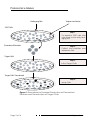
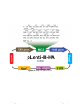
Lenti-III-HA Tag Expression Systems
Lenti-III-HA Tag Vector
LV022
Complete Lenti-III-HA Tag Expression Kit
G327
Partial Lenti-III-HA Tag Expression Kit
G328
Supplemental Kit for All Lentivirus Systems
LV098
Supplemental Kit for All Lentivirus Systems
LV099
www.abmGood.com
www.abmGood.com
Table of Contents
Notice to the Purchaser
1
Technical Support
1
Biosafety
2
Protocol at a Glance
3
Lenti-III-HA Tag Expression System
5
General Information about Lentiviral Vectors
How Lentiviruses Work
VSV Glycoprotein Envelope
Packaging Limits
6
6
6
6
Materials
7
Additional Materials Required
Storage
7
7
Protocol
8
Packaging Mix
293T Cells
Positive Control
Transfection Procedure
Concentrating Virus
Long-Term Storage
Viral Titre Assays
Transduction Procedure
8
8
8
9
10
11
11
12
Troubleshooting
14
References
15
Contact Information
16
Lenti-III-HA Tag Handbook
Notice to the Purchaser
The products are for research use only. Use in and/or for diagnostics or therapeutics is strictly prohibited. By opening and using the
product, the purchaser agrees to the following: The components in this kit
may not be distributed, resold, or modified for resale without prior written
approval from Applied Biological Materials (abm) Inc. If you do not agree
to the above conditions, please return the UNOPENED product to abm Inc.
within ten (10) days of receipt for a full refund. The information in this document is subject to change without notice and should not be construed as
a commitment by abm Inc. or its affiliated corporations. In no event shall
abm Inc. or its affiliated corporations be liable for incidental or consequential damages in connection with or arising from the use of this manual and
product.
abm Inc. products are warranted to meet our QC testing standards
at the time of shipment. Notice of problematic products must be made
to abm Inc. within 15 days of receipt of the product. This product warranty
limits abm Inc.’s liability to the replacement of the product only.
Technical Support
For more information on abm Inc. products, please visit our website:
http://www.abmGood.com
For additional information or technical assistance, please call or email us at:
Applied Biological Materials, Inc.
Phone: (604) 247-2416
1-866-757-2414
Fax:
(604) 247-2414
E-mail: [email protected]
Page 1 of 16
Lenti-III-HA Tag Handbook
Biosafety
Our Lenti-III-HA Tag Expression System employs 3rd generation self-inactivating recombinant lentiviral vectors with enhanced biosafety and minimal
relation to the wild-type, human HIV-1 virus. The lentiviral particles produced
with this system are replication-incompetent and designed with a number of
safety features to enhance its biosafety.
All Lentiviral Expression Systems provided from abm Inc. include the
following safety features:
• An enhancer deletion in the U3 region of 3’ΔLTR ensures self-inactivation
of the lentiviral vector following transduction & integration into the target
cell’s genomic DNA.
• Utilization of a RSV promoter upstream of 5’ΔLTR allows efficient Tet-independent production of viral RNA.
• The number of lentiviral genes necessary for packaging, replication and
transduction is limited to three (Gag/Pol/Rev), and their expression is derived from different plasmids, all lacking packaging signals. The plasmids
share no significant homology to the expression vector, preventing the generation of replication-competent virus.
• None of the Gag, Pol, or Rev genes will be present in the packaged viral
genome, thus making the mature virus replication-incompetent.
Despite the safety features discussed above, it is highly recommended that all manipulation with lentiviral vectors, including viral production
and transduction, be performed under Biosafety Level 2 (BL-2). All published
BL-2 guidelines with proper waste decontamination should be strictly followed. In addition, exercise extra caution when creating lentivirus carrying
potentially harmful or toxic genes (e.g. oncogenes). For more information
about the BL-2 guidelines and lentivirus handling, refer to “Biosafety in Microbiological and Biomedical Laboratories,” 5th Edition. This may be downloaded at: www.cdc.gov/biosafety/publications/bmbl5/index.htm
It is also important to consult with the health and safety officer(s) at
your institution for guidelines regarding the use of lentiviruses, and to always
follow standard microbiological practices, which include:
• Wear gloves and a lab coat at all times.
• Always work with pseudoviral particles in a Class II culture facility and that
all procedures are performed carefully to minimize splashes and aerosols.
• Work surfaces are decontaminated at least once a day and after any
spills of viable material.
• All cultures, stocks and other regulated wastes are decontaminated before disposal by an approved decontamination method, like autoclaving.
Lenti-III-HA Tag Handbook
Page 2 of 16
Protocol at a Glance
Packaging Mix
Expression Vector
293T Cells
Step 1
Co-transfect 293T cells with
a lentiviral vector and packaging mix.
Pseudoviral Particles
Step 2
Collect viral particles and
determine titre.
Target Cells
Step 3
Infect Target Cells
Target Cells Transduced
Step 4
Assay Cells
Figure 1: Procedure for Transient Production of Pseudoviral
Particles and Transduction of Target Cells.
Page 3 of 16
Lenti-III-HA Tag Handbook
Figure 2: Map of pLenti-III-HA.
Lenti-III-HA Tag Handbook
Page 4 of 16
Lenti-III-HA Tag Expression System
Lenti-III-HA Tag
The Lenti-III-HA Tag Expression System allows production of
replication-incompetent 3rd generation lentivirus that can stably
transduce both dividing and non-dividing mammalian cells with
high efficiency (Naldini, 1998 and Dull et al., 1998). Our lentiviral expression vector has been fully optimized for simple manipulations
such as subcloning the gene of interest into our pLenti-expression vector,
and easy viral DNA production including maxi DNA purification. The
vector simply works like any other plasmid. In fact, our vectors are so
stable that non-specific recombination or rearrangement in regular
DH5a bacteria cells is rarely observed. This is a significant advantage
compared to similar lentiviral vectors offered by other companies,
which are associated with substantial adversity in subcloning and
DNA production. Because of our vector stability, there is no need for
special competent cells during transformation. A general procedure
for lentiviral production is shown in figure 1 on page 3.
Our lentiviral vectors are 3rd-generation and are compatible
with packaging mixes that support the production of both 2nd and
3rd-generation vectors. Our optimized Lenti-Combo Mix (LV003) supplies all the structural and replication proteins in-trans that are required for high-titre lentivirus production in packaging cells (Titres can
be obtained up to 107IU/ml). Figure 2 on page 4 shows the vector map
for the pLenti-III-HA vector used in this kit.
Page 5 of 16
Lenti-III-HA Tag Handbook
General Information about Lentiviral Vectors
Morphology: Virions consist of an envelope, a nucleocapsid, a nucleoid,
and matrix proteins. The enveloped virions assume a spherical to pleomorphic shape of 80-100nm in diameter. The virion surface is covered with dense
inconspicuous spikes of 8 nm in length.
Physical Properties: Virions have a buoyant density of 1.13-1.18g/cm3 in sucrose. Virions are sensitive to treatment with heat, detergents, and formaldehyde. The infectivity is not affected by irradiation.
How Lentiviruses Work
Once target cells are transduced with a recombinant lentivirus, the
viral RNA is reverse-transcribed, actively imported into the nucleus (Lewis &
Emerman, 1994; Naldini, 1999), and undergoes stable integration into the host
genome (Buchschacher & Wong-Staal, 2000; Luciw, 1996). One or two days
after the lentiviral genome is integrated into the host genome, you may begin
to assay for the transient expression of your recombinant protein or use appropriate selection markers to generate a stable cell line for long-term expression studies.
VSV Glycoprotein Envelope
Most commercial retroviral vectors are limited in gene delivery applications because of their restricted tropisms and generally low titres. For recombinant lentiviral vectors, these limitations are resolved by pseudotyping
the vector with the G glycoprotein gene from Vesicular Stomatitis Virus Glycoprotein (VSV-G) envelope. The significant advantages associated with the
use of VSV-G envelope include:
• allowing production of high titre lentiviral vectors
• increasing viral particle stability
• broadening target cell ranges
• generating highly efficient transduction efficiency (Burns et al., 1993; Emi et
al., 1991; Yee et al.,1987, 1994, 1999).
Packaging Limits
Recombinant lentiviral titres will decrease with increasing insert gene
size. The packaging limit for our Lenti-III-HA Tag expression system is approximately 5.5 kb; above these limits, little to no virus will be produced.
Lenti-III-HA Tag Handbook
Page 6 of 16
Lenti-III-HA Tag
Lenti-III-HA Tag Expression System
Lenti-III-HA Tag
Materials
Scientists at abm have successfully developed a comprehensive
product line for HA tagged gene expression, and reagents for packaging
viral particles. In addition, ready-to-use lentiviral particles are also available
for immediate transduction of any target cells as in shown in Table I.
Table I. Lenti-III-HA Tag Vector and Kits
Component
Cat. No.
pLenti-III-HA Vector
LV022
10ug
Packaging Mix
LV003
100ul
LentifectinTM
G074
1.0ml
293T Cells
LV010
1x10⁶
LV011-a
10ug
Lenti-GFP Vector
Quantity G327
Kit Cat. No.
G328
LV098
LV099
Additional Materials Required
The following materials and reagents are required but not provided:
• Dulbecco’s Modified Eagle’s Medium (Invitrogen Cat: 11995)
• Fetal bovine serum (FBS) (Cat. No. TM999-500) Note: does not need to be
heat-inactivated.
• 200 mM L-Glutamine (Sigma Cat. No. G7513)
• Solution of 10,000 units/ml Penicillin G sodium and 10,000 μg/ml Streptomycin sulphate (Sigma Cat. No. P0781)
• Complete Medium: DMEM supplemented with 100 units/ml penicillin G
sodium, 100 μg/ml streptomycin and 10% fetal bovine serum (FBS)
• Puromycin (Cat. No. C021)
• Polybrene (Hexadimethrine Bromide; Cat. No. G062)
• Trypsin-EDTA (Trypsin; Sigma Cat. No. T3924)
• Dulbecco’s phosphate buffered saline (DPBS; VWR Cat. No. 82020-066)
• Tissue culture plates and flasks
Storage
• 293T Cells in Liquid Nitrogen.
• Lentifectin at 4°C.
• All other components at -20°C.
• Spin briefly to recover contents and avoid repeated freeze-thaw cycles.
Page 7 of 16
Lenti-III-HA Tag Handbook
NOTE: The following protocol is broken into sections for convenience. However,
time should be taken to read through the full procedure before attempting.
Lenti-III-HA Construct Generation
Generate a pLenti-III-HA expression vector containing your gene of interest. For information on subcloning using the multiple cloning sites available in
our lentiviral vectors, refer to a standard molecular cloning manual (Sambrook,
J.& Russell, D.W.). Once the expression construct has been produced, perform a
Maxi DNA purification for transfection.
Packaging Mix
All plasmids required for the production of recombinant lentiviruses are
provided in optimized mixtures. We have developed 2 different packaging
mixes for producing recombinant lentiviral particles in either 2nd or 3rd generation lentiviral vectors. 2nd Generation Packaging System Mix (Cat. No. LV003) is
used for the production of 2nd generation lentiviral particles and 3rd Generation
Packaging System Mix (Cat. No. LV053) is used for the packaging of 3rd generation lentiviral particles. All lentiviral expression vectors provided by abm Inc. are
of 3rd generation and can be packaged by either 2nd or 3rd Generation Packaging Mixes. In general, relatively higher titres can be achieved with the 2nd
Generation Packaging System Mix, but lentiviral particles packaged with the 3rd
Generation Mix have a higher safety profile.
293T Cells
The 293T cell line is widely used for optimal cell line for lentivirus production (Naldini et al., 1996). The health of 293T cells at the time of transfection is a
critical factor for the success of lentivirus production. The use of “unhealthy” cells
will negatively affect the transfection efficiency, resulting in lower titre lentiviral
stocks. For optimal lentivirus production, follow the guidelines below to culture
293T cells before use in transfection:
• ensure cell viability is greater than 90%.
• subculture and maintain cells in complete medium containing 0.1mM MEM,
Non-Essential Amino Acids, 4mM L-Glutamine, 1mM sodium pyruvate,
• 10% FBS.
• do not allow cells to overgrow before passaging.
• use cells that have been subcultured for less than 16 passages.
Positive Control
We recommend using a positive control vector such as pLenti-GFP to
generate a control lentiviral stock that can be used to help you optimize expression conditions in your target cells.
Lenti-III-HA Tag Handbook
Page 8 of 16
Lenti-III-HA Tag
Protocol
Lenti-III-HA Tag
Protocol
A. Transfection Procedure
We produce lentiviral stocks in 293 cells using the following optimized transfection conditions. The amount of lentivirus produced using
these recommended conditions (10ml of virus at a titre of at least 1x10⁵ transducing units (TU)/ml) is generally sufficient to transduce at least 1x10⁶ cells
at a multiplicity of infection (MOI) of 1; higher titre lentivirus can be produced
by scaling up transfection. For example, 10 wells of cells plated at 1x10⁵ cells/
well in 6-well plates could each be transduced with 1ml of a 1x10⁵ TU/ml virus
stock to achieve an MOI of 1.
1. One day before transfection (Day 1), plate 293T cells in a 10cm
tissue culture plate so that they will be 90-95% confluent on the day of
transfection (i.e. 5x10⁶ cells in 10ml of growth medium containing serum). As a rule, one 15cm culture dish at 95% confluence can be subcultured into five 10cm dishes; while one 10cm dish at 95% confluence
can be subcultured to two 10cm dishes.
2. On the day of transfection (Day 2), set up the transfection
mix:
a. In a sterile 15ml culture tube, dilute 15μg of Lenti-Combo Mix
and 10μg of pLenti expression plasmid DNA in 1.0ml of medium without
serum. Mix gently.
b. In a separate sterile 15ml tube, dilute 80μl of Lentifectin (mix
gently before use) in 1.0ml of medium without serum. Mix gently and incubate for 5 minutes at room temperature.
c. After the 5 minutes of incubation, combine the diluted DNA with
the diluted Lentifectin. Mix gently.
d. Incubate for 20 minutes at room temperature to allow the
Lentifectin/DNA complexes to form.
e. Add 4.5ml serum-free medium to the complexes followed by
gently mixing.
f. Remove the medium from the cells, and then add Lentifectin/
DNA complexes carefully to culture dishes without dislodging cells. Incubate the cells for 5-8 hours at 37°C in a humidified 5% CO2 incubator.
Note: 293T cells are poorly adhesive to most culture dishes. It is
always recommended to add or change medium against the wall of
culture dishes to avoid dislodging cells.
Page 9 of 16
Lenti-III-HA Tag Handbook
g. Add 0.65ml serum to each transfected culture dish and return
the dishes to incubator. Incubate overnight.
3. The following day (Day 3), remove the medium containing the Lentifectin/DNA complexes and replace with 10ml complete culture medium.
Incubate at 37°C in a humidified 5% CO2 incubator. Note: Expression of
the VSVG glycoprotein can cause 293T cells to fuse, resulting in the appearance of large, multinucleated cells known as syncytia. This morphological change is normal and does not affect production of the lentivirus.
4. Harvest virus-containing supernatants 48-72hrs post-transfection
(Day 4-5) by collecting medium into to a 15ml sterile, capped, conical
tube. Caution: Remember that you are now working with infec-
tious virus at this stage. Follow the recommended guidelines
for working with BL-2 organisms (see page 3 for more information).
5. Centrifuge supernatants at 3000rpm for 15 min. at 4°C to pellet debris. Optional: Filter the viral supernatant through 0.45μm PVDF syringe
filter (Millipore, Cat. No. SLHVR25LS).
6. Aliquot viral supernatants into cryovials in 1.0ml portions and
store viral stocks at -80°C. Proceed to titre your lentiviral stock (page
10). Note: If you plan to use your lentiviral construct for in vivo applications, we recommend filtering your viral supernatant through a sterile,
0.45μm low protein binding filter after the low-speed centrifugation step
to remove any remaining cellular debris. The viral supernatant can be
concentrated using the protocols discussed in the following section if
higher titre virus is required.
B.Concentrating Virus
There are many protocols that have been established to concentrate
VSVG pseudotyped lentiviruses without significantly affecting their ability to
transduce target cells. These include ultracentrifugation (Yee, 1999), filterbased ion exchange chromatography (Ultra-Pure Cat. No. LV998), and size
exclusion chromatography (Speedy Lentivirus Purification Cat. No. LV999).
However, we would strongly recommend using abm’s Ultra-Pure Lentivirus Purification Kit (Cat. No. LV998) for pLenti-miR vector concentration and purification based on our in-house testing data.
Lenti-III-HA Tag Handbook
Page 10 of 16
Lenti-III-HA Tag
Protocol
Lenti-III-HA Tag
Protocol
C. Long Term Storage
Viral stocks stored at -80°C should be stable for at least one year. Repeated freeze/thaw cycles will result in a loss of viral titre. Based on our in-house
data, each freeze/thaw will lead to a 25% loss of viral titre.
D.Viral Titre Assays
It is useful to titre the viral supernatant before proceeding with the transduction experiments for the following reasons:
• To ensure that viral stock is viable.
• To determine the percentage of target cells that can be transduced with
the pseudoviral stock.
• To control the number of copies of viral constructs per target cell.
The commonly used protocol for measuring relative titres uses a
positive control expression plasmid (i.e. GFP mixed with expression construct)
as an internal control at a ratio of 1:100 and is packaged into pseudoviral particles. In an alternative approach, the GFP control plasmid can
be packaged separately but in parallel with your construct, as an external control. In this scenario, the control plasmid can be used to check and optimize
the transfection/packaging steps (see transfection procedure). Recently, other
in vitro protocols including qRCR and HIV p24 protein-based ELISA have been
developed for quick assays.
To determine the relative titre, transduce a target cell line, like MDAMB-468, in the presence of Polybrene (2μg/ml) for 12-16 hrs, and then count
the number of cells expressing GFP either by fluorescence microscopy or FACS.
1. For each viral stock, plate MDA-MB-468 cells one day prior to viral
infection in a 24-well plate at a density of 0.6-1x10⁵ cells per well. Add 1ml
of complete D-MEM medium (with serum and antibiotics) and incubate
cells at 37°C with 5% CO2 overnight. Note: It is possible to use bigger culture dishes for transduction, especially when a large number of cells is
needed for FACS analysis. In this case, the amount of cells should be adjusted based on the growth area of the dish.
2. Prepare complete D-MEM medium plus 10% FBS with Polybrene
to a final concentration of 2μg/ml. Note: Polybrene is a polycation that
neutralizes charge interactions to increase binding between the pseudoviral capsid and the cellular membrane. The optimal concentration
of Polybrene depends on cell type and may need to be empirically determined (usually in the range of 1–8μg/ml). Excessive exposure to Polybrene (>12 hr) can be toxic to some cells.
Page 11 of 16
Lenti-III-HA Tag Handbook
3. Remove culture medium and replace with 0.5ml of complete
DMEM medium with 10% serum and Polybrene (from Step 2). For each
viral stock, use three wells. Infect MDA-MB-468 cells by adding 1ul of
viral stock into the first well (dilution factor of 500), 10μl of viral stock into
the second well (dilution factor of 50), and 100μl of viral stock into the
last well (dilution factor of 5). For controls, add 0.5ml of DMEM medium with Polybrene (from Step 2). Incubate at 37°C with 5% CO2 overnight.
4. Remove culture medium and replace with 1ml of complete DMEM
medium (without Polybrene). Incubate the cells at 37°C with 5% CO2
overnight.
5. The following day, split the cells 1:3 to 1:5, if necessary, depending on the growth rate of cells. Incubate in complete D-MEM for an additional 24-48 hours.
6. Count the fraction of fluorescent cells by FACS analysis. You may
also count the GFP positive cells under a fluorescent microscope, but the
results may be less accurate due to inconsistencies in counting methods.
Use an average of the fraction of GFP+ cells in 5 – 10 random fields to
estimate the overall percentage of GFP+cells on the plate. Multiply the
number of infected cells by 1.5×10⁵ (in this example, the expected number of MDA-MB-468 cells on the plate at the moment of infection) and by
the corresponding dilution factor, then divide by 0.5ml to determine the
relative titre of the virus in the supernatant.
Alternatively, the viral titre can also be estimated by real time
PCR using ABM’s Lentiviral qPCR Titre Kit (Cat. No. LV500) or p24-based
ELISA titre kit (Cat. No. LV501).
E.Transduction Procedure
The following information should be considered before one attempts
target cell transduction:
- The transduction efficiency of target cells varies significantly under
different experimental conditions, including virus concentration, exposure
time to virus, and growth area of cells. To determine the viral concentration
required to provide the desired multiplicity of infection (MOI) for your target
cells, perform several transductions with different concentrations of viral particles containing GFP control plasmid. Results from these test transductions
should be used to determine an optimal concentration that yields the highest percentage of infected cells.
Lenti-III-HA Tag Handbook
Page 12 of 16
Lenti-III-HA Tag
Protocol
Lenti-III-HA Tag
Protocol
- Recombinant gene expression can be measured directly 48 – 72
hours after transduction (“transient transduction”), but selecting stably transduced cells will require additional time after transduction. The decision to
use “transiently transduced” cells or selected stable cells will depend on
the nature of your target cells, biological assay, and transduction efficiency.
For efficient transducable cells (e.g., 293, HT1080, HeLa, MDA-MB-468 cells,
etc), most biological assays can be performed following transient transduction. However, for “difficult-to-transduce” cells, it is desirable to select the
clones that stably expresses the Lentivector construct for experimental assays.
The following provides general guidelines as a starting point for determining optimal conditions for target cell transduction:
1. Plate target cells in a 24-well plate 24 hours prior to viral infection
at a density of 0.5×10⁵ cells per well. Add 0.5 ml of complete optimal
medium (with serum and antibiotics) and incubate cells at 37°C with 5%
CO2 overnight. Note: It is possible to use other plate formats for transduction. In this case, the amount of cells should be adjusted depending on the growth area of the well/plate.
2. Prepare a mixture of complete media with Polybrene at a
concentration of 2μg/ml. Remove media from the wells and replace with 0.5ml of the Polybrene/media mixture per well (for 24-well
plate).
3. Infect target cells by adding several different amounts of viral
stock (example: 1μl, 5μl , 10μl, and 100μl of virus). In addition, include a
transduction well with GFP positive control virus and other appropriate
positive and negative control viral constructs. Incubate cells at 37°C
with 5% CO2 overnight.
4. Remove the culture medium and replace with 1ml of complete
medium. Incubate cells at 37°C with 5% CO2 overnight.
5. The following day, split the cells 1:3 to 1:5, depending on the
growth rate of your target cells, and continue incubating for 48 hours
in complete DMEM.
6. The infected target cells can be either analyzed for transient expression or selected for stable expression using appropriate selection
markers.
Page 13 of 16
Lenti-III-HA Tag Handbook
Troubleshooting
Problem
Possible Cause
Solution
No Viral Particles Lenti-III-HA Tag DNA modified (e.g. Re-transform plasmid into
acetylation or methylation).
an authetic DH5a.
Low Viral Titre
No Transgene
Expression
Low transfection efficiency:
-poor quality DNA.
-low 293T viability.
-transfection media containing antibiotics and serum.
-Purify DNA with an endotoxin-free Maxi colomn.
-Use 293T cells under passage 16.
-Reduce transfection antibiotic or serum efficiency.
Low transfection efficiency.
Optimize calcium transfection procedure.
Insufficient DNA used
for transfection.
Use 30-50ug of expression vector
and 30-50ug of packaging mix.
293T cell density too low.
Optimal cell density is 90-95%.
Viral supernatan harvested too early.
Optimal viral titres can be collected
48-72 hours post-transfection.
Viral supernatant subjected to
multiple freeze-thaw cycles.
Each freeze/thaw cycle can
lose 25% of the titre. Make aliquots for long-term storage.
Polybrene not used during transduction.
Transduce cells in the presence of polybrene.
Promoter silencing.
Lentiviral vector may integrate into a
chromosomal region that silences the
promoter. Screen multiple antibioticresistant clones and select the one
with the highest expresssion levels.
MOI too low.
Use maximum MOI for cell transduction.
This is very critical for Tet-expression vectors.
Viral stocks stored incorrectly.
Aliquot and store at -80C.
Avoid freeze/thaw cycles.
Target cells not transducible with lentiviral vectors.
Transduce target cells in the
presence of polybrene.
Antibiotic concentration too high. Determine antibiotic sensitivity of target
cells by performing a killing curve. Use minimum antibiotic concentration required.
Cells harvested too early for assay. Perform expression assay 3-4 days posttransduction and induction to allow the
accumulation of expressed protein.
Cytoxic Effects
of Target Cells
Large volume of viral supernatant used for transduction.
Dilute viral supernatant 1:2 to
1:3 during transduction.
Polybrene concentration too high. Use less or omit polybrene during transduction.
Antibiotic concentration too high. Use minimum antibiotics for effective selection.
Gene of interest toxic to cells.
Lenti-III-HA Tag Handbook
Try a different cell line.
Page 14 of 16
References
Buchschacher, G. L., Jr., and Wong-Staal, F. (2000) Development of Lentiviral
Vectors for Gene Therapy for Human Diseases. Blood 95, 2499-2504
Burns, J. C., Friedmann, T., Driever, W., Burrascano, M., and Yee, J.-K. (1993)
Vesicular Stomatitis Virus G Pseudotyped Retroviral Vectors: Concentration
to a Very High Titer and Efficient Gene Transfer into Mammalian and Nonmammalian Cells. Proc. Natl. Acad. Sci. USA 90, 8033-8037
Dull, T., Zufferey, R., Kelly, M., Mandel, R. J., Nguyen, M., Trono, D., and Naldini, L. (1998) A Third-Generation Lentivirus Vector with a Conditional Packaging System. J. Virol. 72, 8463-8471
Emi, N., Friedmann, T., and Yee, J.-K. (1991) Pseudotype Formation of Murine
Leukemia Virus with the G Protein of Vesicular Stomatitis Virus. J. Virol. 65,
1202-1207
Lewis, P. F., and Emerman, M. (1994) Passage Through Mitosis is Required for
Oncoretroviruses but not for the Human Immunodeficiency Virus. J. Virol.
68, 510-516
Luciw, P. A. (1996) in Fields Virology (Fields, B. N., Knipe, D. M., Howley, P. M.,
Chanock, R. M., J. L., Monath, T. P., Roizman, B., and Straus, S. E., eds), 3rd Ed.,
pp. 1881-1975, Lippincott-Raven Publishers, Philadelphia, PA
Naldini, L. (1998) Lentiviruses as Gene Transfer Agents for Delivery to Nondividing Cells. Curr. Opin.Biotechnol. 9, 457-463
Naldini, L. (1999) in The Development of Human Gene Therapy (Friedmann,
T., ed), pp. 47-60, Cold Spring Harbor Laboratory Press, Cold Spring Harbor,
NY
Sambrook, J.& Russell, D.W. (2001) Molecular Cloning: A Laboratory Manual
(Cold Spring Harbor Laboratory Press, Cold Spring Harbor, NY).
Yee, J.-K., Miyanohara, A., LaPorte, P., Bouic, K., Burns, J. C., and Friedmann,
T. (1994) A General Method for the Generation of High-Titer, Pantropic Retroviral Vectors: Highly Efficient Infection of Primary Hepatocytes. Proc. Natl.
Acad. Sci. USA 91, 9564-9568
Yee, J. K. (1999) in The Development of Human Gene Therapy (Friedmann, T.,
ed), pp. 21-45, Cold Spring Harbor Laboratory Press, Cold Spring Harbor, NY
Yee, J. K., Moores, J. C., Jolly, D. J., Wolff, J. A., Respess, J. G., and Friedmann, T. (1987) Gene Expression from Transcriptionally Disabled Retroviral
Vectors. Proc. Natl. Acad. Sci. USA 84, 5197-5201
Page 15 of 16
Lenti-III-HA Tag Handbook
Contact Information
Applied Biological Materials Inc.
Website:
Email:
www.abmGood.com
General Information: [email protected]
Phone:
Order Products:
[email protected]
(8:30am-4:30pm PST M-F)
Toll Free: 1-866-757-2414
Local: (604) 247-2416
Fax: (604) 247-2414 (24Hr.)
Address:
Technical Support:
[email protected]
siRNA:
Suite #8-13520 Crestwood Place
Richmond, BC
Canada V6V 2G2
[email protected]
Business Development:
[email protected]
Distributors
North America
Canada
Applied Biological Materials Inc.
Tel: (604) 247-2416 / 1-866-757-2414
Fax: (604) 247-2414
www.abmGood.com
United States
Applied Biological Materials Inc.
Tel: (604) 247-2416 / 1-866-757-2414
Fax: (604) 247-2414
www.abmGood.com
Mexico
Quimica Lavoisier S.A. de C.V.
Tel: 52-333-848-8484
Email: [email protected]
www.lavoisier.com.mx
Australia
Biosensis Pty Ltd.
Tel: +61 43 166 5519
Email: [email protected]
www.biosensis.com
Belgium
Gentaur
Tel: 32 2 732 5688
Email: [email protected]
www.gentaur.com
France
Gentaur
Tel: 01 43 25 01 50
Email: [email protected]
www.gentaur.com
Germany
BioCat GmbH
Tel: +49 (0) 6221-714-1516
Email: [email protected]
www.biocat.com
India
G Biosciences
Tel: 0120-432-3330
Email: [email protected]
www.GBiosciences.com
Israel
BioConsult
Tel: 972-(0)2-566-7043
Email: [email protected]
www.bioconsult.co.il
Italy
MICROTECH s.r.l.
Tel: +39-0816107435
Email: [email protected]
www.microtech.eu
Japan
Cosmo Bio Co. Ltd.
Tel: 03-5632-9610/9620
Email: [email protected]
www.cosmobio.co.jp
Singapore
Bio-REV PTE
Tel: (65) 6273-3022
Email: [email protected]
bio-rev.com
South Korea
CMI Biotech
Tel: 02 444 7101
Email: [email protected]
www.cmibio.com
Taiwan
Interlab Co. Ltd.
Tel: +886-2-2736-7100
Email: [email protected]
www.interlab.com.tw
United Kingdom
NBS Biologicals Ltd.
Tel: +44 (0)1480 433875
Email: [email protected]
www.nbsbio.co.uk
International
Lenti-III-HA Tag Handbook
Page 16 of 16