1
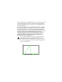
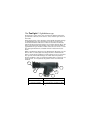
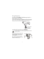
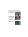
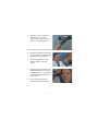
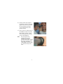
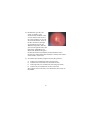
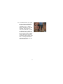
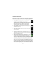
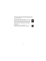
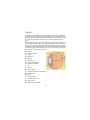
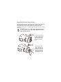
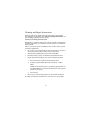
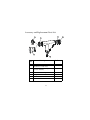
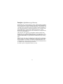
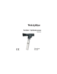
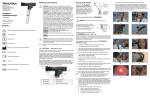
PanOptic™ Ophthalmoscope
118 Series
Operating
Instruction
Manual
This product is protected by the following US patents: 6,065,837; 6,390,625;
6,409,341; 6,527,390; U.S. Pub. No. US20020097379; other patents pending.
AU App. No. 16331/100; AU App. No. 2001263366; CA App. No. 2,352,148; CA
App. No. 2,409,596; PRC App. No. 99813584.4; EP App. No. 99959085.4; EP App.
No. 01937653.2; JP App. No. 2000-583418.
©2008 by Welch Allyn, Inc. All rights reserved. No part of this manual
may be reproduced or transmitted in any form or by any means, electronic or mechanical, including photocopy, without prior consent in writing from Welch Allyn. Printed in the U.S.A.
ii
Table of Contents
Symbols. . . . . . . . . . . . . . . . . . . . . . . . . . . . . . . . . . . . . . . . 1
Warnings and Cautions . . . . . . . . . . . . . . . . . . . . . . . . . . . 2
The PanOptic™ Ophthalmoscope . . . . . . . . . . . . . . . . 4
Set-up Instructions . . . . . . . . . . . . . . . . . . . . . . . . . . . . . . . 5
Conducting an Examination with the PanOptic
Ophthalmoscope. . . . . . . . . . . . . . . . . . . . . . . . . . . . . . . . . 7
Apertures and Filters . . . . . . . . . . . . . . . . . . . . . . . . . . . . 12
The Eye . . . . . . . . . . . . . . . . . . . . . . . . . . . . . . . . . . . . . . . 14
Lamp Replacement Instructions . . . . . . . . . . . . . . . . . . . 15
Cleaning and Repair Instructions. . . . . . . . . . . . . . . . . . . 16
Troubleshooting . . . . . . . . . . . . . . . . . . . . . . . . . . . . . . . . 17
Accessory and Replacement Parts List . . . . . . . . . . . . . . 19
Service and Warranty Information. . . . . . . . . . . . . . . . . . 20
Specifications . . . . . . . . . . . . . . . . . . . . . . . . . . . . . . . . . . 22
iv
Symbols
Attention. Read Operating Manual for Cautions and Instructions for Use.
TYPE BF – Indicates this is a product with Type BF applied
parts (the patient eyecup).
The CE mark on this product indicates it has been tested to
and conforms with the provisions noted within the 93/42/
EEC Medical Device Directive.
1
Warnings and Cautions
CAUTION: To minimize lamp housing temperatures, ontime should not exceed 2 minutes with off-time not less than
10 minutes.
CAUTION: No acute optical radiation hazards are identified
for ophthalmoscopes. However, Welch Allyn recommends
limiting the intensity of the light directed into the patient’s
eye to the minimum level that is necessary for diagnosis.
Infants, aphakes, and persons with diseased eyes are at a
greater risk. The risk is also increased if the examined person has had any exposure with the same instrument or any
other ophthalmic instrument using a visible light source
within the previous 24 hours. This will apply particularly if
the eye has had exposure to retinal photography. The
intended use of this device is for routine ophthalmic exams
on the order of typically less than 60 seconds per eye.
Although there is a benefit versus risk factor in any medical
procedure, these more complicated exams should not
exceed a three minute exam time in 24 hours. Significant
use of this device beyond its intended use is not recommended; it may cause harm to the eyes.
Use only PanOptic Patient Eyecups (part number 118092)
with this product.
CAUTION: Federal Law restricts this device to sale by or
order of a physician.
CAUTION: Spectrally weighted photochemical radiance LB
and LA give a measure of the potential hazard that exists for
a beam of light to cause photochemical damage to the retina.
LB gives the measure for eyes in which a crystalline lens is
not in place. LA gives this measure either for eyes in which
the crystalline lens has been removed (aphakes) and has not
been replaced by a UV-blocking lens or for the eyes of very
young children.
2
The value stated for this ophthalmoscope gives a measure of hazard
potential when the instrument is operated at maximum intensity
and maximum aperture. Values of LB or LA over 684mW/(cm2*sr)
are considered high.
The retinal exposure dose for a photochemical hazard is a product
of the radiance and the exposure time. For instance, at a radiance
level of 684mW/(cm2*sr), a 3-minute exposure would produce a
retinal exposure dose level at the recommended limit. If the value
of radiance were reduced to 342mW/(cm2*sr), twice that time (i.e. 6
minutes) is needed to reach the recommended limit. The
recommended exposure dose is based on calculations using the
American Conference of Governmental Industrial Hygienists
(ACGIH) Threshold Limit Values for Chemical Substances and
Physical Agents (1995-1996 Edition).
The 118 Series Ophthalmoscope has spectrally weighted
photochemical radiances at maximum intensity and
maximum aperture of .281 W/(cm2*sr) for LB and .300 W/
(cm2*sr) for LA (aphake). The relative spectral output of the
device is shown below.
305
340
375
410
445
480
515
550
585
620
655
690
725
760
W a ve le ngth (nm )
3
795
830
865
900
935
970 1005 1040 1075
The PanOptic™ Ophthalmoscope
Transparency of the cornea, lens and vitreous humor permits the
practitioner to directly view arteries, veins, and the optic nerve of
the retina.
Direct observation of the structures of the fundus through a PanOptic Ophthalmoscope may show disease of the eye itself or may
reveal abnormalities indicative of disease elsewhere in the body.
Among the most important of these are vascular changes due to diabetes or hypertension and swelling of the optic nerve head due to
papilledema or optic neuritis. In this sense, the eye serves as a window through which many valuable clinical evaluations may be
made.
When a preliminary diagnosis of an imminently dangerous eye condition, such as acute (angle-closure) glaucoma or retinal detachment, is made by the examiner, prompt referral to an eye specialist
may prevent irreversible damage. Or, when distressing but less
urgent conditions, such as visual impairment due to cataract or vitreous floaters, are recognized, the patient can be reassured and
referred.
A
F
B
E
C
D
A Patient Eyecup
D Aperture/Filter Dial
B Patient’s Side
E Focusing Wheel
C Soft Grip Handle
F Practitioner’s Side Brow Rest
4
Set-up Instructions
Attaching the PanOptic Ophthalmoscope head to a
Welch Allyn Power Source
Align cutouts in the PanOptic Ophthalmoscope base with lugs on
power source. (The PanOptic Ophthalmoscope fits all 3.5v Welch
Allyn power sources.) Push in slightly and turn 90 degrees.
TURN HANDLE
COUNTERCLOCKWISE
Attaching the Patient Eyecup to PanOptic
Use only PanOptic Patient Eyecups (part number 118092)
with this product.
Insert eyecup into the patient’s side
of the instrument. Push and twist in
one motion until you feel the two
“lock” together.
See “Accessory and Replacement
Parts List” on page 19 to reorder new
PanOptic Patient Eyecups.
5
TWIST
Attaching Corneal Viewing Lens to PanOptic (Model
11820 only)
Insert Corneal Viewing Lens into patient’s side of the instrument.
Push and twist in one motion until you feel the two “lock” together
as shown on page 5, except replacing Patient Eyecup with the Corneal Viewing Lens. Verify that the line on the lens housing is
aligned with the top seam of the PanOptic instrument.
6
Conducting an Examination with the PanOptic
Ophthalmoscope
1.
To examine the right eye, stand or sit to the patient’s right side.
2.
Remove Spectacles (preferred,
but not required).
3.
Look through the scope (from
the doctor’s side) with your
thumb on the focusing wheel.
Focus the scope on an object
roughly 15 feet away. (This
procedure will make any
adjustments for your own eye’s
corrective needs.)
4.
Start with the small aperture
(green line position on the
aperture dial).
7
5.
Turn the scope on (using any
Welch Allyn 3.5V power
source). Adjust light intensity
rheostat to desired position.
You are now ready to begin the examination:
6.
Examiner should be positioned
approximately 15 degrees to the
temporal side of the patient.
7.
Instruct the patient to look
straight ahead at a distant
object.
8.
Begin from about 6 inches away,
looking through the scope and
shining the light at the patient’s
eye while looking for the red
retinal reflex.
9.
Rest your left hand on the
patient’s forehead. (optional)
8
10. Slowly follow the red reflex
toward the patient and into the
pupil. Get as close as possible
for the optimum view (the
eyecup should contact the
patient’s brow).
11. Once you have a stable view of
the fundus rotate the focusing
wheel until you have a crisp
view of the optic disc and the
vessels surrounding it.
Note:
For the largest view,
compress the eyecup
halfway against the
patient’s brow. This enables
the user to view the entire
optic disc plus many
surrounding vessels (about
25° field of view) at one
time.
9
12. Examine the optic disc for
clarity of outline, color,
elevation and condition of the
vessels. Follow each vessel as
far to the periphery as you can.
To locate the macula, focus on
the disc, then move the light
approximately one (1) disc
diameter temporally. You may
also have the patient look at the
light of the ophthalmoscope,
which will automatically place
the macula in full view. Examine for abnormalities in the
macular area. The red-free filter facilitates viewing of the center
of the macula, or the fovea.
13. To examine the extreme periphery instruct the patient to:
A
B
C
D
look up for examination of the superior retina
look down for examination of the inferior retina
look temporally for examination of the temporal retina
look nasally for examination of the nasal retina.
This routine will reveal almost any abnormality that occurs in
the fundus.
10
14. To examine the left eye, repeat
the procedure outlined above.
However, unlike traditional
ophthalmoscopy, a unique
feature of PanOptic is that the
examiner does not have to
switch to his/her left eye. The
practitioner can use either eye
to examine either patient eye
because of the greater working
distance between patient and
practitioner. This permits those
who are strongly dominant in
one eye to always use that eye
in the fundus exam.
11
Apertures and Filters
There is a wide range of practical apertures and filters to select
from: small spot, large spot, micro spot, slit aperture, red-free filter,
cobalt blue filter (optional), and half-moon aperture (optional).
1. Small Aperture: Provides easy view of the fundus
through an undilated pupil. Always start the
examination with this aperture and proceed to micro
aperture if pupil is particularly small and/or sensitive to
light. This position is the “Home” position on the
aperture dial and is denoted by the green marking.
2.
Large Aperture: Standard aperture for dilated pupil
examination of the eye.
3.
Micro Spot Aperture: Allows easy entry in very small,
undilated pupils.
4.
Slit Aperture (Models 11810, 11820 only): Helpful in
determining various elevations of lesions, particularly
tumors and edematous discs.
5.
Red-Free Filter (Model 11810, 11820 only): This filter
excludes red rays from the examination field: this is
superior to ordinary light in viewing slight alterations
in vessels, minute retinal hemorrhages, ill-defined exudates
and obscure changes in the macula. The nerve fibers become
visible and the observer may note the disappearance of such
fibers, as in optic nerve atrophy. The background appears gray,
the disc appears white, the macula appears yellow, the fundus
reflex is intense and the vessels appear almost black. This filter
is also used to help distinguish veins from arteries; veins stay
12
relatively blue, but oxygenated arterial blood makes arteries
appear blacker. This greater contrast makes differentiation
easier for the examiner.
6.
Cobalt Blue Filter (Model 11820 only): In conjunction
with fluorescein dye applied topically to the cornea
and an add-on magnifying lens (included), this filter is
helpful in detecting corneal abrasions and foreign
bodies. In this way, it can be used as a surrogate to a
Woods Light.
7.
Half-Moon Aperture (11810 Only): Provides a
combination of depth perception and field-of view.
13
The Eye
In addition to examination of the fundus, the PanOptic Ophthalmoscope is a useful diagnostic aid in studying other ocular structures.
The light beam can be used to illuminate the cornea and the iris for
detecting foreign bodies in the cornea and irregularities of the
pupil.
The practitioner can also easily detect lens opacities by looking at
the pupil through an add-on corneal viewing lens. In the same manner, vitreous opacities can be detected by having the patient look up
and down, to the right and to the left. Any vitreous opacities will be
seen moving across the pupillary area as the eye changes position or
comes back to the primary position.
A) Macula
B) Vitreous humor
C) Sclera
D) Choroid
E) Retina
F) Ora Serrata
G) Canal of Schlemm
H) Anterior chamber
I) Iris
J) Cornea
K) Ciliary body
L) Zonule (Suspensory Ligament)
M) Conjunctiva
N) Lens
O) Hyaloid canal
P) Central retinal vein
Q) Optic nerve
R) Central retinal artery
14
Lamp Replacement Instructions
Lamp Replacement Caution: Halogen lamps are pressurized to provide maximum efficiency and illumination. Mistreatment may
cause shattering. Protect lamp surface against abrasion and
scratches. Verify power is off when replacing lamp. Dispose of lamp
with care.
CAUTION: Lamps may be hot. Lamp should be allowed to
cool before removal. Use only Welch Allyn model 03800
lamp with this product
Use your fingernail or
a small prying instrument to remove the
lamp from the ophthalmoscope head
base.
ALIGN TAB
15
To insert the new
lamp, align tab on
lamp with notch in
the base of the ophthalmoscope head
and push inward
until firmly seated.
Cleaning and Repair Instructions
Cleaning and repairs (with exception of the lamp replacement)
beyond what is specified in the “general cleaning instructions” section should be performed at the factory.
General Cleaning Instructions
NOTE: Excess solution entering the optical assembly could damage
internal components. Use caution to ensure cloth is not saturated
with solution.
This is a precision optical instrument. Please follow these specific
cleaning requirements:
• Do not use any solvent based cleaners on the lenses. Use only a
clean/soft optical grade cleaning cloth on the lenses.
•
The PanOptic Ophthalmoscope is Non-sterilizable.
•
Disinfect housing using soft cloth moistened with alcohol.
•
Clean the Patient Eyecup by any of the following methods:
1.
Soft cloth/cotton swab moistened with alcohol.
2.
Soaking in gluteraldehyde based solutions (i.e. Cidex
OPA).
NOTE: For disinfection time / procedure requirements it is
recommneded that you follow the directions of the specific
brand you are using.
3.
•
Autoclaving.
Do not use acetone based products or other harsh chemicals.
No other preventative maintenance is necessary for this product.
16
Troubleshooting
Trouble Area
Possible Cause
Corrective
Action
Aperture dial is
in-between
positions
Rotate the aperture
dial.
Lamp burned out.
Replace the lamp
using Welch Allyn
part number
03800.
Wrong lamp/
incorrect Welch
Allyn lamp/nonWelch Allyn lamp.
Replace lamp
using Welch Allyn
part number
03800.
The battery handle
has completely
discharged.
Charge the handle,
check the battery,
and/or the charger.
Spot is not
centered.
The aperture dial
is not centered.
Move aperture dial
to the full detent
position.
Not achieving full
or expected view.
Unit is not in
proper operating
position.
Ensure that the
eyecup is slightly
compressed
during the
procedure.
No Light output
17
Trouble Area
Can not obtain
sharp focus/hazy
view.
Possible Cause
Corrective
Action
The lenses are
dirty.
Clean the lenses
with a clean/soft
optical grade
cleaning cloth.
Lamp pin not
engaged in slot.
Insert the lamp
with pin firmly
seated in slot.
Film on Optics.
Clean the lenses
with a clean/soft
optical grade
cleaning cloth.
The handle is not
fully charged.
Charge the handle,
check the battery
and/or the charger.
Glare in view
Fingerprints or
dirt on the
objective (Patient’s
Side) lens.
Clean the objective
(Patient’s Side)
lens with a clean/
soft optical grade
cleaning cloth.
Cannot attach to
power handle.
Lamp is not fully
inserted.
Insert the lamp
with pin firmly
seated in slot.
Dim Light Output
18
Accessory and Replacement Parts List
1
5
3
OR
2
4
Item
No.
Description
Product #
1
Corneal Viewing Lens
(model no. 11820 only)
11875
2
Patient Eyecups (Package of five
(5) each 118092)
11870
3
Patient’s Side Bumper
118051
4
Lamp
03800
5
Chrome Ring
118027
6
Practitioner’s Side Brow Rest
118052
19
6
Service and Warranty Information
Service Centers
For information about any Welch Allyn product, call Welch Allyn
Customer Support:
USA +1 800 535 6663
+1 315 685 4560
Australia +61 2 9638 3000
Canada +1 800 561 8797
China +86 21 6327 9631
European +353 46 90 67790
Call Center
France +33 1 60 09 33 66
Germany +49 7477 9271 70
Latin +1 305 669 9003
America
Japan +81 3 3219 0071
Netherlands +31 157 505 000
Singapore +65 6419 8100
South +27 11 777 7555
Africa
United +44 207 365 6780
Kingdom
Sweden +46 85 853 6551
20
PanOptic Ophthalmoscope Warranty
Welch Allyn, Inc. warrants the No. 11810, 11820 PanOptic Ophthalmoscope to be free of original defects in material and workmanship
and to perform in accordance with manufacturer’s specifications for
a period of one year from the date of purchase. If this instrument or
any component thereof is found to be defective or at variance with
the manufacturer’s specifications during the warranty period,
Welch Allyn will repair or replace the instrument or component(s)
at no cost to the purchaser.
This warranty only applies to instruments purchased new from
Welch Allyn or its authorized distributors or representatives. The
purchaser must return the instrument directly to Welch Allyn or an
authorized distributor or representative and bear the costs of shipping.
This warranty does not cover breakage or failure due to tampering,
misuse, neglect, accidents, modification or shipping, and is void if
the instrument is not used in accordance with manufacturer’s recommendations or if repaired or serviced by other than Welch Allyn
or a Welch Allyn authorized representative.
No other express or implied warranty is given.
21
Specifications
Model Number
11810 PanOptic Ophthalmoscope without Cobalt Blue Filter
11820 PanOptic Ophthalmoscope with Cobalt Blue Filter and
Add-on Corneal Viewing Lens
Dimensions
5.125”L x 1.4”W x 3.750”H without eyecup
Eyecup 1.45”L
Weight
.48 lbs without eyecup
.50 lb with eyecup
Approvals
EN60601-1
IEC 60601-1-2
CAN/CSA-C22.2 No. 601.1-M90
UL 2601-1, Second Edition, 1997
Equipment Classification
IPXØ
Equipment not protected against the ingress of water.
Equipment not suitable for use in the presence of a flammable anesthetic.
Environment
Transport/
Storage:
-20°C - 49°C, 95% Rh max, 500hPa - 1060 hPa,
altitude
Operating:
10°C - 49°C, 95% Rh max, 500 hPa - 1060 hPa,
altitude
CAUTION: Federal Law restricts this device to sale by or order of
a physician.
22
Welch Allyn, Inc.
4341 State Street Road
Skaneateles, NY 13153-0220
Tel: (800) 535-6663
Printed in U.S.A.
Part No. 118088 Rev. F