1
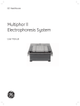
instructions
Immobiline DryPlate
User Manual
i 71-7030-01 Edition AD
Important user information
Reading this entire manual is recommended
for full understanding of the use of this
product.
The exclamation mark within an equilateral
triangle is intended to alert the user to the
presence of important operating and
maintenance instructions in the literature
accompanying the instrument.
Should you have any comments on this
manual, we will be pleased to receive them
at:
Amersham Biosciences AB
S-751 82 Uppsala
Sweden
Amersham Biosciences AB reserves the
right to make changes in the specifications
without prior notice.
Warranty and Liability
Amersham Biosciences AB guarantees that
the product delivered has been thoroughly
tested to ensure that it meets its published
specifications. The warranty included in the
conditions of delivery is valid only if the
• p2
product has been installed and used
according to the instructions supplied by
Amersham Biosciences AB.
Amersham Biosciences AB shall in no event
be liable for incidental or consequential
damages, including without limitation, lost
profits, loss of income, loss of business
opportunities, loss of use and other related
exposures, however caused, arising from
the faulty and incorrect use of the product.
Trademarks
Immobiline, Multiphor, MultiTemp,
PlusOne, NovaBlot, Sephadex and NAP
are the exclusive trade marks of Amersham
Biosciences AB. In view of the risk of trade
mark degeneration, it is respectfully
suggested that authors wishing to use these
designations refer to their trade mark
status at least once in each article.
Copyright© 1995 Amersham Biosciences AB
All rights reserved. No part of this
publication may be reproduced, stored in a
retrieval system or transmitted in any form
or by any means, without permission in
written form from the company.
Reading this entire manual is recommended
for full understanding of the use of this
product.
Contents
1. Introduction ....................................................................... 5
1.1 Package contents and technical data ......................... 6
2. Preparing the gel .............................................................. 7
2.1 Preparing the rehydration solution .............................. 7
2.2 Opening the package ................................................. 8
2.3 Rehydrating Immobiline DryPlate ................................ 9
3. Sample treatment ............................................................. 10
3.1 Sample preparation ................................................... 10
3.2 Sample concentration ................................................ 10
4. Isoelectric focusing .......................................................... 11
4.1 Preparing the experiment .......................................... 11
4.2 Sample application .................................................... 11
4.3 Running conditions .................................................... 12
5. Detection .......................................................................... 14
5.1 Silver staining ............................................................. 14
5.2 Coomassie staining .................................................... 17
5.3 Electrophoretic transfer ............................................. 18
6. Evaluation ......................................................................... 19
6.1 Determination of the isoelectric point ......................... 19
6.2 Densitometric evaluation ............................................ 19
7. References ...................................................................... 20
8. Ordering information ......................................................... 22
• p3
• p4
1. Introduction
Immobiline™ DryPlate offers a convenient and reliable way
to obtain the utmost separation power of isoelectric
focusing.
The Immobline system has indefinitely stable pH gradients
allowing high voltages for maximal separation and, when
necessary, long focusing times (1, 2).
The rehydratable dry gels facilitate the use of additives
such as urea, detergents, carrier ampholytes etc, for
optimal performance, even for samples with poor
solubility.
This manual gives general instructions on how to use
Immobiline DryPlate for isoelectric focusing. Please consult
the Application Notes and/or the articles in the reference
list (3–21) for detailed instructions on specific
applications.
Immobiline DryPlate is a polyacrylamide gel with an
immobilized pH gradient. It is bound to plastic backing
and is ready to use for isoelectric focusing after
rehydration. The product is available with various pH
gradients (See Table 1.) The pH gradients are linear over
the stated interval.
Table 1
Code No.
pH
interval
Major application
Appl.Note
80-1128-28
80-1128-29
80-1128-30
80-1128-31
80-1128-32
4–7
4.2–4.9
4.5–5.4
5.0–6.0
5.6–6.6
General purpose
α1-antitrypsin
Group specific component
Transferrin
Phosphoglucomutase
—
470
471
472
473
• p5
1.1. Package contents and technical data
Package contents
Each gel package contains 3 gels, filter papers,
experimental result forms and instructions.
Designation
Code No.
Immobiline DryPlate
Filter paper
Experimental result form
Instructions
(See label)
71-7030-01
No. per pack.
3
50
3
1
Technical data
Gel dimensions:
Gel matrix:
Buffering capacity:
Gel backing:
Storage:
Shelf life:
• p6
Approx. 250×110 x 0.5 mm
Polyacrylamide T=4%, C=3%
3 meqv/pH/L
Polyester film
–20° C
18 months from manufacturing. Please
observe “Expiry date” printed on each kit.
2. Preparing the gel
2.1. Preparing the rehydra-tion solution
One of the advantages of the dry gel format is the
opportunity to include different additives in the reswelling
solution (10, 14–16, 18–19). The three options given in
Table 2 should therefore only be regarded as typical
examples that will give good results for most applications.
However, whenever required these recipes can easily be
modified for further optimization. Consult the relevant
Application Note (Table 1 and Reference list) or any of the
cited references for instructions about specific
applications. The rehydration process itself has also been
investigated (20, 21).
Table 2.
Application areas:
Alt.1
Alt.2
Alt.3
Water soluble
proteins
Proteins with
reduced solubility
Proteins with low solubility,
e. g. Membrane proteins
Lipoproteins
20.0 ml
19.5 ml
12.0 ml
0.5 ml
Composition:
Distilled water
Pharmalyte 3–10/
Ampholine pH 3.5–9.5
Urea
Triton X-100
DTT
Total volume:
20.0 ml
20.0 ml
0.5 ml
9.6 g
0.1 ml
60 mg
20.0 ml
Rehydration time:
1–2 h
1–2 h
15–18 h
Comments
Note:
All chemicals should be of the highest purity. PlusOne
chemicals are highly recommended. Double-distilled
water should be used.
The presence of carrier ampholytes not only increases
protein solubility but also their electrophoretic migration
velocity resulting in shorter focusing times.
• p7
Mercaptoethanol (2%) or dithiothreitol (15–50 mmol/l)
can be added to avoid oxidation of sensitive proteins.
Glycerol (20–25%) improves solubility of hydrophobic
proteins and reduces the risk for urea crystallization.
Lateral band spreading can be reduced by adding acetic
acid (2 mmol/l) and applying the sample at the anodic side
or adding Tris (2 mmol/l) and applying the sample at the
cathodic side.
Triton X-100 can be replaced with other non-ionic or
zwitterionic detergents, e.g. CHAPS. Other carrier
ampholytes than Pharmalyte 3–10/Ampholine
pH 3.5–9.5 may also be used.
Alt. 3 in table 2 corresponds to what is used in the first
dimension focusing in 2-D electrophoresis. This
alternative can be regarded as a standard choice for
focusing under denaturing conditions and will normally
give high quality results with all kind of samples.
2.2. Opening the package
Note 1: Wear clean gloves to avoid contamination of the gel
surface, particularly when using silver stain.
Note 2: The gel is packed so that it is faced down to the
aluminium foil backing of the package, and the gel
support is uppermost.
Note 3: If only half of the gel is to be used, cut the package in
half with sharp scissors, reseal the portion to be saved
with tape, and store it at –20°C. Remember to identify
the polarity of the remaining part.
Open the gel package from the transparent side. Use
scissors to cut around all four sides of the package,
taking care not to cut either the gel or its transparent
backing film.
To simplify gel handling later on, identify the polarity of
the pH gradient. The support film has a precut corner
which indicates the anodic side of the pH gradient.
• p8
2.3. Rehydrating Immobiline DryPlate
For this procedure the specially designed Reswelling
Cassette is highly recommended. It allows fast, convenient,
even and reproducible rehydration of the gel. It also
facilitates including the additives necessary for optimal
performance for each application. Proceed as follows:
(See the instruction manual for the Reswelling Cassette or
the Multiphor II Electrophoresis System user manual for
detailed instructions).
1. To prevent the gel from adhering to the glass plate fitted with
the U-frame, coat the plate with Repel-Silane.
2. Mark the cathodic side of the gel.
3. Wet a clean thick glass plate with a few drops of water and
place the gel on the glass plate with the gel side up.
4. Roll the gel with a clean rubber roller (Code No. 80-1106-79)
to remove all air bubbles from between the glass plate and
the support film.
5. Mount the gel in the cassette taking particular care that the
U-frame gasket seals also over the cut-off corner of the
supporting plastic foil and that the clamps are mounted
correctly to avoid leakage.
6. Fill the cassette with the desired rehydrating solution.
7. Leave the gel to rehydrate for the recommended time.
8. Open the cassette and check the gel surface. Remove
excess liquid by placing a filter paper moistened in distilled
water on top of the gel followed by a dry filter paper on top.
9. Blot the gel by gently rolling the rubber roller under slight
pressure over the dry filter paper. Finally remove the filter
papers carefully from the gel.
(Since gels rehydrated in detergent containing solutions
have less tendency to stick to dry filter paper, they can be
dried with a simpler procedure: Place a piece of dry tissue
paper (e.g. Kleenex) on the gel, press gently to ensure
contact between tissue and gel, and remove the paper
carefully.)
• p9
3. Sample treatment
3.1 Sample preparation
Even if Immobiline DryPlate is exceptionally tolerant
towards impure samples, best results are still obtained
with samples that are free from precipitates. Should
aggregation occur at the application point, this can often
be overcome by diluting the sample or changing the
sample application position.
Best results are generally obtained when the samples are
solubilized in the rehydration solution. If this is not possible,
the concentration of salt and buffer ions should still be kept
at a minimum and, as a general rule, preferrably below
50 mmol/l. Excess buffer and salt ions will cause local
overheating due to high local currents, which can result in
protein denaturation and/or prolonged running times.
Desalting and buffer exchange can be carried out by
dialysis, or, more easily, by gel filtration using a prepacked
Sephadex G-25 column (Choose NAP-5 Column,
NAP-10 Column or PD-10 Column depending on the
sample size. See Ordering information).
3.2. Sample concen-tration
In general, Immobiline gels can take much higher sample
loads than corresponding gels with carrier ampholytes.
Several factors will determine the optimal sample
concentration and volume:
1. pH range
2. Number and relative proportions of the components in the
sample
3. Sensitivity of the detection method used.
Guide lines: PhastGel™ Blue R stain detects proteins
down to the µg range. Normally 15–20 µl of sample
with a concentration of 0.5–10 mg/ml will give good
results. Silver staining has about 20 times higher sensitivity.
A suitable load in a narrow pH gradient is normally
2–3 times higher than the load in a pH 4–7 gradient.
• p10
4. Isoelectric focusing
4.1. Preparing the experiment
Setting the cooling temperature
Connect Multiphor II electrophoresis unit to MultiTemp
II thermostatic circulator and set the desired running
temperature. A running temperature of 10 °C is often used
except for gels containing urea, which are preferably run
at somewhat higher temperatures (15 °C or more) to
avoid precipitation of the urea.
Switch on the thermostatic circulator 15 minutes before
starting the run.
Positioning the gel on the cooling plate
Pipette a few milliliters of insulating fluid (kerosene or
light paraffin oil) on the cooling plate of Multiphor II.
Position the gel on the cooling plate so that the polarity of
the gel corresponds with the polarity marked on the
cooling plate. Ensure that no air bubbles are trapped
between the gel and the cooling plate.
Electrode strips are used to ensure good electrical contact
between the gel and the electrodes. This prevents sparking and
allows salt ions from the gel to migrate into the electode strips
where they will stay and not interfere with the separation.
Soak the electrode strips evenly with approximately 3 ml
distilled water and remove the excess by using a filter
paper. The electrode strips should appear very dry before
they are applied to the gel.
Lay the electrode strips along the long edges of the gel. Cut
off the parts of the strips which protrude beyond the
short ends of the gels using a pair of sharp scissors.
4.2. Sample application
There are basically three different methods for sample
application. Which method to choose is determined
primarily by the sample volume.
• p11
1. For 5–20 µl sample volumes: Apply sample directly on the
gel, using Immobiline applicator strip (Code No. 18-100276). This applicator strip makes it possible to use a multiple
syringe which allows quick and simple sample loading,
especially when working with large numbers of samples.
Check that the contact between the gel and the applicator
strip is uniform. Leave the applicator strip on the gel during
focusing.
2. For 15–20 µl (and larger) sample volumes: Use sample
application pieces (Code No. 80-1129-46). Apply the dry
pieces to the gel surface at the desired position(s) in the
gradient. Using a micropipette, apply 15–20 µl volumes of
sample solution on each piece. To apply larger volumes, use
2 or 3 pieces stacked or laid end-to end. If you want to apply
smaller volumes with by this method, trim the paper
proportionally before applying it to the gel. Remove the
pieces of paper after completing half the total focusing time.
3. For 2–10 µl sample volumes: Apply the sample as droplets
directly on the gel surface.
4.3. Running conditions
Place the electrode holder on the Multiphor II
electrophoresis unit and align the electrodes with the
center of the electrode strips. Finally, connect the two
electrodes to the base unit and place the safety lid in
position. Connect the electrode leads to the power supply.
Typical running conditions are listed in Table 3.
Table 3.
pH range
Voltage (V)
4–7
4.2–4.9
4.5–5.4
5.0–6.0
5.6–6.6
3 500
3 500
3 500
3 500
3 500
• p12
Current(mA)
5.0
5.0
5.0
5.0
5.0
(1.0)*
(2.0)
(2.0)
(1.0)
(1.0)
Power (W)
Time (KVh)
15.0
15.0
15.0
15.0
15.0
7–15
15–25
15–25
15–25
15–25
Time (h)
2–4
4–7
4–7
4–7
4–7
Comments:
– Decrease the power and current settings proportionally if
only part of the plate is being used.
– The settings above are only to be regarded as guidelines.
Some proteins focus very slowly and may require as much
as 50–60 KVh to give optimal sharp bands. This must be
determined experimentally in each case: Run the sample for
different times.
– Since there is no gradient drift in the Immobiline DryPlate
there is no limitation in the electrophoresis system as such
as to how long the experiment can be continued. The limits
are set only by the risk of drying out the gel, oxidising or
denaturing the sample.
These risks can be minimized by placing a plastic foil on top
of the gel, running at low tempera-tures, flushing the unit with
inert gas (N2) and/or including a reducing agent (DTT or
b-mercapto-ethanol) in the rehydration mixture. The surface
can also be protected with DryStrip Cover Fluid (22).
• p13
5. Detection
All currently used detection methods can be applied on
Immobiline DryPlate gels, including Coomassie Blue
(4, 14), silver staining (23). Possible problems from
extensive swelling of the gel can be reduced by adding
ethanol (30%) to the washing solutions.
Enzymatic- and immunologically-based staining procedures as well as blotting can also be used (10–12).
5.1 Silver Staining
This silver staining method is according to Heukeshoven
and Dernick (23) with some modifications.
Silver staining solutions
Note: 250 ml of solutions are needed per gel. All steps should
be done with gentle shaking of the tray.
• p14
Fixing solution:
60 min
Trichloroacetic acid
Ethanol
Dissolve in distilled water
and make up to 250 ml.
30.0 g
125 ml
Wash:
2 × 15 min
Ethanol
Acetic acid
Make up to 500 ml with
distilled water
150 ml
50 ml
Incubation solution:
minimum 40 min
Ethanol
Sodiumacetate • 3 H2O
Glutardialdehyde (25%w/v)*
Sodium thiosulfate,
Na2S2O3 • 5 H2O
Make up to 250 ml with
distilled water
75 ml
17.0 g
1.3 ml
0.50 g
Wash:
3 × 5 min
Distilled water
Silver solution:
20 min
Silver nitrate
Formaldehyde*
Make up to 250 ml with
distilled water
0.25 g
50 µl
Developing solution: Sodium carbonate
5–15 min
Ethanol
Formaldehyde*
Make up to 250 ml with
distilled water
6.25 g
75 ml
25 µl
Stop solution:
10 min
EDTA-Na2 • 2 H2O
Ethanol
Make up to 250 ml with
distilled water
3.0 g
75 ml
Wash:
5 min
Ethanol
Make up to 500 ml with
distilled water
150 ml
Preserving solution:
20 min
Glycerol (87% (w/w)
Ethanol
Make up to 250 ml with
distilled water
25 ml
75 ml
* Note: Add these components immediately before use.
• p15
Fixation
Remove the electrode strips by using forceps, thereafter
immediately immerse the gel in the fixing solution for 60
minutes. This solution precipitates the proteins and allow
detergents (if used) to diffuse out.
Washing
Thereafter wash the gel in washing solution for 2 × 15
minutes.
Incubation
Place the gel in incubation solution for a minimum of 40
minutes. The gel can be left over night in the incubation
solution.
Washing
Thereafter, wash three times in distilled water, each time
for 5 minutes.
Silver reaction
Put the gel in silver solution for 20 minutes.
Developing
Develop the gel in developing solution for 5–15 minutes.
The protein bands should become intensively dark.
Stopping
Stop the reaction by placing the gel in stop solution for 10
minutes.
Washing
Wash in washing solution for 5 minutes.
Preserving
To preserve the silver stained gel, place the gel in
preserving solution for 20 minutes. Then place the gel on
a glass plate with the gelside up, and cover the gel with
cellophane preserving sheet soaked in preserving solution.
Allow the gel to dry in room temperature (Do not put the
gel in a heating cabinett, the silver stain will bleach due to
the increased temperature).
• p16
5.2 Coomassie staining
This is a general protein stain which detects protein
concentrations down to the µg level. 250 ml of solution is
used in each step.
Fixing solution:
30–60 min
Destaining solution:
5 min
Coomassie solution:
10 min
Trichloroacetic acid
Sulphosalicylic acid
Make up to 400 ml with
distilled water
46 g
14 g
1. Ethanol
Make up to 1000 ml
with distilled water
2. Acetic acid
Make up to 1000 ml
with distilled water
Mix 1:1 before use
500 ml
PhastGel™ Blue R
Dissolve 1 tablet in 400 ml
destaining solution. Stir
with a magnetic stirrer and
heat the solution to 60 °C.
Filter before use. Use only
once.
160 ml
1 tablet
See above
Destaining solution:
Until the background
is clear
Preserving solution:
Glycerol
Add 360 ml destaining
solution and mix well.
40 ml
Fixation
Remove the elexctrode strips with forceps. Immediately
place DryPlate in Staining Kit containing fixing solution
for 30–60 minutes. This solution precipitates the proteins.
• p17
Destaining
Before staining, wash DryPlate in destaining solution for
5 minutes.
Staining
Pour off the destaining solution and stain DryPlate for
10 minutes in staining solution which has been preheated
to 60 °C.
Destaining
Destain DryPlate with several changes of destaining
solution until the background is clear.
Preserving
Place the destained DryPlate in the glycerol preserving
solution for 30–60 minutes. Then place the gel on a glass
plate with the gel side up, and cover the gel with a
cellophane preserving sheet soaked in preserving solution.
Allow it to dry at room temperature.
5.3 Electro-phoretic transfer
Before electrophoretic blotting can take place, the support
film must be removed to allow the current to pass through
the gel. FilmRemover is ideal for this purpose. After the
film and the gel have been separated, the proteins can be
transferred to an immobilizing membrane by using the
Multiphor II NovaBlot transfer kit. Complete information
on running conditions is given in Multiphor II User
Manual (Code No. 18-1103-43).
• p18
6. Evaluation
6.1 Determination of the isoelectric point
Because of the low ionic strength in the gel, the pH
gradient cannot be directly measured with a standard
surface pH electrode unless carrier ampholytes have been
included in the rehydrating solution (21).
An alternative to direct pH measurement is to run pI
calibration proteins in parallel with the experimental
samples.
For details about the use of pI calibration proteins see the
Instruction enclosed with each pI Calibration Kit.
6.2 Densitometric evaluation
ImageMaster 1D Software (Code No. 80-6350-37) is a
powerful software package for protein quantitation and
data analysis. By using ImageMaster 1D Software together
with ImageScanner (Code No. 18-1134-45), you can
capture, store, evaluate, and report all the information
contained in your electrophoresis gels. ImageMaster 1D
Software automatically selects your lanes, bands, subtracts
the background, corrects the smiling, and integrates areas
and band volume (OD × mm2). The software calculates
relative amounts, percentages of totals, and amounts of
proteins in real quantity units using a calibration curve.
ImageMaster 1D Software also calculates isoelectric points
or molecular weights, compares bands across different
lanes or gels, and produces hierarchial clustering
information. Further lane comparision, databasing, and
identification using an internal library can be done by
using ImageaMaster 1D Database (Code No 80-6350-94)
and an add-on package to ImageMaster 1D Software.
• p19
7. References
1.
Isoelectric focusing in immobilized pH gradients. Principle, methodology and some
applications. J. Biochem. Biophys. Meth. 6 (1981) 317–339, Bjellqvist B., Ek,
K., Righetti, P.G., et al.
2.
Righetti, P. G.. Immobilized pH gradients: Theory and Methodology. Vol. 20,
Laboratory techniques in Biochemistry & Molecular Biology. Elsevier, 1990.
3.
Analysis of alpha1-Antitrypsin phenotypes in Immobiline electrofocusing gels.
Application Note 470, Amersham Biosciences AB.
4.
Analysis of Gc phenotypes by IEF in Immobiline gels. Application Note 471,
Amersham Biosciences AB.
5.
Analysis of the Transferrin phenotypes in Immobiline electrofocusing gels.
Application Note 472, Amersham Biosciences AB.
6.
Analysis of Phosphoglucomutase (PGM1) phenotypes in Immobiline isoelectric
focusing gels. Application Note 473, Amersham Biosciences AB.
7.
Subtyping of group specific component (Gc) in human semen, blood and vaginal fluid
by isoelectric focusing in immobilized pH gradients. Electrophoresis 9 (1988)
602–605, Pötsch-Schneider, L. and Klein, H.
8.
Carbohydrate analysis of transferrin subfractions isolated by preparative isoelectric
focusing in immobilized pH gradients. Electrophoresis 4 (1992) 225–229,
G. de Jong, W. L. van Noort and H. G. van Eijk.
9.
Isoelectric focusing of apolipoproteins on immobilized pH gradients: Improved
determination of apolipoprotein E phenotypes. Electrophoresis 9 (1988) 576–579,
Baumstark, M. W., Berg, A., Halle, M. and Keul, J.
10. Microheterogeneity of apolipoprotein D as revealed by electroblotting following
isoelectric focusing in Immobiline DryPlates. Electrophoresis 4 (1992) 262–264,
Holmquist, L.
11. Phenotyping of Apolipoprotein-E. Immunoblotting After Isoelectric Focussing in
Immobilized pH Gradients Electrophoresis 12 (1991) 59–63, Marz, W., Cezanne, S.
and Gros, W.
12. Immobilized pH gradient isoelectric focusing and immunoblotting for investigations
of anti-Borrelia burgdorferi IgG antibodies. Electrophoresis 4 (1992) 229–234,
M. Cruz and Å. Sidén.
• p20
13. Isoelectric focusing in Immobilized pH Gradients – applications in Clinical Chemistry
and Forensic Analysis (review). J. Chromatogr. 569 (1991) 197–228, Righetti,
P. G., Gianazza, E., Bianchibosisio, A., et al.
14. The application and optimisation of solubilising additives in Immobiline gels.
Application Note 345, Amersham Biosciences AB.
15. Membrane protein analysis by isolectric focusing in immobilized pH gradients.
Electrophoresis 6 (1985) 419–422, Rimpilainen, M. A. and Righetti, P. G.
16. Hybrid isoelectric focusing in rehydrated immobilized pH gradients with added
carrier ampholytes: Demonstration of human globulins. Electrophoresis 6 (1985)
314–325, Altland, K. and Rossman, U.
17. Analysis of recombinant proteins by isoelectric focusing in immobilized pH
gradients. Electrophoresis 4 (1992) 214–220,R. Bischoff, D. Roecklin and
C. Roitsch.
18. Apolipoprotein E phenotyping by isoelectric focusing in immobilized pH gradients and
silver staining. Electrophoresis 4 (1992) 252–258, R. Cartier and A. Sassolas.
19. Rapid and simple method for the identification of apolipoprotein E isomorphic
phenotypes from whole serum. Electrophoresis 4 (1992) 258–262, M. Kohlmeier,
H.-J. Drossel, P. Sinha and E. Köttgen.
20. Swelling kinetics of Immobiline gels for isoelectric focusing. Electrophoresis 5
(1984) 257–262, Gelfi, C and Righetti, P. G.
21. Improved rehydration procedure for polyacrylamide gels in presence of urea:
Demonstration of inhereted presence of prealbumin variants by isoelectric focusing
in an immobilized pH gradient. Electrophoresis 5 (1984) 379–381, Altland, K.,
Bantzhoff, A., Hackler, R. and Rossman, U.
22. Immobiline™ DryStrip Kit Instructions (Code No. 18-1038-63).
23. Simplified method for silver staining of proteins in polyacrylamide gels and the
mechanism of silver staining. Electrophoresis 6, (1985) 103–112, Heukeshoven, J.
and Dernick, R.
24. pH measurements in ultranarrow immobilized pH gradients. J. Biol. Biophys. Meth.
(1986) 113–124, Gelfi, C., Morelli, A., Rovida, E., and Righetti, P. G.
• p21
8. Ordering information
Code No.
Designation
80-1128-28
80-1128-29
80-1128-30
80-1128-31
80-1128-32
Immobiline
Immobiline
Immobiline
Immobiline
Immobiline
80-1106-79
18-1004-40
18-1002-76
80-1129-46
Roller
IEF electrode strips (100)
Immobiline applicator strip (5)
IEF sample application pieces (200)
DryPlate pH 4–7
DryPlate pH 4.2–4.9
DryPlate pH 4.5–5.4
DryPlate pH 5.0–6.0
DryPlate pH 5.6–6.6
17-0851-01
PD-10 column,
Desalting samples <= 2.5 ml (30)
17-0853-01/02 NAP-5 column,
Desalting samples <= 0.5 ml (20/50)
17-0854-01/02 NAP-10 column,
Desalting samples <= 1.0 ml (20/50)
18-1018-06
18-1130-05
18-1102-77
18-1102-78
18-1016-86
18-1013-75
80-1129-38
80-6350-37
18-1134-45
17-0518-01
17-0471-01
17-0472-01
17-0473-01
• p22
Multiphor II electrophoresis unit
EPS 3500 XL Power Supply
MultiTemp III Thermostatic Circulator,
115 V AC
MultiTemp III Thermostatic Circulator,
220 V AC
NovaBlot electrophoretic transfer kit
FilmRemover
Cellophane preserving sheets,
210×320 mm (50)
ImageMaster 1D Software
ImageScanner
PhastGel Blue R
Broad pI Calibration kit, pH 3–10
Low pI Calibration kit, pH 2.5–6.5
High pI Calibration kit, pH 5–10.5
Code No.
Designation
PlusOne chemicals
17-1319-01
Urea
17-1315-01
Triton X-100
17-1325-01
Glycerol 87% (w/w)
17-1317-01
2-Mercaptoethanol
17-1318-01
Dithiothreitol (DTT)
• p23
Important Information
Immobiline, Multiphor, MultiTemp, PlusOne, NovaBlot, Sephadex and
NAP are trademarks of Amersham Biosciences Limited. Amersham and
Amersham Biosciences are trademarks of Amersham plc.
© Amersham Biosciences AB 2002 – All rights reserved
All goods and services are sold subject to the terms and conditions of sale
of the company within the Amersham Biosciences group that supplies
them. A copy of these terms and conditions is available on request.
Amersham Biosciences AB
Björkgatan 30
SE-751 84 Uppsala
Sweden
Amersham Biosciences UK Limited
Amersham Biosciences Corporation
800 Centennial Avenue
P.O. Box 1327
Piscataway, NJ 08855
USA
Amersham Biosciences Europe GmbH
Munzinger Strasse 9
D-79021 Freiburg
Germany
Amersham Biosciences K.K.
Sanken Building, 3-25-1
Shinjuku-ku, Tokyo 169-0073
Japan
Produced by Wikströms, Sweden 1021704, 12.2002
Printed matter. Licence 341 051
Amersham Place, Little Chalfont
Buckinghamshire, England HP7 9NA


![Nutrition dans l`espace_Espace Explorat[...] - dubel](http://vs1.manualzilla.com/store/data/006394729_1-25072548e357e668aee2caa2d25cad9f-150x150.png)