1
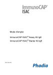
®
Genarrayt Microarray 200+ Food IgG 16pt
Quantitative assay for investigation of IgG-mediated food sensitivity
Product code GD201-4 (16 patients)
For in vitro diagnostic use
266-201pt16-01
1. Intended use
®
6.2 Technical Precautions
The Genarrayt Microarray 200+ Food IgG kit is a rapid colorimetric
microarray-based ELISA for the measurement of IgG antibodies to 221
foods in human whole blood, serum or plasma.
1.
2. Explanation of the Test
3.
4.
5.
6.
7.
8.
Many people exhibit chronic food sensitivity reactions to specific food
antigens. Unlike the immediate effects of IgE-mediated allergy, IgGmediated food sensitivity reactions may take several days to appear.
Controlled removal of the problem foods from the patient’s diet will, in many
cases, rapidly improve the patient’s condition. General lethargy, weight gain,
dermatitis, arthritis and tiredness are associated with food allergies. Irritable
bowel syndrome may also be linked to food sensitivity.
3. Principle of the test
221 food extracts have been microarrayed onto nitrocellulose pads on a
glass microscope slide. Food extracts are incubated with either diluted
patient whole blood, serum or plasma in sample diluent. After washing away
unbound proteins, anti-human IgG conjugated to horseradish peroxidase is
added to the pads and this binds to food extract-bound antibodies in the
primary incubation. Unbound conjugate is removed by washing and a
solution containing 3,3’,5,5’-tetramethylbenzidine (TMB) and enzyme
substrate is added to trace specific antibody binding. After washing with
distilled water, the slides are dried by centrifugation prior to scanning. The
optical densities of the standards, positive and negative controls and
samples are measured using a high resolution flat-bed scanner with
associated software.
4. Materials included in the kit
•
•
•
•
•
•
•
•
4 x 4 Pad Food microarrays: on modified glass slides in a slide
carrier; sealed in a foil bag with desiccant. Each microarray is
comprised of 221 food extracts.
Sample Diluent: 10mM Tris-buffered saline, pH 7.2 with 0.09%
sodium azide and proteins; 10ml, ready to use; blue cap
Wash Buffer: 10mM Tris-buffered saline, pH 7.2, with detergent
100ml; ready to use
Conjugate: Goat anti-human IgG conjugated to a horseradish
peroxidase; 10ml, ready to use; red cap
TMB Substrate: aqueous solution of TMB and mild oxidising agent,
10ml, ready to use
1 x 96 well microtitre plate
Instructions for Use
Genarrayt® Reporting Software
5. Other equipment required
• 8 channel pipette and tips to deliver 100µl and 120µl • Single channel
pipettes and tips to deliver 5µl to 250µl • distilled or deionised water •
powder-free examination gloves • 4 reagent reservoirs • slide frame holder
• slide centrifuge • wash station • high resolution flat-bed scanner with
associated computer and spot finding software • high speed (14,000 x g)
centrifuge • Ethanol or iso-propyl alcohol
2.
9.
10.
11.
12.
The microarrays should not be used if the foil bag is damaged
and solutions should not be used if liquids have leaked.
Allow all reagents and the microarrays to reach room
temperature before use.
Do not touch the microarrays.
Strictly observe the indicated incubation times.
Ensure that no cross-contamination occurs between pads.
Never pour unused reagents back into the original bottles.
Do not allow the microarrays to dry between incubation steps.
Strictly follow the described wash procedure.
Insufficient
washing may cause high background signal.
Avoid direct sunlight and exposure to heat sources during all
incubation steps.
The use of lint-free wipes is essential in reducing potential dust
contamination of the arrays.
Replace colour-coded caps on their correct vials to avoid crosscontamination
Wear powder-free nitrile gloves when handling the microarrayed
slides and when handling patient samples
7. Shelf life and storage conditions
On arrival, store the kit at 2 – 8°C. Do not use kits beyond their expiry date.
Do not freeze any kit component. Once opened use the kit within 3 months.
8. Specimen collection and storage
Serum, plasma or whole blood samples may be used. Serum and plasma
should be stored at -20°C for long-term storage. Repeated freezing and
thawing can affect results. Addition of preservatives to any sample type
may adversely affect the results. Microbially contaminated, heat-treated or
specimens containing particulate matter should not be used. Grossly
haemolysed, icteric or lipaemic specimens should be avoided.
9. Preparation of samples
Serum or plasma samples should be gently thawed, if necessary, mixed
well - DO NOT VORTEX - and centrifuged at 14,000g for 10 minutes. If the
sample is not used immediately, repeat the mix and centrifugation step prior
to testing.
Dilute samples 1:49 in sample diluent by adding 5µl serum/plasma to 245µl
Sample Diluent. For whole blood, dilute samples 1:24 by adding 10µl whole
blood to 240µl Sample Diluent.
Samples can be diluted directly into the 96-well microtitre plate or into
microtubes and then transferred to the 96-well microtitre plate if
required. When transferring samples to the microtitre plate, record the
well position for each sample as this will dictate the position of the
sample on the microarrayed slides. For example, well A1 corresponds
to Slide 1, Pad 1.
Pad 1
Pad 2
Pad 3
Pad 4
6. Precautions
6.1 Safety Precautions
1.
2.
3.
4.
5.
Only experienced laboratory personnel should use this test. The
test protocol must be followed strictly.
Human IgG used in the preparation of the Standard and the
Positive Control for this product has been tested and found
negative for antibodies to HIV, HBsAg and HCV. No test
method, however, can offer complete assurance that infectious
agents are absent. Therefore, all reagents containing human
material should be handled as if potentially infectious.
Operators should wear powder-free examination gloves and
protective clothing when handling any patient sera or serum
based products and throughout the assay procedure.
The Sample Diluent contains 0.09% sodium azide. Avoid
contact with the skin and eyes. Rinse immediately with plenty of
water if any contact occurs. Flush sinks with copious amounts
of water after discarding these reagents.
Diluted samples and any liquid that has been brought into
contact with potentially infectious material has to be discarded in
a container with a disinfectant. Disposal must be performed in
accordance with local legislation.
The slide frame holder should be disinfected after use by
immersing it in ethanol or iso-propyl alcohol for no longer than
ten minutes.
X
10. Assay Procedure
1.
Load slides into the slide frame holder with the membrane side
facing up. The “X” or dot on the slide should be on the bottom
left.
2.
Using a single or 8- channel pipette, add 100µl of diluted patient
sample (in Sample Diluent) to the pad. Agitate, cover and
incubate for 30 minutes.
3.
Vigorously flick out the slide contents, add 120µl of wash buffer
and agitate by tapping the side of the frame. Repeat step 3
once more.
4.
Vigorously flick out the slide contents, add 120µl of wash buffer
and agitate by tapping the side of the frame. Cover and incubate
for 5 minutes. Repeat step 4 once more. Flick off the wash
buffer. Do not allow the slide to dry.
5.
Using a single or 8-channel pipette, add 100µl of conjugate to
each pad. Agitate, cover and incubate for 30 minutes.
6.
Vigorously flick out the slide contents, add 120µl of wash buffer
and agitate by tapping the side of the frame. Repeat step 6 once
more.
7.
8.
9.
Vigorously flick out the slide contents, add 120µl of wash buffer
and agitate by tapping the side of the frame. Cover and incubate
for 5 minutes. Repeat step 7 once more. Flick off the wash
buffer. Do not allow the slide to dry.
Using a single or 8-channel pipette, add 100µl of TMB substrate
to each pad. Agitate, cover and incubate for 10 minutes.
Carefully flick off the slide contents and gently remove slides
from the slide frame holder and carefully place into the wash
station containing 400ml distilled/de-ionised water for 2 minutes.
DO NOT AGITATE.
10.
Carefully remove slides and centrifuge in the slide centrifuge for
30 seconds. Remove from centrifuge and leave for at least 30
minutes before scanning (>30 minutes if > 60% RH)
11.
Scan using the high resolution flat-bed scanner.
12.
Process the data using the Genarrayt® reporting software
following the user manual provided.
15. 221 Food IgG – Food Antigen Layout
See the food reporting software provided with the kit.
Method Summary
•
Pipette diluted sample onto the microarray
•
Cover and incubate for 30 minutes at room temperature
•
Wash the microarrays twice with120µl wash buffer
•
Flick off buffer and add 120µl wash buffer and incubate for 5
minutes. Repeat once. Flick off
•
Dispense 100µl of conjugate onto each microarray
•
Incubate at room temperature for 30 minutes
•
Wash the microarrays twice with120µl wash buffer
•
Flick off buffer and add 120µl wash buffer and incubate for 5
minutes. Repeat once. Flick off
•
Incubate with 100µl TMB substrate for 10 minutes
•
Load slides into a wash station containing water and incubate for
2 minutes
•
Centrifuge the slides in a centrifuge for 30 seconds
•
Remove from centrifuge and leave for 30 minutes before scanning
•
Scan the microarray using a high resolution flat-bed scanner and
apply associated spot-finding software
•
®
Process the data using the Genarrayt reporting software following
the user manual provided
11. Quality control
Further reading
The microarrays include positive and negative controls which are intended
to monitor for substantial reagent failure.
James M. Toward an understanding of allergy and in vitro testing. Nat.
Med. Journal, 1999; 2 (4): 7-15.
12. Interpretation of Results
Results are derived from internal IgG standards included in the array.
Response
Range (U/ml) 1
Negative
Borderline
Positive
<24
24 - 30
>30
1
Units are arbitrary
Genesis units.
These are suggested ranges based on in-house studies at Genesis
Diagnostics Ltd. Users of the kit should verify these ranges in their own
laboratory under local conditions and adjust as required
13. Limitations of the Procedure
1.
2.
Results must always be correlated to the clinical condition of the
patient since a raised food IgG level need not manifest as
specific symptoms.
It should be noted that results from this kit give no information
about IgE mediated allergy.
14. Assay characteristics
Within assay imprecision <20%
Between assay imprecision <22%
Atkinson et al. IgG antibodies in IBS, Gut 2004;53:1459-1464
Gaby AR. The role of hidden food allergy/intolerance in chronic disease.
Alt. Med. Review, 1998; 3(2): 90-100.
Hofman T. IgE and IgG antibodies in children with food allergy. Rocz.
Akad. Med. Bialmyst, 1995; 40 (3): 468-473
Sampson HA, Metcalfe DD. Food allergies. JAMA, 1992; 268 (20): 28402844.
El Rafei A. et al. Diagnostic value of IgG4 measurement in patients with
food allergy. Ann. Allergy, 1989; 62: 94-99.