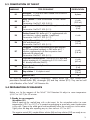
1
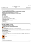
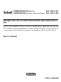
PURIFICATION KIT (768 tests)
TeSeE™ DETECTION KIT
(768 tests) - Short Assay Protocol
Ref.: 355-1181
Ref.: 355-1182
REAGENT KITS FOR IN VITRO PURIFICATION AND DETECTION OF
PrPSc
Within the European Union, this test is approved as rapid test for the BSE
and scrapie testing programmes on cattle, sheep and goats which are set up
in accordance with Annex III, chapter A to Regulation (EC) No 999/2001.
User’s manual
User’s manual
TABLE OF CONTENTS
1 - GENERAL INFORMATION
2 - TeSeE™ PURIFICATION KIT
2-1
Principle of purification of PrPSc
2-2
Samples
2-3
Composition of the TeSeE™ Purification Kit
2-4
Preparation of reagents
2-5
Storage, shelf-life
2-6
Procedure
2-7
Limits of the purification protocol
3 - TeSeE™ SAP DETECTION KIT
3-1
Principle of PrPSc detection by EIA
3-2
Samples
3-3
Composition of the TeSeE™ SAP Detection Kit
3-4
Preparation of reagents
3-5
Storage, shelf-life
3-6
Preparation of samples for PrPSc detection by EIA
3-7
Procedure
3-8
Calculation and interpretation of the results
3-9
Limits of the test
4 - MATERIAL REQUIRED BUT NOT SUPPLIED
5 - PRECAUTIONS
6 - HYGIENE AND SAFETY INSTRUCTIONS
7 - REFERENCES
2
1 - GENERAL INFORMATION
Transmissible Spongiform Encephalopathies (TSE’s) are slow degenerative diseases of the central
nervous system induced by unconventional transmissible agents (UTAs) routinely called prions.
TSEs are generally classified according to their etiology, as iatrogenic, familial and/or
sporadic. Sheep scrapie has been reported in the 18th century and transmission demonstrated
(including to goats). However, the modes of contamination within flocks remain obscure. TSEs
were also described in deer and elk (chronic wasting disease, CWD) and in cow (Bovine
Spongiform Encephalopathy, BSE).
Humans are also susceptible to certain forms of TSE. There is compelling evidence supporting
that Bovine Spongiform Encephalopathy (BSE) has passed from cattle to human, probably
through consumption of contaminated meat.
In addition to this variant form of Creutzfeldt-Jakob disease (vCJD), other forms in humans
include the Kuru and the iatrogenic Creutzfeldt-Jakob disease.
Pure hereditary forms (such as the Gerstmann-Sträussler-Scheinker syndrome [GSS]) and/or
sporadic CJD have been demonstrated in humans, but their incidences are low. We do not
know if similar sporadic TSE cases exist in animals.
The main characteristics of these diseases are:
• a slowly progressive but always fatal course,
• absence of conventional infectious agents,
• progressive accumulation in the central nervous system of an abnormal isoform of the natural
prion protein (PrP) called PrPSc. This isoform is characterized by particular biochemical
properties and especially by an increased resistance to proteases.
The strikingly long incubation period that precedes the neurological symptoms suggests that
important events of TSE pathogeneses might take place in extra nervous sites and especially in
peripheral lymphoid tissues.
In spite of many unknown and/or incertitudes, the detection of abnormal PrP Sc is now
established as the method, to confirm TSE diagnosis. This detection is mainly achieved from
post mortal collected nervous tissues.
Abnormal PrPSc has also been detected in a number of lymphoid tissue and organs: in the germinal
centres of spleen, lymph nodes, tonsils, and/or mucosa-associated lymphoid tissue (but at the
research area), in animal models or in scrapie sheeps, CWD deers and elks and vCJD patients.
Reagent designed by the “Commissariat à l’Énergie Atomique - CEA” (French Atomic Energy
Commission), developed, produced and marketed by Bio-Rad, allow PrPSc detection on
samples of nervous tissues taken from animals.
3
This determination comprises the following reaction steps:
• Purification of PrPSc (768 tests)
Step performed with the following reagents and accessories:
Ref.: 355-1181
- TeSeE™ Purification Kit (768 tests)
- Calibration syringe and needle (x 200)
Ref.: 355-1174
or Filter plates (x 50)
Ref.: 355-1179
- Deepwell microplates (x 50)
Ref.: 359-0132
- Medium beads (x 2000)
Ref.: 355-1171
• PrPSc detection (768 tests)
Step performed with the following reagents:
- TeSeE™ Detection Kit (768 tests) - Short Assay Protocol
4
Ref.: 355-1182
TeSeE™ PURIFICATION KIT
768 TESTS
355-1181
REAGENT KIT FOR IN VITRO PURIFICATION OF PrPSc
User’s manual
5
2-1 PRINCIPLE OF PURIFICATION OF PrPSc
The reagents of the TeSeE™ Purification Kit allow purification, concentration and solubilisation
of PrPSc from samples of tissues obtained from infected animals.
Processing of the samples comprises the following steps:
• Grinding of samples
• Treatment of samples with Proteinase K
• Concentration of PrPSc by precipitation
• Solubilisation of PrPSc for immunoenzymatic assay using the reagents of the TeSeE™ SAP Detection
Kit (Ref.: 355-1182).
2-2 SAMPLES
Bovine: purification of PrPSc is performed on samples from Central Nervous System (CNS). The BSE
extraction tool (Ref.: 355-1130) can be used to collect brainstem.
Since distribution of PrPSc is heterogeneous in central nervous system, obex area from brainstem must
be preferably sampled for optimal detection.
Sampling syringe (Ref.: 355-1175) allows easy and rapid sampling of obex area in a secure way.
Please refer to sampling protocol for detailed instructions on good sampling procedure.
Small ruminants and cervids: purification of PrPSc is performed on samples from Central Nervous
System (CNS) or peripheral tissues (lymphoïd nodes, spleen,…). The small ruminant extraction tool
(Ref.: 355-1184) can be used to collect both brainstem and cerebellum.
Since distribution of PrPSc is heterogeneous in central nervous system, obex area from brainstem must
be preferably sampled for optimal detection.
Samples are cut and weighed individually.
Note: other tissues (tonsils, ileum, eyelid…) can be used for research purposes only.
Samples are stored at +2°C to +8°C when purification is performed within 24 hours or can be
stored frozen for several months. They can only be submitted to 3 freezing/thawing cycles. If these
samples must be transported, they should be packaged in accordance with current local regulations.
2-3 COMPOSITION OF THE TeSeE™ PURIFICATION KIT
LABELLING
TYPE OF REAGENTS
PRESENTATION
STORAGE
Grinding
Tube
Grinding tube containing ceramic beads
in a buffer solution
Preservative: ProClin™ 300 (0.1%)
8 bags
(8 x 96 tubes)
+2°C to +25°C
Reagent A
Denaturing solution
Reagent B
Reagent C
PK
Clarifying solution
Colouring: bromophenol blue
Resolving buffer
Colouring: malachite green
Proteinase K
Colouring: phenol red
4 vials
(55 ml)
4 vials
(55 ml)
4 vials
(7 ml)
4 vials
(0.5 ml)
+2°C to +8°C
+2°C to +8°C
+2°C to +8°C
+2°C to +8°C
Reagent A, reagent B and grinding tubes are generic components. They can be used with all
batches of the TeSeE™ Purification Kits.
6
2-4 PREPARATION OF REAGENTS
All reagents of the TeSeE™ Purification Kit except proteinase K are ready for use.
Reagent A is the dilution buffer for proteinase K.
The solution must be prepared in the following way (4 μl of proteinase K in 1 ml of reagent A):
NUMBER
OF SAMPLES
REAGENT A
PROTEINASE K
2
1 ml
4 μl
10
3 ml
12 μl
18
5 ml
20 μl
26
7 ml
28 μl
34
9 ml
36 μl
42
11 ml
44 μl
50
13 ml
52 μl
58
15 ml
60 μl
66
17 ml
68 μl
74
19 ml
76 μl
82
21 ml
84 μl
90
23 ml
92 μl
The volumes must be pipetted exactly. The tip containing the PK has to be correctly rinsed by
successive aspiration/distribution cycles in reagent A.
After reconstitution, homogenize the solution by successive inversions until you obtain a red
homogeneous solution.
2-5 STORAGE, SHELF-LIFE
Store the TeSeE™ Purification Kit (Ref.: 355-1181) at +2°C to +8°C. All reagents are stable at this
temperature until the expiry date indicated on the kit (before and after opening of the vials).
After dilution, the reconstituted proteinase K solution when stored at room temperature (+18°C
to +30°C) must be used within 6 hours.
2-6 PROCEDURE
For the semi-automatic processing of the purification protocol, please refer to the TeSeE™ NSP
operator’s manual.
Procedure for manual processing:
1. Sampling:
For peripheral tissues (lymph nodes, spleen,…) insert one medium bead (Ref.: 355-1171) in
the grinding tube, before to add the sample.
Take a mass of 350 mg ± 40 mg of nervous tissue (preferably in the obex area) or 200 mg
± 20 mg of peripheral tissue.
Deposit the samples in grinding tubes, close firmly and proceed to the grinding step in the
homogenizer (Ribolyser®, TeSeE™ PRECESS 24™ or TeSeE™ PRECESS 48™ systems).
7
2. Sample grinding:
Place the tubes in the crown of the homogenizer (Ribolyser®, TeSeE™ PRECESS 24™ or TeSeE™
PRECESS 48™ systems). Perform one agitation cycle with the following instrument parameters:
Ribolyser®
TeSeE™ PRECESS 24™ or 48™
Nervous tissues Peripheral tissues Nervous tissues Peripheral tissues
Time (sec.)
45
2 x 45*
-
-
Speed
6.5
6.5
-
-
-
-
Program 1
Program 2
Program
* Wait a 5 minutes pause between the 2 agitation cycles.
When grinding is insufficient, another 1 or 2 agitation cycles can be performed, by ensuring
that the temperature of the tube returns to room temperature (+18°C to +30°C) between each
cycle (using crushed ice for example).
3. Sample transfer:
Remove the grinding tubes from the homogenizer, resuspend the homogenate by inversion
before opening the tubes.
Transfer the homogenate with one of the following methods:
• Calibration syringe method
Take 250 μl with the calibration syringe (Ref.: 355-1174) taking care to immerse the needle in
the pellet of beads to avoid sampling poorly homogenized tissue fragments.
Transfer each 250 μl sample into a 2 ml Eppendorf micro test-tube or Deepwell (Ref.: 359-0132).
• Filter plate method
The transfer and the filtration are done separately using a filter plate (Ref.: 355-1179) and a
Deepwell plate (Ref.: 359-0132), with either one of the two following filtration techniques.
- Vacuum technique:
Fit the Deepwell plate (Ref.: 359-0132) (the master plate) in the bottom of the vacuum
manifold, place the lead of the manifold and then the filter plate (Ref.: 355-1179). Take at
least 400 μl (≤ 1000 μl) with a 1000 μl tip and transfer in one well of the filter plate (Ref.: 3551179), exclude the first 6 positions (from A1 to F1). Place a plastic sealing film on top the filter
plate. Set the vacuum gauge of the pump (Ref.: 359-0350) to 25.4 cm Hg (± 2.5%). Switch the
pump on and check the gauge for correct vacuum, then open manifold valve for 1 minute ± 6
seconds. Close the valve, switch off the pump and release the vacuum from the manifold.
- Centrifugation technique:
Take at least 400 μl (≤ 1000 μl) with a 1000 μl tip and transfer in one well of the filter plate
(Ref.: 355-1179) priorly fitted on a Deepwell plate (Ref.: 359-0132) (the master plate ),
exclude the first 6 positions (from A1 to F1). Place a plastic sealing film on top the filter plate.
Centrifuge the complete system (filter plate and Deepwell plate) for 1 min at 500 g. Taking
care to keep the filtration plate securely in position on the Deepwell plate.
Note:
Centrifuge must be equipped with Deepwell microplate rotor (Ref.: 359-0136), for 5804R
Eppendorf centrifuge (Ref.: 359-1396).
After either technique discard the filter plate and transfer 250 μl of filtered samples into another
Deepwell (the purification plate) for the manual protocol or directly place the master plate on
board the NSP (refer to the TeSeE™ NSP operator’s manual).
8
Note:
At this stage, grinding tubes after homogenisation, micro test-tubes and Deepwell plate after
sample transfer can be stored, closed:
At room temperature
(+18°C to +30°C)
for 8 hours
At +2°C to 8°C
(in ice or refrigerator)
for 15 hours
At -20°C
for 1 year*
Grinding tubes and
micro test-tubes
Yes
Yes
Yes
Deepwell plate
Yes
Yes
No
* Frozen samples must be thawed at room temperature (+18°C to +30°C). Samples can be
submitted to a maximum of 3 freezing/thawing cycles. Samples must always be
homogenized by inversion before use.
4. PK treatment:
Distribute 250 μl (± 10%) of reconstituted proteinase K solution [see paragraph 2.4] into
each micro-tube or Purification plate well. Do not exceed intervals of 5 minutes for
distribution of reconstituted proteinase K between the first and the last sample. Immediately
homogenise closed tubes or Deepwell sealed with aluminium film 10 times by inversion.
Do not exceed 2 minutes between the homogenization and the incubation at 37°C.
Incubate at 37°C ± 2°C in a heating block incubator for 10 ± 1 minute.
Note:
If using Deepwell, heating block must be equipped with a Deepwell rack adaptor for
heating block (Ref.: 359-0134).
5. Precipitation of PrPSc with reagent B:
Remove the micro test-tubes or Deepwell plate from the heating block incubator. Open the
tubes and distribute 250 μl (± 10%) of reagent B into all micro test-tubes or Deepwell wells.
Observe the same order of distribution as described in step 4. Do not exceed intervals of 2
minutes between the exit of the incubator and the homogenization. Homogenization is
performed under the same conditions as in step 4.
6. Concentration of the PrPSc (centrifugation):
Within 30 minutes, after reagent B distribution and mixing : centrifuge the micro test tubes
or purification plate as follows:
Centrifugation
Micro test-tubes
Deepwell plate
Speed (g)
20 000
15 000
2 000
Time (mm)
5
7
10
20
20
4
Temperature (°C)
Note:
For Deepwell plate allow a 5 minute delay at 37°C or a 10 minute delay at room temperature
(+18°C to +30°C) before centrifugation.
7. Sample clarifying:
Discard the supernatant by inverting the micro test-tubes over a waste container. Dry the
micro test-tubes by inverting onto absorbent paper for 5 minutes.
Or load the Deepwell plate on DW40 unit (Ref.: 359-0137). Select “TSE DW” program
and select number of strips to be performed. Deepwell plate wells must be dried at the end
of the DW40 process, by inverting the plate on absorbent paper for 5 minutes.
9
Distribute 25 μl (± 10%) of reagent C into all micro test-tubes or Deepwell wells.
Do not exceed an interval of 10 minutes between the end of the drying operation and
distribution of buffer C.
Incubate immediately for 5 ± 1 minute at 100°C ± 5°C. Do not exceed 2 minutes between
the reagent C distribution and the beginning of the incubation. Do not seal the Deepwell
plate during incubation.
Note:
If using Deepwell, heating block must be equipped with a Deepwell rack adaptor for
heating block (Ref.: 359-0134).
Remove the micro test-tubes or the Deepwell from the incubator, and homogenate the tubes
with a vortex (5 ± 2 seconds).
Samples in micro test-tubes or Deepwell can be stored for 5 hours at +2°C to +8°C or
frozen for 72 hours at -20°C. Frozen samples must be thawed at room temperature (+18°C
to +30°C) and homogenized with a vortex (5 ± 2 seconds).
Please refer to information on TeSeE™ SAP Detection package insert (Ref.: 355-1182) for
detailed detection assay protocol.
2-7 LIMITS OF THE PURIFICATION PROTOCOL
Difficulties can be encountered during the grinding step when using dehydrated samples or
peripheral tissues. If necessary, the grinding step (step No.2 of the procedure) may need to be
repeated several times for this type of sample.
10
TeSeE™ DETECTION KIT -
Short Assay Protocol
355-1182
768 TESTS
REAGENTS FOR IN VITRO DETECTION OF PrP AFTER PURIFICATION
Sc
User’s manual
11
3-1 PRINCIPLE OF PrPSc DETECTION BY EIA
The TeSeE™ SAP Detection Kit is an immuno-enzymatic technique (sandwich format) using 2
monoclonal antibodies for the detection of the abnormal prion protein, resistant to proteinase K, in
tissues collected from infected animals. The kit contains sufficient reagents to assay 768 tests
(including controls).
The solid phase is composed of 12 strips of 8 polystyrene wells coated with the first
monoclonal antibody. The second monoclonal antibody is bound to peroxidase.
The assay comprises the following reactive steps:
1. Distribution of negative (R3) and positive controls (R4), samples prepared with the reagents
of the TeSeE™ Purification Kit (Ref.: 355-1181) in the wells of the microplate coated with
the first monoclonal antibody. This distribution can be visually controlled, as there is a
marked colour difference between an empty well and a well containing a sample.
2. Incubation.
3. Washing, then distribution of the peroxidase-labelled antibody. This distribution can also
be visually controlled by the colour difference between an empty well and a well
containing the conjugate solution.
4. Incubation.
5. Washing, then revelation of enzymatic activity bound to the solid phase by addition of the
substrate.
6. Stopping of the colour development, determination of optical density at 450 nm - 620 nm
(bichromatism mode) and interpretation of the results.
3-2 SAMPLES
The assay can only be performed on samples obtained from collected tissues treated with the
reagents and under the conditions of use of the TeSeE™ Purification Kit (Ref.: 355-1181).
Purified samples must be diluted with reagent R6 of the TeSeE™ SAP Detection Kit.
12
3-3 COMPOSITION OF THE KIT
LABELLING
R1
R2
R3
R4
R6
R7
TYPE OF REAGENT
Microplate: 12 strips of 8 wells coated with an anti-PrP
monoclonal antibody
Wash solution: 10-fold concentrated Tris-NaCl buffer
pH 7.4.
Preservative: ProClin™ 300 (0.01%)
Negative Control: PBS buffer pH 7.2 supplemented with
BSA
Preservative: ProClin™ 300 (0.1%)
Positive Control: PBS buffer pH 7.4 supplemented with
non infectious synthetic peptide. Lyophilized.
Preservative: ProClin™ 300 (0.1%)
Sample diluent: PBS buffer pH 7.2 supplemented with
BSA and phenol red
Preservative: ProClin™ 300 (0.1%)
Conjugate: 10-fold concentrated peroxidase-labelled
anti-PrP monoclonal antibody in PBS buffer pH 7.1
solution supplemented with bovine proteins and
coloured with phenol red
Preservative: ProClin™ 300 (0.1%)
PRESENTATION
8 plates
4 vials
(250 ml)
4 vials
(4 ml)
4 vials
4 vials
(35 ml)
4 vials
(2.8 ml)
R8
Peroxidase Substrate Buffer: Solution of citric acid and
sodium acetate pH 4.0 containing 0.015% H2O2 and
4% dimethylsulfoxide (DMSO)
4 vials
(60 ml)
R9
Chromogen: Tetramethylbenzidine
(TMB) solution.
4 vials
(5 ml)
R10
Stop solution: 1 N sulphuric acid.
Adhesive films
4 vials
(28 ml)
16
The following reagents are generic components: sample diluent (R6), wash solution (R2),
peroxidase substrate buffer (R8), chromogen (R9) and stop solution (R10). They can be used
with all batches of the TeSeE™ SAP Detection Kits.
3-4 PREPARATION OF REAGENTS
Before use, let the reagents of the TeSeE™ SAP Detection Kit adjust to room temperature
(+18°C to +30°C) for 30 minutes.
1- Ready to use reagents
Microplates (R1):
Before opening the sealed bag with a desiccant, let the microplate adjust to room
temperature (+18°C to +30°C) in its protective packaging to avoid any water condensation
in the wells. Open at the solder point and immediately return the unused rows to the sachet.
Tightly close the bag after expelling any air, then store at +2°C to +8°C.
The negative control (R3), sample dilution solution (R6) and stop solution (R10) are ready to use.
13
2- Reagents to reconstitute
Wash solution (R2):
Dilute wash solution R2 to 1/10 in distilled or ultrapure water (example 100 ml of reagent
R2 in 900 ml of distilled water).
Positive control (R4):
Gently tap the vial of positive control (R4) on the laboratory bench to detach any
substance adherent to the rubber stopper. Open the vial and dissolve the content in 4 ml
of diluent R6. Reseal the vial and let stand for approximately 1 minute, homogenizing
gently and occasionally to facilitate dissolution.
Conjugate (R7):
Dilute reagent R7 to 1/10 in the freshly reconstituted wash solution (example: 0.1 ml of
reagent R7 in 0.9 ml of reconstituted wash solution) bearing in mind that 1 ml of ready-for-use
conjugate is sufficient for 1 row. Homogenize gently. Avoid using a vortex agitator.
Enzymatic development solution (R8 + R9):
Dilute reagent R9 to 1/11 in reagent R8 (example: 0.1 ml of reagent R9 in 1 ml of reagent
R8) bearing in mind that 1.1 ml of enzymatic revelation solution is sufficient for 1 row.
Homogenize gently. Avoid using a vortex agitator.
3-5 STORAGE, SHELF-LIFE
Store the kit at +2°C to +8°C before use; all reagents are stable at this temperature until the
expiry date indicated on the kit.
The shelf-lives of the reagents after preparation are as follows:
LABELLING
REAGENT
R1
Microplate in tightly closed sachet
1 month at +2°C to +8°C
SHELF-LIFE
R2
Diluted wash solution
24 hours at room temperature (+18°C to + 30°C)
2 weeks at +2°C to +8°C
R4
Reconstituted positive control
2 hours at room temperature (+18°C to + 30°C)
4 hours at +2°C to +8°C
6 months at -20°C
It is recommended to divide the reconstituted solution into
0.5 ml aliquots and to store them immediately at -20°C.
Can be submitted to 3 successive freezing/thawing
cycles.
R7
Reconstituted conjugate solution
(with diluted wash solution)
8 hours at room temperature (+18°C to + 30°C)
R8 + R9
Development solution
6 hours at room temperature (+18°C to + 30°C)
always protected from light
3-6 PREPARATION OF SAMPLES FOR PrPSc DETECTION BY EIA
Purified samples (chapter 2.6) must be diluted with 125 μl (± 10%) of reagent R6.
Diluted samples must be homogenized with vortex (5 sec. ± 2 sec.) just before distribution into the
plate (R1).
14
3-7 PROCEDURE
TeSeE™ Detection Kit - Short assay Protocol (Ref.: 355-1182) Procedure
Procedure for manual processing:
1. Remove the microplate rack and the required number of rows (R1) from the protective
packaging. Replace the unused rows with the desiccated bag in the microplate sachet and
hermetically close it.
2. Prepare the positive control (R4), as described in chapter 3.4.2.
3. For each series of tests and every single plate, distribute 100 μl (± 10%) of control/sample
into wells in the following order:
- Wells A1, B1, C1, D1: negative control (R3)
- Wells E1, F1:
positive control (R4)
- Wells G1, H1, etc… : sample diluted with reagent (R6)
Samples are performed in singulate.
4. Cover with adhesive film and incubate for 30 mn ± 2 mn at 37°C ± 2°C.
5. Prepare wash solution (R2).
6. Prepare conjugate solution (R7).
7. Remove the adhesive film, perform 3 wash cycles.
Optimal washing conditions are obtained with PW40, PW41 or 1575 Bio-Rad plate washers
with program TSE 3.
Do not let the microplate stand for more than 5 minutes after the last wash cycle. Dry by
inversion on absorbent paper before the following step.
8. Distribute 100 μl (± 10%) of conjugate solution (R7) into each well.
9. Cover with adhesive film and incubate 30 mn ± 2 mn at +2°C to +8°C.
10. Prepare the enzymatic revelation solution (R8+R9).
11. Remove the adhesive film, perform 5 wash cycles.
Optimal washing conditions are obtained with PW40, PW41 or 1575 Bio-Rad plate washers
with program TSE 5.
Do not let the microplate stand for more than 5 minutes after the last wash cycle. Dry by inversion on
absorbent paper before the following step.
12. Distribute 100 μl (± 10%) of revelation solution (R8+R9) into each well and incubate the plate in
darkness and at room temperature (+18°C to +30°C) for 30 mn ± 2 mn. Do not use adhesive film
during this incubation.
13. Add 100 μl (± 10%) of stop solution (R10) to each well according to the same sequence
and same distribution rate as for the revelation solution.
14. Thoroughly wipe the bottom of the plate and determine the optical density at 450 nm - 620 nm
(bichromatism mode) within 30 minutes after stopping the reaction (the rows must always be
protected from light before reading).
15
16
-
Method 2
STRIPS
Manifold
-
-
STRIPS
-
-
-
Method 2
-
-
-
-
Plate
-
Plate
-
Yes
Yes
-
MODE CROS
SW
ASP.
BOTTOM ASP. Plate
WASH
-
Met.
(Method)
Yes
Yes
-
MODE CROS
SW
ASP.
BOTTOM ASP. Plate
WASH
-
Met.
(Method)
Flat
BOT:
SHAPE
1,4
ASP.
HOR. POS.
0,3
CENTERING
13,5
ASP.
VERT. POS.
9,5
BOT.
VERT. POS.
PLATE NAME: FLAT 01 (PW40/PW41) - FLAT 03 (1575)
-
Flat 01 (PW40/PW41) 1*8 (PW40/1575) 1,2,3,4,
Flat 03 (1575)
2*8 (PW41)
5,6,7,8,9,
10,11,12
Method 1
Main
parameter
PLATE
NAME: TSE 5
-
EDIT
mode
function
Manifold
Flat 01 (PW40/PW41) 1*8 (PW40/1575) 1,2,3,4,
Flat 03 (1575)
2*8 (PW41)
5,6,7,8,9,
10,11,12
PLATE
Method 1
Main
parameter
EDIT
mode
function
NAME: TSE 3
Microplate washer parameters
9,5
-
2,5
-
-
W1
-
-
-
2,5
-
6
-
-
W1
HORIZONTAL
SPEED
800
-
8
-
-
-
-
1
-
-
-
-
-
-
-
-
6
9
DISP.
UPW.
SPEED
-
-
-
1
-
-
6
BOT.
DOWNW.
SPEED
-
-
-
BOT. BOTTOM
BOT.
SHAKE
WASH
TIME
ASP.
TIME
NUMBER
NUMBER
-
-
-
BOT. BOTTOM
BOT.
SHAKE
WASH
TIME
ASP.
TIME
NUMBER
NUMBER
ASP.
DOWNW.
SPEED
0 (PW40/1575)
5 (PW41)
-
FLOW
-
0 (PW40/1575)
5 (PW41)
-
FLOW
VERTICAL
SPEED
VOLUME OVER LIQUID
FLOW
-
800
-
VOLUME OVER LIQUID
FLOW
B.W.
VERT. POS.
0,3
0,3
-
ASP.
TIME
0,3
0,3
-
ASP.
TIME
9
-
SOAKING
0
30 (PW41)
45 (PW40/1575)
-
SOAKING
1
SHAKING
AMPLITUDE
0
30 (PW41)
45 (PW40/1575)
BOT.
UPWARD
SPEED
1
5
-
Nr OF
CYCLES
1
3
-
Nr OF
CYCLES
-
0
-
MET.
INTER
-
0
-
MET.
INTER
-
-
-
9
SHAKING
SPEED
-
-
1
-
-
-
Nr OF KIT
KITS INTER
-
-
1
Nr OF KIT
KITS INTER
3-8 CALCULATION AND INTERPRETATION OF THE RESULTS
1) Calculation of the mean optical density (OD) of the negative control
OD R3 = mean of the four OD of R3 wells
2) Calculation of the cut-off value
The cut-off value is equal to : OD R3 + 0.210
Example
OD R3 = 0.020
Cut-off value = 0.020 + 0.210 = 0.230
3) Condition of validation of the test
• Negative control (R3):
a) Validation of the individual negative control values:
The optical density of each individual negative control must be lower than 0.150.
However, a maximum of one individual aberrant value can be eliminated when its optical
density is higher or equal to 0.150.
The test must be repeated if more than one of the negative control lies outside of this limit.
b) Homogeneity of the negative control values:
Calculate the mean of the negative controls with the individual remaining values.
Values higher than the mean of the negative controls + 40% (OD R3 + 40%) must be
eliminated.
- If one individual value is eliminated in a), one additional value can be eliminated in b).
- If no negative control value is eliminated in a), two values maximum can be eliminated in b).
The test must be repeated if more than two values of the negative control are eliminated
[criteria a)+b)].
• Positive control (R4):
The mean of the positive control optical densities (R4 ODs) must be higher or equal than
1.000.
The test must be repeated if the mean of the positive control optical densities (R4 ODs) is
strictly lower than this limit.
4) Interpretation of the results
Samples with an optical density lower than the cut-off value are considered to be negative
according to the TeSeE™ SAP Detection Kit.
However, results situated just below the cut-off value (cut-off value - 10%) must be interpreted
cautiously, and the corresponding samples should be retested in duplicate, starting from the
original homogenate.
Samples with an optical density greater than or equal to the cut-off value are considered to be
initially reactive according to the TeSeE™ SAP Detection Kit and should be retested in duplicate,
starting from the original homogenate, before the final interpretation.
After repeating the test, the sample is considered to be positive according to the TeSeE™ SAP
Detection Kit when at least one of the 2 measurements is positive (greater than or equal to the cutoff value). The sample is considered to be negative according to the TeSeE™ SAP Detection Kit
when these two values are less than the cut-off value.
Samples retested in duplicate and found to be negative according to the TeSeE™ SAP
Detection Kit, but for which one of the 2 values is close to the cut-off value (cut-off value 10%) must be interpreted cautiously.
17
3-9 LIMITS OF THE TEST
A negative result means that the test sample does not contain any PrPSc detectable by the
TeSeE™ SAP Detection Kit. However, as very low levels of PrPSc may not be detected, such a
negative result does not exclude the possibility of infection.
Any sample with a reproducible positive result according to the test interpretation criteria must
be confirmed in accordance with the countries national reference laboratory for TSEs or
community reference laboratory in exceptional circumstances.
4 - MATERIAL REQUIRED BUT NOT SUPPLIED
• Distilled or ultrapure water.
• 20 000 ppm sodium hypochlorite (final concentration) and 1 M sodium hydroxide (final
concentration).
• Absorbent paper.
• Disposable gloves.
• Protective glasses or mask with visor.
Purification step:
• 2 ml polypropylene micro test-tubes with caps and appropriate tube rack.
• Automatic or semiautomatic adjustable pipettes able to distribute volumes between 20 μl and 500 μl.
• Tissue homogenizer : Ribolyser®, TeSeE™ PRECESS 24™ or TeSeE™ PRECESS 48™.*
• Centrifuge* adapted to micro test-tubes.
• One micro test-tube heating block* thermostated at 37°C ± 2°C and one micro test-tube
heating block* thermostated at 100°C ± 5°C.
For the semi-automatic purification of the sample: TeSeE™ NSP System.
Detection step:
• Automatic or semiautomatic adjustable or fixed pipettes able to distribute 50 μl, 100 μl,
200 μl and 1000 μl.
• 10 ml, 20 ml, 100 ml graduated test tubes.
• Contaminated waste containers.
• Microplate incubator thermostated at 37°C ± 2°C.
• Refrigerated chamber at +2°C to +8°C.
• Automatic or semiautomatic microplate washing system.*
• Microplate reading apparatus* (equipped with 450 nm and 620 nm filters).
• Microplate system* for the automation of the assay protocol stages. The performances of the system
must conform with the requirements of the test protocol.
* Contact Bio-Rad for the list of available instruments.
5 - PRECAUTIONS
The quality of the results depends on compliance with the following good laboratory practices:
• Reagents must be stored at +2°C to +8°C.
• Do not use reagents whose shelf-life has expired.
• Do not use the reconstituted and stored at room temperature (+18°C to +30°C) proteinase K
over 6 hours.
• Do not mix reagents derived from different batches of the TeSeE™ SAP kits during the same
manipulation, with the exception of generic reagents: wash solution (R2), sample diluent (R6),
peroxidase substrate buffer (R8), chromogen (R9), stop solution (R10), grinding tubes, reagent A
and reagent B.
18
• Wash solution (R2), sample diluent (R6), peroxidase substrate buffer (R8), chromogen (R9), stop
solution (R10) and grinding tubes can be used with all kits from the TeSeE™ product line (TeSeE™
and TeSeE™ sheep/goat).
• Allow the reagents to adjust to room temperature (+18°C to +30°C) for 30 minutes before use.
• Thoroughly reconstitute reagents, avoiding any contamination.
• Do not perform the test in the presence of reactive vapors (acids, alkalis, aldehydes) or dust,
which could alter the enzymatic activity of the conjugate.
• Only use polypropylene tubes.
• Use perfectly washed glassware, rinsed in distilled water, or preferably disposable material.
• Do not let the microplate more than 5 minutes between the end of washing and distribution of
the reagents.
• The enzymatic reaction is very sensitive to all metals or metallic ions. Consequently, no metallic
element must enter in contact with the various solutions containing the conjugate or the substrate.
• The revelation solution (substrate buffer + chromogen) must be colorless. The appearance of
a colour few minutes after reconstitution indicates that the reagent cannot be used and must
be replaced. The revelation solution should preferably be prepared with disposable plastic
containers and distribution material or glassware previously washed in 1 N hydrochloric
acid, rinsed in distilled water and dried. Store this solution protected from light.
• Use a new pipette tip for each sample.
• Washing of the wells is an essential step of the procedure: respect the recommended
number of washing cycles and ensure that all wells are completely filled, then completely
emptied. Inadequate washing can give incorrect results.
• Never use the same container and pipette to distribute the conjugate and the revelation solution.
6 - HYGIENE AND SAFETY INSTRUCTIONS
Generally, hygiene conditions, biosafety mesures and good laboratory practices must be in
agreement with recommendation of regular authorities of the country.
• All reagents of the kit are intended for use in “in vitro” diagnosis.
• Wear disposable gloves when handling reagents and samples and wash your hands
thoroughly after handling them.
• Do not pipette with the mouth.
• Use polypropylene containers to avoid any wounds with broken glass.
• All the materials directly in contact with the samples and the wash solutions must be
considered as contaminated.
• Avoid splashing samples or solutions containing samples.
• Contaminated surfaces must be cleaned with 20 000 ppm sodium hypochlorite solution (bleach).
When the contaminating liquid is an acid, contaminated surfaces must be first neutralized with
sodium hydroxide before using bleach. Surfaces must be rinsed with distilled water, dried with
ethanol and wiped with absorbent paper. The material used for cleaning must be discarded in a
special container for contaminated wastes.
• Samples, material and contaminated products must be eliminated after decontamination:
- either by soaking in 1 M sodium hydroxide (final concentration) for 1 hour at room temperature
(+18°C to +30°C),
- or by soaking in 20 000 ppm sodium hypochlorite solution for 1 hour at room temperature
(+18°C to +30°C),
- or by autoclaving at 134°C minimum for at least 18 minutes, under 3 bars of pressure.
Note: never autoclave solutions containing sodium hypochlorite solution or reagent B.
• All operations involved in Transmissible Spongiform Encephalopathy (TSE) screening tests are
subject to regulations and must be performed in an isolated, limited and controlled access
laboratory devoted exclusively to this activity. A laboratory coat, overshoes, gloves, mask
with visor or simple mask with safety glasses are required to ensure the operator's safety.
19
• Operators must receive specific training concerning the risks related to TSEs agents or prions
and the validated modes of decontamination for unconventional agents. Biosafety measures
must be in agreement with recommendations of regular authorities of the country.
• Avoid any contact of the substrate buffer, chromogen and stopping solution with the skin
and mucous membranes.
• Neutralize and/or autoclave all wash solutions or wash wastes or any liquid containing
biological samples prior to their elimination.
• Reagent B is a dangerous substance classified as nocive (> 25% alcohol) according to
European legislation.
• Reagents containing 0.1% ProClin™ 300 are classified as irritating preparations according
to European legislation.
Xn
(Alcohol > 25%)
(0.1% ProClin™ 300)
R : 10-22-37/38-41-43-67 Flammable. Harmful if swallowed. Irritating to respiratory system
and skin. Risk of serious damage to eyes. May cause sensitisation by skin contact. Inhalation
of vapour may cause drowsiness and dizziness.
S : 7/9-13-26-28-37/39-46 Keep container tightly closed and in a well ventilated place. Keep
away from food, drink and animal feed. In case of contact with eyes, rinse immediately with
plenty of water and seek medical advice. After contact with skin wash immediately with plenty
of water. Wear suitable protecting clothings, gloves and eye/face protection. If swallowed,
seek medical advice immediately and show this container or label.
20
7 - REFERENCES
1. J. GRASSI, E. COMOY, S. SIMON, C. CREMINON, Y. FROBERT, S. TRAPMANN,
H. SCHIMMEL, S.A.C. HAWKINS, J. MOYNAGH, JP DESLYS, G.A.H. WELLS (2001)
Rapid Test for the preclinical postmorten diagnosis of BSE in central nervous system tissue.
The Veterinary Record (149) 577-582.
2. JP. DESLYS, E. COMOY, S. HAWKINS, S. SIMON, H. SCHIMMEL, G. WELLS, J. GRASSI,
J. MOYNAGH (2001)
Screening slaughtered cattle for BSE - Nature (409) 476-477.
3. E. COMOY (2000)
Contribution au développement d’un test de diagnostic post mortem des bovins atteints
d’Encephalopathie Spongiforme Bovine.
Thèse de doctorat vétérinaire (Ecole Nationale Vétérinaire d’Alfort).
4. EUROPEAN COMMISSION
Directorate General DG XXIV (1999).
Preliminary Report : The evaluation of tests for the diagnosis of transmissible Spongiform
Encephalopathy in bovines.
5. JP. DESLYS (1999)
Prevention du risque d’Encephalopathie Spongiforme Subaiguë Trans-missible.
La Revue du Praticien (49) 966-970.
6. R. KNIGHT (1999)
The relationship between new variant Creutzfeldt-Jakob Disease and Bovine Spongiform
Encephalopathy - Vox sanguinis (76) 203-208.
7. D. DORMONT (1997)
Les Agents Transmissibles Non Conventionnels ou prions - Virologie (1) 11-22
8. F. HILLA, M. DESBRULAIS, S. JOINER, KCL SIDLE, I. GOWLAND, J. COLLINGE, LJ. DOEY,
P. LANTOS (1997)
The same prion strain causes CJ disease and BSE - Nature (389) 448-450.
9. CI. LASMEZAS, JP. DESLYS, O. ROBAIN, D. DORMONT (1997)
L’agent secret des maladies à prions - La Recherche 46-53.
10. AM. HAYWOOD (1997)
Transmissible Spongiform Encephalopathies.
The New England Journal of Medecine (337-25) 1821-1828.
11. J. COLLINGE, KC. SIDLE, J. MEADS, J. IRONSIDE, AF. HILL (1996)
Molecular analysis of prion strain variation and the aetiology of “new variant” CJD.
Nature (383) 685-690.
12. RG. WILL, J. IRONSIDE, M. ZEIDLER, SN. COUSENS, K. ESTIBEIRO, A. ALPEROVITCH,
S. POSER, M. POCCHIARI, A. HOFMAN, PG. SMITH (1996)
A new variant of Creutzfeldt-Jakob disease in the U.K. - Lancet (347) 911-925.
13. SB. PRUSINER & AL (1993)
Immunologic and molecular biologic studies of prion protein in Bovine Spongiform Encephalopathy.
The Journal of Infectious Diseases (167) 602-613
21
Sample syringe
355-1175
SAMPLING METHOD FOR BIO-RAD TSE SCREENING ASSAYS
(PLATELIA® AND TeSeE™ SAP)
23
TABLE OF CONTENTS
1 - GENERAL INFORMATION
1-1
Sample collection at the abattoir
1-2
Sample procedure at the laboratory
2 - BIO-RAD SAMPLE SYRINGE
3 - SAMPLE MASS REQUIRED FOR THE TEST
4 - OPERATING PROCEDURE
5 - PRECAUTIONS/ADVICE
6 - HEALTH AND SAFETY PROCEDURES
24
1 - GENERAL INFORMATION
The Bio-Rad TSE screening assays are performed on a sample of 350 ± 40 mg of central
nervous tissues (CNS). The specific anatomical region for detecting PrPSc in infected animals is
the brain stem, more precisely in the area of the vagal nerve nucleus, in the obex region. This
is the area of the brainstem where PrPSc is most concentrated.
Brain
Sampling region
(right or left)
Obex
Spinal cord
Cross section of the brain stem at the level of the obex
identifying the key target sites for diagnosis by
histopathology and immunohistochemistry in BSE (nucleus of
the solitary tract [1] and the nucleus of the trigeminal tract V
[2]) and scrapie (dorsal nucleus of the vagus). [3]).
(Source: OIE - Manual of Diagnostic Tests
and Vaccines for Terrestrial Animals)
1 - 1 Sample collection at the abattoir
The brain stem is easily and quickly collected with an appropriate tool or sample collection
spoon, via the occipital foramen, without opening the cranial cavity.
Sample collection with the Bio-Rad collection spoon
25
1 - 2 Sampling procedure at the laboratory
The whole brain stem sample is sent to the testing laboratory ensuring that appropriate bio-safety
measures recommended by the regulatory authorities of the particular country are followed. In the
laboratory, the appropriate amount of cerebral material is cut (scalpel blade,…) from the obex region
or collected with the Bio-Rad sample syringe (Ref: 355-1175) which makes it possible to sample the
required amount of the appropriate area quickly and safely, without any risk of sharps injuries.
The following describes the procedure to effectively collect the sample from the obex region
using the Bio-Rad sample syringe, without damaging the tissue.
2 - BIO-RAD SAMPLE SYRINGE
The Bio-Rad sample syringe consists of a green piston and a transparent syringe barrel. The
syringe barrel is labelled with a series of geometric shapes. (
l)
Marks (black)
Piston (green)
Cutting wire
Barrel of the syringe
(transparent)
3 - SAMPLE MASS REQUIRED FOR THE TEST
The sample mass should occupy the space between two symbols of the same shape which
corresponds to a mass (m) of 350 +/- 40 mg.
m
m
m
m
m
4 - OPERATING PROCEDURE
• Take a sample syringe and pull out the green piston to approximately 1 cm from its home
position then push home again.
• Firmly grasp the brain stem in one hand, using a disposable wrapper (plastic bag, glove, etc.) in order
to avoid possible cross-sample contamination. The end of the brain stem should remain accessible. If
the brainstem received has a cord too long, the user should trim it. Samplers should received proper
training regarding the précise location of the targetted area.
• Use the other hand to position the open end of the sampling syringe on the right or left side
of the caudal end of the brain stem.
Note: a complete hemi-section of brain stem with an intact obex region must remain available
after sample collection for confirmatory testing.
26
• Insert the syringe barrel gradually into the brain stem whilst holding the green piston stationary
(relative to the brain stem).
Note: While collecting the sample from the obex region, take care that the syringe barrel
remains within the selected side of the brain stem.
• Stop this movement when the top of the syringe barrel has reached the upper limit of the
sampling zone.
• Cut the sample core by twisting the syringe barrel through one complete turn.
• Slowly remove the sample syringe from the brain stem, taking care not to damage
surrounding tissues. The remaining brain stem can be placed in its original sample container.
• Check whether there are any air gaps in the core sample collected. If needed, compress the
sample core by closing the top of the syringe barrel and pushing the green piston until the
air gaps have been eliminated. At the same time ensure that the tissue nearest the opening
of the syringe barrel is retained.
• Holding the top of the syringe barrel still, move the green piston to the nearest symbol.
• Check that the sample core covers at least one zone corresponding to “m”, as described in
the previous section of this document (sample mass required for the test).
• Take a grinding tube and remove the lid, with the sample syringe carefully depress the green
piston to the next identical symbol to ensure that the correct mass of tissue (“m”) is dispensed
in the grinding tube. Remember that you must move the piston to the corresponding position of
the next symbol as indicated in “Sample mass required for the test”.
• Cut the sample core by gripping the top of the sample syringe against the inner edge of the
grinding tube.
• Samples of extremely bad quality should be either disected or if very autolysed pipetted up.
• The unused part of the sample core can be stored by placing the sample syringe in the
original container with the remaining piece of brain stem.
27
5 - PRECAUTIONS/ADVICE
As for any pipetting device, Bio-Rad recommends that operators using the sample syringe
should be periodically monitored, for a representative statistical population of samples taken,
so ensuring that sample weights are within range.
The sample syringes are to be used only once, and then discarded in order to prevent any
cross-sample contamination.
The sample must be taken with all due precautions in order to ensure that risk of contamination
for operators is minimized.
The syringes used are to be discarded after being decontaminated (see Health & Safety instructions).
If the sample core does not fill the entire syringe barrel despite carrying out the procedure
correctly, it is advisable to weigh the sample.
6 - HEALTH & SAFETY PROCEDURES
The hygiene conditions, bio-safety measures and good laboratory practices must comply with
the guidelines of the regulatory authorities in the country.
The sample syringe is intended for use in “in-vitro” diagnostic procedures only.
Wear disposable gloves when handling reagents and samples, and wash your hands
thoroughly after handling them.
Any equipment that has come into direct contact with the samples must be considered to have
been contaminated.
Contaminated surfaces must be cleaned with 20 000 ppm sodium hypochlorite solution. When
the contaminating liquid is an acid, contaminated surfaces must first be neutralized with
sodium hydroxide before using sodium hypochlorite. Surfaces must be rinsed with distilled
water, dried with ethanol and wiped with absorbent paper. The material used for cleaning
must be discarded in a specific container for contaminated waste.
Samples, equipment and contaminated products must be discarded after decontamination
using one of the following methods:
- by soaking in 1 M sodium hydroxide (final concentration) for 1 hour at room temperature
(+18°C to +30°C).
- by soaking in 20 000 ppm sodium hypochlorite solution for 1 hour at room temperature
(+18°C to +30°C).
- by autoclaving at a temperature of at least 134°C for a minimum of 18 minutes, at 3 bar
pressure.
- Note: never autoclave solutions containing bleach.
All operations involved in Transmissible Spongiform Encephalopathy (TSE) screening tests are
subject to local safety guidelines and must be performed in an isolated, limited and controlledaccess laboratory devoted exclusively to this activity. A laboratory coat or boiler suit,
overshoes, gloves (two pairs), mask with visor or simple mask with safety glasses are required
to ensure the Operator's safety.
Operators must receive specific training concerning the risks related to TSE agents or prions,
and the validated methods of decontamination for unconventional agents. Bio-safety measures
must comply with the Guidelines of the regulatory authorities of the country concerned.
28
Group Headquarters
Bio-Rad Laboratories
2000 Alfred Nobel Drive
Hercules California 94547
Phone: (510) 741-1000
Toll-Free Phone:
1-(800) 424-6723
Fax: (510) 741-5800
Subsidiaries of Bio-Rad
Laboratories:
Australia
Bio-Rad Laboratories Pty., Ltd.
PO Box 210 Regents Park
Block Y, Unit 1
Regents Park Industrial Estate
393 Park Road
Regents Park, New South Wales
2143
Phone: 02 9914 2800
Toll Free: 1800-224 354 (within
Australia only)
Fax: 02 9914 2889
email: [email protected]
Austria
Bio-Rad Laboratories Ges.m.b.H.
Hummelgasse 88/3-6,
A-1130 Wien
Phone: (01) 877 89 01
Fax: (01) 876 56 29
Belgium
Bio-Rad Laboratories S.A.-N.V.
Begoniastraat 5
B-9810 Nazareth EKE
Phone: 09-385 55 11
Toll-Free Phone: 0800/97032
Fax: 09-385 65 54
email: [email protected]
Brazil
Bio-Rad Laboratórios Brasil Ltda
Av. Padre Antônio José dos Santos,
449 / 5º andar
Brooklin - São Paulo - SP
CEP.: 04563-011
Brazil
Phone: (55) 11 5044 5699
Fax: (55) 11 5543 4383
Praia de Botafogo
440 / 3° andar
Botafogo - Rio de Janeiro - RJ
CEP: 22250-040
Phone: (55) 21 3237 9400
Fax: (55) 21 2527 3099
Canada
Bio-Rad Laboratories (Canada) Ltd.
5671 McAdam Road
Mississauga, Ontario L4Z 1N9
Phone: (905) 712-2771
Toll-Free Phone: 1-(800) 268-0213
Fax: (905) 712-2990
Germany
Bio-Rad Laboratories GmbH
Heidemannstraße 164
D-80939 München
Postfach 45 01 33
D-80901 München
Phone: 49 89 318 84-0
Fax: 49 89 318 84-123
Czech Republic
Bio-Rad s.r.o.
nad ostrovem 1119/7
147 00, Praha 4
Czech Republic
Phone: 420 242 430 532
Fax: 420 242 431 642
email: [email protected]
Greece
Bio-Rad Laboratories EPE
2-4 Mesogion Ave. (Athens Tower)
155 27 Ampelokipi - Athens
Phone: 0030 210 7774396 7774345
Fax: 0030 210 7774376
People’s Republic of China
HuiZhong Bio-Rad Technologies Ltd.
Beijing Office
14 Zhi Chun Road, Hai Dian District
Beijing 100 008
Phone: 86-10-62051850
and 86-10-6204662 ext 3401-06
Fax: 86-10-62051876
Bio-Rad Technologies (Shanghai) Ltd.
10/F Ascendas Building,
333 Tian Yao Qiao Road,
Shanghai, 200030
Phone: 86-21-6305-2255
Fax: 86-21-5396-4775
Denmark
Bio-Rad Laboratories
Generatorvej 8 C
2730 Herlev
Phone: 44 52 10 00
Fax: 44 52 10 01
email: [email protected]
Finland
Bio-Rad Laboratories
Pihatörmä 1A
Fin-02240 Espoo
Phone: 09 804 22 00
Fax: 09 804 22 00
email: [email protected]
France
Bio-Rad S.A.
3 Boulevard Raymond Poincaré
92430 Marnes-la-Coquette
Phone: 01 47 95 60 00
Fax: 01 47 95 61 81
Hong Kong
Bio-Rad Pacific Ltd.
Unit 1101, 11/F,
DCH Commercial Center,
25 Westlands, Quarry Bay,
Hong Kong
Phone : 852-2789-3300
Fax : 852-2789-1257
Hungary
Bio-Rad Hungary Ltd.
Tuzolto u. 59.
H-1094 Budapest
Phone: (361) 455 8800
Fax: (361) 455 8809
e-mail: [email protected]
India
Bio-Rad Laboratories (India) Pvt. Ltd.
B&B-1, Enkay Towers, Vanijya Nikunj
Udyog Vihar, Phase V
Gurgaon 122016
Phone: 91-1242398112/113/114,
91-124-5018111
Fax: 91-124-2398115, 2450095
email: [email protected]
Israël
Bio-Rad Laboratories Ltd
14 Homa Street
PO Box 5044
Rishon Le Zion 75150
Phone: 03 951 4127
Fax: 03 951 4129
email: [email protected]
Italy
Bio-Rad Laboratories S.r.l.
Via Cellini, 18/A
20090 Segrate - Milano
Phone: 39-02-21609-1
Fax: 39-02-21609-399
57
Japan
Nippon Bio-Rad Laboratories
7-18 Hogashi Nippori 5-Chome,
Arakawa-ku, Tokyo 116-0014
Phone: 03-5811-6270
Fax: 03-5811-6272
Korea
Bio-Rad Korea Ltd.
10F, Hyunjuk BLDG,
832-41 Yeoksam dong Gangnam
gu, Seoul 135-080
Phone: 82-2-3473-4460
Fax: 82-2-3472-7003
Latin America
Bio-Rad Latin America
14100 Palmetto Frontage Road
Suite 101
Miami Lakes, Florida 33016
Phone: (305) 894-5950
Fax: (305) 894-5960
Web address: latinamerica.biorad.com
Mexico
Bio-Rad, S.A.
Adolfo Prieto No. 1653
Col. del Valle
México, DF C.P. 03100
Phone. 525-55-200-0520
Fax 525-55-524-7940
Netherlands
Bio-Rad Laboratories B.V.
Fokkerstraat 2-8
3905 KV Veenendaal
Phone: 31 318-540 666
Fax: 31 318-542 216
email: [email protected]
New Zealand
Bio-Rad Laboratories Pty Ltd.
PO Box 300-571
Albany, Auckland
Phone: 64 9 415 2280
Toll Free: 0508 805 500 (within
New Zealand only)
Fax: 64 9 443 3097
email: [email protected]
Norway
Bio-Rad Laboratories
Johan Scharffenbergs vei 91
N-0694 Oslo
Phone: 23 38 41 30
Fax: 23 38 41 39
email: [email protected]
58
Poland
Bio-Rad Polska Sp. zo.o.
ul. Nakielska 3
01-106 Warszawa
Phone: 48 22 331 99 99
Fax: 48 22 331 99 88
email: [email protected]
Portugal
Bio-Rad Laboratories Lda
Rua do Entreposto Industrial,
N3-1 Esq
2724-513 Amadora
Phone: 351 21 472.7700
Fax: 351 21 472.7777
Romania
Bio-Rad Laboratories
52, Spatarului St.
020776 Bucharest 2
Romania
Phone: (4021) 210 1703
Fax: (4021) 210 1507
email: [email protected]
Russia
Bio-Rad Laboratorii
Leningradsky Prospect, 37A, Bld.14
RF 125167 Moscow
Phone: 7-095-721-14-04
Fax: 7-095-721-14-12
email: [email protected]
Singapore
Bio-Rad Laboratories Singapore
Pte Ltd.
27 International Business Park
#01-02 Singapore 609924
Phone: (65) 6415 3188
Fax: (65) 6415 3189
email: [email protected]
South Africa
Bio-Rad Laboratories Ltd.
34 Bolton Road, Rosebank
Johannesburg 2195
Phone: 00 27 11 4428508
Fax: 00 27 11 4428525
email: [email protected]
Spain
Bio-Rad Laboratories S.A.
Edificio “M”, Miniparc II
C/Caléndula, 95
El Soto de La Moraleja
28109 - Madrid
Phone: 34 91 590 52 22
Fax: 34 91 590 52 17
Sweden
Bio-Rad Laboratories AB
Ekensbergsvägen 128,
Box 1097
S-172 22 Sundbyberg
Phone: 46 8 555 12700
Fax: 46 8 555 12780
email: [email protected]
Switzerland
Bio-Rad Laboratories AG
Nenzlingerweg 2 - Postfach
CH-4153 Reinach
Phone: 01-809 55 55
Fax: 01-809 55 00
email: [email protected]
Taiwan
Bio-Rad Laboratories (Taiwan) Ltd.
3/F-A2, No. 126 Nan King Road,
Section 4,
Taipei 10567, Taiwan
Republic of Taiwan
Phone: 886-2-2578-7189
Fax: 886-2-2578-6890
email: [email protected]
Thailand
Bio-Rad Laboratories Ltd.
1st, and 2nd Floor, Lumpini I Building
239/2, Rajdamri Road, Lumpini,
Pathumwan, Bangkok 10330
Phone: (662) 6518311
Fax: (662) 6518312
United Kingdom
Bio-Rad Laboratories Ltd.
Bio-Rad House
Maylands Avenue
Hemel Hempstead
Hertfordshire HP2 7TD
Phone: 44 20 8328 2000
Fax: 44 20 8328 2550
Freephone: 0 800 181134
email: [email protected]
Vietnam
Bio-Rad Laboratories Vietnam
Room B, 3rd Floor, Mansion Pasteur
180 Pasteur Street, District 1,
Ho Chi Minh City
Vietnam
Phone: (848) 8236757
Fax: (848) 8236755
email: [email protected]
Bio-Rad
3, boulevard Raymond Poincaré
92430 Marnes-la-Coquette - France
Tel.: +33 1 47 95 60 00
Fax.: +33 1 47 41 91 33
Rev. A - 01/2009
Code: 862193GV