1
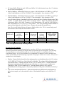
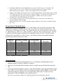
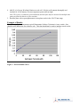
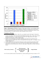
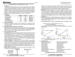
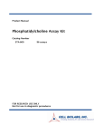
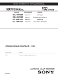
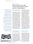
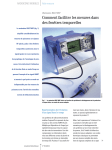
Product Manual OxiSelect™ Monoamine Oxidase Assay Kit (Colorimetric) Catalog Number XPX-5006 96 assays XPX-5006-5 5 x 96 assays FOR RESEARCH USE ONLY Not for use in diagnostic procedures Introduction Monoamine Oxidases (MAO) are a collection of flavin adenine dinucleotide oxidoreductase enzymes found in the outer mitochondrial membrane. MAOs catalyze the oxidative deamination of a variety of biogenic and xenobiotic amines. MAOs include MAO-A and MAO-B, which are two isoforms of the enzyme in mammals that have been distinguished based on localization, inhibitor, and substrate specificity. Both MAO-A and MAO-B are omnipresent throughout brain, liver, and other tissues. MAO-A is predominantly found in the liver, intestine, brain, and placenta, whereas MAO-B is found in the liver, brain, and platelets. MAOs primary function is to regulate neurotransmitters such as dopamine, noradrenaline, or serotonin. Dysfunction of MAO enzymes has been associated with many neurological disorders such as depression, drug abuse, migraines, schizophrenia, Attention Deficit Disorder (ADD), Parkinson’s disease, Alzheimer’s disease, as well as other disorders. Cell Biolabs’ OxiSelect™ Monoamine Oxidase Assay Kit is a simple HTS-compatible assay for measuring amine oxidase activity. The assay can detect both MAO activity and semicarbizidesensitive amine oxidases (SSAO). In order to discriminate between MAO-A and MAO-B, the MAO-A inhibitor Clorgyline and MAO-B inhibitor Pargyline are included. In addition, two substrates, Tyramine, and Benzylamine Hydrochloride, are included. Tyramine will react with MAO-A, MAO-B, and SSAO, while Benzylamine will react with MAO-B and SSAO. The two amine oxidase substrates are interchangeable in the assay, and coupled with the provided MAO inhibitors, can be used to determine MAO activity. Applications for the kit include measuring amine oxidase in tissues, blood samples, and screening for amine oxidase inhibitors or substrates. The kit has a detection sensitivity limit of 0.05 U/L. Each kit provides sufficient reagents to perform up to 96 assays, including standard curve and unknown samples. Assay Principle The OxiSelect™ Monoamine Oxidase Assay Kit is a simple and sensitive quantitative colorimetric assay for measuring amine oxidase activity in biological samples. The assay can be utilized for both end point and kinetic measurements of Monoamine Oxidase (MAO) activity, as well as semicarbazidesensitive amine oxidase (SSAO). Monoamine Oxidase reacts with its substrate and generates hydrogen peroxide (H2O2). In the presence of HRP, the Colorimetric Probe reacts with the H2O2 to produce a red/pink colored product. This product can be easily read by a microplate reader in the 540570 nm range. Absorbance values are proportional to the amine oxidase levels within the samples. Unknown samples are determined by comparison with a hydrogen peroxide standard curve. Clorgyline, a specific inhibitor of MAO-A, and Pargyline, a specific inhibitor of MAO-B, are included in the kit to differentiate between the two enzymes. The assay is simple, sensitive, and adaptable to high throughput testing. Related Products 1. STA-320: OxiSelect™ Oxidative DNA Damage ELISA Kit (8-OHdG Quantitation) 2. STA-341: OxiSelect™ Catalase Activity Assay Kit 3. STA-342: OxiSelect™ Intracellular ROS Assay Kit (Green Fluorescence) 4. STA-344: OxiSelect™ Hydrogen Peroxide/Peroxidase Assay Kit (Fluorometric) 5. STA-347: OxiSelect™ In Vitro ROS/RNS Assay Kit (Green Fluorescence) 2 6. STA-600: Phosphatidylcholine Assay Kit 7. STA-601: Sphingomyelin Assay Kit 8. STA-602: Acetylcholine Assay Kit (Fluorometric) 9. STA-844: OxiSelect™ Hydrogen Peroxide/Peroxidase Assay Kit (Colorimetric) 10. XPX-5000: OxiSelect™ Monoamine Oxidase Assay Kit (Fluorometric) Kit Components 1. 96-well Protein Binding Plate (Part No. 231001): One 96-well strip plate 2. 200X Colorimetric Probe (Part No. 239804): One 55 μL vial 3. HRP (Part No. 234402): One 100 μL tube of 100 U/mL solution in glycerol 4. Hydrogen Peroxide (Part No. 234102): One 100 μL amber tube of an 8.82 M solution 5. 100X Tyramine (Part No. 50001C): One 100 μL amber tube (substrate for MAO-A, MAO-B, and SSAO) 6. 100X Benzylamine (Part No. 50004C): One 100 μL amber tube (substrate for MAO-B and SSAO) 7. MAO-A Inhibitor (Part No. 50002C): One 50 μL amber tube of 20 mM Clorgyline solution 8. MAO-B Inhibitor (Part No. 50003C): One 50 μL amber tube of 20 mM Pargyline solution 9. 10X Assay Buffer (Part No. 234403): One 25 mL bottle Materials Not Supplied 1. Distilled or deionized water 2. 1X PBS for sample dilutions 3. 10 μL to 1000 μL adjustable single channel micropipettes with disposable tips 4. 50 μL to 300 μL adjustable multichannel micropipette with disposable tips 5. Multichannel micropipette reservoir 6. Spectrophotometric microplate reader capable of reading in the 540-570 nm absorbance range 7. Reagents and equipment necessary for sample preparation Storage Upon receipt, aliquot and store the, HRP, 100X Tyramine, 100X Benzylamine, MAO-A Inhibitor, and MAO-B Inhibitor at -20ºC. The Colorimetric Probe is light sensitive and must be protected accordingly; it may be stored at either -20ºC or -80ºC. Avoid multiple freeze/thaw cycles. Store all remaining kit components at 4ºC. Preparation of Reagents Note: All reagents must be brought to room temperature prior to use. 3 • 1X Assay Buffer: Dilute the stock 10X Assay Buffer 1:10 with deionized water for a 1X solution. Stir or vortex to homogeneity. • MAO-A Inhibitor: Immediately before use, prepare a 100 µM solution in 1X PBS (e.g. Add 5 µL of the 20 mM inhibitor to 0.995 mL 1X PBS). Vortex thoroughly. Store solutions at -20ºC. • MAO-B Inhibitor: Immediately before use, prepare a 100 µM solution in 1X PBS (e.g. Add 5 µL of the 20 mM inhibitor to 0.995 mL 1X PBS). Vortex thoroughly. Store solutions at -20ºC. • Assay Working Solution: Immediately before use, prepare an Assay Working Solution using Table 1 below as a guide based on the number of assays needed. Prepare by diluting the 200X Colorimetric Probe 1:100, 100X Tyramine or 100X Benzylamine 1:50, and 100 U/mL HRP to a final concentration of 0.2 U/mL in 1X Assay Buffer. The Assay Working Solution should be protected from light and used within 2 hours. Prepare only enough for immediate use. Note: The Assay Working Solution will appear slightly pink in color. This is normal background and should be subtracted from all absorbance values. 1X Assay Buffer (mL) 4.840 2.420 0.968 100X Tyramine or 100X Benzylamine (µL) 100 50 20 HRP (µL) 200X Colorimetric Probe (µL) 10 5 2 50 25 10 Total Volume of Assay Working Solution (mL) 5 2.5 1 Number of Assays in 96-well Plate (50 µL/well) 100 50 20 Table 1. Preparation of Assay Working Solution. Preparation of Samples Note: Samples should be assayed immediately or stored at -80ºC prior to performing the assay. Optimal experimental conditions for samples must be determined by the investigator. The following recommendations are only guidelines and may be altered to optimize or complement the user’s experimental design. A set of serial dilutions is recommended for samples to achieve optimal assay results and minimize possible interfering compounds. Run proper controls as necessary. Always run a standard curve with samples. • • Platelets: Prepare freshly drawn blood into polypropylene or polyethylene tubes with 1/10 volume of 1.5% EDTA/0.7% NaCl (e.g. 20 mL blood, 2 mL buffer). The volume of blood depends on the experimental needs. Centrifuge the tubes at 200 x g for 15 minutes at 4ºC. Carefully transfer the supernatant containing platelet rich plasma into clean tubes. Avoid carry over of erythrocytes. Centrifuge at 2000 x g for 10 minutes at 4ºC. Store the platelet pellet at -80ºC until use. Immediately before testing, prepare cell lysate by sonication of the pellet in 1X PBS (Youdim, Ref. 4). Cell or tissue mitochondrial fractions: Isolate mitochondria using differential centrifugation for cell or tissue samples, or by the method of choice. Mitochondrial samples can be diluted in 1X PBS. Notes: 4 • • • • All samples should be assayed immediately or stored at -80°C for up to 1-2 months. Run proper controls as necessary. Optimal experimental conditions for samples must be determined by the investigator. Always run a standard curve with samples. Samples with NADH concentrations above 10 μM and glutathione concentrations above 50 μM will oxidize the probe and could result in erroneous readings. To minimize this interference, it is recommended that superoxide dismutase (SOD) be added to the reaction at a final concentration of 40 U/mL (Tatyana et al, Ref. 2). Avoid samples containing DTT or β-mercaptoethanol since the probe is not stable in the presense of thiols (above 10 μM). Maintain pH between 7 and 8 for optimal working conditions as the probe is unstable at high pH (>8.5). Preparation of Standard Curve To prepare the H2O2 standards, first perform a 1:1000 dilution of the stock H2O2 in deionized water. Prepare only enough for immediate use (e.g. Add 5 μL of H2O2 to 4.995 mL deionized water). This solution has a concentration of 8.8 mM. Next, further dilute the 8.8 mM in deionized water to prepare a 1 mM solution (e.g. Add 114 μL of the 8.8 mM H2O2 solution to 886 μL deionized water). Use this 1 mM H2O2 solution to prepare standards in the concentration range of 0 µM – 100 µM by further diluting in 1X Assay Buffer (see Table 2 below). H2O2 diluted solutions and standards should be prepared fresh each time the assay is tested. Standard Tubes 1 2 3 4 5 6 7 9 1 mM H2O2 Standard (µL) 100 500 of Tube #1 500 of Tube #2 500 of Tube #3 500 of Tube #4 500 of Tube #5 500 of Tube #6 0 1X Assay Buffer (µL) 900 500 500 500 500 500 500 500 H2O2 (µM) 100 50 25 12.5 6.25 3.125 1.56 0 Table 2. Preparation of H2O2 Standards. Assay Protocol 1. Prepare and mix all reagents thoroughly before use. Each sample, including unknowns and standards, should be assayed in duplicate or triplicate. 2. Add 50 µL of each sample (H2O2 standard, control or sample) into an individual microtiter plate well. 3. If assaying with MAO inhibitors, add 5 µL of the 100 µM inhibitor to the appropriate MAO sample wells. Add 5 µL Assay Buffer to the H2O2 standards and samples without inhibitor. Mix the well contents thoroughly by pipetting or on a horizontal shaker and incubate 30 minutes at room temperature to allow the inhibitor to react with the enzyme. Note: The concentration of MAO-A or MAO-B inhibitors may be adjusted by the user. 5 4. Add 50 µL of Assay Working Solution to each well. Mix the well contents thoroughly and incubate for 45-60 minutes at room temperature protected from light. Note: This assay is continuous (not terminated) and therefore may be measured at multiple time points to follow the kinetics of the reactions. 5. Read the plate with a spectrophotometric microplate reader in the 540-570 nm range. Example of Results The following figures demonstrate typical Monoamine Oxidase Colorimetric Assay results. One should use the data below for reference only. This data should not be used to interpret actual results. Figure 1. H2O2 Standard Curve. 6 Figure 2. Measurement of MAO-A. 50 µg/mL of Monoamine Oxidase A was incubated with the MAO-A Inhibitor (Clorgyline) or MAO-B Inhibitor (Pargyline) according to the Assay Protocol. These were subsequently incubated with the substrates Tyramine or Benzylamine within the Assay Working Solution for 45 minutes and read with a microplate reader at 540 nm. Calculation of Results 1. Calculate the average absorbance values for every standard, control, and sample. Subtract the average zero standard value from itself and all standard and sample values. This is the corrected background absorbance. If sample background control value is high, subtract the sample background control value from the sample reading. 2. Plot the corrected absorbance for the H2O2 standards against the final concentration of the hydrogen peroxide standards from Table 2 to determine the best slope (µM-1). See Figure 1 for an example standard curve. 3. Use the standard curve to determine the hydrogen peroxide concentration generated by MAO. 4. Determine the monoamine oxidase enzyme activity of the samples using the equation below. Substitute the corrected fluorescence values for each sample. Remember to account for dilution factors. [H2O2] generated MAO Activity (Units/L) = x Sample dilution Reaction Time (minutes) Note: One unit of MAO catalyzes the formation of 1 µmole of hydrogen peroxide per minute at 25ºC. 7 References 1. Hall, D.W.R., et al. Biochem. Pharmacol. (1969) 18: 1447-1454. 2. Tatyana, V., et al. Neurochem. (2001) 79: 266. 3. Tipton, K. F., et al. Biochem. J. (1968) 108: 95-99. 4. Youdim, M.B. (1976) Preparation of Human Platelets. In Monoamine Oxidase and It’s Inhibition North-Holland, N.Y., 405-406. 5. Youdim, M.B., et al. Methods Enzymol. (1987) 142: 617-627. Warranty These products are warranted to perform as described in their labeling and in Cell Biolabs literature when used in accordance with their instructions. THERE ARE NO WARRANTIES THAT EXTEND BEYOND THIS EXPRESSED WARRANTY AND CELL BIOLABS DISCLAIMS ANY IMPLIED WARRANTY OF MERCHANTABILITY OR WARRANTY OF FITNESS FOR PARTICULAR PURPOSE. CELL BIOLABS’ sole obligation and purchaser’s exclusive remedy for breach of this warranty shall be, at the option of CELL BIOLABS, to repair or replace the products. In no event shall CELL BIOLABS be liable for any proximate, incidental or consequential damages in connection with the products. Contact Information Cell Biolabs, Inc. 7758 Arjons Drive San Diego, CA 92126 Worldwide: +1 858-271-6500 USA Toll-Free: 1-888-CBL-0505 E-mail: [email protected] www.cellbiolabs.com 2015: Cell Biolabs, Inc. - All rights reserved. No part of these works may be reproduced in any form without permissions in writing. 8