1
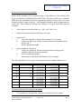
UNCONTROLLED DOCUMENT
IF PRINTED
15. DNA Quantitation
Purpose
This procedure provides a method for the quantitation of human DNA using the
Quantifiler® Duo kit and HID Real-Time PCR Analysis Software v1.0. This system is
capable of efficiently determining the relative quantities of male and human DNA in a
single reaction while detecting the presence of PCR inhibitors. The quantification results
can help guide downstream analyses and maximize the chances of obtaining optimal STR
results.
Background
The Quantifiler® Duo Quantitation Kit was designed for the simultaneous estimation of
human genomic and human male genomic DNA in forensic samples using real-time
PCR, and validated for use on the ABI PRISM® 7500 instrument. This assay detects the
Ribonuclease P RNA Component H1 (RPPH1) (human target) and the sex-determining
region Y gene (SRY) (human male target).
This system is based on sets of oligonucleotide PCR primers specific for a target DNA
sequence, plus a TaqMan® probe, labeled with a 5’ fluorescent reporter dye and a 3’ nonfluorescent quencher, that is homologous to the amplicon region between the PCR
primers. At the start of PCR thermal cycling, all TaqMan® probes are intact, and the
close proximity of the reporter dye and quencher suppress the fluorescence of the dye
molecules by Förster-type energy transfer. During the course of synthesizing new DNA
strands from the target template, the Taq polymerase enzyme encounters the TaqMan®
probe annealing to the template DNA and hydrolyzes it with its 5’ to 3’ exonuclease
activity. The hydrolysis of the probe separates the reporter dye and quencher, which
allows the dye molecules to fluoresce more intensely. This hydrolysis occurs in
proportion to the amplification of the target sequence, and therefore, the fluorescence
signal. Eventually the fluorescence signal crosses a specific threshold, defined as the
“Threshold Cycle,” or CT. It is at this point that the fluorescent signal has accumulated to
the point where it can be detected over background fluorescence. There is an inverse
mathematical relationship between the starting copy number of target sequence molecules
and the resulting CT, which allows samples to be quantitated reliably.
Safety
1.
Appropriate safety precautions should be taken and proper personal protective
equipment worn when working with hazardous or potentially hazardous materials.
The MSDS sheets and the laboratory safety manual should be consulted prior to
conducting these tests.
Santa Clara County Crime Laboratory
Forensic Biology Procedures Manual
DNA Quantitation
Page 1 of 8
Issue date: 11/16/2010
Rev. 2
UNCONTROLLED DOCUMENT
IF PRINTED
2.
All samples can potentially contain biological hazards and should be treated as
such.
Materials Required
Quantifiler® Duo kit
Teknova TE buffer
96-well reaction plate
Adhesive sealing supplies
Computer with HID Real-Time PCR Software v1.0
7500 Sequence Detection System
UV crosslinker
Quality Control/Calibration
1.
Quantifiler® Duo - Kits are subjected to a quality control test prior to use in
casework. This test evaluates the chemistry’s ability to accurate estimate DNA
quantities such that the profiles generated downstream are of sufficient amplitude
and quality. Details regarding this test can be found in the DNA QA/QC Manual.
2.
7500 Sequence Detection System - Routine calibration and maintenance is
performed on the 7500 instruments. The Instrument Maintenance Manager
provides step-by-step wizard-based instructions for each calibration; ROI,
Background, Optical, System Dye, and RNase-P Verification. The software
automatically analyzes the calibration and displays the results as pass or fail. The
software automatically records the date and time of each calibration. Details
regarding calibration and maintenance can be found in the DNA QA/QC Manual,
and in the Instrument Maintenance Manager.
3.
96-well plates mush be UV crosslinked at 250,000µJ for 12 minutes prior to use.
Procedure
Aliquoting Samples/Sample-Plate Association
1.
Aliquot 2-3 µL of each extract to be quantitated in 0.5 mL tubes labeled with the
well number that they are to be loaded in, and then place them in a rack that is
labeled with the run name in the set-up room freezer. Unless specified otherwise
by the analyst any remaining aliquot will be disposed of after the run.
2.
Samples placed in the rack should be added to the corresponding wells on the
Quant Duo Plate Template Excel Worksheet located on the network: Q:\7500
Runs\7500 Templates and Forms\HID v1 templates. Save the file with a unique
run name.
Santa Clara County Crime Laboratory
Forensic Biology Procedures Manual
DNA Quantitation
Page 2 of 8
Issue date: 11/16/2010
Rev. 2
UNCONTROLLED DOCUMENT
IF PRINTED
Preparation of Human DNA Standards
Human DNA standards will be prepared using a serial dilution of the standard (200
ng/μL) provided in the Quantifiler® kits and Teknova TE buffer (sterile water or dilution
buffer from the Quantifiler® Duo kit are appropriate options as long as they were tested
during the quality control of the kit). Fresh standards should be made up approximately
every week. These standards will range from 50 ng/μL to 23 pg/μL and will be prepared
as follows:
1.
Label eight microcentrifuge tubes (e.g., Std.1, Std.2, Std.3, etc.)
2.
Dispense the required amount of diluent to each tube.
3.
Prepare Std. 1:
a.
Vortex the Quantifiler® Human DNA Standard 3 to 5 seconds
b.
Using a new pipette tip, add the calculated amount of the standard to the
tube for Std. 1.
c.
Mix the dilution thoroughly.
4.
Prepare Standards 2 through 8:
a.
Using a new pipette tip, add the calculated amount of the prepared
standard to the tube for the next standard
b.
Mix the standard thoroughly.
c.
Repeat steps 4a and 4b until the dilution series is complete.
The following table serves as a guide in the preparation of the standard dilution series:
Standard
Concentration (ng/μL)
Std. 1
50
Std. 2
16.7
Std. 3
5.56
Std. 4
1.85
Std. 5
0.62
Std. 6
0.21
Std. 7
0.068
Std. 8
0.023
Recommended Amounts
50 μL standard +
150 μL TE buffer
50 μL standard +
100 μL TE buffer
50 μL standard +
100 μL TE buffer
50 μL standard +
100 μL TE buffer
50 μL standard +
100 μL TE buffer
50 μL standard +
100 μL TE buffer
50 μL standard +
100 μL TE buffer
50 μL standard +
100 μL TE buffer
Minimum Amounts
10 μL standard +
30 μL TE buffer
10 μL standard +
20 μL TE buffer
10 μL standard +
20 μL TE buffer
10 μL standard +
20 μL TE buffer
10 μL standard +
20 μL TE buffer
10 μL standard +
20 μL TE buffer
10 μL standard +
20 μL TE buffer
10 μL standard +
20 μL TE buffer
Dilution Factor
4X
3X
3X
3X
3X
3X
3X
3X
Preparing the Reactions/Loading the Instrument
1.
UV crosslink a 96-well plate for 12 minutes at 250,000 µJ for 12 minutes.
Santa Clara County Crime Laboratory
Forensic Biology Procedures Manual
DNA Quantitation
Page 3 of 8
Issue date: 11/16/2010
Rev. 2
UNCONTROLLED DOCUMENT
IF PRINTED
2.
To prepare the master mix (primer mix plus reaction mix) calculate the volume of
each component needed to prepare the reactions, using the table below. Include
additional reactions in your calculations to provide excess volume for the loss
during reagent transfers.
Component
Volume Per Reaction (μL)
Quantifiler® Primer Mix
10.5
Quantifiler® PCR Reaction Mix
12.5
3.
If necessary, thaw the primer mix completely, then vortex 3 to 5 seconds and
centrifuge briefly before opening the tube. Swirl the Quantifiler® PCR Reaction
Mix gently before using. Do not vortex the Quantifiler® PCR Reaction Mix.
4.
Pipette the required volumes of components into an appropriately sized
microcentrifuge tube.
5.
Vortex the master mix 3 to 5 seconds, then centrifuge briefly.
6.
Dispense 23 μL of the PCR mix into each assigned reaction well of a 96-well
reaction plate.
7.
Add 2 μL of sample, standard, or control to the appropriate wells. The standards
are to be run in duplicate.
8.
Seal the reaction plate with an Optical Adhesive Cover using an appropriate tool.
9.
Centrifuge the plate at 3000 rpm for approximately 5 minutes in the tabletop
unit equipped with plate holders to remove any bubbles.
10.
Place the sealed tray in the pass-through.
11.
Once in the post-amplification room, remove the tray from the pass-through.
12.
To load the plate, push in the button on the tray at the front of the instrument. The
tray will move out to accept the reaction plate. Ensure the plate is orientated
correctly with well A1 in the upper-left hand corner. Close the tray by pushing it
in until it clicks into position.
Initiating the Run
1.
Before a run is initiated, open the completed Quant Duo Plate Template Excel
Worksheet and select the tab at the bottom left corner that is labeled “Quant
Santa Clara County Crime Laboratory
Forensic Biology Procedures Manual
DNA Quantitation
Page 4 of 8
Issue date: 11/16/2010
Rev. 2
UNCONTROLLED DOCUMENT
IF PRINTED
Duo_data”. Save the document a final time as a Text (tab delimited) (*.txt) file
on the network.
2.
Open the HID Real-Time PCR Analysis v1.0 software and select the Quantifiler®
Duo assay icon from the home screen. This will open template with the standards
and negative control orientated in a horizontal fashion in the first two rows of the
plate. The run method, targets, and passive reference have been predefined in this
template and should not be altered.
3.
Specify an Experiment Name in the “Experiment Properties” section of the
“Setup” tab.
4.
Select “Import” from the file menu and Browse to locate the text file on the
network. Once located, select “Start Import”.
5.
When the import is complete, view the “Plate Setup” section of the “Setup” tab in
to confirm the sample information and well assignments.
6.
Any well that does not have a sample should be cleared. This can be
accomplished by selecting the well(s) in the “View Plate Layout” section, rightclicking, and selecting “Clear”.
7.
Save the document before initiating the run.
8.
Select the green “Start Run” icon in the top left quadrant of the “Run” tab.
Analyzing the Run and Assessing the Quality Indicators
1.
After the run is complete, turn off the instrument. Plates should be disposed of in
the biohazard bins in the post-amplification room.
2.
Analyze the run by selecting the green “Analyze” icon at the upper right hand
corner of the “Analysis” tab. Be sure the default analysis settings are used (i.e.,
Threshold: 0.2; Baseline Start Cycle: 3; Baseline End: 15).
3.
Verify the three quality indicators as specified below:
a.
b.
Slope – this quality indicator indicates the amplification efficiency of the
standard reactions. The slope should range from -2.9 to -3.6 (-3.3
indicates 100% amplification efficiency). If the slope falls outside this
range, the run may need to be repeated.
R2 – the correlation coefficient indicates the statistical significance of the
standard curve. A passing run should have an R2 value greater than 0.98.
Santa Clara County Crime Laboratory
Forensic Biology Procedures Manual
DNA Quantitation
Page 5 of 8
Issue date: 11/16/2010
Rev. 2
UNCONTROLLED DOCUMENT
IF PRINTED
c.
4.
If the correlation coefficient falls below this value, the run may need to be
repeated.
Y-intercept – this value indicates the expected CT value for a sample with
quantity of 1. A running list of Y-intercept values is kept in logs adjacent
to each instrument. The Y-intercept for a given run should be similar to
those from previous runs. This list can be used as a tool to troubleshoot
potential quantitation issues.
Up to two nonconcordant points may be removed from the standard curve in order
to make minor adjustments should the slope and/or R2 fall out of range. The two
points removed should be of differing concentrations. Any standards removed
should be noted on the Quantifiler® Worksheet in the Comments section.
Exporting the Results
1.
Following data analysis, the results should be exported to the Quantifiler®
Worksheet, which is located on the network: Q:\7500 Runs\7500 Templates and
Forms\HID v1 templates.
2.
Select “Export” from the application toolbar.
3.
On the “Export Properties” tab, specify the “Export File Name,” set the file type
to “*.xls” and designate the Export File Location on the network. The default
settings (i.e., “Results” in the field “Select Data to Export,” “One File” in the field
“Select One File or Separate Files”) in this tab should not be altered.
4.
On the Customize Export Tab, make sure “Well, Sample Name, Target Name,
Task, CT, CT SD, Quantity and M:F Ratio” in the “Select Result Content” are
checked, and appear in that order in the “Results Export” table. These are the
default settings and should not need alteration.
5.
Select “Start Export”.
6.
Open the “*.xls” export file from the network and select the complete set of cells
or rows to be copied to the Quantifiler® Worksheet.
7.
Open the Quantifiler® Worksheet template, select the tab titled “Put SDS Data
Here”, and paste the data from the export file beginning in row A16.
8.
Select the tab titled “Quantitation” to view the results.
9.
The fields at the top of the Quantifiler® Worksheet need to be manually entered.
This information includes the quality indicator values, lot numbers, case number,
analyst, date, run name, comments section, and instrument name.
Santa Clara County Crime Laboratory
Forensic Biology Procedures Manual
DNA Quantitation
Page 6 of 8
Issue date: 11/16/2010
Rev. 2
UNCONTROLLED DOCUMENT
IF PRINTED
10.
Print the results for inclusion in the case note packet. It is only necessary that this
record include the applicable case information.
Limitations/Notes
1.
When preparing the standards keep in mind that DNA quantification standards are
crucial for accurate analysis of run data. Any mistakes or inaccuracies in making
the dilutions directly affect the quality of results. The quality of pipettors and tips
and the care used in measuring and mixing dilutions affect accuracy.
2.
Samples with a quantitation value less than 200 ng/µL may be used to estimate
target volumes for amplification. Samples with values greater than 200ng/µL
should be diluted and requantitated.
3.
All samples that are potentially degraded and/or inhibited should be amplified
regardless of the quantitation value
4.
All samples selected for Yfiler® testing will be amplified regardless of the
quantitation value.
5.
Samples with a male quantitation value but a zero human quantitation should still
be amplified with Identifiler® Plus.
6.
It may be appropriate in certain circumstances to terminate analysis when the
human and male quantitation values are zero. When making this decision, the
analyst should carefully consider the sample type, expected levels of DNA,
probative nature of the sample, and the results of other samples in the case. The
decision to terminate analysis must be approved by a Supervising Criminalist in
the DNA Unit. The Supervising Criminalist will document this approval by
initialing and dating the quantitation worksheet. Additionally, the justification for
terminating analysis must be clearly documented on the worksheet.
7.
Samples with IPC CT values of 31 or greater will be automatically highlighted on
Quantifiler® Worksheet to indicate that the extract may be inhibited. Samples
with high concentrations may also have IPC CT values of 31 or greater and should
not be confused with an inhibited sample. If sample volume is not a limiting
factor, the inhibited sample should be diluted and requantitated. If requantitation
will limit amplification opportunities, the inhibition should be carefully
considered during the amplification step. It should be noted that inhibition seen
during real-time PCR is not an absolute indicator that there will be inhibition seen
in downstream amplifications.
Santa Clara County Crime Laboratory
Forensic Biology Procedures Manual
DNA Quantitation
Page 7 of 8
Issue date: 11/16/2010
Rev. 2
UNCONTROLLED DOCUMENT
IF PRINTED
8.
The Quantifiler® Duo kit simultaneously quantitates male and female DNA in
DNA mixtures. The male:female ratio can be used to dictate case approach.
Validation studies indicated that the minor component typically drops out in
male:female ratios greater than 1:20.
References
Quantifiler Duo User’s Manual
7500 User’s Manual
HID Software version 1.0 User’s Manual
Documentation
Quantifiler® Duo plate setup worksheet
Quantifiler® Duo worksheet
Revision history:
Revision No.
0
1
2
Date
11/16/2010
2/16/2011
3/15/2011
Approved By
L Burley
L Burley
L Burley
2
3/15/2011
L Burley
Santa Clara County Crime Laboratory
Forensic Biology Procedures Manual
DNA Quantitation
Revisions
Original
Specified current version of HID Real-time software (v1.0)
Added UV crosslinker to materials; specified 96-well plates
must be crosslinked prior to use.
Changed Identifiler®to Identifiler®Plus on page 6.
Page 8 of 8
Issue date: 11/16/2010
Rev. 2