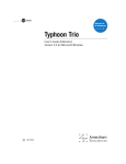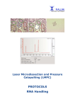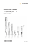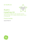
1
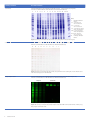
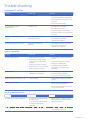
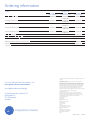
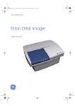
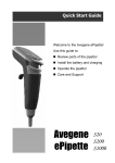
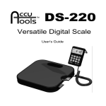
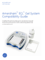
GE Healthcare Life Sciences Amersham™ ECL™ Gel System Compatibility Guide This guide provides useful hints and tips on how to use Amersham ECL Gel system for SDS-PAGE. It contains illustrative examples of system performance, optimized Western blotting transfer protocols, as well as trouble shooting guidelines to help you achieve optimal results. imagination at work Compatibility Guide Electrophoresis applications Recommendations Protein SDS-PAGE The content of this guide will focus on SDS-PAGE conditions Amersham ECL Gel is available in 10, 15 and 2 well formats, and as either homogeneous (10 and 12%), or gradient (4-12%, 8-16%, and 4-20%) gels. Homogenous and 8-16% gradient gels have a 4% stacking gel. Gel buffer is Tris-HCL pH 6.3. %C=2.6% (w/w); %T=Total concentration (w/v) of acrylamide+ bisacrylamide is given as percentage in the respective Amersham ECL Gel product name. Running buffer: Amersham ECL Gel Running Buffer; 25 mM Tris, 192 mM Glycine, 0.1% SDS, pH 8.3. Protein Native PAGE Running buffer: 25 mM Tris, 192 mM glycine, pH 8.3. See figure 16 for example result. DNA/RNA PAGE Running buffer: 25 mM Tris, 192 mM glycine, pH 8.3. Preparing Amersham ECL Gel Box Recommendations Power supply Recommended power supply is EPS 301. See ECL Gel Box User Manual (28-9943-33) for more information on power supply compatibility. Running buffer Running buffer for SDS-PAGE: Amersham ECL Gel Running buffer ; 25 mM Tris, 192 mM glycine, 0.1% SDS, pH 8.3. Note: It is important to follow buffer volume recommendations; 90 ml running buffer in each buffer chamber (Figure 1), and 6 ml running buffer in the sample well chamber (Figure 2). Figure 1. Add 90 ml of Tris-GlycineSDS running buffer to each buffer container. Figure 2. Add 6 ml of Tris-Glycine-SDS running buffer to the sample well container. Note: Use of non-Tris Glycine based buffers such as MES or MOPS buffer for Tris-HCL based Amersham ECL Gels will have a negative impact on results (Figure 3). 1 2 3 4 5 6 7 8 9 10 1 2 3 4 5 6 7 8 9 10 Lanes 1. Full-Range Rainbow™ Marker 2-4. E. coli cell Lysate (10ug) 5-7. CHO cell lysate (10ug) 8-10.HeLa cell lysate (10ug) Figure 3. E. coli, CHO cell, and HeLa lysates separated on Amersham ECL Gel, 4-20%, using Amersham ECL Gel running buffer (Tris-Glycine-SDS) (a) and NuPAGE® MES SDS buffer (Invitrogen) (b). MES buffer is suitable for Bis-Tris gels only, and has a negative impact on separation using Tris-HCL based Amersham ECL Gel. Running temperature Amersham ECL Gel should be stored at +2 to +8°C. Amersham ECL Gel Running Buffer can be stored and used at room temperature (+15 to +25 °C), (Figure 4). Note Remove gel from refrigerator just prior use. For optimal resolution, gels should be cold when run in Amersham ECL Gel system. 1 2 3 4 5 6 7 8 9 10 1 2 3 4 5 6 7 8 9 10 Lanes 1-4. HeLa cell lysate 6-10. Low Molecular Weight (LMW) Marker Figure 4. Separation on Amersham ECL Gel, 4-20% using cold (left) and room tempered (right) running buffer. The temperature of the running buffer does not affect the separation. 2 29-0251-27 AA Sample loading Recommendations Sample loading buffer Tris-HCl-SDS or other buffer suitable for the application. 1 2 3 4 5 6 7 8 Lanes 1-2. CHO cell Lysate, Tris-HCL-SDS sample loading buffe 3-4. E. coli lysate, Tris-HCL-SDS sample loading buffe 5-6. CHO cell lysate, Novex Tricine Sample buffer 7-8. E. coli lysate, Novex Tricine Sample buffer Figure 5. CHO cell, E. coli, and HeLa cell lysates (10 µg total protein) separated on Amersham ECL Gel, 10%, using Tris-HCL-SDS sample loading buffer, and Novex® Tricine SDS Sample Buffer (Invitrogen). The results demonstrate that protein in Tricine based sample loading buffer may result in slightly affected resolution of protein bands compared to proteins in Tris-HCL based SLB. Salt concentration High salt concentration in sample may result in vertical streaking of bands. Use gel filtration (e.g. Vivaspin Sample Concentrators) for buffer exchange if needed. Note: In general, concentration of imidazole up to 20 mM does not affect protein separation. 1 2 3 4 5 6 7 8 Lane 1. Low molecular weight (LMW) markers 2. Start material, MBP-(His)6 in E. coli lysate 3. Flow through, 5 mM imidazole 4. Flow through, 10 mM imidazole 5. Flow through, 20 mM imidazole 6. Eluted sample, 5 mM imidazole during binding 7. Eluted sample, 10 mM imidazole during binding 8. Eluted sample, 20 mM imidazole during binding Figure 6. Samples from flow through and elution fractions containing different concentrations of imidazole are separated on Amersham ECL gels. The result shows that concentrations of imidazole up to 20 mM do not affect the protein separation or cause vertical streaking. Sample well volume 10 wells; 35 µl, 15 wells; 20 µl, 2 wells; 100 µl. Protein amount Maximum of 0.5 µg protein/band per well can be loaded. Overloading may cause smearing and distortion (Figure 7). Figure 7. Example of band broadening and smearing due to overloading. Plasma fractions in an IgG purification process, stained with Deep Purple™ Total Protein Stain. Pipette tips Use regular pipette tips for sample loading. Flexible gel loading tips should not be used. Note: Be careful not to thrust the pipette tip down into the well when loading. This may cause the well bottom to break and leak. Recommendations Use a prestained marker, e.g. Rainbow Molecular Weight markers, to monitor separation, and confirm transfer from gel to membrane. Empty wells Load an equal volume of sample loading buffer into any unused well to ensure uniform mobility. 29-0251-27 AA 3 Staining methods Recommendations Coomassie Amersham ECL Gel works well with standard Coomassie® staining protocols (Figure 8). See also recommendations in ECL Gel Box User Manual, (28-9943-33). 1 2 3 4 5 6 7 8 9 10 11 12 13 14 15 Lanes 1. Full-Range Rainbow Marker 3-4. CHO cell Lysate, Tris-HCL-SDS SLB 5-6. E. coli lysate, Tris-HCL-SDS SLB 7-8. CHO cell lysate, Novex Tricine Sample buffer 9-10. E. coli lysate, Novex Tricine Sample buffer 11-13.HeLa cell lysate, Tris-HCL-SDS SLB 15. Full-Range Rainbow Marker Figure 8. Staining of Amersham ECL Gel, 4-20%, using SimplyBlue™ Safestain (Invitrogen). Silver stain Amersham ECL Gel works well with PlusOne™ Silver Staining Kit, Protein (Figure 9). Follow recommendations in ECL Gel Box User Manual (28-9943-33). A B C D E F G H I J Figure 9. Staining of Amersham ECL Gel, 4-20%, using PlusOne™ Silver Staining Kit, Protein. Dilution series from 15.6-0.03 ng protein in 50 kDa band. Deep Purple™ Stain Amersham ECL Gel works well with Deep Purple Total Protein Stain, RPN6305 (Figure 10). Follow recommendations in ECL Gel Box User Manual (28-9943-33). Cell lysate Transferrin 500 ng 250 ng 125 ng 62.5 ng 500 ng 250 ng 125 ng 62.5 ng Figure 10. Staining of complex and purified samples after SDS-PAGE on Amersham ECL Gel, 4-20%, using Deep Purple Total Protein Stain. 4 29-0251-27 AA Western blotting transfer protocols Tank transfer Recommended running conditions using TE 22 Tank transfer unit Parameter Value Voltage Constant voltage: 25 V Current (A) and power (W): Non-limited Note: Voltage may be adjusted depending on protein size and characteristics. Temperature 4°C Note: Time 2.5 h Note: Time may be adjusted depending on protein size and characteristics. Perform the transfer in a cold room. Use a magnetic stirrer to ensure buffer circulation. 1. Use pre-chilled transfer buffer Tris-glycine buffer: 25 mM Tris, 192 mM glycine, 20% methanol, pH 8.3. Prepare fresh, and pre-chill to reduce heating. Hints & Tips: Lower methanol concentration (10%), and addition of 0.1% SDS may improve transfer of large proteins. 2. Equilibrate the gel Equilibrate the gel in cold transfer buffer for >20 minutes. 3. Follow step-by-step protocol for tank transfer outlined in Amersham ECL Gel Box User Manual (28-9943-33). Gel ERK ½ (42, 44 kDa) Gel Stat3 (79, 86 kDa) Amersham ECL Gel, 10% Amersham ECL Gel, 10% Novex Tris-Glycine Gel, 12% (Invitrogen) Novex Tris-Glycine Gel, 12% (Invitrogen) Samples: HeLa cell lysate in a 2-fold dilution series starting at 5 µg. Samples: HeLa cell lysate in a 2-fold dilution series starting at 5 µg. Figure 11. Tank transfer with subsequent Western blotting detection of ERK1/2 (left) and Stat3 (right). Comparison shows similar performance between Amersham ECL Gel, 10%, and Novex Tris-Glycine Gel, 12% (Invitrogen). Gel electrophoresis and transfer was performed in triplicates according to the manufacturer´s instructions. Semidry transfer Recommended running conditions using TE 77 semidry transfer unit Parameter Value Current 0,8 mA/cm2 Time 80 minutes Note: Recommended transfer time is for mid-size proteins. The time may be longer for larger proteins (90min), and shorter for small proteins (60min). 1. Transfer buffer Tris-glycine buffer: 25 mM Tris, 192 mM glycine, 20% methanol, 0.1% SDS, pH 8.3. Prepare fresh. Hints & Tips: A discontinuous buffer system may improve transfer efficiency. Use 25 mM Tris, 192 mM glycine, 20% methanol in anode buffer, and 25 mM Tris, 192 mM glycine 0.1% SDS in cathode buffer. 2. Equilibrate the gel Equilibrate the gel in cold transfer buffer for 20 minutes. Note: Equilibration in transfer buffer removes SDS from the gel This preserves the shape of the gel, and prevents the gel from sticking to the membrane. 3. Follow step-by-step protocol outlined in TE 77 User Manual (28-4025-91). Transfer method Small to mid-size protein ERK ½ (42 kDa) Transfer method Mid-size to large protein Transferrin (79 kDa) Semidry transfer using TE 77;80 min Semidry transfer using TE 77; 90 min Tank transfer using TE 22; 25 V, 2.5 h Tank transfer using TE 22; 25 V, 2.5 h Samples: HeLa cell lysate in a 2-fold dilution series starting at 2,5 µg. Samples: Transferrin in a 2-fold dilution series starting at 5 ng. Figure 12. A comparison of semidry and tank transfer using the Amersham ECL gels shows similar results in Western blotting detection of ERK1/2 (left) and transferrin (right). 29-0251-27 AA 5 Typical results In this section, typical appearance of Amersham ECL Gel, before, during, and after electrophoresis is illustrated. Examples from protein separation under native and denatured conditions respectively are provided, as well as an example of DNA separation. Removing the comb: Excess gel pieces Native conditions: Blue Native PAGE using Amersham ECL Gel Figure 14. As the comb is removed, excess gel pieces can be detatched from the well container. This will not affect gel performace, but pieces should be removed to facilitate sample loading. Figure 16. Blue Native data kindly provided by Prof. Dr. H.P. Braun, Institute of Plant Genetics, Leibniz University Hannover. Performing the separation: Appearance of front during run • Ready-made gel: Amersham ECL gel 4-12%, 10 wells • Cathode buffer: 50 mM Tricine, 15 mM Bis-Tris, 0.02% w/v Coomassie G-250 (90 ml/run) • Anode buffer: 50 mM Tricine, pH 7.0 (90 ml/run) • Thylakoid membrane protein complexes of Arabidopsis thaliana were solubilized with 5 % (w/v) digitonin in solubilization buffer (150 mM K-Acetate, 30 mM HEPES, 10 % v/v glycerol, pH 7.4). Detergent:clorophyll ratio was 50:1. Unsolubilized material was removed by centrifugation at 18000 g for 10 min. Each well was loaded with solubilized membrane protein complexes corresponding to 10 μg chlorophyll. Prior loading 1 μl of 5 % (w/v) Coomassie G-250 in 750 mM amino-caproic acid was added to each sample and the sample volume was adjusted to 21 μl using solubilization buffer without digitonin. All step were carried out on ice/at 4 °C. • Running conditions: Pre-run 12 min at 160 V prior to sample loading, then 120 min at 160 V at room temperature • Poststain: Fixation solution [40% (v/v) Methanol, 10% (v/v) acetic acid in ddwater] over night. Visualization of proteins colloidal Coomassie staining (Neuhoff 1988) acc. Wittig, I., Braun, H.P. and Schägger, H. (2006): Blue-Native PAGE. NATURE Protocols 1, 418-428. Nucleic acid separation on Amersham ECL Gel 2 500 bp Figure 15. As the gel is run in Amersham ECL Gel box, the front may appear less straigth than expected (upper). This typically has no effect on resulting separation after completion of the electrophoresis run (lower). 512 bp 500 bp 36 bp Figure 17. Example of DNA separation on Amersham ECL Gel, 4-12%, run at native conditions with Tris-Glycine buffer. 6 29-0251-27 AA Trouble shooting Amersham ECL Gel Box Symptom Possible cause Remedies Low or no current during run Incomplete circuit • Add 90 ml running buffer to each tank, and 6 ml running buffer in well chamber • Ensure that the tape is removed from the gel cassete • Ensure that the lid is in place No green light, low current (<20 mA) at start Incomplete circuit • Check all power connections • Check buffer level in tanks • Ensure that the gel cassette is in place • Ensure that the tape is removed from the gel cassete Green light, but only approx 35 mA at start Leakage between tanks, with or without gel cassette • Check that the tanks are not overfilled Green light, but approx 65 mA at start Leakage between tanks, with gel cassette • Check that the tanks are not overfilled Symptom Possible cause Remedies Vertical streaking of bands • Sample overload, see Figure 4 • Load maximum of 0.5 μg protein/band per well • Check if the unit is damaged and needs to be replaced • Check if the unit is damaged and needs to be replaced Protein separation • Poorly soluble or weakly charged particles (such as carbohydrates) in sample Poor separation • Cetrifuge the amples before loading • Change pH of sample buffer • High salt concentration in sample • Heat sample together with SDS • Contaminants such as cell membrane or DNA complexes in sample • Decrease salt concentration using gel filtration • Incorrect ECL Gel type selected • Use an ECL Gel type that separates in the desired molecular weight range • Separation time not sufficient • Increase the electrophoresis run time Bands spreading horizontally across gel • Sample overload • Load maximum of 0.5 µg protein/band per well Smiling effects across gel • Uneven heating of gel • Use cold gels and pre-chilled running buffer to reduce heating effects • Run gels at lower voltage for longer time; e.g. 125 V for 90 minutes Western blotting transfer Symptom Possible cause Remedies No or little protein transfer to membrane • Too short transfer time/ low voltage; Proteins left in gel • Optimize transfer conditions, starting from recommended transfer conditions outlined in this guide • Too long transfer time/high voltage; Proteins passed through the membrane Gel sticking to membrane after transfer • Unsufficient equilibration of gel • Equilibrate gel in transfer buffer >20 min before tranfsfer step 29-0251-27 AA 7 Ordering information Product Product code 2 wells Pack of 10 gels 10 wells Pack of 10 or 2 gels 15 wells Pack of 10 gels Amersham ECL Gel 10% 28-9901-60 28-9898-04 28-9898-08* 28-9901-55 Amersham ECL Gel 12% 28-9901-61 28-9898-05 28-9898-09* 28-9901-56 Amersham ECL Gel 4-12% 28-9901-62 28-9898-06 28-9901-51* 28-9901-57 Amersham ECL Gel 8-16% 28-9901-63 28-9898-07 28-9901-52* 28-9901-58 Amersham ECL Gel 4-20% 28-9901-64 28-9901-54 28-9901-53* 28-9901-59 Amersham ECL Gel Box 28-9906-08 Amersham ECL Gel Running Buffer 29-9902-52 PlusOne Silver Staining Kit, Protein 17-1150-01 Full-Range Rainbow Molecular Weight marker RPN800E EPS 301 18-1130-01 * 2 gel pack For local office contact information, visit www.gelifesciences.com/contact www.gelifescience.com/eclgel GE, imagination at work, and GE monogram are trademarks of General Electric Company. Amersham, Deep Purple, ECL, Rainbow, and PlusOne are trademarks of GE Healthcare companies. Deep Purple Total Protein Stain is exclusively licensed to GE Healthcare from Fluorotechnics Pty Ltd. Deep Purple Total Protein Stain may only be used for applications in life science research.Deep Purple is covered under a granted patent in New Zealand entitled “Fluorescent Compounds”, patent number 522291 and equivalent patents and patent applications in other countries. Coomassie is a trademark of Imperial Chemical Industries Limited. NuPAGE, Novex and Simply Blue are trademarks of Life Technologies Corporation. © 2012 General Electric Company – All rights reserved. First published July 2012. GE Healthcare Bio-Sciences AB Björkgatan 30 751 84 Uppsala Sweden All goods and services are sold subject to the terms and conditions of sale of the company within GE Healthcare which supplies them. A copy of these terms and conditions is available on request. Contact your local GE Healthcare representative for the most current information. GE Healthcare UK Limited Amersham Place Little Chalfont Buckinghamshire, HP7 9NA UK GE Healthcare Europe, GmbH Munzinger Strasse 5 D-79111 Freiburg Germany imagination at work GE Healthcare Bio-Sciences Corp. 800 Centennial Avenue, P.O. Box 1327 Piscataway, NJ 08855-1327 USA GE Healthcare Japan Corporation Sanken Bldg., 3-25-1, Hyakunincho Shinjuku-ku, Tokyo 169-0073 Japan 29-0251-27 AA 07/2012