1
APPENDICES
TECHNICAL ARTICLES
RECORDING ARTERIAL AND VENOUS FLOW VELOCITIES
Although the following recordings were made with a Model 806-C Doppler, they all apply to all
directional Dopplers of our manufacture using the 8 MHz probe. It is usually better to use stereo
earphones when doing carotid and small-vessel work because the stereo effect lets you know when
the ultrasound beam is intercepting more than one vessel. To minimize this, keep the length of
the probe crystals in line with the vessel being studied. The probe body must NOT be
perpendicular to the vessel. An adjacent vein may unavoidably interfere with a small artery and
reduce the quality of the recording.
The zero-flow line is obtained by pressing down the ZERO/CAL button. Reposition the stylus if
necessary while holding this button down. If you change the OUTPUT FILTER-Hz control, check
the zero-flow line again.
If you are pointing the probe toward the direction of flow which may reverse during the cardiac cycle,
it is suggested you set the zero-flow line two major divisions from the bottom (right side) of the
paper. The DOPPLER RECORDING button should be in the NORMAL position so that flow toward
the probe causes an upstroke and flow away from the probe causes a downstroke (below the zeroflow line).
If you want the recording reversed in polarity, push the DOPPLER RECORDING selector to indicate
INVERT. Use the 28 Hz position of the OUTPUT FILTER-Hz selector for best representation of
velocity changes, but the recording will be ragged. Too much filtering will eliminate recording of a
momentary reversal of flow. If the flow is not going to reverse during the cardiac cycle, use the
NORMAL or INVERT position of the DOPPLER RECORDING selector, depending on whether flow
is toward or away from the probe. The quality of the recordings will be better.
Please keep in mind that these recordings are primarily meant to help you understand and therefore
effectively use our instruments. Diagnostic information MUST come from other doctors and the
scientific literature.
SUPRAORBITAL ARTERY
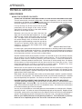
The most difficult vessel of interest to record from is the supraorbital artery. This recording is
of a normal right supraorbital artery using the 10 MHz pencil probe almost flat against the cheek.
With pathology, the recording would not be so clean. Three things are important in assessing
this recording. First, a rapid upstroke is indicative of blood moving very quickly, something
that doesn’t happen when small-vessel collaterals supply the flow. Second, the presence of a
dicrotic notch is important, for the same reasons just cited. Third, the flow is in the right directioncoming out of the eye socket. Fourth, there is considerable flow. The flow is the area under
the curve to the zero flow line, not just the pulse. No augmentation can be obtained with temporal
artery compression on this side. Lack of augmentation does not necessarily mean carotid
obstruction.
2100-SX, US 05M Appendices, Basics, Tech Arts-1.0 10/25/06
PARKS Flo-Lab Operating Manual
V . 13