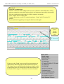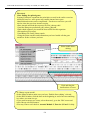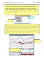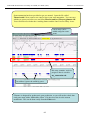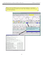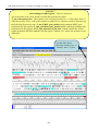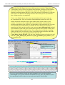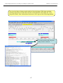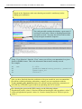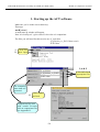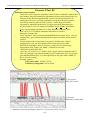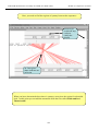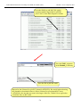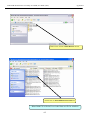
1
Index
WHO/TDR Bioinformatics Workshop at ICGEB, New Delhi (2005)
WHO/TDR Bioinformatics Workshop at ICGEB, New Delhi (2005)
Index
Page
Index ……………………………………………………………………………. 1
Module 1: Artemis Prokaryotic………………………………………………. 2
Exercise 1
3
Module 2:
Artemis Eukaryotic……………………………………………….. 10
Exercise 1
11
Module 3:
Artemis Advanced ……………………………………………….. 15
Exercise 1
16
Module 4:
Gene Prediction ………………………………………………… 28
Exercise 1
29
Exercise 2
30
Exercise 3
33
Exercise 4
34
Exercise 5
36
Exercise 6
41
Exercise 7
45
Module 5:
Small Scale Annotation ………………………………………… 49
Exercise 1
51
Exercise 2
52
Module 6:
Comparative Genomics …………………………………………
Exercise 1
Exercise 2
Exercise 3
Exercise 4
53
56
60
65
68
Module 7:
Generating ACT comparison files using BLAST ………..……
Exercise 1
Exercise 2
72
72
79
References ………………………………………………………………...
84
Appendices …………………………………………………………………
85
-1-
WHO/TDR Bioinformatics Workshop at ICGEB, New Delhi (2005)
Module 1: Artemis Prokaryotic
Module 1
Artemis: Prokaryotic
Introduction
Artemis (Rutherford et al., (2000) is a DNA viewer program, written by Kim Rutherford, and
used for both Prokaryotic and Eukaryotic annotations. It allows the user to get away from the
relatively faceless EMBL and Genbank style database files and view the sequence in a graphical
and highly interactive format. Artemis is designed to present multiple lines of information within
a single context. This manifests itself as being able to zoom in to look for fine DNA motifs as
well as being able to zoom out and bring into view operons, several kilobases of a genome or in
fact to view an entire genome in one screen. It is also possible to perform quite sophisticated
analyses and store the output within the 'Artemis environment' to be accessed later.
Aims
The aim of this Module is for you to become familiar with the basic functioning of Artemis by
using a series of worked examples. These examples are designed to take you through the most
immediately useful functions. However, there will be time, and encouragement, for you to
explore other menus and gain a basic understanding of Artemis. Like all the Modules in this
workshop, the key is ‘if you don’t understand please ask’.
-2-
Module 1: Artemis Prokaryotic
WHO/TDR Bioinformatics Workshop at ICGEB, New Delhi (2005)
Artemis Exercise 1 Part I
1.
Starting up the Artemis software
Navigate your way into the correct directory for this module
Then type:
art & [return]
A small start-up window will appear (see below).
Now follow the sequence of numbers to load up the Salmonella typhi chromosome sequence.
Ask a demonstrator for help if you have any problems.
1
2
Click ‘File’
then ‘Open
In the ‘Options’ menu
you can switch between
prokaryotic and
eukaryotic mode.
For simplicity it is a good idea to
open a new start up window for
each Artemis session and close
down any sessions once you have
finished an exercise.
3
Single click
to select
DNA file
4
Single click to open file in Artemis then wait
-3-
DNA sequence files
will have the suffix
‘.dna’. Annotation
files end with ‘.tab’.
WHO/TDR Bioinformatics Workshop at ICGEB, New Delhi (2005)
Module 1: Artemis Prokaryotic
2. Loading annotation files (entries) into Artemis
Hopefully you will now have an Artemis window like this! If not, ask a demonstrator for
assistance.
Now follow the numbers to load up the annotation file for the Salmonella typhi chromosome.
1
Click ‘File’ then
‘Read an Entry’
Entry = file
2
Single click
to select tab
file
3 Single click to open file in Artemis then wait
What’s an “Entry”? It’s a file of DNA and/or amino acid features which can be overlaid
onto the sequence information displayed in the main Artemis view panel.
-4-
WHO/TDR Bioinformatics Workshop at ICGEB, New Delhi (2005)
Module 1: Artemis Prokaryotic
3. The basics of Artemis
Now you have an Artemis window open let’s look at what’s in there.
1
2
3
6
4
7
8
5
1.
2.
3.
4.
5.
6.
7.
8.
Drop-down menus. There’s lots in there so don’t worry about them right now.
Shows what entries are currently loaded (bottom line) and gives details regarding the
feature selected in the window below; in this case gene STY0003 (top line).
This is the main sequence view panel. The central 2 grey lines represent the forward
(top) and reverse (bottom) DNA strands. Above and below those are the 3 forward
and 3 reverse reading frames. Stop codons are marked as black vertical bars. Genes
and other features (eg. Pfam and Prosite matches) are displayed as coloured boxes.
We will refer to genes as coding sequences or CDSs from now on.
This panel has a similar layout to the main panel but is zoomed in to show
nucleotides and amino acids. Double click on a gene in the main view to see the
zoomed view of the start of that gene. Note that both this and the main panel can be
scrolled left and right (7, below) zoomed in and out (6, below).
This panel lists the various features in the order that they occur on the DNA with the
selected gene highlighted. The list can be scrolled (8, below).
Sliders for zooming view panels.
Sliders for scrolling along the DNA.
Slider for scrolling feature list.
-5-
WHO/TDR Bioinformatics Workshop at ICGEB, New Delhi (2005)
Module 1: Artemis Prokaryotic
4. Getting around in Artemis
The 3 main ways of getting to where you want to be in Artemis are the Goto dropdown menu,
the Navigator and the Feature Selector. The best method depends on what you’re trying to do
and knowing which one to use comes with practice.
4.1 The ‘Goto’menu
The functions on this menu (ignore the Navigator for now) are shortcuts for getting to locations
within a selected feature or for jumping to the start or end of the DNA sequence. This one’s
really intuitive so give it a try!
Click ‘Goto’
It may seem that ‘Goto’ ‘Start of Selection’ and ‘Goto’ ‘Feature Start’ do the same thing. Well
they do if you have a feature selected but ‘Goto’ ‘Start of Selection’ will also work for a region
which you have highlighted by click-dragging in the main window. So yes, give it a try!
Suggested tasks:
1.
Zoom out, highlight a large region of sequence by clicking the left hand button and
dragging the cursor then go to the start and end of the highlighted region.
2.
Select a gene then go to the start and end.
3.
Go to the start and end of the genome sequence.
4.
Select a gene. Within it, go to a base (nucleotide) and/or amino acid of your choice.
-6-
WHO/TDR Bioinformatics Workshop at ICGEB, New Delhi (2005)
Module 1: Artemis Prokaryotic
4.2 Navigator
The Navigator panel is fairly intuitive so open it up and give it a try.
Click ‘Goto’
then Navigator
Check that
the search
button is on
Suggestions of where to go:
1.
Think of a number between 1 and 4809037 and go to that base (notice how the
cursors on the horizontal sliders move with you).
2.
Your favourite gene name (it may not be there so you could try ‘fts’).
3.
Use ‘Goto Feature With This Qualifier value’ to search the contents of all
qualifiers for a particular term. For example using the word ‘pseudogene’ will
take you to the next feature with the word ‘pseudogene’ in any of its qualifiers.
Note how repeated clicking of the ‘Goto’ button takes you through the
pseudogenes as they occur on the chromsome.
4.
tRNA genes. Type ‘tRNA’ in the ‘Goto Feature With This Key’.
5.
Regulator-binding DNA consensus sequence (real or made up!). Note that
degenerate base values can be used (Appendix VIII).
6.
Amino acid consensus sequences (real or made up!). You can use ‘X’s. Note
that it searches all six reading frames regardless of whether the amino acids are
encoded or not.
What are Keys and Qualifiers? See Appendix III
-7-
WHO/TDR Bioinformatics Workshop at ICGEB, New Delhi (2005)
Module 1: Artemis Prokaryotic
Clearly there are many more features of Artemis which we will not have time to explain
in detail. Before getting on with this next section it might be worth browsing the menus.
Hopefully you will find most of them easy to understand.
Artemis Exercise 1 Part II
This part of the exercise uses the files and data you already have loaded into Artemis
from Part I. By a method of your choice go to the region located between bases
2188349 to 2199512 on the DNA sequence. This region is bordered by the fbaB
gene which codes for fructose-bisphosphate aldolase. You can use either the
Navigator, Feature Selector or Goto functions discussed previously to get there. The
region you arrive at should look similar to that shown below.
CDS
features
Misc
features
-8-
WHO/TDR Bioinformatics Workshop at ICGEB, New Delhi (2005)
Module 1: Artemis Prokaryotic
Once you have found this region have a look at some of the information that is
available to you:Information to view:
Annotation
If you click on a particular feature you can view the annotation attached to it:
select a CDS feature (or any other feature) and click on the Edit menu and select Edit
Selected Feature. A window will appear containing all the annotation that is associated
with that CDS. The format for this information is constrained by that which can be
submitted to the EMBL database as seen in Module 1.
Viewing amino acid or protein sequence
Click on the view menu and you will see various options for viewing the bases or
amino acids of the feature you have selected, in two formats i.e. EMBL or FASTA.
This can be very useful when using other programs that are not integrated into Artemis
e.g. those available on the Web that require you to cut and paste sequence into them.
Plots/Graphs
Feature plots can be displayed by selecting a CDS feature then clicking ‘View’ and
‘Show Feature Plots’. The window which appears shows plots predicting
hydrophobicity, hydrophilicity and coiled-coil regions for the protein product of the
selected CDS.
Load additional files
The results from Prosite searches run on the translation of each CDS should already be
on display as pale-green boxes on the grey DNA lines. The results from the Pfam
protein motif searches are not shown, but can be viewed by loading the appropriate
file. Click on ‘File’ then ‘Read an Entry’ and select the file PF.tab. Each Pfam match
will appear as a coloured blue feature in the main display panel on the grey DNA lines.
To see the details click the feature then click ‘View’ then ‘View Selection’ or click
‘Edit’ then Edit Selected Features’. Please ask if you are unsure about Prosite and
Pfam.
Viewing the results of database searches
Click the ‘View’ menu, then select ‘Search Results’ and then ‘Fasta results’. The
results of the database search will appear in a scrollable window. If you click on the
button at the bottom of this window labelled ‘view in browser’, then the results will be
posted into an internet browser window. Within this window there are many active
links (coloured blue), to external sources of information such as the original database
entries for all those aligning to your sequence, as well as information stored in
PubMed, PFAM and many others. This is your opportunity to explore some of the
other features of Artemis whilst we are here to help.
Further information on specific Prosite or Pfam entries can be found on the web at
http://www.expasy.ch/prosite and http://www.sanger.ac.uk/software/Pfam/tsearch.shtml
-9-
WHO/TDR Bioinformatics Workshop at ICGEB, New Delhi (2005)
Module 2: Artemis Eukaryotic
Module 2
Artemis: Eukaryotic
Introduction
Following a similar format to Module 1, this Module will introduce Eukaryotic sequence
analysis using Artemis. This exercise will look at a section of the Malaria genome. Your task is to
assess the gene models that we have given you and to assess whether they are acceptable or in
need of modification. To do this you will use G+C content to identify possible missing exons and
then run database searches in order to see if there are similar CDS in the public databases. Note
that there is not always a perfect answer when creating gene-models and a certain amount of
subjectivity can be involved.
Aims
The aim of this Module is for you to become familiar with creating CDS features and merging
them to create multi-exon gene models for this region of sequence. You will also find out how to
run database searches against a locally installed public sequence database.
-10-
WHO/TDR Bioinformatics Workshop at ICGEB, New Delhi (2005)
Module 2: Artemis Eukaryotic
Exercise 1
This exercise will look at a section of the Malaria genome. You will need to close down
the last Artemis exercise if you haven’t already done so. Then start a new Artemis
Session, as before, using the file ‘Malaria.embl’ in the current directory
(Module_2_Artemis). Unlike the Salmonella exercise, in this instance the annotation and
sequence are contained within the same file ‘Malaria.embl’
The sequence you are going to look at is a small region of contrived sequence (~21 kb)
taken from Plasmodium falciparum chromosome 13. You will see 7 CDSs, some with
multiple exons. As a gentle introduction to splicing we would like you to look at the
genes named , PF13_0119, MAL13P1.294 and PF13_0061. They have only been
partially characterised and may in fact be missing exons. Have a look at these CDSs and
confirm, edit or dismiss the proposed gene models by using G+C content, database
searches and looking for splice sites (Appendix IX).
G+C content is a very good indicator of coding capacity in Malaria. On average, the
coding regions are ~23% G+C and the non-coding regions are ~19%. Have a look at the
G+C content for this region by selecting the appropriate graph. Left click within the
graph window and then select by clicking on the exons to see how this relates to the
G+C peaks on the graph.
Note, we will cover the principals and methods of gene prediction in much more
detail in a module 3.
fasta banner
-11-
WHO/TDR Bioinformatics Workshop at ICGEB, New Delhi (2005)
Module 2: Artemis Eukaryotic
To compare the three CDS with others currently in the public databases run a fasta search.
Left click the CDS, click on the ‘Run’ menu and then ‘Run fasta on selected features’.
When the search is finished, a banner will appear saying ‘fasta process completed’ (see
above). The search may take a couple of minutes to run.
To view the search results click ‘View’ then ‘Search Results’ then ‘fasta results’. The
results will appear in a scrollable window. You could also view these results in your
Netscape Browser window as in the previous exercise.
How does your predicted gene model for this CDS compare with proteins pulled out of the
public databases? Is it possible that there are additional exons not featured in the current
model.
If you think that there are additional exons that should have been included in the gene
model you should add them to it. Using GC content and results from your database search
as guides roughly draw in where you think the additional exon(s) lie:
To create additional exons:
Select the region you think represents the exon by holding down the left mouse button and
dragging the curser over the region of interest. Then click the ‘Create’ menu and select
‘Create feature from base range’. A new blue CDS feature will appear on the appropriate
frame line (See below).
2
Click Edit
3
Merge Features
1
Select both the original gene-model and the new CDS feature,
which is to be merged with it to form a new exon.
Tip, to select more than one feature (of any type) you must hold the shift key down.
-12-
WHO/TDR Bioinformatics Workshop at ICGEB, New Delhi (2005)
Module 2: Artemis Eukaryotic
The new CDS feature can then be merged with the original gene model as shown above.
A small window will appear asking you whether you are sure you want to merge these
features. Another window will then ask you if you want to ‘delete old features’. If you click
‘yes’ the CDS features you have just merged will disappear leaving the single merged CDS.
If you select ‘no’ all of the three CDS features (the two CDSs that you started with plus the
merged feature) will be retained.
Artemis startup window
Click here to
enable direct
editing
You may noticed after you performed the merge function that one of the exons has subsequently
jumped into another reading frame. Artemis automatically splices the CDS and so if the exon
boundaries have an additional partial codon then any following exon will be pushed into another
reading frame to account for this. To correct this you can edit the exon boundaries directly by
turning on manual editing in the options menu of the Artemis start-up window, ( as shown
above). This will now allow you to edit the start and end positions of the feature boxes by using
the left mouse button. Click and hold down the curser over the first or last base of any feature
and then drag the mouse. The feature box should move as you drag it (see below. This can be a
little tricky so please ask)
1
Click to select
exon to edit
2
Click and drag with the cursor
here to manually edit.
-13-
WHO/TDR Bioinformatics Workshop at ICGEB, New Delhi (2005)
Module 2: Artemis Eukaryotic
When manually editing your exons you should look out for appropriate splice donor and
acceptor sites. See below for a small list and Appendix IX for details of known acceptor and
donor motifs for Malaria splice sites.
Once you are happy with your newly created exon re-run the fasta search and see how this
compares with the other hits in the public databases. If there are more exons to mark up try and
complete the gene model.
The three example CDS to analyse were selected because they have very good database hits.
This obviously makes the task of making the gene model far easier. However, several of the
other CDS in this region have no significant database hits. If you have time you may want to
have a look at these too.
-14-
WHO/TDR Bioinformatics Workshop at ICGEB, New Delhi (2005)
Module 3: Artemis Advanced
Module 3
Artemis Advanced
Introduction
This Module builds on the Prokaryotic exercise we completed in Module 1. Like Module 1 you
will be looking at the Salmonella typhi genome sequence. Salmonella typhi is the causative agent
of Typhoid fever. It has been known for some time that S. typhi has evolved into a potent
pathogen by acquiring large regions of DNA from other bacteria by a process called lateral genetransfer. Many of these laterally acquired DNA regions encode genes that are important for
virulence and consequently some of these regions have been called Salmonella pathogenicity
islands.
Aims
The aim of this Module is extend your knowledge of Artemis. You will identify regions within
the Salmonella genome that may have been acquired by lateral gene-transfer and then edit one of
these regions as a subsequence and to save this information to a newly created file.
-15-
WHO/TDR Bioinformatics Workshop at ICGEB, New Delhi (2005)
Module 3: Artemis Advanced
Artemis Exercise 1
Follow the same procedures for starting Artemis as described in Module 1. All the files
(S_typhi.dna and S_typhi.tab) you will need are contained in the directory:
Module_3_Whole_genome_analysis.
By a method of your choice (i.e. use Navigator, Feature Selector or Goto) go to the
region located between bases 2188349 to 2199512 on the DNA sequence. This region
is bordered by the fbaB gene which codes for fructose-bisphosphate aldolase. You can
use either the Navigator, Feature Selector or Goto functions discussed previously to get
there. The region you arrive at should look similar to that shown below.
In addition to looking at annotation for this region it is also possible to look at the
characteristics of the DNA displayed. This can be done by adding in to the display
various plots showing different characteristics of the DNA. This information is
generated dynamically by Artemis and although this is a relatively speedy exercise for
a small region of DNA, on a whole genome view (we will move onto this later) this
many take a little time so be patient.
To view the graphs:
Click on the ‘Graph’ menu to see all those available. Perhaps some of the most useful
plots are the ‘GC Content (%)’ (1) ‘GC Deviation’ (2) and ‘Karlin signature plots’ (3)
as shown below. To adjust the smoothing of the graph you change the window size
over which the points on the graph are calculated, using the sliders shown below. If you
are not familiar with any of these please ask.
1
DNA plots
Sliders for
smoothing
2
3
Notice how several of the plots show a marked deviation around the region you
are currently looking at. To fully appreciate how anomalous this region is move
the genome view by scrolling to the left and right of this region. The apparent
unusual nucleotide content of this region is indicative of laterally acquired
DNA that has inserted into the genome.
-16-
WHO/TDR Bioinformatics Workshop at ICGEB, New Delhi (2005)
Module 3: Artemis Advanced
As well as looking at the characteristics of small regions of the genome, it is possible to
zoom out and look at the characteristics of the genome as a whole. To view the entire
genome use the sliders indicated below. However, be careful zooming out quickly with
all the features being displayed, as this may temporarily lock up the computer. To make
this process faster, and clearer, switch off stop codons by clicking with the right mouse
button in the main view panel. A menu will appear with an option to de-select stop
codons (see below). If you have any problems ask a demonstrator.
To de-select the annotation
click here.
Menu item for de-selecting
stop codons
No stop codons
shown on frame
lines
You will also need to temporarily remove all of the annotated features from the Artemis
display window. In fact if you leave them on, which you can, they would be too small to
see when you zoomed out to display the entire genome. To remove the annotation click
on the S_typhi.tab entry button on the grey entry line of the Artemis window shown
above.
Your Artemis window should now look similar to the one shown below.
-17-
WHO/TDR Bioinformatics Workshop at ICGEB, New Delhi (2005)
Module 3: Artemis Advanced
Graph scaling menu
Slider for
zooming out
One final tip is to adjust the scaling for each graph displayed before zooming out. This
increases the maximum window size over which a single point for each plot is calculated.
To adjust the scaling click with the right mouse button over a particular graph window. A
menu will appear with a series of values for the maximum window size (see above), select
20000. You should do this for each graph displayed.
You are now ready to zoom out by dragging or clicking the slider indicated above.
Once you have zoomed out fully to see the entire genome you will need to adjust the
smoothing of the graphs using the vertical graph sliders as before to have a similar view
to that shown below.
Click with the left
mouse button in a
graph window. A
line and a number
will appear. The
number is the
relative position
within the genome
(bps).
Click and drag to
highlight a region
on the main DNA
line. Notice that the
boundaries of this
region should now
be marked in the
graph windows that
you previously
clicked in.
-18-
Module 3: Artemis Advanced
WHO/TDR Bioinformatics Workshop at ICGEB, New Delhi (2005)
Artemis Exercise 1 Part III
Third region
to investigate
First region
to investigate
Second region
to investigate
There are many examples where these anomalous regions of DNA within a genome have
been shown to carry laterally acquired DNA. In this part of the exercise we are going to
look at several of these regions in more detail. Starting with the whole genome view, note
down the approximate positions and characteristics of the three regions shown above.
Remember the locations of the peaks are given in the graph window if you click with the
left mouse button within it.
Genome location
Region 1 : 2,860,000 bps
Characteristics of DNA plots
peak - karlin, troughs for G+C and CG deviation
Region 2 :
Region 3 :
We will now zoom back into the genome to look in more detail at the first of these three
peaks. Zoom into this position by first clicking on the DNA line at approximately the
correct location. If you then use the vertical side slider to zoom back in, Artemis will go
to the location you selected. Remember that in order to see the CDS features lying within
this region you will need to turn the annotation (S_typhi.tab) entry back on.
-19-
WHO/TDR Bioinformatics Workshop at ICGEB, New Delhi (2005)
Module 3: Artemis Advanced
The region you should be looking at is shown below and is a classical example of
what is referred to as a Salmonella pathogenicity island (SPI). The definitions of what
actually constitutes a pathogenicity island are quite diverse. However, below is a list
of characteristics which are commonly seen within these regions, as described by
Hacker et al., 1997.
1. Often inserted alongside stable RNA’s
2. Atypical G+C contents.
3. Carry virulence-related functions
4. Often carry genes encoding transposase or integrase-like proteins
5. Unstable and self-mobilisable
6. Of limited phylogenetic distribution
Have a look in and around this region and look for some of these features.
Region 1 SPI-1
-20-
WHO/TDR Bioinformatics Workshop at ICGEB, New Delhi (2005)
Module 3: Artemis Advanced
Region 2
Use one of the methods you have already used to take you to the second region of
interest that you noted down.
Region two acts as a cautionary note when looking at anomalous regions within a
genome. Have a look at the CDSs within this region.
Does this region:
• have any of the characteristics of pathogenicity island
• are the genes within this region essential or dispensable.
Is it possible that the atypical base composition of this region is not a
consequence of having originated from a foreign host. The base composition may
actually be reflective of the tight sequence constraints under which this region has
been maintained, in contrast to the background level sequence variation in the rest
of the genome.
-21-
WHO/TDR Bioinformatics Workshop at ICGEB, New Delhi (2005)
Module 3: Artemis Advanced
Region 3
Go to region 3 as before.
Like region 1, this region is also referred to as a Salmonella pathogenicity island
(SPI). SPI-7, or the major Vi pathogenicity island, is ~ 134 kb in length and contains
~30 kb of integrated bacteriophage. Have a look at the CDSs within this region. As
before notice any stable RNAs that may have acted as the phage integration site.
Artemis Exercise 1 Part IV
Continuing on from the analysis of Region 3 or SPI-7 (the major Vi-antigen
pathogenicity island) we are going to extract this region from the whole genome
sequence and perform some more detailed analysis on it. We will aim to write and
save new EMBL format files which will include just the annotations and DNA for
this region.
-22-
Module 3: Artemis Advanced
WHO/TDR Bioinformatics Workshop at ICGEB, New Delhi (2005)
2
Click ‘Edit
subsequence (and
features). ’
Click ‘Edit’
1
Select region by
clicking with the
left mouse button
& dragging
Note the entry names have
changed
4
A new Artemis window will appear
displaying only the region that you
have highlighted
Note the bases
have been
renumbered from
the first base you
selected.
-23-
3
WHO/TDR Bioinformatics Workshop at ICGEB, New Delhi (2005)
Module 3: Artemis Advanced
Note that the two entries on the grey Entry line are now denoted ‘no name’, they
represent the same information in the same order as the original Artemis window but
simply have no assigned name. Because the sub-sequence is now viewed in a new
Artemis session, this prevents the original files from being over written
(i.e. S_typhi.dna and S_typhi.tab). We will now save them as new files to avoid
confusion. So click on the File menu then ‘Save an entry as’ and then ‘New file’.
Another menu will ask you to choose one of the entries listed. At this point they will
both be called ‘no name’. Left click on the top entry in the list. A window will
appear asking you to give this file a name. Save this file as spi7.dna
Do the same again for the other unnamed entry and save it as spi7.tab
We are going to look at this region in more detail and to attempt to define the limits
of the bacteriophage that lies within this region. Luckily for us all the phage-related
genes within this region have been given a colour code number 12 (pink). We are
going to use this information to select all the relevant phage genes using the Feature
selector as shown below and then to define the limits of the bacteriophage.
First we need to create a new entry (click ‘Create’ then ‘New Entry’). Another entry
will appear on the entry line called, you guessed it, ‘no name’. We will eventually
copy all our phage-related genes into here.
-24-
WHO/TDR Bioinformatics Workshop at ICGEB, New Delhi (2005)
1
Module 3: Artemis Advanced
Click ‘Select’ then
‘Feature Selector’
Make sure the
buttons are down
2
Type search term
3
4
Set Key to
‘CDS’ and
Qualifier to
‘colour’
Click to select features
containing search term
5
Click to view
selected features
6
Double click to
bring feature into
main view window
The genes listed in 6 are only those fitting your selection criterion. They can be
copied or moved in to a new entry so we can view them in isolation from the rest of
the information within spi7.tab.
Firstly in window 6 select all of the CDS shown by clicking on the ‘select ‘ menu
and then selecting ‘All’. All the features listed in window 6 should now be
highlighted. To copy them to another entry (file) click ‘Edit’ then ‘move selected
Features To’ then ‘no name’. Close the two smaller feature selector windows and
return to the SPI-7 Artemis window. You could rename the ‘ no name ‘ entry as
you did before. Temporarily remove the features contained in ‘spi7.tab’ file by left
clicking on the entry button on the grey entry line. Only the phage genes should
remain.
-25-
WHO/TDR Bioinformatics Workshop at ICGEB, New Delhi (2005)
Module 3: Artemis Advanced
Additional methods of selecting/extracting features using the Feature Selector
It is worth noting that the feature selector can be used in many other ways to select
and extract subsets of features from the genome. If you have a closer look at the
Feature selector you will also see that you can use search terms to select a class or
all those features with a particular amino acid motif.
Space for a search
term or amino acid
motif
Defining the extent of the prophage.
Even from this very cursory analysis it is clear from the selection that the prophage
occupies a fairly discrete region within SPI-7 (see below). It is often useful to create
a DNA feature to define the limits of this type of genome landmark. To do this use
the left mouse button to click and drag over the region that you think defines the
prophage. Click on the create menu and select ‘Create feature from base range’. A
feature edit window will appear. The default ‘ key ’ value given by Artemis when
creating a new feature is ‘CDS’. With this ‘ key ’ the newly created feature would
automatically be put on the translation line. However, if we change this it to
‘misc_feature’ (an option in the key menu top left hand corner at the edit window)
Artemis will place this feature on the DNA line. This is perhaps more appropriate
and is easier to visualise. If you also add in a qualifier, such as ‘ /label ’ and add text
following the /label= ????, then click ok. That text will be used as a feature label to
be displayed in the main sequence view panel.
-26-
WHO/TDR Bioinformatics Workshop at ICGEB, New Delhi (2005)
Module 3: Artemis Advanced
To see how well you have done turn back on the spi7.tab and have a look at the
genes located at either side of your selection. Go to and look at the CDS samA. In
reality this gene was disrupted by the insertion of this bacteriophage. If you look at
the FASTA results for this CDS you may be able to track the bases between which
this phage inserted.
Your final task is to write out these files in EMBL format and create a merged
annotation and sequence file in EMBL format:
1
Click ‘File’ then
‘Save An Entry As’
EMBL Format
2
3
Select a file
to save
This will create two files one with the sequence and the other with the annotation in
the directory within which you started Artemis. To create a complete EMBL file use
the UNIX you covered earlier and ‘cat’ the files together
-27-
WHO/TDR Bioinformatics Workshop at ICGEB, New Delhi (2005)
Module 4: Gene Prediction
Module 4
Gene Prediction
Introduction
There are many automated gene prediction programs commonly used for both Manual and
Automated annotation protocols. Most of these programs use different algorithms, data sets and
criteria for gene-calling. Consequently, if you ran all of these different gene prediction programs
on the same piece of DNA they would all come up with different solutions (sometimes markedly
different) describing the coding capacity of that section of DNA. The importance of this should
not be underestimated when you consider that many of these automatically assigned genes may
find their way into the public databases and subsequently influence experimental design.
Aims
The aim of this module is to compare the results generated by several gene prediction programs.
We will also use several other metrics with which to validate the output of these programs and
finally generate a gene model for a given region of DNA. We will cover both Prokaryotic and
Eukaryotic worked examples.
-28-
WHO/TDR Bioinformatics Workshop at ICGEB, New Delhi (2005)
Module 4: Gene Prediction
Gene Identification
Exercise 1
Finding the open reading frames
This exercise is designed to introduce the different methods used to identify genes in
genomic sequence
To start open the file TB.fasta (Mycobacterium tuberculosis genome) in Artemis.
A quick method to identify all possible genes is to identify all possible open reading
frames. To do this select ‘Create’ and then ‘Mark open reading frames’ (see below)
You can choose a minimum size of open reading frame that you want to create. Try
typing 100. Notice that a new entry will appear on the entry line called ORFS 100+. This
will contain all the ORFS you have just created. Turn it on and off to check.
Then go to ‘Select’ and ‘All’. Then ‘Edit’ and ‘Trim Selected Features to Any’ this will
give you all the possible open reading frame with a bacterial start codon.
Go to Mark
Open Reading
Frames
1
Type 100
2
Delete features that can’t be trimmed to a start codon. These will be selected.
Trimmed
ORFs
-29-
WHO/TDR Bioinformatics Workshop at ICGEB, New Delhi (2005)
Module 4: Gene Prediction
Exercise 2
Using DNA content plots
The G+C content in each reading frame of a piece of DNA is often different in coding
regions compared to non-coding regions due to the limitations imposed by codon usage.
You can visualise the codon usage for a DNA sequence in Artemis.
Go to ‘Graph’ and ‘GC frame plot’.
Compare this to the overall GC content by going to ‘Graph’ and ‘Choosing G+C
content’.
Try to optimise the graphs by moving the slide bar on the right.
Move slide
bar to
change
window
size
GC frame plot
If you go to the ‘Graph’ menu you will see that Artemis can
display many different graphs of different DNA properties.
Try as many as you can and decide which ones may be useful
for predicting genes. A description of what each of these can
be found at www.sanger.ac.uk/software/artemis/v4/manual.
-30-
WHO/TDR Bioinformatics Workshop at ICGEB, New Delhi (2005)
Module 4: Gene Prediction
Codon usage
Most organisms have a preferred set of codons for protein coding DNA. These
are represented by codon usage tables (see below) and can be used to predict
what regions of a genome are coding using a codon usage plot. To do this:
• go to ‘Graph’ and ‘Add usage plots’
•Choose the file TB_cu in the current directory
•Two graphs will appear (see below) use the vertical slider to scroll the graphs
•Decide which CDSs agree with the codon usage and delete those that
definately do not.
Codon
Frequency
Total
Number
Forward
codon
usage plot
Reverse
codon
usage plot
-31-
WHO/TDR Bioinformatics Workshop at ICGEB, New Delhi (2005)
Module 4: Gene Prediction
Introduction
Automated Gene Prediction
Gene finding software (Glimmer and Orpheus) that has been trained for M.
tuberculosis has been pre run for you. To see the gene finding predictions:
•Go to the ‘File’ menu and choose ‘Read an entry’
•Read in both files called TB_orpheus.tab and TB_glimmer.tab.
•You can show all of the evidence (ORFS +100, orpheus and glimmer) on
separate lines by right clicking on the frame lines and selecting ‘One line per
entry’ from the menu that appears (see below; 1)
•Compare the different predictions and using the plot information. Remove any
genes that you think are not ‘ real ’ from the ORFS_100+ entry you created
earlier. Left click on them and press delete.
•You can be conservative at this stage.
Orpheus
predictions
Glimmer
predictions
1
Delete genes that you don’t think
are real from the ORFS_100+ entry
-32-
WHO/TDR Bioinformatics Workshop at ICGEB, New Delhi (2005)
Module 4: Gene Prediction
Exercise 3
Using homology data
The tools you have used so far have only looked at sequence properties to
predict if a region is coding. However you may also use homology evidence
to identify if it is coding. Evidence from Blast searches can be read into
Artemis for this purpose. To do this:
•Go to ‘Read an entry’ in the ‘File’ menu and select TB_v_swall.blastx. This
file contains BLASTX hits from the M.tuberculosis DNA sequence searched
against the SWALL non-redundant protein database.
•From this evidence you will be able to remove more genes that are incorrect
from your ORFS_100+ file.
•At this point you can run FASTA searches of the remaining ORF sequences
using the ‘Run’ menu. Use this evidence to help you predict which genes are
real and remove any others. Also remember that bacterial CDSs rarely
overlap by more than 3-5 codons
BlastX
results
Check your predictions against the Sanger annotations by reading the entry
TB.tab.
Gene-prediction for M. tuberculosis was a relatively simple, although time
consuming, task. Once you have predicted several CDS for this bacterium, repeat
the same steps for M. leprae. All the files that you will need are in the current
directory and named using the same conventions as the M. tuberculosis files e.g.
LEPRAE.fasta and LEPRAE_glimmer.tab etc. The exception is the BlastX file
(LEPRAE_v_TB.blastx) which is the results of a search of the M. leprae proteins
against those of M. tuberculosis. The reason for this is that the M. leprae genome
has undergone reductive evolution leaving many pseudogenes and gene
fragments that remain intact in the closely related and larger M. tuberculosis
genome sequence. Many of these can be seen using the BlastX comparison data.
The region you will look at is equivalent to that you have just been looking at in
M. tuberculosis. Note gene-prediction in M. leprae is very difficult.
-33-
WHO/TDR Bioinformatics Workshop at ICGEB, New Delhi (2005)
Module 4: Gene Prediction
Exercise 4
Gene finding for spliced genes
In many Eukaryotic organisms the principles covered in the earlier exercises
still hold, however, some genes may contain introns hence gene
identification becomes more complicated. For the next exercises you will
need to close the previous Artemis session
•Start Artemis and load the sequence file Pfal_subseq.embl
•Load the Phat gene predictions pfal_subseq_phat.tab
•Find which sequence plot would be most useful for this organism
(Plasmodium falciparum).
•Load Blastx file swall_blastx.crunch.
•Using the Fasta searches and information you have loaded edit the gene
models to fit the evidence you have
Select enable direct
editing
Click and drag the
last/first base of exon
To Change a gene model
•In the startup window make sure you have ‘Enable direct editing’ selected.
•In the DNA window click the base at the beginning or end of an exon and drag
it to where you want it to go.
•If you want to merge two exons, select them and, go to the ‘Edit’ menu and
select ‘Merge selected features’
•You may want to refer back to Artemis Module 2, Exercise 2, Part I for help.
-34-
WHO/TDR Bioinformatics Workshop at ICGEB, New Delhi (2005)
Module 4: Gene Prediction
ESTs
ESTs (Expressed Sequence Tags) are sequences of cDNA derived from
mature transcripts, hence they give useful information about splice site
boundaries. Remember that they will contain UTRs and hence will not help
find start and stop sites.
You can see the Plasmodium ESTs by reading in the entry EST.blastn.tab.
This compares BlastN results of the Plasmodium sequence against a DNA
database of all Plasmodium EST sequences. View BlastN hits from
Plasmodium ESTs by reading the entry EST_blastn.tab. Try and use this
information to help refine your gene models. Remember, ESTs are clear
evidence that this region is transcribed and is useful for finding missing
exons.
Check quality of
hit here
EST Blast Hit
Once you have finished you may check how your gene prediction of this region
compares to the final Sanger annotation by reading in the file Pfal_subseq.tab
-35-
WHO/TDR Bioinformatics Workshop at ICGEB, New Delhi (2005)
Module 4: Gene Prediction
Exercise 5
Gene finding for spliced genes (malaria)
You will need to start a new Artemis Session, as before, using the files
malaria.sequence, malaria.annotation, malaria.glimmer and malaria.phat in the current
directory.
The sequence you are going to look at is a small region of contrived sequence (~24 kb)
taken from Plasmodium falciparum. The file malaria.annotation contains two annotated
CDSs with multiple exons. See if you agree with them – one has only been partially
characterised and may in fact be missing exons. You will also see predicted CDSs from
the algorithms Glimmer and Phat. Make you own gene models based on the predictions
in the tab file called malaria.annotation. Use the strong G+C bias of malaria to guide
your decisions
G+C content is a very good indicator of coding capacity in Malaria. On average, the
coding regions are ~23% G+C and the non-coding regions are ~19%. Have a look at the
G+C content for this region by selecting the appropriate graph. Left click within the
graph window and then select by clicking on the exons to see how this relates to the
G+C peaks on the graph.
fasta banner
-36-
WHO/TDR Bioinformatics Workshop at ICGEB, New Delhi (2005)
Module 4: Gene Prediction
To compare CDSs with others currently in the public databases run a fasta search. Left
click the CDS, click on the ‘Run’ menu and then ‘Run fasta on selected features’. When
the search is finished, a banner will appear saying ‘fasta process completed’ (see above).
The search may take a couple of minutes to run.
To view the search results click ‘View’ then ‘Search Results’ then ‘fasta results’. The
results will appear in a scrollable window. You could also view these results in your
Netscape Browser window as in the previous exercise.
How does your predicted gene model for this CDS compare with proteins pulled out of the
public databases? Is it possible that there are additional exons not featured in the current
model.
If you think that there are additional exons that should have been included in the gene
model you should add them to it. Using G+C content and results from your database
search as guides roughly draw in where you think the additional exon(s) lie:
To create additional exons:
Select the region you think represents the exon by holding down the left mouse button and
dragging the curser over the region of interest. Then click the ‘Create’ menu and select
‘Create feature from base range’. A new blue CDS feature will appear on the appropriate
frame line (See below).
2
Click Edit
3
Merge Features
1
Select both the original gene-model and the new CDS feature,
which is to be merged with it to form a new exon.
Tip, to select more than one feature (of any type) you must hold the shift key down.
-37-
WHO/TDR Bioinformatics Workshop at ICGEB, New Delhi (2005)
Module 4: Gene Prediction
The new CDS feature can then be merged with the original gene model as shown above.
A small window will appear asking you whether you are sure you want to merge these
features. Another window will then ask you if you want to ‘delete old features’. If you click
‘yes’ the CDS features you have just merged will disappear leaving the single merged CDS.
If you select ‘no’ all of the three CDS features (the two CDSs that you started with plus the
merged feature) will be retained.
Artemis startup window
Click here to
enable direct
editing
You may noticed after you performed the merge function that one of the exons has subsequently
jumped into another reading frame. Artemis automatically splices the CDS and so if the exon
boundaries have an additional partial codon then any following exon will be pushed into another
reading frame to account for this. To correct this you can edit the exon boundaries directly by
turning on manual editing in the options menu of the Artemis start-up window, ( as shown
above). This will now allow you to edit the start and end positions of the feature boxes by using
the left mouse button. Click and hold down the curser over the first or last base of any feature
and then drag the mouse. The feature box should move as you drag it (see below. This can be a
little tricky so please ask)
1
Double-click to
select exon to
edit
2
Click and drag with the cursor
here to manually edit.
-38-
WHO/TDR Bioinformatics Workshop at ICGEB, New Delhi (2005)
Module 4: Gene Prediction
When manually editing your exons you can should look out for appropriate splice donor and
acceptor sites.
Once you are happy with your newly created exon re-run the fasta search and see how this
compares with the other hits in the public databases. If there are more exons to mark up try and
complete the gene model.
To compare the output of different algorithms alongside each other, it is necessary to use a
different view in Artemis – “One line per entry”.
right-click on
feature view
panel
select one
line per
entry
-39-
WHO/TDR Bioinformatics Workshop at ICGEB, New Delhi (2005)
Module 4: Gene Prediction
Now feature coordinates can be directly compared against each other. After running
fasta, you can copy a feature that you are happy with to the malaria.annotation file
-40-
WHO/TDR Bioinformatics Workshop at ICGEB, New Delhi (2005)
Module 4: Gene Prediction
Exercise 6
Gene finding in kinetoplastid parasite Trypanosoma brucei
You will need to start a new Artemis session and open the file called Tbrucei.dna. The
sequence you are going to look at is a large region from chromosome 9 of T. brucei (~242kb).
Add to the sequence a graph of G+C content, as before, and open up the file
Tbrucei.glimmer.tab, which contains Glimmer prediction for this region. What can you
already see about the sequence that will help you decide which genes are real?
use this slider bar to
adjust the window size to
“smooth” the G+C plots
-41-
WHO/TDR Bioinformatics Workshop at ICGEB, New Delhi (2005)
Module 4: Gene Prediction
Some annotation has been provided to get you started. Open the file called
Tbrucei.embl. Now create a new entry to store your own annotation. You can copy
whichever genes you believe are real from Tbrucei.embl and Tbrucei.glimmer.tab.
You will need to use bothe the 6-reading frames and the One Line Per Entry views.
a new entry can be
added using the create
menu
this entry contains ‘work in
progress’ that is saved in
my_annotation.tab
be ruthless! ignore the unlikely genes.
Glimmer is designed for prokaryotic gene prediction, so you will need to check that
each gene starts with a Methionine codon. If it does not, trim it to the nearest
methionine. This can be done easily from the Edit menu
-42-
WHO/TDR Bioinformatics Workshop at ICGEB, New Delhi (2005)
Module 4: Gene Prediction
When you have decided that a gene is real, you need to annotate it. If you haven’t
already run searches do so and view the results. More information on this will be given
in the following Module.
view the search result for genes
that you think are real
-43-
WHO/TDR Bioinformatics Workshop at ICGEB, New Delhi (2005)
Module 4: Gene Prediction
Based on the alignment, make sure that the gene model is satisfactory before
adding your annotation
For each gene add a product description, a gene name if
you know it and a note, if there is anything unusual that
you wish to record. You may also wish to add a colour
to help navigate around the sequence.
Using “view selection” from the view menu, you will see your annotation for a given
feature in EMBL format. This is the information that Artemis actually records.
For example:
FT
FT
FT
CDS
153060..154385
/product="nucleoside trransporter"
/colour=4
-44-
Module 4: Gene Prediction
WHO/TDR Bioinformatics Workshop at ICGEB, New Delhi (2005)
Exercise 7
Gene finding for spliced genes ( Theileria annulata)
You will need to start a new Artemis session and open the file called
T_ann_subsequence.dna. The sequence you are going to look at is a ~17 kb region from cT.
annulata genomic DNA. Add to the sequence a graph of G+C content, as before, and open up
the following files one-by-one: T_ann_PHAT.gene_model (which contains PHAT gene
predictions for this region), T_ann_genefinder.gene_model (which contains genefinder gene
predictions for this region), and T_ann_blastsearch_SWALL. (which contains the blastx
results against the SWALL database) for this region. Add the G+C plot to the window as you
did before.
use this slider bar to
adjust the window size to
“smooth” the G+C plots
-45-
WHO/TDR Bioinformatics Workshop at ICGEB, New Delhi (2005)
Module 4: Gene Prediction
Select ‘One Line Per Entry view as you did in previous exercises. Then select all
‘CDS Features’ from ‘Select’ drop-down menu and after selecting all CDS features
select ‘Run Fasta (%L) on selected features’ under the ‘Run’ drop-down menu.
Similarly run blastp against SWALL on all of the selected CDS features. It will take
some time to run the Fasta and blastp searches and it will report to you when the
fasta / blastp searches are completed.
Create a new blank entry to store your own annotation and save your entry as
my_annotation2.tab. Now for check the Fasta and blastp results for each CDS
feature and decide about the correct gene model, based on the results of your
searches. You may need to combine the results from both the automated gene
prediction algorithms (such as PHAT and genefinder in this particular case) to reach
to a consensus gene model which you think is the most likely gene model for a
particular gene and copy the model to your own entry my_annotation2.tab and add
your annotation (such as the gene product and a specific colour, based on the colour
scheme mentioned later in the exercise). You will need to use both the 6-reading
frames and the One Line Per Entry views as and when required and also check the
blastx hits (if any) for a given gene prediction from the file
T_ann_blast_search_SWALL. You can copy whichever genes you believe are real
from the T_ann_PHAT.gene_model and T_ann_genefinder.gene_model to your
own annotation file..
a new entry can be
added using the create
menu
For every gene prediction, check the splice-boundaries are correctly predicted
(following the GT-AG rule) and also check that the every gene you predict in you’re
my_annotation2.tab file starts at a start codon and ends at a stop codon.
-46-
WHO/TDR Bioinformatics Workshop at ICGEB, New Delhi (2005)
Module 4: Gene Prediction
You view the fasta or blastp search results by first selecting a CDS feature and then
selecting appropriate search results from the ‘Search Results’ menu under the ‘View’
drop-down menu. More information about these will be given in the following Module.
view the search result for genes
that you have selected
-47-
WHO/TDR Bioinformatics Workshop at ICGEB, New Delhi (2005)
Module 4: Gene Prediction
Based on the alignment, make sure that the gene model is satisfactory before
adding your annotation
For each gene add a product description, a gene name if
you know it and a note, if there is anything unusual that
you wish to record. You may also wish to add a colour
to help navigate around the sequence.
Using “View Selection” from the “View” menu, you will see your annotation for a given
feature in EMBL format. This is the information that Artemis actually records.
For example:
FT
FT
FT
FT
CDS
5..1552
/product=“RNA-binding protein, putative“
/gene=“gene_01”
/colour=7
After you have finalised about the prediction of the gene models in your own annotation
file and have added preliminary annotation, compare your own annotation with the
T_ann_curated_gene_model annotation file (which contains preliminary annotation of
corrected (by an annotator) gene models) by uploading the file in Artemis.
For colouring the genes on this DNA contig, use the following scheme:
Hypothetical protein: colour 8; Protein with known homologues other organisms: colour 7;
conserved hypothetical protein: colour 10; Protein, already known in Theileria species:
colour 2
-48-
WHO/TDR Bioinformatics Workshop at ICGEB, New Delhi (2005)
Module 5: Small Scale Annotation
Module 5
Small Scale Annotation
Introduction
In this short Module you will attempt to annotate a small region of genomic DNA. Using the
web analysis tools covered in the previous Module (such as Prosite and Pfam) cut and paste
the nucleotide or amino acid sequence into the submission box of the relevant web page(s).
Aims
This Module is also your opportunity to have a go at annotating one , or hopefully more
genes that have been predicted in the genomic segments detailed over the page.
Note:
It is not practical to rely on cut and paste searches for the analysis of whole genomes and so
for large scale genome analysis these programs must be installed and run locally on your
own computer. This has the added advantage of allowing you to feed the input to these
programs in batch i.e. sending off hundreds of CDS/proteins in one operation. This also
makes it possible to convert the output of these searches into a form that can be read directly
into Artemis, examples to which will be included.
Unfortunately, local installation of these software falls outside of the scope of this
workshop. To do this you need to have systems administrators clearances at your home
institute and a detailed knowledge of your computer operating environment. However, so as
not to dodge this important issue we can give you details of how to approach doing this and
so I urge you to speak to the demonstrators about this during the course
-49-
WHO/TDR Bioinformatics Workshop at ICGEB, New Delhi (2005)
Module 5: Small Scale Annotation
Exercises:
The choices are:
Own Sequence Please ask a demonstrator.
Exercise 1
Aspergillus
- Eukaryotic filamentous fungi
Or
Exercise 2
Yersinia
- Gram-negative Prokaryote
Once in the correct directory start up an Artemis session using the appropriate files (detailed
in the exercise details themselves).
Although you have only been given minimal 'Manual-based' direction for these exercises
there will be plenty of demonstrators around to help you out if you are stuck, so please ask.
The details of the file for each exercise and what they contain are given below.
An example of a partially annotated CDS (figure 1). Aim to fill in some of the qualifier
fields: gene, product etc. For the Prokaryotic exercise there is a colour code and classification
system you may want to use in Appendix VI and VII. Eukaryotic protein classification will
be mentioned in the final Module of this course.
To add more qualifiers look here
Figure 1
Qualifiers
/product etc.
For each gene add a product description, a gene name if
you know it and a note, if there is anything unusual that
you wish to record. You may also wish to add a colour
to help navigate around the sequence.
-50-
WHO/TDR Bioinformatics Workshop at ICGEB, New Delhi (2005)
Module5: Small Scale Annotation
Exercise 1
Aspergillus fumigatus is the most common mould pathogen of human and usually causes
both invasive aspergillosis and allergies in immunocompromised patients and
allergic diseases in patients with atopic immune systems. We have provided you
with a part of Aspergillus genomic DNA sequence that originated from a pilot
project to sequence part of A. fumigatus genome by construction of a bacterial
artificial chromosome (BAC) library and subsequent BAC-end sequencing and
analysis (done at the Sanger Institute in collaboration with the University of
Manchester).
All you have to do is to open the main DNA file (containing the DNA sequence and the
curated gene models) and annotate at least one gene. To extract the sequence click
on the CDS feature you wish to annotate and click on ‘view’ and ‘view bases of
selection’ or ‘view amino acids of selection’. Note that you can view the sequence
in different formats. By cutting & pasting the sequence into the ‘Web tools you
were introduced to in one of the previous modules.
The file you will need is within the exercise directory:
1.
Af_2004.genemodels.embl (The A. fumigatus DNA with gene models).
As mention in the introduction to this Module for larger scale analysis we cannot use the ‘Cut
and Paste’ approach and need to install and run these search programs locally. The
output can then be converted directly into a format that Artemis can read. There are
additional files in the current directory which contain this type of search so have a
look after you have had a bash at cut and paste.
Pre-run search files:
1.
2.
3.
4.
5.
6.
Af_2004_blastx_swall.crunch (Blastx comparison file against all proteins in the
public database)
Af_2004_blastx_nidulans.crunch (Blastx comparison file against all A. nidulans
proteins
Af_2004_signalp.tab (The SignalP output file).
Af_2004_tmhmm.tab (The TMHMM output file).
Af_2004_pfam.tab (The Pfam output file).
Af_2004_tigr.tab (The TIGRfam output file).
-51-
WHO/TDR Bioinformatics Workshop at ICGEB, New Delhi (2005)
Module 5: Small Scale Annotation
Exercise 2
This exercise is centred on a segment of bacterial DNA taken from the genome sequence of
Yersinia sp. X. The file Yersinia_2004.embl contains the sequence and predicted CDS
for this region. All you have to do is to open the main DNA file (containing the DNA
sequence and the curated gene models) and annotate at least one gene. To extract the
sequence click on the CDS feature you wish to annotate and click on ‘view’ and ‘view
bases of selection’ or ‘view amino acids of selection’. Note that you can view the
sequence in different formats. By cutting & pasting the sequence into the ‘Web tools
you were introduced to you in one of the previous modules.
The file you will need is within the exercise directory:
1.
Yersinia_2004.embl (The Yersinia DNA with gene models).
As mentioned in the introduction to this Module for larger scale analysis we cannot use the
‘Cut and Paste’ approach and need to install and run these search programs locally. The
output can then be converted directly into a format that Artemis can read. There are
additional files in the current directory which contain this type of search so have a look
after you have had a bash at cut and paste.
Pre-run search files:
1.
2.
3.
4.
5.
6.
7.
Yersinia_2004.embl
SignalP_2004.tab
Prosite_2004.tab
TMHMM_2004.tab
PF_2004.tab
Yersinia.cod
Blastx_2004.tab
The Yersinia DNA with gene models.
The output of a SignalP search (signal sequences)
The output of a Prosite search (protein motifs)
The output of a TMHMM search (membrane domains)
The output of a Pfam search (protein motifs)
The Yersinia codon usage table
The output of a blastX search of Yersinia against SWALL
-52-
WHO/TDR Bioinformatics Workshop at ICGEB, New Delhi (2005)
Module 6: Comparative Genomics
Module 6
Comparative Genomics
Introduction
The Artemis Comparison Tool (ACT), also written by Kim Rutherford, was designed to extract
the additional information that can only be gained by comparing the growing number of genomes
from closely related organisms.
ACT is based on Artemis, and so you will already be familiar with many of its core functions.
ACT, is essentially composed of three layers or windows. The top and bottom layers are mini
Artemis windows (with their inherited functionality), showing the linear representations of the
genomes with their associated features. The middle window shows red blocks, which span this
middle layer and link conserved regions within the two genomes, above and below.
Consequently, if you were comparing two identical genome sequences you would see a solid red
block extending over the length of the two sequences in this middle layer. If insertions were
present in either of the genomes, they would show up as breaks between the solid red conserved
regions. Data used to draw these red blocks and link conserved regions is generated by running
pairwise BlastN or tBlastX comparisons of the genomes (details of how this is done are outlined
in Appendix II and can be obtained from the ACT user manual:
http://www.sanger.ac.uk/Software/ACT /manual/).
Aims
The aim of this Module is for you to become familiar with the basic functioning of ACT by using
a series of worked examples. Some of these examples will touch on exercises that were used in
previous Modules, this is intentional. Hopefully, as well as introducing you to the basics of ACT
this Module will also show you how ACT can be used for not only looking at genome evolution
but also to backup, or question, gene models and so on.
-53-
WHO/TDR Bioinformatics Workshop at ICGEB, New Delhi (2005)
Module 6: Comparative Genomics
1. Starting up the ACT software
Make sure you’re in the correct directory.
Then type
act & [return]
A small start up window will appear.
Now let’s load up a S. typhi versus Escherichia coli comparison.
The files you will need for this exercise are: S_typhi.dna
S_typhi.dna_vs_EcK12.dna.crunch
EcK12.dna
1
2
Click ‘File’
then ‘Open
S_typhi.dna
S_typhi.dna_vs_EcK12
EcK12.dna
For comparing
more than two
genomes!
6
Click ‘Apply’
and wait……
Comparison files end
with ‘.crunch’. For more
info on comparison files
see Appendix II.
-54-
3, 4 & 5
Click and select
appropriate files
WHO/TDR Bioinformatics Workshop at ICGEB, New Delhi (2005)
Module 6: Comparative Genomics
2. The basics of ACT
You should now have a window like this so let’s see what’s there.
1
2
5
3
4
1.
2.
3.
4.
5.
Drop-down menus. These are mostly the same as in Artemis. The major difference
you’ll find is that after clicking on a menu header you will then need to select a
DNA sequence before going to the full drop-down menu.
This is the Sequence view panel for ‘Sequence file 1’ (Subject Sequence) you
selected earlier. It’s a slightly compressed version of the Artemis main view panel.
The panel retains the sliders for scrolling along the genome and for zooming in and
out.
The Comparison View. This panel displays the regions of similarity between two
sequences. Red blocks link similar regions of DNA with the intensity of red colour
directly proportional to the level of similarity. Double clicking on a red block will
centralise it.
Artemis-style Sequence View panel for ‘Sequence file 2’ (Query Sequence).
Right button click in the Comparison View panel brings up this important ACTspecific menu which we will use later.
-55-
WHO/TDR Bioinformatics Workshop at ICGEB, New Delhi (2005)
Module 6: Comparative Genomics
1
Right button
click here
2
De-select
stop codons
3. Exercise 1
Introduction & Aims
In this first exercise we are going to explore the basic features of ACT. Using the ACT
session you have just opened we firstly are going to zoom outwards until we can see the
entire S. typhi genome compared against the entire E. coli K12 genome. As for the
Artemis exercises we should turn off the stop codons to clear the view and speed up the
process of zooming out.
The only difference between ACT and Artemis when applying changes to the sequence
views is that in ACT you must click the right mouse button over the specific sequence
that you wish to change, as shown above.
Now turn the stop codons off in the other sequence too. Your ACT window should
look something like the one below:
Use the vertical sliders to
zoom out. Drag or click
the slider downwards
from one of the genomes.
The other genome will
stay in synch.
-56-
WHO/TDR Bioinformatics Workshop at ICGEB, New Delhi (2005)
Module 6: Comparative Genomics
Once zoomed out your ACT window should look similar to the one shown above. If the genomes
in view fall out of view to the right of the screen, use the horizontal sliders to scroll the image
and bring the whole sequence into view, as shown below. You may have to play around with the
level of zoom to get the whole genomes shown in the same screen as shown below.
-57-
WHO/TDR Bioinformatics Workshop at ICGEB, New Delhi (2005)
Module 6: Comparative Genomics
Notice that when you scroll along with either slide both genomes move together. This is
because they are ‘locked’ together. Right click over the middle comparison view panel. A
small menu will appear, select Unlock sequences and then scroll one of the horizontal sliders.
Notice that ‘LOCKED’ has disappeared from the comparison view panel and the genomes will
now move independently
LOCKED
You can optimise your image by either removing ‘low scoring’ (or percentage ID) hits
from view, as shown below 1-3 or by using the slider on the the comparison view panel
(4). The slider allows you to filter the regions of similarity based on the length of
sequence over which the similarity occurs, sometimes described as the “footprint”.
1
Right button click
in the Comparison
View panel
2
4
Select either Set
Score Cutoffs or
Set Percent ID
Cutoffs
3
Move the sliders to
manipulate the comparison
view image
-58-
WHO/TDR Bioinformatics Workshop at ICGEB, New Delhi (2005)
Module 6: Comparative Genomics
4. Things to try out in ACT
Load into the top sequence (S.typhi) a ‘.tab’ file called ‘laterally.tab’. You
will need to use the ‘File’ menu and select the correct genome sequence
(‘S.typhi.dna’) before you can read in an entry. If you are zoomed out and
looking at the whole of both genomes you should see the above. The small
white boxes are the regions of atypical DNA covering regions that we looked
at in the first Artemis exercise. It is apparent that there is a backbone
sequence shared with E. coli K12. Into this various chunks of DNA, specific
the S. typhi (with respect to E. coli K12) have been inserted.
5. More things to try out in ACT
1.
2.
3.
4.
5.
Double click red boxes to centralise them.
Zoom right in to view the base pairs and amino acids of each sequence.
Load annotation files into the sequence view panels.
You could load in the appropriate ‘.tab’ files for each genome
(S_typhi.tab and EcK12.tab) and view the annotation of a particular region.
Also try using some of the other Artemis features eg. graphs etc.
Find an inversion in one genome relative to the other then flip one of the
sequences.
Once you have finished this exercise remember to close this ACT
session down completely before starting the next exercise
-59-
WHO/TDR Bioinformatics Workshop at ICGEB, New Delhi (2005)
Module 6: Comparative Genomics
6. Exercise 2 Part I:
Plasmoidum falciparum and Plasmodium knowlesi: Genome Comparison
Introduction
The parasite P. falciparum is responsible for hundreds of millions of cases of malaria
and causes over 1 million deaths every year. Treatment and control have become
difficult with the spread of drug-resistant malaria strains across the endemic countries
in the world and there has been a major emphasis on research as part of our search for
new drugs / vaccine candidates to fight against malaria. The analysis of the whole
genome of P. falciparum has been completed and is made publicly available by the
Malaria Genome Sequencing Consortium . Several animal models of malaria have
also been used by researchers to study several aspects of malaria biology / hostparasite interactions. Sequences representing partial genomes of some of these model
malaria parasites are also available now. This allows us to perform comparative
analysis of the genomes of malaria parasites and understand the basic biology of their
parasitism, based on the similarities / dissimilarities between the parasites at DNA /
predicted protein level.
Aim
You will be looking at the comparison between a genomic DNA fragment of the
primate malaria P. knowlesi and the previously annotated chromosome 13 of P.
falciparum . By comparing the two genomic fragments you will be able to study the
degree of conservation of gene order and identify new genes in P. knowlesi genome.
As part of the exercise you will also identify any gross dissimilarity visible between
the the two genomic fragments and finally, predict/ modify the gene model for one
multi-exon gene in P. knowlesi genomic fragment.
The files that you are going to need are:
Pfal_chr13.embl
- annotation file with sequence
Pknowlesi_contig.seq
- sequence file (without annotation)
Pknowlesi_contig.embl
- annotation file with sequence
Plasmodium_comp.crunch - tblastx comparison file
P. falciparum
chr 13 (fragment)
P. knowlesi
contig
Comparison of P. knowlesi contig and the annotated chromosome 13 fragment of P. falciparum
-60-
WHO/TDR Bioinformatics Workshop at ICGEB, New Delhi (2005)
Module 6: Comparative Genomics
Exercise 2 Part II
Conservation of gene order (synteny)
•
•
•
•
•
•
•
•
In the ACT start up window load up the files Pfal_chr13.embl,
Pknowlesi_contig.seq and the comparison file Plasmodium_comp.crunch
Use the slider on either sequence view panel to obtain a global view of the
genome comparison. Also used the slider on the comparison view panel to remove
the ‘shorter’ similarity hits. What effects does this have?
Can you see conserved gene order between the 2 species?
Can you see any region where similarity is broken up? Zoom in and look at some
of the genes encoded within this unique region in file: Pfal_chr13.embl (top
sequence)
Example location: Pfal_chr13.embl, 815823..829969
What are the predicted products of the genes assigned to this unique location?
View the details by clicking on the feature, and then select `Edit selected feature’
from the ‘Edit’ menu after selecting the appropriate CDS feature.
Can you identify a few putative genes in P. knowlesi contig, based on their
conserved and syntenic nature with P. falciparum chromosome 13? Activate /
inactivate stop / start codons in an entry, using the right click button on the mouse.
This will allow you to see any potential ORFS.
Any thoughts about the possible biological relevance of the comparison?
P. falciparum
Pfal_chr13.embl
What is the gene
product?
P. knowlesi
Pknowlesi_contig.embl
-61-
WHO/TDR Bioinformatics Workshop at ICGEB, New Delhi (2005)
Module 6: Comparative Genomics
Exercise 2 Part III
Prediction of gene models:
There are several computer algorithms covered earlier in Module 3 that predict
gene models, based on training the algorithm with previously known gene sets
with previously known experimentally verified exon-intron structures (in
eukaryotes). However, no single programme can predict the gene structure
with 100% accuracy and one needs to curate / refine the gene models,
generated by automated predictions. We have generated automated gene
models for the P. knowlesi contig, using PHAT (Pretty Handy Annotation
Tool, a gene finding algorithm, see in Mol. Biochem. Parasitol. 2001
Dec;118(2):167-74) and the automated annotation is saved in
Pknowlesi_contig.embl.
•
Zoom into the P. falciparum gene labelled PFM1010w shown below. Can you
compare the 2 gene models and identify the conserved exon(s) between the 2
species?
•
Use the slider on the comparison view panel to include some ‘shorter’
similarity hits. Can you now identify all the conserved exons of the
PFM1010w orthologue in the P. knowlesi contig? (For the time being,
disregard the misc_feature for ‘Phat4’, coloured in red in the
‘Pknowlesi_contig.embl’ file )
•
Open the ‘GC Content ( %)’ window from ‘graph’ menu for both the entries.
Can you relate the exon-intron boundaries to GC-content for the P. falciparum
gene labelled PFM1010w? Is it also applicable to the gene model ‘Phat4’ in
the P. knowlesi contig?
•
Example regions:
Pfal_chr13.embl, 789034..793351
Pknowlesi_contig.embl, 15618..20618
P. falciparum
Pfal_chr13.embl
P. knowlesi
Pknowlesi_contig.embl
Comparison between orthologous genes in P. falciparum and P. knowlesi
-62-
WHO/TDR Bioinformatics Workshop at ICGEB, New Delhi (2005)
Module 6: Comparative Genomics
Exercise 2 Part IV
Gene models for multi-exon genes in P. falciparum:
•
•
•
•
•
•
•
Use ‘File’ menu to select entry ‘Pfal_chr13.embl’ and select ‘Edit In Artemis’ to
bring up an Artemis window.
In Artemis window, use ‘Graph’ menu and switch ‘on’ the ‘GC Content (%)’
window.
Use ‘Goto’ menu to select ‘Navigator’ window and within the Navigator window,
select ‘Goto Feature With This qualifier value’ and type ‘PFM1010w’, click then
close the dialogue box.
Go through the annotated gene model for ‘PFM1010w’ and have a look at the the
exon-intron boundaries and compare with the splice site sequences from P.
falciparum given in Appendix IX.
Also have a glance through a few other gene models for multi-exon genes and
have a look at the intron sequences as well. Can you find any common pattern in
the putative intron sequences? Hint – look at the complexity of the sequence
You can delete exon(s) of any gene by selecting the exon(s) and then choosing
‘Delete Selected Exons’ from ‘Edit’ menu. Similarly, you can add an exon to a
particular gene by co-selecting the exon and the gene (CDS features) followed by
selecting ‘Merge Selected Features’ from the ‘Edit’ menu.
Example regions:
Pfal_chr13.embl, 789034..793351, 657638..660023, 672361..673753
Example location: 789034..793351, in Pfal_chr13.embl
-63-
WHO/TDR Bioinformatics Workshop at ICGEB, New Delhi (2005)
Module 6: Comparative Genomics
Exercise 2 Part V
Curation of gene models in P. knowlesi:
We are now going to edit the gene model for P. knowlesi.
•Use ‘File’ menu from the ACT displaying P. falciparum and P. knowlesi to select entry
‘Pknowlesi_contig.embl’ and select ‘Edit In Artemis’ to bring up an Artemis window.
•Within the Artemis window, use ‘Graph’ menu and switch ‘on’ the ‘GC Content (%)’
window.
•Use ‘Goto’ menu to select ‘Navigator’ window and within the Navigator window,
select ‘Goto Feature With This Text’ and type ‘Phat4’.
•Go to the first ACT window, and use the ‘Options’ menu to select ‘Enable Direct
Editing’
•Go through the gene model of ‘Phat4’ and have a glance through the exon-intron
boundaries. Can you suggest any alternative gene model, after consulting the Table
provided in Appendix IX, containing several examples of experimentally verified splice
site sequences for P. falciparum?
•Example modifications:
Have a look at the ‘misc_feature’, coloured in red (location: 15618..20618). Can
you spot any difference in the red gene model of ‘Phat4’ at the exon-intron
boundaries? Select the red feature, click on ‘Edit’ menu and select ‘Edit Selected
Features’ and in the new window that pops out, change the ‘Key’ from miscfeature to ‘CDS’ and click on ‘OK’ button to close the window. Now you can
compare the automatically created blue gene model and the curated red gene
models at protein level and predict any alternative splicing pattern.
Automated gene
prediction for
hypothetical
gene ‘phat4’
Can you curate the
‘Phat4’ gene model
and suggest any
alternative splicing
pattern such as the red
model?
Example location: 15618..20618, in Pknowlesi_contig.embl
-64-
WHO/TDR Bioinformatics Workshop at ICGEB, New Delhi (2005)
Module 6: Comparative Genomics
Exercise 3
Introduction
Having familiarised yourselves with the basics of ACT, we are now going to use it to look
at a region of synteny between T. brucei and Leishmania.
Aim
By looking at a comparison of the annotated sequences of T. brucei and L.major you will
be able to analyse, in detail, those genes that are found in both organisms as well as spot
the differences. You will also see how act can be used to study the different chromosome
architecture of these two parasite species.
The files that you are going to need are:
Tbrucei.dna
Tbrucei.embl
Leish_vs_Tbrucei.tblastx
Leish.dna
Leish.embl
- T. brucei sequence
- T. brucei annotation
- comparison file
- L. major sequence
- L. major annotation
First, load up the sequence files for T. brucei and L. major and the comparison file in ACT.
65
-65-
WHO/TDR Bioinformatics Workshop at ICGEB, New Delhi (2005)
Module 6: Comparative Genomics
Next, you need to find the regions of synteny between the sequences.
zoom out &
switch off stop
codon to
clarify the
display
an “hour-glass”
shape indicates an
inversion
When you have determined where there is synteny, zoom in to the region for a detailed
look. At this point you can add the annotation from the files called Leish.embl and
Tbrucei.embl.
-66-
WHO/TDR Bioinformatics Workshop at ICGEB, New Delhi (2005)
Module 6: Comparative Genomics
Can you see conserved gene order between the 2 species?
Can you see any region where similarity is broken up? Zoom in and look at some of the
genes encoded within theses regions.
What are the predicted products of the genes assigned to these locations? View the
details by clicking on the feature, and then select `Edit selected feature’ from the ‘Edit’
menu after selecting the appropriate CDS feature.
Can you identify any genes in one organism that don’t appear to be predicted in the
other? If so, add these to your annotation.
-67-
WHO/TDR Bioinformatics Workshop at ICGEB, New Delhi (2005)
Module 6: Comparative Genomics
Exercise 4
Introduction
The quinic acid gene cluster (the qut cluster) is present among many filamentous fungi
including including Aspergillus fumigatus, Neurospora crassa, Aspergillus nudulans
and Podospora anserina. Although these fungi belong to the same fungal taxonomic
family (Ascomycetes), they vary greatly in their biological characteristics. In this
exercise you will be studying and comparing the organisation of qut gene cluster among
these 4 fungi, using ACT.
Aim
By looking at a comparison of the annotated sequences of N. crassa, A. fumigatus and A.
nidulans you will be able to first, add annotations to qut cluster genes in P. anserina
sequence and second compare those genes that are found in all 4 organisms as well as
spot the differences and study the synteny.
The files that you are going to need are:
1) N_crassa_qut.embl
- sequence & annotated file for N. crassa
2) A_fum_qut.embl
- sequence & annotation file for A. fumigatus
3) A_nid_qut.embl
- sequence & annotation file for A. nidulans (artificially
joined contig)
4) P_anserina_qut.embl
- sequence & gene model file for P. anserina (without
annotation)
5) A_fum_N._crassa.comp - tblastx comparison file of A. fumigatus & N. crassa
6) A_fum_A_nid.comp
- tblastx comparison file of A. fumigatus & A. nidulans
7) A_nid_P_anserina.comp - tblastx comparison file of A. nidulans & P. anserina
8) P_anserina_N_crassa.comp- tblastx comparison file of P. anserina & N. crassa.
First, open an ACT window and then open the annotation and the appropriate
comparison files in the order of 1 – 5 – 2 – 6 – 3 – 7 – 4 – 8 – 1 (the numbers are
designated above).
You will need to click on ‘more files’ to upload more than 2 sequences and the
comparison flies.
Click on ‘apply’ after you have uploaded all the files.
68
-68-
WHO/TDR Bioinformatics Workshop at ICGEB, New Delhi (2005)
Module 6: Comparative Genomics
Upload the files in sequential order as described in the previous page
Click on here to
load more files
and select the
appropriate file
Click on here to
read all the files
that you have
selected.
Click on ‘yes’ if any small dialogue window appears while reading / opening the files.
-69-
WHO/TDR Bioinformatics Workshop at ICGEB, New Delhi (2005)
Module 6: Comparative Genomics
Can you see any conserved gene order between the A. fumigatus & A. nidulans in the
qut gene cluster?
Can you obtain a clearer picture of the ACT 4-way comparison figure by filtering out
the low scoring segments, using the blast score cut off feature which you have used
previously.
Zoom in and look at some of the genes encoded within theses regions. View the details
by clicking on the feature, and then select `Edit selected feature’ from the ‘Edit’ menu
after selecting the appropriate CDS feature.
By comparing the blast similarity matches, assign your own annotation (gene product)
to the predicted gene models (the blue genes) on the P. anserina gene model file.
Can you identify any gene NOT present in the qut cluster of ALL four fungi?
Note down the gene order (and direction of transcription) in each after you have
completed annotation of the P. anserina gnes in the qut cluster.
Use the right click
on your mouse
and select score
cutoff window to
appear. Scroll
along the bar to
screen out low
scoring hits
-70-
Module 6: Comparative Genomics
WHO/TDR Bioinformatics Workshop at ICGEB, New Delhi (2005)
After filtering out the low-scoring blast matches, you should be able to see a figure like the
image below.
After comparing the arrangement of genes in the qut cluster in these fungi, do you agree
with the schematic diagram (not in scale) below where each colour represents a specific type
of gene in the quinic acid utilisation gene cluster and each set of clustered genes represents
the qut cluster one of the organsims. Before you do this you need to annotate the P.
anserina genes shown as black arrows.
Organism?
Organism?
P. anserina
Organism?
What are
these genes?
qut ?
qut ?
qut ?
qut ?
qut ?
qut ?
qut ?
qut ?
-71-
WHO/TDR Bioinformatics Workshop at ICGEB, New Delhi (2005)
Module 7: ACT comparison files
Module 7
Generating ACT comparison files
using BLAST
Introduction
In the previous module you used ACT to visualize pairwise BlastN or TBlastX
comparisons between DNA sequences. In order to use ACT to investigate your own
sequences of interest you will have to generate your own pairwise comparison files.
ACT is written so that it will read the output of several different comparison file
formats; these are outlined in appendix II. Two of the formats can be generated using
Blast software freely downloadable from the NCBI (appendix X). Both Windows and
Linux versions of the software are available which can be loaded onto a PC or Mac.
For the purposes of this module the NCBI Blast distribution software has already been
installed locally and therefore ready to use. To give you an idea of how easy it is to
download and install the software on a PC we have included a step-by-step guide in the
appendixes (Appendix X). The example shown in appendix X is for downloading onto
a PC with Windows XP. The exercises in this module are based on the Linux version
of the Blast software. Although the operating systems are different, the command lines
used to run the programs are the same. One of the main differences between the two
operating systems is that in Windows the Blast program command line is run in the
DOS Command Prompt window, whereas in Linux it is run from a Xterminal window.
Aims
The aim of this module is to demonstrate how you can generate you own comparison
files for ACT from a stand-alone version of the Blast software. In this module you will
use Blast to generate comparison files for sequences that you have downloaded from
the EBI genomes web resource. A copy of the Blast software has been installed locally.
You will run Blast from the command-line using two different programs from the
NCBI Blast distribution to generate ACT-readable comparison file for two small
sequences (plasmids), and for two large sequences (whole genomes).
Exercise 1
In this exercise you are going to download two plasmid sequences in EMBL format
from the EBI genomes web page. You are then going to use Artemis to write out the
DNA sequences of both plasmids in FASTA format. These two FASTA format
sequences will then be compared using BlastN to identify regions of DNA-DNA
similarity and write out a ACT readable comparison file.
The plasmids chosen for this comparison are the multiple drug resistance incH1
plasmid pHCM1 from the sequenced strain of Salmonella typhi CT18 originally
isolated in 1993, and R27, another incH1 plasmid first isolated from S. typhi in the
1960s.
-72-
WHO/TDR Bioinformatics Workshop at ICGEB, New Delhi (2005)
Module 7: ACT comparison files
Downloading the S. typhi plasmid sequences
Go to the EBI genomes web page (http://www.ebi.ac.uk/genomes)
Click on the Plasmid hyperlink
Scroll down the page
to the Salmonella
plasmids
-73-
WHO/TDR Bioinformatics Workshop at ICGEB, New Delhi (2005)
Module 7: ACT comparison files
Press the Shift key and left Click on the
accession number hyperlink for pHCM1
(AL513383) in the Plain Sequence column
Save the EMBL sequence
in the Module_7 directory
Save the file as pHCM1.embl
Repeat for the Salmonella typhi R27 plasmid (AF250878). Be careful when choosing
the plasmid to download as there is also a Salmonella typhi plasmid R27 entry
(AF105019), the one that you want is the larger of the two, 180,461 kb as opposed to
38,245 kb. Save as R27.embl.
-74-
WHO/TDR Bioinformatics Workshop at ICGEB, New Delhi (2005)
Module 7: ACT comparison files
In order to run BlastN you require two DNA sequences in FASTA format. The
pHCM1 and R27 sequences previously downloaded from the EBI are EMBL format
files, i.e. they contain protein coding information and the DNA sequence. In order to
generate the DNA files in FASTA format, Artemis can be used as follows.
Load up the plasmid EMBL files in Artemis (each plasmid requires a separate
Artemis window), select Write, Write All Bases, FASTA format.
Save the DNA sequence in
the Module_7 directory
Save as pHCM1.dna
Also do this for R27.embl
-75-
WHO/TDR Bioinformatics Workshop at ICGEB, New Delhi (2005)
Module 7: ACT comparison files
Running Blast
There are several programs in the Blast package that can be used for generating
sequence comparison files. For a detailed description of the uses and options see the
appropriate README file in the Blast software directory (see appendix X).
In order to generate comparison files that can be read into ACT you can use the Blastall
program running either BlastN (DNA-DNA comparison) or TBlastX (translated DNAtranslated DNA comparison) protocols.
As an example you will run a BlastN comparison on two relatively small sequences; the
pHCM1 and R27 plasmids from S. typhi. In principle any DNA sequences in FASTA
format can be used, although size becomes and issue when dealing with sequences such
whole genomes of several Mb (see exercise 2 in this module). When obtaining
nucleotide sequences from databases such as EMBL using a server such as SRS
(http://srs.ebi.ac.uk), it is possible to specify that the sequences are in FASTA format.
To run the blast software you will need an Xterminal window like the one below. If you
do not already have one opened, you can open a new window by clicking on the
Xterminal icon on the menu bar at the bottom of your screen.
$:
Make sure you are in the Module_7 directory. You should now see both the new
FASTA files for the pHCM1 and R27 sequences in the Module_7 directory as well as
their respective EMBL format files.
(Hint: You can use the pwd command to check the present working directory, the cd
command to change directories, and the ls command will list the contents of the
present working directory).
When comparing sequences in Blast, one sequence is designated as a database
sequence, and the other the query sequence. Before you run Blast you have to format
one of the sequences so that Blast recognises it as a database sequence. formatdb is a
program that does this and comes as part of the NCBI Blast distribution.
-76-
Module 7: ACT comparison files
WHO/TDR Bioinformatics Workshop at ICGEB, New Delhi (2005)
You will treat pHCM1.dna as the database
sequence and R27.dna as the query sequence
At the Command Prompt type:
formatdb –i pHCM1.dna –p F
Press Return
formatdb is the
database format program
$: formatdb –i pHCM1.dna –p F
–p designates the sequence type:
DNA is F (protein would be T)
–i designates the input
sequence: pHCM1.dna
Now you can run the Blast on the two plasmid sequences. The program that you are going
to use is blastall. In addition to the standard command line inputs we have to add an
additional flag (-m 8) to the command line so that the Blast output can be read by ACT.
This specifies that the output of Blast is in one line per entry format (see appendix II).
At the Command Prompt type:
blastall –p blastn –m 8 –d pHCM1.dna –i R27.dna –o pHCM1_vs_R27
Press Return
tblastx could be substituted here if a
translated DNA-translated DNA
comparison was required
–o designates the
output file:
pHCM1_vs_R27
$: blastall –p blastn –m 8 –d pHCM1.dna –i R27.dna –o pHCM1_vs_R27
blastall is the
Blast program
–p designates the flavour of
Blast: blastn (in this instance
a DNA-DNA comparison)
–m 8 designates the
ACT readable
output
-77-
–d designates the
database sequence:
pHCM1.dna
–i designates the
query sequence:
R27.dna
WHO/TDR Bioinformatics Workshop at ICGEB, New Delhi (2005)
Module 7: ACT comparison files
The pHCM1_vs_R27 comparison file can now be read into ACT along
with the pHCM1.embl and R27.embl (or pHCM1.dna and R27.dna)
sequence files.
The result of the BlastN comparison shows that there are regions of DNA shared
between the plasmids; pHCM1 shares 169 kb of DNA at greater than 99% sequence
identity with R27. Much of the additional DNA in the pHCM1 plasmid appears to
have been inserted relative to R27 and encodes functions associated with drug
resistance. What antibiotic resistance genes can you find in the pHCM1 plasmid that
are not found in R27?
The two plasmids were isolated more than 20 years apart. The comparison suggest
that there have been several independent acquisition events that are responsible for
the multiple drug resistance seen in the more modern S. typhi plasmid.
-78-
WHO/TDR Bioinformatics Workshop at ICGEB, New Delhi (2005)
Module 7: ACT comparison files
Exercise 2
In the previous exercise you used BlastN to generate a comparison file for two
relatively small sequences (>500,000 kb). In the next exercise we are going to use
another program from NCBI Blast distribution, megablast, that can be used for
nucleotide sequence alignment searches, i.e. DNA-DNA comparisons. If you are
comparing large sequences such as whole genomes of several Mb, the blastall program
is not suitable. The Blast algorithms will struggle with large DNA sequences and
therefore the processing time to generate a comparison file will increase dramatically.
Megablast uses a different algorithm to Blast which is not as stringent which therefore
makes the program faster. This means that it is possible to generate comparison files
for genome sequences in a matter of seconds rather than minutes and hours.
There are some drawbacks to using this program. Firstly, only DNA-DNA alignments
(BlastN) can be performed using megablast, rather than translated DNA-DNA
alignments (TBlastX) as can be using blastall. Secondly as the algorithm used is not as
stringent, megablast is suited to comparing sequences with high levels of similarity
such as genomes from the same or very closely related species.
In this exercise you are going to download two Staphylococcus aureus genome
sequences from the EBI genomes web page and use Artemis to write out the FASTA
format DNA sequences for both as before in exercise 1. These two FASTA format
sequences will then be compared using megablast to identify regions of DNA-DNA
similarity and write out an ACT readable comparison file.
The genomes that have been chosen for this comparison are from a hospital-acquired
methicillin resistant S. aureus (MRSA) strain N315 (BA000018), and a communityacquired MRSA strain MW2 (BA000033).
-79-
WHO/TDR Bioinformatics Workshop at ICGEB, New Delhi (2005)
Module 7: ACT comparison files
Downloading the S. aureus genomic sequences
Go to the EBI genomes web page (http://www.ebi.ac.uk/genomes) as before in
exercise 1, and click on the Bacteria hyperlink
Scroll down the
page to the
Staphylococcus
aureus genomes
Press the Shift key and left
Click on the S. aureus
N315 accession number
hyperlink (BA000018) in
the Plain Sequence column
-80-
WHO/TDR Bioinformatics Workshop at ICGEB, New Delhi (2005)
Module 7: ACT comparison files
Save the EMBL sequence
in the Module_7 directory
Save the file as N315.embl
Repeat for the S. aureus MW2 genome (BA000033). Be careful when choosing the
genome to download as there is another S. aureus genome entry for strain Mu50
(BA000017). Save as MW2.embl.
Generate DNA files in FASTA format using Artemis for both the genome sequences
as previously done in exercise 1.
(Hint: In Artemis (each genome requires a separate Artemis window), select Write,
Write All Bases, FASTA format).
Save the DNA sequences as N315.dna and MW2.dna for the respective genomes.
Running Blast
In the previous exercise you used the blastall program to run BlastN on two plasmid
sequences. As the genome sequences are larger (~2.8 Mb) you are going to run
megablast, another program from the NCBI Blast distribution that can generate
comparison files in a format that ACT can read (see appendix II). For a detailed
description of the uses and options in megablast see the megablast README file in
the Blast software directory (appendix X).
As before you will run the program from the command line in an Xterminal window.
Like Blast, megablast requires that one sequence is designated as a database
sequence and the other the query sequence. Therefore one of the sequences has to be
formatted so that Blast recognises it as a database sequence. This can be done as
before using formatdb.
-81-
Module 7: ACT comparison files
WHO/TDR Bioinformatics Workshop at ICGEB, New Delhi (2005)
We will treat N315.dna as the
database sequence and MW2.dna
as the query sequence
At the Command Prompt type:
formatdb –i N315.dna –p F
Press Return
$: formatdb –i N315.dna –p F
–i designates the input
sequence: N315.dna
–p designates the sequence type:
DNA is F (protein would be T)
Now we can run the megablast on the two MRSA genome sequences. The default output
format is one line per entry that ACT can read, therefore there is no need to add an
additional flag to the command line (see appendix II).
At the Command Prompt type:
megablast –d N315.dna –i MW2.dna –o N315_vs_MW2
Press Return
megablast is the program
–o designates the output file:
N315_vs_MW2
$: megablast –d N315.dna –i MW2.dna –o N315_vs_MW2
–d designates the database
sequence: N315.dna
-82-
–i designates the query
sequence: MW2.dna
WHO/TDR Bioinformatics Workshop at ICGEB, New Delhi (2005)
Module 7: ACT comparison files
The N315_vs_MW2 comparison file can now be read into ACT along
with the N315.embl and MW2.embl (or N315.dna and MW2.dna)
sequence files.
A comparison of the N315 and MW2 genomes in ACT using the megablast
comparison reveals a high level of synteny (conserved gene order). This is perhaps
not unsurprising as both genomes belong to strains of the same species. Using
results of comparisons like these it is possible to identify genomic differences that
may contribute to the biology of the bacteria and also investigate mechanisms of
evolution.
Both N315 and MW2 are MRSA, however N315 is associated with disease in
hospitals, and MW2 causes disease in the community and is more invasive.
Scroll rightward in both genomes to find the first large region of difference.
Examine the annotation for the genes in these regions. What are the encoded
functions associated with these regions? What significance does this have for the
evolution of methicillin resistance in these two S. aureus strains from clinically
distinct origins?
-83-
WHO/TDR Bioinformatics Workshop at ICGEB, New Delhi (2005)
References
References
Abbot, J. C. et al. (2005) Bioinformatics 21(18) 3665-3666
Web AT – an online companion for the Artemis Comparison Tool
Carver et al. (2005)
Hacker, J., Blum-Oehler, G., Muhldorfer, I., and Tschape. (1997) Pathogenicity islands
of virulent bacteria:structure, function and impact on microbial evolution. Mol Microbiol
23 1089-97.
Berriman, M., and K. Rutherford (2003) Brief Bioinform 4 (2) 124-132
Viewing and annotating sequence data with Artemis.
Hacker, J., Blum-Oehler, G., Muhldorfer, I., and Tschape. (1997) Pathogenicity islands
of virulent bacteria:structure, function and impact on microbial evolution. Mol Microbiol
23 1089-97.
Majoros et al. (2003) Nucleic Acids Research 31 (13) 3601-3604
GlimmerM, Exonomy and Unveil: three ab initio eukaryotic genefinders
Parkhill, J. (2002) Methods in Microbiology 33 1-26
Annotation of Microbial Genomes
Rutherford et al.(2000) Bioinformatics 16 (10) 944-945
Artemis: sequence visualization and annotation
-84-
WHO/TDR Bioinformatics Workshop at ICGEB, New Delhi (2005)
References
References
Hacker, J., Blum-Oehler, G., Muhldorfer, I., and Tschape. (1997) Mol. Microbiol. 23: 1089-97.
Pathogenicity islands of virulent bacteria:structure, function and impact on microbial evolution.
Berriman, M., and K. Rutherford (2003) Brief Bioinform. 4 (2): 124-132.
Viewing and annotating sequence data with Artemis.
Majoros et al. (2003) Nucleic Acids Research 31 (13): 3601-3604.
GlimmerM, Exonomy and Unveil: three ab initio eukaryotic genefinders
Parkhill, J. (2002) Methods in Microbiology 33: 1-26.
Annotation of Microbial Genomes
Rutherford et al. (2000) Bioinformatics 16 (10): 944-945.
Artemis: sequence visualization and annotation
Abbot, J. C. et al. (2005) Bioinformatics 21(18): 3665-3666.
Web ACT – an online companion for the Artemis Comparison Tool
Carver, T. J. et al. (2005) Bioinformatics 21(16): 3422-3423.
ACT: the Artemis Comparison Tool
-85-
WHO/TDR Bioinformatics Workshop at ICGEB, New Delhi (2005)
Appendices
Appendix I: Artemis minimum hardware and software requirements.
Artemis and ACT will, in general, work well on any standard modern
machine and with most common operating systems. It is currently used on
many different varieties of UNIX and Linux systems as well as Apple
Macintosh and Microsoft Windows systems.
Note that the ability to run external programs (such as BLAST and FASTA)
from within Artemis and ACT is available only on UNIX and Linux systems.
Minimum memory requirements for people working on whole genomes are
approximately 128 megabytes for Artemis and 128 megabytes per genome
for ACT. Analysis of cosmid sized sequences can comfortably be achieved
with less memory.
Appendix II: ACT comparison files
ACT supports three different comparison file formats:
1) BLAST version 2.2.2 output: The blastall command must be run with
the -m 8 flag which generates one line of information per HSP.
2) MEGABLAST output: ACT can also read the output of MEGABLAST,
which is part of the NCBI blast distribution.
3) MSPcrunch output: MSPcrunch is program for UNIX and GNU/Linux
systems which can post-process BLAST version 1 output into an
easier to read format. ACT can only read MSPcrunch output with
the -d flag.
Here is an example of an ACT readable comparison file generated by
MSPcrunch -d.
1399 97.00 940 2539 sequence1.dna 1 1596 AF140550.seq
1033 93.00 9041 10501 sequence1.dna 9420 10880 AF140550.seq
828 95.00 6823 7890 sequence1.dna 7211 8276 AF140550.seq
773 94.00 2837 3841 sequence1.dna 2338 3342 AF140550.seq
The columns have the following meanings (in order): score, percent
identity, match start in the query sequence, match end in the query
sequence, query sequence name, subject sequence start, subject sequence
end, subject sequence name.
The columns should be separated by single spaces.
-86-
Appendices
WHO/TDR Bioinformatics Workshop at ICGEB, New Delhi (2005)
Appendices
Appendix III: Feature Keys and Qualifiers – a brief explanation of what they are
and a sample of the one’s we use.
1 – Feature Keys: They describe features with DNA coordinates and once marked,
they all appear in the Artemis main window. The ones we use are:
Î CDS: Marks the extent of the coding sequence.
Î RBS: Ribosomal binding site
Î misc_feature: Miscellaneous feature in the DNA
Î rRNA: Ribosomal RNA
Î repeat_region
Î repeat_unit
Î stem_loop
Î tRNA: Transfer RNA
2 – Qualifiers: They describe features with protein coordinates. Once marked they
appear in the lower part of the Artemis window. They describe the gene whose
coordinates appear in the ‘location’ part of the editing window. The ones we
commonly use for annotation at the Sanger Institute are:
Î Class: Classification scheme we use “in-house” developed from Monica Riley’s
MultiFun assignments (see Appendix VI).
Î Colour: Also used in-house in order to differentiate between different types of
genes and other features.
Î Gene: This qualifier either gives the gene a name or a systematic gene number.
Î Label: Allows you to label a gene/feature in the main view panel.
Î Note: This qualifier allows for the inclusion of free text. This could be a
description of the evidence supporting the functional prediction or other notable
features/information which cannot be described using other qualifiers.
ÎPartial: When a region in the DNA hits a protein in the database but lacks start
and/or stop codons and the match does not include the whole length of the protein, it
can be considered as a partial gene.
Î Product: The assigned possible function for the protein goes here.
Î Pseudo: Matches in different frames to consecutive segments of the same protein in
the databases can be linked or joined as one and edited in one window. They are
marked as pseudogenes. They are normally not functional and are considered to have
been mutated.
The list of keys and qualifiers accepted by EMBL in sequence/annotation submission
files are list at the following web page:
http://www3.ebi.ac.uk/Services/WebFeat/
-87-
Appendices
WHO/TDR Bioinformatics Workshop at ICGEB, New Delhi (2005)
Appendix IV: Schematic of workshop files and directories
Key:
Directories and subdirectories
Module_1+2_Artemis
Module_3_Artemis
Module_4_Gene_Prediction
Home directory
(position at login)
Aspergillus
Module_5_Small_scale_annotation
Yersinia
Module_6_Comparative_Genomics
Module_7_Generating_ACT_Comparison_files
-88-
WHO/TDR Bioinformatics Workshop at ICGEB, New Delhi (2005)
Appendices
Appendix V: Useful Web addresses
Major Public Sequence Repositories
DNA Data Bank of Japan (DDBJ)
EMBL Nucleotide Sequence Database
Genomes at the EBI
GenBank
http://www.ddbj.nig.ac.jp
http://www.ebi.ac.uk/embl.html
http://www.ebi.ac.uk/genomes/
http://www.ncbi.nlm.nih.gov/
Microbial Genome Databases Resources
Sanger Microbial Genomes
TIGR Microbial Database
Institute Pasteur GenoList databases
Including: SubtiList, Colbri, TubercuList,
Leproma, PyloriGene, MypuList, ListiList,
CandidaDB,
Pseudomonas Genome Database
Clusters of Orthologous Groups of proteins (COGs)
SCODBII (S. coelicolor database)
http://www.pseudomonas.com/
http://www.ncbi.nlm.nih.gov/COG/
http://www.jiio16.jic.bbsrc.ac.uk/S.coelicolor
Protein Motif Databases
Prosite
Pfam
BLOCKS
InterPro
PRINTS
SMART
InterPro
http://www.expasy.ch/prosite/
http://www.sanger.ac.uk/Software/Pfam/index.shtml
http://blocks.fhcrc.org
http://www.ebi.ac.uk/interpro/
http://www.bioinf.man.ac.uk/dbbrowser/PRINTS/
http://smart.embl-heidelberg.de
http://www.ebi.ac.uk/interpro/index.html
Protein feature prediction tools
TMHMM Prediction of transmembrane
helices in proteins
SignalP Prediction Server
PSORT protein prediction
http://www.sanger.ac.uk/Projects/Microbes/
http://www.tigr.org/tdb/mdb/mdbcomplete.html
http://genolist.pasteur.fr
http://www.cbs.dtu.dk/services/TMHMM-2.0/
http://www.cbs.dtu.dk/services/SignalP/
http://psort.ims.u-tokyo.ac.jp/form.html
Metabolic Pathways and Cellular Regulation
EcoCyc
http://ecocyc.org/
ENZYME
http://www.expasy.ch/enzyme/
Kyoto Encyclopedia of Genes and Genomes (KEGG)http://www.genome.ad.jp/kegg
MetaCyc
http://ecocyc.org/
Miscellaneous sites
NCBI BLAST website
The tmRNA website
tRNAscan-SE Search Server
Codon usage database
RNAgenie RNA gene prediction
GO Gene Ontology Consortium
Artemis homepage
ACT homepage
Glimmer
Orpheus
http://www.ncbi.nlm.nih.gov/BLAST/
http://www.indiana.edu/~tmrna/
http://www.genetics.wustl.edu/eddy/tRNAscan-SE/
http://www.kazusa.or.jp/codon/
http://rnagene.lbl.gov/
http://www.geneontology.org/
http://www.sanger.ac.uk/Software/Artemis/
http://www.sanger.ac.uk/Software/ACT/
http://www.tigr.org/software/glimmer/
http://pedant.gsf.de/orpheus
-89-
WHO/TDR Bioinformatics Workshop at ICGEB, New Delhi (2005)
Appendices
Appendix VI: Prokaryotic Protein Classification Scheme used within the PSU
This scheme was adapted for in-house use from the Monica Riley’s protein classification
<http://genprotec.mbl.edu/riley-lab.html>).
More classes can be added depending on the microorganism that is being annotated (e.g secondary metabolites,
sigma factors (ECF or non-ECF), etc).
0.0.0 Unknown function, no known homologs
0.0.1 Conserved in Escherichia coli
0.0.2 Conserved in organism other than Escherichia coli
1.0.0 Cell processes
1.4.0 Protection responses
1.1.1 Chemotaxis and mobility
1.4.1 Cell killing
1.2.1 Chromosome replication
1.4.2 Detoxification
1.3.1 Chaperones
1.4.3 Drug/analog sensitivity
1.4.4 Radiation sensitivity
1.5.0 Transport/binding proteins
1.6.0 Adaptation
1.5.1 Amino acids and amines
1.6.1 Adaptations, atypical conditions
1.5.2 Cations
1.6.2 Osmotic adaptation
1.5.3 Carbohydrates, organic acids and alcohols
1.6.3 Fe storage
1.5.4 Anions
1.5.5 Other
1.7.1 Cell division
2.0.0 Macromolecule metabolism
2.1.0 Macromolecule degradation
2.1.1 Degradation of DNA
2.1.3 Degradation of polysaccharides
2.1.2 Degradation of RNA
2.1.4 Degradation of proteins, peptides, glycoproteins
2.2.0 Macromolecule synthesis, modification
2.2.01 Amino acyl tRNA synthesis; tRNA modification
2.2.07 Phospholipids
2.2.02 Basic proteins - synthesis, modification
2.2.08 Polysaccharides - (cytoplasmic)
2.2.03 DNA - replication, repair, restriction./modification
2.2.09 Protein modification
2.2.04 Glycoprotein
2.2.10 Proteins - translation and modification
2.2.05 Lipopolysaccharide
2.2.11 RNA synthesis, modif., DNA transcrip.
2.2.06 Lipoprotein
2.2.12 tRNA
3.0.0 Metabolism of small molecules
3.1.0 Amino acid biosynthesis
3.1.01 Alanine
3.1.08 Glutamine
3.1.15 Phenylalanine
3.1.02 Arginine
3.1.09 Glycine
3.1.16 Proline
3.1.03 Asparagine
3.1.10 Histidine
3.1.17 Serine
3.1.04 Aspartate
3.1.11 Isoleucine
3.1.18 Threonine
3.1.05 Chorismate
3.1.12 Leucine
3.1.19 Tryptophan
3.1.06 Cysteine
3.1.13 Lysine
3.1.20 Tyrosine
3.1.07 Glutamate
3.1.14 Methionine
3.1.21 Valine
3.2.0 Biosynthesis of cofactors, carriers
3.2.01 Acyl carrier protein (ACP)
3.2.09 Molybdopterin
3.2.02 Biotin
3.2.10 Pantothenate
3.2.03 Cobalamin
3.2.11 Pyridine nucleotide
3.2.04 Enterochelin
3.2.12 Pyridoxine
3.2.05 Folic acid
3.2.13 Riboflavin
3.2.06 Heme, porphyrin
3.2.14 Thiamin
3.2.07 Lipoate
3.2.15 Thioredoxin, glutaredoxin, glutathione
3.2.08 Menaquinone, ubiquinone
3.2.16 biotin carboxyl carrier protein (BCCP)
-90-
WHO/TDR Bioinformatics Workshop at ICGEB, New Delhi (2005)
Appendices
Appendix VI (cont):
3.3.0 Central intermediary metabolism
3.3.01 2'-Deoxyribonucleotide metabolism
3.3.11 Nucleotide interconversions
3.3.02 Amino sugars
3.3.12 Oligosaccharides
3.3.03 Entner-Douderoff
3.3.13 Phosphorus compounds
3.3.04 Gluconeogenesis
3.3.14 Polyamine biosynthesis
3.3.05 Glyoxylate bypass
3.3.15 Pool, multipurpose conversions of intermed. metabol’m
3.3.06 Incorporation metal ions
3.3.16 S-adenosyl methionine
3.3.07 Misc. glucose metabolism
3.3.17 Salvage of nucleosides and nucleotides
3.3.08 Misc. glycerol metabolism
3.3.18 Sugar-nucleotide biosynthesis, conversions
3.3.09 Non-oxidative branch, pentose pathway 3.3.19 Sulfur metabolism
3.3.10 Nucleotide hydrolysis
3.3.20 Amino acids
3.3.00 other
3.4.0 Degradation of small molecules
3.4.1 Amines
3.4.4 Fatty acids
3.4.2 Amino acids
3.4.5 Other
3.4.3 Carbon compounds
3.4.0 ATP-proton motive force
3.5.0 Energy metabolism, carbon
3.5.1 Aerobic respiration
3.5.5 Glycolysis
3.5.2 Anaerobic respiration
3.5.6 Oxidative branch, pentose pathway
3.5.3 Electron transport
3.5.7 Pyruvate dehydrogenase
3.5.4 Fermentation
3.5.8 TCA cycle
3.6.0 Fatty acid biosynthesis
3.6.1 Fatty acid and phosphatidic acid biosynthesis
3.7.0 Nucleotide biosynthesis
3.7.1 Purine ribonucleotide biosynthesis
3.7.2 Pyrimidine ribonucleotide biosynthesis
4.0.0 Cell envelop
4.1.0 Periplasmic/exported/lipoproteins
4.1.3 Outer membrane constituents
4.1.1 Inner membrane
4.1.4 Surface polysaccharides & antigens
4.1.2 Murein sacculus, peptidoglycan
4.1.5 Surface structures
4.2.0 Ribosome constituents
4.2.1 Ribosomal and stable RNAs
4.2.3 Ribosomes - maturation and modification
4.2.2 Ribosomal proteins - synthesis, modification
5.0.0 Extrachromosomal
5.1.0 Laterally acquired elements
5.1.1 Colicin-related functions
5.1.3 Plasmid-related functions
5.1.2 Phage-related functions and prophages
5.1.4 Transposon-related functions
6.0.0 Global functions
6.1.1 Global regulatory functions
7.0.0 Not classified (included putative assignments)
7.1.1 DNA sites, no gene product
7.2.1 Cryptic genes
-91-
WHO/TDR Bioinformatics Workshop at ICGEB, New Delhi (2005)
Appendix VII: List of colour codes
0 (white) - Pathogenicity/Adaptation/Chaperones
1 (dark grey) - energy metabolism (glycolysis, electron transport etc.)
2 (red) - Information transfer (transcription/translation + DNA/RNA
modification)
3 (dark green) - Surface (IM, OM, secreted, surface structures
4 (dark blue) - Stable RNA
5 (Sky blue) - Degradation of large molecules
6 (dark pink) - Degradation of small molecules
7 (yellow) - Central/intermediary/miscellaneous metabolism
8 (light green) - Unknown
9 (light blue) - Regulators
10 (orange) - Conserved hypo
11 (brown) - Pseudogenes and partial genes (remnants)
12 (light pink) - Phage/IS elements
13 (light grey) - Some misc. information e.g. Prosite, but no function
Appendix VIII: List of degenerate nucleotide value/IUB Base Codes.
R = A or G
S = G or C
B = C, G or T
Y = C or T
W = A or T
D = A, G or T
K = G or T
N = A, C, G or T
H = A, C or T
M = A or C
V = A, C or G
-92-
Appendices
Appendices
WHO/TDR Bioinformatics Workshop at ICGEB, New Delhi (2005)
Appendix IX Splice site information
Gere
41-3
RhopH3
RNA pol III
SERA
SERP H
Ag15
PfGPx
Calmodulin
No.
Exon
Intron
Exon
Size (bp)
1
GAA|GTACACA..CCTTCTTTTTCCATATTTAG|CAA
152
2
AAT|GTTAAAA...TTTTTTTTTTTAAACTTAG|CCG
208
3
GAG|GTAAGAA...ATTCATTATATATTTATAG|GGA
86
4
TCG|GTATGGA...TTTTGAAATACTTCCTCAG|TTA
152
5
ACT|GTAATAT..TTTTTTTTTTTATTTCCTAG|ATG
112
6
CAG|GTAAATA..ATAATGACATTTTGATACAG|ATT
120
7
AAT|GTACATT..TTATTTTTATTTATTTATAG|AAA
81
8
TAG|GTATTTG..ATATTTTTTACTTATGATAG|TTA
96
1
AGG|GTAATAT..TTTATTTTATTTTTTTTTTA|TTT
150
2
GGA|GTAAGAG..TTTTTATTATTTTATTGTAG|TCC
442
3
GGA|GTAAGAG..TTTTTATTATTTTATTGTAG|TCC
199
4
CAG|GTAYGCT..TTTAATTTTTTTTTCCTTCA|TCA
160
5
AAA|GTAAGAA..TATTTTTTTACAATTTTTAG|TTC
206
6
AAG|GTAAAAG..TTTTTTTTTTTTTGTTTCAG|TTT
142
1
CAG|GTACATA..TTTTTTTTTTTTTTTTTTAG|GTG
158
2
CAA|GTAATTA..TATATTTTATTTTTTCTTAG|GTT
113
3
TAC|GTTAGTT..TTTTTTTTTTTTTTTTTTAG|TGG
169
4
ATT|GTAAGTT..TATTTTTTTTTTTTTTTTAG|TGA
112
1
TGT|GTAAGAA..TTGTCATTATTTTTTTTTAG|GTG
158
2
AAA|GTATAAA..TTTATTTATTTTTTTTTTAG|ATA
175
3
CAG|GTAAATA..TTTTAATTTTTTTGTTTTAG|AAA
129
1
CTG|GTTTGTC..CATATATTTCTTTATTTTAG|ATA
345
2
AGA|GTAAAAA..TTTCTTATATTTTCTTTTAG|GTG
92
3
CTG|GTTTGTC..CATATATTTCTTTATTTTAG|ATA
116
1
ATG|GTAAGAG..TATTTTTGATACCTTTATAG|AGT
214
2
AAA|GTAATTA..CAATCATATTAACACAAAAG|ATG
280
1
GAG|GTATACA..TTATTATTCCCTTGCTTTAG|ATC
208
2
TCG|GTTAGTA..TATTTATCATTTTTTTCCAG|ATG
168
1
GAA|GTAAATC..TTTTTTATTTTTCTCATTAG|CTA
480
PfPK1
1
TAG|GTGTGTT..TCATTACATTTTTACCTTAG|GAT
101
MESA
1
TTA|GTAAGTT..CGTAATATATTTTTTTTTAG|GAT
122
Aldolase
1
ATG|GTAAGAA..TATTTTTATATTTTTTTTAG|GCT
452
KAHRP
1
AAC|GTAAGTT..TTATTTTTTTTTTCATATAG|TGC
430
GBPH2
1
TTG|GTATGCC..TTTGTATTATTTAATTTTAG|AAT
157
GBP
1
TTG|GTATG....TGTGTATTGTTTATTTTTAG|AAT
179
FIRA
1
TGT|GTAAGGA..TTTTTATATTTTTTCTTTAG|CGA
175
GARP
1
AAG|GTAACAA..TATATGTATTTTTTTTTTAG|TGC
214
Donor motif
Acceptor motif
The splice acceptor and donor sequences for several P. falciparum genes: adapted from
Coppel and Black(1998). In "Malaria:Parasite Biology, Pathogenesis and Protection", I.W.
Sherman (ed.); ASM Press; Washington DC; pp185-202
-93-
WHO/TDR Bioinformatics Workshop at ICGEB, New Delhi (2005)
Appendices
Appendix X: Downloading and installing BLAST on a Windows PC
The following pages describe downloading BLAST onto a computer running Windows XP. Downloading onto
computers with other versions of Windows should be essentially the same but the windows will look different to
the screen shots used here.
Go to NCBI home page (http://www.ncbi.nlm.nih.gov/)
Scroll to bottom, Click on FTP Site (left hand side of the screen;
http://www.ncbi.nlm.nih.gov/Ftp/index.html)
Click on BLAST Basic Local Alignment Search Tool (ftp://ftp.ncbi.nih.gov/blast/)
Double click on Executables (ftp://ftp.ncbi.nih.gov/blast/executables/)
This page may appear slightly different if you are using Netscape
-94-
Appendices
WHO/TDR Bioinformatics Workshop at ICGEB, New Delhi (2005)
Double click on the LATEST-BLAST shortcut
Double click on blast-20040725-ia32-win32.exe
Blast-20040725-ia32win32.exe is the blast exe file for windows
-95-
Appendices
WHO/TDR Bioinformatics Workshop at ICGEB, New Delhi (2005)
You now need to save the blast-20040725-ia32-win32.exe file in a new directory,
blast, on to the hard drive of your PC
Click on Save
Click on local disk C:
Click on new directory icon
Type blast in the name box,
press Enter key.
Double click on the new blast
directory
-96-
Appendices
WHO/TDR Bioinformatics Workshop at ICGEB, New Delhi (2005)
Click on Save
Once downloaded view the
contents of the blast directory by
clicking on the open folder
button
blast-2.2.6-ia32-win32.exe is a
compressed file that contains a
host of other files.
Now double click on the blast2.2.6-ia32-win32.exe file to
extract and unpack the rest of
the BLAST download files
Included in the directory that has now
been unpacked are several README files
that describe the various programs in the
BLAST software package. These files also
provide descriptions of the command line
options that you can set when you run the
programs. To read these files double click
on the icon or view them in notepad.
The README.BLS file contains details
of the main BLAST program and how to
format DNA sequences prior to running
BLAST
Before you can run BLAST you will need to create an
ncbi.ini file containing the following lines:
[NCBI]
Data=C:\blast\data
Open Notepad (All
programs, Accessories
menu). Type in the text:
[NCBI]
Data=C:\blast\data
-97-
Appendices
WHO/TDR Bioinformatics Workshop at ICGEB, New Delhi (2005)
Save the file as ncbi.ini in
the Windows directory
(C:\Windows\)
Running BLAST
The BLAST software does not run in Windows, but DOS, an operating
system that Windows runs in. When you want to run blast you will need a
DOS window a.k.a. Command Prompt
To get to a DOS window select from the Start menu,
Run and type Command
Alternatively the Command prompt icon can be
found in Accessories, under All programs
-98-
Appendices
WHO/TDR Bioinformatics Workshop at ICGEB, New Delhi (2005)
Type cd \blast
Press Return
This changes the directory
to the blast folder which you
have just down loaded and
unpacked blast-2.2.6-ia32win32.exe
Now that that you are
in the blast directory
you can start to run
BLAST from the
command line
There are several programs in the BLAST package that you have now downloaded
that can be used for sequence comparison. For a detailed description of the uses and
options see the appropriate README file.
-99-