1
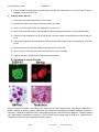
Mouse iPS Cell Lines (Matched Sets) Cat # SC201A-1 User Manual Store kit at -80 C on receipt A limited-use label license covers this product. By use of this product, you accept the terms and conditions outlined in the Licensing and Warranty Statement contained in this user manual. (ver. 3 - 112113) iPS Cell Lines Cat. # SC201A-1 Contents I. Mouse iPS Cells ......................................................................3 A. Description ..............................................................................3 B. Culture Conditions for MEF Feeder Cells ...............................4 C. Growth Conditions for Mouse iPS Cells .................................8 D. Validation of Mouse iPS Cells.................................................8 II. References ........................................................................11 III. Technical Support .............................................................12 IV. Licensing and Warranty Statement .......................................13 888-266-5066 (Toll Free) 650-968-2200 (outside US) Page 1 System Biosciences (SBI) User Manual List of Components Each iPS cell line set comes as one vial of MEF-derived mouse iPS cells and one vial of MEF at 2 x 10⁵ cells each. SC201A-1 Mouse iPS Cell Line Set [MEF & iPS cells] 2x10^5 cells each The product is shipped on dry ice and should be immediately stored in the gas phase of liquid nitrogen. In general, iPS cells are challenging to culture and should only be operated by researchers experienced in the intricacies of mouse embryonic stem (mES) cell culture. The methods for culture are nearly identical to mES cell culture, although more careful maintenance will be required. The MEF cells provided in the iPSC kit are meant to be used as source cells only and not as feeder cells for culturing iPS cells. MEF cells for feeder cells can be obtained from Applied Stemcell, Inc (http://www.appliedstemcell.com). Page 2 ver. 3-112113 www.systembio.com iPS Cell Lines I. Cat. # SC201A-1 Mouse iPS Cells A. Description Mouse induced pluripotent stem cells (iPSCs, Cat# SC201A-iPSC) were generated by transducing genetically unmodified mouse embryonic fibroblasts with viruses individually encoding the four murine transcription factors (Oct4, Sox2, Klf4, and c-Myc) that have been shown to induce the reprogramming of somatic cells to a pluripotent state. The cells were derived using morphological selection criteria and without the use of fluorescent markers or drug selection. When cultured under standard mouse ES cell culture conditions, the morphology of SBI mouse iPSCs are identical to that of mouse ES cells. The cells also express the pluripotency markers SSEA-1 and Nanog, and demonstrate strong endogenous AP activity. Mouse iPS cells should be grown on a feeder cell layer. Appropriate feeder cells for mouse iPS cells are mouse embryonic fibroblasts (MEFs, available from Applied Stemcell, Inc (http://www.appliedstemcell.com/). Mouse iPS cells from SBI are provided at passage 10 and can be passaged 50 times before differentiation. B. Culture Conditions for MEF Feeder Cells 1. Required media and reagents Reagent Information MEF Medium DMEM containing 10% FBS, 2 mM glutamine, 1x10⁻⁴ M nonessential amino acids and 50 U and 50 µg/ml penicillin and streptomycin. 2x Cold Freezing Media 20% DMSO and 80% FBS Mitomycin C solution 1 mg/ ml 2. Gelatin treatment of plates for MEF feeder cells 1) Add enough sterile/ autoclaved 0.1% gelatin to cover the bottom of the wells. Approximate amounts: 10cm plate – 5 ml 6 well – 1.5 ml/ well 24 well – 0.5 ml/ well 96 well – 100 µl/ well 888-266-5066 (Toll Free) 650-968-2200 (outside US) Page 3 System Biosciences (SBI) User Manual 2) Incubate the gelatin-coated dishes for at least 15 min at 37°C. 3) Aspirate excess gelatin solution before use. 3. Thawing MEF cells To insure the highest level of viability, be sure to warm medium to 37°C before using it on the cells. Cells should be plated at a minimum cell density of 104 cells/ cm2. 1) Remove the vial from liquid nitrogen and thaw quickly in a 37°C water bath. 2) Remove the vial from the water bath as soon as the cells are half-thawed, and sterilize the tube by spraying with 70% ethanol. 3) Transfer the cells with 10 ml of MEF medium to a 15 mL conical tube and pellet the cells by centrifugation at 200 g for 5 min. 4) Discard the supernatant and resuspend the cells with 10 ml fresh MEF medium and plate the cells at seed density of 104 cells/ cm2. 5) Incubate at 37°C with 5% CO2, until the cells reach 80-90% confluency. 6) Change medium twice a week or when pH decreases. 4. Passaging MEF cells Cells should be split when they reach confluency. We recommend splitting the cells based on 0.5x10⁴ cells/ cm2. 1) Discard the medium and wash the cells once with PBS. 2) Aspirate PBS, and add 2 ml per T75 flask of 0.25% trypsin-EDTA, and incubate for 2 min. 3) Add 5 ml of MEF medium, and break up the cell clumps by gently pipetting up and down several times. 4) Transfer cells into a conical tube and centrifuge at 200 g for 5 min. 5) Discard the supernatant and resuspend the cell pellet in 10 ml MEF medium. 6) Count the number of cells, plate cells at 0.5x10⁴ cells/ cm2 and incubate at 37°C with 5% CO2. 5. Freezing MEF cells 1) Follow steps 1-4 from the Passaging MEF cells protocol (above). 2) Discard the supernatant, and resuspend the pellet in MEF medium. 3) Count the number of cells and adjust the cell suspension to 4 x 10⁶ cells/ ml. 4) Add equal volume of cold 2X Freezing Media to the cell suspension. 5) Aliquot 1 ml of suspension into each cryovial (2 x 10⁶ cells/ vial). 6) Place the vials in a cell-freezing container and keep it at -80°C overnight. 7) Transfer the vials to a liquid nitrogen tank for long-term storage. Page 4 ver. 3-112113 www.systembio.com iPS Cell Lines Cat. # SC201A-1 6. Mitomycin C treatment of MEF Mitomycin C acts to halt the division of MEF cells so that they can be used to condition the medium for human iPS cells. MEF cells should be at confluence when treated with mitomycin C. 1) Add 6 ml of fresh MEF medium contain 50 µl of mitomycin C solution (1 mg/ ml) to one T75 flask of confluent MEF cells, and swirl it briefly. The final concentration of mitomycin C is 8 µg/ ml. 2) Incubate at 37°C for at least 3 hrs. 3) Aspirate the mitomycin C-containing medium off the cells and wash the cells twice with 10 ml PBS. 4) Aspirate PBS and add 1 ml of 0.25% trypsin-EDTA, swirl to cover the entire surface, and incubate for 2 min at room temperature. 5) Add 5 ml MEF medium and break up the cells to a single-cell suspension by pipetting up and down. Count the number of cells. 6) Seed the cells on gelatin-coated dishes (3 x 10⁶ cells per 100-mm dish, or 5 x 10⁵ cells per well of a 6-well plate). 7) Cells should be ready to use by the next day. 888-266-5066 (Toll Free) 650-968-2200 (outside US) Page 5 System Biosciences (SBI) User Manual C. Growth Conditions for mouse iPS cells 1. Required Media and Reagents Reagent Information Mouse ES Medium DMEM containing 15% FBS, 2 mM glutamine, 0.1M nonessential amino acids, 0.1 mM 2-mercaptoethanol, 103 U/ml LIF, 50 U and 50 µg/ ml Penicillin and Streptomycin 2x Cold Freezing Media 20% DMSO and 80% FBS Trypsin-EDTA GIBCO 1. Thawing Mouse iPS cells To insure the highest level of viability, be sure to warm medium to 37°C before using it on the cells. 1) Remove the vial from liquid nitrogen and thaw quickly in a 37°C water bath. 2) Remove the vial from the water bath as soon as the cells are half-thawed, and sterilize by spraying with 70% ethanol. 3) Transfer the cells with 10 ml of mouse ES medium to a 15 mL conical tube and pellet the cells by centrifugation at 200 g for 5 min. 4) While centrifuging, remove MEF medium from the feeder cell plates, and wash the wells twice with DMEM. Then add 1 ml of mouse ES Medium. 5) Discard the supernatant from the mouse iPS cells, and resuspend cells with 1 ml fresh mouse ES medium. Plate the cells on MEF feeder cells in a 6-well plate. 6) Incubate at 37°C with 5% CO2 until the cells reach 80% confluency. The ES media must be changed every day. 2. Maintenance of mouse iPS cells It is important not to keep mouse iPS cells in culture for long period of time without passaging, to maintain the pluripotency. 1) Aspirate the medium and wash the cells twice with 1 ml PBS. 2) Remove PBS completely and add 0.5 ml of 0.25% trypsin-EDTA solution, and incubate at 37°C for 2 min. 3) While incubating, remove a 6-well plate of feeder cells (mitomycin C-treated MEFs). Aspirate the medium and add 2 ml of mouse ES medium to each well. 4) Remove the plate containing mouse iPS cells from the incubator and swirl to dislodge the cells from the bottom of the plate. Page 6 ver. 3-112113 www.systembio.com iPS Cell Lines Cat. # SC201A-1 5) Add 2 ml of mouse ES medium, and suspend the cells by pipetting up and down to single cell suspension. 6) Transfer the cell suspension to a 15 ml conical tube and spin the cells at 200 g for 5 min. 7) Add 2 ml of ES medium to the plate and suspend the cells by pipetting up and down to single cell suspension. 8) Distribute ~0.2 ml of the mouse iPS cell suspension to each well of the 6-well plate. Right after plating iPS cells, gently swirl the plate back-and-forth and side-to-side and incubate at 37°C with 5% CO2 until the cells reach 80% confluency. 888-266-5066 (Toll Free) 650-968-2200 (outside US) Page 7 System Biosciences (SBI) User Manual 9) The ES media must be changed every day and mouse iPS cells subcultured ~1:10 every 2-3 days. Track the passage number of the iPS cells. 3. Freezing mouse iPS cells 1) Grow cells to the exponential phase in a 6-well plate. 2) Aspirate the medium, and wash the cells twice with 2 ml of PBS. 3) Add 0.5 ml 0.25% trypsin-EDTA and incubated for 2 min at 37°C. 4) Add 2 ml of mouse ES medium, and suspend the cells by pipetting up and down to single cell suspension. 5) Transfer the cell suspension to a 15 ml conical tube, count the number of cells and spin the cells at 200 g for 5 min. 6) Discard the supernatant, and resuspend the cells with mouse ES medium to the concentration of 1x10⁶ cells per ml. 7) Add equal volume of 2x freezing medium and aliquot it at 0.5 ml per vial. 8) Put the vials in a cell-freezing container, and store them at -80°C overnight. 9) Transfer the vials to a liquid nitrogen tank for long-term storage. D. Validation of mouse iPS cells Stem cell markers for SSEA-1 and Nanog were determined by immunocytochemistry using primary antibodies for SSEA1 (Millipore) and Nanog (Abcam) followed by Alexa Fluor fluorescent-labeled secondary antibodies (Invitrogen). Detection of Alkaline Phosphatase activity was performed using the AP Detection Kit (Millipore). Teratoma assays were performed by subcutaneous injection of 1x10⁶ iPS cells into the dorsal flank of SCID mice. Teratomas were visualized 4 weeks later with hematoxylin and eosin staining. Page 8 ver. 3-112113 www.systembio.com iPS Cell Lines II. Cat. # SC201A-1 References Rossant, J. 2007. Stem cells: The magic brew. Nature 448, 260–262. Takahashi, K. and Yamanaka, S. 2006. Induction of pluripotent stem cells from mouse embryonic and adult fibroblast cultures by defined factors. Cell 126: 663–676. Takahashi K. et al. 2007. Induction of pluripotent stem cells from adult human fibroblasts by defined factors. Cell. 131: 861–72. Park, IH et al. 2008. Reprogramming of human somatic cells to pluripotency with defined factors. Nature. 451:141–6. Baker, Monya 2007. Adult cells reprogrammed to pluripotency, without tumors. Nature Reports Stem Cells. 2007– 12–11. Nakagawa, M et al. 2008. Generation of induced pluripotent stem cells without Myc from mouse and human fibroblasts. Nature Biotechnology. 26: 101–106. Okita, K, et al. 2007. Generation of germline-competent induced pluripotent stem cells. Nature. 448:313–7 Yu, J. et al. 2007. Induced Pluripotent Stem Cell Lines Derived from Human Somatic Cells. Science 318: 1917–1920. III. Technical Support For more information about SBI products or to download manuals in PDF format, please visit our website: http://www.systembio.com For additional information or technical assistance, please call or email us at: [email protected] 650-968-2200 IV. Licensing and Warranty Statement Limited Use License Use of the human and mouse iPS cells (i.e., the “Product”) is subject to the following terms and conditions. If the terms and conditions are not acceptable, return all components of the Product to System Biosciences (SBI) within 7 calendar days. Purchase and use of any part of the Product constitutes acceptance of the above terms. The purchaser of the Product is granted a limited license to use the Product under the following terms and conditions: The Product shall be used by the purchaser for internal research purposes only. The Product is expressly not designed, intended, or warranted for use in humans or for therapeutic or diagnostic use. The Product may not be resold, modified for resale, or used to manufacture commercial products without prior written consent of SBI. This Product should be used in accordance with the NIH guidelines developed for stem cell research. 888-266-5066 (Toll Free) 650-968-2200 (outside US) Page 9 System Biosciences (SBI) User Manual SBI has pending patent applications related to the Product. For information concerning licenses for commercial use, contact SBI. Purchase of the product does not grant any rights or license for use other than those explicitly listed in this Licensing and Warranty Statement. Use of the Product for any use other than described expressly herein may be covered by patents or subject to rights other than those mentioned. SBI disclaims any and all responsibility for injury or damage which may be caused by the failure of the buyer or any other person to use the Product in accordance with the terms and conditions outlined herein. Limited Warranty SBI warrants that the Product meets the specifications described in this manual. If it is proven to the satisfaction of SBI that the Product fails to meet these specifications, SBI will replace the Product or provide the purchaser with a refund. This limited warranty shall not extend to anyone other than the original purchaser of the Product. Notice of nonconforming products must be made to SBI within 30 days of receipt of the Product. SBI’s liability is expressly limited to replacement of Product or a refund limited to the actual purchase price. SBI’s liability does not extend to any damages arising from use or improper use of the Product, or losses associated with the use of additional materials or reagents. This limited warranty is the sole and exclusive warranty. SBI does not provide any other warranties of any kind, expressed or implied, including the merchantability or fitness of the Product for a particular purpose. SBI is committed to providing our customers with high-quality products. concerns about any SBI products, please contact us at (888) 266-5066. If you should have any questions or © 2013 System Biosciences (SBI). Page 10 ver. 3-112113 www.systembio.com