1
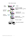
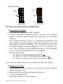
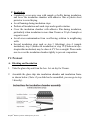
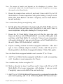
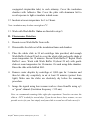
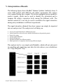
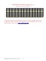
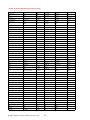
RayBio® Human Cytokine Antibody Array
G Series
Patent Pending Technology
User Manual (Revised February 18, 2009)
RayBio® Human Cytokine Antibody Array G Series 3 (Cat# AAH-CYT-G3-4)
RayBio® Human Cytokine Antibody Array G Series 5 (Cat# AAH-CYT-G5-4)
RayBio® Human Cytokine Antibody Array G Series 6 (Cat# AAH-CYT-G6-4)
RayBio® Human Cytokine Antibody Array G Series 7 (Cat# AAH-CYT-G7-4)
RayBio® Human Cytokine Antibody Array G Series 8 (Cat# AAH-CYT-G8-4)
RayBio® Human Cytokine Antibody Array G Series 7 (Cat# AAH-CYT-G9-4)
RayBio® Human Cytokine Antibody Array G Series 8 (Cat# AAH-CYT-G10-4)
RayBio® Human Inflammation Antibody Array 3 (Cat# AAH-INF-G3-4)
RayBio® Human Growth Factor Antibody Array 1 (Cat# AAH-GF-G1-4)
RayBio® Human Cytokine Antibody Array G Series 3 (Cat# AAH-CYT-G3-8)
RayBio® Human Cytokine Antibody Array G Series 5 (Cat# AAH-CYT-G5-8)
RayBio® Human Cytokine Antibody Array G Series 6 (Cat# AAH-CYT-G6-8)
RayBio® Human Cytokine Antibody Array G Series 7 (Cat# AAH-CYT-G7-8)
RayBio® Human Cytokine Antibody Array G Series 8 (Cat# AAH-CYT-G8-8)
RayBio® Human Cytokine Antibody Array G Series 7 (Cat# AAH-CYT-G9-8)
RayBio® Human Cytokine Antibody Array G Series 8 (Cat# AAH-CYT-G10-8)
RayBio® Human Inflammation Antibody Array 3 (Cat# AAH-INF-G3-8)
RayBio® Human Growth Factor Antibody Array 1 (Cat# AAH-GF-G1-8)
RayBio® Custom Human Cytokine Antibody Array (Cat# AAH-CUS-G)
RayBio® Human Cytokine Antibody Array Service (Cat# AAH-SER-G)
Please read the manual carefully before you start your experiment
RayBiotech, Inc.
We Provide You With Excellent
Protein Array Systems and Service
Tel:(Toll Free) 1-888-494-8555 or 770-729-2992; Fax: 1-888-547-0580;
Website:www.raybiotech.com Email: [email protected]
RayBiotech, Inc.
RayBio® Human Cytokine Antibody Array G Series Protocol
TABLE OF CONTENTS
I.
Introduction……..……………………………...
2
How It Works………………………..…………
5
Materials Provided……………………………..
6
Additional Materials Required…………………
6
III. Overview and General Considerations…………
7
A. Preparation of Samples………………………
7
B. Handling Glass Chips ……………………….
7
C. Incubation……………………………………
8
IV. Protocol…………………………………………
9
A. Blocking and Incubation…………………….
9
B. Detection…………………………………….
11
Interpretation of Results………………………..
12
VI. Troubleshooting Guide…………………………
21
VII. Reference List………………………………….
22
II.
V.
Cytokine Antibody Arrays are RayBiotech patent-pending technology.
RayBio® is the trademark of RayBiotech, Inc.
RayBio® Human Cytokine Antibody Array G series
1
I. Introduction
All cell functions, including cell proliferation, cell death and differentiation,
as well as maintenance of health status and development of disease, are
controlled by many genes and signaling pathways. New techniques such as
cDNA microarrays have enabled us to analyze the global gene expression 1-3.
However, almost all cell functions are executed by proteins, which cannot be
studied by DNA and RNA alone. Experimental analysis clearly shows a
disparity between the relative expression levels of mRNA and their
corresponding proteins 4. Therefore, it is critical to analyze the protein
profile. Currently, two-dimensional polyacrylamide SDS page coupled with
mass spectrometry is the mainstream approach to analyzing multiple protein
expression levels 5,6. However, the requirement of sophisticated devices and
the lack of quantitative measurements greatly limit its broad application.
Thus, no simple, cost effective, and rapid method of analysis of multiple
protein expression levels has been available to researchers until now.
Our RayBio® Human Cytokine Antibody Array is the first commercially
available protein array system 7-11. By using the RayBiotech system,
scientists can rapidly and accurately identify the expression profiles of
multiple cytokines in several hours inexpensively.
The RayBiotech kit (G series) is a glass slide format. The kit provides a
highly sensitive approach to simultaneously detect multiple cytokine
expression levels from cell culture supernatant, patient’s serum, tissue lysate
and other sources. The arrays are manufactured using non-contact arrayer.
The experimental procedure is simple and can be performed in any
laboratory. The signals from G series arrays are detected using a laser
scanner.
Besides the products listed in this manual, RayBiotech also provides
RayBio® Human Cytokine Antibody Array G series 1000 for detection of
120 human cytokines in single experiment, RayBio® Human Cytokine
Antibody Array G series 2000 for detection of 174 human cytokines in single
experiment and RayBio® Human Cytokine Antibody Array G series 4000 for
detection of 274 human cytokines in single experiment.
RayBio® Human Cytokine Antibody Array G series
2
Pathway-specific array systems allow investigators to focus on the specific
problem and are becoming an increasingly powerful tool in cDNA
microarray system. RayBiotech’s first protein array system, known as
RayBio® Human Cytokine Antibody Array, is particularly useful compared
with the human cytokine cDNA microarray system. Besides the ability to
detect protein expression, RayBiotech’s system is a more accurate reflection
of active cytokine levels because it only detects secreted cytokines, and no
amplification step is needed. Furthermore, it is much simpler, faster,
environmentally friendlier, and more sensitive.
Simultaneous detection of multiple cytokines undoubtedly provides a
powerful tool to study cytokines. Cytokines play an important role in innate
immunity, apoptosis, angiogenesis, cell growth and differentiation 12.
Cytokines are involved in most disease processes, including cancer and
cardiac diseases. The interaction between cytokines and the cellular immune
system is a dynamic process. The interactions of positive and negative
stimuli, and positive as well as negative regulatory loops are complex and
often involve multiple cytokines.
Reference List
1. LPS induces the interaction of a transcription factor, LPS-induced TNF-a factor,
and STAT6(B) with effects on multiple cytokines. Xiaoren Tang, Deborah Levy
Marciano, Susan E. Leeman and Salomon Amar. PNAS. April 5, 2005 vol. 102 no.
14 5132-5137
2. HIV-1-mediated apoptosis of neuronal cells: Proximal molecular mechanisms of
HIV-1-induced encephalopathy. Yan Xu, Joseph Kulkoshy, Roger j. Pomerantz.
PNAS. 2004 May 4, 2004 Vol. 101 No. 18.
3. Synergistic increases in intracellular Ca(2+), and the release of MCP-1, RANTES,
and IL-6 by astrocytes treated with opiates and HIV-1 Tat. GLIA. 2005 Apr
15;50(2):91-106.
4. Bone Marrow Stroma Influences Transforming Growth Factor-β Production in
Breast Cancer Cells to Regulate c-myc Activation of the Preprotachykinin-I Gene
in Breast Cancer Cells. Hyun S. Oh, Anabella Moharita… Pranela Rameshwar.
CANCER RESEARCH. 64, 6327-6336, September 1, 2004
RayBio® Human Cytokine Antibody Array G series
3
5. Recombinant Herpes Simplex Virus Type 1 (HSV-1) Codelivering Interleukin12p35 as a Molecular Adjuvant Enhances the Protective Immune Response against
Ocular HSV-1 Challenge JOURNAL OF VIROLOGY. Mar. 2005 Vol. 79, No. 6.
6. Dysregulated Inflammatory Response to Candida albicans in a C5-Deficient
Mouse Strain. Alaka Mullick, Miria Elias, Serge Picard, Philippe Gros. Infection
and Immunity, Oct. 2004, p. 5868-5876, DOI: 10.1128/IAI.72.10.5868-5876.2004
7. Leukotriene B4 Strongly Increases Monocyte Chemoattractant Protein-1 in Human
Monocytes Li Huang, Annie Zhao, Frederick Wong, Julia M. Ayala, Jisong Cui
Arteriosclerosis, Thrombosis, and Vascular Biology. 2004;24:1783-1788
8. Human CD1d-unrestricted NKT cells release chemokines upon Fas engagement.
Martin Giroux and François Denis. Yan Xu, Joseph Kulkoshy, Roger j. Pomerantz.
Blood. prepublished online September 2, 2004; DOI 10.1182/blood-2004-04-1537
9. Monitoring the response of orthotopic bladder tumors to granulocyte macrophage
colony-stimulating factor therapy using the prostate-specific antigen gene as a
reporter. Wu Q, Esuvaranathan K, Mahendran R. Clinical Cancer Research. 2004
Oct 15; 10(20):6977-84.
10. Neuroglial activation and neuroinflammation in the brain of patients with autism
(p NA). Diana L. Vargas, Caterina Nascimbene, Chitra Krishnan, Andrew W.
Zimmerman, Carlos A. Pardo. Annals of Neurology. 2005 Jan 1; DOI:
10.1002/ana.20315
11. Cytokine profiling of macrophages exposed to Porphyromonas gingivalis, its LPS
or its FimA. Qingde Zhou, Tesfahun Desta, Dana T. Graves and Salomon Amar.
Infection and Immunity (IAI). 2005 Feb;73(2):935-43.
12. Veto-like activity of mesenchymal stem cells: functional discrimination between
cellular responses to alloantigens and recall antigens. Rameshwar P. Journal of
Immunology. 2003 Oct 1;171(7):3426-34.
13. The promise of cytokine antibody arrays in drug discovery process. Huang RP,
Yang W, Yang Y, Flowers L, et al. Expert Opinion on Drug Discovery. (2005)
9:601-615.
RayBio® Human Cytokine Antibody Array G series
4
Here’s how it works
Array
support
Sample
Incubation of Sample
With arrayed antibody
Supports
Cocktail of
Biotin-Ab
Incubation with
Biotinylated Ab
1-2 hrs
1-2 hrs
Labeled –
streptavidin
Incubation with
Labeled- streptavidin
2 hrs
Detection of signals
Data analysis and graph
RayBio® Human Cytokine Antibody Array G series
5
II. Materials Provided
Upon receipt, all components of the RayBio® Mouse Cytokine Antibody
Array kit should be stored at -20°C. At -20°C the kit will retain complete
activity for up to 6 months. Once thawed, the glass chips, Fluorescent dyestreptavidin, Internal Control and 2X Blocking Buffer should be kept at –
20°C and all other component should be stored at 4°C. Use within three
months after reagents have been thawed. Please use within six months of
purchase.
• RayBio® Mouse Cytokine Antibody Microarray slides (1 slide with 4
or 8 subarrays each)
• Biotin-Conjugated Anti-Cytokines (1 tube per 4 arrays: 1 tube for 4
subarrays, 2 tubes for 8 subarrays)
• 1,500X Fluorescent Dye-conjugated Streptavidin (Cy3 equivalent, 1
tube)
• 2X Blocking Buffer (10 ml)
• 20X Wash Buffer I (30 ml)
• 20X Wash Buffer II (30 ml)
• Internal control (powder, 1 tube)
• 2X Cell Lysis Buffer (10 ml)
• RayBio® G series antibody array accessory (including slide incubation
chamber, Gasket, Protective cover, Snap-on sides and adhesive film)
• 30 ml tube
• Manual
Additional Materials Required
•
•
•
•
•
Orbital shaker
Laser scanner for fluorescence detection
Aluminum foil
Distilled water
Plastic box
RayBio® Human Cytokine Antibody Array G series
6
Layout of G series
Antibody
Array
Antibody
Array
Blank
Blank
Barcode
Barcode
8 arrays in one glass chip
4 arrays in one glass chip
III. Overview and General Considerations
A. Preparation of Samples
• Use serum-free conditioned media if possible.
• If serum-containing conditioned media is required, use uncultured
media as a negative control sample, since many types of sera contain
cytokines.
• For cell lysates and tissue lysates, we recommend using RayBio® Cell
Lysis Buffer to extract proteins from cell or tissue (e.g. using
homogenizer). Dilute 2X RayBio® Cell Lysis Buffer with H2O (we
recommend adding proteinase inhibitors to Cell Lysis Buffer before
use). After extraction, spin the sample down and save the supernatant
for your experiment. Determine protein concentration.
• We recommend using:
o 50–100 µl of Conditioned media (undiluted), or
o 50–100 µl of 2-fold to 5-fold diluted sera or plasma, or
o 10–200 µg of total protein for cell lysates and tissue lysates.
If you experience high background, you may further dilute your sample.
B. Handling glass chips
• The microarray slides are sensitive, do not touch the surface. Hold the
slides by the edges only.
• Handle all buffers and slides with powder-free gloves.
• Avoid breaking glass slide.
• Handle glass chip in clean environment.
RayBio® Human Cytokine Antibody Array G series
7
C. Incubation
• Completely cover array area with sample or buffer during incubation,
and cover the incubation chamber with adhesive film or plastic sheet
protector to avoid drying.
• Avoid foaming during incubation steps.
• Perform all incubation and wash steps under gentle rotation.
• Cover the incubation chamber with adhesive film during incubation,
particularly when incubation is more than 2 hours or 50 µl of sample or
reagent is used.
• Avoid cross-contamination from overflowing solution to neighboring
wells.
• Several incubation steps such as step 3 (blocking), step 4 (sample
incubation), step 9 (biotin-Ab incubation) or step 12 (Fluorescent dyestreptavidin incubation) may be done at 4°C for overnight. Please make
sure to cover the incubation chamber tightly to prevent evaporation.
IV. Protocol
A. Blocking and Incubation
1. Take the glass chip out from the box. Let air dry for 2 hours.
2. Assemble the glass chip into incubation chamber and incubation frame
as shown below. (Note: if you slide has be assembled, you can go to step
3 directly).
RayBio® Human Cytokine Antibody Array G series
8
3. Add 100 µl 1 X Blocking Buffer into each well and incubate at room
temperature for 30 min to block slides. Dilute 2X Blocking Buffer with
H2O. Make sure no bubbles are in the well.
Note: only add reagents to wells printed with antibodies.
4. Decant Blocking Buffer from each well. Add 50 to 100 µl of each
sample to array 6 and array 7. Incubate arrays with sample at room
temperature for 1 to 2 hours. Dilute sample using 1X Blocking Buffer if
necessary. We strongly recommend including Internal Control (IC) in
your assay. Add 100 µl of Blocking Buffer to IC tube, mix well and
transfer 1 µl of IC to each well (50 to 100 µl of sample).
Note: when you transfer IC, use 0.1 µl to 2 µl pipettor.
Note: Incubation may be done overnight at 4°C.
Note: We recommend using 50 to 100 µl of undiluted conditioned media or 50 to 100 µl
of 2-5 fold diluted serum or plasma or 10-200 µg of protein for cell lysates and
tissue lysates. Dilute the lysate at least 10 fold with 1X blocking buffer to make a
total volume of 50 to 100 µl. Make sure there are no bubbles in the wells.
RayBio® Human Cytokine Antibody Array G series
9
Note: The amount of sample used depends on the abundance of cytokines. More
concentrated sample can be used if signals are too weak. If signals are too strong,
the sample can be diluted further.
5. Decant the samples from each well, and wash 3 times with 150 µl of 1X
Wash Buffer I at room temperature with gentle shaking. 2 min per wash.
Dilute 20X Wash Buffer I with H2O. Completely remove Wash Buffer I
in each wash step.
Note: avoid solution flowing into neighboring wells.
6. Put the glass chip with frame into a box with 1X Wash Buffer I (cover
the whole glass slide and frame with Wash Buffer I), and wash 2 times at
room temperature with gentle shaking for 10 min per wash.
7. Decant the 1X Wash Buffer I from each well, Put the glass chip with
frame into the box with 1XWash Buffer II (cover the whole glass slide
and frame with Wash Buffer II), and wash 2 times at room temperature
with gentle shaking for 5 min per wash. Remove all of Wash Buffer II in
the well. Dilute 20X Wash Buffer II with H2O.
8. Prepare working solution for biotin-conjugated antibodies. After brief
spin to force antibody aliquot to bottom of tube, add 300 µl of 1x
blocking buffer to the Biotin-Conjugated Antibody VI tube. Mix gently.
Note: the diluted biotin-conjugated antibodies can be stored at 40C for 2-3 days.
9. Add 70 µl of diluted biotin-conjugated antibodies to each corresponding
well. Incubate at room temperature for 2 hours.
Note: incubation may be done overnight at 4°C.
10. Wash as directed in steps 5 and then wash 3 times with 150 µl of 1X
Wash Buffer II at room temperature with shaking. 2 min per wash.
Completely remove wash buffer II in each wash step.
11. Add 70 µl of 1,500 fold diluted Fluorescent dye-conjugated streptavidin
(after brief spinning, add 1.5 ml of Blocking Buffer to Fluorescent dyeRayBio® Human Cytokine Antibody Array G series
10
conjugated streptavidin tube) to each subarray. Cover the incubation
chamber with Adhesive film. Cover the plate with aluminum foil to
avoid exposure to light or incubate in dark room.
12. Incubate at room temperature for 1 to 2 hours.
Note: incubation may be done overnight at 4°C.
13. Wash with Wash Buffer I twice as directed in steps 5.
B. Fluorescence Detection
1. Decant excess Wash Buffer from wells.
2. Disassemble the slide out of the incubation frame and chamber.
3. Place the whole slide in 30 ml centrifuge tube provided, add enough
Wash Buffer I (about 20 ml) to cover the whole slide and gently shake at
room temperature for 10 minutes. Decant Wash Buffer I. Repeat Wash
Buffer I once. Wash with Wash Buffer II (about 20 ml) with gentle
shake at room temperature for 10 minutes. Or wash using slide chamber.
Rinse the slide with distilled H2O.
4. Remove water droplets by centrifuge at 1,000 rpm for 3 minutes and
then let slide dry completely in air at least 20 minutes (protect from
light). Make sure the slides are absolutely dry before the scanning
procedure.
5. Image the signals using laser scanner such as Axon GenePix using cy3
or “green” channel (Excitation frequency = 532 nm).
Note: we recommend scanning slides right after experiment. You also can store the
slide at –20°C in dark for several days. If you do not have a laser scanner, we can
provide service for you. Just simply send your slide to us and we will take care of it.
RayBio® Human Cytokine Antibody Array G series
11
V. Interpretation of Results:
The following figure shows RayBio® Human Cytokine Antibody Array G
series 2000 probed with different cell culture supernatant. The images
were captured using laser scanner. The biotin-conjugated protein produces
positive signals, which can be used to identify the orientation and to
compare the relative expression levels among the different wells. The
internal control (IC) can also be used to normalize the signal intensities
among array membranes in different experiments.
The signal intensities obtained from laser scanner can simply be imported
into our analysis tool. The analysis tool will help you:
•
•
•
•
•
•
•
Locate your signal intensities to antibody array map
Link the protein to website for more detailed information on the particular protein
Protein list sorting
Average signal intensities
Subtract background
Normalize the data from different samples
Obtain protein level comparison charts among different samples
This analysis tool is very simple and affordable, which will not only assist
in compiling and organizing your data, but also reduces your calculations
to a “copy and paste” step.
RayBio® Human Cytokine Antibody Array G series
12
If you do not use our RayBio® Analysis Tool, you can locate the cytokines
by referring to corresponding RayBio® Human cytokine Antibody Array G
series map.
Normalization and comparison
For biomarker discovery or for analysis of large number of arrays, great
attention must be paid to the normalization. Our antibody array design
includes several controls for normalization and comparison of arrays
performing in different membranes and different experiments (for more
information please read the reference 13 in the introduction).
Positive control. Positive control is biotinylated protein. It can be used to
normalize the streptavidin incubation step. If the positive signals from
different array membranes are similar, positive control is a simple and
effective way for normalization.
Internal control. RayBio® antibody arrays also include spiking-in protein
serving as internal control (IC). The spiking-in proteins do not have crossreactivity with protein in the array. It can be used to normalize the entire
process.
Negative control. Negative control is BSA. Normally, it should only give a
background reading.
Data Extraction Tips:
• Ignore any comet tails
• Define the area for signal capture for all spots as 110-120 micron diameter,
using the same area for every spot.
• Use median signal value, not the total or the mean
• Use local background correction (also median value).
• Exclude obvious outlier data in its calculations.
• Scan all slides at same PMT
Using these guidelines, along with using PMT, brightness and contrast
settings that reduce the background as much as possible, we get very good
results with interassay and intraassay CV <20%, even with some
imperfections in the antibody spots.
RayBio® Human Cytokine Antibody Array G series
13
Threshold of significant difference in expression: Any ≥1.5-fold increase or
≤0.65-fold decrease in signal intensity for a single analyte between samples,
provided that both signals are well above background (Mean background + 2
standard deviations, accuracy ≈ 95%).
Array Map Notes:
o
o
o
o
o
o
o
IL-12 reacts with both IL-12p40 and IL-12p70.
IL-12p70 only recognizes IL-12p70.
GRO reacts with CXCL1, CXCL2 and CXCL3 (GRO alpha, beta, and gamma,
respectively).
GRO alpha reacts only with CXCL1.
VEGF reacts with VEGF-165 and VEGF-121
TGF-beta 1 reacts only with active form of TGF-beta 1
All MMP antibody pairs detect both pro and mature forms
Abbreviations: IP-10, Interferon-inducible protein-10; LAP, latency associated peptide (TGF-β1);
LH, leutinizing hormone; LIF, leukocyte inhibitory factor. MMP, Matrix Metalloproteinase; PF, plasma factor 4;
Pos, positive control; Neg, negative control. All other are used standard abbreviations.
RayBio® Human Cytokine Antibody Array G series 3 (42)
a
b
c
d
e
f
g
h
I
j
k
l
J
1
Pos 1
Pos 2
Pos 3
Neg
Neg
ENA-78
GCSF
GM-CSF
GRO
GRO-α
I-309
IL-1α
IL-1 β
2
Pos 1
Pos 2
Pos 3
Neg
Neg
ENA-78
GCSF
GM-CSF
GRO
GRO-α
I-309
IL-1α
IL-1 β
3
IL-2
IL-3
IL-4
IL-5
IL-6
IL-7
IL-8
IL-10
IL-12 p40p70
IL-13
IL-15
IFN-γ
MCP-1
4
IL-2
IL-3
IL-4
IL-5
IL-6
IL-7
IL-8
IL-10
IL-12 p40p70
IL-13
IL-15
IFN-γ
MCP-1
5
MCP-2
MCP-3
MCSF
MDC
MIG
MIP-1 δ
RANTES
SCF
SDF-1
TARC
TGF- β1
TNF-α
TNF-β
6
MCP-2
MCP-3
MCSF
MDC
MIG
MIP-1 δ
RANTES
SCF
SDF-1
TARC
TGF- β1
TNF-α
TNF-β
7
EGF
IGF-I
Angiogenin
Oncostatin M
Thrombopoietin
VEGF
PDGF BB
Leptin
IC 1
IC 2
IC 3
Neg
Neg
8
EGF
IGF-I
Angiogenin
Oncostatin M
Thrombopoietin
VEGF
PDGF BB
Leptin
IC 1
IC 2
IC 3
Neg
Neg
RayBio® Human Cytokine Antibody Array 5 (80)
A
B
C
D
E
F
G
H
I
J
K
L
M
1
Pos 1
Pos 2
Pos 3
Neg
Neg
Neg
ENA-78
GCSF
GM-CSF
GRO
GRO-α
I-309
IL-1α
2
Pos 1
Pos 2
Pos 3
Neg
Neg
Neg
ENA-78
GCSF
GM-CSF
GRO
GRO-α
I-309
IL-1α
3
IL-1 β
IL-2
IL-3
IL-4
IL-5
IL-6
IL-7
IL-8
IL-10
IL-12 p40p70
IL-13
IL-15
IFN- γ
4
IL-1 β
IL-2
IL-3
IL-4
IL-5
IL-6
IL-7
IL-8
IL-10
IL-12 p40p70
IL-13
IL-15
IFN- γ
5
MCP-1
MCP-2
MCP-3
MCSF
MDC
MIG
MIP-1β
MIP-1δ
RANTES
SCF
SDF-1
TARC
TGF-β1
6
MCP-1
MCP-2
MCP-3
MCSF
MDC
MIG
MIP-1β
MIP-1δ
RANTES
SCF
SDF-1
TARC
TGF-β1
7
TNF-α
TNF-β
EGF
IGF-I
Angiogenin
Oncostatin M
Thrombopoietin
VEGF
PDGF-BB
Leptin
BDNF
BLC
Ck β 8-1
8
TNF-α
TNF-β
EGF
IGF-I
Angiogenin
Oncostatin M
Thrombopoietin
VEGF
PDGF-BB
Leptin
BDNF
BLC
Ck β 8-1
9
Eotaxin
Eotaxin-2
Eotaxin-3
FGF-4
FGF-6
FGF-7
FGF-9
Flt-3 Ligand Fractalkine
GCP-2
GDNF
HGF
IGFBP-1
10
Eotaxin
Eotaxin-2
Eotaxin-3
FGF-4
FGF-6
FGF-7
FGF-9
Flt-3 Ligand Fractalkine
11
IGFBP-2
IGFBP-3
IGFBP-4
IL-16
IP-10
LIF
LIGHT
GCP-2
GDNF
HGF
IGFBP-1
MCP-4
MIF
MIP-3 α
NAP-2
NT-3
NT-4
IGFBP-3
IGFBP-4
IL-16
IP-10
LIF
LIGHT
MCP-4
MIF
MIP-3 α
NAP-2
NT-3
NT-4
13
Osteopontin
Osteoprotegerin
PARC
PIGF
TGF- β 2
TGF- β 3
TIMP-1
TIMP-2
IC1
IC2
IC3
Neg
Neg
14
Osteopontin
Osteoprotegerin
PARC
PIGF
TGF- β 2
TGF- β 3
TIMP-1
TIMP-2
IC1
IC2
IC3
Neg
Neg
12
IGFBP-2
RayBio® Human Cytokine Antibody Array G series
14
RayBio® Human Cytokine Antibody Array G series 6 (60)
RayBio® Human Cytokine Antibody Array G series 7 (60)
RayBio® Human Cytokine Antibody Array G series 8 (54)
RayBio® Human Cytokine Antibody Array G series
15
RayBio® Human Cytokine Antibody Array G series 9 (51)
a
b
c
d
e
f
g
h
i
j
k
l
1
POS1
POS2
POS3
NEG
NEG
Adiposin
BCAM
CD30
CD40
Fcr RIIB/C
Ferritin
FLRG
2
POS1
POS2
POS3
NEG
NEG
Adiposin
BCAM
CD30
CD40
Fcr RIIB/C
Ferritin
FLRG
3
Follistatin
Furin
Galectin-7
GDF-15
Growth
Hormon
IL-10 R alpha
IL-22
IL-28A
IL29
IL-31
Insulin
Luteinizing
hormone
4
Follistatin
Furin
Galectin-7
GDF-15
Growth
Hormon
IL-10 R alpha
IL-22
IL-28A
IL29
IL-31
Insulin
Luteinizing
hormone
5
LIMPII
LYVE-1
Marapsin
MICA
MICB
MMP-2
MMP-7
MMP-8
MMP-10
NCAM-1
Nidogen-1
NrCAM
6
LIMPII
LYVE-1
Marapsin
MICA
MICB
MMP-2
MMP-7
MMP-8
MMP-10
NCAM-1
Nidogen-1
NrCAM
7
NRG1-beta1
Osteopontin
PAI-I
Platelet factor 4
PSA-total
RAGE
RANK
Resistin
SAA
Siglec-9
TACE
TIM-1
8
NRG1-beta1
Osteopontin
PAI-I
Platelet factor 4
PSA-total
RAGE
RANK
Resistin
SAA
Siglec-9
TACE
TIM-1
9
TRAIL R2
Trappin-2
TREM-1
TSH
TSLP
VCAM-1
VEGF-C
XEDAR
NEG
IC 1
IC 2
IC 3
10
TRAIL R2
Trappin-2
TREM-1
TSH
TSLP
VCAM-1
VEGF-C
XEDAR
NEG
IC 1
IC 2
IC3
RayBio® Human Cytokine Antibody Array G series 10 (49)
a
b
c
d
e
f
g
i
j
k
l
1
POS1
POS2
POS3
NEG
NEG
4-1BB
ACE-2
Alpha-fetoprotein
Angiopoietin-1
Angiostatin
ANGPTL4
Bate2 M
2
POS1
POS2
POS3
NEG
NEG
4-1BB
ACE-2
Alpha-fetoprotein
Angiopoietin-1
Angiostatin
ANGPTL4
Bate2 M
CA19-9
Carbonic
Anhydrase IX
Cathepsin S
CCL14a
CCL21
CD23
CD40 Ligand
CEA
3
BCMA
beta IG-H3
CA125
CA15-3
h
4
BCMA
beta IG-H3
CA125
CA15-3
CA19-9
Carbonic
Anhydrase IX
Cathepsin S
CCL14a
CCL21
CD23
CD40 Ligand
CEA
5
CEACAM-1
Cripto-1
CRP
DAN
Decorin
DKK-1
DKK-3
DKK-4
DPPIV
E-Cadherin
EDA-A2
EG-VEGF
6
CEACAM-1
Cripto-1
CRP
DAN
Decorin
DKK-1
DKK-3
DKK-4
DPPIV
E-Cadherin
EDA-A2
EG-VEGF
7
EpCAM
ErbB2
Erythropoietin R
FSH
HB-EGF
hCGa, intact
HVEM
IL-13 R alpha1
IL-17B
IL-17C
IL-17F
IL-17R
8
EpCAM
ErbB2
Erythropoietin R
FSH
HB-EGF
hCGa, intact
HVEM
IL-13 R alpha1
IL-17B
IL-17C
IL-17F
IL-17R
9
Procalcitonin
PSA-free
S-100b
Shh N
Thyroglobulin
Ubiquitin+1
Blank
IC 1
IC 2
IC 3
NEG
NEG
10
Procalcitonin
PSA-free
S-100b
Shh N
Thyroglobulin
Ubiquitin+1
Blank
IC 1
IC 2
IC 3
NEG
NEG
RayBio® Human Inflammation Antibody Array G series 3 (40)
A
B
C
D
E
H
I
J
K
L
M
1
POS 1
POS 2
POS 3
NEG
NEG
EOTAXIN EOTAXIN-2
F
G
GCSF
GM-CSF
ICAM-1
IFN-γ
I-309
IL-1α
2
POS 1
POS 2
POS 3
NEG
NEG
EOTAXIN EOTAXIN-2
GCSF
GM-CSF
ICAM-1
IFN-γ
I-309
IL-1α
3
IL-1β
IL-2
IL-3
IL-4
IL-6
IL-6sR
IL-7
IL-8
IL-10
IL-11
IL-12 p40
IL-12 p70
IL-13
4
IL-1β
IL-2
IL-3
IL-4
IL-6
IL-6sR
IL-7
IL-8
IL-10
IL-11
IL-12 p40
IL-12 p70
IL-13
5
IL-15
IL-16
IL-17
IP-10
MCP-1
MCP-2
M-CSF
MIG
MIP-1α
MIP-1β
MIP-1δ
RANTES
TGF-β1
6
IL-15
IL-16
IL-17
IP-10
MCP-1
MCP-2
M-CSF
MIG
MIP-1α
MIP-1β
MIP-1δ
RANTES
TGF-β1
7
TNF-α
TNF-β
s TNF RI
s TNF RII
PDGF-BB
TIMP-2
NEG
NEG
IC 1
IC 2
IC 3
NEG
NEG
8
TNF-α
TNF-β
s TNF RI
s TNF RII
PDGF-BB
TIMP-2
NEG
NEG
IC 1
IC 2
IC 3
NEG
NEG
RayBio® Human Cytokine Antibody Array G series
16
RayBio® Human Growth Factor Antibody Array 1
Detect 41 Growth Factors in one experiment
a
b
c
d
e
f
g
h
i
j
k
l
m
1
Pos 1
Pos 2
Pos 3
Neg
Neg
AR
bFGF
b-NGF
EGF
EGF R
FGF-4
FGF-6
FGF-7
2
Pos 1
Pos 2
Pos 3
Neg
Neg
AR
bFGF
b-NGF
EGF
EGF R
FGF-4
FGF-6
FGF-7
3
GCSF
GDNF
GM-CSF
HB-EGF
HGF
IGFBP-1
IGFBP-2
IGFBP-3
IGFBP-4
IGFBP-6
IGF-I
IGF-I SR
IGF-II
4
GCSF
GDNF
GM-CSF
HB-EGF
HGF
IGFBP-1
IGFBP-2
IGFBP-3
IGFBP-4
IGFBP-6
IGF-I
IGF-I SR
IGF-II
5
M-CSF
M-CSF R
NT-3
NT-4
PDGF R a
PDGF R b
PDGF-AA
PDGF-AB
PDGF-BB
PlGF
SCF
SCF R
TGF-a
6
M-CSF
M-CSF R
NT-3
NT-4
PDGF R a
PDGF R b
PDGF-AA
PDGF-AB
PDGF-BB
PlGF
SCF
SCF R
TGF-a
7
TGF-b
TGF-b 2
TGF-b 3
VEGF
VEGF R2
VEGF R3
VEGF-D
neg
IC 1
IC 2
IC 3
Neg
Neg
8
TGF-b
TGF-b 2
TGF-b 3
VEGF
VEGF R2
VEGF R3
VEGF-D
neg
IC 1
IC 2
IC 3
Neg
Neg
We also offer Custom Human Cytokine Antibody Arrays. You can select the cytokines of interest from
the following list and we will produce the customized array at an affordable price. For more
information, please visit our website, www.raybiotech.com.
RayBio® Human Cytokine Antibody Array G series
17
Human Custom Antibody Array List (285 proteins)
4-1BB/TNFRSF9
CNTF
GDNF
IL-18 R alpha
MIP-1 alpha
SCF
ACE-2
Cripto-1
GITR
IL-18 R beta
MIP-1 beta
SCF R
Activin A
CRP
GITR Ligand
IL-1ra
MIP-1 delta
SDF-1 alpha
Adiponectin/Acrp30
CTACK/CCL27
GM-CSF
IL-2
MIP-3 alpha
SDF-1 beta
Adipsin/Factor D
CTLA-4
GRO
IL-2 R alpha
MIP-3 beta
sgp130
AFP
CXCL16
GRO-a
IL-2 R beta
MMP-1
Shh N
AgRP(ART)
DAN
Growth Hormom
IL-2 R gamma
MMP-2
Siglec-5
ALCAM
Decorin
HB-EGF
IL-21 R
MMP-3
Siglec-9
Angiogenin
DKK-1
HCC-4/CCL16
IL-22
MMP-7
sTNF RII
Angiopoietin-1
DKK-3
hCGa, intact
IL-28A/IFN-lambda
MMP-8
sTNT RI
Angiopoietin-2
DKK-4
HGF
IL29/IFN-lambda 1
MMP-9
TACE
Angiostatin
DPPIV/CD26
HVEM
IL-3
MMP-10
TARC
ANGPTL4
DR6
I-309
IL-31
MMP-13
TECK/CCL25
AR (amphiregulin)
Dtk
ICAM-1
IL-4
MPIF-1
TGF-alpha
Axl
E-Cadherin
ICAM-2
IL-5
MSP a Chain
TGF-beta 1
B7-1(CD80)
EDA-A2
ICAM-3
IL-5 R alpha
NAP-2
TGF-beta 2
Bate2 M
EGF
IFN-gamma
IL-6
NCAM-1
TGF-beta 3
BCAM
EGF R
IGFBP-1
IL-6 sR
NGF R
Thyroglobulin
BCMA/TNFRSF17
EG-VEGF/PK1
IGFBP-2
IL-7
Nidogen-1/Entactin
Tie-1
BDNF
ENA-78
IGFBP-3
IL-8
NrCAM
Tie-2
beta IG-H3
Endoglin
IGFBP-4
IL-9
NRG1-beta 1/HRG1-beta 1
TIM-1
Betacellulin (BTC)
Endostatin
IGFBP-5
IL-9 R
NT-3
TIMP-1
bFGF
Eotaxin
IGFBP-6
Insulin
NT-4
TIMP-2
BLC
Eotaxin-2
IGF-I
IP-10
Oncostatin M
TIMP-4
BMP-4
Eotaxin-3
IGF-I sR
I-TAC/CXCL11
Osteopontin
TNF-alpha
BMP-5
EpCAM/TROP1
IGF-II
LAP(TGF-b1)
Osteoprotegerin
TNF-beta
BMP-6
ErbB2
IL-1 alpha
Leptin R
PAI-I
TPO
BMP-7
ErbB3
IL-1 beta
LEPTIN(OB)
PARC
TRAIL R1
b-NGF
Erythropoietin R (EPO R)
IL-1 R4/ST2
LH
P-Cadherin
TRAIL R2
BTC
E-Selectin
IL-1 sRI
LIF
PDGF R alpha
TRAIL R3
CA125
Fas Ligand
IL-1 sRII
LIGHT
PDGF R beta
TRAIL R4
CA15-3
Fas/TNFRSF6
IL-10
LIMPII/SR-B2
PDGF-AA
Trappin-2/Elafin
TREM-1
CA19-9
Fcr RIIB/C
IL-10 R alpha
Lipocalin-2/NGAL
PDGF-AB
Carbonic Anhydrase IX(CA9)
Ferritin
IL-10 R beta
L-Selectin
PDGF-BB
TROY
Cardiotrophin-1 (CT-1)
FGF-4
IL-11
Lymphotactin
PECAM-1
TSH
Cathepsin S
FGF-6
IL-12 p40
LYVE-1
Platelet Factor 4
TSLP
CCL14a/HCC-1
FGF-7
IL-12 p70
Marapsin/Pancreasin
PlGF
u PAR
CCL21/6ckine
FGF-9
IL-13
MCP-1
Procalcitonin/Calcitonin
Ubiquitin+1
CCL28/VIC
FLRG
IL-13 Ra1
MCP-2
Prolactin
VCAM-1
CD14
Flt-3 Ligand
IL-13 Ra2
MCP-3
PSA-free
VE-Cadherin
CD23/Fc epsilon RII
Follistatin
IL-15
MCP-4
PSA-total
VEGF
CD27
Fractalkine
IL-16
MCSF
P-selectin
VEGF R2
CD30
FSH
IL-17
M-CSF R
RAGE
VEGF R3
CD40
Furin
IL-17B
MDC
RANK
VEGF-C
CD40 Ligand
Galectin-7
IL-17C
MICA
RANTES
VEGF-D
CEA
GCP-2
IL-17F
MICB
Resistin
CEACAM-1
GCSF
IL-17R
MIF
S-100b
CK beta 8-1
GDF-15/MIC-1
IL-18 BPa
MIG
SAA
RayBio® Human Cytokine Antibody Array G series
18
RayBiotech, Inc., the protein array pioneer company, strives to research and develop
new products to meet demands of the biomedical community. RayBio’s patent-pending
technology allows detection of over 180 cytokines, chemokines and other proteins in a
single experiment. Our format is simple, sensitive, reliable and cost effective. Products
include: Cytokine Arrays, Chemokine Arrays, ELISA kits, Phosphotyrosine kits,
Recombinant Proteins, Antibodies, and custom services.
1. Antibody arrays
Cytokine antibody array
Human cytokine antibody arrays
Mouse cytokine antibody arrays
Rat cytokine antibody arrays
Pathway- or disease-focused antibody arrays
Inflammation antibody array
Angiogensis antibody array
Chemokine antibody array
Growth factor antibody array
MMP antibody array
Atherosclerosis antibody array
Quantibody arrays for quantitative measurement of cytokine and other protein
concentraton
Phosphorylation antibody arrays
Biotin label-based antibody arrays for high density antibody arrays.
Antibody analysis tool, software
2.
3.
4.
5.
6.
7.
8.
9.
ELISA
Cell-based phosphorylation assay
Custom antibody arrays
Antibody
Recombinant protein
Peptide
Protein arrays
EIA
RayBiotech also provides excellent custom service:
1. Antibody arrays
2. Protein arrays
3. Peptide synthesis
4. Production of recombinant protein and antibody
RayBio® Human Cytokine Antibody Array G series
19
5.
6.
7.
8.
Peptide arrays
Phosphorylation arrays
ELISA
EIA
Just simply send your samples and we will do the assay for you.
Technology transfer program
Have you developed technologies or reagents interested to the scientific and research
community? RayBiotech can help you commercialize your technologies, reagents and
dream.
RayBio® Human Cytokine Antibody Array G series
20
VI. Troubleshooting guide
Problem
Weak signal
High background
Cause
Recommendation
Inadequate detection
Check laser power
and PMT parameters
Inadequate reagent volumes
or improper dilution
Check pipetters and
ensure correct preparation
Short incubation times
Ensure sufficient incubation
Time and change sample
incubation step to overnight
Too low protein concentration
in sample
Don’t make too low dilution
Or concentrate sample
Improper storage of kit
Store kit at suggested
temperature
Make sure correct
amount of antibodies
Excess of biotinylated antibodies
Excess of streptavidin
Make sure correct
amount of streptavidin
Inadequate detection
Check laser power
And PMT parameters
dust
Work in clean environment
Increase wash time and
use more wash buffer
Insufficient wash
Uneven signal
Bubbles formed during
incubation
Avoid bubble formation
during incubation
Arrays are not completed
Covered by reagent
Completely cover arrays
with solution
RayBio® Human Cytokine Antibody Array G series
21
VII. Selected References Using RayBiotech Products:
Human AB Serum and Thrombin-Activated Platelet-Rich Plasma Are Suitable
Alternatives to Fetal Calf Serum for the Expansion of Mesenchymal Stem Cells from
Adipose Tissue. Kocaoemer A, Kern S, Kluter H, Bieback K. Stem Cells. 2007;
25:1270-1278
ATP Binding by Monarch-1/NLRP12 Is Critical for Its Inhibitory Function. Ye Z,
Lich JD, Moore CB, Duncan JA, Williams KL, Ting JP-Y. Mol Cell Biol.
2008;28:1841-1850..
Monocyte Chemotactic Protein-1 Mediates Prostate Cancer-Induced Bone Resorption.
Lu Y, Cai Z, Xiao G, Keller ET, et al. Cancer Res. 2007; 67(8):3646-3653.
Antibody array-generated profiles of cytokine release from THP-1 leukemic
monocytes exposed to different amphotericin B formulations. Turtinen LW, Prall DN,
Bremer LA, Nauss RE, Hartsel SC. Antimicrob Agents Chemother. 2004
Feb;48(2):396-403.
Reduced T-cell and dendritic cell function is related to cyclooxygenase-2
overexpression and protaglandin e(2) secretion in patients with breast cancer. Pockaj
BA, Basu GD. Annals of Surgical Oncology. 3:327-344, 2004.
Airway cytokine expression measured by means of protein array in exhaled breath
condensate: Correlation with physiologic properties in asthmatic patients. Matsunaga
K, Yanagisawa S, Ichikawa T, Ueshima K, et al. J Allergy Clin Immunol. 2006;
188:84-90.
Classification and prediction of clinical Alzheimer's diagnosis based on plasma
signaling proteins. Ray S, Britschgi M, Herbert C, Takeda-Uchimura Y, et al. Nature
Med. 2007; 13(11):1359-1362.
Systemic Endocrine Instigation of Indolent Tumor Growth Requires Osteopontin.
McAllister SS, Gifford AM, Greiner AL, Kelleher SP, Saelzer MP, et al. Cell.
2008;133:994-1005.
Chemokine Signaling via the CXCR2 Receptor Reinforces Senescence. Acosta JC,
O'Loghlen A, Banito A, Guijarro MV, Augert A, et al. Cell. 2008;133: 1006-1018.
RayBio® Human Cytokine Antibody Array G series
22
Note:
RayBio® is the trademark of RayBiotech, Inc.
Cytokine protein arrays are RayBiotech patent-pending technology.
This product is intended for research only and is not to be used for clinical
diagnosis. Our produces may not be resold, modified for resale, or used to
manufacture commercial products without written approval by RayBiotech,
Inc.
Under no circumstances shall RayBiotech be liable for any damages arising
out of the use of the materials.
Products are guaranteed for three months from the date of purchase when
handled and stored properly. In the event of any defect in quality or
merchantability, RayBiotech’s liability to buyer for any claim relating to
products shall be limited to replacement or refund of the purchase price.
HiLytePlus™ is a trademark of AnaSpec, Inc.
RayBio® Human Cytokine Antibody Array G series
23
RayBio® Human Cytokine Antibody Array G series
24
This product is for research use only.
©2009 RayBiotech, Inc.
RayBio® Human Cytokine Antibody Array G series
25