1
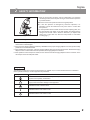
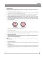
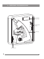
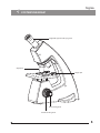
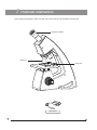
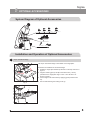
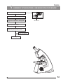
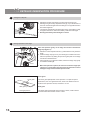
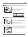
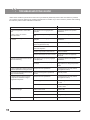
LABOMED sigma User Manual Educational Microscopy To ensure proper use of this instrument as well as to avoid injury while operating instrument, understanding this manual completely before use is highly recommended. CONTENTS 1 INTRODUCTION 1 2 SAFETY INFORMATION 3 UNPACKING YOUR MICROSCOPE 5 4 SYSTEM DIAGRAM 6 5 STANDARD COMPONENTS 7 6 OPTIONAL ACCESSORIES 7 INITIAL SETUP 10 8 ASSEMBLY 11 9 SUMMARY OF BRIGHTFIELD OBSERVATION PROCEDURE 12 10 DETAILED OBSERVATION PROCEDURE 11 TROUBLESHOOTING GUIDE 15 12 SPECIFICATIONS 16 2-4 8-9 13-14 1 INTRODUCTION The Sigma is an educational microscope reflecting a modern design as well as the latest in optical and mechanical advancements. Designed for professionals as well as students, this microscope offers many features and functions for a diverse set of applications. Extra clarity and contrast is provided through an integrated monocular tube, inclined at 45°. The pressure die cast stand housed in plastic covers consists of Ball bearing ‘friction less’ sideways focusing to avoid any loss in motion. The coarse and fine focusing is CAM driven for fatigue free operation. The sturdy new stylish design provides comfort as well as stability. The high powered objectives are spring loaded to prevent accidental damage to specimen slides. The quadruple nosepiece has a comfortable ribbed grip for easy rotation that also safeguards the turret system against any damage. All positions are par-centered and par-focalised ensuring the highest level of accuracy. The LED illumination is operational with an in-built rechargeable battery P/N 2124000-901.The battery is rechargeable with an external adapter with a Universal Power Supply operating at 100V-240V AC input. This ensures continuous operation even under fluctuating voltages. Our LED has an average life span of up to 100,000 hours. 1 2 Sigma SAFETY INFORMATION 1. After the microscope has been used for observation of a specimen containing bacteria, clean all parts coming in contact with the specimen to prevent infection. ? Be sure to remove the specimen before moving this product. ? In case the specimen is damaged by erroneous operation, it is important to clean all surfaces that may have come in contact with the specimen. 2 To avoid potential shock hazards and burns when replacing LED, turn the microscopes main switch to the OFF position and disconnect the charger from the wall outlet in advance. Whenever you replace LED during use or right after use, allow the lamp socket to cool before touching (Fig. 1) Fig. 1 Applicable LED replacement: LED P/N 2124000-950 3. Install the microscope on a sturdy, level table or bench. Do not place the microscope on a flexible surface, as this could result in overheating/fire. 4. Always use the charging adapter provided by LABOMED. If the proper charging adapter is not used, product safety performance cannot be warranted. 5 When installing the microscope, route the charging adapter away from the microscope frame. Should the charging adapter come in contact with the microscopes base, the charging adapter could short. 6 After operation of microscope has ceased, be sure to disconnect the charging adapter from the connector on the microscope or from the wall power outlet. Safety Symbols The following symbols are found on the microscope. For optimal use, it is recommended that users understand these symbols and always use the equipment as prescribed. Symbol Explanation Indicates that the surface has a tendency to heat up and should not be touched unless system has completely cooled down. ! Before use, carefully read the instruction manual. Improper use could result in injury to the user and/or damage to the equipment. Indicates the risk of electric shock. | Indicates that the main switch is ON. Indicates that the main switch is OFF. 2 Warning Label A warning indication label is attached to every part where special precaution is required while handling and using the microscope. Always read the warnings. Warning label position Bottom of microscope frame [Warning against high temperature in battery compartment] [Warning against risk of electric shock] [Warning against damage in noncompliance with this manual] ! If the warning label is stained or peeled off, contact your LABOMED distributor. 1 Getting Ready 1. A microscope is a precision instrument with delicate glass components. Please handle with care. 2. Do not use the microscope where it is subjected to direct sunlight, high temperature, humidity, dust and vibrations. (For operating conditions, see chapter 12, "SPECIFICATIONS" on Page 16) 3. The microscope is ventilated by natural convection. Be sure to leave enough space (10 cm or more) around body when installing it. To prevent damage, do not hold the microscope by the stage. Be sure to remove the specimen from the stage clip while transporting unit to avoid damage to slide. Fig. 2 2 Caution If the microscope is used in a manner not specified by this manual, the safety of the user may not be warranted. In addition, the equipment may also suffer damage. Always use the equipment as outlined in this instruction manual. 3 Care & Maintenance Your microscope has been engineered for a long and safe operational life with the least amount of maintenance required. In general, routine maintenance is limited to keeping the microscope working parts lubricated and optics clean. Always cover the microscope with the provided dust cover when not in use. 1. Cleaning the lenses : To clean the lens surfaces, remove dust using a soft brush or gauze (compressed air dust cans are ideal). For removing finger marks or grease, soft cotton cloth/ lens tissue or gauze lightly moistened with cleaning solution (85% petroleum ether and 15% isopropanol) should be used. For cleaning the objective optics, use xylene. Observe sufficient caution in handling xylene. 3 Sigma Cleaning procedure: Place the objectives and/or eyepieces on a dust-free surface (e.g. fresh aluminum foil). All other optical components to be cleaned should be as accessible as possible. 1. Blow all loose dust particles away with a dust blower. 2. Remove all water-soluble dirt with distilled water. If this is unsuccessful repeat using a solution of diluted hand soap liquid. Remove any remaining residue with a dry cotton swab. 3. To remove oil, use a solution of diluted hand-soap liquid initially. If this does not produce a satisfactory result, repeat the cleaning using a solvent (Optical Cleaning Solution 85% petroleum ether and 15% isopropanol). 4. Grease must always be removed using a solvent. 5. Cleaning is achieved by using a spiral motion from the center to the rim. Never wipe using zig-zag movements as this will only spread the dirt. With larger optical surfaces (e.g. tube lenses) the spiral motion starts initially at the rim before moving to the middle and is only then followed by a center to rim cleaning motion. Normally several spiral wipes are recommended. We recommend pure, volatile petroleum ether or Optical Cleaning Solution as explained in point 3 above. zig-zag motion (X) spiral motion ( ) Wipe using a spiral movement. Do not use a zig-zag motion! 2. Cleaning of painted surfaces : Avoid the use of any organic solvent ( e.g. thinner, xylene, ether, alcohol etc.) for cleaning of painted surfaces of the instrument. Painted surfaces can be cleaned with a very lightly moistened micro fiber cloth. Loose dust and other dirt can be removed using a brush of soft hair used exclusively for this purpose. 3. Cleaning of plastic surfaces: Sigma microscope frame is made up of special grade plastic which can be cleaned with mild soap solution. Do not use acetone for cleaning stage condenser lens. ! Caution: Do not use aggressive organic solvent such as acetone for cleaning painted surfaces and plastic parts of the microscope. 4. Never attempt to dismantle : Never attempt to dismantle the instrument so as to avoid the possibility of impairing its operational efficiency and accuracy. 5. Periodical checking : To maintain the performance of the instrument, we recommend customers have their microscopes serviced periodically by a factory authorized dealer/rep. For details, contact your nearest dealer or Labo America’s main office in California. 4 3 UNPACKING YOUR MICROSCOPE Power Adapter / Battery charger Mechanical stage (optional) Microscope Slides (optional) 5 4 Sigma SYSTEM DIAGRAM Integrated eyepiece with eye guard Objectives Stage clips Fine focusing knob Coarse focusing knob 6 5 STANDARD COMPONENTS After opening the package, make sure that the correct units for the selected set are present. Integrated eyepiece Objectives Stage clips LED Adapter 7 6 Sigma OPTIONAL ACCESSORIES System Diagram of Optional Accessories EP 4x EP 10x EP 40x, spring EP 100x, Spring, oil Clip-on mechanical stage LED Assembly Rechargeable Battery Installation and Operation of Optional Accessories 1 clip-on mechanical stage The clip-on mechanical stage is mountable on the stage plate. Procedure for installation of mechanical stage: 1. Rotate the stage clips outwards (in direction of arrows) as shown in figure 3. 2. Align the indexing pins (A and B) to the index holes (1 and 2) provided on the stage plate. Align ‘A’ with ‘1’ and ‘B’ with ‘2’ as shown in figure 4. 3. Fix the stage in the index holes by applying gentle pressure from top. 4. Secure with fastening the locking screw (3). Fig. 3 3 A 1 B 2 Fig. 4 8 4 3 2 1 Using clip-on mechanical stage (figure 5): 1. Open the bow-shaped lever (4) by pulling lever handle (3) and place specimen slide on the stage. 2. After positioning your specimen slide, return the bow-shaped lever (4) gently by slowly releasing lever handle (3). 3. Use X-axis movement control knob (2) and Y-axis movement control knob (1) for horizontal and vertical movements respectively. ! Fig. 5 9 Do not adjust the specimen holder directly by hand, otherwise it will damage the rotary mechanisms. 7 Sigma INITIAL SET UP The microscope must be charged for at least eight hours before initial use. To charge microscope, plug-in the adapter (1) in the input socket provided at rear of the microscope. See figure 6. 1 Fig. 6 Objectives are factory set. Objectives are par-centered and parfocalised during assembly phase. All objectives have been secured for a tight fit to prevent them from coming loose during transit. To remove an objective, rotate it counterclockwise while holding it with a rubber sheet, etc. to avoid any slippage. Fig. 7 10 8 ASSEMBLY Each standard set can be assembled by simply charging the microscope. 1 Installing or Replacing the LED 5 3 6 1 2 4 7 Fig. 8 Before changing the LED, remove specimen from the microscope frame, and move to the empty objective position. In case all objectives are mounted, move to the 4x objective. 1. Raise the stage to the highest position by rotating the coarse focus knob (7) clock wise. See figure 8. 2. Use allen key 3mm (2) to unlock the screw (1) from the stage to unsecure LED assembly. 3. Detach the LED assembly from connectors (3 & 4) by gently pulling them apart. Secure the lower part of the connector (3) with the stage clip to avoid it from slipping inside. ! Caution: Do not pull the LED assembly excessively as it may damage the wire harness inside the system. 4. Remove the condenser system (5) by rotating it anti clockwise. ! Caution: Remove the LED by holding it with a soft tissue paper or a cloth to avoid finger prints. 5. Remove the LED assembly (6) and replace with new LED assembly. 6. Reverse the procedure from 4 to 2 to secure the system. Applicable LED replacement: LED P/N 9135000-950 Always use the designated parts. Using an LED other than those specified by LABOMED may lead to a fire hazard or improper light level. If contamination occurs, wipe bulb surface with a cloth slightly moistened with alcohol. ! Caution: For LED Replacement During Use or Right After Use The LED socket and areas near these will be hot during and right after use. Set the knob to" O" (OFF), disconnect the charging adapter from the wall outlet, and allow LED assembly to cool before replacing it with a new LED of the designated type. Cooling time may vary with ambient temperature. 2 Battery Replacement Rest the microscope safely aside and follow the following procedure: 1. Peel the sticker showing electrical information. 2. Open the screws (1) as shown in figure 9. 3. Replace the battery. 4. Reverse the operation from point 3 to 2, to complete the process. 5. Replacement battery comes with a sticker which is to be replaced after charging battery to make the system tamper-proof. 1 Fig. 9 11 9 Sigma SUMMARY OF BRIGHTFIELD OBSERVATION PROCEDURE Rotate the knob towards “I” (ON) Place the specimen on the stage Engage the 10X objective in the light path ON/OFF Knob Bring the specimen in focus Engage the objective to be used in the light path and bring the specimen in focus Adjust the brightness Observe Specimen 12 10 DETAILED OBSERVATION PROCEDURE 1 Turning the Lamp ON 1. Rotate the On/OFF control knob to ”I” (ON) as shown in figure 10. 2. Rotating the light intensity adjustment knob (fig. 10) in the direction of the arrow increases brightness and rotating it in the opposite direction decreases brightness. 3. The intensity adjustment knob illuminates “Green” when battery is fully charged. It starts to turn “Red” as the battery needs to be recharged. Recharge the battery when Red light is noticed. 1 Fig. 10 2 Placing specimen on the stage Place the specimen gently on the stage and secure it beneath the stage clips (figure 11). 1. Rotate the coarse adjustment knob (1) anticlockwise to fully lower the stage. 2. Press the stage clips (2) one by one with finger to lift them from front. Place the specimen by sliding the specimen glass plate on the stage from the front toward the rear. 3. After positioning your specimen slides, release the stage clips gently on the slide to hold it tight. 2 1 When the specimen is placed, do not move it with the stage clips Fig. 11 resting on it. Lift the stage clips and move the specimen to avoid damage to stage surface and the specimen slide. Cover glass Cover glass Slide glass Slide glass Placing Specimen This glass plate should ideally have a length of 76mm, width of 26mm ±1mm and thickness between 0.9 and 1.4mm. Fig. 12 13 This is the glass plate placed on the specimen. For optimum optical performance, the cover glass thickness, which is the distance from its surface to the specimen surface, should be 0.17 mm. Sigma 3 Adjusting the Focus Focusing Procedure (Fig. 13) 1. Rotate the coarse adjustment knob (1) clockwise so that the objective (3) is as close as possible to the specimen (We recommend starting with 10X). 2. While observing the specimen through the eyepiece, slowly rotate the coarse adjustment knob (1) counterclockwise to lower the stage. 3. When coarse focusing of the specimen is obtained (an image is detected), rotate the fine adjustment knob (2) for fine focusing. 3 WD 1 Working Distance (WD) 2 Fig. 13 4 The WD refers to the distance between each objective and the specimen, when precise focus of the specimen is obtained. Objective Magnification 4X 10X 40X 100X WD (mm) 22 10.5 0.56 0.13 Using the Eye Shades Using the Eye Shades When Wearing Eyeglasses Use with the eye shades in the normal, folded-down position. This will prevent the eyeglasses from being scratched. When Not Wearing Eyeglasses Extend the folded eye shades outwards (direction of the arrow) to prevent extraneous light from entering into your line of vision. 5 Switching the Objectives Hold and rotate the revolving nosepiece turret (1) so that the objective to be used is in line above the specimen. Always use the ribbed grip to rotate the objective nosepiece. 1 Fig. 15 14 11 TROUBLESHOOTING GUIDE Under certain conditions, performance of the unit may be adversely affected by factors other than defects. If problems occur, please review the following list and take remedial action as needed. If you cannot solve the problem after checking the entire list, please contact Labomed for assistance. Trouble 1. Uneven brightness in observation field The objective is not engaged in the light path Engage the objective into position until it clicks 2. Dust or stains are visible in observation field The eyepiece or specimen glasses are dirty Clean them thoroughly 3. Observation image is whitish-blurred or unclear The objective is not engaged in the light path Engage the objective into position until it clicks The objective, eyepiece and/or specimen glasses are dirty Clean them thoroughly Immersion oil is not used with an immersion objective Use immersion oil Bubbles are present in immersion oil Remove bubbles The specified immersion oil is not used Use the immersion oil supplied by Labomed The objective is not properly engaged in the light path Engage the objective into position until it clicks The specimen is not set properly on the stage Set the specimen correctly on the stage and secure using the specimen holder 4. Part of image is defocused or image looks like it’s flowing 5. High-magnification objective touches The specimen is upside down specimen just before coming into focus Set the specimen correctly with the cover glass on the top 6. Focusing is impossible (because the stage cannot be raised) The pre-focusing knob is positioned too low Raise its position 7. Objective hits the specimen when an objective is switched to a higher magnification objective The specimen is upside down Set the specimen correctly with the cover glass on the top The cover glass is too thick Use a cover glass with thickness of 0.17mm Slide is of excessive thickness Use slide having thickness between 0.9 and 1.4mm LED is not mounted Attach a LED LED is blown Replace the LED Battery is low Charge battery The specified LED is not used Replace with a specified LED 8. LED does not light 9. LED blows easily 15 Remedy Cause Sigma 12 SPECIFICATIONS 1. Illumination Built-in illumination system LED type 2. Focusing mechanism Stage height adjustment mechanism Fine adjustment stroke: 0.2mm per turn Total stroke: 8mm Co-axial coarse and fine focusing with gear movements 3. Revolving nosepiece Quadruple nosepiece (reverse angle) 4. Integrated Monocular head Field number 18 Tube tilting angle 45° Size 125 x 120mm (with mechanical stage) Specimen holder Holds a single specimen 5. Stage 6. Dimensions 267.0mm (L) x 179.6mm (W) x 368.0mm (H) 7. Electrical Battery 3.7V, 500mAH Charging time up to 3 hours (with totally consumed battery) Back up time up to 8 hours 8. Operating environment Indoor use Altitude: Max. 2000 meters Ambient temperature: 5° to 40°C (41° to 104° F) Maximum relative humidity: 80% for temperature up to 31°C (88°F), decreasing linearly through 70% at 34°C (93°F), to 50% relative humidity at 40°C (104°F) Supply voltage fluctuations: Not to exceed ±10% of the normal voltage. Pollution degree: 2 (in accordance with IEC60664) Installation/Overvoltage category: II (in accordance with IEC60664) 16 www.laboamerica.com Our policy is one of continuous development. Labo America, Inc., reserves the right to change design and specifications without prior notice. Labo America Inc. 920 Auburn Court Fremont CA 94538 U.S.A. Telephone: 510 445 1257 Fax: 510 445 1317 [email protected] LABOMED and Sigma are registered trademarks of Labo America, Inc. With a policy of continuous development, Labo America, Inc. reserves the right to change design and specifications without prior notice. © 2009 Labo America, Inc. | 2124000-990A 12-2009