1
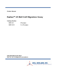
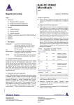
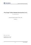
Product Manual CytoSelect™ 96-Well Cell Transformation Assay (Soft Agar Colony Formation), Trial Size Catalog Number CBA-130-T 24 assays FOR RESEARCH USE ONLY Not for use in diagnostic procedures Introduction Neoplastic transformation occurs via a series of genetic and epigenetic alterations that yield a cell population that is capable of proliferating independently of both external and internal signals that normally restrain growth. For example, transformed cells show reduced requirements for extracellular growth promoting factors, are not restricted by cell-cell contact, and are often immortal. Anchorageindependent growth is one of the hallmarks of transformation, which is considered the most accurate and stringent in vitro assay for detecting malignant transformation of cells. Traditionally, the soft agar colony formation assay is a common method to monitor anchorageindependent growth, which measures proliferation in a semisolid culture media after 3-4 weeks by manual counting of colonies. Standard soft agar assays are usually performed in 100-mm or 60 mm dishes, where cells are allowed to grow inside a semisolid culture media for 3-4 weeks before sizable colonies appear. This method is quite cumbersome, time-consuming, and difficult when testing a large number of samples. Additionally, the manual counting of colonies is highly subjective; with varying colony sizes, it’s difficult to determine meaningful results. Cell Biolabs CytoSelect™ 96-well Cell Transformation Assay does not involve subjective manual counting of colonies or require a 3-4 week incubation period. Instead cells are incubated only 6-8 days in a semisolid agar media before being solubilized, lysed and detected by the patented CyQuant® GR Dye in a fluorescence plate reader (see Assay Principle below). This format provides a quantitative, high-throughput method to accurately measure cell transformation. Additionally, the short incubation time (6-8 days) makes it possible to assay cells transiently transfected with oncogenes or siRNA. The CytoSelect™ 96-well Cell Transformation Kit provides a robust system for screening oncogenes and cell transformation inhibitors. Each Trial Size kit provides sufficient quantities to perform 24 tests in a 96-well microtiter plate. 2 Assay Principle Related Products 1. CBA-106: CytoSelect™ 96-Well Cell Migration Assay (8µm, Fluorometric) 2. CBA-106-C: CytoSelect™ 96-Well Cell Migration and Invasion Assay (8µm, Fluorometric) 3. CBA-110: CytoSelect™ 24-Well Cell Invasion Assay (Basement Membrane, Colorimetric) 4. CBA-112: CytoSelect™ 96-Well Cell Invasion Assay (Basement Membrane, Fluorometric) 5. CBA-135: CytoSelect™ 96-Well Cell Transformation Assay (Cell Recovery, Colorimetric) 6. CBA-140: CytoSelect™ 96-Well Cell Transformation Assay (Cell Recovery, Fluorometric) 7. CBA-145: CytoSelect™ 384-Well Cell Transformation Assay 8. CBA-150: CytoSelect™ In Vitro Tumor Sensitivity Assay 9. CBA-155: CytoSelect™ Clonogenic Tumor Cell Isolation Kit 10. CBA-320: CytoSelect™ 96-Well Hematopoietic Colony Forming Cell Assay 3 Kit Components 1. CytoSelect™ Agar Powder (Part No. 113001-T): One 0.24 g tube 2. 5X DMEM Solution (Part No. 113002): One 1.5 mL sterile tube 3. Agar Solubilization Solution (Part No. 113003-T): One 1.5 mL amber tube 4. 8X Lysis Buffer (Part No. 113004-T): One 1 mL tube 5. CyQuant GR Dye (Part No. 10103-T): One 10 µL tube Materials Not Supplied 1. Cells and Culture Medium 2. 1X PBS 3. 37ºC Incubator, 5% CO2 Atmosphere 4. Light Microscope 5. 96-well Fluorometer 6. Microwave or Heating Block 7. Water bath 8. (Optional) Positive Control cells such as NIH 3T3 (Ras G12V) Storage Store all components at 4ºC until their expiration dates. Preparation of Reagents 1.2% Agar Solution: Place 0.24 g of Agar Powder in a sterile bottle, then add 20 mL of sterile cell culture grade water. Microwave or boil until agar is completely dissolved. 2X DMEM/20% FBS Medium: In a sterile tube, dilute the provided 5X DMEM in sterile cell culture grade water to 2X containing 20% FBS. For example, to prepare a 2.5 mL solution, add 1 mL of 5X DMEM, 0.5 mL of FBS and 1 mL of sterile cell culture grade water. Sterile filter the 2X media to 0.2 µm. Note: You may substitute your own medium in place of the DMEM we provide, but ensure that it is at a 2X concentration. CyQuant Working Solution: Immediately before use, prepare sufficient amount of the CyQuant Working Solution by diluting the CyQuant GR Dye 1:400 with 1X PBS. For example, add 10 µL to 4 mL of 1X PBS. Use the solution immediately; do not store the CyQuant Working Solution. 4 Assay Protocol (must be under sterile conditions) I. Preparation of Base Agar Layer 1. Melt 1.2% Agar Solution in a microwave and cool to 37ºC in a water bath. 2. Warm 2X DMEM/20% FBS medium to 37ºC in a water bath. Allow at least 30 minutes for the temperature to equilibrate. 3. Mix equal volumes of 1.2% Agar Solution and 2X DMEM/20% FBS medium in a sterile, prewarmed tube by inverting several times. Immediately transfer 50 µL of the mixture to each well of a 96-well sterile flat-bottom microplate. Gently tap the plate a few times to allow the agar solution to evenly cover the wells. Notes: Work quickly with the agar solution to avoid gelation. Also, try to avoid adding air bubbles to the well. To avoid fast and uneven evaporation that leads to aberrant results, we suggest not using the wells on the plate edge, or filling the edge wells with medium to reduce evaporation. 4. Transfer the plate to 4ºC for 30 minutes to allow the base agar layer to solidify. 5. Prior to adding the Cell Agar Layer (Section II), allow the plate to warm up for 15 minutes at 37ºC. II. Preparation of Cell Agar Layer (samples should be assayed in triplicate) 1. Melt 1.2% Agar Solution in a microwave and cool to 37ºC in a water bath. 2. Warm 2X DMEM/20% FBS medium to 37ºC in a water bath. Allow at least 30 minutes for the temperature to equilibrate. 3. Harvest and resuspend cells in culture medium at 0.4 - 4 x 10⁵ cells/mL, keep the cell suspension warm in a 37ºC water bath. 4. Mix equal volumes of 1.2% Agar Solution, 2X DMEM/20% FBS media, and cell suspension (1:1:1) in a sterile, pre-warmed tube by inverting several times. Immediately transfer 75 µL of the mixture to each well of the 96-well flat-bottom microplate already containing the solidified base agar layer (25 µL of cell suspension containing 1000-10000 cells/well will be seeded). Note: Work quickly with the agar solution to avoid gelation, but gently pipette as not to disrupt the base layer integrity. Also, try to avoid adding air bubbles to the well. Always include negative control wells that contain no cells in the cell agar layer. 5. Transfer the plate to 4ºC for 15 minutes to allow the cell agar layer to solidify. III. Quantitation of Anchorage-Independent Growth 1. Add 100 µL of culture medium containing cell growth activator(s) or inhibitor(s) to each well. 2. Incubate the cells for 6-8 days at 37ºC and 5% CO2. Examine the cell colony formation under a light microscope. 5 3. Remove culture medium by inverting the plate and blotting on paper towel. Gently tap several times. 4. Add 50 µL of Agar Solubilization Solution to each well of the 96-well plate. Incubate for 1 hr at 37ºC. 5. Pipette each well 5-10 times to ensure complete agar solubilization. 6. Add 25 µL of 8X Lysis Buffer to each well. Pipette each well 5-10 times to ensure a homogeneous mixture. 7. Incubate the plate at room temperature for 15 minutes. 8. Transfer 10 µL of the mixture to a 96-well plate suitable for fluorescence measurement. 9. Add 90 µL of the CyQuant Working Solution to each well. Incubate 10 minutes at room temperature. 10. Read the plate in a 96-well fluorometer using a 485/520 nm filter set. Cell Dose Curve (optional) 1. Harvest and resuspend cells in culture medium at 1 - 5 x 10⁶ cells/mL. 2. Prepare a 2-fold serial dilution with culture medium, including a medium blank. 3. Transfer 125 µL of each cell dilution to a microfuge tube. Add 50 µL of Agar Solubilization Solution and 25 µL of 8X Lysis Buffer to each tube. Vortex each tube to ensure a homogeneous mixture. Incubate the tubes at room temperature for 15 minutes. 4. Transfer 10 µL of the mixture to a 96-well plate suitable for fluorescence measurement. 5. Add 90 µL of the CyQuant Working Solution to each well. Incubate 10 minutes at room temperature. 6. Read the plate in a 96-well fluorometer using a 485/520 nm filter set. 6 Example of Results 50 50 40 40 30 30 RFU RFU The following figures demonstrate typical results with the CytoSelect™ 96-well Cell Transformation Assay Kit. Fluorescence measurement was performed on SpectraMax Gemini XS Fluorometer (Molecular Devices) with a 485/538 nm filter set and 530 nm cutoff. One should use the data below for reference only. This data should not be used to interpret actual results. 20 20 10 10 0 0 0 1000 2000 3000 4000 0 5000 10000 15000 20000 25000 Cells/mL (x1000) Cells Figure 1. HeLa Cell Dose Curve. Cervical carcinoma HeLa cells were resuspended at 4 x 10⁶ cells/mL and titrated 1:2 in culture medium, followed by addition of Agar Solubilization Solution, Lysis Buffer, and Cyquant® GR Dye detection (as described in the Cell Dose Section). Results were shown by cell concentration or by actual cell number in CyQuant Detection. 20 HeLa NIH3T3 RFU 15 10 5 0 20000 10000 5000 2500 1250 625 313 0 Cells Seeded/Well Figure 2. Anchorage-Independent Growth of HeLa Cells. HeLa cells were seeded at various concentrations and cultured for 6 days. HeLa cell transformation is determined according to the assay protocol. 7 Figure 3. HeLa Colony Formation. HeLa cells were cultured for 14 days according to the assay protocol. Colonies were visualized by 0.1% p-iodonitro tetrazolium violet (INT) staining. Calculation of Anchorage-Independent Growth 1. Compare RFU values with the Cell Dose Curve and extrapolate the cell concentration in soft agar. 2. Calculate the Total Transformed Cell Number/Well Total Transformed Cells/Well = cells/mL in soft agar x 0.125 mL/well For example: If you extrapolate your RFU value from your cell dose curve and determine you have 500,000 cells/mL in your soft agar sample. Total Transformed Cells/Well = 500,000 cells/mL x 0.125 mL/well = 62,500 cells/well References 1. Shin SI, Freedman VH, Risser R, and Pollack R. (1975) Proc Natl Acad Sci U S A. 72:4435-9. 2. Hahn WC, Counter CM, Lundberg AS, Beijersbergen RL, Brooks MW and Weinberg RA. (1999) Nature 400:464-8. Recent Product Citations 1. Ercan, D. et al. (2015). EGFR mutations and resistance to irreversible pyrimidine based EGFR inhibitors. Clin Cancer Res. 21:3913-3923. 2. Mayr, C. et al. (2015). 3-Deazaneplanocin A may directly target putative cancer stem cells in biliary tract cancer. Anticancer Res. 35:4697-4705. 3. He, C. et al. (2015). The Hippo/YAP pathway interacts with EGFR signaling and HPV oncoproteins to regulate cervical cancer progression. EMBO Mol Med. doi:10.15252/emmm.201404976. 4. Hua, G. et al. (2015). YAP induces high-grade serous carcinoma in fallopian tube secretory epithelial cells. Oncogene. doi:10.1038/onc.2015.288. 5. Ukaji, T. et al. (2015). Inhibition of IGF-1-mediated cellular migration and invasion by migracin A in ovarian clear cell carcinoma cells. PLoS One. 10:e0137663. 8 6. Fujita, T. et al. (2015). Identification and characterization of CXCR4-positive gastric cancer stem cells. PLoS One. 10:e0130808. 7. Bon, H. et al. (2015). Salt-inducible Kinase 2 regulates mitotic progression and transcription in prostate cancer. Mol Cancer Res. 13:620-635. 8. Yonesaka, K. et al. (2015). Anti-HER3 monoclonal antibody patritumab sensitizes refractory nonsmall cell lung cancer to the epidermal growth factor receptor inhibitor erlotinib. Oncogene. doi: 10.1038/onc.2015.142. 9. Mayr, C. et al. (2015). Cytotoxic effects of chemokine receptor 4 inhibition by AMD3100 in biliary tract cancer cells: Potential drug synergism with gemcitabine. Mol Med Rep. doi: 10.3892/mmr.2015.3589. 10. He, C. et al. (2015). YAP forms autocrine loops with the ERBB pathway to regulate ovarian cancer initiation and progression. Oncogene. doi: 10.1038/onc.2015.52. 11. Kim, T. et al. (2015). Role of MYC-regulated long noncoding RNAs in cell cycle regulation and tumorigenesis. J Natl Cancer Inst. 107:dju505. 12. Galvan, A. et al. (2015). Germline polymorphisms and survival of lung adenocarcinoma patients: a genome-wide study in two european patient series. Int J Cancer. 136:E262-E271. 13. Nakanishi, Y. et al. (2015). Mechanism of oncogenic signal activation by the novel fusion kinase FGFR3-BAIAP2L1. Mol Cancer Ther. doi: 10.1158/1535-7163.MCT-14-0927-T. 14. Westbom, C. M. et al. (2014). CREB-induced inflammation is important for malignant mesothelioma growth. Am J Pathol. 184:2816-2827. 15. Kong, L. Y. et al. (2014). Therapeutic targets in subependymoma. J Neuroimmunol. 277:168-175. 16. Takiguchi, S. et al. (2014). Involvement of CXCL14 in osteolytic bone metastasis from lung cancer. Int J Oncol. 44:1316-1324. 17. Wang, L. et al. (2014). Ectopic over-expression of miR-429 induces mesenchymal-to-epithelial transition (MET) and increased drug sensitivity in metastasizing ovarian cancer cells. Gynecol Oncol. 134:96-103. 18. Sun, Y. et al. (2014). Cholesterol-induced activation of TRPM7 regulates cell proliferation, migration, and viability of human prostate cells. Biochim Biophys Acta. 1843:1839-1850. 19. Priya, S. et al. (2014). Diethyl maleate inhibits MCA+ TPA transformed cell growth via modulation of GSH, MAPK, and cancer pathways. Chem Biol Interact. 219:37-47. 20. Nakamura, T. et al. (2014). Osteopontin-integrin αvβ3 axis is crucial for 5-fluorouracil resistance in oral squamous cell carcinoma. FEBS Lett. 589:231-239. 21. Eckerdt, F. et al. (2014). Regulatory effects of a Mnk2-eIF4E feedback loop during mTORC1 targeting of human medulloblastoma cells. Oncotarget. 5:8442-8451. 22. He, X., et al. (2014). Involvement of polypyrimidine tract-binding protein (PTBP1) in maintaining breast cancer cell growth and malignant properties. Oncogenesis. 3:e84. 23. Lin, L. et al. (2014). A large-scale RNAi-based mouse tumorigenesis screen identifies new lung cancer tumor suppressors that repress FGFR signaling. Cancer Discov. 4:1168-1181. 24. Zecchini, V. et al. (2014). Nuclear ARRB1 induces pseudohypoxia and cellular metabolism reprogramming in prostate cancer. EMBO J. 33:1365-1382. 25. Eisfeld, A.K. et al. (2014). NRAS isoforms differentially affect downstream pathways, cell growth, and cell transformation. PNAS 111:4179-4184. 26. Gong, J. et al. (2014). Combined targeting of STAT3/NF-kB/COX-2/EP4 for effective management of pancreatic cancer. Clin. Cancer Res. 20:1259-1273. 27. Kegelman, T. et al. (2014). MDA-9/Syntenin is a key regulator of glioma pathogenesis. Neuro Oncology. 16:50-61. 9 28. Giacoia, E.G. et al. Targeting Plasminogen Activator Inhibitor-1 inhibits angiogenesis and tumor growth in a human cancer xenograft model. Mol. Cancer Ther. 12:2697-2708. 29. Hatano, K. et al. (2013). Residual prostate cancer cells after docetaxel therapy increase the tumorigenic potential via constitutive signaling of CXCR4, ERK1/2 and c-Myc. Mol. Cancer Res. 11:1088-1100. 30. Gupta, P. et al. (2013).Oncrasin targets the JNK-NF-B axis to sensitize glioma cells to TNFαinduced apoptosis. Carcinogenesis. 34:388-396. 31. Xing, C. et al. (2013). Reversing effect of ring finger protein 43 inhibition on malignant phenotypes of human hepatocellular carcinoma. Mol. Cancer Ther. 12:94-103. 32. Rai, V. et al. (2012). Lysophosphatidic acid targets vascular and oncogenic pathways via RAGE signaling. J. Exp. Med. 209: 2339-2350. 33. Ghantous, A. et al. (2012). Inhibition of tumor promotion by parthenolide: epigenetic modulation of p21. Cancer Prevention Research. 5: 1298-1309. 34. Shin, S.Y. et al. (2012). Transcriptional regulation of the interleukin-11 gene by oncogenic Ras. Carcinogenesis. 10.1093/carcin/bgs297. 35. Tan, X. et al. (2012). cAMP response element-binding protein promotes gliomagenesis by modulating the expression of oncogenic microRNA-23a. PNAS. 109: 15805-15810. 36. Maccario, C. et al. (2012).The resveratrol analog 4,4'-dihydroxy-trans-stilbene suppresses transformation in normal mouse fibroblasts and inhibits proliferation and invasion of human breast cancer cells. Carcinogenesis. 10.1093/carcin/bgs244. License Information This product is provided under an intellectual property license from Life Technologies Corporation. The purchase of this product conveys to the buyer the non-transferable right to use the purchased product and components of the product only in research conducted by the buyer (whether the buyer is an academic or for-profit entity). The sale of this product is expressly conditioned on the buyer not using the product or its components, or any materials made using the product or its components, in any activity to generate revenue, which may include, but is not limited to use of the product or its components: (i) in manufacturing; (ii) to provide a service, information, or data in return for payment; (iii) for therapeutic, diagnostic or prophylactic purposes; or (iv) for resale, regardless of whether they are resold for use in research. For information on purchasing a license to this product for purposes other than as described above, contact Life Technologies Corporation, 5791 Van Allen Way, Carlsbad CA 92008 USA or [email protected]. Warranty These products are warranted to perform as described in their labeling and in Cell Biolabs literature when used in accordance with their instructions. THERE ARE NO WARRANTIES THAT EXTEND BEYOND THIS EXPRESSED WARRANTY AND CELL BIOLABS DISCLAIMS ANY IMPLIED WARRANTY OF MERCHANTABILITY OR WARRANTY OF FITNESS FOR PARTICULAR PURPOSE. CELL BIOLABS’ sole obligation and purchaser’s exclusive remedy for breach of this warranty shall be, at the option of CELL BIOLABS, to repair or replace the products. In no event shall CELL BIOLABS be liable for any proximate, incidental or consequential damages in connection with the products. 10 Contact Information Cell Biolabs, Inc. 7758 Arjons Drive San Diego, CA 92126 Worldwide: +1 858-271-6500 USA Toll-Free: 1-888-CBL-0505 E-mail: [email protected] www.cellbiolabs.com 2014-2015: Cell Biolabs, Inc. - All rights reserved. No part of these works may be reproduced in any form without permissions in writing. 11