1
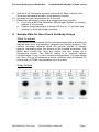
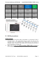
Q ™™ Exosome Antibodies, Array & ELISA Kits Cat #s EXOAB,EXORAY, EXOEL User Manual See page #2 for product storage conditions A limited-use label license covers this product. By use of this product, you accept the terms and conditions outlined in the Licensing and Warranty Statement contained in this user manual. Exosome Antibody and ELISA Kits Cat.#s EXOAB, EXORAY, EXOEL Series Contents List of components 2 Exosome Antibodies I. Recommended protocol 3 II. Serum exosome Western data 5 Exosome Antibody Arrays (Exo-Check I. Protocol…………………………………………………………….6 II. Example data………………………………………………………7 ExoELISA Kits I. ExoELISA principle 8 II. Equipment supplied by user 9 III. Protocol 9 A. Exosome precipitation with ExoQuick/ExoQuick-TC 9 B. Exosome isolated with ultracentrifugation method...10 C. Exosome protein standard 10 D. ELISA procedures 11 E. Sample Data 13 IV. Citations………………………………………………………….14 V. Technical references…………………………………………..15 VI. Technical Support 16 VII. Licensing and Warranty Statement 18 888-266-5066 (Toll Free) 650-968-2200 (outside US) Page 1 System Biosciences (SBI) User Manual List of Components EXOAB Series Exosome Antibodies Components Exosome specific primary antibody (CD63,CD9 ,CD81 or Hsp70 ) Exosome validated secondary antibody (Goat anti-Rabbit HRP) Amount 25 µl 5 µl The Exosome Antibodies are shipped in blue ice and should be stored at +4°C upon receipt. . Properly stored antibodies are stable for 6 months from the date received. They can be place at -20°C for long term storage. EXORAY Series Exo-Check Exosome Antibody Arrays Antibody Membrane Array (PVDF membrane) – 4, 8 or 12 array membranes Exosome Lysis buffer Exosome Array Binding buffer Array Wash buffer Detection buffer (HRP-conjugated secondary mixture) Disposable blue forceps to handle membrane arrays The Exosome Antibody array kits are shipped in blue ice and should be stored at +4°C upon receipt. Properly stored antibody array kits are stable for 6 months from the date received. * Not supplied: HRP developer solution, we recommend the SuperSignal West Femto Chemi-luminescent Substrate, THERMO catalog# 34095. Page 2 ver. 6-2014-01-02 www.systembio.com Exosome Antibody and ELISA Kits Cat.#s EXOAB, EXORAY, EXOEL Series EXOEL Series ExoELISA kit Components Amount Exosome binding buffer 20 ml 20X Wash buffer 18 ml Blocking buffer 30 ml ExoELISA protein standard* 400 µl Exosome specific primary antibody (CD63,CD9 or CD81) 2 x 25 µl Exosome validated secondary antibody (Goat anti-Rabbit HRP) 10 µl ELISA substrate (Super–sensitive TMB) 6 ml Stop buffer 6 ml 96 well ExoELISA plate (12x8 well strips) 1 plate The ExoELISA™ kits are shipped in blue ice and should be stored at +4°C upon receipt. Properly stored kits are stable for 6 months from the date received. * ExoELISA protein standards should be stored at -20°C upon receipt. We recommend making single-use aliquots to avoid repeated freezethawing. Exosome Antibodies (Cat# EXOAB series) I. Recommended protocol -for isolating serum exosomes for Western blotting analysis 1. 2. 3. 4. 5. 6. 7. 8. 9. If frozen, thaw 250 µl serum on ice Centrifuge at 1500 × g for 15 minutes to remove cells and cell debris Transfer sample supernatant to a centrifuge tube Combine 250 µl sample + 63 µl ExoQuick™ Mix well by inversion three times Place at 4ºC for at least 30 minutes Centrifuge at 1500 × g for 5 minutes Remove supernatant, keep exosome pellet Centrifuge at 1500 × g for 5 minutes to remove all traces of fluid (take great care not to disturb the pellet) 888-266-5066 (Toll Free) 650-968-2200 (outside US) Page 3 System Biosciences (SBI) User Manual 1 10. Add 200 µl RIPA buffer (with appropriate protease inhibitor cocktail added) to exosome pellet and vortex 15 seconds 11. Place at room temperature for 5 minutes (to allow complete lysis) 12. Perform standard Bradford protein assay to determine yield. 2 13. Add Laemmli buffer (with Beta-mercaptoethanol) and heat at 95⁰C for 5 minutes. 14. Chilled on ice for 5 minutes before loading onto gel 15. Perform standard SDS-PAGE electrophoresis and Western transfer onto PVDF membrane 16. Block with 5% dry milk in Tris Buffered Saline + 0.05% Tween (TBS-T) for 1 hour 17. Incubate blot overnight at 4°C with Exosome specific primary antibody (e.g. CD9) at 1:1000 dilution (5% dry milk in TBS-T) 18. Wash 3X with TBS-T 19. Incubate one hour at room temperature with Exosome validated secondary antibody (Goat-Rabbit-HRP) antibody at 1:20,000 dilution (5% dry milk in TBS-T) 20. Wash 3X with TBS-T 21. Incubate blot with chemi-luminescence substrate and visualize on film or other imaging equipment. We recommend the SuperSignal West Femto Chemi-luminescent Substrate, THERMO catalog# 34095. 1 1X RIPA buffer contains: 25mM Tris-HCl pH 7.6 150mM NaCl 1% NP-40 1% sodium deoxycholate 0.1% SDS 2 2X Laemmli buffer contains: 4% SDS 20% glycerol 10% 2-mercaptoethanol 0.004% bromophenol blue 0.125 M Tris-HCl pH 6.8 Page 4 ver. 6-2014-01-02 www.systembio.com Exosome Antibody and ELISA Kits II. Cat.#s EXOAB, EXORAY, EXOEL Series Serum Exosome Western Data Western blot analysis of serum exosomes using the recommended protocol. 20 µg of serum exosome proteins were loaded into each lane. All Exosome specific primary antibodies were used at 1:1,000 dilution and Exosome validated secondary antibody was used at 1:20,000 dilution. We recommend the SuperSignal West Femto Chemi-luminescent Substrate, THERMO catalog# 34095. Exosome Antibody Arrays (Cat# EXORAY series) The Exo-Check antibody array has 12 pre-printed spots and features 8 antibodies for known exosome markers (CD63, CD81, ALIX, FLOT1, ICAM1, EpCam, ANXA5 and TSG101) and a GM130 cisGolgi marker to monitor any cellular contamination in your exosome isolations. The array is compatible with ultracentrifugation and ExoQuick methods. The positive control spot is derived from human serum exosome proteins and are printed directly onto the membrane. Your exosome preparations are lysed and then incubated with the array for the pre-printed antibodies to capture their respective exosome proteins. The GM130 antibody spot on each array is included to help identify and cellular contamination in your exosome preparations. The kits come complete with a secondary detection mixture conjugated to HRP. The arrays are semi-quantitative and can be used to evaluate relative abundance of certain exosome protein 888-266-5066 (Toll Free) 650-968-2200 (outside US) Page 5 System Biosciences (SBI) User Manual markers from a set of given samples. The orientation of the Array is identified as using the upper left hand corner that is notched. I. Protocol for Exo-Check Antibody Arrays Sample preparation and antibody array capture 1. 2. 3. 4. 5. 6. 7. 8. Precipitate sample exosomes from serum or culture media using ExoQuick or ExoQuick-TC (also compatible with exosome pellets from ultracentrifugation) to obtain an exosome pellet. We suggest when using ExoQuick, start with 500 ul serum sample 300 ul PBS. For culture media, we suggest starting with 10 ml media and then resuspend exosome pellet with 300 ul PBS. Use a small volume of resuspended exosomes and perform a protein assay to determine the recovery. Add 300-500 ug exosome proteins with 600 ul Exosome Lysis buffer. Vortex for 15 seconds to prepare exosome protein lysate mixture. In a fresh 15 mL Falcon tube, combine the 600 ul exosome lysate with 9.4 mL Exosome Array Binding buffer and mix by inverting 3 times. In a small, clean tray, briefly wet the antibody array in 5 mL distilled water for 2 minutes at room temperature. Decant the water from the membranes. Pipette the 10 mL Exosome lysate / binding mixture to one Exo-Check antibody membrane array. Place the membrane “face-up” by positioning the membrane such that the upper left notched corner can be seen in the left hand side on top. Incubate the tray/membrane mixture shaking at 2-8°C overnight on a shaker or a rocker. Antibody array washing and signal detection 1. 2. 3. 4. 5. 6. 7. 8. Decant the Exosome lysate binding mixture from the membrane carefully. Add 10 mL Array wash buffer (made into 1X from 20X stock) and shake gently for 5 minutes at room temperature. Decant the Array wash buffer. Add 10 mL Detection buffer to Array membrane and gently shake at room temperature for 2 hours. Prepare the Developer mixture (not included) about 5 minutes before the end of Step #5 by combining 1 mL solution A with 1 mL solution B together. We recommend the SuperSignal West Femto Chemiluminescent Substrate, THERMO catalog# 34095. Decant the Detection buffer Add 10 mL Array wash buffer (made into 1X from 20X stock) and shake gently for 5 minutes at room temperature. Decant the Array wash buffer, repate twice for a total of three washes. Page 6 ver. 6-2014-01-02 www.systembio.com Exosome Antibody and ELISA Kits Cat.#s EXOAB, EXORAY, EXOEL Series 9. Add the 2 mL Developer solution mixture from Step 5 directly onto the Array membrane so that it is completely covered. 10. Incubate at room temperature for 2 minutes. 11. Decant the developer solution and image the Array signals. a. If you use a Bio-Rad ChemiDoc XRS Imager System (or similar), expose 4-10 seconds. b. Expose Array membrane to Kodak XR film for 1-3 minutes and develop in a film developer machine. II. Sample Data for Exo-Check Antibody Arrays What to expect The positive control signals should provide a bright signal and this will indicate that all of the detection reagents are working properly. The various exosome antibody spots will provide signals of varying degree, depending upon the source of the isolated exosomes. The Blank spot should not have any signal and this serves as a background control. The GM130 control is to evaluate cellular contamination (if any) in your exosome preparations. The data below are from 300 ug of exosome proteins isolated using ExoQuick-TC from human HT1080 lung sarcoma cell line media. Array format 888-266-5066 (Toll Free) 650-968-2200 (outside US) Page 7 System Biosciences (SBI) User Manual Information about the exosome protein targets ExoELISA™ - (Cat# EXOEL series) I. ExoELISA principle The ExoELISA kit is designed as a direct Enzyme-Linked ImmunoSorbent Assay (ELISA). The lysed exosome particles and their proteins are directly immobilized onto the wells of the microtiter plate. After binding, the wells are coated with a block agent to prevent non-specific binding of the primary detection antibody. The detection (primary) antibody is added to the wells for binding to a specific antigen (e.g. CD63) protein present in the exosome lysates. A Horseradish Peroxidase enzyme linked secondary antibody (Goat anti-Rabbit) is used for signal amplification and to increase assay sensitivity. A colorimetric substrate (Extra-sensitive TMB) is used for the assay read-out. The accumulation of the colored product is proportional to the specific antigen present in each well. The results are quantitated by a microtiter plate reader at 450 nm absorbance. Page 8 ver. 6-2014-01-02 www.systembio.com Exosome Antibody and ELISA Kits II. Cat.#s EXOAB, EXORAY, EXOEL Series Equipment to be supplied by user 1. 2. 3. 4. 5. III. Microtiter plate sealing film/cover 37°C incubator Microtiter plate shaker Microtiter plate spectrophotometer with 450 nm absorbance capability Multichannel pipets (recommended) Protocol A. Exosomes precipitation with ExoQuick/ExoQuick-TC For simple and quick isolation of exosomes from serum, we recommend using the ExoQuick precipitation solution (Catalog# EXOQ5A-1 or EXOQ20A-1) and the ExoQuick-TC for isolation of exosomes from tissue culture media and urine samples (EXOTC10A-1 or EXOTC50A-1). 1. 2. 3. If frozen, thaw sample on ice Centrifuge at 3000 × g for 15 minutes to remove cells and cell debris. Transfer supernatant to a sterile vessel and add the appropriate volume of ExoQuick or ExoQuick-TC. Incubation time 30 minutes Overnight Overnight Overnight Overnight 4. 5. 6. 7. 8. 9. Bio-fluid Sample volume Serum 250 l 250 l 5 ml 5 ml 5 ml Ascites fluid Culture Media Urine Spinal fluid ExoQuick volume 63 l 63 l - ExoQuick-TC voulme 1 ml 1 ml 1 ml Mix well by inversion three times Place at 4 ºC from 30 minutes to overnight according to table. Centrifuge at 1500 × g for 30 minutes Remove supernatant, keep exosome pellet Centrifuge at 1500 × g for 5 minutes to remove all traces of fluid (take great care not to disturb the pellet) Add 200 µl Exosome Binding buffer to exosome pellet and vortex 15 seconds 888-266-5066 (Toll Free) 650-968-2200 (outside US) Page 9 System Biosciences (SBI) User Manual 10. Incubate at 37 ºC temperature for 20 minutes to liberate exosome proteins 11. Centrifuge at 1500 × g for 5 minutes to remove all residual precipitation solution 12. Transfer supernatant to new centrifuge tube on ice 13. Exosome protein is now ready for immobilization onto micro-titer plate Proceed to section - C. Exosome protein standard curve B. Exosomes isolated with ultracentrifugation method 1. 2. 3. 4. 5. Performed exosome isolation by the user’s preferred ultracentrifugation protocol Carefully remove all traces of supernatant, keep exosome pellet Resuspend exosome pellet in 200 µl of Exosome binding buffer and vortex 15 seconds Incubate at room temperature for 10 minutes and keep on ice. Exosome protein is now ready for immobilization onto micro-titer plate Proceed to section - C. Exosome protein standard curve C. Exosome protein standard curve A standard curve should be prepared each time the assay is performed 1. Thaw ExoELISA protein standard on ice 2. Dilute ExoELISA protein standard by performing serial dilutions with Exosome Binding buffer in microcentrifuge tubes 3. Suggested dilutions for standard curve: Page 10 ver. 6-2014-01-02 www.systembio.com Exosome Antibody and ELISA Kits Tube # Exosomes 0 1 2 3 4 5 6 7 0 1.35x10¹⁰ 6.75x10⁹ 3.37x10⁹ 1.68x10⁹ 8.44x10⁸ 4.21x10⁸ 2.10x10⁸ Cat.#s EXOAB, EXORAY, EXOEL Series Dilution factor Blank 1 1:2 1:4 1:8 1:16 1:32 1:64 ExoELISA protein standard 60 l 60 l 60 l 60 l 60 l 60 l 60 l Exosome binding buffer 60 60 l 60 l 60 l 60 l 60 l 60 l D. ELISA procedures Before starting 1. The ExoELISA micro-titer plate is provided in a convenient 8 well X 12 strip format. We recommend using at least one strip for the standard curve and additional strips depending on the number of samples tested. Unused 8-well strips can be removed and stored at room temperature for later use. 2. Make sure to warm the Super-sensitive TMB ELISA substrate to room temperature before adding to the ELISA plate wells in step #10. 888-266-5066 (Toll Free) 650-968-2200 (outside US) Page 11 System Biosciences (SBI) 3. User Manual Dilute stock 20X Washing buffer into 1X working Wash buffer with purified water (each 8-well strip requires approximately 10 ml of 1X Washing solution) ELISA assay 1. Add 50 µl of prepared protein standards and exosome protein sample to the appropriate well of the micro-titer plate 2. Cover plate with sealing film/cover 3. Incubate the plate at 37ºC from 2 hours to overnight (recommended) 4. After incubation step, invert the plate to empty all contents. 5. Wash the plate 3 times for 5 minutes each with 100 µl 1X Wash buffer - A micro-titer plate shaker is recommend for all subsequent the washing and incubation steps - Residual liquid should be removed by hard-tapping the plate on fresh paper towels, while taking care not to let the wells dry out completely 6. Dilute Exosome specific primary antibody (CD63, CD9 or CD81) 1:100 in 1X blocking buffer and add 50 µl of to each well and incubate at room temperature for 1 hour with shaking 7. Wash the plate 3 times for 5 minutes each with 100 µl 1X Wash buffer 8. Dilute Exosome validated secondary antibody 1:5,000 1X blocking buffer and add 50 µl to each well and incubate at room temperature for 1 hour with shaking 9. Wash the plate 3 times for 5 minutes each with 100 µl 1X Wash buffer 10. Add 50 µl of Super-sensitive TMB ELISA substrate and incubate at room temperature for 15 to 45 minutes with shaking -15 to 45 minutes substrate incubation time is optimized for the recommended exosome protein standard curve Page 12 ver. 6-2014-01-02 www.systembio.com Exosome Antibody and ELISA Kits Cat.#s EXOAB, EXORAY, EXOEL Series - Further optimization maybe required by the user for individual sample. 11. Add 50 µl of Stop buffer to provide a fixed endpoint for the assay - Note that the initial color of a positive sample is blue and the color changes to yellow when Stop buffer is added 12. Quantitate results with a spectrophotometric plate reader at 450 nm absorbance E. Sample Data 888-266-5066 (Toll Free) 650-968-2200 (outside US) Page 13 System Biosciences (SBI) IV. User Manual Product Citations Ramteke A, Ting H, Agarwal C, Mateen S, Somasagara R, Hussain A, Graner M, Frederick B, Agarwal R, Deep G. Exosomes secreted under hypoxia enhance invasiveness and stemness of prostate cancer cells by targeting adherens junction molecules. Mol Carcinog. 2013 Dec 17. doi: 10.1002/mc.22124. Dubovsky JA, Chappell DL, Harrington BK, Agrawal K, Andritsos LA, Flynn JM, Jones JA, Paulaitis ME, Bolon B, Johnson AJ, Byrd JC, Muthusamy N. Lymphocyte cytosolic protein 1 is a chronic lymphocytic leukemia membrane-associated antigen critical to niche homing. Blood. 2013 Nov 7;122(19):3308-16. Masato Mitsuhashi, Dennis D. Taub, Dimitrios Kapogiannis, Erez Eitan, Linda Zukley, Mark P. Mattson, Luigi Ferrucci, Janice B. Schwartz, and Edward J. Goetzl. Aging enhances release of exosomal cytokine mRNAs by Aβ1-42-stimulated macrophages. FASEB J. 2013; 27:51415150. Sohel MMH, Hoelker M, Noferesti SS, Salilew-Wondim D, Tholen E, et al. Exosomal and Non-Exosomal Transport of Extra-Cellular microRNAs in Follicular Fluid: Implications for Bovine Oocyte Developmental Competence. PLoS ONE 8(11): e78505. Alhasan, A. H., Patel, P. C., Choi, C. H. J. and Mirkin, C. A. Exosome Encased Spherical Nucleic Acid Gold Nanoparticle Conjugates as Potent MicroRNA Regulation Agents. Small. doi: 10.1002/smll.201302143. Lee HK, Finniss S, Cazacu S, Bucris E, Ziv-Av A, Xiang C, Bobbitt K, Rempel SA, Hasselbach L, Mikkelsen T, Slavin S, Brodie C. Mesenchymal stem cells deliver synthetic microRNA mimics to glioma cells and glioma stem cells and inhibit their cell migration and self-renewal. Oncotarget. 2013 Feb;4(2):346-61. Ariza ME, Rivailler P, Glaser R, Chen M, Williams MV. Epstein-Barr Virus Encoded dUTPase Containing Exosomes Modulate Innate and Adaptive Immune Responses in Human Dendritic Cells and Peripheral Blood Mononuclear Cells. PLoS ONE 8(7): e69827. Chugh PE, Sin S-H, Ozgur S, Henry DH, Menezes P, et al. Systemically Circulating Viral and Tumor-Derived MicroRNAs in KSHV-Associated Malignancies. PLoS Pathog 9(7): e1003484. doi:10.1371/journal.ppat.1003484. Bashratyan R, Sheng H, Regn D, Rahman MJ, Dai YD. Insulinomareleased exosomes activate autoreactive marginal zone-like B cells that expand endogenously in prediabetic NOD mice. Eur J Immunol. 2013 Jul 2. doi: 10.1002/eji.201343376. Page 14 ver. 6-2014-01-02 www.systembio.com Exosome Antibody and ELISA Kits Cat.#s EXOAB, EXORAY, EXOEL Series Epple LM, Griffiths SG, Dechkovskaia AM, Dusto NL, White J, Ouellette RJ, Anchordoquy TJ, Bemis LT, Graner MW. Medulloblastoma Exosome Proteomics Yield Functional Roles for Extracellular Vesicles. (2012) PLoS ONE 7(7): e42064. doi:10.1371/journal.pone.0042064. Singh PP, Smith VL, Karakousis PC, Schorey JS. Exosomes Isolated from Mycobacteria-Infected Mice or Cultured Macrophages Can Recruit and Activate Immune Cells In Vitro and In Vivo. J Immunol. 2012 Jun 20. [Epub ahead of print]. Alvarez ML, Khosroheidari M, Kanchi Ravi R, Distefano JK. Comparison of protein, microRNA, and mRNA yields using different methods of urinary exosome isolation for the discovery of kidney disease biomarkers. Kidney Int. 2012 Jul 11. doi: 10.1038/ki.2012.256. V. Technical References Adachi T, Nakanishi M, Otsuka Y, Nishimura K, Hirokawa G, Goto Y, Nonogi H, Iwai N. Plasma microRNA 499 as a biomarker of acute myocardial infarction. Clin Chem. 2010 Jul;56(7):1183-5. De Smaele E, Ferretti E, Gulino A. MicroRNAs as biomarkers for CNS cancer and other disorders. Brain Res. 2010 Jun 18;1338:100-11. Mitchell PS, Parkin RK, Kroh EM, Fritz BR, Wyman SK, PogosovaAgadjanyan EL, Peterson A, Noteboom J, O'Briant KC, Allen A, Lin DW, Urban N, Drescher CW, Knudsen BS, Stirewalt DL, Gentleman R, Vessella RL, Nelson PS, Martin DB, Tewari M. Circulating microRNAs as stable blood-based markers for cancer detection. Proc Natl Acad Sci U S A. 2008 Jul 29;105(30):10513-8. Laterza OF, Lim L, Garrett-Engele PW, Vlasakova K, Muniappa N, Tanaka WK, Johnson JM, Sina JF, Fare TL, Sistare FD, Glaab WE. Plasma MicroRNAs as sensitive and specific biomarkers of tissue injury. Clin Chem. 2009 Nov;55(11):1977-83. Valadi H, Ekström K, Bossios A, Sjöstrand M, Lee JJ, Lötvall JO. Exosome-mediated transfer of mRNAs and microRNAs is a novel mechanism of genetic exchange between cells. Nat Cell Biol. 2007 Jun;9(6):654-9. 888-266-5066 (Toll Free) 650-968-2200 (outside US) Page 15 System Biosciences (SBI) User Manual Pegtel DM, Cosmopoulos K, Thorley-Lawson DA, van Eijndhoven MA, Hopmans ES, Lindenberg JL, de Gruijl TD, Wordinger T, Middeldorp JM. Functional delivery of viral miRNAs via exosomes. Proc Natl Acad Sci USA; 2010 Apr 6; 107(14):6328-33. Mathivanan, S. and Simpson, R.J. ExoCarta: A compendium of exosomal proteins and RNA. Proteomics. 2009.21, 4997-5000. Thery C, Ostrowski M, Segura E. Membrane vesicles as conveyors of immune responses. Nat Rev Immunol. 2009. 8, 581-93. Michael A, Bajracharya SD, Yuen PS, Zhou H, Star RA, Illei GG, Alevizos I. Exosomes from human saliva as a source of microRNA biomarkers. Oral Dis; 2010 Jan; 16(1):34-8. Luo SS, Ishibashi O, Ishikawa G, Ishikawa T, Katayama A, Mishima T, Takizawa T, Shigihara T, Goto T, Izumi A, Ohkuchi A, Matsubara S, Takeshita T, Takizawa T. Human villous trophoblasts express and secrete placenta-specific microRNAs into maternal circulation via exosomes. Biol Reprod; 2009 Oct; 81(4):717-29. Taylor DD, Gercel-Taylor C. MicroRNA signatures of tumor-derived exosomes as diagnostic biomarkers of ovarian cancer. Gynecol Oncol; 2008 Jul; 110(1):13-21. Simpson RJ, Lim JW, Moritz RL, Mathivanan S. Exosomes: proteomic insights and diagnostic potential. Expert Rev Proteomics. 2009 Jun;6(3):267-83. Review. VI. Technical Support For more information about SBI products and to download manuals in PDF format, please visit our web site: http://www.systembio.com For additional information or technical assistance, please call or email us at: System Biosciences (SBI) 265 North Whisman Road. Page 16 ver. 6-2014-01-02 www.systembio.com Exosome Antibody and ELISA Kits Cat.#s EXOAB, EXORAY, EXOEL Series Mountain View, CA 94043 Phone: (650) 968-2200 (888) 266-5066 (Toll Free) Fax: (650) 968-2277 E-mail: General Information: [email protected] Technical Support: [email protected] Ordering Information: [email protected] 888-266-5066 (Toll Free) 650-968-2200 (outside US) Page 17 System Biosciences (SBI) User Manual VII. Licensing and Warranty Statement Limited Use License Use of the ExoAB antibodies, Exo-Check antibody Arrays and ExoELISA Kits (i.e., the “Product”) is subject to the following terms and conditions. If the terms and conditions are not acceptable, return all components of the Product to System Biosciences (SBI) within 7 calendar days. Purchase and use of any part of the Product constitutes acceptance of the above terms. The purchaser of the Product is granted a limited license to use the Product under the following terms and conditions: The Product shall be used by the purchaser for internal research purposes only. The Product is expressly not designed, intended, or warranted for use in humans or for therapeutic or diagnostic use. The Product may not be resold, modified for resale, or used to manufacture commercial products without prior written consent of SBI. This Product should be used in accordance with the NIH guidelines developed for recombinant DNA and genetic research. SBI has pending patent applications related to the Product. For information concerning licenses for commercial use, contact SBI. Purchase of the product does not grant any rights or license for use other than those explicitly listed in this Licensing and Warranty Statement. Use of the Product for any use other than described expressly herein may be covered by patents or subject to rights other than those mentioned. SBI disclaims any and all responsibility for injury or damage which may be caused by the failure of the buyer or any other person to use the Product in accordance with the terms and conditions outlined herein. Limited Warranty SBI warrants that the Product meets the specifications described in this manual. If it is proven to the satisfaction of SBI that the Product fails to meet these specifications, SBI will replace the Product or provide the purchaser with a refund. This limited warranty shall not extend to anyone other than the original purchaser of the Product. Notice of nonconforming products must be made to SBI within 30 days of receipt of the Product. SBI’s liability is expressly limited to replacement of Product or a refund limited to the actual purchase price. SBI’s liability does not extend to any damages arising from use or improper use of the Product, or losses associated with the use of additional materials or reagents. This limited warranty is the sole and exclusive warranty. SBI does not provide any other warranties of any kind, expressed or implied, including the merchantability or fitness of the Product for a particular purpose. SBI is committed to providing our customers with high-quality products. If you should have any questions or concerns about any SBI products, please contact us at (888) 266-5066. © 2014 System Biosciences (SBI), All Rights Reserved Page 18 ver. 6-2014-01-02 www.systembio.com