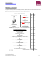
1
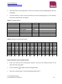
www.clondiag.com www.alere-technologies.com User guide for the Escherichia coli combined Assay Array Hybridisation Assay for DNA-based serogenotyping and detection of resistance and virulence genes of Escherichia coli For Research Use Only. Not Intended for Use in Clinical Diagnostics. www.clondiag.com www.alere-technologies.com CONTENT BACKGROUND ................................................................................................................................. 1 Intended Use .............................................................................................................................. 2 Specifications .............................................................................................................................. 2 Technical Support ....................................................................................................................... 2 Safety Precautions ...................................................................................................................... 3 Material Safety Data Sheets (MSDS) .......................................................................................... 3 Shipping Precautions .................................................................................................................. 3 REAGENTS AND DEVICES ................................................................................................................. 4 Assay Components, Storage and Stability .................................................................................. 4 Cell Lysis (optional) ................................................................................................................... 4 DNA Labelling and Amplification .............................................................................................. 4 Hybridisation and Detection ..................................................................................................... 5 Instrumentation & Software .................................................................................................... 6 Components required but not provided .................................................................................. 6 PROTOCOL ....................................................................................................................................... 8 Culturing and Harvesting Bacterial Cells .................................................................................... 8 Extraction of DNA ....................................................................................................................... 8 Extraction of DNA by Spin Columns (e.g. Qiagen) .................................................................... 9 Linear Amplification and Internal Biotin Labelling ................................................................... 11 Hybridisation ............................................................................................................................ 12 General Remarks - Handling of Arrays ................................................................................... 12 General Remarks - Handling of Liquids .................................................................................. 12 General Remarks – the Substrate (Precipitating Dye) D1 ...................................................... 13 General Remarks - Thermoshakers ........................................................................................ 14 Protocol for Quantifoil’s BioShake iQ ..................................................................................... 14 Protocol for Eppendorf’s Thermomixer Comfort with microtitre plate adapter ................... 15 Data Analysis............................................................................................................................. 17 Starting the ArrayMate Reader .............................................................................................. 17 Worklist................................................................................................................................... 18 Data Acquisition in the ArrayMate Reader ............................................................................ 19 Results..................................................................................................................................... 21 Export of E. coli combined Test Reports ................................................................................. 22 TROUBLESHOOTING ...................................................................................................................... 24 Staining Control ........................................................................................................................ 24 Image Quality............................................................................................................................ 24 DNA Quality and RNA contamination control .......................................................................... 25 Physical Damage to the Array .................................................................................................. 25 Report Unavailable ................................................................................................................... 25 ADDITIONAL INFORMATION ......................................................................................................... 26 Warranty ................................................................................................................................... 26 Disclaimer ................................................................................................................................. 26 Quality Control ......................................................................................................................... 27 Legal Manufacturer .................................................................................................................. 27 Contact...................................................................................................................................... 27 E. coli combined Assay User Guide 13-04-16-001-V1 www.clondiag.com www.alere-technologies.com LITERATURE ................................................................................................................................... 28 UPDATES & SOFTWARE ................................................................................................................. 28 APPENDIX 1 – FLOW CHARTS ........................................................................................................ 29 APPENDIX 2 – PROBE TO TARGET TABLE ...................................................................................... 31 APPENDIX 3 – TYPING INFORMATION .......................................................................................... 41 Definitions & Explanations ....................................................................................................... 41 E. coli combined Assay User Guide 13-11-11-001-V1 2 www.clondiag.com www.alere-technologies.com BACKGROUND The CLONDIAG Escherichia coli (E. coli) combined Assay allows DNA-based serogenotyping of most known O- and H-antigens as well as, simultaneously, the detection of important antimicrobial resistance (i.e. blaOXA) and virulence genes (i.e stxA/stxB). RNA-free, unfragmented genomic DNA from pure and monoclonal E. coli colony material is amplified approximately 50-fold and internally labelled with biotin-11-dUTP using a linear amplification protocol. In contrast to standard PCR, only one antisense primer per target is used resulting in producing single stranded (ss) DNA reaction products. This allows simultaneous sequence-specific labelling and amplification of an essentially unlimited number of targets. However, sensitivity is lower than in a standard PCR (whereas contamination with amplicons is nearly impossible) and for that reason the method is restricted to clonal colony material and cannot be performed on samples such as swabs or other patient samples (e.g. faeces). Resulting biotin labelled ssDNA is transferred and hybridised to DNA oligonucleotide microarrays with 393 probes for different genetic markers and a biotin staining control. Probes for serogenotyping are printed once, whereas probes for antimicrobial resistance (AMR) and virulence genes are printed in two spots. Targets include a variety of species and serotyping markers including genes that code for 23 different O-antigens and 47 H-antigens. Additionally, 102 probes targeting AMR genes as well as 88 probes for virulence genes were included. Spot recognition is performed automatically based on a digital image of the arrays and results are given as an html-file with a description of each analysed target. E. coli combined Assay User Guide 13-11-11-001-V1 1 www.clondiag.com www.alere-technologies.com GENERAL INSTRUCTIONS FOR USE Intended Use For Research Use Only. Not Intended for Use in Clinical Diagnostics. This assay allows genotypic characterisation of E. coli isolates for research and epidemiological applications. It must not be used as a substitute for phenotypic susceptibility tests and for the guidance of antibiotic therapy. Specifications Upon receipt, the assay components need to be stored at different temperatures as specified on the package insert. The assay is to be performed at an ambient temperature of 18°C to 28°C. Technical Support If you require any further information on this product please contact: Alere Technologies GmbH Löbstedter Straße 103 -105 D-07749 Jena - Germany phone: +49 (0) 36 41 / 3111-0 fax: + 49 (0) 36 41 / 3111-120 [email protected] E. coli combined Assay User Guide 13-11-11-001-V1 2 www.clondiag.com www.alere-technologies.com Safety Precautions The assay is intended for use by personnel that are trained in microbiological and molecular methods. Preparation of DNA from pure E. coli colonies (clones) requires expertise in microbiology and the local regulations for handling of pathogenic microorganisms (biosafety level 2) are to be obeyed. Isolated, cell-free E. coli DNA may be processed without further biosafety precautions, although contamination with E. coli or other bacteria needs to be ruled out. Always wear protective clothes as required for laboratory work by your local regulations. Material Safety Data Sheets (MSDS) According to OSHA 29CFR1910.1200, Commonwealth of Australia [NOHSC: 1005, 1008(1999)] and the latest amendments to the European Union Directives 67/548/EC and 1999/45/EC, the enclosed reagents do not require a Material Safety Data Sheet (MSDS). They do not contain more than 1% of a component classified as hazardous and do not contain more than 0.1 % of a component classified as carcinogenic. MSDS therefore are not provided. Nevertheless, the buffers may cause irritation if they come into contact with eyes or skin, and may cause harm if swallowed. The regular precautions associated with laboratory work should be obeyed (e.g., wear protective goggles, gloves and lab coat and avoid contact with the reagents). In case, any liquids are spilled, clean with disinfectant and/or laboratory detergent and water. Alere assumes no liability for damage resulting from handling or contact with these products. If you have any questions please contact our Technical Support (see above). Shipping Precautions RID/ADR: Kein Gefahrgut / No dangerous goods IMDG: No dangerous goods IATA: No dangerous goods E. coli combined Assay User Guide 13-11-11-001-V1 3 www.clondiag.com www.alere-technologies.com REAGENTS AND DEVICES Assay Components, Storage and Stability All reagents are provided in a certain surplus amount (see below). In case of need, all components may also be ordered separately; please refer to the order numbers at the end of this user guide. For pricing please contact your local representative or our customer service, respectively. The expiry date can be found on each bottle and on the outer package. All components have been tested for stability for short term shipment (<1 week) at ambient temperature (< 37 °C). The assay components with a rather limited stability are D1 and C3. The other components have proven to be stable even six months after the assay expiry date has passed. Cell Lysis (optional) A1: Lysis Buffer (cat# 245101000) Store at 18-28°C (ambient temperature). Surplus: 50%. A2: Lysis Enhancer (lyophilised, cat# 245102000) Store at 18-28°C (ambient temperature). Centrifuge A2 tubes shortly prior to opening. Add 200 µL Buffer A1 to Lysis Enhancer before use. Mix well and store for less than 1 week at 28°C. Sufficient for 96 isolations. DNA Labelling and Amplification B1+: Labelling Buffer, Store at 2-8°C. Surplus: 25%. B2: Labelling Enzyme, Store at 2-8°C. Surplus: 50%. Ecoli-PM1: lyophilized Labelling-Primer-Mix, dilute in 50 µl molecular grade water, Store at -20°C, E. coli combined Assay User Guide 13-11-11-001-V1 4 www.clondiag.com www.alere-technologies.com Hybridisation and Detection ArrayStrips (12 x 8 samples), Protected against light and sealed under inert gas. Store at 18°C to 28°C. After opening to be used within two weeks. Close the unused wells with caps, protect them against humidity and dust and store them at a dark place. Avoid any touching or scratching of the surface of the microarray at the bottom of the well. Do not store or handle unused wells at an air humidity of more than 60% since this may irreversibly corrode the spots. CapStrips (24 strips) C1: Hybridisation Buffer Store at 18-28 °C, protect against sunlight. Surplus: 100%. C2: Washing Buffer 1 Store at 18 °C - 28 °C, protect against direct sunlight. Surplus: 100%. C3: HRP Conjugate 100x Store at 2-8 °C, protect against direct sunlight. Surplus: 100%. C4: Conjugate Buffer Store at 18°C to 28°C, protect against direct sunlight. Surplus: 200%. C5: Washing Buffer 2 Store at 18°C to 28°C, protect against direct sunlight. Surplus: 200%. D1: Horseradish Peroxidase Substrate Store at 2-8°C, protect against direct sunlight. Surplus: 50%. CM: Reference DNA from E. coli EDL933 (GenBank accession number NC_002655.2), cDNA = 0.1-0.4 µg/µL. Store at 2-8 °C. Sufficient for 5-6 tests. E. coli combined Assay User Guide 13-11-11-001-V1 5 www.clondiag.com www.alere-technologies.com Instrumentation & Software ArrayMate Reader (to be ordered separately, for details see below) The E. coli combined assay may be used on the ArrayMate reader only. The alternative devices ATR01/03 are not suitable for reading ArrayStrip based assays. In case of any questions please contact us. Iconoclust software (provided with the reader) Test specific software plug-in (can be downloaded from Alere Technologies’/CLONDIAG´s website, check periodically for updates, for details see below). Information (such as spot names, marker names, location of the spots on the array, size of the image taken by the reader’s specific camera) is delivered with the reader or can be downloaded from our website. These test-specific plug-ins will occasionally be updated. Please check the NEWS section of our website http://www.alere-technologies.com. Support is available via [email protected]. Components required but not provided Growth media for the cultivation of E. coli. The test should be performed with colonies harvested from 2xTY or Columbia Blood Agar. Other rich media (e.g. Standard 1 or LB) may also suffice, but have not systematically been tested. Liquid media should not been used because contaminations or mixed cultures cannot easily be ruled out. Equipment and consumables needed for the cultivation of E. coli (incubator, inoculation loops, Petri dishes) DNA preparation kits: The assay has been tested with the DNeasy Blood & Tissue Kit from Qiagen (cat# 69504) and High Pure DNA Isolations Kit from Roche (Cat. No. 11796828001). Please note: The DNA specimen needs to be free of RNA. Recommendation: a pretreatment with the Cell Lysis components A1/A2 (see below) or a standard RNase A treatment while DNA preparation. E. coli combined Assay User Guide 13-11-11-001-V1 6 www.clondiag.com www.alere-technologies.com Equipment needed for DNA isolation, e.g. pipettes, centrifuge, thermoshaker or automated device (see above) Photometer (OD 260 nm) for measuring the concentration of DNA Equipment for non denaturing agarose DNA gel electrophoresis for quality control of DNA Thermocycler for PCR Thermoshaker We strongly recommend the BioShake iQ by Quantifoil Instruments (http://www.qinstruments.com/) equipped with a customised heating block designed to fit ArrayStrips. Alternatively, you may use Eppendorf’s Thermomixer Comfort, equipped a heating block for microtitre plates. Pipettes: suitable for 1µL-5µL volumes, 90µL, 100µL, 200µL, 1000µL Multichannel Pipettes for 100-200 µL Reaction vials suitable for PCR Ultrapure (PCR grade) water RNase A (we recommend Qiagen’s RNase A solution, 100 mg/mL, Qiagen order# 19101). Pasteur pipettes (BRANDT / Cat. No. 612-2856). E. coli combined Assay User Guide 13-11-11-001-V1 7 www.clondiag.com www.alere-technologies.com PROTOCOL Culturing and Harvesting Bacterial Cells Members of the genus Escherichia are potential pathogens. All procedures for cultivation of the bacterium and DNA preparation need to be performed by properly trained staff in a biosafety level 2 facility. Grow E. coli on 2xTY or Columbia Blood agar (overnight at 37°C or 48 hrs at room temperature). Obtain confirmation of the identification as E. coli (API, VITEK, MALDI) and make sure that you have a pure, monoclonal culture of E. coli. Contamination with other bacteria, especially with other Enterobacteriaceae needs to be strictly avoided as the might carry the same resistance genes as certain E. coli strains and thus can introduce false positive signals and patterns. Extraction of DNA The required sample type for the E. coli combined assay is 0.5-2 µg (cDNA = 0.1-0.4 µg/µl) of intact genomic DNA from a single clone. The DNA specimen needs to be free of RNA and it should not be fragmented. This can be determined by agarose gel electrophoresis. Additionally, the microarray includes probes as an internal control for RNA contamination. The automatic software analysis will give a “failed” if a RNA contamination occurred while DNA isolation. DNA should not be prepared by disrupting E. coli cells using bead beaters, ultrasonication or aggressive chemicals such as in alkaline lysis protocols. Most performance problems with the E. coli combined assay are due to insufficient amounts or quality of DNA preparation. We therefore strongly recommend following the protocols outlined below. Please note: To yield more genomic DNA, we recommend the optional cell lysis step with A1/A2 reagent. E. coli combined Assay User Guide 13-11-11-001-V1 8 www.clondiag.com www.alere-technologies.com Extraction of DNA by Spin Columns (e.g. Qiagen) Add an inoculating loop full of monoclonal colony material of the E. coli isolate to 0.2 ml 1xPBS and vortex thoroughly. Loop empty Loop full It is important to harvest enough bacteria; this is a prerequisite for extraction of a sufficient amount of DNA. Take an inoculating loop of 1mm diameter filled with bacteria as shown in the right picture. Optional cell lysis with A1/A2 reagent (instead of 1xPBS): Centrifuge A2 tube shortly, open it, add 0.2 ml of Lysis Buffer A1 to Lysis Enhancer A2 and dissolve. Add an inoculating loop full of monoclonal colony material of the E. coli isolate to this A1/A2 reagent and vortex thoroughly. Incubate the colony material of the E. coli isolate in A1/A2 for 30-60 min at 37°C and 550 rpm in the thermoshaker. Proceed with the DNA preparation protocol of the DNA preparation kit. For the Qiagen DNeasy Blood&Tissue Kit that is as follows: Add 20 µL proteinase K (Qiagen Kit, or equivalent) and add 200 µL buffer AL (Qiagen Kit) Vortex shortly or shake vigorously. Incubate for 30-60 min at 56°C and 550 rpm in the thermoshaker. important: if A1/A2 reagent not used, add now 4 μl RNase A (100 mg/ml), mix by vortexing, and incubate for 2 min at room temperature before continuing Add 200 µl ethanol (96-100%) Vortex the sample and centrifuge shortly. E. coli combined Assay User Guide 13-11-11-001-V1 9 www.clondiag.com www.alere-technologies.com Transfer the complete content of the tube (including any precipitate) into a spin column that is placed in a 2 ml collection tube. Centrifuge (8000 rpm, 1 min) at room temperature, time and speed need to be determined depending on viscosity of the sample and type of centrifuge used. All liquid should be collected in the collection tube afterwards. Discard collection tube with liquids. Place the spin column in a new 2 ml collection tube (provided with the kit). Add 500 µl Buffer AW1. Centrifuge (8.000 rpm, 1 min) at room temperature. Discard collection tube with liquids. Place the spin column in a new 2 ml collection tube (provided with the kit). Add 500 µl Buffer AW2. Centrifuge (14.000 rpm, 3 min) at room temperature, the membrane of the spin column should be dry, and all liquid should be in the collection tube. Discard collection tube with liquids. Place the spin column in a clean 1.5 ml tube (not provided with the kit). Add 100 µl Buffer AE (or PCR grade distilled water) directly onto the membrane of the spin column. Incubate at room temperature for 1 min to elute DNA. Centrifuge (8000 rpm, 1min) at room temperature. Optional: add another 100 µl Buffer AE (or PCR grade distilled water) directly onto the membrane, incubate at room temperature for 1 min and centrifuge again. Discard the spin column. Please note: Ethanol from Washing Buffers strongly inhibits the enzymes used in the assay. E. coli combined Assay User Guide 13-11-11-001-V1 10 www.clondiag.com www.alere-technologies.com A contamination with Washing Buffer might occur during elution of prepared DNA by drops adhering to the funnel of the spin columns. Thus these funnels should be gently touched and tried with sterile filter paper or wipes prior to the elution step. Alternatively, prepared DNA can shortly be heated to evaporate ethanol (e.g., 10 min at 70 C). Check for DNA integrity and absence of RNA (e.g., agarose gel). If necessary, you might perform another digestion step with additional RNase A (not provided). Measure DNA concentration (A260 method), it shouldn´t be less than 0.1 µg/µl. The concentration might be increased by heating and evaporation of water, or by using a speed vac centrifuge. Linear Amplification and Internal Biotin Labelling Please keep in mind the limited surplus of reagents whilst pipetting. The surplus of B1+ labelling reagent is 25 %. Prepare a Master Mix by combining 3.9 µL of B1+ labelling reagent, 1 µl Ecoli-PM1 Labelling-Primer-Mix and 0.1 µL of B2 (DNA polymerase) per sample. Add 5 µl of E. coli DNA (cDNA = 0.1 - 0.4 µg/µl) prepared as described above to 5 µl of the Master Mix (B1+/B2). Do not forget to label the vial! Perform amplification in a pre-programmed thermocycler (e.g., Eppendorf Mastercycler gradient with heated lid, VWR, cat No. 460-0108) according to following protocol: Pre-heat cover/lid to 105°C 300 sec at 96°C 20 sec at 50°C 50 cycles with 40 sec at 72°C 60 sec at 96°C Cool down to 4°C, hold the samples can be stored frozen until usage Please note: When using another device, some adaptations might be necessary. Before starting routine use, please test the protocol with a few known reference strains. E. coli combined Assay User Guide 13-11-11-001-V1 11 www.clondiag.com www.alere-technologies.com Hybridisation General Remarks - Handling of Arrays Never touch the array surface! Avoid complete drying of the array surface during processing! Do not allow it to stay without liquid for more than two minutes! Never rinse the wells with distilled water after the hybridisation step, use only C2 Washing Buffer! Unused wells should be capped during the whole procedure. The strips may be processed up to three times without a loss of quality of properly capped unused arrays. Close all wells that will not be used with a cap und leave it there until you use these wells (for storage conditions after use: see section “Assay components, storage and stability/Hybridisation and Detection”). Always label your array strips with a laboratory marker at the recommended position. Never label them on the bottom or across the data matrix barcode! This may cause an error. Barcode label HERE (keep it clean!) Avoid contact of data matrix barcode with organic solvents! The ArrayMate needs the information encoded in the data matrix to perform the assay and the analysis afterwards. Avoid touching the bottom of the microarray strip and keep it clean. General Remarks - Handling of Liquids We recommend the use of a multichannel pipette and reagent reservoirs. Please keep in mind the limited surplus of C1 (100%). We strongly recommend that the liquid is removed by pipetting rather than by inverting the strips and flicking the liquids out. Fine tipped soft, disposable Pasteur pipettes are suited best (such as BRANDT / Cat. No. 612-2856). Always place the pipette tip at the cavity between the E. coli combined Assay User Guide 13-11-11-001-V1 12 www.clondiag.com www.alere-technologies.com array and the wall of the reagent well. If you touch the array surface, probes may be scratched off and this may cause an error. Pipette tip use the cavity between array and the wall of the tube do never touch the array Array General Remarks – the Substrate (Precipitating Dye) D1 An appropriate amount of substrate (precipitating dye) should be filled into an Eppendorf tube and taken out of the refrigerator when starting the procedure allowing it to pre-warm to room temperature/25°C. Cold D1 may yield weak signals. D1 should shortly be centrifuged prior to use to remove bubbles as well as possible precipitates. Triggered by peroxidase, in case of positive reactions, the dye precipitates but it is not covalently bound. The precipitate can be dissolved by vigorous shaking. Thus the arrays must not be shaken, dropped or moved abruptly during the staining procedure and afterwards. E. coli combined Assay User Guide 13-11-11-001-V1 13 www.clondiag.com www.alere-technologies.com After completion of staining, remove and discard reagent D1 as completely as possible and scan immediately. The dye precipitate fades slowly in presence of liquids. General Remarks - Thermoshakers The correct temperature within the vessels is essential; therefore always use the appropriate equipment for heating. Because of a possibly inhomogeneous distribution of the temperature within the heating block and because of possible differences between displayed and actual temperatures, the use of different brands of thermoshakers might affect test performance. We tested the assay with BioShake iQ by Quantifoil Instruments (http://www.qinstruments.com/) equipped with a customised heating block designed to fit ArrayStrips and Eppendorf’s Thermomixer Comfort, equipped with a heating block for microtitre plates.When using other devices, some modifications to the protocol might be necessary. Before starting routine use, please test the protocol with a few known reference strains or the control DNA (E. coli EDL933). Protocol for Quantifoil’s BioShake iQ Switch on the thermoshaker and let it pre-heat to 50°C. Remove the ArrayStrip(s) from the pouch. Insert the ArrayStrip(s) into the white frame. Assure the correct orientation (data matrix barcode close to row (A) and proper fit. Pre-wash the array in two steps: First, PCR-grade distilled water, 200 µl per well at 50°C, 5 min and 550 rpm Second, C1 Hybridisation Buffer, 150 µl per well at 50°C, 5 min and 550 rpm Add 90 µL of buffer C1 to each tube with (10 µL) labelled amplification product, mix gently Remove the buffer from the well with the array and add the mixture of C1 and labelled amplification product Incubate at 50°C, 60 min and 550 rpm. E. coli combined Assay User Guide 13-11-11-001-V1 14 www.clondiag.com www.alere-technologies.com Remove liquid and add 200 µl C2 Washing Buffer. Incubate at 45°C, 10 min and 550 rpm, remove and discard. Add another 200 µl C2 Washing Buffer. Incubate at 45°C, 10 min and 550 rpm, remove and discard. Meanwhile, prepare conjugate: for each experiment add 1 µl conjugate 100xHRP to 100 µl C4 Conjugation Buffer. This mixture is stable for around one working day at room temperature; C3 is delivered with a surplus of 100%, C4 is delivered with a surplus of 200%. Suggested pipetting scheme: C3 C4 1 well 1.5 µL 150 µL 2-3 wells 3.5 µL 350 µL 4-6 wells 7 µL 700 µL 7-10 wells 11 µL 1100 µL 11-15 wells 16 µL 1600 µL 16-20 wells 21 µL 2100 µL 21-30 wells 32 µL 3200 µL 31-40 wells 42 µL 4200 µL Remove and discard Washing Buffer, and add 100 µl diluted conjugate to each well, incubate at 30 °C, 10min and 550rpm. Add 200 µl C5 Washing Buffer. Incubate at 30°C, 5 min and 550rpm. Remove and discard Washing Buffer, add 100 µl of D1 substrate (precipitating dye, at 25°C, see above) per well. Incubate at 25°C, 10 min but do not shake ! Remove liquid completely. The bottom of the ArrayStrips may be cautiously be cleaned with wipes, bubbles may be removed by removing and adding D1. Scan and process (see below). Protocol for Eppendorf’s Thermomixer Comfort with microtitre plate adapter Switch on the thermoshaker and let it pre-heat to 55°C. Remove the ArrayStrip(s) from the pouch. E. coli combined Assay User Guide 13-11-11-001-V1 15 www.clondiag.com www.alere-technologies.com Insert the ArrayStrip(s) into the white frame. Assure the correct orientation (data matrix barcode close to row (A) and proper fit. Pre-wash the array in two steps: First, PCR-grade distilled water, 200 µl per well, just pipette up and down four times, remove and discard. Second, C1 Hybridisation Buffer, 150 µl per well at 55°C, 5 min and 550 rpm Add 90 µL of buffer C1 to each tube with (10 µL) labelled amplification product, mix gently Remove the buffer from the well with the array and add the mixture of C1 and labelled amplification product Incubate at 55°C, 60 min and 550 rpm. Remove liquid and add 200 µl C2 Washing Buffer. Incubate at 40°C, 12 min and 550 rpm, remove and discard. Add another 200 µl C2 Washing Buffer. Incubate at 40°C, 12 min and 550 rpm, remove and discard. Meanwhile, prepare conjugate: for each experiment add 1 µl conjugate 100xHRP to 100 µl C4 Conjugation Buffer. This mixture is stable for around one working day at room temperature; C3 is delivered with a surplus of 100%, C4 is delivered with a surplus of 200%. Suggested pipetting scheme: C3 C4 1 well 1.5 µL 150 µL 2-3 wells 3.5 µL 350 µL 4-6 wells 7 µL 700 µL 7-10 wells 11 µL 1100 µL 11-15 wells 16 µL 1600 µL 16-20 wells 21 µL 2100 µL 21-30 wells 32 µL 3200 µL 31-40 wells 42 µL 4200 µL Remove and discard Washing Buffer, and add 100 µl diluted conjugate to each well, incubate at 30°C, 10min and 550rpm. Remove liquid and wash with 200 µl C5 Washing Buffer, just pipette up and down four times, remove and discard. E. coli combined Assay User Guide 13-11-11-001-V1 16 www.clondiag.com www.alere-technologies.com Add another 200 µl C5 Washing Buffer, just pipette up and down four times. Remove and discard Washing Buffer, add 100 µl of D1 substrate (precipitating dye, at 25°C, see above) per well. Incubate at 25°C, 10 min but do not shake ! Remove liquid completely. The bottom of the ArrayStrips may be cautiously be cleaned with wipes, bubbles may be removed by removing and adding D1. Scan and process (see below). Data Analysis Starting the ArrayMate Reader We recommend starting the ArrayMate Reader after having started the hybridisation; this allows you to conveniently start the device and to import the worklist file (see below) Please note that this is a short instruction only. For more detailed information please refer to the ArrayMate User Manual. Switch on the ArrayMate (main switch on the rear below the electric cable plug, operating switch on the bottom/left corner of the front side). Switch on the screen (switch right hand below the screen). Log-in as “R&D User” (Research and Development User) for full access to test specific software (a default password will be provided together with the ArrayMate device). If you log-in as “User”, you will obtain only raw values, but no interpretation as positives/negatives and no strain assignment. “Administrator” log-in will allow manipulation of file folders and software; and this should be done only upon direct advice of Clondiag´s IT team. E. coli combined Assay User Guide 13-11-11-001-V1 17 www.clondiag.com www.alere-technologies.com The user interface will be loaded, ArrayMate performs internal testing. This will require slightly less than a minute. Click on the icon “New Run” (left upper edge of the screen). A suggestion for a run name / folder name for the new run appears in the top line of the screen). You may modify or change the experiment name at your convenience. Type in your operator ID (optional). You may optionally enter a comment into the “memo” field. Worklist A “Worklist” file allows linking an identifier such as a laboratory/sample number to a position of an array within the ArrayStrip. For privacy reasons, arrays should not be identified by patient names. Worklists can be generated using spreadsheet software such as EXCEL (see below) but must be saved in the *.txt file format that can be imported into the test specific ArrayMate software. Do not use special characters (such as : ; ()[] / \ ä ü etc.). Create a list with at least three columns that have headers written into the first line. The following headers are obligatory (in this order): position / sample ID / assay ID (Table 1). Positions are continuously numbered from 1 to, maximal, 96. Position 1 would correspond to A1, 8 to H1, 9 to A2 and 96 to H12 (Table 2). Do not leave empty lines in the worklist. If you use EXCEL, position numbers should be typed into column A. Sample ID are strain/sample/laboratory numbers such as exported from your LIMS (or assigned in any different way). Patient´s names should not be used as Sample IDs. Assay IDs allow the system to identify the actual test and to correctly use information on layout, spot number and identity etc. E. coli combined Assay has the Assay ID: 10313. Assay ID numbers must not be confused as this could lead to errors or loss of data. You may add further columns and headers with notes and comments at your convenience Information from these columns will not appear on the result screens or the Test Report. We recommend using a printout of the worklist as template for pipetting. E. coli combined Assay User Guide 13-11-11-001-V1 18 www.clondiag.com www.alere-technologies.com Safe the worklist as tab separated *.txt file on the memory stick provided together with the ArrayMate. To avoid confusion, make sure that worklists are named unambiguously or that worklists from earlier experiments are deleted. Table 1: Example worklist: Position 1 2 3 4 5 6 7 8 sampleID 2013-12345 2013-12346 2013-12347 2013-12348 2013-12349 2013-12350 987654 E. coli EDL933 assayID 10313 10313 10313 10313 10313 10313 10313 10313 comment Isolate referred from Dr. J. Doe. Control strain Table 2: Positions in the 96 well format A B C D E H G H 1 1 2 3 4 5 6 7 8 2 9 10 11 12 13 14 15 16 3 17 18 19 20 21 22 23 24 4 25 26 27 28 29 30 31 32 5 33 34 35 36 37 38 39 40 6 41 42 43 44 45 46 47 48 7 49 50 51 52 53 54 55 56 8 57 58 59 60 61 62 63 64 9 65 66 67 68 69 70 71 72 10 73 74 75 76 77 78 79 80 11 81 82 83 84 85 86 87 88 12 89 90 91 92 93 94 95 96 Data Acquisition in the ArrayMate Reader Insert your memory stick containing the worklist. Use any of the USB ports down to the right side of the ArrayMate. Press the button: Select your worklist (path: “My Computer/Removable Disk”). ; a folder selection dialog will open. E. coli combined Assay User Guide 13-11-11-001-V1 19 www.clondiag.com www.alere-technologies.com Open your selected worklist with “Enter” or the button “Open”. Press the button: (your imported worklist opens in a separate window). Proofread. If the new window is empty or if it was the wrong worklist, repeat the import. Press the button “OK”; the worklist window will close. Leave the memory stick attached to the ArrayMate if you intend to export E. coli combined Test Reports afterwards. Press the button “next” (bottom/right on the screen; reader is opening). Carefully insert the appropriate metallic adapter/frame into the ArrayMate. Do not apply any strong force. Assure proper fit, otherwise the images may be out of focus. Carefully insert the white frame with the array strips into the metallic adapter. Assure the correct orientation (Position A1 in the frame next to the data matrix barcode on the adapter) and proper fit, otherwise the images may be out of focus. ArrayStrip frame with inserted strips. Strips are inserted in accordance to the worklist. Please note: ArrayStrips must be clean. They should not contain any liquids by now. Barcodes must be clean. There must be no lids on the wells that are to be analysed (however, unused wells should remain capped). Press the button “Next” (bottom/right on the screen; reader is closing, analysis program starts, it takes ca. 2-10 min dependent on the number of strips; reader takes images and automatically analyses the data). The progress of the reading is indicated by the following symbols: E. coli combined Assay User Guide 13-11-11-001-V1 20 www.clondiag.com www.alere-technologies.com photographed: in analysis: ready: The reader indicates the end of the entire process with an acoustic signal (beep). Press the button “Next” (bottom/right on the screen; reader is opening). Remove the white frame with the ArrayStrip(s). Press the button “Next” (bottom/right on the screen; reader is closing). Results On the left hand site of the screen you will see a list showing all runs stored on the ArrayMate´s hard disk. A run contains the results from all arrays analysed together within one frame. If this list is not visible: Press the button “Archive” (left hand) and activate the Flag “Browse” (top left). The runs are organised like folders in “Windows Explorer” and, by default, named according to the date of acquisition. Example: there is one reading in this archive: E. coli combined Assay User Guide 13-11-11-001-V1 21 www.clondiag.com www.alere-technologies.com If you click on the plus symbol left on the run name, the folder opens and you will see a list of the individual arrays alphabetically ordered by Sample ID. Click on a Sample ID and the E. coli combined Test Report for this array is shown in the window on the right: Export of E. coli combined Test Reports The generated result files in an html format will show information of all target genes. Possible invalid controls that might display in this report will be explained below (see Troubleshooting). Other files that are generated and that can be exported include a *.txt file with the raw measurements, an image file (*.bmp) with the actual photo of the array, E. coli combined Assay User Guide 13-11-11-001-V1 22 www.clondiag.com www.alere-technologies.com a second image file (*.png) in which the coordinate grid is superimposed and the recognised spots are circled and a *.xml files that contains the same information as the html result sheets for future export into databases etc. Please note: only complete runs can be exported. The export of individual E. coli combined Test Reports is not possible Right-Click on the reading (a menu appears with the option “Export Run Reports”). Right-Click on “Export Run Reports” (a file browser opens). Click on “My Computer”, then on “Removable Disk” and choose the folder where to store or click on the button “Make New Folder” (on the bottom; a new folder icon appears). Rename the new folder (e.g. with the experiment name or date). Click on the “Ok” button (data are exported now into the new folder on your memory stick). Do NOT remove the memory stick as long as the hourglass symbol is visible. Switch off the device by clicking on the “Power”-button (left/down on the screen): Switch off the Screen. There is no need to physically switch off the ArrayMate. E. coli combined Assay User Guide 13-11-11-001-V1 23 www.clondiag.com www.alere-technologies.com TROUBLESHOOTING In case of trouble always make sure that reagents are within the recommended shelf-life and stored in the appropriate way. In case of trouble we are always happy to support. Please contact, [email protected] and please include a description of the problem as well as the array images (*.bmp files) in question. Staining Control A staining control is included to check whether possible problems originate from the hybridisation or the staining procedure. If the staining control has “Failed” proceed as follows: Horseradish peroxidase conjugate may have degraded during storage. Add 1µL buffer C3/C4 to 9 µL D1 (substrate). If the solution turns green within 3-5 seconds, the horseradish peroxidase still has sufficient enzymatic activity. Enzymatic reaction is inhibited by carryover of buffer C1. Ensure proper washing of the wells to remove all of Buffer C1 prior to adding horseradish peroxidase conjugate. If the Staining Control has “Passed”, refer to the following hints. Image Quality In case of poor image quality we recommend to re-check DNA quantity and quality first by loading leftover DNA on an agarose gel. In order to determine whether any problems originated from the DNA preparation, perform an experiment with the CM / Control material. This is DNA from the reference strain E. coli EDL933 (GenBank accession number NC_002655.2) and should be identified by the assay as E. coli with O:146 and H:20. If the control experiment yields a valid result and a correct identification, there was probably an issue with DNA preparation. If the control experiment also fails, an error affecting later steps or a degradation of reagents from later steps is likely. E. coli combined Assay User Guide 13-11-11-001-V1 24 www.clondiag.com www.alere-technologies.com DNA Quality and RNA contamination control The amount of DNA is crucial because of the linear kinetics of amplification (see Introduction). DNA should be free of RNA, as free RNA reduces the efficiency of amplification and labelling by effectively removing primer from the reaction mix due to competitive hybridisation. A260 readings will cover RNA and other contaminants as well. Therefore pure DNA preparations without RNA contaminations are a prerequisite for proper DNA concentration measurement. RNase treatment prior to A260 reading therefore is necessary (component A2 contains RNAse). Additionally, the microarray includes probes as an internal control for RNA contamination. The automatic software analysis will give a “failed” if a RNA contamination occurred while DNA isolation. DNA must be unfragmented, as fragmentation reduces the efficiency of amplification and labelling due to the distance between primer and probe binding sites. DNA should for this reason not be prepared by disrupting E. coli cells using bead beaters, ultrasonication or aggressive chemicals such as in alkaline lysis protocols. We made good experiences with the manual QIAGEN DNeasy kit and the Roche High Pure Kit. DNA must be free of any traces of ethanol, as ethanol strongly influences the amplification. It is possible to heat the sample prior to adding it to the labelling mix (5-10 minutes at 70°C). Physical Damage to the Array Scratching of the array surface with a pipette tip can lead to the damage of array spots that prohibits the acquisition of a valid signal. In this case the respective marker is not assigned as “negative”, but instead the message “none” appears next to the marker name. Report Unavailable If the ArrayMate indicates that no report is available for an array (or multiple arrays on one strip), please check that the strip was positioned properly into the frame. Scratches or drops of condensed water might render the barcode identifier unreadable, please wipe it carefully or try to manually identify the test. If no obvious reason for the fault can be discovered, please contact the technical service. E. coli combined Assay User Guide 13-11-11-001-V1 25 www.clondiag.com www.alere-technologies.com ADDITIONAL INFORMATION Warranty Alere guarantees the performance as described in this user guide. Usage of the Assay was successfully tested at ambient temperatures up to 37°C, a guarantee is limited to ambient temperatures in the laboratory between 18°C and 28°C. Assay components comprise the arrays and their caps, the Lysis Enhancer, the reagents for DNA labelling and for detection of labelled DNA products on the array, the ArrayMate reader and its software. In case one of these components fails within the expiry date due to other reason than misuse, contact Alere for replacement or refund. Terms and conditions apply. If you have any problem or question, please contact the technical service. Disclaimer This system is for research use only. We do not accept any liability for damages caused by misuse. Misuse comprises, especially but not exclusively, of a use of the system for the detection of resistance genes in order to predict phenotypic antibiotic resistances or susceptibilities for the guidance of an antibiotic chemotherapy. Since resistances might be caused by genes or mutations not covered by this array or by hitherto unknown genes or mutations, any antibiotic chemotherapy MUST be guided by phenotypic susceptibility tests. Furthermore, we do not accept any liability for damages caused by inappropriate use of the device as a personal computer, for instance related to the use of additional software, to network connections, or to a breach of privacy related to the storage of confidential information (such as names of patients from whom E. coli was isolated) on its hard disk and/or to the use of external storage devices that might be contaminated with spyware. E. coli combined Assay User Guide 13-11-11-001-V1 26 www.clondiag.com www.alere-technologies.com Quality Control Each batch is stringently tested with the use of standard E. coli DNA preparations for good performance and correctness of results. List of Components for Separate Order If required, these reagents for the E. coli combined Assay may be ordered separately: component A1 A2 Ecoli-PM1 Category Buffer lyophylised enzymes Labelling Primer amount cat# storage 30 ml 245101000 18-28 °C 96 units 245102000 18-28 °C 50 µl/tube on request 2-8 °C B1+ name Lysis Buffer Lysis Enhancer PrimerMix E. coli_combined (= E. coli_gesamt) Labelling Buffer buffered reagents 700 µl 245103000 2-8 °C B2 C1 C2 C3 C4 C5 D1 CM Labelling Enzyme Hybridisation Buffer Washing Buffer 1 HRP Conjugate 100x Conjugate Buffer Washing Buffer 2 HRP Substrate Control Material 20 µl 30 ml 120 ml 200 µl 30 ml 120 ml 15 ml 30 µl 245104000 245105000 245106000 245107000 245108000 245109000 245110000 on request 2-8 °C 18-28 °C 18-28 °C 2-8 °C 18-28 °C 18-28 °C 2-8 °C 2-8 °C ArrayStrips E.coli-combined-AS-2 (=E.coli-gesamt-AS-2) StripCaps buffered enzyme buffered reagents buffer buffered enzyme buffered reagents buffer buffered reagents E. coli EDL933 DNA (cDNA = 0.1-0.4 µg/µl) plugged microarrays 12 St 240008929 15-28 °C plasticware 24 St 245112000 15-28 °C StripCaps For pricing please contact us: Legal Manufacturer Alere Technologies GmbH Loebstedter Str. 103-105 07749 Jena, Germany Contact If you require any further information on this product please contact: [email protected] E. coli combined Assay User Guide 13-11-11-001-V1 27 www.clondiag.com www.alere-technologies.com LITERATURE Anjum MF, Mafura M, Slickers P, Ballmer K, Kuhnert P, et al. (2007) Pathotyping Escherichia coli by using miniaturized DNA microarrays. Appl Environ Microbiol 73: 5692-5697. Ballmer K, Korczak BM, Kuhnert P, Slickers P, Ehricht R, et al. (2007) Fast DNA serotyping of Escherichia coli by use of an oligonucleotide microarray. J Clin Microbiol 45: 370-379. Geue L, Schares S, Mintel B, Conraths FJ, Muller E, et al. (2010) Rapid microarray-based genotyping of enterohemorrhagic Escherichia coli serotype O156:H25/H-/Hnt isolates from cattle and clonal relationship analysis. Appl Environ Microbiol 76: 5510-5519. Korczak B, Frey J, Schrenzel J, Pluschke G, Pfister R, et al. (2005) Use of diagnostic microarrays for determination of virulence gene patterns of Escherichia coli K1, a major cause of neonatal meningitis. J Clin Microbiol 43: 1024-1031. Monecke S, Mariani-Kurkdjian P, Bingen E, Weill FX, Baliere C, et al. (2011) Presence of enterohemorrhagic Escherichia coli ST678/O104:H4 in France prior to 2011. Appl Environ Microbiol 77: 8784-8786. Schilling AK, Hotzel H, Methner U, Sprague LD, Schmoock G, et al. (2012) Zoonotic agents in small ruminants kept on city farms in southern Germany. Appl Environ Microbiol 78: 3785-3793. Wu G, Ehricht R, Mafura M, Stokes M, Smith N, et al. (2012) Escherichia coli isolates from extraintestinal organs of livestock animals harbour diverse virulence genes and belong to multiple genetic lineages. Vet Microbiol 160: 197-206. UPDATES & SOFTWARE Notifications on database/software updates and freeware tools can be found at : http://alere-technologies.com/en/science-technologies/publications/downloads.html. and/or http://alere-technologies.com/en/news.html. E. coli combined Assay User Guide 13-11-11-001-V1 28 www.clondiag.com www.alere-technologies.com APPENDIX 1 – Flow charts The figures summarise the test procedure. However, please refer to the text section of this user guide at any step of the test protocol for further important details. Protocol : Quantifoil BioShake iQ processing time handsontime Grow CLONAL E. coli isolate (not part of the kit) over night 5 min isolate genomic DNA (not part of the kit) 3-4 h 40 min 2h 5 min 2 min 2 min 2 min 2 min hybridise; 50 °C, 550 rpm; 60 min 60 min 0 min discard labeled DNA; incubate twice in 200 µl Buffer C2; 45 °C, 550 rpm, 10 min; prepare C3/C4-conjugate (C3:C4=1:100), preheat Substrat D1 (25°C) 20 min 5 min discard Buffer C2; incubate in 100 µl C3 /C4-conjugate; 30 °C, 550 rpm, 10 min 10 min 2 min discard C3/C4-conjugate; incubate once in 200 µl Buffer C5; 30 °C, 550 rpm, 5 min 5 min 2 min discard Buffer C5; incubate with 100 µl Substrate D1; 25 °C, 10 min 10 min 2 min discard Substrate D1; analyse (ArrayMate) 15 min prepare ArraysStripes rinse ArrayStripes 200 µl water; 50 °C, 550 rpm, 5 min discard water; 150 µl Buffer C1; 50 °C, 550 rpm, 5 min discard C1, process promptly prepare DNA label RNA free DNA in thermocycler 5 µl DNA (cDNA = 0.1 - 0.4 µg/µl) plus MM (3.9 µL B1+ + 0.1 µL B2 + 1 µl PM Ecoli-PM1) preparing labeled DNA to 10 µL of labeled DNA add 90 µL of Buffer C1 transfer 100 µl labeled DNA to ArrayStripes Barcode MM - MasterMix PM - PrimerMix a) with Label here total time requirement : over night + 7-8h 10 min app. 120 min heating block for microtitre plates E. coli combined Assay User Guide 13-11-11-001-V1 29 www.clondiag.com www.alere-technologies.com Protocol : Eppendorf Thermoshaker a) processing time handsontime Grow CLONAL E. coli isolate (not part of the kit) over night 5 min isolate genomic DNA (not part of the kit) 3-4 h 40 min 2h 5 min 10 min 5 min 2 min 2 min hybridise; 55 °C, 550 rpm; 60 min 60 min 0 min discard labeled DNA; incubate twice in 200 µl Buffer C2; 40 °C, 550 rpm, 12 min; prepare C3/C4-conjugate (C3:C4=1:100), preheat Substrat D1 (25°C) 25 min 5 min discard Buffer C2; incubate in 100 µl C3 /C4-conjugate; 30 °C, 550 rpm, 10 min 10 min 2 min discard C3/C4-conjugate; incubate once in 200 µl Buffer C5; 30 °C, 550 rpm, 5 min 5 min 2 min discard Buffer C5; incubate with 100 µl Substrate D1; 25 °C, 10 min 10 min 1 min discard Substrate D1; analyse (ArrayMate) 15 min 10 min prepare ArraysStripes prepare DNA rinse ArrayStripes 200 µl water; pipette up and down (4x) label RNA free DNA in thermocycler 5 µl DNA (cDNA = 0.1 - 0.4 µg/µl) plus MM (3.9 µL B1+ + 0.1 µL B2 + 1 µl PM Ecoli-PM1) discard water; 150 µl Buffer C1; 55 °C, 550 rpm, 5 min discard C1, process promptly preparing labeled DNA to 10 µL of labeled DNA add 90 µL of Buffer C1 transfer 100 µl labeled DNA to ArrayStripes Barcode MM - MasterMix PM - PrimerMix a) with Label here total time requirement : over night + 7-8h app. 120 min heating block for microtitre plates E. coli combined Assay User Guide 13-11-11-001-V1 30 www.clondiag.com www.alere-technologies.com APPENDIX 2 – PROBE TO TARGET TABLE Target genes Probes Function Family and Species Marker gapA prob_gapA_611 ihfA prob_ihfA_611 gad gad_10 dnaE hp_dnaE_612, hp_dnaE_613 rrs hp_rrs_611, hp_rrs_612 glyceraldehyde 3-phosphate dehydrogenase A (CP000468.1, locus tag APECO1_847)- genetic marker for family Enterobacteriaceae integration host factor subunit alpha (U00096.2)genetic marker for family Enterobacteriaceae glutamate decarboxylase (AE014075.1, locus tag c4328) - genetic marker for genus Escherichia/Shigella DNA polymerase III subunit alpha - genetic marker for species Escherichia spec. and Shigella spec. (U00096.2) 16S rRNA - genetic marker for species E. coli (U00096.2) O4 wzy-O4_11 O-antigen - O4 (U39042.1) O6 wzx-O6_11, wzy-O6_11 O-antigen - O6 (AJ426045.2) O7 wzx-O7_11, wzy-O7_11 O-antigen - O7 (AB490074.1) O8 wzx-O8_11, wzy-O8_11 O9 wzx-O9_11, wzy-O9_11 O-antigen - O8 (AF013583.1) O-antigen - O9 (wbdA_O9: D43637.1, wzx_O9: AF104912.2, wzy_09: AB031867.1) O15 wzx-O15_11, wzy-O15_11 O-antigen - O15 (CP002291.1) O26 wzx-O26_11, wzy-O26_11 O-antigen - O26 (AF529080.1) O52 wzm-O52_11 O-antigen - O52 (AY528413.1) O53 wzy-O53_11 O-antigen - O53 (AF402312.1) O55 wzx-O55_11, wzy-O55_11 O-antigen - O55 (AF461121.1) O79 wbdU-O79_11, wzx-O79_11 O-antigen - O79 (EU294162.1) O86 wzx-O86_11, wzy-O86_11 O-antigen - O86 (AY670704.1) O91 wzx-O91_11, wzy-O91_11 O-antigen - O91 (AY035396.1) O101 wz-O101_11, wbdA-O9_11 O-antigen - O101 (X59852.1) O103 wzx-O103_11, wzy-O103_11 O-antigen - O103 (AY532664.1) O104 O111 wzx-O104_11, wzy-O104_11 O-antigen - O104 (AF361371.1) wbdH-O111_11, wbdM-O111_11, wzx-O111_11, wzy-O111_11 O-antigen - O111 (AF078736.1) O113 wzx-O113_11, wzy-O113_11 O-antigen - O113 (AF172324.1) O114 wzx-O114_11, wzy-O114_11 O-antigen - O114 (AY573377.1) O121 wzx-O121_11, wzy-O121_11 O-antigen - O121 (AY208937.1) O128 wzx-O128_11, wzy-O128_11 O-antigen - O128 (AY217096.1) O157 rfbE-O157_11, wzx-O157_11 O-antigen - O157 (AB008676.1) O172 wzx-O172_11, wzy-O172_11 O-antigen - O172 (AY545992.1) O-serotyping E. coli combined Assay User Guide 13-11-11-001-V1 31 www.clondiag.com www.alere-technologies.com H-serotyping fliC H01 fliC-H01_11, fliC-H01_12 flagellin C H-antigen H01 (AE014075.1) fliC H02 fliC-H02_11 flkA H03 flkA-H03_11 flagellin C H-antigen H02 (AF543692.1) flagellin A H-antigen H03 (AB128916.1) (Note: If repressor FljA "positive", the gene fliC H16 is repressed; H-phenotype is H03) fliC H04 fliC-H04_11 flagellin C H-antigen H04 (AY249989.1) fliC H05 fliC-H05_11 flagellin C H-antigen H05 (AY249990.1) fliC H06 fliC-H06_11 flagellin C H-antigen H06 (AY249991.1) fliC H07 fliC-H07_11, fliC-H07_12 flagellin C H-antigen H07 (AB028474.1) fliC H08 fliC-H08_11 flagellin C H-antigen H08 (AJ884571.1) fliC H09 fliC-H09_11 flagellin C H-antigen H09 (AY249994.1) fliC H10 fliC-H10_11 flagellin C H-antigen H10 (AY249995.1) fliC H11 fliC-H11_11 flagellin C H-antigen H11 (AY337465.1) fliC H12 fliC-H12_11 flagellin C H-antigen H12 (AY249997.1) fliC H14 fliC-H14_11 flagellin C H-antigen H14 (AY249998.1) fliC H15 fliC-H15_11 fliC H16 fliC-H16_11 flagellin C H-antigen H15 (AY249999.1) flagellin C H-antigen H16 (AB128919.1), Note: If repressor fljA "positive", fliC H16 might be repressed, H-phenotype is than determined by flkA H03. fliC H18 fliC-H18_11 flagellin C H-antigen H18 (AY250001.1) fliC H19 fliC-H19_11 flagellin C H-antigen H19 (AY250002.1) fliC H20 fliC-H20_11 fliC H21 fliC-H21_11 flagellin C H-antigen H20 (AY250003.1) flagellin C H-antigen H21 (DQ862122.1), Note: If repressor fljA "positive", fliC H21 might be repressed, H-phenotype is than determined by flmA H54. fliC H23 fliC-H23_11 flagellin C H-antigen H23 (AY250005.1) fliC H24 fliC-H24_11 flagellin C H-antigen H24 (AY250006.1) fliC H25 fliC-H25_11 flagellin C H-antigen H25 (ADUP01000024.1) fliC H26 fliC-H26_11 flagellin C H-antigen H26 (AY250008.1) fliC H27 fliC-H27_11 flagellin C H-antigen H27 (CU928162.2) fliC H28 fliC-H28_11 flagellin C H-antigen H28 (AY250010.1) fliC H29 fliC-H29_11 flagellin C H-antigen H29 (AY250012.1) fliC H30 fliC-H30_11 flagellin C H-antigen H30 (AY250011.1) fliC H31 fliC-H31_11 flagellin C H-antigen H31 (AY250013.1) fliC H32 fliC-H32_11 flagellin C H-antigen H32 (AY250014.1) fliC H33 fliC-H33_11 flagellin C H-antigen H33 (AY250015.1) fliC H34 fliC-H34_11 flagellin C H-antigen H34 (AY250016.1) fliC H37 fliC-H37_11 flagellin C H-antigen H37 (AY250017.1) fliC H38 fliC-H38_11 flagellin C H-antigen H38 (AY250018.1) fliC H39 fliC-H39_11 fliC H40 fl-H40_11 flagellin C H-antigen H39 (AY250019.1) flagellin C H-antigen H40 (AJ865464.1), Note: If repressor fljA "positive", fliC H40 might be repressed, H-phenotype is than determined by flkA H53. E. coli combined Assay User Guide 13-11-11-001-V1 32 www.clondiag.com www.alere-technologies.com fliC H41 fliC-H41_11 flagellin C H-antigen H41 (AY250020.1) fliC H42 fliC-H42_11 flagellin C H-antigen H42 (AY250021.1) fliC H43 fliC-H43_11 flagellin C H-antigen H43 (AY250022.1) fliC H45 fliC-H45_11 flagellin C H-antigen H45 (AY250023.1) fliC H46 fliC-H46_11,fliC-H46_12 flagellin C H-antigen H46 (AY250024.1) fliC H48 fliC-H48_11 flagellin C H-antigen H48 (AY250025.1) fliC H49 fliC-H49_11 flagellin C H-antigen H49 (AY250026.1) fliC H51 fliC-H51_11 flagellin C H-antigen H51 (AY250027.1) fliC H52 fliC-H52_11 flkA H53 flkA-H53_11 flmA H54 flmA-H54_11 flagellin C H-antigen H52 (AY250028.1) flagellin A H-antigen H53 (AB128917.1) (Note: If repressor FljA "positive", the gene fliC H40 is repressed; H-phenotype is H53) flagellin A H-antigen H54 (AB128918.1) (Note: If repressor FljA "positive", the gene fliC H21 is repressed; H-phenotype is H54) fliC H56 fliC-H56_11 flagellin C H-antigen H56 (AY250029.1) H-serotyping (additional marker) fliC-5p_11, fliC-5p_12, fliC-5p_13, fliC Marker fliC-5p_14, fliC-5p_15 fljA Repressor fljA_11 fliC marker - consensus sequence for all fliC genes FljA represses the expression of fliC genes, if "positive" the genes flkA (H03 or H53) or flmA (H54) represent the H-serotype (AB128916.1) fliC non-motile fl-H-NM_11 flagellin C from non-motile isolates (AY337480.1) streptogramin A resistance vatE hp_vatE_611, hp_vatE_612 acetyltransferase;streptogramin A acetyltransferase; associated with resistance to streptogramin A (AF242872.1) aminoglycoside resistance strA, strB prob_strA_611, prob_strB_611 aac_aph aac3 hp_aac_aph_611 hp_aac3_611, hp_aac3_612, hp_aac3_613, hp_aac3_614, hp_aac3_615 aac3Ia prob_aac3Ia_1 aac3IVa prob_aac3IVa_1 hp_aac6_611, hp_aac6_612, hp_aac6_613, hp_aac6_614, hp_aac6_616, hp_aac6_617, hp_aac6_618 aac6 E. coli combined Assay User Guide 13-11-11-001-V1 aminoglycoside-3''-phosphotransferase (locus A) and aminoglycoside-6''-phosphotransferase; associated with resistance to streptomycin (EF090911.1) 3-N-aminoglycoside acetyltransferase; associated with resistance to gentamycin (AE017171.1) 3-N-aminoglycoside acetyltransferase; associated with resistance to gentamycin (U90945.1) 3-N-aminoglycoside acetyltransferase; associated with resistance to astromicin; gentamicin; sisomicin (U90945.1) 3-N-aminoglycoside acetyltransferase; associated with resistance to apramycin; dibekacin; gentamicin; netilmicin; sisomicin; tobramycin (EU784152.1) aminoglycoside 6'-N-acetyltransferase, associated with resistance to amikacin; dibekacin; isepamicin; netilmicin; sisomicin; tobramycin (AF162771.1) 33 www.clondiag.com www.alere-technologies.com aac6Ib prob_aac6Ib_1 aadA1 prob_aadA1_1 aadA2 prob_aadA2_1 aadA4 prob_aadA4_1 aadB hp_aadB_611, hp_aadB-2_611 ant2 aphA prob_ant2Ia_1 hp_aphA_611, hp_aphAvar4_611, hp_aphA-var5_611 grm hp_grm_611 armA hp_armA_611 rmtA hp_rmtA_611 rmtB hp_rmtB_611 rmtC hp_rmtC_611 rmtD hp_rmtD_611 npmA hp_npmA_611 aminoglycoside 6'-N-acetyltransferase; associated with resistance to streptomycin, spectinomycin (M21682.1) aminoglycoside adenyltransferase; associated with resistance to streptomycin, spectinomycin (EU704128.1) aminoglycoside adenyltransferase; associated with resistance to streptomycin, spectinomycin (EU704128.1) aminoglycoside adenyltransferase; associated with resistance to streptomycin, spectinomycin (Z50802.3) 2''-aminoglycoside nucleotidyltransferase (L06418.4) aminoglycoside (2'') adenylyltransferase; associated with resistance to dibekacin; gentamicin; kanamycin; sisomicin; tobramycin (L06418.4) aminoglycoside 3'-phosphotransferase; kanamycin resistance protein (AY260546.3) 16S rRNA methylase, associated with gentamicin resistance (M55521.1) 16S rRNA methylase, associated with aminoglycoside resistance (AB117519.1) 16S rRNA methylase, associated with aminoglycoside resistance (AB083212.2) 16S rRNA methylase, associated with aminoglycoside resistance (DQ345788.1) 16S rRNA methylase, associated with aminoglycoside resistance (AB194779.2) 16S rRNA methylase, associated with aminoglycoside resistance (DQ914960.2) 16S rRNA methylase, associated with aminoglycoside resistance (AB261016.1) beta lactam resistance blaACC prob_acc2_11, prob_acc1_11 blaACT prob_act1_11 blaCMY hp_blaCMY_611, hp_blaCMY_612, prob_cmy_11 blaKHM blaMOX-CMY9 blaCTX-M1, blaCTXM15 blaCTX-M2, blaCTXM8, blaCTX-M26 ctxM9 class C beta-lactamase blaACC-1/blaACC2(EF554600.1) class C beta-lactamase blaACT-1(U58495.2) consensus sequence for blaCMY-13, blaCMY-2, blaCMY-24, blaCMY-35, blaCMY-CFE1, blaCMY-CFE2 (Citrobacter spp.), blaCMY-Cmur (Citrobacter murliniae), blaCMY-Cwer (Citrobacter werkmanii), blaCMY-Cyou (Citrobacter youngae), blaCMY-HG3, blaCMY-HG4 hp_blaKHM-1_611 hp_blaMOX-CMY9_611, hp_blaMOX-CMY9_612, hp_blaMOX-CMY9_613 class B metallo beta-lactamase (AB364006.1) class C extended-spectrum beta-lactamase precursor, associated with resistance to cephalosporins (AF381617.1) class A extended-spectrum-beta-lactamase prob_ctxM1_11, prob_ctxM1_12 (X92506.1), including blaCTX-M15 (HQ202266.1) prob_ctxM2_11, class A extended-spectrum-beta-lactamase prob_ctxM26_11, prob_ctxM8_11 (AM040709.1, AF518567.2, AY750914.2) prob_ctxM9_11, prob_ctxM9_12 E. coli combined Assay User Guide 13-11-11-001-V1 class A beta-lactamase (AF174129.3) 34 www.clondiag.com www.alere-technologies.com blaDHA-1 prob_dha1_1 class C beta-lactamase (EF406115.1) blaFOX prob_fox_11, prob_mox_1mm class C beta-lactamase blaFOX (consensus) blaGES-1 hp_ges-1_611 class A beta-lactamase (AY219651.1) blaGIM-1 hp_gim1_611 blaIMI-3 blaIMP hp_imi3_611 hp_imp_611, hp_imp_612, hp_imp_613, hp_imp_614, hp_imp_615, hp_imp_616, hp_imp_617 class B metallo beta-lactamase (AJ620678.1 ) class A metallo beta-lactamase - carbapenemase, associated with imipenem resistance (AY780889.1) blaKPC-4 hp_kpc4_611 class A beta lactmase - carbapenemase (EU447304.1.) blaLAP-1 hp_lap1_611 class A beta-lactamase (EF026092.1) blaLEN-1 prob_len1_11 class A beta-lactamase (AY743416.1) blaMOX prob_mox_1pm blaOXA-1 prob_oxa1_21 blaOXA-2 prob_oxa2_11 blaOXA-7 prob_oxa7_11 blaOXA-9 prob_oxa9_11 class C beta-lactamase blaMOX (consensus) oxacillinase - class D beta-lactamase blaOXA-1 (AY458016.1) oxacillinase - class D beta-lactamases blaOXA2/blaOXA-15 (U63835.1) oxacillinase - class D beta-lactamase blaOXA-7 (AY866525.1) oxacillinase - class D beta-lactamase blaOXA-9 (M55547.1) blaOXA-23 hp_oxa_611 oxacillinase - class D beta-lactamase (AJ132105.1) blaOXA-40 hp_oxa_612 blaOXA-48 hp_oxa_613 oxacillinase - class D beta-lactamase (AF509241.1) oxacillinase - class D carbapenem-hydrolyzing betalactamase (AY236073.2 ) blaOXA-51 hp_oxa_614 oxacillinase - class D beta-lactamase (CP000863.1) blaOXA-54 hp_oxa_615 oxacillinase - class D beta-lactamase (AY500137.1) blaOXA-55 hp_oxa_616 oxacillinase - class D beta-lactamase (AY343493.1) blaOXA-58 hp_oxa_617 oxacillinase - class D beta-lactamase (AY665723.1) blaOXA-60 hp_oxa_618 blaPER-1 hp_per1_611 blaPER-2 blaPSE-1 hp_per2_611, prob_per2_1 prob_pse1_1pm, prob_pse1_1mm oxacillinase - class D beta-lactamase (AF525303.2) class A beta-lactamase PER-1; extended-spectrum beta-lactamase (Z21957.1) class A beta-lactamase PER-2; extended-spectrum beta-lactamase (X93314.1) blaSHF-1 hp_sfh1_611 blaSHV prob_shv1_11 blaSME-1 hp_sme1_611 blaSPM-1 hp_spm1_611 blaTEM prob_tem1_1 E. coli combined Assay User Guide 13-11-11-001-V1 class B metallo beta-lactamase (consensus), associated with imipenem resistance carbenicillinase (Z18955.1) class B metallo beta-lactamase (AF197943.1) class A beta-lactamase - consensus sequence for blaSHV genes, including extended-spectrum betalactamases carbapenem hydrolysing beta-lactamase (Serratia marcescens)(Z28968.1) metallo beta-lactamase, carbapenemase (AY341249.1) class A beta-lactamase - consensus sequence for blaTEM genes, including extended-spectrum betalactamases 35 www.clondiag.com www.alere-technologies.com blaVEB-1 extended-spectrum beta-lactamase (AF010416.1) blaVIM hp_veb1_611 hp_vim_611, hp_vim_612, hp_vim_613 ble hp_ble_611 bleomycin resistance (X01702.1) class B metallo beta-lactamase (Y18050.2) chloramphenicol resistance chloramphenicol acetyltransferase (group A) (V00622.1) chloramphenicol acetyltransferase (group B) (AJ009818.1) catA1 prob_catA1_11 catB3 prob_catB3_11 catB8 prob_catB8_12 catIII prob_catIII_1 chloramphenicol acetyltransferase (AF227506.1) chloramphenicol acetyltransferase (type III) (AJ249249.1) cmlA1 prob_cmlA1_11 chloramphenicol transporter (EF113389.1) floR prob_floR_11 florfenicol export protein (AF252855.1) erythromycin resistance ereA hp_ereA_611, hp_ereA_612 type I erythromycin resistance (AY183453.1) ereB hp_ereB_611, hp_ereB_612 type II erythromycin resistance (AB207867.1) macrolide resistance ermB mphA hp_ermB_611, hp_ermB_612 hp_mph2_611, hp_mphA_611, hp_mphB_611, hp_mphBM_611, hp_mphD_611, mrx hp_mrx_611 rRNA adenine N-6-methyltransferase, lincosamide and streptogramin B resistance protein (AB089505.1) macrolide 2'-phosphotransferase (EF102240.1) member of macrolide inactivation gene cluster mphAmrx-mphR (AB038042.1) quinolione resistance qepA hp_qepA_611 qnrA1 prob_qnr_11, prob_qnr_12 qnrB prob_qnrB_11, prob_qnrB_12 qnrD hp_qnrD_611 qnrS prob_qnrS_11 QepA - fluoroquinolone/quinolone efflux pump (AM886293.1) quinolone or fluoroquinolone resistance protein (AY931018.1) quinolone or fluoroquinolone resistance protein (AB281054.1) quinolone or fluoroquinolone resistance protein (FJ228229.1) quinolone or fluoroquinolone resistance protein (AM234722.1) hp_arr-1_611, hp_arr-4_611, hp_arr-5_611, hp_arr-6_611 ADP-ribosyltransferase, associated with resistance to rifampin (AF078527.1) rifampin resistance arr streptomycin resistance sph hp_sph_611 streptomycin 3''-phosphotransferase (U00004.1) streptomycin resistance sul1 prob_sul1_11 dihydropteroate synthetase type 1 (AJ698325.1) sul2 prob_sul2_11 dihydropteroate synthetase type 2 (DQ464881.1) sul3 prob_sul3_11 dihydropteroate synthetase type 3 (AJ459418.2) E. coli combined Assay User Guide 13-11-11-001-V1 36 www.clondiag.com www.alere-technologies.com tetracycline resistance tet37 hp_tet37_611 tetA prob_tetA_11 tetracycline resistance protein (AF540889.1) tetracycline resistance protein A, tetracycline efflux protein (CP000971.1) tetB prob_tetB_11 tetracycline resistance protein A, class B (V00611.1) tetC prob_tetC_11 tetracycline resistance protein A, class C (EU751612.1) tetD prob_tetD_1 tetracycline resistance protein A, class D (X65876.1) tetE prob_tetE_11 tetracycline resistance protein A, class E (L06940.1) tetG prob_tetG_11, prob_tetG_12 tetracycline resistance protein A, class G (AF261825.2) tetX hp_tetX_611 tetracycline resistance protein (M37699.1) trimethoprim resistance dfrA1 prob_dfrA1_21, prob_dfrA1_22 dihydrofolate reductase type 1 (AJ884723.1) dfrA5 prob_dfrV_21 dfrA7 prob_dfrA7_11, prob_dfrA7_12 dihydrofolate reductase type 5 (AB188269.1) dihydrofolate reductase type 7 (AB161450.1, AM237806.1) dfrA12 prob_dfr12_11 dfrA13 prob_dfr13_11 dihydrofolate reductase type 12 (AB154407.1) dihydrofolate reductase type 13 (synonym A21) (Z50802.3) dfrA14 prob_dfrA14_21 dihydrofolate reductase type 14 (AJ313522.1) dfrA15 prob_dfrA15_1 dihydrofolate reductase type 15 (Z83311.1) dfrA17 prob_dfrA17_11 dihydrofolate reductase type 17 (AF169041.1) dfrA19 prob_dfrA19_1 dihydrofolate reductase type 19 (AJ310778.1) virulence factor - adhesins eae_consensus_10, eae_consensus_20, eae_consensus_30, eae - consensus eae_consensus_40 efa1 hp_efa1_611 an outer membrane protein important for the attachment to host cells; pathogenesis factor (M58154.1) lymphocyte inhibitory factor A - adherence factor (AF159462.2) fasA fasA_10 adhesin - fimbrial major subunit (M35257.1) fedA fedA_10 adhesin - fimbrial major subunit (M61713.1) fedF fedF_10 adhesin - fimbrial protein (Z26520.1) fim41a fim41a_10 adhesin - fimbrial protein (X14354.1) iha hp_iha_611 nfaE nfaE_10 espB_O157 espB_O157_20 espB_O26 espB_O26_40 adherence-conferring protein (BA000007.2) chaperone protein - required for the expression of aggregative adherence fimbria II (S61968.1) EspB - protein (type III secretion system; O157:H7) (BA000007.2) EspB - protein (type III secretion system, 26:H- and O15:H-) (AJ287768.1) saa hp_saa_611 STEC autoagglutinating adhesin (AF399919.3) E. coli combined Assay User Guide 13-11-11-001-V1 37 www.clondiag.com www.alere-technologies.com virulence factor - fimbrae bfpA bfpA_10 BfpA protein - essential for apoptosis signalling (AB024946.1) cfaC cfa_c_10 outer membrane usher protein (M55661.1) cofA cofA_10 major pilin subunit - CFA/III pilin (D37957.1) f17-A f17-A_40, f17-A_50, f17-A_60 major fimbrial subunit protein (L77091.1) f17-G f17-G_20 major fimbrial subunit protein (pilin G) (L43372.1) fanA fanA_10 regulatory protein (X05797.1) K88ab K88ab_10 major subunit of K88 fimbriae (V00292.1) lngA lngA_20 longus pilus structural subunit (EF595770.1) lpfA hp_lpfA_611 major fimbrial subunit (AY057066.1) perA perA_10, perA_20 prfB prfB_30 transcriptional activator (AF255772.1) major pilu subunit operon regulatory protein (X76613.1) sfaS sfaS_10 adhesin - minor Shigella fimbriae subunit (X16664.4) virulence factor - secretion systems cif hp_cif_611 espA_C_rodentium espA hp_espA_Crod_611 hp_espA_O103H2_611, hp_espA_O119H6_611, hp_espA_O127H7_611, hp_espA_O157H11_611, hp_espA_O49H12_611, hp_espA_O55H7_611, hp_espA_O8_611 espC hp_espC_611 espF hp_espF_611, hp_espF_612 espF_C_rodentium espF_O103H2 hp_espF_Crod_611 hp_espF_O103H2_611, hp_espF_O103H2_612 espI hp_espI_611 espJ hp_espJ_611, hp_espJ_612 etpD nleA hp_etpD_611 hp_nleA_611, hp_nleA_612, hp_nleA_613, hp_nleA_614 nleB hp_nleB_611 nleB O157:H7 hp_nleB_O157H7_611 E. coli combined Assay User Guide 13-11-11-001-V1 cell cycle inhibiting factor (type III secretion system) (AY128535.1) EspA - protein (type III secretion system), associated with Citrobacter rodentium (AF311901.1) EspA - protein (type III secretion system) (AF054421.1) EspC - extracellular serine protease (type III secretion system) (AF297061.1) EspF - effector protein (type III secretion system) (AE005174.2) EspF - effector protein (type III secretion system) (AF311901.1) EspF - effector protein (type III secretion system) (AJ277443.1) EspI - non-LEE encoded effector protein (LEE - EPEC Locus of Enterocyte Effacement) (AJ278144.1) EspJ - non-LEE encoded effector protein (LEE - EPEC Locus of Enterocyte Effacement) (AE005174.2) EtpD - type II secretion pathway related protein (AF074613.1) NleA - non-LEE-encoded effector protein A (type III secretion system) (AM421997.1) NleB - non-LEE-encoded effector protein B (type III secretion system (BA000007.2) NleB - non-LEE-encoded effector protein B (type III secretion system), associated with serotype O157:H7 (BA000007.2) 38 www.clondiag.com www.alere-technologies.com nleB Salmonella hp_nleB_Styp_611 nleC hp_nleC_611 tccP hp_tccP_611, hp_tccP_612 NleB - non-LEE-encoded effector protein B (type III secretion system), associated with Salmonella enterica (AE008894.1) NleC - non-LEE-encoded effector protein C (type III secretion system) (AY485823.1) Tir - cytoskeleton coupling protein (type III secretion system) (AB275113.1) virulence factor - SPATE (serin protease autotransporters) EspP - serine protease autotransporter of Enterobacteriaceae (SPATE) (AF074613.1) Pic - serin protease autotransporter of Enterobacteriaceae (SPATE) (U35656.1) RpeA - serin protease autotransporter of Enterobacteriaceae (SPATE) (AY552473.1) SepA - serine protease autotransporter of Enterobacteriaceae (SPATE) (AY604009.1) SigA - serine protease autotransporter of Enterobacteriaceae (SPATE) (AF200692.2) Tsh - hemoglobin-binding protease (SPATE) (AJ223631.1) espP hp_espP_611 pic hp_pic_611 rpeA hp_rpeA_611 sepA hp_sepA_611 sigA hp_sigA_611 tsh hp_tsh_611 vat hp_vat_611 eaaA hp_eaaA_611 eatA hp_eatA_611 epeA hp_epeA_611 Vat - haemoglobin protease (SPATE) (AF242872.1) EaaA - serine protease autotransporter of Enterobacteriaceae (SPATE)(AF151674.1 ) EatA - serine protease autotransporter of Enterobacteriaceae (SPATE) (AY163491.2) EpeA - serine protease autotransporter of Enterobacteriaceae (SPATE) (AY258503.2) astA astA_consens_10 heat stable enterotoxin (consensus sequence) virulence factor - SPATE (serin protease autotransporters) cba cba_10 colicin B activity protein (M16816.1) ccl ccl_10 colicin activity protein (AF540491.1 ) cdtB cdtB_40, cdtB_50, cdtB_60 cytolethal distending toxin subunit B (AJ508930.1) celB celb_10 colicin lysis protein (X03632.1) cma cma_20 colicin M activity protein (CP000971.1) cnf1 cnf1_20 cytotoxic necrotizing factor type 1 (CP000243.1) hlyA hlyA_20 hemolysin A (AB011549.2) hlyE hlyE_10 hemolysin E (AF052225.1) ipaD ipaD_10 IpaD - invasin (CP000035.1) ipaH ipaH9.8_20 IpaH - invasion plasmid antigen (CP000039.1) ltcA ltcA_20 heat labile enterotoxin subunit A (AB011677.1) mchB mchB_10 microcin H47 activity protein (AJ515252.1) mchC mchC_20 member of the microcin operon (AJ515252.1) mchF mchF_10 putative microcin L transport protein (AJ515252.1) mcmA mcmA_10 microcin M truncated protein (AJ515252.1) pet pet_20 enterotoxin (SPATE) (AF056581.1) sat hp_sat_611 Sat serine protease (SPATE) (AJ586888.1) E. coli combined Assay User Guide 13-11-11-001-V1 39 www.clondiag.com www.alere-technologies.com senB senB_20 enterotoxin (Z54195.1) sta1 sta1_110 heat stable enterotoxin I (AJ555214.1) sta2 sta2_210 stb stb_10 heat stable enterotoxin II (CP000795.1) heat stable enterotoxin Stb - enterotoxin B (M35729.1) stx1 - consensus shiga toxin 1 stx2 - consensus stx1A_10 hp_stxA2_611, hp_stxA2_613, hp_stxA2_614, hp_stxA2_615, hp_stxA2_616, hp_stxA2_617, hp_stxA2_618, stx2A_10, hp_stxB2_612, hp_stxB2_613, hp_stxB2_614, hp_stxB2_615 stx2b hp_stxB2_612 shiga toxin 2 variant b (AB012101.1) stx2e hp_stxA2_616 shiga toxin 2 variant e (X81415.1) stx2f hp_stxA2_611, hp_stxA2_613 shiga toxin 2 variant f (AJ270998.1) stx2g hp_stxA2_617 hp_stxA2_614, hp_stxA2_618, hp_stxB2_614, stx2A_10 shiga toxin 2 variant g (AJ966782.1) shiga toxin 2 variant a, shiga toxin 2 variant c or shiga toxin 2 variant d (X61283.1) subtilase cytotoxin, subunit A (AF399919.3) toxB hp_subA_611 hp_toxB_611, hp_toxB_612, hp_toxB_613 virF virF_20 stx2a,c,d subA shiga toxin 2 (NOTE: variant classification by Scheutz et al. 2012) cytotoxin B (AB011549.2) transcriptional regulator - required for transcription of virB and icsA (AF348706.1) virulence factor - miscellaneous hemL hp_hemL_612 glutamate-1-semialdehyde aminotransferase (U00096.2) intI1 prob_intI1_1 class 1 integron integrase (AY260546.3) intI2 prob_intI2_11 ireA ireA_20 class 2 integron integrase (AY183453.1) siderophore receptor - iron-regulated outer membrane protein (AF320691.1) iroN iroN_10 outer membrane siderophore receptor (AF449498.2) iss iss_10 increased serum survival (AF042279.1) katP hp_katP_611 hp_tir_4051.6_611, hp_tir_MPEC_611, hp_tir_O103H2_611, hp_tir_O111_611, hp_tir_O157H45_611, hp_tir_O157H7_611, hp_tir_NTH19_611 peroxidase and catalase (AB011549.2) tir E. coli combined Assay User Guide 13-11-11-001-V1 translocated intimin receptor (consensus) 40 www.clondiag.com www.alere-technologies.com APPENDIX 3 – TYPING INFORMATION Definitions & Explanations The displayed result will yield following typing information: Discrimination of the 23 described O-serotypes is mainly determined by the genes wzy (polymerase) and wzx (flippase). The 47 known H-antigens are encoded by the gene fliC. The probes immobilized on the current array version can discriminate 23 O-antigens: O:4, O:6, O:7, O:8, O:9, O:15, O:26, O:52, O:53, O:55, O:79, O:86, O:91, O:101, O:103, O:104, O:111, O:113, O:114, O:121, O:128, O:157 and O:172 The following flagellar antigens can be identified on the array: H:01, H:02, H:03, H:04, H:05, H:06, H:07, H:08, H:09, H:10, H:11, H:12, H:14, H:15, H:16, H:18, H:19, H:20, H:21, H:23, H:24, H:25, H:26, H:27, H:28, H:29, H:30, H:31, H:32, H:33, H:34, H:37, H:38, H:39, H:40, H:41, H:42, H:43, H:45, H:46, H:46, H:48, H:49, H:51, H:52, H:53, H:54, H:56 The mix culture control is based on 5 probes located in consensus sequences on the 5’ end of the fliC genes. These probes were divided in two groups which are correlated with two groups of fliC genes (fliC-5p_11, fliC-5p_12 and fliC-5p_13, fliC-5p_14, fliC-5p_15). Only one group should be positive, if not an E. coli mix culture was probed. The gene fljA encodes for a repressor protein which repressed some of fliC genes. If the fljA probe positive may two H- serotype probes are positive. In this case the genes flkA and flmA encodes for the H-serotype. Such strains are described as biphasic (Wang et al. 2003, Tominaga 2004). Probes specifying gapA, gad, ihfA, and dnaE that were introduced to confirm the identity of E. coli and to serve as genus controls. Probes used to detect the following antimicrobial resistance and virulence genes: see Appendix 2 -Probe to Target Table E. coli combined Assay User Guide 13-11-11-001-V1 41