1
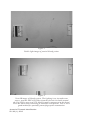
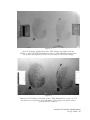
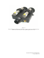
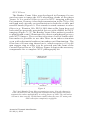
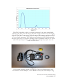
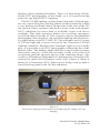
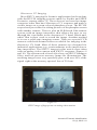
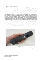
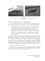
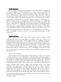
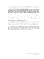
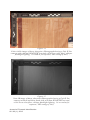
Technical Note
Forensic Reflected Ultraviolet Imaging
Austin Richards 1
Rachel Leintz 2
Abstract: The use of ref lected ultraviolet (UV) imaging has long
been documented in the forensic literature, but little has been written about the “how” and “why” elements of using this technique in
the field. This paper examines the different types of UV light and
explains how and when to use different imaging techniques to visualize hidden evidence. The authors explain, in detail, the wavelength
of light required, the image capturing equipment, and the type of
evidence that can be examined using these techniques.
Introduction
The technique of ref lected ultraviolet (UV) photography has
been around for more than 100 years. During that time, some
interesting applications have been developed for it–particularly in the field of forensics. In some instances, ref lected UV
imaging is the only way to visualize certain phenomena, such
as faint traces of substances on surfaces or the surface texture
on certain transparent materials [1]. These characteristics of
ref lected UV imaging would seem to make it a required part
of forensic investigation, but its use has been inconsistent over
the years. There are two reasons for this. First, it is hard to
get good results with f ilm-based ref lected UV photographic
methods without a lot of trial and error because of the difficulties involved with determining correct exposure, achieving
the appropriate application angle for the ultraviolet light source
without a live UV image preview, and setting the focus and field
1
2
Oculus Photonics, Santa Barbara, CA
Phoenix Police Department, Crime Scene Response Unit, Phoenix, AZ
Received Febr uary 3, 2012; accepted June 21, 2012
Journal of Forensic Identification
46 / 63 (1), 2013
of view properly. Second, as police departments worldwide have
migrated to digital photographic equipment, they have discovered that the standard digital cameras they have purchased do
not respond to ultraviolet light well at all [1, 2]. Even a special
line of Fuji digital cameras that were converted to be more
responsive in the UV and infrared (IR) wavebands did not have
much sensitivity in the UV waveband. This is because of their
Bayer color filter arrays that made it possible to shoot color
images but also tended to block UV light and because silicon
sensors were more responsive to infrared light than to UV light.
There was also additional blocking of UV light from the optics.
The camera systems were sold with a standard Nikon lens that
was not ideal for UV imaging. This is due to the antiref lection coating on the lens as well as absorption characteristics of
the optical glass. This made the transmission less effective in
the UV band than if the lens were designed for UV work and
built out of the best UV optical materials. But the worst part
was that these cameras were sold as part of forensic camera
packages with a UV bandpass filter that unfortunately passed a
substantial amount of infrared (IR) light as well. When forensic
photographers attempted to record ref lected UV images, they
invariably recorded an almost purely IR image. This is because
the camera is much more responsive to IR light than it is to UV
light, and because common photographic light sources contain
very little UV light.
Some forensic photographers assumed that the camera just
did not work correctly in the UV waveband, because the resulting images looked nothing like UV images they had previously
taken with film. The user’s manual and other documentation
provided by Fuji did not shed any light on the matter. Other
photographers incorrectly assumed that the images they were
capturing were UV, when the images they obtained were really
IR. A number of incorrectly designated images were published
in the forensic literature, which further added to the confusion,
because IR images of common scenes generally look nothing
like their UV counterparts.
One of the primary motivations of this paper is to describe
how to take UV digital images correctly, using the right equipment, in the hopes that forensic photographers will buy the
equipment they need and start using the technique again. After
all, if one follows the correct procedure, it is much easier, faster,
and less expensive than UV film photography ever was. This
may lead to a resurgence of these techniques, which are also
made easier and more powerful than ever by the recent developJournal of Forensic Identification
63 (1), 2013 \ 47
ment of extremely bright near-UV light sources based on light
emitting diodes. These lights are much more intense than traditional light sources that are based on gas-discharge tubes and
have a number of other advantages as well.
Light Theory Explained
U lt r av iole t r a d iat ion i s ch a r a c t e r i z e d a s l ig ht w it h
wavelengths between 10 nm and 400 nm. For the purposes of
forensic work, there are two ranges of wavelength (wavebands)
that are of interest: the near-UV band (300 to 400 nm) and the
shortwave UV band (200 to 300 nm). These two wavebands,
which we will refer to as NUV and SWUV, respectively, are very
useful because they allow the user to visualize evidence that is
usually difficult or impossible to record with other photographic
techniques. (In the forensic community, near-UV radiation is
also called longwave UV. The authors prefer calling it near-UV,
because the band is right next to the visible spectrum of light.)
For reference, the infrared light detected by silicon sensors used
in forensic cameras is in the near-infrared (NIR) band, with a
wavelength range of 750 to 1100 nm. There are many interesting
forensic applications for this band, and these applications will
be discussed by the authors in a future article.
The strong absorption of UV radiation by many substances
makes it possible to image forensic evidence that is not readily
apparent to the unaided eye [1, 2]. Ref lected UV photographs
can often reveal otherwise invisible traces of material on a given
surface if those materials absorb UV, which is a common characteristic for many materials, particularly organics. UV imaging
can be used to see substances that are translucent or transparent to the unaided eye and to NIR imaging systems. Because
of the strong absorption of UV radiation, normally transparent material, such as very faint blood stains, will have opacity,
enabling visualization of a bloody shoeprint, for example. The
rule of thumb for using ref lected UV techniques is this: If the
unaided eye can only see a faint pattern of some substance on a
surface, the chances are very good that the pattern will “pop” and
show with a much higher contrast when imaged in the near-UV
band. If one uses shortwave UV imaging, the chances are even
better of getting a good record of the pattern. In general, organic
materials will be even better absorbers of SWUV radiation than
NUV. However, a caveat for the forensic photographer needs
to be given when considering the use of SWUV for imaging
purposes. SWUV is often used in germicidal lamps and is also
Journal of Forensic Identification
48 / 63 (1), 2013
commonly used to “clean” DNA work stations. This is because
this particular wavelength is capable of destroying DNA [3]. If an
investigator plans to collect DNA samples from a photographic
subject, he or she needs to be aware that potential DNA profiles
could be destroyed by using SWUV imaging. This is generally
not the case with NUV imaging, unless the NUV exposure is
quite prolonged and intense. The longer wavelengths of NUV
radiation have less energetic photons that are not as likely to
damage DNA.
Some applications for UV imaging include:
1. Imaging bite marks and other patter n injuries
to skin by increasing the contrast of the surface
impressions or by visualizing the faint darkening
of the skin that appears over time in response to
pressure trauma (NUV) [2].
2. Imaging shoeprints, footprints, and handprints
made with substances that are transparent to the
eye and near-IR imaging, such as faint bloodstains
or faint dust (NUV and SWUV).
3. Searching for and documenting bone fragments
(NUV).
4. Imaging latent fingerprints (SWUV).
5. Imaging explosives residue on surfaces (NUV and
SWUV).
This paper will cover the first two applications. The latter
three will be covered in a future paper by the authors.
Figures 1 through 4 are images of partial bloody fingerprints
taken in different wavebands of light.
Journal of Forensic Identification
63 (1), 2013 \ 49
Figure 1
Visible light image of partial bloody prints.
Figure 2
Near-IR image of bloody prints. The lighting is an incandescent
source, and the PECA 910 filter from the forensic kit built around
the Fuji IS Pro was used. The blood is quite transparent in this band.
Even the inked letters denoting A and B are transparent. This is not a
good method for partially patent fingerprint examination.
Journal of Forensic Identification
50 / 63 (1), 2013
Figure 3
Near-UV image of bloody prints. This image was taken with an
IS/Pro, a 365 nm LED illumination source, and a Baader Venus filter.
The prints are definitely easier to see than in the visible image.
Figure 4
Shortwave UV image of bloody prints. The illumination source is 254
nm from a low-pressure mercury lamp. The image was taken with a
special SWUV camera.
Journal of Forensic Identification
63 (1), 2013 \ 51
Procedures and Equipment for Photography
Near-UV and Shortwave UV Imaging: How Do You Do It?
Photography using ref lected NUV radiation is essentially
the same as visible-light photography. Digital sensors can be
made reasonably sensitive to NUV and can be used with suitable
optics, filters, and illumination to take photographs. But most
digital cameras are designed with NUV blocking filters that
are not easily removable. Attempts to record N U V images
with these cameras usually yield disappointing results. Some
specially modified digital single-lens ref lex cameras (DSLRs)
can be used for NUV photography. Silicon sensors in DSLRs
are wide spectrum and have to be used with bandpass filters to
record NUV unless the lighting is pure NUV, with little or no
visible or IR light present. Many forensic photographers have
tried using their traditional UV pass filters (e.g., Kodak Wratten
18A) on their digital cameras. Because the Wratten 18A has a
substantial transmission peak in the IR band, the resulting image
is often more of a near-IR image than anything else. Fortunately,
there is at least one filter on the market that solves this problem:
the Baader Venus f ilter (Baader Planetarium, Mammendorf,
Germany), which we will describe in more detail shortly.
Shortwave UV imaging requires special types of detectors.
The RUVIS systems use an image intensifier with a photocathode that is responsive to shortwave UV light. There are also
silicon CCD sensors that can be processed in a special way to
make them sensitive in the SWUV band. This CCD technique
was used to take the SWUV images in this article.
NUV Camera Systems
At the time that this article was written, there was a fairly
limited selection of equipment that was optimal for NUV photography with a DSLR. These were the best options:
1. Fuji S-3 UVIR or Fuji IS Pro camera or other aftermarket
DSLR conversion with NUV response (such as those
done by LDP LLC., Carlstadt, NJ).
2. JENOPTIK Coastal Optics 60 mm UV-VIS-IR lens or
105 mm UV lens, Universe Kogaku 78 mm lens with
F-mount adapter, or legacy Nikon U V-Nik kor 105
mm lens. In a pinch, a conventional Nikon F-mount
lens should work, though it will have reduced NUV
transmission.
Journal of Forensic Identification
52 / 63 (1), 2013
3. Baader Venus filter with 52 to 48 mm stepper ring
4. NUV illumination source (LED-based illuminator, gasdischarge blacklight, or direct sunlight)
The Fuji S-3 UVIR and the Fuji IS Pro DSLRs were marketed
for forensic work. Their “hot mirrors” have been removed so
that NIR and NUV light can reach the sensor. The end result is
a camera that has reasonable response to NIR light and substantially less response in the NUV band. Still, the NUV response
is enough to make the cameras capable of NUV imaging, as
long as one achieves sufficient exposure, either through bright
NUV lighting or long exposure times. Cameras with hot mirrors
typically have negligible NIR or NUV response1.
Invisible-light images should be presented in black and white,
and ideally, all the pixels in any camera used for UV imaging
should be monochrome. Unfortunately, the Fuji cameras are like
every other DSLR on the market and have a color filter array on
the sensor. There are three colors of filters–red, blue, and green–
and each has a distinctive spectral response in the NUV band.
Because of this, NUV images taken using direct sunlight as an
illumination source will have a different color cast to them than
images taken by the light of a narrowband NUV illuminator.
After much experimentation, we recommend setting the camera
to shoot in black and white mode. This is a setting within the
Fuji’s menus that will combine the R, G, and B channels in such
a way as to produce grey-level pixel values with no color. This
technique will exploit all the available pixels in the sensor in
the final image.
Nikon made a 105 mm lens for 35 mm cameras called the
UV-Nikkor, but it is long out of production. JENOPTIK Coastal
Optics (formerly Coastal Optics) has designed a replacement for
this lens, with improvements. Their 60 mm CoastalOpt lens is
designed to have visible, NUV, and NIR transmission and color
correction. They also make a clone of the 105 mm UV-Nikkor
lens that works down into the SWUV band. These lenses are
expensive but of high quality. A more economical option is the
1
The hot mirror is specifically designed to keep out near-IR light, which
will substantially degrade digital photos. Unlike color film, which has very
little near-IR response, silicon sensors have plenty of response in that range.
Because most scenes are lit with light sources that contain enough nearIR light, and that light transmits reasonably well through most lenses, it
must be stopped before the sensor. The hot mirror is really an optical coating applied to the sensor window. It is designed to eliminate near-UV light,
another unwanted waveband of light that gets through lenses well enough to
soften images or throw off color balance.
Journal of Forensic Identification
63 (1), 2013 \ 53
78 mm Universe Kogaku quartz lens. The elements include fused
silica, which is often incorrectly called quartz, a lower purity
mineral for m. Fused silica transmits below the glass cutoff
wavelength of 310 nm. The authors have found that it is not
strictly necessary to use an expensive fused silica lens for NUV
work, because standard BK7 lens glass has some transmission
at 365 nm and below. We have used standard Nikon lenses on
the Fuji S-3 UVIR with reasonable results. Compared to the
Coastal Optics lenses or the UV-Nikkor, the transmission and
color correction of standard lenses is mediocre in the NUV band.
Fused silica lenses are mandatory for shortwave UV imaging,
because absolutely no radiation below about 300 nm band passes
through glass lenses–it is all absorbed.
It is often convenient to search for evidence in the NUV
band using an N U V viewer. The Fuji cameras can be used
in this fashion, but with limited utility. Both cameras can be
commanded to retract the mirror and generate a live video image.
When a Baader filter is in place, and sufficient NUV illumination is present, the user will see live NUV video on the rear LCD
screen. But this live video uses up the batteries rapidly, and the
user has to keep pressing the button every 30 seconds to restart
the live preview. It is also hard to see the viewfinder image in the
presence of ambient lighting without a loupe on the LCD screen.
A useful tool for a quick search of a scene in the NUV band is
the Oculus Photonics UVScanner (Figure 5). This is a battery
powered, handheld viewer with excellent spectral purity and a
built-in pair of NUV illuminators. This unit has rubber eyecups
on the back to block out ambient light. The operator sees an NUV
video image on the dual LCD displays and can then walk around
looking for evidence at a scene while using stick-on evidence
markers to mark it for later photography at high resolution with
an NUV DSLR system.
Journal of Forensic Identification
54 / 63 (1), 2013
Figure 5
Oculus Photonics UVScanner. This device images in the near-UV band, from
about 320 nm to 440 nm, with a peak response at 370 nm.
Journal of Forensic Identification
63 (1), 2013 \ 55
NUV Filters
The Baader Venus filter was developed in Germany for use
on telescopes to image the NUV-absorbing clouds of the planet
Venus. It is a perfect filter to use for NUV imaging with the
Fuji cameras, because it has high transmission in the 330 to 380
nm band and virtually no transmission in both the visible and
near-IR bands (Figure 6). This stands in stark contrast to other
filters (e.g., Wratten 18A, PECA 900) that suffer from large red
leaks and are generally unsuitable for use with a DSLR for NUV
imaging (Figure 7) [1]. The Baader Venus filter makes it possible
to photograph scenes illuminated by direct sunlight and get very
pure NUV images. The excellent rejection of out-of-band radiation makes it possible to use this filter in an indoor situation
even with substantial ambient incandescent illumination. The
filter has a 48 mm ring thread on it, which requires a 52 to 48
mm stepper ring to allow it to be screwed onto the front of the
Coastal Optics lens or UV Nikkor. Figure 8 depicts the necessary
components for a complete NUV photography kit.
Figure 6
The 2-inch Baader Venus filter transmission curve. Note the change in
scale. Beyond 418 nm, the magenta transmission data points have their
transmission values multiplied by a scaling factor of 1000. The red leak at
950 nm is thus roughly 10,000 times smaller than the near-UV main peak
and is therefore completely negligible.
Journal of Forensic Identification
56 / 63 (1), 2013
Figure 7
The PECA 900 filter, which is virtually identical to the now unavailable
Wratten 18A filter. Note the large red leak, which is almost 12% the height
of the NUV peak. For a sensor with a lot more NIR response relative to NUV
response, this filter will heavily bias the image towards the NIR, unless there
is a very large amount of NUV light present relative to NIR. Unfortunately,
this filter was part of a forensic package sold by Fujifilms with the S-3 UVIR
and IS Pro packages. Many other NUV filters designed for film have the
same red-leak issue.
Figure 8
NUV digital imaging system. Fuji IS Pro, Universe Kogaku quartz lens
with Nikon F-mount adapter, 2-inch Baader Venus filter, and adapter ring.
Journal of Forensic Identification
63 (1), 2013 \ 57
NUV Illumination
Having a sufficient level of near-UV lighting is crucial, and
here again, specialized equipment should be used. Without
fairly bright NUV light sources, photos will tend to be dramatically underexposed for two reasons: NUV is scarce in indoor or
nighttime environments, and most materials appear very dark in
the NUV band because of their high absorption. Trying to use
conventional indoor lighting for NUV illumination is doomed
to failure–there is little or no NUV emitted by these lights by
design (to prevent furniture and paint from fading). Even with a
filter like the Baader with its tiny red leak, the exposures have
to be so long that the dark noise of the CCD sensor in the Fuji
cameras becomes the dominant feature in the image.
Several ALS units (including the Rof in Polilight) have
“UV” settings, but we have found that these are not nearly
bright enough for digital NUV photography without very long
exposures of relatively small areas of the scene. There are also
special f lash units that are designed to emit more NUV light
than typical f lashes. These f lashes have strobe tubes that do not
have a UV-absorbing coating on them. We have found the QFlash
brand unit to be relatively weak in the NUV band, and multiple
f lashes of the unit were required to get a decent exposure for
typical conditions. This, of course, requires that the camera be
mounted on a tripod. An additional complication to using a f lash
for near-UV photography is that the NUV light is not present in
the scene until the f lash is actually fired. This makes it impossible to focus the image using the live-preview function of the
camera. Because the Baader filter does not transmit any visible
light, the user must take a picture, look at the results on the
camera’s LCD screen, then adjust the focus blindly and take
another picture. This is very time consuming and will quickly
use up the batteries in the f lash unit and camera. The photographer can, in principal, take off the Baader filter, adjust focus
using visible light, replace the filter, and shoot images using
the f lash. In practice, this does not work well, because all but
very expensive UV lenses will have a different focus setting for
visible light and near-UV radiation.
The authors have had excellent results with NUV lighting
from Clearstone Technologies in Minnesota. They manufacture
illumination systems composed of arrays of very bright LEDs
at a wavelength of 365 nm and others. The 365 nm wavelength
is no accident–it corresponds to a strong line of high-pressure
mercury vapor discharge lamps that the LEDs are intended to
Journal of Forensic Identification
58 / 63 (1), 2013
displace in the commercial market. There is a large body of filmbased NUV photography in this band, so it is a good starting
point for any digital NUV imaging.
The NUV LED lighting systems from Clearstone Technologies
are very spectrally pure, having almost all their radiation within
a very narrow waveband–about 10 nm wide in the case of the 365
nm system. These systems can f lood a crime scene with intense
NUV radiation–far more than is available from even direct
sunlight. This high intensity makes for excellent exposures
even with shutter speeds of 1/60 second or faster that handheld
photography often requires. An excellent lighting head for forensic applications is the JL3-365E-42. This unit puts out 10.5 watts
of 365 nm light and it can be driven by the Clearstone CF2000
lighting controller. Because the Clearstone light is so spectrally
pure, it is possible to do NUV photography without the use of the
Baader filter. The authors think that this is inadvisable because
often there may be visible f luorescence in the scene that will
record and spoil the spectral purity of the resulting image. Any
visible or IR ambient light must be eliminated or at least greatly
reduced to make this technique work well. Figure 9 shows a
photo of a Clearstone NUV light source being used to light a
wood f looring panel with 365 nm radiation.
Figure 9
Clearstone lighting system used on wood flooring for oblique 365 nm
imaging.
Journal of Forensic Identification
63 (1), 2013 \ 59
A much less expensive (but much less effective) alternative to
the Clearstone system is an array of blacklights like the ones sold
at hardware stores. These lamps are sold as 18-inch or 48-inch
f luorescent tubes with dark purple glass envelopes. Like the
Clearstone, these lamps radiate around 365 nm, but they are
much less intense than LED-based solutions. We have found
it necessary to arrange six or eight lamps in banks of lighting
fixtures within 18 inches of the subject to get usable illumination
levels. Because the tubes are bulky and long, it is not possible
to get an intense spotlight of near-UV light out of them. Even at
these close ranges, exposures of multiple seconds are necessary
to achieve adequate illumination, so a tripod is required. 2
A third option is a Crime-Lite 82S UV lighting system by
Foster and Freeman. This is a battery-powered handheld NUV
source in a convenient form factor. We do not have data on the
total power emitted by the illuminator, but it is less than the
Clearstone system. Nevertheless, this forensic light source is
quite suitable for the imaging of smaller regions of interest at a
crime scene. There are several other handheld near-UV f lashlights on the market, including products made by Rof in and
Labino, that emit on the order of a few watts of NUV radiation
power. These are based on LED emitters, and thus are ver y
spectrally pure–perfect for ref lected-UV photography. It should
be mentioned that the same vendors also sell alternative light
sources (ALS) that are based on xenon lamps, and these ALS
units can be ordered with a UV filter option. A forensic specialist looking to purchase such a light source for UV illumination
needs to be cautious. We have found that the UV setting on some
of these ALS units has near-infrared leakage through the UV
bandpass filter. If they are used with the Baader filter, there
will not be a problem with this contamination, but generally,
these lights are quite weak in the NUV band compared to the
LED units.
2
Another caveat to consider when using this lighting source is that the ends of
the bulbs emit near-IR light. This is due to the bulb’s hot cathode filaments.
Blacklight tubes should always be used with a Baader filter on the camera’s
lens, unless the last three inches of each end of the tube is covered with black
electrical tape to block the near-IR emission.
Journal of Forensic Identification
60 / 63 (1), 2013
Shortwave UV Imaging
Most SWUV imaging for forensic applications has been done
with the RUVIS imaging systems made by Sirchie and SPEX
Forensics, among others [1]. These devices are based on image
converter tubes that have shortwave UV response and make a
visible image on a green-colored phosphor screen. The RUVIS
scopes have provisions for taking pictures of the phosphor screen
with another camera. There is also an SLR-based film camera
system with an image intensif ier that allows the user to see
through the viewfinder in the shortwave UV band. Black-andwhite film is then used to record the image. Another method
is to use a solid-state imaging sensor. There are special CCD
cameras with back-thinned sensors available for imaging in the
shortwave UV band. Most of these cameras are designed for
industrial applications (e.g., semiconductor wafer inspection or
lens inspection). The SWUV images in this article were taken
with an analog video camera with SWUV response (Figures 4
and 10). The system used a fused silica lens, an SWUV bandpass
filter, and low-pressure mercury lamps for illumination. The
resulting images were very spectrally pure, with over 99% of the
signal right at the mercury spectral line of 254 nm.
Figure 10
SWUV image of fingerprints on analog video monitor.
Journal of Forensic Identification
63 (1), 2013 \ 61
SWUV Light Sources
The most common light source for SW U V imaging is a
low-pressure mercury vapor discharge lamp (Figure 11). The
low-pressure mercury vapor spectrum is dominated by a strong
emission line at 254 nm wavelength. This lamp looks like a
f luorescent tube with no white phosphor coating inside. The
tube is made of fused silica, because ordinary glass will not
transmit shortwave UV light at all. The lamps are inexpensive
and relatively powerful, but the output angular pattern is quite
diffuse and it is hard to make a really intense focused beam
of radiation with long skinny tubes. We have used a handheld
germicidal lamp like the one in Figure 12 as a SWUV source
with reasonably good results, although the lamp needs to be held
close to the object of interest. Future sources will undoubtedly
be based on arrays of LEDs. At present though, SWUV LEDs
are very expensive and very low power. It may be some time
before there are good alternatives to mercury lamps, though
there is also development work being done into plasma illumination sources in the SWUV band.
Figure 11
254 nm germicidal lamp in handheld battery powered package.
Journal of Forensic Identification
62 / 63 (1), 2013
Figure 12
Visible-light (left) and reflected near-UV (right) images of a fresh
bitemark.
Near-UV and Shortwave UV Applications
Forensic applications for NUV and SWUV imaging hinge on
two characteristics of UV radiation. These characteristics are:
1. The strong absorption of UV by many visibly transparent
or translucent materials and substances. The absorption
tends to be greater for SWUV compared to NUV.
2. The tendency of shorter UV lightwaves to scatter more
strongly off of tiny surface features, defects, and texture
relative to visible light. Very fine details are revealed
by NUV and SWUV imaging, with SWUV giving the
best results.
The first characteristic is due to the high energy of UV photons
that causes them to be absorbed by atoms and molecules, rather
than ref lected, and the second is due to the short wavelength of
UV lightwaves. Shorter wavelengths of light “feel” tiny bumps
and surface texture that longer wavelengths (visible and NIR)
tend to average out. The next section shows examples of NUV
imaging revealing features that are not apparent to the unaided
eye because of high UV absorption of materials in the scene.
Bitemarks and Pattern Injuries
The NUV band is commonly used for photographing bitemarks
or pattern injuries. These injuries can further be classified into
two classes: fresh injuries and aged injuries.
Journal of Forensic Identification
63 (1), 2013 \ 63
Fresh Injuries
NUV light rays barely penetrate the skin when compared
to visible light, so they can allow a crime scene specialist to
record just the surface topology of the skin. Using this type of
NUV lighting will not capture bruising under the skin. This is
an important distinction to note because if bruises are viewed
with visible light, the underlying bruises can obscure the image
and make it diff icult to see the minute details of the bite or
pattern impression. An example is shown in Figure 12. Although
we do not have photographic data to back up the assertion, we
believe that SWUV imaging can further enhance fresh bitemarks
because of its very minimal penetration into skin. Thus, very
fine changes in the surface topology can be captured. The skin
of living individuals should not be exposed to shortwave UV
light for long durations, because it is a strong skin irritant, so
this technique is best reserved for imaging bitemarks on the
deceased.
Aged Injuries
When bite marks age, their individual characteristics tend
to fade and often become barely visible to the eye. However,
melanin can concentrate in the injured areas, leading to hyperpigmentation of the skin, especially in Caucasian skin. Melanin
absorbs NUV light much more strongly than visible light, so it
is almost as though the bitemark “develops” in the NUV band
like a Polaroid picture [4, 5]. Observation of this hyper pigmentation can be enhanced by NUV imaging, because of this
strong absorption. Figure 13 shows an excellent example of this
phenomenon.
Shoeprint Imaging
Shoeprints made with light-colored dirt or dust on a lightcolored surface can be difficult to photograph with any degree
of contrast using conventional color photography. To enhance
the print-to-surface contrast, these prints are typically collected
using electrostatic dust print lifters [5]. The dust print is transferred from the crime scene surface to a special jet-black mylar
sheet, which then shows light-colored dust with ver y high
contrast. The process of obtaining these lifts is time consuming and uncomfortable for the crime scene specialist, who is
required to be on his or her hands and knees for several hours.
Other disadvantages of using this technique are that the placement of the Mylar film and the possible lateral movement caused
by the static charge place the fragile dust print at risk, and the
Journal of Forensic Identification
64 / 63 (1), 2013
lifters are expensive and not reusable. Sometimes it is not clear
to the investigator where to place the EDPLs because the prints
have such low contrast to the unaided eye.
The NUV and SWUV imaging methods can sometimes greatly
increase contrast in this situation and give the user a nondestructive means of searching and recording that does not compromise
the print or remove the evidence from its original substrate. This
technique can be used with excellent results, particularly when
the substrate is an organic material (e.g., wood or cloth). In this
case, the organic substrate records as very dark, because UV is
absorbed by it; inorganic dirt or dust often tends to ref lect and
scatter UV, causing the substance to appear lighter. Figures 14
through 17 depict a dusty shoeprint on a wood panel, visualized
using different wavelengths of light.
As a point of reference, NIR techniques are definitely not
recommended for this application, because the dust print tends
to be even less apparent than to the unaided eye. Figure 15 is
an NIR image of the shoeprint, which is almost undetectable.
We note the two fundamental rules of forensic imaging in
these two wavebands: NIR imaging is good for seeing through
thin layers of substances, whereas NUV and SWUV are good
for seeing the thin layers of substances themselves.
Journal of Forensic Identification
63 (1), 2013 \ 65
Figure 14
Color visible image of dusty shoeprint. (Photographed using a Fuji IS Pro
camera with a 60 mm JENOPTIK lens and a 916 Peca color filter; oblique
flashlight lighting; 1 second at f/8 exposure; ISO setting of 400.)
Figure 15
Near-IR image of dusty shoeprint.(Photographed using a Fuji IS Pro
camera in black and white mode with a 60 mm JENOPTIK lens and
a 916 Peca color filter; oblique flashlight lighting; 1/4 second at f/4
exposure; ISO setting of 100.)
Journal of Forensic Identification
66 / 63 (1), 2013
Figure 16
365 nm near-UV image of dusty shoeprint. (Photographed using a Fuji
IS Pro camera in black and white mode with a 78 mm Kogaku lens and
a Baader filter; oblique Clearstone lighting; 2 seconds at f/3.8 exposure;
ISO setting of 100.)
Figure 17
254 nm SWUV image of dusty shoeprint. (Photographed using an Oculus
Photonics SWUV camera, Kogaku 25 mm quartz lens, 254 nm bandpass
filter; oblique lighting with twin germicidal lamps; analog video.)
Journal of Forensic Identification
63 (1), 2013 \ 67
Conclusion
NUV and SWUV imaging has some very interesting forensic applications that have been traditionally quite difficult to
achieve with film-based photography. These applications hinge
on the tendency for UV light to be absorbed by many visibly
transparent materials (because of the high photon energy of UV)
and the tendency for UV light to be scattered by minute surface
texture (because of the short wavelength of UV waves).
Ref lected UV imaging works best for forensic applications
where a faint pattern needs to be enhanced, for example in the
case of dusty or bloody prints, when the print itself is transparent to visible light. Materials that are transparent to visible light
will tend to be even more transparent to NIR radiation, so that
method is not the right choice either. NUV imaging may work
well if the substance has decent absorption.
If NUV imaging does not yield good results, then shortwave
UV imaging should be used, because SWUV light tends to be
even more strongly absorbed and scattered than NUV by many
substances.
There are specialized digital camera systems that can image
in these UV wavebands. The most critical aspect of any UV
imaging system is the spectral purity of the light signal that
reaches the imaging sensor. High purity can be achieved by
using a very bright, monochromatic, LED-based light source.
These intense lights compensate for transmission losses in the
optics and color filter arrays on converted DSLRs, making it
possible to photograph a crime scene rapidly and without a
tripod in many cases.
For further information, please contact:
Dr. Austin Richards
Oculus Photonics
2542 Mesa School Lane
Santa Barbara, CA 93109
www.uvcorder.com
[email protected]
or
Rachel Leintz
621 West Washington Street
Phoenix, AZ 85003
[email protected]
Journal of Forensic Identification
68 / 63 (1), 2013
References
1.
Sanfilippo, P.; Richards, A.; Nichols, H. Ref lected Ultraviolet
Digital Photography: The Part Someone Forgot to Mention.
J. For. Ident. 2010, 60 (2), 181–198.
2. Krauss, T. C.; Warlen, S. C. The Forensic Science Use of
Ref lective Ultraviolet Photography. J. For. Sci. 1985, 30 (1),
262–268.
3. Schrier, W.; Schrader, T.; Koller, F.; Gilch, P.; CrespoHe r n á ndez , C.; Swa m i n at h a n , V.; C a r el l , T.; Zi nt h ,
W.; Kohler, B. T hy m i ne Di mer izat ion is an U lt rafast
Photoreaction. Science 2007, 315 (5812), 625–629.
4. David, T. J.; Sobel, M. N. Recapturing a Five-Month-Old
Bite Mark by Means of Ref lective Ultraviolet Photography.
J. For. Sci. 1994, 39 (6), 1560 –1567.
5. Robinson, E. M. Crime Scene Photography, 1st ed.; Soucy,
J., Weaver, K., Eds.; Elsevier: Burlington, MA, 2007; pp
291–295, 393–396.
Journal of Forensic Identification
63 (1), 2013 \ 69