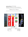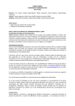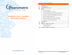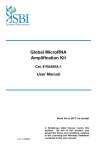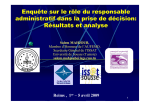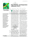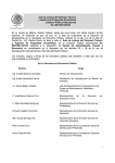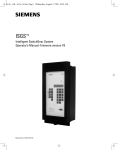
1
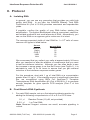
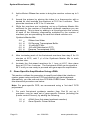
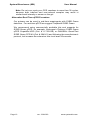
Interferon Response Detection Kit
for validation of siRNA experiments
(Cat. # SI300A-1)
User Manual
Store kit at -20°C on receipt
(ver. 4-082410)
A limited-use label license covers this
product. By use of this product, you
accept the terms and conditions outlined
in the Licensing and Warranty Statement
contained in this user manual.
Interferon Response Detection Kit
Cat. # SI300A-1
Contents
I.
Introduction and Background
A.
B.
C.
D.
E.
F.
G.
H.
Overview
siRNA and Interferon Induction
Detection of Interferon Pathway Activation
Variability of the Interferon Response
Overview of Protocol
List of Components
Additional Required Materials
Procedural Guidelines
2
2
3
4
6
7
7
7
II. Protocol
A. Isolating RNA
B. First-Strand cDNA Synthesis
C. Gene-Specific Amplification Using cDNA
9
9
10
III. References
13
IV. Troubleshooting
14
V. Appendix
A. Related Products
B. Technical Support
VI. Licensing and Warranty Statement
888-266-5066 (Toll Free)
650-968-2200 (outside US)
15
15
16
Page 1
System Biosciences (SBI)
User Manual
I. Introduction and Background
A. Overview
This manual provides details and information necessary to use the
Interferon Response Detection Kit to identify and reproducibly confirm
non-specific stress response of cells to the introduction of siRNA. This
kit may also be used for other applications, such as for the detection of
retroviral infection. To ensure optimal results, please read the entire
manual before using the reagents and material supplied with this
system.
B. siRNA and Interferon Induction
Mammalian hosts have evolved defense mechanisms against doublestranded RNA (dsRNA), typically present as a consequence of viral
infection. Upon viral infection, cytokine interferon is induced. Interferon
then activates a signaling cascade, culminating in transcriptional
activation of hundreds of interferon stimulated genes (ISGs). Many of
these ISGs function in stress response pathways to mediate the
cellular antiviral response (Haque and Williams, 1998; Stark et al.,
1998).
The use of siRNA to selectively knockdown genes of interest has
become a powerful tool to study gene function. Recently, it was
discovered that, in addition to gene-specific silencing, siRNA can also
induce nonspecific effects in cells by activating ISGs involved in the
stress response (Bridge et al., 2003; Persengiev et al., 2004; Scacheri
et al., 2004; Sledz et al., 2003, Pebernard, 2004). The observation that
treatment of mammalian cells with siRNA molecules can trigger nonspecific cellular responses raises a serious concern about specificity of
RNA interference responses and complicates the interpretation of
siRNA knockdown results. To address this issue, new tools are
needed that help confirm the specificity of responses to the knockdown
of genes targeted by experimental siRNA and the absence of off-target
effects induced by the presence of the siRNA molecules.
SBI’s Interferon Response Detection Kit provides an easy and accurate
method to ensure that synthetic siRNA or siRNA constructs in plasmid
or viral vectors do not induce significant interferon-related responses
when expressed in the cell system of choice. Also, the system can be
used to assess the effects of reagents (such as transfection reagents)
and other procedural steps that may produce, enhance, or otherwise
affect cellular interferon responses.
While not all siRNA molecules will activate non-specific cellular stress
response pathways, the only way to confirm the specificity of an siRNA
knockdown phenotype or response is to also confirm the absence of
Page 2
ver. 3-061023
www.systembio.com
Interferon Response Detection Kit
Cat. # SI300A-1
any nonspecific cellular effects. SBI’s Interferon Response Detection
Kit provides an objective and unambiguous procedure to ensure nonspecific activation of interferon-related pathways do not perturb your
siRNA knockdown studies.
C. Detection of Interferon Pathway Activation
SBI’s Interferon Response Detection Kit uses a PCR-based approach
to enable researchers to easily measure relative expression levels of
five genes involved in the interferon response. The use of these five
genes helps ensure the kit will accurately detect interferon-related
stress responses in a variety of cell types and under many different
conditions. Different cell types may show different levels of increase
for these genes. Also, the level of interferon-related stress response
that is observed will vary depending on the concentration of the siRNA
molecule (Persengiev et al., 2004) and the cell line.
The OAS1 (NM_016816) and OAS2 (NM_016817.1) genes:
The 2',5'-oligoadenylate synthetases (OAS) represent a family of
interferon-induced proteins implicated in the mechanism of the antiviral
action of interferon. When activated by double-stranded RNA, these
proteins polymerize ATP into 2'-5'-linked oligomers that bind and
activate RNase L, which plays a significant role in the inhibition of
cellular protein synthesis and viral infection resistance.
Three forms of human OAS have been described (Hovnanian A, et al.,
1998). This kit contains primers to detect two of the three forms:
OAS1 and OAS2. There are two isoforms of the OAS1 gene which
share identical N-terminal sequence but diverge at exon 7 and have
molecular weights of 40 and 46 KDa. The Primer Mixture for OAS1
amplifies a 144 base region present in both isoforms. Similarly, there
are two isoforms of OAS2 gene which also share a common 5' region
and have molecular weights of 69 and 71KDa. The OAS2 Primer
Mixture amplifies a 234 base region present in both isoforms.
The MX1 (NM_002462.2) gene:
Human MX1 protein is a member of the interferon-induced myxovirus
(influenza virus) resistance protein family (MX proteins) and an
important component of the innate host defense against RNA viruses.
The MX family belongs to a superfamily of large GTPases that also
includes the dynamins and the interferon-regulated guanylate-binding
proteins. MX1, by interacting with a component of the nucleocapsid,
prevents replication of viral RNA and thereby inhibits the production of
new infectious virus particles. (Kochs G, et al., 2002). The MX1 Primer
Mixture amplifies a 402 nucleotide region of this gene.
The IFITM1 (NM_003641.2) gene:
Interferon-inducible
trans-membrane
proteins
(IFITMs)
of
approximately 17 kDa have been suggested to play a role in the
888-266-5066 (Toll Free)
650-968-2200 (outside US)
Page 3
System Biosciences (SBI)
User Manual
antiproliferative activity of interferons based on their pattern of
induction in interferon-sensitive and interferon-resistant cell lines and
their ability to inhibit cell growth when a membrane fraction enriched in
17-kDa proteins is added to cells in culture. IFITM1 has been
demonstratively shown to associate with other proteins at the cell
surface to form a complex relaying growth inhibitory and aggregation
signals. (Deblandre G.A, et al., 1995). The IFITM1 Primer Mixture
amplifies a 128 base fragment of this gene.
The ISGF3γ (NM_006084.3) gene:
Alpha interferon stimulates transcription by converting the positive
transcriptional regulator ISGF3 from a latent to an active form. This
receptor-mediated event occurs in the cytoplasm, with subsequent
translocation of the activated factor to the nucleus. ISGF3 has two
components, termed ISGF3α and ISGF3γ. ISGF3γ serves as the DNA
recognition subunit, while ISGF3α is the target for interferon signaling
and serves as a regulatory component. (Veals S.A., et al., 1992). The
ISGF3γ Primer Mixture amplifies a 333 base portion of the gene.
D. Variability of the Interferon Response
The sensitivity of different cell lines to double-stranded RNA activation
of interferon-induced stress genes and pathways can vary significantly.
Cell type, as well as growth conditions and passage number, can affect
the susceptibility, level, and extent of activation of the interferon
response. The interferon response is readily detected in the numerous
human cell lines that activate stress response pathways when doublestranded RNA is introduced. This response can be clearly seen by the
robust increase in OAS1, OAS2, MX1, ISGF3γ and IFITM1 expression
levels after treatment with concentrations as low as 250 ng/ml of a long
poly(I)-poly(C) dsRNA sequence (Sigma, Cat. # P9582) in cell lines
such as HEK 293 and MRC-5 (see Figure 1). Higher concentrations of
the double-stranded RNA (e.g. 2.5 μg/ml) can trigger rapid apoptosis
and cytotoxicity within 24 to 48 hours.
Page 4
ver. 3-061023
www.systembio.com
Interferon Response Detection Kit
Cat. # SI300A-1
MRC-5 Cells
HEK 293 Cells
2 hours
8 hours
24 hours
48 hours
90
80
70
60
50
40
30
20
10
0
OAS1
OAS2
MX1
ISGF3γ
40
Ratio Induced to Control
Ratio Induced to Control
100
2 hours
8 hours
24 hours
48 hours
35
30
25
20
15
10
5
0
IFITM1
OAS1
OAS2
MX1
ISGF3γ
IFITM1
Interferon Induction in HEK 293 Cells
INF ELISA (pg/ml)
0.7
0.6
control
INF induced
0.5
0.4
0.3
0.2
0.1
0
2 hours
8 hours
24 hours
Fig. 1. Interferon response detected in two human cell lines. MRC-5 and HEK 293
cells were grown to confluency in a T-75 format flask, and then transfected with 500 ng/ml
of a long poly(I)-poly(C) dsRNA sequence (Sigma, Cat. # P9582). Total RNA was
prepared from treated (+) and mock transfected (-) control cells at 2, 8, 24, and 48 hours
after transfection. The expression levels of OAS1, OAS2, MX1, ISGF3γ and IFITM1
genes were determined by end-point RT-PCR (27 cycles) using the protocol described in
the kit. As is evident from the results shown for MRC-5 cells (on the left) and HEK293
cells (on the right), the interferon response was readily detected by examination of the
expression levels of these genes. Also shown is the level of interferon response in HEK
293 cells as detected by ELISA.
888-266-5066 (Toll Free)
650-968-2200 (outside US)
Page 5
System Biosciences (SBI)
User Manual
E. Overview of Protocol
As can be seen in the flowchart in Figure 2, the procedure for the
Interferon Response Detection Kit is straightforward, convenient, and
provides clear, measurable results.
Fig. 2. Flowchart for IRD Kit Procedure.
Page 6
ver. 3-061023
www.systembio.com
Interferon Response Detection Kit
Cat. # SI300A-1
F. List of Components
The Interferon Detection Kit provides enough primer, in each mix, to
perform 20 PCR reactions (100 total reactions) in 50 µl.
20 µl
Primer Mixture for OAS1
144 nt*
10 µM ea.
20 µl
Primer Mixture for OAS2
234 nt
10 µM ea.
20 µl
Primer Mixture for MX1
402 nt
10 µM ea.
20 µl
Primer Mixture for IFITM1
128 nt
10 µM ea.
20 µl
Primer Mixture for ISGF3γ
333 nt
10 µM ea.
20 µl
Control Primer Mixture for β-actin
110 nt
6 µl
Control cDNA (INF induced cells)
10 µM ea.
50 ng/µl
*indicates size of the amplicon.
The kits are shipped on blue ice and should be stored at -20°C upon
receipt. Properly stored kits are stable for 1 year from the date
received.
G. Additional Required Materials
•
•
•
•
•
•
•
RNeasy Mini Total RNA Purification Kit (QIAGEN, Cat. # 74104)
Reverse Transcription Kit (SuperScript™ III, Invitrogen, Cat. #
18080-051) or, for small quantities of RNA, use SBI’s Full
Spectrum™ Complete Transcriptome RNA Amplification Kit (Cat. #
RA101A-1)
PCR Reagents (Titanium™ Taq DNA Polymerase, Clontech, Cat.
# 639208)
Thermocycler (with heated lid)
3% Agarose Gel in Tris-Acetate EDTA (TAE) Buffer
DNA Size Ladder with markers from 50 to 2,000 bp (AmpliSize™
DNA Ladder, Bio-Rad, Cat. # 170-8200)
Optional for samples from sources with high RNase activity:
Ribonuclease Inhibitor (SUPERase-IN™, Ambion, Cat. # 2694)
H. Procedural Guidelines
Basal and induced levels of OAS1, OAS2, MX1, ISGF3γ, and IFITM1
may vary from human cell line to human cell line. In addition, basal
and induced levels of the ISGs may also vary in the same cell line
depending on growth conditions. Thus, it is critical to ensure that you
include a non-induced control that is treated exactly the same as
888-266-5066 (Toll Free)
650-968-2200 (outside US)
Page 7
System Biosciences (SBI)
User Manual
samples where you introduce siRNA—this includes mock transfecting
the control cells.
Since siRNA reagents are delivered to cells via transfection, it is crucial
to consider the transfection efficiency of the cells when assessing the
sensitivity of a particular cell line to the interferon-related response to
siRNA.
In addition to the specific procedural notes above, be sure to follow
these general practices to ensure quality results:
•
•
•
•
•
Page 8
Before dispensing, completely thaw all reagents. Vortex, to mix
thoroughly, all reagents except for the enzymes. After adding
reagents to the mixture, pipette up and down 5-10 times to ensure
mixing.
Briefly centrifuge each mixture once all the components have been
added to ensure there are no reagents left on the sides of the
tube, separated from the reaction mixture.
When setting up multiple reactions, we recommend that you
prepare a master mix.
cDNA is relatively stable and can be stored for a few hours at
room temperature or 4°C. For longer storage, place at -20°C.
It is important to perform the amplification with the control cDNA
provided with this kit. The amplified products generated with this
control cDNA should be saved and used in subsequent
experiments to verify correct amplimer sizes. Without this control
reaction, it will be difficult to troubleshoot any unexpected results.
ver. 3-061023
www.systembio.com
Interferon Response Detection Kit
Cat. # SI300A-1
II. Protocol
A. Isolating RNA
In general, you can use any procedure that provides you with high
quality total RNA. In our labs, the QIAGEN RNeasy Total RNA
Purification Kit (Cat. # 74104) provides consistent and reproducible
yields.
If possible, confirm the quality of your RNA before starting the
amplification. The Agilent BioAnalyzer offers a convenient, sensitive,
and reliable method to test small amounts of RNA. Alternatively, you
can run the RNA on an agarose gel to make sure it is intact.
The average expected yields of total RNA for 1 x 10⁶ cells of some
common cell types is listed below:
Source
NIH/3T3
HeLa
COS-7
LMH
Huh
µg total RNA
10
15
35
12
15
We recommend that you collect your cells at approximately 24 hours
after you transfect or after the addition of components that you want
to test for interferon induction. Although you can often begin to see
the increase in some genes in as few as 8 hours (see Figure 1), it is
advisable to wait 12 to 24 hours to ensure that you get maximal
induction of the interferon response marker genes.
For this procedure, start with 1 µg of total RNA at a concentration
greater than 0.5 µg/µl. If the starting amount is significantly less than
this, we recommend using SBI’s Full Spectrum™ Complete
Transcriptome RNA Amplification Kit (Cat. # RA101A-1) to generate
enough template for gene specific PCR analysis (Part C of this
protocol).
B. First-Strand cDNA Synthesis
1.
For each RNA sample, set up a first-strand synthesis reaction by
adding the following components to a 0.2 or 0.5 ml PCR tube:
1.0 µl
Random Primer (10 µM, not provided)
0.5-2 µl
1 µg Total RNA
Note: Because reagent volumes are small, accurate pipetting is
critical.
888-266-5066 (Toll Free)
650-968-2200 (outside US)
Page 9
System Biosciences (SBI)
User Manual
2.
Add sufficient RNase-free water to bring the reaction volume up to 3
μl.
3.
Anneal the primers by placing the tubes in a thermocycler with a
heated lid, and incubate the reactions at 70°C for 2 minutes. Then
place the reactions at 25°C for 10 minutes.
4.
While the reactions are incubating, set up a Synthesis Master Mix
sufficient for the number of first-strand synthesis reactions you are
processing. This is done by adding to a microfuge tube the volume
of each of the following components multiplied by the number of
reactions you are processing for each first strand reaction mix:
Synthesis Master Mix:
3.0
2.0
1.0
0.5
0.5
7.0
μl
μl
μl
μl
μl
μl
RNase-free Water
5X Reverse Transcriptase Buffer
10 mM dNTP Mix
100 mM Dithiothreitol (DTT)
Reverse Transcriptase (20X)
Total volume per reaction
5.
After incubating each of the first-strand reactions from step 3 for 10
minutes at 25°C, add 7 μl of the Synthesis Master Mix to each
reaction tube.
6.
Incubate the first-strand reactions for 1 hour at 42°C, then place
them at 70°C for 4 minutes. The first-strand cDNA can be stored at 20°C until you are ready to proceed with gene-specific amplification.
C. Gene-Specific Amplification Using cDNA
This section outlines the procedure to amplify and detect the interferon
marker genes using end-point PCR and agarose gel electrophoresis.
Alternatively, you can use real-time PCR with SYBR Green. Details on
this alternative procedure are below.
Note: For gene-specific PCR, we recommend using a “hot start” PCR
polymerase.
1.
For each first-strand synthesis reaction from Part B, set up 6
reactions—one for each set of gene-specific Primers (including the
β-actin control Primers), by adding the following.
1.0 μl
1.0 μl
Page 10
cDNA (from first-strand synthesis reaction)
Gene-Specific Primer Mixture
ver. 3-061023
www.systembio.com
Interferon Response Detection Kit
2.
Cat. # SI300A-1
Set up an Amplification Master Mix by adding to a microfuge tube the
volume of each of the following components multiplied by the
number of first-strand synthesis reactions from Part B:
Amplification Master Mix:
30
6
6
246
288
μl
μl
μl
μl
μl
10X PCR Buffer
50X dNTP Mix
PCR Polymerase (50X)
RNase-free Water
Total volume per first-strand synthesis reaction
3.
Add 48 μl from the Amplification Master Mix to each of the tubes
contain gene-specific primers and cDNA (from Step 1).
4.
Place the reactions in a thermal cycler and cycle using the following
program:
• step 1. 94°C for 3 min
• step 2. 68°C for 1 min
• step 3. (94°C for 30 sec; 68°C for 30 sec) for 20-30 cycles (see note)
• step 4. 15°C hold
Note: You will need to optimize the number of cycles for step 3 of
the program, depending on the amount of starting RNA. Refer to
the table below to determine the approximate number of times you
should cycle for samples that show a positive stress response to
siRNA:
Starting RNA (ng)
300
100
50
25
# Cycles
20
23
26
30
Note: Some genes may not normally be expressed—or may be
expressed at very low levels—in some cell types, so you may not
see a band in some control samples.
5.
After amplification, run 5 μl of each reaction on a 3% agarose gel in
1X TAE Buffer. Include a DNA size ladder with markers in the range
of 50-2,000 bp (e.g., Bio-Rad AmpliSize™ DNA Ladder). You should
see results similar to those shown in Figure 1.
Depending on your particular RNA sample (or cell type), more cycles
may be necessary. If so, perform an additional 3 cycles and check
your product again. You do not need to add additional PCR
Polymerase, even if your reaction was cycled overnight, as long as
you held the reaction at 15°C after cycling. You can continue adding
two-cycle increments, up to 30 cycles, until you see sufficient
product from your amplification reaction.
888-266-5066 (Toll Free)
650-968-2200 (outside US)
Page 11
System Biosciences (SBI)
User Manual
Note: Do not over cycle your PCR reactions to more than 30 cycles,
because both induced and non-induced samples may result in
similar band intensity or smear on the gel.
Alternative Real-Time qPCR Procedure.
Our primers can be used in real-time experiments with SYBR Green
detection. For real-time qPCR we suggest TaqMan® SYBR Green.
We recommend using commercially available kits and reagents for
SYBR Green qPCR, for example, Invitrogen’s Platinum SYBR Green
qPCR SuperMix-UDG (Cat. # 11733-038) or QIAGEN’s QuantiTect
SYBR Green PCR Kit (Cat. # 204143) and following the manufacturer’s
protocol, but increase the extension time to at least 45 seconds.
Page 12
ver. 3-061023
www.systembio.com
Interferon Response Detection Kit
Cat. # SI300A-1
III. References
Bridge et al., Nature Genet.;2003, 34, 263-264.
Deblandre GA et al. J Biol Chem. 1995;270(40):23860-6.
Haque and Williams, Oncol.;1998, 25,14-22.
Hovnanian A et al., Genomics. 1998;52(3):267-77.
Kochs G et al., J Biol Chem. 2002;277(16):14172-6.
Pebernard, S., Iggo, R.D. Differentiation (2004) 72:1–9.
Persengiev et al., RNA;2004, 10, 12-18.
Scacheri et al., Proc. Natl. Acad. Sci. USA; 2004, 101, 1892-1897.
Sledz et al., Nature Cell Biol.;2003,5, 834-839.
Stark et al., Annu. Rev. Biochem. 1998; 67, 227-264.
Veals SA et al., Mol Cell Biol. 1992;12(8):3315-24.
888-266-5066 (Toll Free)
650-968-2200 (outside US)
Page 13
System Biosciences (SBI)
User Manual
IV. Troubleshooting
A. No Product from cDNA Amplification with Gene-Specific
Primers
If you do not see bands of the expected size for any samples,
including the Control cDNA and β-actin Control Primer Mix…
• One or more of the reagents were omitted during the procedure or
the volume of the reactions is incorrect. Calibrate your pipette and
try amplifying the Control RNA again using primers for β-actin.
• There may be a problem with your RT or PCR reagents. Replace
all the reagents or use commercially available kits for RT and PCR
and try to amplify with the control β-actin primers.
If you see bands for the Control cDNA, but not for your RNA
samples…
• You may have less starting RNA than measured. Place the
amplification reactions back in the thermocycler and perform an
additional three cycles (94°C for 30 sec; 68°C for 30 sec). If, after
additional cycles, there are still no bands or very weak bands
compared with the Control RNA reaction, your RNA may either be
(1) degraded or (2) contain an inhibitor.
Try the reverse
transcription again after purifying new RNA. If you still do not get
sufficient yield, try a different RNA purification kit (For example
Invitrogen’s TRIzol Reagent, Cat. # 15596-026) or try using SBI’s
Full Spectrum™ Complete Transcriptome RNA Amplification Kit
(Cat. # RA101A-1) to increase the amount of starting template.
• Your Reverse Transcription reaction may not have worked well.
To check this, you can run the remaining portion of your reverse
transcriptase reaction on a 1% agarose gel containing 0.01%
ethidium bromide. You should see a smear as shown in Figure 3.
If you do not see a relatively bright smear, there was a problem
with your reverse transcription reaction. Do this reaction again,
and check 3 μl of the product on a gel before continuing with the
amplification. If you do not see sufficient product again, contact
SBI.
Fig. 3. Agarose gel
showing product of a
cDNA synthesis reaction.
Page 14
ver. 3-061023
www.systembio.com
Interferon Response Detection Kit
Cat. # SI300A-1
If you see smears or non-specific products with your genespecific primers…
• Confirm that you are using some form of “hot-start” PCR system
that ensures specific priming.
• If you are using “hot start” and get smearing, you may be over
cycling your PCR reaction, making a mistake in mixing reagents,
or failing to use optimized PCR reagents.
• Reduce the number of cycles or use recommended reagents. As
noted, the volume of the amplified cDNA added to the genespecific reaction should not exceed 5%.
V. Appendix
A. Related Products
•
RNAi Cloning and Expression Lentivectors
These FIV and HIV-based single- and double-promoter shRNA
and siRNA cloning vectors allow you to clone siRNA templates
and efficiently transduce these siRNA constructs in a wide range
of cells. For a list of currently available vectors, please visit our
website at http://www.systembio.com.
B. Technical Support
For more information about SBI products and to download manuals in
PDF format, please visit our web site:
http://www.systembio.com
For additional information or technical assistance, please call or email
us at:
System Biosciences (SBI)
1616 North Shoreline Blvd.
Mountain View, CA 94043
Phone: (650) 968-2200
(888) 266-5066 (Toll Free)
Fax:
(650) 968-2277
E-mail:
General Information: [email protected]
Technical Support: [email protected]
Ordering Information: [email protected]
888-266-5066 (Toll Free)
650-968-2200 (outside US)
Page 15
System Biosciences (SBI)
User Manual
VI. Licensing and Warranty Statement
Limited Use License
Use of SBI’s Interferon Response Detection Kit (i.e., the “Product”) is subject to the
following terms and conditions. If the terms and conditions are not acceptable,
return all components of the Product to System Biosciences (SBI) within 7
calendar days. Purchase and use of any part of the Product constitutes
acceptance of the above terms.
Purchase of the product does not grant any rights or license for use other than
those explicitly listed in this Licensing and Warranty Statement. Use of the
Product for any use other than described expressly herein may be covered by
patents or subject to rights other than those mentioned. SBI disclaims any and all
responsibility for injury or damage which may be caused by the failure of the buyer
or any other person to use the Product in accordance with the terms and
conditions outlined herein.
SBI may have pending patent applications related to the Product. For information
concerning licenses for commercial use, contact SBI.
Limited Warranty
SBI warrants that the Product meets the specifications described in the
accompanying Product Analysis Certificate. If it is proven to the satisfaction of SBI
that the Product fails to meet these specifications, SBI will replace the Product or
provide the purchaser with a refund. This limited warranty shall not extend to
anyone other than the original purchaser of the Product. Notice of nonconforming
products must be made to SBI within 30 days of receipt of the Product.
SBI’s liability is expressly limited to replacement of Product or a refund limited to
the actual purchase price. SBI’s liability does not extend to any damages arising
from use or improper use of the Product, or losses associated with the use of
additional materials or reagents. This limited warranty is the sole and exclusive
warranty. SBI does not provide any other warranties of any kind, expressed or
implied, including the merchantability or fitness of the Product for a particular
purpose.
SBI is committed to providing our customers with high-quality products. If you
should have any questions or concerns about any SBI products, please contact us
at (888) 266-5066.
© 2006 System Biosciences (SBI).
Page 16
ver. 3-061023
www.systembio.com