1
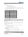
In situ Proximity Ligation Assay protocols
!! PLEASE READ BEFORE USE !!
The test protocol is a guideline, user need to determine their optimal experimental condition for
best performance.
The following protocol is modified from Duolink™ in situ PLA in situ Proximity Ligation
Assay user manual, please refer to this manual for detail information. It is available to
download from Olink Biosciences website (http://www.olink.com/) on Support & Downloads
section.
SECTION 1 – Introduction of in situ PLA Technology
DuolinkTM in situ Proximity Ligation Assay (PLA) technology is capable of detecting single protein events such as
protein interactions (e.g. protein dimerization) and modifications (e.g. protein phosphorylation) in tissue and cell
samples prepared for microscopy. The target is detected using one or two primary antibodies depending on the
application. In the case that two primary antibodies are used, they must have been raised in different species. A pair of
oligonucleotide labeled secondary antibodies (PLA probes) is applied on the sample, and a signal is generated only
when the two PLA probes have bound in close proximity, either to the same primary antibody or two primary
antibodies that have bound to the sample in close proximity. The signal from each detected pair of PLA probes is
visualized as an individual fluorescent dot. These PLA signals can be quantified (counted) and assigned to a specific
subcellular location based on microscopy images. In principle, with appropriate primary antibodies, in situ PLA
technology can detect any antigen with proximate epitopes at the single molecule level.
SECTION 2 - Reagent & Equipment
2.1 Antibody pair for detecting phospho-protein or protein-protein interaction
Abnova have two type of Ab pair products for PLA™ application, Ab pair for detecting protein phosphorylation
(Catalog # format: DPxxxx) or protein-protein interaction (Catalog # format: DIxxxx).
2.2 DuolinkTM in situ PLA reagents
Catalog # 90301 PLA probe 100 Mouse PLUS
Catalog # 90202 PLA probe 100 Rabbit MINUS
Catalog # 90103 Duolink® 100 Detection Kit 613
Catalog # 80100 Duolink® Mounting Medium 10 ml
These reagents are available to purchase from Abnova or Olink Biosciences
(http://www.olink.com/webshop.php).
2.3 Reagents required for fixation and antigen retrieval of the sample (according to your own protocol).
2.4 Ethanol
Tel: +886-2-87511888
Fax: +886-2-87511166
9th Fl., No.108, Jhouzih St., Neihu District,
Taipei City, 114 Taiwan.
Page: 1/5
Revision: 1.1
2.5 Equipment needed:
Fluorescence microscope equipped as follows:
i.
Excitation/emission filters compatible with fluorophore and nuclear stain excitation/emission.
ii.
Software for image acquisition and evaluation.
Staining jars.
Pen or mask for delimitation of reaction area (e.g. grease pen or silicon mask).
Shaker.
Humidity chamber (moist chamber).
Freeze block for enzymes.
Incubator, +37°C.
Pipettes (covering the range from 1 µl to 1000 µl).
Cover slips compatible with fluorescence microscopy.
High purity water (sterile filtered, MilliQ® or similar)
2.6 Software for image analysis
The result from a Duolink experiment is typically a number of distinct fluorescent dots (“PLA signals” or ”Blobs”) of
sub-micrometer size in various locations of the studied cells. To analyze the result, any image analysis software can
be used using suitable algorithms. A freeware optimized for image analysis of individual PLA signals has been
developed by the Centre for Image Analysis, Uppsala University. The freeware “BlobFinder” is available for download
from http://www.cb.uu.se/~amin/BlobFinder.
SECTION 3 - Preparation of Reagents & samples
3.1 Reagents
Primary antibodies
Use Antibody Diluent stock (5x) include in the DuolinkTM in situ PLA reagents to dilute the antibody. We
recommend to dilute Phospho-Ab 1:1200 (v/v) & Non-phospho-Ab 1:50 (v/v) as a starting dilutions for Ab pair
detecting protein phosphorylation, or to dilute rabbit Ab 1:1200(v/v) & mouse Ab 1:50(v/v) as a starting dilutions
for Ab pair detecting protein-protein interaction. However, each antibody may require some optimization in
customer’s own specific experiment.
DuolinkTM in situ PLA reagents
Please prepare the reagent according to Duolink™ in situ PLA in situ Proximity Ligation Assay user
manual.
3.2 Samples
Before you start the assay protocol, the sample should have been deposited on a glass slide, sufficiently
pre-treated to fit the primary antibodies with respect to fixation, retrieval and permeabilization, please see section
5.2 in Duolink™ in situ PLA in situ Proximity Ligation Assay user manual. Your reaction area must be
Tel: +886-2-87511888
Fax: +886-2-87511166
9th Fl., No.108, Jhouzih St., Neihu District,
Taipei City, 114 Taiwan.
Page: 2/5
Revision: 1.1
delimited. You can use chamber slides or delimit your sample with e.g. a grease pen or silicon mask.
3.3 Reaction volume
Use open droplet reactions.
Perform the incubations without a cover slip.
Perform all incubations in a pre-heated humidity chamber.
Use volumes corresponding to your reaction area. Never use less than 15 µl of total reaction volume.
Note: It is important that all incubations are performed in a humid environment to prevent excessive evaporation.
If the sample goes dry, this will give rise to severe artifacts.
Area
Total reaction volume
0.2 cm2
15 µl
2
40 µl
2 cm2
80 µl
1 cm
2
120 µl
4 cm2
160 µl
6 cm2
240 µl
2
320 µl
3 cm
8 cm
10 cm2
400 µl
Table 1, Suitable reaction volume for different reaction areas.
SECTION 4 - Assay Procedure
Before you start, your samples shall be deposited on glass slides and pre-treated with respect to fixation, retrieval
and/or permeabilization.
Use open droplet reactions without a cover slip and perform all incubations in a humidity chamber. The volume
examples are based on 40 µl reaction volume suitable for 1 cm2. Use volumes corresponding to your reaction area,
see section 5.3 and the Reaction Volume Guide.
4.1
Blocking
a)
Dilute the Duolink Blocking stock 1:5 in high purity water. E.g. For a 40 µl reaction take 8 µl of the 5x
Blocking stock and 32 µl of high purity water.
4.2
b)
Add the Blocking solution to each sample.
c)
Incubate the slides in a pre-heated humidity chamber for 30 min at +37°C.
Primary Antibodies
a)
Mix and dilute primary antibodies at suitable concentration in 1x Antibody Diluent.
b)
Tap off the Blocking solution from the slides. Try to obtain equal residual volume for each slide as this will
affect reproducibility.
Tel: +886-2-87511888
Fax: +886-2-87511166
9th Fl., No.108, Jhouzih St., Neihu District,
Taipei City, 114 Taiwan.
Page: 3/5
Revision: 1.1
4.3
c)
Do not allow the samples to dry before addition of the primary antibodies as this will cause background.
d)
Immediately add the primary antibody solution.
e)
Incubate in a humidity chamber at 4°C, overnight.
PLA probes
a)
Mix and dilute the two PLA probes in 1x Antibody Diluent. E.g. For a 40 µl reaction take 2.7 µl of PLA probe
MINUS stock, 2.7 µl of PLA probe PLUS stock and 34.6 µl of 1x Antibody Diluent.
4.4
b)
Tap off the primary antibody solution from the slides.
c)
Wash the slides in a wash buffer (1x TBS-T) for 5 min, twice.
d)
Add the PLA probe solution.
e)
Incubate the slides in a pre-heated humidity chamber for 1 hour at +37°C.
Hybridization
a)
Dilute the Duolink Hybridization stock 1:5 in high purity water and mix. E.g. For a 40 µl reaction take 8 µl of
the 5x Hybridization stock and 32 µl of high purity water.
4.5
b)
Tap off the PLA probe solution from the slides.
c)
Wash the slides in a wash buffer (1x TBS-T) for 5 min, twice.
d)
Add the Hybridization solution to each sample.
e)
Incubate the slides in a pre-heated humidity chamber for 15 min at +37°C.
Ligation
a)
Dilute the Duolink Ligation stock 1:5 in high purity water and mix.
b)
Wait to add the Ligase until immediately before addition to the samples. Take the addition of Ligase into
account when calculating the amount of water added. E.g. For a 40 µl reaction take 8 µl of the 5x Ligation
stock and 31 µl of high purity water.
c)
Tap off the Hybridization solution from the slides.
d)
Wash the slides with 1x TBS-T for 1 min under gentle agitation.
e)
Add Duolink Ligase to the Ligation solution from step a) at a 1:40 dilution and vortex. E.g. For a 40 µl
reaction add 1µl of Ligase to 39 µl of Ligation solution.
4.6
f)
Add the Ligation solution to each sample.
g)
Incubate the slides in a pre-heated humidity chamber for 15 min at +37°C.
Amplification
a)
Dilute the Duolink Amplification stock 1:5 in high purity water and mix.
b)
Wait to add the Polymerase until immediately before addition to the sample. Take the addition of
Polymerase into account when calculating the amount of water added. E.g. For a 40 µl reaction take 8 µl of
the 5x Amplification stock and 31.5 µl of high purity water.
c)
Tap off the Ligation solution from the slides.
Tel: +886-2-87511888
Fax: +886-2-87511166
9th Fl., No.108, Jhouzih St., Neihu District,
Taipei City, 114 Taiwan.
Page: 4/5
Revision: 1.1
d)
Wash the slides with 1x TBS-T for 2 x 2 min under gentle agitation. Tap off all wash solution after the last
washing.
e)
Add Duolink Polymerase to the Amplification solution from step a) at a 1:80 dilution and vortex. E.g. For a
40 µl reaction add 0.5 µl of Polymerase to 39.5 µl of Amplification solution.
4.7
f)
Add the Amplification solution to each sample.
g)
Incubate the slides in a pre-heated humidity chamber for 90 min at +37°C.
Detection
Please note! Light sensitive reagents – keep protected from light.
a)
Dilute the Duolink Detection stock 1:5 in high purity water and mix. E.g. For a 40 µl reaction take 8 µl of the
5x Detection stock and 32 µl of high purity water.
4.8
b)
Tap off the Amplification solution from the slides.
c)
Wash the slides with 1x TBS-T for 2 x 2 min under gentle agitation.
d)
Add the Detection solution to each sample.
e)
Incubate the slides in a pre-heated humidity chamber for 30 min at +37°C.
Final wash step
Please note! Light sensitive reagents. Wash and dry the slides protected from light:
4.9
a)
2x SSC for 2 min.
b)
1x SSC for 2 min
c)
0.2x SSC for 2 min.
d)
0.02x SSC for 2 min.
e)
70% EtOH for 1 min.
f)
Let the slides dry.
Preparation for imaging
Use a minimal volume of Duolink Mounting Medium (Art no. 80100) and apply a cover slip on top of your sample.
Preferably add the mounting media to the cover slip and then gently slip over the sample slide to ensure no air
bubbles get caught under the cover slip. Analyze in a fluorescence microscope as soon as possible.
4.10
Image acquisition and analysis
Please refer to Duolink™ in situ PLA in situ Proximity Ligation Assay user manual, for image acquisition and
analysis detail.
Tel: +886-2-87511888
Fax: +886-2-87511166
9th Fl., No.108, Jhouzih St., Neihu District,
Taipei City, 114 Taiwan.
Page: 5/5
Revision: 1.1