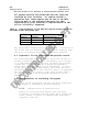
1
AVPRO1505.03
Page 1 of 33
National Veterinary Services Laboratories
Testing Protocol
Real-Time RT-PCR for Detection of
Virulent Newcastle Disease Virus in Clinical Samples
Date:
February 24, 2005
Supersedes:
AVPRO1505.02, September 29, 2004
Number:
AVPRO1505.03
Contact Person:
Janice C. Pedersen, (515) 663-7551
Approvals:
/s/ B. Panigrahy____________
B. Panigrahy, Head
Avian Viruses Section
Date:_2-24-05_
/s/ Beverly Schmitt_________
Beverly J. Schmitt, Chief
Diagnostic Virology Laboratory
Date:_2-24-05
United States Department of Agriculture
Animal and Plant Health Inspection Service
P. O. Box 844
Ames, IA 50010
Mention of trademark or proprietary product does not constitute a guarantee or
warranty of the product by USDA and does not imply its approval to the exclusion
of other products that may be suitable.
AVPRO1505.03
Page 2 of 33
NVSL
Testing Protocol
Real-Time RT-PCR Test for Detection of Virulent Newcastle Disease Virus
Table of Contents
1.
Introduction
1.1
1.2
1.3
2.
Materials
2.1
2.2
3.
Facilities/equipment/instrumentation
Reagents/supplies
Preparation for the test
3.1
3.2
3.3
3.4
4.
Background
Keywords
Collaboration
Personnel qualifications/training
Preparation of equipment/instrumentation
Preparation of reagents/control procedures
Preparation of samples
Performance of the test
4.1 Extraction of RNA from swab specimens (Qiagen®
RNeasy method)
4.2 Trizol® LS extraction for tissue samples
4.3 High throughput magnetic bead RNA extraction from
swab specimens (Ambion® MagMAX method)
4.4 Reverse-transcription and PCR
5.
Data Analysis Settings for the Cepheid® Smart Cycler
6.
Analysis of test results
7.
References
8.
Appendix 1
9.
Summary of revisions
NVSL
Testing Protocol
AVPRO1505.03
Page 3 of 33
Real-Time RT-PCR Test for Detection of Virulent Newcastle Disease Virus
1.
Introduction
1.1
Background
Virulent Newcastle disease (vNDV) is an acute, highly
contagious and fatal disease of chickens of all ages. The
disease also affects turkeys and numerous species of wild
and captive birds. Virulent Newcastle disease is caused by
avian paramyxovirus-1 (APMV-1) of the family
Paramyxoviridae, genus Avulavirus. Acute vNDV in
susceptible chickens can cause sudden death and high
mortality with few or no clinical signs. Surviving birds
may show respiratory distress, diarrhea, depression and
central nervous system signs. The clinical signs of vNDV
may resemble those of other diseases. Therefore, the signs
are not considered pathognomonic. Virulent Newcastle
disease is reportable to the World Organization for Animal
Health (OIE). The causative virus must meet one of the
following criteria for virulence: a virus that has an
intracerebral pathogenicity index (ICPI) in day-old chicks
of 0.7 or greater or presence of multiple basic amino acids
at the C-terminus of the F2 protein and phenylalanine at
residue 117, which is the N-terminus of F1 protein.
The real-time reverse transcriptase-polymerase chain
reaction (RRT-PCR) assay was developed to aid in the rapid
diagnosis of avian paramyxovirus-1 infections in poultry.
The technique utilizes a one step protocol with specific
primers designed to amplify a portion of the genome that
contains a target PCR sequence. Non-extendible fluorogenic
hydrolysis/Taqman probes monitor the target PCR product
formation at each cycle during the PCR reaction. The probes
are labeled at the 5’ end with a reporter dye (e.g. FAM) and
a quencher dye (e.g. blackhole quencher [BHQ-1]) at the 3’
end. The proximally located quencher dye absorbs the
emission of the reporter dye as long as the probe is intact
and not hybridized to the target. When the probe is
hybridized to the target, the 5’ nuclease activity of Taqpolymerase will cause hydrolysis of the probe, separating
the quencher from the reporter dye. This separation results
in an increase in fluorescence emission of the reporter dye,
which is detected spectrophotometrically and recorded. The
amount of fluorescence recorded is proportional to the
amount of target template in the samples.
NVSL
Testing Protocol
AVPRO1505.03
Page 4 of 33
Real-Time RT-PCR Test for Detection of Virulent Newcastle Disease Virus
The APMV-1 (matrix) assay described in this protocol will
detect viral RNA from both virulent and avirulent APMV-1
strains. Due to the lower diagnostic sensitivity of the
CalMex assay (the fusion gene primer set described in
AVPRO1505.01), new primer combinations were developed for
the detection of vNDV viral RNA. This assay will be
referred to as the vNDV assay and the primer set is the
Creelan/CalMex primers. A description of the equivalency
study for the validation of the Creelan/CalMex primers has
been included in appendix 1. The vNDV (fusion gene) assay
will detect most vNDV strains, including the one that
infected U.S. poultry in CA, AZ and NV in 2002-03. The
matrix assay respectively has a diagnostic sensitivity and
specificity of 96.7% and 97.3% for the detection of APMV-1
RNA from chicken cloacal and tracheal swabs and 97.8% and
95.6%, for the detection of vNDV in chicken tissues. The
vNDV assay respectively has a diagnostic sensitivity and
specificity of 91.26% and 97.5%, for the detection of vNDV
viral RNA from chicken cloacal and tracheal swabs.
Tissues from more than one bird should not be pooled
together. However, tracheal swabs from up to 5 birds from
one premises can be pooled together in 2-3.5 ml of brain
heart infusion (BHI) broth, but should not be pooled with
cloacal swabs. Tracheal swabs are the preferred specimen for
the isolation of RNA. Validation data has demonstrated a
decreased efficiency in the isolation of RNA from cloacal
swabs due to the heavy load of organic material. It should
be emphasized that the RRT-PCR technique will detect viral
nucleic acid from infectious as well as noninfectious virus.
For this reason, the RRT-PCR would not be the test of choice
to determine if infectious APMV-1 was present in
environmental samples.
Any specimen from outside a USDA quarantine zone that is
positive with the APMV-1 (matrix) assay, regardless of the
vNDV (fusion) assay results, should be referred to the
National Veterinary Services Laboratories for further
testing and characterization. Virus isolation and
characterization is required to officially diagnose a
foreign animal disease (FAD).
Virulent NDV is a zoonotic agent and causes
conjunctivitis in humans. Personnel handling clinical
samples or live virus should take appropriate safety
precautions to avoid accidental introduction of the virus
into the eyes. Appropriate safety precautions should
include wearing disposable gloves, laboratory coat, and
safety glasses. Care should be taken not to rub or touch
the eyes before removing gloves and washing hands with
soap and water.
NVSL
Testing Protocol
AVPRO1505.03
Page 5 of 33
Real-Time RT-PCR Test for Detection of Virulent Newcastle Disease Virus
The procedure described here is used in the Diagnostic
Virology Laboratory (DVL) of the National Veterinary
Services Laboratories (NVSL). The brands of equipment
listed in the protocol are used in the DVL; however,
comparable equipment may also be used. Laboratories using
this protocol should follow quality assurance procedures as
they pertain to equipment maintenance, receiving specimens,
and recording/reporting results.
1.2
Keywords
Virulent Newcastle disease virus (vNDV), real-time reversetranscriptase polymerase chain reaction (RRT-PCR), avian
paramyxovirus-1 (APMV-1), room temperature (RT), biological
safety cabinet (BSC).
1.3
Collaboration
This protocol was developed in cooperation with the
Southeast Poultry Research Laboratory, Agriculture
Research Services, USDA, Athens, GA, and the
California Animal Health and Food Safety Laboratory,
University of California, Davis, CA.
2.
Materials
2.1
Facilities/equipment/instrumentation
2.1.1
Surveillance samples originating from outside a
known vNDV quarantine zone can be processed in a
biosafety level 2 (BSL-2) laboratories. However,
samples originating from inside a known vNDV quarantine
zone or considered suspect for vNDV, should be handled
under increased biosecurity. This includes restricted
access to where the clinical samples are being handled
until the samples have been rendered non-infectious.
Once a clinical sample has been treated with lysis
buffer for RNA extraction, the sample can be moved to a
less restrictive environment to complete the RNA
extraction and RRT-PCR analysis. Once a sample is
biologically amplified (isolated in cell culture or
chicken embryo) and has been confirmed or is suspected
of being a vNDV virus, the virus should be handled at a
higher containment level, specifically biosafety level
3 or preferably biosafety level 3-ag.
AVPRO1505.03
Page 6 of 33
NVSL
Testing Protocol
Real-Time RT-PCR Test for Detection of Virulent Newcastle Disease Virus
2.1.2
Class II, HEPA filtered biological safety
cabinets (BSCs) with UV germicidal lights are required
(preferably 3, minimum of 2) to maintain sample
integrity during processing and testing. In addition,
the Trizol® extraction procedure should be performed in
a class II BSC that is connected to an external exhaust
plenum to minimize exposure to organic chemical fumes.
2.1.3
Refrigerator (4 C + 2º)
2.1.4
-20 C (+ 3º) freezer (not frost-free)
2.1.5
-70 C (+ 5º) freezer
2.1.6
Micro centrifuge (non-refrigerated
[International Equipment Co., MicroMax, Needham
Heights, MA] and refrigerated [Hermle, Z 360K, Germany]
or [Sorval® Heraeus Biofuge Fesco, Germany])
2.1.7
Vortex mixer
2.1.8
Assorted test tubes and Eppendorf tube racks
2.1.9
An integrated DNA/RNA amplification and
detection instrument system that has the capability to
detect specific sequences using hybridization probes.
Instrumentation should be capable of exciting and
detecting fluorescein-based probes (450-495nm,
500-550 nm, 565-590nm, and 630-750nm ranges). The
current protocol was developed using the Cepheid Smart
Cycler [(Cepheid® Smart Cycler, P#SC2000N1-1,
Sunnyvale, CA). However, most if not all of the
commercially available real-time PCR machines can
detect the fluorescein-based probes, and likely can be
used with this test. Based on past experience of
transferring protocols between different machines,
changes in cycling times and probe concentrations are
often required to get equivalent sensitivity.
Therefore, optimization of the assay on alternative
machines is required. This optimization data needs to
demonstrate that the alternative machine provides
comparable sensitivity and limit of detection to the
assay described in the current protocol with the Smart
Cycler® machine. This data should be available for
review by APHIS.
2.1.10 25μl Smart Cycler® tubes (Catalog #900-0022
AVPRO1505.03
Page 7 of 33
NVSL
Testing Protocol
Real-Time RT-PCR Test for Detection of Virulent Newcastle Disease Virus
or 900-0003, Cepheid® Smart Cycler, Sunnyvale, CA)
2.1.11 PCR reaction tube refrigerated tube holder and
mini-microcentrifuge to spin tubes. Both items are
supplied with the Smart Cycler® real-time PCR machine.
2.1.12 QiaVac® 24 vacuum manifold (Valencia, CA) and
vacuum pump with a capacity of 18-20 liter/min. (Gast
MFG Corp., St. Louis, MO). Use of insufficient vacuum
pressure may reduce RNA yield and purity. The vacuum
manifold system with vacuum pump is optional, but is a
highly recommended product for processing large numbers
of samples for RRT-PCR.
2.2
Reagents/supplies
2.2.1
Molecular biology grade RNase-free sterile
distilled water
2.2.2
In vitro transcribed NDV matrix-fusion gene
positive control RNA (supplied by the NVSL, Ames, IA)
2.2.3
*Ethanol, absolute, ACS grade or better
(Caution: irritant, flammable)
2.2.4
*Isopropanol, 99+% pure ACS grade or better
(Caution: irritant, flammable)
2.2.5
*Chloroform, 99+% pure (Caution:
toxic)
2.2.6
*Trizol® LS reagent (Caution: toxic in contact
with skin and if swallowed; causes burns.) (Cat.
#15596 or 10296, Life Technologies, Grand Island, NY)
2.2.7
Qiagen® RNeasy Extraction Kit (Cat. # 74103 20
preps, #74104 50 preps or #74106 250 preps, Qiagen®,
Valencia, CA).
2.2.8
One-Step RT-PCR (Cat. #210210 or 210212,
Qiagen®, Valencia, CA). Many one-step RT-PCR kits are
commercially available. However, the Qiagen system has
been tested extensively with good results. Other kits
can be considered for use, but a minimum level of
equivalency testing is required before substituting any
reagents in the approved protocol. Currently, the only
alternative kit that has been tested that has
comparable test results to the Qiagen system is the
Superscript One-Step RT-PCR System with Platinum Taq
NVSL
Testing Protocol
AVPRO1505.03
Page 8 of 33
Real-Time RT-PCR Test for Detection of Virulent Newcastle Disease Virus
DNA Polymerase (Cat #10928-034 or 10928-042,
Invitrogen, Carlsbad, CA). Appropriate changes to
optimize the protocol for use with alternative reagents
or RT-PCR kits, including cycling parameters, must be
supported by an equivalency evaluation as previously
noted.
2.2.9
Hydrolysis probes and primers
(oligonucleotides) (Table 1) for the detection of vNDV.
Suggested probe sources: Biosearch Technologies
(http://blackholequenchers.com) and Operon
(http://oligos.qiagen.com/). Suggested primer sources:
Integrated DNA Technologies, (http://www.idtdna.com/),
or Operon (http://oligos.qiagen.com/). Other companies
can be used to order both primers and probes. It is
strongly recommended that the probes and primers be
purified to a high level to reduce nonspecific
reactions. Purification is typically performed with
HPLC purification as the minimum.
2.2.10 Ambion MagMAX®-96 Viral RNA Isolation Kit
(Cat. # 1835-plate kit or 1929 for single tube kit)
(Ambion®, Austin, TX 78744-1832)
AVPRO1505.03
Page 9 of 33
NVSL
Testing Protocol
Real-Time RT-PCR Test for Detection of Virulent Newcastle Disease Virus
Table 1.
NDV real-time RT-PCR probe and primer sequences.
Specificity
target gene
and
APMV-1 (matrix)
vNDV (fusion)*
Primer/probe
name-Genomic
target
M+4100**
(forward primer)
Sequence
5’-AGTGATGTGCTCGGACCTTC-3’
M+4169**
(matrix probe)
5’-[FAM]TTCTCTAGCAGTGGGACAGCCTGC[BHQ]-3’
M-4220**
(reverse primer)
5’-CCTGAGGAGAGGCATTTGCTA-3’
F+4829**
(forward primer)
F+4894**
Probe-1 (VFP-1)
(virulent
fusion)
F-4939**
(reverse primer)
5’-GGTGAGTCTATCCGGARGATACAAG-3’
5’-[FAM]AAGCGTTTCTGTCTCCTTCCTCCA[BHQ]-3’
5’-AGCTGTTGCAACCCCAAG-3’
*Validation testing with the vNDV primers and VFP-1 probe set has
demonstrated that these reagents will detect the vast majority of
mesogenic and velogenic APMV-1 viruses.
**Refers to the nucleotide position where the 5’ end of the probe
or primer anneals to the NDV genome. The FAM/TAMRA hybridizing
probes were validated with the Smart Cycler I system. When using
the Smart Cycler II system it is recommended that all hybridizing
probes be labeled with FAM as a reporter dye and quenched with
either Dabcyl or Black Hole™ quencher I. The Smart Cycler II
system is not calibrated to use the TAMRA dye as a quencher. A
TAMRA dye is read as background noise in channel 2 for the Smart
Cycler II.
2.2.11 RNase Inhibitor, 40 units/µl (Promega, catalog
#N2511 or N2515, Madison, WI)
2.2.12 MgCl2, 25 mM (Promega, catalog #A3511 or A3513,
Madison, WI)
2.2.13 TE buffer pH 8.3 (Promega #V6231 or V6232)
2.2.14 Qiagen® RNeasy Mini Kit (Qiagen #74104 or
74106)
2.2.15 14.3 M *β-mercaptoethanol (β-ME) (Sigma,
M 6250) Caution: toxic – dispense in a fume hood, wear
gloves
NVSL
Testing Protocol
AVPRO1505.03
Page 10 of 33
Real-Time RT-PCR Test for Detection of Virulent Newcastle Disease Virus
2.2.16 Sterile, aerosol-resistant pipette tips of
various sizes (1.0ml, 200-50µl, 100-10µl, 10-0.5µl)
2.2.17 1.7 ml microcentrifuge tubes (sterile)
2.2.18 Either powder-free latex or nitrile gloves
may be used for any procedure requiring gloves
2.2.19 Calibrated pipettors from 0.5 µl to 1000 µl,
2 sets, one extra pipettor for DNA transfer
2.2.20 Ambion® Magnetic Stand-96 (Ambion Inc. catalog
#10027, Austin, TX or an O-ring 96 well Magnetic-Ring
Stand (Cat.# 10050) (Ambion, Austin, TX). Product
#10027 has 96 ring magnets that pellet beads in a donut
shape. Product #10050 has 24 big magnetic rods that
pellet beads to one side of the wells.
2.2.21 Orbital shaker for 96 well plates (Lab-Line
Titer Plate Shaker Model #4625, Melrose Park, ILL)
2.2.22 5-200 µl 12 channel pipetting tool (Matrix
Technologies Corp., catalog #6012, Lowell, MA)
*Use caution when using these reagents. Refer to the
individual Material Safety Data Sheet (MSDS) before
handling any of these reagents.
NVSL
Testing Protocol
AVPRO1505.03
Page 11 of 33
Real-Time RT-PCR Test for Detection of Virulent Newcastle Disease Virus
3
Preparation for the test
For this procedure, it is critical to have separate preparation
areas and equipment for nucleic acid extraction, “clean”
procedures, and work with amplified nucleic acid. The “clean”
area is used for preparing reagents for use in the PCR procedure.
Amplified c-DNA or sample RNA should never be introduced into
this area. One biological safety cabinet should be designated
for “clean” work only. There should also be a separate set of
“clean” pipettors and tips, RNase-free water, tubes for reagent
preparation, racks, and ice container which are designated for
“clean” use only and never leave the area. A -20 C freezer
should also be designated as “clean” for storage of reagents. A
second biological safety cabinet, set of pipettors, and other
equipment and reagents should be used for extraction procedures.
Ideally, a third biological safety cabinet should be used for
transfer of RNA to amplification tubes. Latex or nitrile gloves,
in particular, must be worn throughout the procedure and must be
changed frequently. RNA is very labile and easily degraded by
RNases that are ubiquitous, including on human skin. Gloves also
help protect the reagents and samples from other contaminating
agents and cross-contamination that can adversely affect results.
Always change gloves after working with sample RNA or amplified
DNA. Always wear fresh gloves when working with “clean”
reagents. Protective eyewear, gloves, and lab coats should be
worn as some of the reagents used are toxic. It is recommended
that the Trizol extraction procedure is conducted in a vented
class II BSC.
3.1
Personnel qualifications/training
Personnel performing PCR procedures should be familiar with:
3.1.1
Preparation and proper handling of samples and
reagents
3.1.2
Calibration, maintenance, and use of
instruments included in this protocol
AVPRO1505.03
Page 12 of 33
NVSL
Testing Protocol
Real-Time RT-PCR Test for Detection of Virulent Newcastle Disease Virus
3.2
Preparation of equipment/instrumentation
Refrigeration equipment, incubators, centrifuges, pipettors
and thermal cyclers are calibrated and certified according
to the respective institution standard operating procedures.
3.3
Disinfectants
Several classes of disinfectants, e.g. iodophores,
phenolics, quaternary ammonia compounds, 70% alcohol, 10%
sodium hypochlorite, and peroxigen compounds, will
inactivate APMV-1 by destroying the lipid envelope of the
virus. However, of the disinfectants listed above, only
sodium hypochlorite (bleach) and peroxygen compounds
(Virkon-S) have been shown to degrade nucleic acid as well
as destroy infectivity of APMV-1. This is important when
selecting a disinfectant to rid surfaces of contaminating
nucleic acids.
3.4
Preparation of reagents/control procedures
3.4.1
Oligonucleotide primers
Prepare primers in a “clean” hood (see 3.0).
wear gloves when dispensing primers.
Always
Dilute primers to 200 pmol/µl (200μM) in 1X TE for the
stock dilution and 20 pmol/µl in RNase-free water for
the working dilution. Aliquot primers in small volumes
to avoid excessive freeze/thaw cycles. For short-term
storage of primers (<1 week), 4 C is acceptable. For
longer term storage, -20 C or colder is recommended.
Store stock primer solutions at -20 C or -70 C. 26.8
pmol of the vNDV forward primer, 13.4 pmol of the vNDV
reverse primer, and 10 pmol of the matrix forward and
reverse primers are added per 25 µl reaction.
Additionally it is recommended that the PCR primers be,
at a minimum, HPLC purified.
3.4.2
Hydrolysis Probe
Prepare probe in a “clean” hood (see 3.0). Always wear
gloves when working with hydrolysis probes. Hydrolysis
probes are light sensitive and should be protected from
exposure to direct light. Diluted probes should be
stored in amber sterile RNase free microcentrifuge
tubes or tubes wrapped with foil.
AVPRO1505.03
Page 13 of 33
NVSL
Testing Protocol
Real-Time RT-PCR Test for Detection of Virulent Newcastle Disease Virus
Dilute probes to 120 pmol/µl (120μM) in 1X TE for the
stock dilution and to 6 pmol/µl in RNase-free water for
the working dilution. Aliquot probe in small volumes
to avoid excessive freeze/thaw cycles. Store diluted
probes at -20 C and stock primer solutions at -20 C or
-70 C. Avoid excessive freezing and thaw cycles.
Diluted probe should not be frozen/thawed more than
4 times. A total of 6 pmol of the probe is added per
25 µl reaction.
3.4.3
Handling and dilution of Primers and Probe
Lyophilized primers and probes must be centrifuged
briefly to ensure that the DNA pellet is at the bottom
of the tube before they are opened and reconstituted.
TE buffer should be used for the initial reconstitution
of lyophilized primers and probes. Quantitation
information will be supplied for each oligonucleotide
primer (oligo) by the manufacturer.
An example of calculation for oligo reconstitution:
You have 17786 pmol of oligo (will be on oligo
information sheet from manufacturer).
Need 200pmol/μl for stock concentration.
Divide pmol of oligo by the pmol/μl needed or:
17786 pmol=88.9μl of 1X TE
200 pmol/μl
The calculation for the probe is the same, except
divide the number of probe pmol by 120 pmol/μl. Mix
gently by tapping the tube and allow the oligo to
resuspend for about 10 minutes before use.
Working stocks of primers should be 20 pmol/µl (20μM)
and working stocks of probes should be 6 pmol/µl.
Dilute the primers 1:10 and dilute the probe 1:20 in
nuclease free H2O (do not use TE buffer) for the
working stocks.
Additional information on fluorescent probe
handling and storage can found at: www.operon.com
and www.idtdna.com ; www.idahotech.com.
NVSL
Testing Protocol
AVPRO1505.03
Page 14 of 33
Real-Time RT-PCR Test for Detection of Virulent Newcastle Disease Virus
3.4.4
Prepare a 70% ethanol solution using 100%
absolute ethanol and RNase-free water.
3.4.5
Prepare 80% ethanol solution using 100%
absolute ethanol and RNase-free water.
3.4.6
β-Mercaptoethanol (β-ME) must be added to the
RNeasy RLT lysis buffer before use. Add 10µl β-ME per
1 ml RLT buffer. Buffer RLT is stable for 1 month
after addition of β-ME. Be sure to date buffer after
adding β-ME.
3.4.7
100% absolute ethanol is added to the RNeasy
RPE wash buffer according to the Qiagen® kit
directions.
3.4.8
The template for the positive controls is in
vitro transcribed RNA from the NDV matrix and fusion
protein gene. (200 ADV, NVSL, 1800 Dayton, Ave., Ames,
IA)
3.4.9
Dilute RNase inhibitor to 13.3 units/µl with
RNase free water.
3.4.10 The transcribed RNA positive control (provided
by NVSL, Ames, IA) should be diluted to a working
concentration that will consistently give a target Ct
of approximately 25.0. Dilute the stock RNA to the
suggested working dilution as described in the product
insert sheet that will accompany the transcribed RNA
positive control with sterile RNase free water. Run
the diluted RNA on the RRT-PCR assay to determine if
the positive control has a Ct that is within the
acceptable range (section 6.2).
3.5
Preparation of samples
Perform all procedures with potentially live agents in an
approved Class II biosafety cabinet with HEPA filtration.
Always wear protective clothing and gloves when handling
potentially infected tissues or live virus.
Samples received for testing are compared to accompanying
paperwork to assure correct samples were received and that
Formatted: Indent: Left: 72 pt,
First line: 0 pt
NVSL
Testing Protocol
AVPRO1505.03
Page 15 of 33
Real-Time RT-PCR Test for Detection of Virulent Newcastle Disease Virus
appropriate samples were submitted for the PCR test.
are logged into the logbook.
Cases
Pooled oropharyngeal/tracheal swabs (5 swabs/tube) are the
specimens of choice for the PCR procedure and should be
extracted using either the Qiagen® or Ambion® RNA extraction
procedure. Appropriate tissue (spleen, lung, brain,
intestine) should be processed by preparing a 10-20% tissue
homogenate and extracting RNA using the Trizol® extraction
procedure. Alternatively, 5mm3 piece tissues are added to
2.0 ml of brain heart infusion broth (BHI), frozen solid,
thawed and centrifuged. The supernatant from this tissue
pool (250µl) is extracted using the Trizol® procedure.
Validation/equivalency data indicate that cloacal swab
specimens are significantly less sensitive than
oropharyngeal/tracheal swabs for the detection of APMV-1 by
RRT-PCR. For this reason, cloacal swabs should be tested by
virus isolation and not by RRT-PCR. Tissues from more than
one bird should not be pooled together.
All samples should be processed in a Class II biological
safety cabinet.
4
Performance of the test
Before beginning the RT-PCR test, place “clean” pipettors, racks,
tips, etc. into the “clean” hood and expose to the UV germicidal
light for several hours or overnight. Similarly, place the
sample equipment into a separate hood and expose to the UV
germicidal light.
4.1
Extraction of RNA from swab specimens (Qiagen® RNeasy
method)
4.1.1
Vortex swab specimen fluid and transfer 500µl
of sample into the microcentrifuge tube labeled with
the specimen number.
4.1.2
Place 500 µl of Qiagen RLT with β-ME into the
microcentrifuge tube. Vortex 15 sec. When processing
a large number of specimens the RLT buffer can be mixed
with the specimen by pipetting up and down vigorously 4
to 6 times.
4.1.3
Pulse spin to eliminate liquid specimen in the
lid after vortexing. Add 500µl 70% ETOH and vortex
NVSL
Testing Protocol
AVPRO1505.03
Page 16 of 33
Real-Time RT-PCR Test for Detection of Virulent Newcastle Disease Virus
well. Centrifuge lysed swab specimen for 5 min. at
5,000 X g in a microcentrifuge at RT.
4.1.4
Transfer all of the lysed specimen supernatant
to a RNeasy® Qiagen column that has been marked to
identify the specimen. Centrifuge for 15 sec. at
>8,000 X g at RT*. Check to assure the entire specimen
has flowed through the column. Repeat until all of
specimen has been applied to the column.
*When working with a large quantity of specimens it is
recommended to use a QiaVac® manifold to pull the
specimen and wash solutions through the collection
columns. This will increase the efficiency and
eliminate the need to centrifuge the columns at the
steps 4.1.3, 4.1.4, 4.1.5, 4.1.6 and 4.1.7.
4.1.5
Add 700µl of RW1 buffer to the RNeasy column
and centrifuge for 15 sec. at > 8,000 x g and place the
column in a clean collection tube (the tube with RW1
flow through may be discarded).
4.1.6
Add 500µl RPE buffer to the RNeasy column and
centrifuge for 15 sec. at >8,000 x g. Discard flow
through from the collection tube.
4.1.7
Repeat for a total of 2 washes with RPE buffer
discarding flow through from the collection tube.
Following the last RPE wash place the RNeasy column in
a new 2 ml collection tube.
4.1.8
Centrifuge the empty RNeasy column an extra 2
minutes at full speed and discard the collection tube.
4.1.9
Place the RNeasy column in an elution tube or a
1.5ml microfuge tube that has been marked with the
specimen number and pipet 50μl RNase-free H2O into the
column. Do not touch the silica-gel membrane with the
pipettor tip. Incubate at room temperature for 1
minute.
Elute RNA by centrifuging for 1 minute at
>10,000 rpm. Discard RNeasy column.
4.1.10 Store at 4 C until specimen is tested on RRTPCR. RNA should be stored at 4 C for as short of period
as possible before testing. If the sample cannot be
tested within 24 hours, it should be stored at -20 C or
colder.
AVPRO1505.03
Page 17 of 33
NVSL
Testing Protocol
Real-Time RT-PCR Test for Detection of Virulent Newcastle Disease Virus
4.2
Trizol® LS Extraction for tissue samples
This procedure describes the extraction of total RNA from
tissue using the Trizol® extraction reagent. The reagent is
a mono-phasic solution of phenol and guanidine
isothiocyanate. Addition of chloroform followed by
centrifugation separates the solution into an aqueous phase
and an organic phase. The RNA is recovered from the aqueous
phase by precipitation with isopropanol.
Different systems for RNA isolation are commercially
available and potentially may work as well or better than
the method described here. Alternative methods of RNA
extraction can be substituted when comparison testing of new
and standard methods show equivalency. This data should be
available for review by APHIS.
Always wear protective clothing, eyewear, and gloves when
working with extraction reagents.
4.2.1
Centrifuge tissue specimens (20% homogenates or
tissue pools) at 1,500 x g for 30 min. Collect 250µl
of the tissue supernatant and transfer to a 1.5
microcentrifuge tube. 750µl of Trizol® LS is added to
the tube and sample is vortexed for 15 sec. Incubate
at room temperature for 7 min.
4.2.2
Pulse spin to remove liquid from the tube lid.
4.2.3
Add 200µl 100% chloroform to the sample/Trizol
homogenate. Vortex for 15 sec. Incubate at room
temperature for 7 min.
4.2.4
Centrifuge at 12,000 x g for 15 min at room
temperature.
4.2.5
Transfer 450µl of the upper aqueous layer to a
separate microcentrifuge tube marked with sample
number. Caution: The transfer of organic phase
material with the aqueous layer will inhibit the PCR
reaction. Add 500µl of 100% isopropanol. Invert tube
several times to mix. Hold at room temperature for
10 min.
4.2.6
Centrifuge at 10,000 x g for 10 min at 4 C in
the Hermle Z360K or Sorval Biofuge Fesco centrifuge.
NVSL
Testing Protocol
AVPRO1505.03
Page 18 of 33
Real-Time RT-PCR Test for Detection of Virulent Newcastle Disease Virus
4.2.7
Decant liquid. Care should be taken to assure
that the RNA pellet is not disturbed. Add 1.0 ml of
80% ethanol. Mix gently.
4.2.8
Centrifuge at 10,000 X g for 5 min at 4 C.
4.2.9
Decant ethanol. Invert tube on a clean tissue
wipe and allow to air dry for 10 min. or until all
visible signs of moisture are gone. It is important not
to let the RNA pellet over-dry, as this will decrease
its solubility.
4.2.10 Hydrate pellet in 50µl of RNase free water and
allow to sit at 4 C for 1 hr to overnight. Briefly
vortex to resuspend pellet before pipetting.
4.3. High throughput magnet bead RNA extraction (Ambion®
MagMax method)
This procedure describes the extraction of total RNA from
swab specimens using the Ambion® extraction reagent. The
MagMAX® Viral RNA Isolation Kit is designed for rapid high
throughput purification of total RNA from
oropharyngeal/tracheal swab samples as well as cultured
cells. The addition of guanidinium thiocyanate rapidly
disrupts cellular membranes and inactivates cellular
nucleases. Paramagnetic beads with a nucleic acid binding
surface are added to the lysate to bind nucleic acids. The
beads, containing the RNA, are then captured on magnets and
the supernatant containing cell debris and other
contaminants is removed during the wash procedures.
Different systems for RNA isolation are commercially
available and potentially may work as well as the described
procedure. Alternative methods can be substituted when
comparison testing of new and standard methods show
equivalency. This data should be available for review by
APHIS.
4.3.1
Preparation of Ambion® Viral Isolation Kit
Components
Lysis/Binding Solution. Sub aliquot the lysis/binding
solution from the Ambion viral isolation kit into two
different nuclease free containers. See table below to
determine the quantity of lysis/binding solution
needed. Set one aliquot aside for preparation of wash
AVPRO1505.03
Page 19 of 33
NVSL
Testing Protocol
Real-Time RT-PCR Test for Detection of Virulent Newcastle Disease Virus
solution I mix. Add 1.0 µl Poly (A) RNA (kit provided)
per 50 µl lysis/binding solution to the second aliquot
and mix briefly. Following the addition of Poly (A)
RNA add an equal amount (50 µl) of 100 % isopropanol to
lysis/binding Poly (A) mixture to give a total volume
of 101 µl per sample. Vortex well. Lysis/binding
solution is stable at room temperature for one month.
It is not recommended to store the prepared Viral
Lysis/Binding Solution at 4º C or below as this may
cause the carrier RNA to precipitate; if the solution
is inadvertently stored at 4º C, warm it at 37º and
shake to dissolve any precipitates before use.
Add carrier RNA to Lysis Solution and mix well before adding
isopropanol, or carrier RNA may be very difficult to disperse.
1. Combine the following:
Viral Lysis/Binding Soln.
Concentrate
Carrier RNA
2. Mix briefly, then add:
100 % Isopropanol
3. Mix well by vortexing
1 Plate
6.25 ml
4 Plates
25.0 ml
125 µl
500 µl
6.25 ml
25.0 ml
4.3.1.1 Bead Resuspension Mix. Dilute the Bead
Resuspension Solution with Nuclease-free Water
according to the table below. Be sure to add 100%
isopropanol to the mixture last or beads may clump
together and be more difficult to enter into
solution. Store at RT.
Bead Resuspension Mix
1. Bead Resuspension Solution
2. Nuclease-free Water
Mix briefly then add
3. RNA Binding Beads
Mix briefly then add
4. 100% Isopropanol
Mix well by vortexing
Total Volume of Lysis/Binding Solution
Volume Per
Well
6.0 µl
4.0 µl
Volume Per
Plate
750 µl
500 µl
4.0 µl
500 µl
6.0 µl
750 µl
20 µl
2500 µl
NVSL
Testing Protocol
AVPRO1505.03
Page 20 of 33
Real-Time RT-PCR Test for Detection of Virulent Newcastle Disease Virus
4.3.1.2 Wash Solution I Mix. Mix the second subaliquot of lysis/binding solution in equal parts
with 100% isopropanol.
A total volume of 100 µl
is used per sample. Store at RT.
4.3.1.3 Wash Solution II Mix. Mix 40 µl of Wash
Solution II Concentrate with 160 µl of 100 %
ethanol to gain a total volume of 200 µl per
sample. Store at RT.
4.3.2 Procedure for Extraction of RNA
4.3.2.1 Vortex swab specimen and transfer 50 µl
of sample into the corresponding well on the 96
well processing plate (supplied with kit).
4.3.2.2 Place 101 µl of viral lysis/binding
solution with Poly (A) RNA to each well
containing sample. After addition of the
lysis/binding solution, the processing plate may
be removed from the BSC. All remaining steps
can be performed on the deck at room
temperature. Shake plate on the plate shaker at
moderate speed for 30 sec.
4.3.2.3 Add 20 µl of beads/binding mix to each
well. Shake plate on orbital shaker for 4
minutes at approximately 550-600 rpm.
4.3.2.4 Capture/pellet the RNA Binding Beads on
a magnetic stand. Pellet the beads on the
Ambion Magnetic Stand-96 for 2 minutes and
remove supernatant from beads (with plate still
on magnet). Discard the supernatant. The
mixture should become transparent, indicating
the capture is complete.
4.3.2.5 Remove the plate from the magnetic
stand.
4.3.2.6 Add 100 µl Wash Solution I Mix (with
isopropanol added) to each well. Shake for 30
seconds at approximately 550-660 rpm. The RNA
Binding Beads may not fully disperse during the
step; this is expected, and it will not affect
RNA purity or yield.
AVPRO1505.03
Page 21 of 33
NVSL
Testing Protocol
Real-Time RT-PCR Test for Detection of Virulent Newcastle Disease Virus
4.3.2.7 Pellet the beads for 1 minute and remove
supernatant as in step 4.2.2.4.
4.3.2.8 Add 100 µl Wash Solution II Mix (with
ethanol added) to each well. Shake for 30
seconds at approximately 550-600 rpm.
4.3.2.9 Pellet the beads for 30 seconds and
remove supernatant. Discard the supernatant.
Remove the processing plate from the magnetic
stand.
4.3.2.10 Repeat steps 4.2.2.8-4.2.2.9 to wash a
second time with Wash Solution II.
4.3.2.11 Shake vigorously for 2 minutes (shaker
dial position 9 Lab Line) to briefly dry the
beads. It is important to remove residual
ethanol from the samples. Residual ethanol may
affect RT-PCR efficiency.
4.3.2.12 Add 50 µl of Elution Solution (RT) and
shake for Four minutes at approximately 1000
rpm.
4.3.2.13 Pellet the beads for 2 minutes and
transfer the RNA into sample tube, storage plate
(kit provided), or RRT-PCR amplification plate.
4.3.2.14 Store at 4 C until specimen is tested
on RRT-PCR. RNA should be stored at 4 C for as
short of period as possible before testing. If
the sample cannot be tested within 24 hours, it
should be stored at -70 C.
4.4. Reverse-transcription and PCR
Two work areas are required for this procedure: a “clean”
area with a dedicated BSC, freezer and supplies, and a
thermal cycling area. Never introduce RNA/DNA material into
the “clean” area and always change gloves before entering
the “clean” area.
4.4.1
In the “clean” hood, prepare a master mix of
the following reagents sufficient for the number of
AVPRO1505.03
Page 22 of 33
NVSL
Testing Protocol
Real-Time RT-PCR Test for Detection of Virulent Newcastle Disease Virus
samples being tested.
per sample.
The amount given in the table is
This procedure was designed for the Cephied® SmartCycler (Cepheid®, Sunnyvale, CA).
Information on setting-up and programming the Smart
Cycler can be found in the Smart Cycler user’s manual.
The conditions for the RT-PCR on the Smart Cycler are
shown in tables 2 and 3.
Table 2.
RT step thermocycling for Qiagen® one-step RT-PCR Kit.
RT Step
1 cycle
30
15
min.
min.
50° C
95° C
Table 3.
Thermocycling conditions for gene specific probe and
primer sets.
Probe/Primer
set
APMV-1 matrix
vNDV/VFP-1
Step
Time
Temp
40 cycles denaturation
annealing
extension
10 sec.
30 sec.
10 sec.
94° C
56° C
72° C
40 cycles denaturation
annealing
extension
10 sec.
30 sec.
10 sec.
95° C
58° C
72° C
Note: The fluorescence is acquired at the annealing step.
4.4.2
The real-time RT-PCR reaction should be
prepared with the following components and volumes
using the appropriate primer and probe set and cycling
conditions. Set-up the reactions with the reaction
tubes in the cooling block and use aerosol resistant
pipet-tips.
Prepare the reaction mix (everything but the template)
by pipetting: H2O, kit supplied 5X reaction buffer, kit
supplied dNTP’s, forward and reverse primers, and 25mM
MgCl2 into a nuclease free microcentrifuge tube using
the volumes per reaction for each reagent given in
NVSL
Testing Protocol
AVPRO1505.03
Page 23 of 33
Real-Time RT-PCR Test for Detection of Virulent Newcastle Disease Virus
table 4. Next add the RNase inhibitor and enzyme. Add
the probe last. Mix reagents and centrifuge briefly.
Once the probe has been added minimize exposure of the
reaction mix to light.
4.4.3
Add the reaction mix (17μl) to the Smart Cycler
tubes (add the mix to the bottom of the cup at the top
of the reaction tube).
4.4.4
Add 8 µl of purified test sample RNA to the
Smart Cycler reaction tube using a pipettor designated
for RNA transfer. Close the lid of the tube and number
the reaction tubes according to test worksheet.
Transfer 8µl of diluted transcribed RNA (200 ADV provided by NVSL, Ames, IA) into the positive control
reaction tube and 8µl of clean RNase free water into
negative control reaction tube.
4.4.5
Centrifuge reaction tubes to enable the RNA
reaction mixture to occupy the reaction portion of the
tubes and to remove any air bubbles from the reading
window of the PCR tubes.
4.4.6
Insert reaction tubes into thermal cycler and
select the designated PCR run protocol, start run and
enter sample identification as well as positive and
negative control information into the sample
identification portion of the results table. Save run.
Table 4a. Real-time RT-PCR reaction mix volumes and conditions
for NDV with APMV-1 (matrix) primer/probe sets.
AVPRO1505.03
Page 24 of 33
NVSL
Testing Protocol
Real-Time RT-PCR Test for Detection of Virulent Newcastle Disease Virus
Volume Per
Reaction
6.45 μl
5
1.25
mM 0.8
H 2O
5X buffer
25mM MgCl2
dNTP’s (10
each)
Forward
Primer
(20 pmol/ul)
Reverse
Primer
(20 pmol/ul)
Rnase Inhibitor
(13.3 units/μl)
Enzyme Mix
Probe
(6pmol/ul)
MM per rxn
Template
Total
Final
Concentration
1X
3.75 mM*
320 μM ea. dNTP
0.5
10 pmol/25μl
0.5
10 pmol/25μl
0.5
0.266 units/µl
1.0
1.0
0.24
μM
17
8
25μl
*Qiagen buffer already contains 2.5 mM MgCl2 at 1X concentration
Table 4b. Real-time RT-PCR reaction mix volumes and conditions
for NDV with vNDV primer/probe set
AVPRO1505.03
Page 25 of 33
NVSL
Testing Protocol
Real-Time RT-PCR Test for Detection of Virulent Newcastle Disease Virus
Volume Per
Reaction
5.69 μl
5
1.25
mM 0.8
H 2O
5X buffer
25mM MgCl2
dNTP’s (10
each)
Forward
Primer
(20 pmol/ul)
Reverse
Primer
(20 pmol/ul)
Rnase Inhibitor
(13.3)units/μl
Enzyme Mix
Probe(6pmol/ul)
MM per rxn
Template
Total
Final
Concentration
1X
3.75 mM*
320 μM ea. dNTP
1.34
26.8 pmol/25μl
0.67
13.4 pmol/25μl
0.5
0.266 units/µl
1.0
0.75
17
8
25μl
0.18
μM
*Qiagen buffer already contains 2.5 mM MgCl2 at 1X concentration
5
Data Analysis Settings for the Cepheid® Smart Cycler
The Smart Cycler software provides multiple methods for
determining the cycle threshold (Ct). All data analysis
options discussed below are selected from the Analysis
Settings from the Views screen. Changes in the standard
default settings have been made to customize the analysis
for the detection of vNDV with the matrix and vNDV
primer/probe sets. Edit the Smart Cycler default analysis
settings as describe below so the raw data will be analyzed
according to the analysis parameters described.
Curve Analysis – Accept the “Primary curve” setting – The Ct
is detected and reported at the cycle where the primary
curve crosses the threshold.
Usage – Accept the default setting “Assay” for the FAM
channel.
Background Subtraction – Accept the default value of “ON”
Background Minimum – accept the default value of “5” (Min).
The Smart Cycler calculates the background of the
fluorescence signal within the range of cycles defined by
the Background Min Cycle value and the Background Max Cycle
value for each site to measure the background noise.
NVSL
Testing Protocol
AVPRO1505.03
Page 26 of 33
Real-Time RT-PCR Test for Detection of Virulent Newcastle Disease Virus
Background maximum cycle – Enter “28” as the maximum cycle
for the calculation of the standard deviation of the
fluorescence signal. The default is 40.
Threshold Settings – accept the default of “Manual”.
Manual Threshold Fluorescence units – Enter “25” fluorescent
units. It is critical that the Ct be above the background
fluorescence. The closer the threshold is set to the
background fluorescence the more sensitive the detection
limit. However, if the threshold is set too close to the
background fluorescence, background noise could cross the
threshold and be reported incorrectly as a positive sample.
By lowering the threshold fluorescence units from 30 to 25
the analytic sensitivity of the assay is increased. The
possibility of reporting a false negative is reduced while
the possibility of detecting a false positive is increased.
Boxcar Average – accept the default of “0”
The only changes from the Cepheid® Smart Cycler default were
the Max background and threshold settings. With these
analysis settings a specimen will be called positive
(crosses the Ct) when the fluorescence units exceeds 25
units. These settings were designed to optimize the
discrimination of positive and negative specimens for this
particular protocol, but the results from each run still
need to be verified by the user. The curve should be in the
log-linear phase when crossing the threshold. By lowering
the Max background, the incidence of traces with a “V” shape
are reduced. For samples that still have a “V” shaped
trace, the Max background setting can be incrementally
lowered to 15 until the early cycles in the trace are
approximately horizontal and aligned with zero fluorescence
(Figures 2a and 2b). When lowering the Max background keep
in mind that you want as many cycles as possible used to
calculate the background subtraction. This is a correction
on how the software handles the data and it decreases the
number of cycles used to calculate the background and
correct the curve. If you have questions about the trace,
look at the trace with the background subtraction off. This
shows the raw fluorescence data, and can sometimes aid
interpretation.
The fluorescence trace of each specimen should be reviewed
before accepting the Smart Cycler positive/negative result.
NVSL
Testing Protocol
AVPRO1505.03
Page 27 of 33
Real-Time RT-PCR Test for Detection of Virulent Newcastle Disease Virus
Any specimen whose fluorescence trace has a gradual increase
in fluorescence units and approaches, but does not cross the
Ct, should be sent to the NVSL for further testing. These
specimens are referred to as “late risers” (LS) and on some
occasions may indicate a sample near the threshold of
detection. At this time the significance of “late risers”
samples is not known and testing by virus isolation is
recommended.
6
Analysis of test results
6.1 The matrix APMV-1 assay is designed to detect APMV-1
including vaccine/lentogenic strains as well as vNDV
(mesogenic/velogenic strains). The vNDV assay was designed
specifically for the California 2002 and Mexico 2000
velogenic vNDV, but it has been shown to detect most
mesogenic/velogenic strains of NDV. Because the matrix
APMV-1 test is more sensitive (96.7%) than the vNDV test
(91.26%), the matrix APMV-1 test is used to screen all
samples. For APMV-1 positive samples, the vNDV assay is
used to confirm the matrix test results. If both tests are
positive, it provides strong evidence that vNDV is present
in the sample. In some cases where the sample is at the
limit of detection, a vNDV specimen can test positive with
the APMV-1 (matrix) test and negative by the vNDV assay.
Samples positive by the matrix primer/probe set that cannot
be confirmed with the vNDV primer/probe set should be
considered as suspect and additional samples collected for
further testing. All suspect specimens should be tested by
virus isolation. It is also possible that APMV-1 (matrix)
positive, vNDV negative tests result from lentogenic or
vaccine strain NDV isolates being in the sample. For
samples that have a low cycle threshold (Ct of <30), where
lower Ct means a stronger positive, confirmation that the
APMV-1 (matrix) positive, vNDV negative samples are vaccine
or lentogenic isolates can be made using different primer
and probe sets. These primers and probes sets are still
being optimized and will be included in future versions of
this document.
AVPRO1505.03
Page 28 of 33
NVSL
Testing Protocol
Real-Time RT-PCR Test for Detection of Virulent Newcastle Disease Virus
All samples positive with either RRT-PCR test should be
confirmed by virus isolation. For samples outside a
quarantine zone, these samples must be sent to the NVSL,
since the NVSL is the reference laboratory for vNDV.
Confirmation by virus isolation is required before an FAD
will be “officially” diagnosed.
Table 5. Interpretation of the RRT-PCR results using the matrix
and vNDV primer and probe sets
Matrix
results
Positive
Positive
Negative
vNDV
Results
Positive
Negative
Positive
vNDV RRT-PCR
Interpretation
Positive
Suspect
Suspect
Any questionable samples should be re-tested. If results of
the second test are unsatisfactory additional sampling from
the flock or premises should be considered if possible.
6.2
Expected Ct for the transcribed RNA positive control
The transcribed RNA positive control should be diluted to a
working concentration that will consistently give a target
Ct of approximately 25.0. Any test run where the positive
control has a Ct higher than 29.0 should be repeated to
assure that test reproducibility is maintained. If the Ct
of the positive control consistently runs lower than 20.0,
recalibration/dilution of the positive control is suggested.
Any time the positive control or any other growth curve Ct
is lower than a 14.0 the background subtraction can be
skewed.
6.3
Recommendations for evaluating fluorograms
Evaluation of the fluorogram with the following conditions
may be helpful in determining results manually:
6.3.1.
Identification of weak positives.
Remove all reactions with greater than 100 units
increase in fluoresce from the graph (this changes the
scale, making it easier to identify weak positives).
(Figures 1a and 1b).
NVSL
Testing Protocol
AVPRO1505.03
Page 29 of 33
Real-Time RT-PCR Test for Detection of Virulent Newcastle Disease Virus
Figure 1a. Example of a fluorogram from samples run on the Smart Cycler. All
samples shown. Background subtraction is on. All analysis criteria are set
to the customized default values. Note that scale is from 0 to 1000
fluorescence units (Y axis), making it difficult to evaluate weak positive
samples.
Figure 1b. Same fluorogram as figure 1a, however all samples which increased
greater than 100units in fluorescence were removed from the graph. Note that
the scale is from 0 to 120 fluorescence units (Y axis) making it easier to
recognize weak positives.
NVSL
Testing Protocol
AVPRO1505.03
Page 30 of 33
Real-Time RT-PCR Test for Detection of Virulent Newcastle Disease Virus
6.3.2
If there are samples which have a “V” shaped
fluorescence trace, incrementally lower the “background
maximum cycles” (analysis settings table) by 5 cycles
until the early cycles are approximately horizontal and
aligned with zero fluorescence (Figures 2a and 2b).
Figure 2a.
Example of a “V” shaped fluorescence trace.
The background
maximum cycle is set to the default of 40 (circled).
All other analysis
criteria are set to the default values. The negative control is shown for
reference (horizontal line at zero).
NVSL
Testing Protocol
AVPRO1505.03
Page 31 of 33
Real-Time RT-PCR Test for Detection of Virulent Newcastle Disease Virus
NVSL
Testing Protocol
AVPRO1505.03
Page 32 of 33
Real-Time RT-PCR Test for Detection of Virulent Newcastle Disease Virus
Figure 2b.
Same fluorogram as figure 2a, however the background maximum
cycles have been reduced to 20 (circled) to align the background fluorescence
at 0 units. All other analysis criteria are set to the default values. The
negative control is shown for reference (horizontal line at zero).
NVSL
Testing Protocol
AVPRO1505.03
Page 33 of 33
Real-Time RT-PCR Test for Detection of Virulent Newcastle Disease Virus
7
References
7.1 Aldous, E. W., M. S. Collins, A. McGoldrick, D.J.
Alexander, Rapid pathotyping of Newcastle disease virus
(NDV) using fluorogenic probes in a PCR assay, Veterinary
Microbiology, 80, 2001. pp. 201-21.
7.2 Office of International des Epizooties Manual of
Standards for Diagnostic Tests and Vaccines, Fourth edition
2000. pp. 221-222.
7.3 Spackman, E., Senne, D.A., Myers, T.J. et al. 2002.
Development of a real-time reverse transcriptase PCR assay
for type A influenza virus and the avian H5 and H7
hemagglutinin subtypes. J. Clin. Micro. 40:3256-3260.
7.4 Wise, M.G., D.L. Suarez, B.S. Seal, J.C. Pedersen, D.A.
Senne, D.J. King, D.R. Kapczynski, E. Spackman, Development
of a Real-Time Reverse-Transcription PCR for Detection of
Newcastle Disease Virus RNA in Clinical Samples. J. Clin.
Micro. 2004. 42:329-338.
7.5 U.S. Department of Health and Human Services Public
Health Service, Centers for Disease Control and Prevention
and National Institutes of Health, Manual of Biosafety in
Microbiological and Biomedical Laboratories, Fourth edition
1999.
8
Appendix 1
A temporal equivalency validation for the Creelan/CalMex
assay was conducted with experimentally inoculated chickens.
The equivalency validation included both acutely infected
and pre-acute infected birds. Both tracheal and cloacal
swabs were collected commencing on day one. The original
APMV-1 matrix and CalMex RRT-PCR validation was conducted
with field specimens collected during the 2002-03 CA vNDV
outbreak. The original CalMex assay was shown to have a
diagnostic sensitivity of 56.02% with acute and pre-actute
specimens. This is significantly different from the
original diagnostic sensitivity of 92.9% using field
specimens from acutely infected birds. Due to the decreased
sensitivity of the original CalMex assay, any specimen
testing positive with the CalMex primer/probe was a strong
positive with either the matrix or the Creelan/CalMex (vNDV)
RRT-PCR assays. Diagnostic sensitivity and specificity data
NVSL
Testing Protocol
AVPRO1505.03
Page 34 of 33
Real-Time RT-PCR Test for Detection of Virulent Newcastle Disease Virus
from both validation studies have been included for further
illustration.
AVPRO1505.03
Page 35 of 33
NVSL
Testing Protocol
Real-Time RT-PCR Test for Detection of Virulent Newcastle Disease Virus
vNDV RRT-PCR for Creelan/CalMex
Cree/Cal
+
Virus Neg
Virus
Mex Primers Isolation
Isolation
Pos RRT-PCR 303
4
Neg RRT-PCR 29
157
Diagnostic Specificity = 97.5%
Diagnostic Sensitivity = 91.26%
CalMex RRT-PCR Primer Set Using vNDV Equivalency Data
Virus
CalMex
+
Virus Neg
Isolation
Primers Isolation
Pos
186
RRT-PCR
Neg
146
RRT-PCR
1
160
Diagnostic Specificity = 99.37%
Diagnostic Sensitivity = 56.02%
CalMex RRT-PCR Validation Using Field Data
CalMex
Primers
Pos
RRT-PCR
Neg
RRT-PCR
+ Virus
Isolation
171
Neg
Virus
Isolation
11
13
1266
Diagnostic Specificity = 99.1%
Diagnostic Sensitivity = 92.9%
NVSL
Testing Protocol
AVPRO1505.03
Page 36 of 33
Real-Time RT-PCR Test for Detection of Virulent Newcastle Disease Virus
9
Summary of Revisions
9.1 The concentration of Rnase Inhibitor in Table 4a and 4b
was corrected. Version 1505.02 listed the use dilution of
Rnase Inhibitor as 6.65 units/µl in Table 4a and 6.65
units/50µl in Table 4b. The final concentration of Rnase
Inhibitor was listed as 0.266 units in both Table 4a and 4b.
The correct use dilution of Rnase Inhibitor is 13.3
units/µl. The correct final concentration is 0.266
units/µl.
9.2 Section 2.2.17 was corrected to include either powderfree latex or nitrile gloves for any procedure requiring
gloves. Neither latex nor nitrile gloves were specified for
any of the specific procedures.
9.3 The title of the protocol was changed from Real-Time
RT-PCR for Detection of Exotic Newcastle Disease Virus in
Clinical Samples to Real-Time RT-PCR for Detection of
Virulent Newcastle Disease Virus in Clinical Samples.
9.4 Exotic Newcastle disease (END) was replaced by virulent
Newcastle disease virus (vNDV) throughout the body of the
protocol.
9.5 Version 03, February 24, 2005, supercedes version 02,
September 29, 2004.