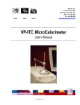
Download VP-DSC Microcalorimeter User`s Manual
Transcript
MicroCal, LLC 22 Industrial Drive East Northampton, MA 01060 USA Phone: +413 586 7720 Fax: +413 586 0149 Binding Stability Kinetics [email protected] VP-DSC MicroCalorimeter User’s Manual MAU120030 Rev B Introduction Key to Symbols ....................................................................................................................................................iv Safety.....................................................................................................................................................................v Instrument Specifications .................................................................................................................................. viii Safety Specifications ............................................................................................................................................ix How to Get Help From MicroCal..........................................................................................................................x Section 1: VP-DSC System Introduction .................................................................1 1.1 VP-DSC Description.......................................................................................................................... 2 1.2 Data Acquisition/Control Description ............................................................................................. 3 1.3 Suitable Laboratory Environment.................................................................................................. 4 1.4 Hardware Connections .................................................................................................................... 5 1.5 ThermoVac ........................................................................................................................................ 7 ThermoVac Accessories ....................................................................................................................................7 Front Panel Controls..........................................................................................................................................8 Degassing Samples ............................................................................................................................................8 Using the ThermoVac for Cleaning.................................................................................................................10 Section 2: Running VPViewer with Origin............................................................13 2.1 VPViewer and Origin Overview .................................................................................................... 14 General Description.........................................................................................................................................14 Operation within a Windows Environment .....................................................................................................14 2.2 Starting VPViewer and the VP DSC ............................................................................................. 15 Opening and Closing VPViewer .....................................................................................................................15 VPViewer and DSC Power Control ................................................................................................................15 2.3 VPViewer and Origin Basics .......................................................................................................... 16 Main Window Display and Description ..........................................................................................................16 Compact Mode ................................................................................................................................................17 Menus and Buttons ..........................................................................................................................................18 Origin For Data Display ..................................................................................................................................19 2.4 VPViewer Cell Status Description ................................................................................................. 20 States of Operation ..........................................................................................................................................20 Section 3: VPViewer and Origin: The Details......................................................23 3.1 The Main Drop Down Menus......................................................................................................... 24 3.2 Main Window Buttons .................................................................................................................... 26 3.3 DSC Control Window ..................................................................................................................... 27 3.4 Thermostat/Calibration Window................................................................................................... 31 3.5 Setup/Maintenance Window .......................................................................................................... 32 3.6 Constants Window .......................................................................................................................... 34 3.7 Origin for Real Time Data Display................................................................................................ 35 Main Window Overview .................................................................................................................................35 VP DSC Customized Buttons..........................................................................................................................36 Section 4: Running The VP-DSC...........................................................................39 4.1: System and Sample Preparation................................................................................................... 40 4.2 Filling/Pressurizing The Cells ........................................................................................................ 41 4.3 Run Parameter Considerations...................................................................................................... 43 i MAU120030 Rev B Introduction 4.4 Carrying Out Scans......................................................................................................................... 44 Starting the Run ...............................................................................................................................................44 Changing Run Parameters While Running......................................................................................................44 Completion of the Run ....................................................................................................................................44 4.5 Cleaning The Cells .......................................................................................................................... 45 Cleaning Overview ..........................................................................................................................................45 Specific Recommendations .............................................................................................................................45 Cleaning Agents ..............................................................................................................................................46 A Word of Caution ..........................................................................................................................................47 4.6 Maximizing Performance of The VP-DSC.................................................................................... 48 4.7 Isothermal Operation...................................................................................................................... 51 Section 5: Experimental Tutorials .........................................................................54 5.1 Scanning Water/Water ................................................................................................................... 55 Introduction .....................................................................................................................................................55 The Tutorial .....................................................................................................................................................55 5.2 Refilling Tutorial ............................................................................................................................. 65 Introduction .....................................................................................................................................................65 The Tutorial .....................................................................................................................................................65 5.3 Running Lysozyme .......................................................................................................................... 66 Introduction .....................................................................................................................................................66 The Tutorial .....................................................................................................................................................66 Section 6: VP DSC Calibration ..............................................................................68 6.1 Y Axis Calibration Check............................................................................................................... 69 Description of Operation .................................................................................................................................69 The Calibration Procedure...............................................................................................................................69 6.2 Temperature Calibration................................................................................................................ 73 Description Of Operation ................................................................................................................................73 The Tutorial .....................................................................................................................................................73 Section 7: Troubleshooting Guide..........................................................................76 7.1 Site Dependent problems and Considerations .............................................................................. 77 Electrical Interference Through Space ............................................................................................................77 Electrical interference Through Power Source................................................................................................77 Ambient Temperature Fluctuations .................................................................................................................78 7.2 Cell Filling Problems and Considerations..................................................................................... 81 Poor Filling Due to Inferior Technique ...........................................................................................................81 Basic Steps and Considerations During Cell Filling .......................................................................................81 Common Causes of Poor Cell Filling and How to Remedy ............................................................................82 Poor Filling Due to Dirty Cells........................................................................................................................82 Partial Filling Technique .................................................................................................................................82 Heat Capacity Dependencies ...........................................................................................................................83 Refilling in Cycle.............................................................................................................................................84 7.3 Controller Settings and Considerations ........................................................................................ 85 Power Management .........................................................................................................................................85 Task Scheduling and Other Real-Time Conflicts ............................................................................................85 Adding PC Peripherals and Hardware Conflicts .............................................................................................85 7.4 Thermal History and Repeatability ............................................................................................... 87 The First Scan of a Series ................................................................................................................................87 The Run Parameters.........................................................................................................................................87 MAU120030 Rev B -ii- Introduction 7.5 Known Application Issues .............................................................................................................. 88 The Use of Reducing Agents...........................................................................................................................88 Fluorides ..........................................................................................................................................................90 Precipitation and Aggregation .........................................................................................................................90 Section 8: Operation Below Freezing ....................................................................92 Section 8.1: Operation Below Freezing ............................................................................................... 93 Warning ...........................................................................................................................................................93 Software and Constants ...................................................................................................................................93 Filling ..............................................................................................................................................................93 Section 9: Extended Temperature Range (ETR) ...................................................94 9.1 General Description ........................................................................................................................ 95 Setting Up Your VP-DSCETR Instrument........................................................................................................95 Locating The External Water Bath ..................................................................................................................95 Connecting The External Water Bath To The DSC Cell Unit.........................................................................95 The Bath Plumbing & Condensation...............................................................................................................95 Connecting The External Water Bath To The PC/ Controller.........................................................................96 9.2 External Water Bath ....................................................................................................................... 97 General Description.........................................................................................................................................97 DSC Range of Operation.................................................................................................................................97 Utilizing The Predefined ‘Modes’ of Operation..............................................................................................98 Changing Between The ‘Modes’ of Operation................................................................................................99 9.3 Pressurizing the DSC Cells ........................................................................................................... 100 9.4 Filling the Calorimetric Cells ....................................................................................................... 101 Total Filling –Non Freezing Applications .....................................................................................................101 Partial Filling—Sub Freezing Applications...................................................................................................101 Using The ‘Trigger’ Syringe For Partial Cell Filling ....................................................................................101 Section 10: Compressibility Studies .................................................................... 104 10.1 Introduction to the PPC.............................................................................................................. 105 Unpacking and PPC Inventory ......................................................................................................................105 Setting Up Your VP-DSC and PPC Accessory .............................................................................................106 PPC Accessory Box – Electrical Connections...............................................................................................106 PPC Accessory Box – Plumbing Connections ..............................................................................................106 Final PPC Accessory Configuration..............................................................................................................107 10.2 Introduction to the PPC Application......................................................................................... 108 The PPC Accessory Box................................................................................................................................108 Activating The PPC Mode.............................................................................................................................110 10.3 Setting the PPC Run Parameters............................................................................................... 112 Experimental Parameters ...............................................................................................................................112 Setting The Scan Parameters & Options........................................................................................................113 Scan Edit Mode .............................................................................................................................................113 PPC Scan Parameters.....................................................................................................................................114 Options ..........................................................................................................................................................115 Isoscan Mode.................................................................................................................................................115 10.4 Loading Run Parameters From File.......................................................................................... 117 10.5 Changing the Auto Equilibration Slope Criterion ................................................................... 119 10.6 Technical Information and Tips ................................................................................................ 120 Different Solvents in the Sample and Reference Cell ...................................................................................120 Changes in Atmospheric Pressure .................................................................................................................120 Combining Data Sets Having Different ∆P ...................................................................................................120 iii MAU120030 Rev B Introduction Key to Symbols Symbols used in this manual: ! Warns the user of possible damage to the unit, draws attention to the risk of injury or contains safety notes and warnings. Symbols used on the VP-DSC instrument: Caution: Read the instruction manual before operating Instrument (supply) ON Instrument (supply) OFF Protective Earth (Ground) Terminal Origin® is a registered trademark of Origin Lab, Northampton, MA VP is a registered trademark of MicroCal, LLC, Northampton, MA MicroCal is a registered trademark of MicroCal, LLC, Northampton, MA Windows™ is a registered trademark of Microsoft Corporation MAU120030 Rev B -iv- Introduction Safety Operator’s Safety The points below are intended to enhance your safety awareness and to draw your attention to risks which only you, the operator, can prevent. MicroCal has set high standards for ourselves in developing and constructing the VP-DSC. Each unit constructed is fully tested to operational and safety standards. It should be noted that no amount of design or constructed safeguards can anticipate or prevent improper handling. It is intended that this instrument be operated only by responsible people trained in basic laboratory protocol in procedures and safety techniques. ! This product is intended to operate from a power source that does not apply more than 260 VAC rms between the supply conductors or between either supply conductor and ground. A protective ground connection, by way of the grounding conductor in the power cord, is essential for safe operation ! To avoid hazard, use only a fuse of the correct type, voltage rating and current rating as specified on the back of the VP-DSC. This is a 1 Amp 250 Volt Time-lag (Time Delay) fuse. ! Repairs, alterations or modifications must only be carried out by specialist personnel. The MicroCal Service Department will be happy to serve you for any repair work or operational questions. ! To enhance safety always plug the instrument into a Ground Fault Circuit Interrupter (GFCI) device. ! A solution can become an electrical conductor when in contact with electricity and can create a hazard with the potential of burns or death. Use caution when using solutions near the instrument and adhere to the following items: ! If any liquid is spilled on or around the instrument unplug the instrument immediately and wipe it up, if there is any possibility that some of the liquid may have leaked into the instrument case contact MicroCal immediately. DO NOT PLUG THE INSTRUMENT INTO ANY POWER MAINS, until the problem is resolved. ! Do not over fill the cell and reservoir. There is a small reservoir located at the top of the access tubes to catch a minimum amount of overflow, do not exceed the capacity of the reservoir and allow any fluid to leak into the instrument cabinet. ! This device is not designed to the Medical Devices Directive 93/42/EEC and should not be used for medical purposes and/or in the diagnosis of patients. ! When using volatile or hazardous solutions in the cells, the operator should always follow proper laboratory procedures in handling and disposing of such materials (e.g. wear safety glasses and protective clothing) and always allow the internal cells to cool down to room temperature before opening the cell port and removing any solutions from the cells. ! All solutions in the cells should be cooled down below 50 ºC before removing from the cells to prevent the glass syringes from breaking due to the hot liquids. v MAU120030 Rev B Introduction Product Safety We have used all means to design and produce the VP-DSC to prevent any damage to the instrument during normal usage. The instrument is solidly built using the latest in micro-technology in its components and design, but of course, this cannot prevent damage due to physical mistreatment. The attentiveness of the operator to the identification of materials which could corrode the Tantalum cells, the prevention of freezing solutions in the cells and the responsible handling and usage of the instrument should insure a long service life for the VP-DSC. ! The VP-DSC cells are constructed out of a tantalum alloy. Tantalum is extremely resistant to corrosion by most acids, but like glass, hydrofluoric acid will readily attack tantalum. Any solution containing the fluoride ion, fuming sulfuric acid or strong bases should be avoided to prevent corrosion of the cells. Please refer to the accompanying booklet “Corrosion Resistance of Tantalum and Niobium Metals” for further information. ! The freezing of solutions in filled cells will cause irreparable damage to the cells due to the expansion of the solution in the cells. If operations below 0 °C are required, the cells should contain a non-freezing solution or at most 230 µl (half filled) of a solution that could freeze. ! When moving the VP-DSC the instrument should always be in its normal operating or its vertical position. The instrument should never be moved in a sideways position, due to the nature of the crystal detectors used for the instruments thermal sensing, which are relatively strong in a compression mode but weak when subjected to shear stresses. The VP-DSC should never be dropped or subject to abrupt acceleration or deceleration forces as these forces could damage the instrument even in its vertical position. MAU120030 Rev B -vi- Introduction European Users: CE Mark MicroCal VP-DSC calorimeters carry the CE marking which confirms that they meet the applicable European Community Technical Directives. Following the provisions of the applicable European council directives: 89/336/EEC, 93/68/EEC Electromagnetic Compatibility 73/23/EEC, 93/68/EEC Low Voltage Is in conformity with the following harmonized standards: EN 61326-1:1997, A1:1998, A2:2001 EN 61000-3-2:2000, A1:2001 EN 61000-3-3:1995 EN 61010-1:2001, IEC 61010-1:2001 EN 61010-2-010:2003, IEC 61010-2-010:2003 Electrical equipment for measurement, control and laboratory use – EMC requirements Limits for harmonic currents emissions Limitation of voltage changes, fluctuations and flicker in lowvoltage systems Safety requirements for electrical equipment for measurement, control and laboratory use Particular requirements for laboratory equipment for the heating of materials Please refer to the manuals that came with the computer controller for information relating to the CE mark for this unit. Instrument Safety Compliance MicroCal VP-DSC calorimeters carry the CUE Safety Certification Mark, authorized by TÜV America, a division of TÜV Süddeutschland, to signify that: 1) the instrument has been tested by an accredited Certification Body and meets applicable Canadian electrical safety standards/requirements (CSA/SCC). 2) the instrument has been tested by an NRTL (Nationally Recognized Testing Laboratory) and meets applicable United States electrical safety standards/requirements (ANSI/UL). 3) the instrument has been tested by a Competent and Notified Body for applicable EU Directives and meets applicable safety standards/requirements (EN/IEC). vii MAU120030 Rev B Introduction Instrument Specifications Model or Type Short Term Noise (RMS average) Baseline Repeatability Minimum Response Time (high gain) Operating Temperature Range : Performance Specifications : : : : Physical Specifications Cell Design Dimensions (L x W x H in inches) Cell Controller Monitor Weight (lbs.) Cell Controller Monitor A VP-DSC 0.5 µcal/°CA 1.25 µcal/°CB 7 seconds -10° to 130°C : Tantalum, 0.5 ml non-capillary type Fixed in place, non-removable : : : 17 x 8 x 15 15½ x 16½ x 7½ 9 x 15 x 15½ : : : 18 25 12 Using upscan mode at 90oC per hour with 30 second filter and over a temperature range of 5o to 110oC. B Average standard deviation between successive upscans at 90oC per hour with 30 second data point filter over the temperature range 5o to 110o C. This specification depends on changes in room temperature and was obtained from multiple overnight scans where room temperature at midnight was lower by ~2.5 oC than the average temperature during daytime. The primary effect of changes in room temperature is to displace baselines by approximately the same amount at all temperatures, thereby maintaining their parallel nature. MAU120030 Rev B -viii- Introduction Safety Specifications Instrument Specifications (excluding Controller) Electrical Specifications Electrical Ratings: Voltage Frequency Power : : : 110 - 240 VAC 50 / 60 Hz 70 watts Fuses (2) : Primary=1.0 A, 250 V, Time Delay Output : Secondary/Data Connection only Protective Earth Terminals : Internal/external marked Mode of Operation : Continuous Classification Installation Category Atmospheric Specifications Operating: Temperature Humidity Atmospheric Pressure Pollution Degree Storage (in original shipping containers with no liquid in cells): Temperature Humidity Atmospheric Pressure : : Class I 2 : : : : 10° to 40° C L.T. 30% to 75% 700 HPa to 1060 HPa Degree 1 : : : -40° to +70° C (no liquid in cells) 10% to 90% 500 HPa to 1060 HPa ix MAU120030 Rev B Introduction How to Get Help From MicroCal The service department at MicroCal, LLC is available to help you during normal business hours, Monday through Friday from 8:00 AM to 5:00 PM (EST). Service personnel may be contacted by e-mail, phone or fax, with the preference being by phone or e-mail. When e-mailing MicroCal for technical assistance it is always best to attach recent data files (*.dsc, raw DSC data files) which exhibit the symptom of concern, whenever possible. Also, please include with the correspondence, all details that may be relevant to the problem. In instances where the problem or question relates to post run data analysis, it is best to attach the raw data file (*.dsc) and the Origin document (*.opj) generated during data analysis. There are 2 general categories of problems associated with the VP-DSC and its operation. The most extreme category is when a system is simply not working, at all. Problems that prevent users from operating the VP-DSC require immediate consultation with a MicroCal technician. Customers should not attempt to repair the VP-DSC hardware or software unless instructed to do so by a MicroCal service representative. The second, and less extreme general category of problems is when a VP-DSC instrument is functioning, but is not operating within its normal performance specifications. Poor repeatability, a detectable change in characteristic baseline position/shape, increase in short term noise level are all examples of performance problems, typically correctable by the operator. For performance issues such as these it is recommended that customers carry out the following minimum diagnostic steps prior to contacting MicroCal for service: 1.) Thoroughly clean cells. Do not assume they are clean. As a minimum, use the provided cell cleaning apparatus to pass 300 ml of a 10-25% ammoniated detergent solution in water, followed by 300 ml of cold water rinse, through both the sample and reference cell. Discretion may call for a more rigorous cleaning procedure as described in Section 4.5 of this manual. 2.) Refill both the reference and sample cells with filtered, degassed water. Cap and pressurize the cells. 3.) Go to the Setup/Maintenance window within VPViewer 2000 and activate the debug mode. While in debug mode, DSC data files will contain all available information produced by the VP-DSC and often times is helpful in diagnosis of problems. 4.) Carry out a minimum of 4 upscans, in debug mode, with freshly degassed water in both cells. The more scans recorded, the better the chance of understanding the problem from the data files. Overnights and weekends are often most convenient for such work. Be sure to use identical run parameters for all scans. Upscans from 10 to 110 degrees, at 90 degrees/hour, with a 15 minute pre-scan thermostat period, are acceptable run parameters. 5.) Consult Section 7 of this manual. If, after completion of the 5 steps listed above, the DSC performance is not corrected, contact the MicroCal service department for help. The water scans, in debug mode, will then be provided to the MicroCal service technician for evaluation. Customers will then be contacted by the service department with comments and recommendations. MicroCal, LLC Contact Information: 413-586-7720 Technical support and service at extension 5 Primary Contact Phone Toll Free Phone Number 800-633-3115 Technical support and service at extension 5 Facsimile 413-586-0149 Web Site: www.MicroCalorimetry.com Please forward corrections or suggestions concerning this manual to [email protected] MAU120030 Rev B -x- Section 1 - VP-DSC System Introduction VP-DSC System Introduction -1- MAU120030 Rev B Section 1.1 - VP DSC Description Section 1.1 VP-DSC Description The VP-DSC is a differential scanning microcalorimeter. The reference and sample cells are fixed in place lollipop shaped vessels with effective volumes of approximately 0.5 ml. The cells are constructed from a tantalum alloy and protrude out of the adiabatic chamber via their 1.5 mm (inner diameter) access tubes. The approximate volume of each cell stem is 0.085mL. The cell material (Tantaloy 61 TM) has chemical properties quite similar to glass. This alloy is immune to attack by almost all acids, having been tested under widely varying concentrations/temperatures involving chlorine, bromine, hydrochloric acid, nitric acid and sulfuric acid. Please refer to the dedicated manual outlining the chemical properties of Tantaloy 61 TM for more detailed information. The VP-DSC operates in the temperature range of -10° to 130° Celsius. It is capable of both upscanning and downscanning with no external heating/cooling source. Scanrates are user selectable and fall in the range of 0°/hr to +90°/hr. in the upscan mode and 0°/hr to -60°/hr in the downscan mode. The VP-DSC can also be used at constant temperature for long periods of time (Isoscan) for shelf life studies and/or for evaluating the stability of drug or other chemical formulations. The duration of such a study is limited only by the available space on the PC hard disk. For scanning solutions above their boiling point, the VP-DSC has a self-contained pressurizing system allowing positive pressures from 0 - 30 p.s.i. Applications requiring higher pressures can be conducted with the addition of an external pressure source and accessory pressure cap, available from MicroCal. Pressure applied to the VP-DSC cells should never exceed 80 p.s.i. or irreversible damage to the cells may be incurred! Temperature differences between the reference cell and the sample cell are measured, calibrated to power units and displayed to the user as well as saved to disk. This data channel is referred to as the DP signal, or the differential power between the reference cell and the sample cell. Calibration of this signal is obtained electrically by administering a known quantity of power through a resistive heater element located on the sample cell. In general, an exothermic (heat is released) event in the sample cell will cause the DP signal to deflect in the negative direction. Likewise, an endothermic (heat is absorbed) event in the sample cell will cause the DP signal to deflect in the positive direction. MAU120030 Rev B -2- Section 1.2 - Data Acquisition/Control Description Section 1.2 Data Acquisition/Control Description The VP-DSC is controlled by two interface boards that reside inside the host PC. The boards are preconfigured and calibrated at MicroCal, and need no adjustments for operating the VP-DSC. The data acquisition is handled by a 16 bit A/D (analog to digital) converter board. There are five differential inputs to the A/D. Jacket temperature, the DP or differential power between the reference and sample cells, the ∆T or the temperature difference between the cells and the adiabatic jacket, the cell pressure as set by the self contained pressurizing system and the heat sink power constitute the analog inputs. All analog input channels may range from -5 volts to +5 volts. Calorimetric control is handled by the second interface board, a 16 bit D/A (digital to analog) converter board. Together with the VPViewer software, this board will facilitate automatic equilibration and repetitive cycling of the VP-DSC with no user interaction. Although other Windows’ programs can be used while the VP-DSC is running without affecting the data acquisition or calorimetric control, it should be realized that there are real time operations taking place during a scan. Performing CPU intensive tasks while a scan is in progress can ‘glitch’ the DSC baseline. DSC users are encouraged to avoid large file copying to disk drives, large print jobs and recursive control loops while the VP-DSC is in the midst of an upscan or downscan. Routine data analysis within Origin® may be performed during DSC runs without regard to compromising the real-time operations. During the system’s idle state (thermostatting) or pre-equilibration state users should feel free to use the P.C./Controller in any manner they wish. -3- MAU120030 Rev B Section 1.4 – Hardware Connections Section 1.3 Suitable Laboratory Environment The VP-DSC with Computer Controller requires about 1 meter length of normal bench space (ca. 70 cm wide). The ThermoVac degassing station will require another 25 cm of bench space. This location should be away from strong drafts, room temperature fluctuations, intense sunlight, vibrations and strong electrical or magnetic fields (as may be produced by an NMR, microwave oven, large motors or refrigeration units). In addition the mains power source (115 to 240 VAC) should be properly grounded and free from voltage fluctuations, harmonic distortions, power dips and spikes. The AC power line should be dedicated to the MicroCal system and should not share that power with additional equipment. Although, the power filtering in the VP-DSC instrument is adequate for most laboratory environments, some disturbances may affect the performance of the instrument and it may be necessary to have the AC Mains power source evaluated (see table below) or install a power conditioner. Since power source problems can be manifested in many different ways, it is not possible to recommend a power conditioner for all situations. It is recommended that you test a power conditioner, at your location, before you purchase it. If you believe you are experiencing power source related problems, please contact a MicroCal engineer. VP-DSC AC Mains Electrical Requirements Specification Requirement 115 to 240 VAC, stable to ± 3% Voltage Regulation 0.5% Maximum Deviation Frequency Stability < 3% Common Mode or Differential Mode at Power Line any Frequency Noise < 5% Total Harmonic Distortion to 1500 Hz Harmonic < 3% For any Single Frequency Content < 1 VAC Peak-To-Peak Ground Noise < 2 VAC Ground to Neutral Peak-To-Peak at any Frequency < 25 Ohm Ground Quality It is emphasized that room temperature fluctuations (i.e. maximum 2.5° C) due to the cycling on/off of heating and cooling systems, strong air currents, sunlight directly on the instrument and through space electromagnetic waves may cause subtle performance problems. VP-DSC Environmental (Operating) Requirements Temperature 10° to 28° C, constant to 2.5° C 0 to 75% Relative Humidity, Humidity non-condensing 70 HPa to 1060 HPa Atmospheric Pressure Section 1.4 MAU120030 Rev B -4- Section 1.4 - Hardware Connections Hardware Connections The cabling necessary to operate the VP-DSC is minimal. Essentially, there is one cable that interfaces the VP-DSC to the host PC. The cable end with just one 37 pin female connector attaches to the rear of the VP-DSC cell and is labeled ‘Cell’. The opposite end of the cable will have two 37 pin female connectors, one for the A/D board, labeled ‘J2’, and the other for the D/A board, labeled ‘J1’. As you look at the rear of the PC (see below) you will notice that there are two connectors visible in the two rightmost I/O expansion slots, both being 37 pin male connectors. Looking from the rear, the rightmost of these connectors is for the A/D board (also labeled ‘J2’) while the left-most is for the D/A board (labeled ‘J1’). Computer Controller Rear View J1 J2 J1 Digital to Analog (D/A) Board Connector J2 Analog to Digital (A/D) Board Connector Now looking at the cable end having two 37 pin male connectors, one of them is labeled J2 (A/D) while the other one is labeled J1 (D/A). Connect each of these two connectors to their appropriate 37 pin female connector at the rear of the host PC. Connect the power cord to the IEC 320 inlet power receptacle (see next page, labeled 3) on the back of the cell. Connect the power plug only to a main power supply receptacle with a 3 wire protective Earth ground and a Ground Fault Circuit Interrupter (GFCI). Once the VP-DSC cell has been cabled to the PC, it is ready to use. At the rear of the cell unit is a power on/off switch. This switch must be in the ‘on’ position for the cell to work. This switch is the master power switch. It can be turned to ‘off’ when the cell will not be used for long periods of time: (i.e. weekends, holidays, etc.) During frequent ‘on’ periods, the master power may be left on, as the power to the cell is also under software control and will be turned ‘on/off’ along with the user interface program, VPViewer. Upon closing the VPViewer application there will be approximately a one minute delay before the VP-DSC power switches off. -5- MAU120030 Rev B Section 1.4 – Hardware Connections In short, the VP-DSC cell power switch can be left on and the VPViewer software left running. The software will ensure that the system is safe and does not incur any damage. As an extra precaution it is recommended that the master power be turned off when the system will not be used for extended periods of time. VP-DSC Cell Rear View 1 5 --- O 2 3 MicroCal, Inc. 22 Industrial Drive East Northampton, MA01060 Product Model Serial No. 1. 2. 3. 4. 5. MAU120030 Rev B Power main switch 2 Power fuses IEC 320 inlet power receptacle Cell Connector Fan -6- 4 Section 1.5-- Thermovac Section 1.5 ThermoVac To facilitate degassing samples and cleaning of the cells, you have been provided with the ThermoVac instrument accessory. The unit is capable of thermostatting a sample at any temperature from 0 to 80 °C, pulling a vacuum of 28.4 inches of mercury and stirring the sample using small magnetic stir bars. SAMPLE TEMPERATURE TEMPERATURE 25.0 ON 250 STIRRER SPEED VACUUM TIMER OFF OFF INCREASE ThermoVac R Sample Degassing and Thermostat MicroCal TM Incorporated ThermoVac Accessories The ThermoVac and accessories may include, but are not limited to the items listed below. A list of the actual items shipped with your order will be included in the documentation included with your shipment. Quantity Description 1 ThermoVac 3 Stir Bars (Small) 3 Stir Bars (Medium) 2 In-line filters 5 Test Tubes (Small) – for conserving sample volume 5 Test Tubes with Cap (Medium) -7- MAU120030 Rev B Section 1.5- ThermoVac 1 Test Tube Holder 1 Flask with Hose & Luer Lock Connector 2 Fuses (1A/250V) 5x20mm Slo-Blo 1 A.C. Mains Power Cord Front Panel Controls VACUUM The ThermoVac is capable of pulling a maximum vacuum of 28.4 inches of mercury. If the VACUUM switch is pushed to the right the vacuum pump will turn on and remain on for approximately 8 minutes, then the pump will shut off. If you desire to remove the vacuum before the preset time you must push the switch to the OFF position. When the switch is pushed to the left the vacuum pump will remain on until it is manually turned off. VACUUM ON TIMER OFF TEMPERATURE Use this dial to set the temperature, in °C, for thermostatting the sample chamber. The rightmost digit sets the temperature value in tenths of a degree. SAMPLE TEMPERATURE Displays the current temperature of the sample chamber. STIRRER SPEED This switch will activate a rotating magnetic field that will stir your sample when a small magnetic stir bar is placed in the tube containing your sample. You may adjust the speed of the Stirrer motor from 0 RPM (OFF) to the maximum speed of 800 RPM (full turn clockwise). Rear Panel 1. 2. 3. 4. 5. Power Main Switch Power Fuses IEC Inlet Power Receptacle Vacuum Port Pressure Port Degassing Samples Top View of ThermoVac MAU120030 Rev B -8- Section 1.5-- Thermovac Tube Holder Vacuum Port Tube Holder Removal Screw Sealing O-ring Vacuum Cap In-line Filter To degas the solutions, before placing them into the cells or injection syringe, please do the following. • Turn on the Power Main Switch. • Set the desired temperature for the solution. • Place your solution to be degassed into a test tube, add a small stir bar and place the tube into one of the open cylinders of the Tube Holder insert. Please Note: To conserve sample for loading into the injection syringe, use the small test tubes provided with the ThermoVac. If you wish to use a tube or beaker larger than will fit into the Tube Holders you may remove the Tube Holder by simply lifting it up. Due to the tight fit of the Tube Holder, it may be difficult to lift it out of the sample chamber. In this case you may use a 3/32” hex (Allen Key) wrench to turn the screw located at the bottom of the center hole of the Tube Holder, to lift the Tube Holder out. • Turn the stirring on and adjust the speed. • Turn on the vacuum. Push the switch to the right to activate the vacuum for a preset (ca. 8 minutes) duration. Push the switch to the left if you wish to manually control the time for the vacuum. • Place the Vacuum cap on top of the sealing o-ring. The sound of the vacuum pump will change pitch to indicate the vacuum has sealed the Cap to the o-ring. Once the vacuum has sealed, the Vacuum Cap will be held firmly in place, till the vacuum pump shuts off. For the first few minutes after the vacuum pump is turned off, the vacuum in the sample degassing chamber may remain fairly tight making the removal of the Vacuum Cap difficult. During this period, the easiest way to release the vacuum is to remove the tubing from the rear Vacuum Port or to twist off (turn counter-clockwise) the in-line filter. -9- MAU120030 Rev B Section 1.5- ThermoVac Using the ThermoVac for Cleaning the DSC Cells The ThermoVac, in conjunction with the cell cleaning device, may be used to circulate cleaning and rinsing solutions through the calorimetric cells. Follow the proceeding steps to conduct the VP DSC cell cleaning procedure. Further information is available in Section 4.7 of this manual. • Insert the long needle into the Sample cell and push down carefully until the o-ring has sealed. • The end of the upper tubing of the Cell Cleaning Apparatus is immersed into a beaker of 200-400 ml of detergent cleaning solution. • The end of the lower tubing is connected to a one liter vacuum flask through the #8 rubber stopper. • The side arm of the vacuum flask is attached to the Vacuum Port of the ThermoVac. Your Setup should look like the picture below. • Turn on the ThermoVac vacuum pump. The vacuum will pull the detergent solution from the beaker, through the cell and into the waste flask. NOTE: DO NOT ALLOW THE VOLUME OF FLUID IN THE WASTE FLASK TO ACCUMULATE SUCH THAT IT WOULD RISE UP TO THE LEVEL OF THE SIDE ARM AND BE SUCKED INTO THE THERMOVAC’S VACUUM PUMP. THIS MAY CAUSE THE PUMP TO BECOME DAMAGED. MAU120030 Rev B -10- Section 1.5-- Thermovac • Once sufficient detergent solution has passed through the cell, the hose is removed from the solution, rinsed free of detergent using a plastic wash bottle, and then placed into another beaker containing ca. 200-400 ml of water for rinsing. • After rinsing with water, remove the tube from the rinse water and allow time for the vacuum to drain the fluid out of the hoses, then remove the cleaning apparatus from the cell. • Remove the remaining water from the cell by using a long needle syringe. If there is any residual soap in the cell reservoir, be sure to rinse it clean with a long needle syringe and water. We do not recommend drying the cell before filling with your sample solution, but it should be rinsed twice with the buffer you are using for the experiment. Finally, it should be completely drained and filled with your sample solution in the manner described in section 4.2. Because a small amount of the buffer used for final rinsing will adhere to the walls of the cell and act to dilute your sample solution, you may wish to correct for this by lowering your sample concentration by 2% if you measured concentration before the sample was introduced into the cell. -11- MAU120030 Rev B Section 1.5- ThermoVac (This page left intentionally blank) MAU120030 Rev B -12- Section 2—Getting Started With VPViewer and Origin Getting Started With VPViewer™ and Origin® -13- MAU120030 Rev B Section 2.1 --VPViewer and Origin Overview Section 2.1 VPViewer and Origin Overview General Description All control of the VP-DSC is handled by the front-end software application, VPViewer. It is within VPViewer that all DSC experimental parameters are defined, DSC scans are started and stopped, real time hardware operations are performed, and DSC data is saved to the hard disk of the PC/Controller. By default, all data that is generated in the scanning state of a DSC scan will be saved to disk, with no user interaction. Operation Within a Windows™ Environment Only one copy of VPViewer will be permitted to run at any given time. If a second instance of the application is detected then VPViewer will immediately terminate itself. Users should feel free, however, to run Origin for data analysis simultaneously with VPViewer, as well as most other Windows applications. In general, VPViewer is a multi-tasking application with some subtle limitations. If VPViewer detects a conflict that may jeopardize its ability to acquire data, it will inform you of this conflict. In such instances you should contact MicroCal. Although other Windows’ programs can be used while the VP-DSC is running without affecting the data acquisition or calorimetric control, it should be realized that there are real time operations taking place during a scan. Performing CPU intensive tasks while a scan is in progress can ‘glitch’ the DSC baseline. Users are encouraged to avoid large file copying to disk drives, large print jobs and recursive control loops while the VP-DSC is in the midst of an upscan or downscan. Routine data analysis within OriginTM may be performed during DSC runs without regard to compromising the real-time operations. During the system’s idle state (thermostatting) or pre-equilibration state users should feel free to use the P.C./Controller in any manner they wish. See the Section 7 of this manual for additional information. MAU120030 Rev B -14- _______ ___ Section 2.2 – Starting VPViewer and the VP-DSC Section 2.2 Starting VPViewer and the VP-DSC Opening & Closing VPViewer The VPViewer application is started via an icon on the Windows desktop or through the start menu in windows. To start VPViewer from the desk top, double click on the icon for VPViewer 2000. To use the start menu, select start/programs/MicroCal’s VPDSC/VPViewer VPDSC. When the VPViewer application is started it will always open a second application, MicroCal’s Origin. This specific instance of Origin will be dedicated to displaying the real time data generated by the VPDSC. Both prescan equilibration data, as well as data obtained from the scan itself will be displayed in real time in this instance of Origin. Users do not need to concern themselves with ‘saving’ any of the data they see within the real-time instance of Origin, as VPViewer will always save experimental data to the hard disk, automatically. VPViewer will also close the real-time instance of Origin when VPViewer itself is closed. If the real-time instance of Origin is closed by accident, an error message will appear and the real-time data display will be lost until VPViewer is closed and reopened. Since all data will still be saved to disk, it is recommended that the user wait for the current series of DSC scans to complete prior to closing and reopening VPViewer. VPViewer and DSC Power Control The main power for the VP-DSC cell unit is controlled by the VPViewer software application. The presence or absence of power to the cell unit is indicated by the Power LED located on the front panel of DSC cell. When the power to the cell unit is on, the LED will shine bright orange and when the power to the cell unit is off, then the LED will also be off. In order for the cell unit power to turn on, the cell must be plugged in, the power switch at the rear of the cell must be in the on position, and VPViewer must be opened and running. Upon opening the VPViewer application, it may take several seconds for the DSC cell power and the Power LED to come on. Likewise, after closing VPViewer it will require a minute or so for the cell power and Power LED to turn off. When the instrument is first powered up, VPViewer will momentarily display erroneous data values. The reason is that upon initial power up, the amplifiers will saturate their outputs for a short period of time independent of the input signal to the amplifiers. As a rule, the temperature display will initially be about 17 degrees higher than the actual temperature. After a minute of so the reading will settle to the actual value. If VPViewer detects a temperature value that could be potentially dangerous to the instrument, it will force the cell into an error state. If this should happen upon the initial power up of the system, simply wait for the temperature value to settle (about 1 minute) and clear the error state from the main menu. Normal operation is now possible. The VP-DSC was designed to be left with its power on for extended periods of time. This will allow for the DSC electronics to remain at the appropriate operating temperature even when the DSC is not actually carrying out scans. It is only recommended that the power to the DSC cell unit be turned off during extended periods of down time, such as holidays and vacations. -15- MAU120030 Rev B Section 2.3 --VPViewer and Origin Basics Section 2.3 VPViewer and Origin Basics Main Window Display and Description The main window of VPViewer for the VP-DSC is depicted below. As can be seen by the above picture, VPViewer displays three data channels by default. The data channels are defined as follows: Temp (Celsius): The current temperature, in degrees Celsius, of the calorimetric cells. DP (mCal/min.): The differential power, in mCal/min., between the reference cell and sample cell. Positive DP values means that the reference cell is hotter than the sample cell while negative DP values means that the sample cell is hotter than the reference cell. In terms of reactions (assuming MAU120030 Rev B -16- _______ ___ Section 2.3 –VPViewer and Origin Basics they take place in the sample cell), exothermic reactions will cause the DP to deflect in the negative direction while endothermic reactions will cause the DP to deflect in the positive direction. The power units for the DP data channel may be displayed in mCal/min., µCal/sec. or µWatts, as can be seen later in this section. Press. (p.s.i.): The current pressure of the cells in pounds per square inch, as set by the VP-DSC self pressurizing system. In addition to the three default data channels, VPViewer makes available to the user three additional data channels. To view the three additional data channels, double click on the Pressure data (the actual numeric data in the display box) and the three additional data channels will be displayed, as pictured below: The additional channels now displayed are the DeltaT, HeatSinkT and the time. These data channels are defined as follows: DT or Delta T (degrees C.): The current temperature difference, in degrees Celsius, between the adiabatic jacket and the calorimetric cells (average of both cells). Negative Delta T values indicate that the cells are hotter than the jacket, while a positive Delta T indicates that the jacket is hotter than the cells. HeatSinkT(no units): A numerical indicator of heat sink function inversely proportional to the actual temperature. This value is only relative and is used for troubleshooting. Time (sec.): If the VP-DSC is in a prescan (equilibration) or a scan, then the current time into that prescan or scan, in seconds, will be displayed. Once the additional data channels are displayed, double clicking on any of the three additional data channels will toggle the display back to the default channels only. Located above the data channel boxes and in the frame which holds all data channel boxes, the current status of the VP-DSC is displayed (Cell Status Indicator). The VP-DSC works by progressing through a series of states, each state having a specific objective. Once the objective of the current state is achieved, the DSC proceeds to the next state. It is a good idea for all users to be generally familiar with the objective of each state as well as the criterion for exiting each of the states. For a detailed definition of each state, please refer to Section 2.4 of this manual. Compact Mode -17- MAU120030 Rev B Section 2.3 --VPViewer and Origin Basics The large buttons at the top of this window provide easy access to commonly used features. The features are unavailable if they are shaded but will become available to the user when the particular function is appropriate to use. An exception to this is the Compact Mode button, which will always be available to the user. This button will shrink VPViewer’s size down and allow it to fit easily with Origin on the desktop. VPViewer’s size is limited to the compact mode size or the standard mode size and can not be manually resized by the user. The compact mode is displayed below. Menus and Buttons VPViewer provides users with three types of controls for accessing all of the application’s functionality. The main drop down menu selections generally control global system functions. The Main Window Buttons allow easy access to features that are frequently used, similar to a toolbar. The main window tab selections toggles the active display between one of the four main windows. The details of each of these sections are provided in Section 3 of this manual. MAU120030 Rev B -18- _______ ___ Section 2.3 –VPViewer and Origin Basics Origin For Data Display All equilibration (prescan) data and actual scan data is automatically plotted in the copy of Origin opened by VPViewer for users to view. The document name within this instance of Origin will always be VPDSCPLOT.OPJ, and that name is visible in the Origin window border. In general, double clicking on any Origin object will give you access to the details of that object. For instance, double clicking on the X Axis will open up the X Axis dialog box where the range, color and various other details of the axis may be changed. Similarly, plot details can be accessed by double clicking on any plotted data set. For more details on changing the characteristics of the plot window and its contents, please refer to the Origin® User’s manual. Further details are also given in Section 3.7 of this manual. The default Origin plot window is displayed below: -19- MAU120030 Rev B Section 2.4-VPViewer Cell Status Descriptions Section 2.4 VPViewer Cell Status Descriptions States Of Operation The VP-DSC is a state driven instrument, with the functionality of the states being controlled by VPViewer. The current state of the DSC (cell status) is always displayed both in VPViewer and Origin, as show in the previous two graphs. To understand the way in which the VP-DSC will progress through a pre-run equilibration and subsequent run (or scan), one need only understand the general purpose of each of the cell states as reported by VPViewer. In general, there are 4 types of states: idle, prerun, run and postrun. When the VP-DSC is not busy equilibrating or scanning, it sits in its idle state, namely the thermostatting state. While in the thermostatting state, the only objective for the VP-DSC is to go to, and maintain, the currently defined thermostat temperature. The VP-DSC will remain in its idle state until the operator starts a scan or a series of scans. Access to the current thermostat temperature is obtained through the Thermostat/Calib. Main window tab. Upon starting a scan or series of scans the VP-DSC will exit the idle state and will proceed to the first prerun state. The objective of the first prerun state will be in getting the VP-DSC cells and adiabatic jacket to the desired starting temperature for the first scan. This will be indicated by a cell temperature that is very close to the requested starting temperature (within 0.5 degrees), and by a DeltaT (temperature difference between cells and adiabatic jacket) which is very close to 0 degrees (< 0.1 Deg.). To save equilibration time it is usually best to set the VP-DSC thermostat temperature to be the same as the starting temperature for scan #1. Once the cells and jacket have settled very close to the requested starting temperature, the DSC will enter the 2nd prerun state which is the prescan thermostat state. The behavior in the prescan thermostat state is the same as in the idle state, in that the VP-DSC will simply maintain the requested starting temperature, for the requested period of time. The purpose of this state is to allow for thermal gradients within the DSC cell unit to minimize so that there is very little heat flow within the thermal core of the DSC. The default prescan thermostat period of 15 minutes is recommended. After the prescan thermostat period has elapsed, VPViewer and the DSC cell will proceed to the final prerun state, the final baseline equilibration. Upon entering this state, the heating (upscan) or cooling (downscan) of the DSC is activated and the equilibration of the DSC is temporarily perturbed. The purpose of the final baseline equilibration state is to allow for re-equilibration of the DSC. Since the DSC is already scanning, several degrees of data will be needed to achieve a stabilized baseline as the DSC heats or cools. In general, it will always require less than 5 degrees of data for re-equilibration. This should be anticipated by the user in the setting of the run parameters. For instance, if the operator specifies that an upscan begin at 10 degrees, the data file will contain equilibrated baseline data beginning at approximately 12-15 degrees (depending on scan rate). There is no avoiding this and users must request starting temperatures which are slightly lower than the temperature at which they wish to save useful data. MAU120030 Rev B -20- _______ ___ Section 2.4 – VPViewer Cell Status Descriptions After the final baseline equilibration period has elapsed the DSC enters the scanning state. Although the DSC cell was scanning in the final baseline equilibration state, now it will be equilibrated and saving DSC data to disk. The scan will continue until the requested ending temperature of the scan is achieved. Upon completion of the actual scan, the VP-DSC can make use of one post run state, the post-run thermostat period. Users may specify time period for post run thermostatting. This state is typically used only for cleaning purposes, in order to incubate the DSC cells at a high temperature for a specified period of time. In general, there are no performance gains to be had by specifying a post-run thermostat period during normal DSC operation. If the recently completed scan is the last scan of a series, the DSC returns to the idle state and resumes thermostatting at the ‘Post Cycle Thermostat’ temperature. If it is not the last scan of the series then the DSC returns to the first prerun state for the next scan and prepares to carry out another equilibration and scan, as specified in the Experimental Control window. -21- MAU120030 Rev B Section 2.4-VPViewer Cell Status Descriptions (This page left intentionally blank) MAU120030 Rev B -22- _______ ___ Section 3 – VPViewer and Origin: The Details VPViewer and Origin: The Details -23- MAU120030 Rev B Section 3.1—The Main Drop Down Menus Section 3.1 The Main Drop Down Menus The main drop down menus at the top of VPViewer will allow for access to some of the less frequently used functions of the application. You will find that the exact sub menu selections may change (DSC menu only) and will depend on the current state of the instrument. Each main menu selection is outlined below with their specific sub menus detailed. System: Quit Program. Selecting Quit Program prompts the user once to see if they want to terminate the VPViewer application. Responding with yes will terminate the program and any scans in progress. All DSC scans, or partial scans that have been completed will already have been saved to disk. Approximately a minute or so after program termination, the power to the VPDSC will also be shut down. Power to the VP-DSC will not be restored until the VPViewer application is run again. DSC: DSC Scan Mode. Allows the user to change between the available modes of operation for the VP-DSC. The DSC Controls window will change according to the mode that is selected. The default mode of operation is Conventional DSC where the cells and jacket are scanned up or down at a constant scan rate. All VP-DSC users also have access to the Isothermal Scan Mode where the DSC is kept at a constant temperature for a specified period of time. This mode of operation is useful for monitoring long, slow thermal processes, common in shelf-life studies, cell metabolism and enzyme kinetics. Please refer to Section 4.7 of this manual for details on the IsoScan Mode of operation. The third mode of DSC operation, Compressibility Mode, is only available to those users who have purchased the PPC (Pressure Perturbation Calorimetry, patent pending) instrument accessory. By using the VP-DSC together with the PPC accessory, scientists can now study the volumetric properties of biopolymers in dilute solution. Please refer to Section 10 of this manual for details on the Compressibility Mode of operation. Print/Save As Text Run Parameters. This function allows users to print or save to a file the run parameters that are being used for a series of DSC scans. While run parameters are always included as part of the DSC data file header, the Print/Save As Text Run Parameters menu option provides the user with a nicely formatted listing of run parameters, ideal for data presentation or general record keeping. MAU120030 Rev B -24- _______ ___ Section 3.1 – The Main Drop Down Menus Start Calibration Run. This option is used to check the accuracy of the DSC y-axis measurements (DP signal). Please refer to Section 6.1 of this manual for details on y-axis calibration. Help: Help. Allows access to the help menus. At the time of release of this User’s Manual the online help was still under development. About. Provides specific details about this version of software and contact information for Microcal, LLC. -25- MAU120030 Rev B Section 3.1—The Main Drop Down Menus Section 3.2 Main Window Buttons The main Window buttons allow access to VPViewer’s frequently used functions. These buttons are displayed below and the details of each button’s function are described in detail. Be aware that this graph shows all the buttons available for use. Normally some of the buttons would be grayed out and not available depending on the current state of the instrument. Specifically, the Start button is only available when the DSC is in its idle state. Similarly, the Stop, Update Run Param. and Display Run Param. buttons are only available when the VP-DSC is active, in a prescan, scan or postscan state. Load Run File: Displays the file open dialog box that allows for loading previously saved run parameters. Run parameters may be loaded from one of two types of files. They may be loaded from a setup file (*.scn, *.iss, *.pps) which was previously saved from the experimental control window. Run parameters may also be loaded from a data file (*.dsc, *.iso, *.(scan#)), with the run parameters being extracted from the data file header. Save Run File. Displays the file save dialog box which allows saving of the currently displayed run parameters. By saving the run parameters to a setup file (*.scn, *.iss, *.pps) they may be reloaded and reused in the future by selecting Load Run File as described above Display Run Parameters. Displays the current run parameters for the scans that are currently being executed. Available only when the VP-DSC is in a non-idle state. Update Run Parameters. Updates the current run parameters for the scans that are currently being executed. This function allows you to change parameters for the scans while they are in progress. Available only when the VP-DSC is in a non-idle state. It should be realized that changing of run parameters while the DSC is in a non-idle state will have no effect on the current series of DSC scans, unless the Update Run Params. button is clicked. Start. This button is used to start a series of DSC scans, as currently defined in the DSC Controls Window. Available only when the VP-DSC is in its idle state. Stop. This button terminates the current scan and series of scans. Available only when the VP-DSC is in a non-idle state. MAU120030 Rev B -26- _______ ___ Section 3.3 – DSC Controls Windows Section 3.3 DSC Controls Window The DSC Controls window is the main access point for operating the VP-DSC instrument. This window will change its content when you change the DSC Scan Mode. The graphic below shows the DSC Controls window as it appears in Conventional DSC mode. Details for the other modes of operation can be found in Section 4.7 (IsoScan mode) and Section 10 (Compressibility mode) of this manual. Found below is a definition for each of the run parameters displayed in the DSC Controls window, while in the Conventional DSC Mode. Number of Scans: Determines the number of scans in the series. There may be between one and two hundred scans in a series, comprised of upscans and/or downscans. All scans of a series will be carried out automatically once the first scan is started from the menus, unless the series is terminated via the menus prior to completion. Post Cycle Thermostat: The temperature that the VP-DSC will go to after the last scan of the series is complete. The cell will remain at this temperature until the currently set Thermostat Temperature is changed, or until another series of scans is started. -27- MAU120030 Rev B Section 3.3—DSC Controls Windows Cell Concentration: This is the concentration (mM) of the solution in the sample cell. The value is stored in the data file header, read by the data analysis program and used in various analytical calculations. This value is only used during data analysis and need not be entered if no data analysis is to be performed (i.e. water/water scans, calibration scans). If the concentration is not entered or is entered incorrectly, it can be changed within the data analysis program. Once the Experimental Parameters have been defined, one would next set the Scan Edit Mode. This determines if all scans of a series are to be identical, or if each scan may potentially have unique scan parameters. The Scan Edit Mode is set by clicking on the appropriate button, with each setting being defined below: Identical Scans: Sets all scan parameters for all scans of the series to be identical to those scan parameters of the currently selected scan. The currently selected scan is that scan number which is highlighted in the list box at the bottom of the Experimental Control window. Additionally, while this button is selected then all scans are assumed identical, and any changes made to a scan parameter will be applied to all scans of the series. Unique Scans: When clicked, sets the Edit Mode to be for unique scans. Unlike the Identical Scans button, clicking on Unique Scans does not change any run parameters but only changes the current edit mode. Additionally, while this button is selected then all scans are assumed unique. Thus, any changes made to a scan parameter will be applied only to the currently highlighted scan or scans. Multiple scan selection can be achieved by clicking on any number of scans in the list, while the Ctrl key is also pressed. Even/Odd: Allows you to easily configure the instrument for alternating up and down scans. Clicking on the Even button will select (highlight) all even numbered scans, and will cause for all ensuing changes to scan parameters to be assigned to all even numbered scans. Clicking on the Odd button will select (highlight) all odd numbered scans, and will cause for all ensuing changes to scan parameters to be assigned to all odd numbered scans. Found below is the definition for all of the scan parameters: Starting Temperature: The starting temperature of a scan. If the Starting Temperature of a scan is less than the Final Temperature of that same scan then the scan will be an upscan. If not, then the scan will be a downscan. Final Temperature: The ending temperature of a scan. If the Final Temperature of a scan is greater than the Starting Temperature of that same scan then the scan will be an upscan. If not, then the scan will be a downscan. ScanRate: The rate at which the VP-DSC will be scanned in temperature. This parameter box only accepts positive values as upscans. Upscans and downscans are distinguished from one another by their Starting Temperature and their Final Temperature and not by the sign of the scanrate. A maximum of 90 Deg./Hr. for upscans and 60 Deg./Hr. for downscans is recommended for optimal results. PreScan Thermostat: The amount of time to thermostat at Starting Temperature, prior to beginning the scan. This allows for thorough equilibration of the thermal core of the VP-DSC before beginning the scan. This parameter can affect the repeatability and the shape of the baseline. The default value of 15 minutes is recommended. MAU120030 Rev B -28- _______ ___ Section 3.3 – DSC Controls Windows PostScan Thermostat: The amount of time to thermostat at Final Temperature prior to beginning the next scan. The PostScan Thermostat period is typically used only for cleaning purposes, in order to incubate the DSC cells at a high temperature for a specified period of time. In general, there are no performance gains to be had by specifying a post-run thermostat period during normal DSC operation. Filtering Period: The time period over which data samples are averaged and a data point is generated and stored. For protein transitions, a filter period in the range of 10-20 seconds is usually appropriate. For sharper transitions such as lipids, a shorter filter period of 1-5 seconds is usually appropriate. Data File Name: The file name under which all experimental data will be saved. All VP-DSC data files will have a .dsc extension. FB Mode/Gain: Determines the method and magnitude of cell-cell compensation used for thermal equilibration of the reference and sample cells. Each of the 4 available FB Modes is precalibrated. When the None (or passive) FB Mode is chosen, then there will be no active cell-cell compensation in response to temperature differences between the reference and sample cells. Equilibration of the cells will be achieved ‘passively’ through thermal conduction and convection. While this method provides for the highest sensitivity (short-term noise), it also provides for the slowest response time of the DP signal. This feedback mode is appropriate for almost all protein studies (broad transitions). When the Low, Mid or High FB Mode is chosen, then there will be active compensation to the cell(s) in response to temperature differences between them. The colder cell will always receive the compensation, and the amount of compensatory heat for a given temperature difference will vary depending on the FB Mode chosen. Low gain will provide for less compensation than will mid gain, which will provide less compensation than will high gain. High gain feedback mode will provide for the greatest amount of cell-cell compensation for any given temperature difference between the cells. The result, when working in High Gain FB Mode will be a faster response time, but with higher short term noise in the DP data. This mode, and its fast response is usually necessary when studying very sharp thermal transitions such as Lipids. The low and mid gain feedback modes will provide intermediate levels of sensitivity and response time characteristics, as compared with the No Gain and High Gain feedback modes. Data File Comments: In addition to the run parameters and data file names, Data File Comments can be assigned for each scan. These comments get saved as part of the data file header and can be viewed from the data analysis program, or from any text editor such as Word or NotePad. When entering data file comments please remember that the Scan Edit Mode will be ignored. Data File Comments will be applied to all selected scans only, or may be applied to all scan by clicking on the Apply Comments To All option. Cell Refill Parameters: The Cell Refill Parameters, available from the DSC Control window are useful for getting the best performance from your VP-DSC. Since the VP-DSC works best while it is cycling (see Sections 4.6 and 5 of this manual), cell refilling must be done during the cooling cycle between scans. The Cell Refill Parameters are provided to help users refill the DSC cell(s) at approximately the same temperature each time, and while the DSC is cycling. You first activate the audible cell filling warning by clicking on Use Audible Fill Indicator. This indicates that you want to be warned, audibly, when the cell temperature is within the specified -29- MAU120030 Rev B Section 3.3—DSC Controls Windows temperature window. Next, you can define the temperature window during which you want to refill the cells. Enter the desired minimum and maximum temperatures during which you want to refill. A 10 degree temperature window, well below any regions of sample transition is suggested. During between scan cooling, and when the cells pass into this temperature window, VPViewer will beep periodically and will indicate that it is time to refill the cells. The beep continues until you clear the message (click on OK in message window), or until the cell temperature passes outside of the refill temperature window. MAU120030 Rev B -30- _______ ___ Section 3.5 – Setup/Maintenance Window Section 3.4 Thermostat/Calibration Window This window allows access to the thermostat control and DP calibration pulse function. The window is displayed below: Thermostat Control: Allows for setting of the thermostat temperature, which will be maintained during the VP-DSC Thermostatting state. Pre-thermostatting the VP-DSC and samples will result in shorter equilibration times. Additionally, high temperature thermostatting during cell cleaning can often times improve the effects of the cleaning. Pulse Control: Allows for manually administering a DP calibration pulse. While this is not the most thorough method of checking the y-axis calibration, it is the quickest and most convenient method. Pulses may be applied any time the DP signal is equilibrated and the resulting deflection used as a crude calibration assessment. Using a pulse which is relatively large ( > +/- 5 mCal/min.) will expose DP calibration errors greater than 1%. Any users who feel that the y-axis calibration requires adjustment should contact Microcal prior to attempting any changes to the VP-DSC calibration constants. -31- MAU120030 Rev B Section 3.5—Setup/Maintenance Window Section 3.5 Setup/Maintenance Window This window allows access to user preferences. Displayed below is the Setup/Maintenance window, along with the default settings for the user preferences. Systems having multiple users may benefit from using customized preferences, linked to a particular user name. Please see Section 3.6 of this manual for more details on Customizing VPViewer. Definitions of the individual preferences can be found immediately below: Data File Path: Indicates the complete path (excluding file name) where all VP-DSC data files will be saved. The specified path should be located on a hard drive and not on any floppy or CD-ROM drives. This setting may only be changed while the VP-DSC is in the idle state, Thermostatting. To change the current data file path, double click on the existing path text and a dialog box will open allowing you to change the current path. The desired data directory must already exist in order to select it from the dialog box. Setup File Path: Indicates the complete path (excluding file name) where all VP-DSC setup files will be saved and loaded. The specified path should be located on a hard drive and not on any floppy or CD-ROM drives. To change the current setup file path, double click on the existing path text and a dialog box will open allowing you to change the current path. The desired setup directory must already exist in order to select it from the dialog box. MAU120030 Rev B -32- _______ ___ Section 3.5 – Setup/Maintenance Window Init. Setup File: Specifies the initial setup file which will be loaded into the experimental control window, the first time it is opened. Specifying a *.scn file here will result in an initial DSC Scan Mode of Conventional DSC. Specifying a *.iso file here will result in an initial DSC Scan Mode of Isothermal Scan Mode. Lastly, specifying a *.pps file (only for those users having the PPC accessory) will result in an initial DSC Scan Mode of Compressability Mode. As an alternative approach, specifying the initial setup file as LastRun1.scn will result in the last run parameters that were executed to be loaded at startup. The initial DSC Scan Mode will then be set to the same mode as was used for the last executed scan. Boot Temperature: Determines the initial VP-DSC thermostat temperature upon launching the VPViewer application. Immediately after starting VPViewer the system will enter the Thermostatting state, go to the specified boot temperature and thermostat. Y-Axis Scale: Allows for changing of the current y-axis (DP) units. Available y-axis units are mCal/min., µCal/sec. and µWatts. By default, y-axis units will be displayed in mCal/min. It must be realized that changing y-axis units in this manner will only change the power units for ‘local’ data displayed in real-time, within VPViewer and Origin (for real-time data display). All data files will be saved with the default units of mCal/Min. Export Current Data: Typically only used for diagnostics. Users may be instructed by a MicroCal technician to Export Current Data, for use in troubleshooting. It provides service personnel with a convenient means of capturing DSC data, which otherwise is not saved to disk. Plot Idle Data: Typically only used for diagnostics. Users may be instructed by a MicroCal technician to Plot Idle Data, for use in troubleshooting. It provides service personnel with a convenient means of capturing DSC data, which otherwise is not saved to disk. Extended Data Mode: Typically only used for diagnostics. This mode of operation will save all of the data channels the VP instrument produces to the data file. This function is used to provide Microcal engineers with extra data for troubleshooting. Save/Add/Erase user: These buttons provide a convenient way of maintaining the desired startup parameters for multiple users. The primary use is for separating data files in different directories. -33- MAU120030 Rev B Section 3.7—Constants Window Section 3.6 Constants Window Provides viewing access to the calibration constants for your particular VP-DSC instrument. Shown below are calibration constants that are typical of the VP-DSC. This window is password protected and should not be changed without authorization by a MicroCal Engineer. MAU120030 Rev B -34- ____________ ___ Section 3.7 – Origin for Real Time Data Display Section 3.7 Origin For Real Time Data Display Main Window Overview When the VPViewer application is opened, a copy of OriginTM will always be opened, as well. The copy of Origin that is opened by VPViewer will always display the text VPDSCPLOT.OPJ in the main window’s border, and has been customized for data display. This instance of Origin must be dedicated for data display only, and should not be used for data analysis. Users should feel free to open a second instance of Origin to carry out all data analysis. This section is intended to detail those features that are contained in the data display version of Origin, only. Please refer to the Origin User Manuals for a complete description of the Origin application. For details on DSC data analysis, please refer to the DSC Data Analysis Manual. Pictured below is the main Origin window for VP-DSC data display. In the picture above there are three areas of customization that are specific to the real-time display of VPDSC data. The VP-DSC Status Display, the VP-DSC Numeric Display and the Buttons For VP-DSC -35- MAU120030 Rev B Section 3.7—Origin For Real Time Data Display____ Data have all been added for user convenience in viewing data generated by the VP-DSC. Found below is a functional description of each of these sets of tools. Numeric Display: Displays the current Temperature, DP and Pressure readings from the VP-DSC instrument. Please refer to Section 2.3 of this manual for a detailed description of those data channels. Status Display: Provides the current state of the VP-DSC instrument. Please refer to Section 2.4 of this manual for further details on the various states of instrument operation Buttons for VP DSC: Provides for convenient viewing of VP-DSC data. VP DSC Customized Buttons A detailed description of each button can be found below. Completed Scans/Current Scan: This button toggles the display between the active scan, and all completed scans. All completed scans within a series of VP-DSC scans will be displayed in the Completed Scans window. Those scans will remain until the next series of scans is started from within VPViewer. Rescale to show all: Causes for automatic rescaling of the x and y-axis ranges so that all data plotted within the window will be displayed on scale. Auto-View 1,2: These buttons re-scale the y-axis display range so that the most recent DP data point is centered on the graph. The centered DP data will be displayed on a full-scale range determined by the value entered in the Auto-View 1(or 2) box (see Edit Range description below). Saved-View 1,2: These buttons re-scale the y-axis to display to one of two preset scales. The minimum and maximum Y-axis settings for these two ranges are determined by the values entered in the Saved View 1 (or 2) boxes (see Edit Range description below). Edit Range: Opens a window that contains parameters to be used with the aforementioned buttons. In addition to the Auto-View and Saved-View entries, which are described above, there is a drop down list box which will control the automatic re-scaling of the x axis. The drop down list is labeled as X Axis Options and contains three choices, as defined below: Disabled: Specifies that no automatic x-axis re-scaling should be performed. New data points which are not within the current x axis display range will not be visible until the x axis is manually re-scaled, or the X Axis Option is changed to be Rescale or Scroll. Rescale: Specifies that the x-axis will be rescaled by 25% when data is received which is beyond the current x-axis display range. This will ensure that all real-time data is within the current xaxis range of display. In rescaling of the x-axis during upscans, the lower limit of the axis will be unchanged and the upper limit will be increased by 25%.In rescaling of the Y-axis durring downscans the lower limit will decrease by 25%. MAU120030 Rev B -36- ____________ ___ Section 3.7 – Origin for Real Time Data Display Scroll: Specifies that the x-axis will scroll when data is received which is beyond the current xaxis display range. In scrolling of the x-axis, the full scale range of the x-axis will be unchanged but both the lower and upper limits of the axis will be increased by 25%. -37- MAU120030 Rev B Section 3.7—Origin For Real Time Data Display____ (This page left intentionally blank) MAU120030 Rev B -38- ______________________ ___ Section 4 – Running the VP-DSC Running The VP-DSC -39- MAU120030 Rev B Section 4.1—System and Sample Preparation____ Section 4.1 System and Sample Preparation Reference or sample solutions must be properly equilibrated with dissolved air before being introduced into the cells. It is important to be aware that the solubility of air in water is higher at low temperature than at high temperature. Solutions or solvents that have been previously stored in the refrigerator for a long time must be degassed before they are entered into the cells. Solutions that have been air-saturated at low temperature and not degassed prior to loading will tend to form air bubbles as they are heated in the cells, and this will produce large aberrations in the baseline. Solutions equilibrated for long times at room temperature will normally not require degassing. The ThermoVac may be used for degassing without excessive boiling (refer to section 1.4 for information about the degassing system included with the instrument). Normally only 5 minutes of vacuum is required to degas if the sample is being stirred but much longer without stirring. The sample should be at room temperature when degassing is started. If volatile buffers or solutes are being used, solutions should then be prepared from degassed or preboiled water and stored air-free. If any solid particles or undissolved solutes are visible in your samples, they should be removed by filtration or centrifugation before degassing the sample. MAU120030 Rev B -40- Section 4.2 - Filling/Pressurizing The Cells Section 4.2 Filling/Pressurizing The Cells Filling of the calorimetric cells is quite simple once the technique has been mastered. The performance of the VP-DSC will greatly depend on one’s cell filling technique, and its importance should be realized. Initially, all users are encouraged to practice cell filling using degassed water in both cells in order to establish the proper technique. All new users of the VP-DSC should be shown the proper cell filling technique by an experienced user. Cells should always be filled from the bottom to the top. The end of the filling syringe needle should be positioned about 1-2 mm from the bottom of the cell during the entire cell filling process. Do not raise the syringe needle during the cell filling process. Slowly depress the syringe plunger until you see solution rise up and out of the top of the cell stem (@ 0.6 ml). This indicates that the entire working volume of the cell and the access tube are completely filled. Once the working volume and cell stems are completely filled, it is a good idea to administer a few (2-5) short abrupt bursts (@ 0.1-0.25 ml) of solution to force any bubbles out of the working volume. Lastly, all excess solution should be removed from the overflow reservoirs using the long needle syringe. Included in your VP-DSC accessories is a Filling Funnel. The Filling Funnel is intended to assist you in the cell filling process. First, insert the funnel into the reference or sample cell reservoir and gently push it down as far as it will go. Next, place the VP-DSC Filling Syringe needle through the hole in the middle of the funnel crossbar and lower it until the syringe glass rests atop the funnel crossbar. The Filling Funnel and VP-DSC Filling Syringe have been machined so that the filling syringe needle will sit just above the bottom of the cell surface when both the funnel and the syringe are seated correctly. The end of the syringe needle is now positioned 1-2 mm above the bottom of the cell, as desired. You can then carry out the cell filling procedure as described above. The picture below shows the proper configuration of the Filling Funnel and the VP-DSC Filling Syringe during cell filling. -41- MAU120030 Rev B Section 4.2 -- Filling/Pressurizing The Cells After the excess solution has been removed from the overflow reservoirs, the operator may remove the top portion of solution (ca. 7/8’’ or 22 mm) from the top of the cell stems. This can be accomplished by inserting the Filling Adjust Syringe into the top of one of the access tubes, and lowering it until the bottom of the syringe collar is resting on the outer diameter of the cell stem. Next, the top 7/8” of solution is drawn out of the cell stem with the syringe. Repeat this procedure on the other cell stem. This step may be considered as optional, but will produce cleaner baselines than if it were not carried out. If positive pressure is to be applied to the cells, it can be done as soon as the cells are filled and excess solution is removed. The pressure is set by installing the cap to the top of the VP-DSC cell port, then compressing the air space inside the cell port. Since the maximum achieved pressure is volume dependent, all excess solution must be removed from the cell reservoirs and cell port prior to setting the pressure. To set the pressure, first secure the cap onto the cell port by turning the lower portion of the cap and mating it with the male threads on the lower portion of the cell port. Turn the cap housing all the way down until it will not go any further. Be sure not to cross-thread the cap/port assembly. Once the cap is all the way on (pressure in VPViewer will still be approximately zero p.s.i.), cell pressure may be achieved by turning the top portion of the pressurizing cap. As the top portion is turned clockwise, an oring sealed piston is compressing the cell port air space and achieving positive pressures up to approximately 30 p.s.i. If at any time while setting the cell pressure it exceeds 40 p.s.i., remove the cap and check for excess solution within the cell reservoirs and/or cell port. During the course of heating or cooling the cells, the pressure may change slightly with temperature. In general, the pressure may change by about 3 p.s.i. over a 120 degree temperature range. Please consult a MicroCal service representative in the event of abnormal pressure leakage. For applications requiring higher cell pressures, MicroCal can provide an auxiliary pressure cap which utilizes an external pressure source. Please call MicroCal to discuss your specific application and the availability of the auxiliary pressure cap. Applying cell pressures in excess of 80 p.s.i. may result in damage to the calorimetric cells and must be avoided. MAU120030 Rev B -42- _______ Section 4.3 - Run Parameters Considerations Section 4.3 Run Parameter Considerations When setting the VP-DSC run parameters there are some general considerations that may prove useful in obtaining the best and most appropriate DSC data. There are also some potential sample considerations that will dictate the appropriate setting for some of the run parameters. Located below are the general considerations for setting of VP-DSC run parameters, followed by some considerations related to the particular samples being scanned. VP-DSC Run Parameters: General Considerations: 1.) The VP-DSC repeatability will be optimized when the individual run parameters for all scans are identical. 2.) The starting and ending temperatures for a DSC scan should allow for both pre-transition and posttransition baselines, necessary for data analysis. 3.) A pre-scan thermostat period of 15 minutes is recommended. 4.) Post-scan thermostat periods are typically set to 0 minutes. Some cell cleaning protocols might make use of post-scan thermostatting at high temperature. VP-DSC Run Parameters: Sample Related Considerations: 1.) Very sharp transitions may require lower scan rates to avoid shifting the transition midpoint in the direction of the scan (i.e. higher transition temperature in upscan, lower transition temperature in downscan). 2.) Solutions that have multiple transitions may benefit from slower scan rates to help ‘separate’ the transitions in the real-time data. 3.) Very dilute samples may benefit from an increase in scan rate, increasing the calorimetric sensitivity of the VP-DSC. 4.) Slow transitions (i.e. protein unfolding) will benefit from the passive feedback mode (feedback gain/mode = None) and will not suffer from the slow DP response time. Faster transitions (i.e. Lipids) may require the high gain feedback mode to obtain the necessary DP response time with the trade-off being a slightly higher short term baseline noise. 5.) Faster scan rates may require smaller filter periods to well resolve the transition. Slower scan rates will not require as many data points, and larger filter periods may be used. -43- MAU120030 Rev B Section 4.4 – Carrying Out Scans Section 4.4 Carrying Out Scans Starting the Run Once all of the run parameters are set, the scan or series of scans can be carried out by selecting the start button at the top of the VPViewer window as shown below. If an already existing data file name has been specified, the user will be asked if he/she wants to overwrite the existing file. Responding with ‘Yes’ will cause the scans to be carried out and any preexisting file to be overwritten. Responding with ‘No’ will return the user to the Experimental Control window with no scans started. Changing Run Parameters While Running To change run parameters for a series of scans already in progress, open the DSC Control window and make the desired changes, then select the Update Run Parameters button. Be aware that changing run parameters in the DSC Control window will have no effect on the current scans until the Update Run Parameters button is clicked. Completion of the Run The scans may be terminated at any time by selecting the stop button. If the scans are not prematurely terminated, the software will complete all the scans, return to its idle state, and thermostat at the post scan thermostat temperature. MAU120030 Rev B -44- Section 4.5 – Cleaning The Cells Section 4.5 Cleaning The Cells Cleaning Overview The VP-DSC uses fixed in place cells in order to provide our users with maximum sensitivity and repeatability. These cells must be cleaned routinely to maintain the high performance of the instrument. Dirty cells will contribute greatly to cell filling problems, poor repeatability and possibly residual thermal effects in the data. Inadequate cleaning is the primary cause of most problems experienced with the VP DSC. The importance of routine effective cell cleaning is critical to the VP-DSC performance! There are three methods of cleaning the calorimetric cell. The simplest is manually rinsing the cells with solution using a filling syringe. The second method involves using the cleaning device and is detailed in section 1.4. The third method involves filling the cells and allowing them to soak in a cleaning solution at an appropriate temperature. The first two methods involve some physical agitation, which is necessary to adequately clean the cells. The soaking method is generally used after the others have failed and uses an appropriate solubilizing/oxidizing cleaner. Adequate rinsing with water after any cleaning agent is important. The selection of a cleaning agent is crucial. All of the components of the sample to be cleaned need to be soluble in your cleaning solution. The cells of the VP DSC are constructed of Tantalum™. This tantalum alloy is highly resistant to acid solutions and partially resistant to alkali solutions. If necessary, please refer to the dedicated manual on the corrosion resistance properties of Tantalum™ or call MicroCal for recommendations. Specific Recommendations As a general rule, the simplest effective method of cleaning is preferred. Short of a very aggressive method it is impossible to give a global recommendation for all samples. Therefore the following comprise the simplest to the more aggressive methods. Please note this list is not all-inclusive and many variations exist. Buffer Rinse: For well behaved (no evidence of precipitation) and related samples, it is best to simply rinse the cells with buffer between scans. Three or more cell volumes of rinsing are usually adequate. Detergent Rinse: The cell(s) are rinsed three or more times with the full volume of a filling syringe containing a detergent. A good general purpose cleaner is a 10% to 25% V/V aqueous Top Job™ solution at ≥ 50°C. The temperature is important as the effectiveness drops greatly as the temperature drops. Top Job is a general purpose ammoniated household cleaner consisting of ionic and non-ionic surfactants. The cells are then extensively rinsed with cold water, completely emptied and then refilled. Detergent Wash: The cleaning device is used in accordance with section 1.4. Two hundred fifty to five hundred milliliters of hot detergent solution is passed through the cells. The same volume of cold water is then passed through the cells to rinse the detergent out and cool the cells. If the detergent is not easily rinsed, hot water may be used. -45- Section 4.5 – Cleaning The Cells Detergent Soak: The cells are filled with a detergent solution such a 10% aqueous Contrad-70™ solution. The cells are not capped but covered with a damp paper towel. The thermostat temperature is then set to 65°C for one hour. The cells are then cooled and rinsed with water using the cleaning device. Do not exceed one hour unless advise by MicroCal personnel to do so. Nitric Acid Soak: The cells are filled with concentrated (90% - 95%) nitric acid. The cells are not capped and a damp paper towel is placed over the cell port. The system is then thermostatted at 80°C. for 1 – 24 hours. After the cells have then been cooled down below 30 degrees, the Nitric Acid may be removed from the cells. Any Nitric Acid cleaning should be immediately followed by a cold water rinse (ca. 250 to 500 ml) using the cell cleaning device connected to the ThermoVac. Important Note: While the calorimetric cells themselves can withstand long exposure to Nitric Acid, other calorimeter components cannot. The pressure transducer will become damaged by the acid and its fumes if it is not protected. You have been provided with a pressure transducer plug to protect the transducer and its sealing from corrosive agents such as nitric acid. The pressure transducer hole is approximately centered between the reference and sample cell access tubes. Insert the plug using the threaded rod and leave it in the hole until all nitric acid has been removed from the cells. The cell port is constructed of 304 stainless steel and is susceptible to attach by nitric acid. Care should be taken to reduce the cell ports exposure to nitric acid. Cleaning Agents Your choice of cleaning agents will depend on the samples being cleaned out of the cells. Below is a partial list of cleaning agents and their general applicability. Ammoniated Detergent (Top Job, Mr. Clean): Prepare a 10% solution of ammoniated detergent (i.e. Top Job, or alternatively a dish detergent) with hot water. This is an acceptable cleaning solution when all other alternatives are unavailable. These detergents may not rinse very well and could leave contamination. Liqui-Nox: This is an excellent choice for most biological contamination. It’s near neutral Ph and excellent rinsibility makes it ideal. A 5% V/V solution should work for non-precipitating samples. Available from Sigma and Thomas Scientific Contrad-70 (Decon-90): Contrad-70 is the standard recommended cleaning agent for the VP-DSC system. A solution of 10-20% V/V should be prepared immediately before use. Contrad-70 is manufactured by Decon Laboratories and contains dodecylbenzensulfonic acid, potassium hydroxide, sodium citrate and sodium laurel ether sulfate. NOTE! CAUTION SHOULD BE USED, ESPECIALLY AT ELEVATED TEMPERATURES, WHEN USING Contrad-70 AS IT IS HIGHLY CAUSTIC. USE OF PROTECTIVE DEVICES FOR EYES, SKIN AND CLOTHING IS REQUIRED. Contrad-70 (also known as Decon-90) may be purchased from the following sources: Decon Laboratories Thomas Scientific Bryn Mawr, PA 01090 P.O. Box 99 Tel: 610 520-0610 Swedesboro, NJ 08085 Tel: 609 467-2000 MAU120030 Rev B -46- Section 4.5 – Cleaning The Cells Catalog # 1003 Catalog # 2901-C30 Pepsin(0.1% solution): Used as a soaking agent to dissolve proteins. EDTA (10% solution): Used as a post cleaning rinsing agent to eliminate metal ion contamination. Organic Solvents: Methanol, hexane, toluene and other solvents are used to remove lipid and some polymer contamination. A Word of Caution Careful consideration should be put into the selection of a cleaning program. As mentioned previously the cells of the VP DSC are constructed of Tantalum™. This tantalum alloy is highly resistant to acid solutions and partially resistant to alkali solutions. With this in mind it is generally best to use an acidic or neutral cleaning solution on a regular basis and revert to an alkalinic solution only when necessary. Please refer to the dedicated Tantalum™ corrosion resistance manual that was included with your system for further information. Care should be taken to avoid exposing the cell port to potentially corrosive agents. The cell port is fabricated from 304 stainless steel and is impervious to attack from most solutions, but extra care should still be taken by removing all excess cleaning solution from the overflow reservoirs of the cell port. You must avoid fluorinated solutions completely. irreparable damage to the instrument. -47- Fluoride compounds will cause severe and Section 4.6 – Maximizing Performance of the VP-DSC Cells Section 4.6 Maximizing Performance of the VP-DSC The VP-DSC is a highly sensitive instrument capable of detecting heat capacity changes of several parts-per-million in the absolute heat capacity of the sample solution. Being highly automated through the use of the VPViewer software, very few demands are placed on the operator while the instrument is running during an experiment. Between experiments, however, there are certain routine procedures that must be carried out manually. These consist of preparing the sample solution and determining concentration, degassing the sample and reference solutions, cleaning and/or rinsing the cells, and loading the solutions into the cells. For routine experiments not requiring maximum performance of the instrument, no particular burden is placed on the operator in carrying out these procedures. However, when sample solutions are extremely dilute (e.g., protein solutions at ∼0.1 mg/ml and below) the best results will only be obtained once appropriate techniques are mastered. Based on our experience, there are several points that should be emphasized. 1.) Often times, proteins and other biological solutes do not dissolve completely or may precipitate slightly during the degassing procedure. Visual inspection under appropriate lighting conditions will reveal small particles suspended in the sample solution whenever this happens. These may be conveniently removed using a disposable syringe filter which has a low binding capacity for biological solutes (e.g., Cole-Parker # H-02915-10; cellulose acetate, 25 mm diameter, 0.8 µm pore size). To avoid precipitation during degassing, use short degassing times (3 min with stirring is usually adequate if solutions are at room temperature) or avoid degassing altogether by preparing sample solutions from degassed buffer solution. 2.) When carrying out a series of related experiments where it is important to get the best comparative results, each sample solution should be treated exactly the same (i.e., same starting temperature, same ending temperature, same scan rate, same elapsed time between the end of one experiment and the start of the next experiment). Results can be improved if the first experiment of the day is a “dummy experiment” with buffer in both cells, since this will put the instrument into the same cycle of thermal history for the first sample experiment as for the later sample experiments. 3.) So long as precipitation does not occur during an experiment, it is not necessary or even desirable to clean the cells with detergent after each experiment in a series of related experiments. After completion of one experiment and after the instrument has cycled to low temperature (i.e. audible refill indicator has beeped), drain both the sample and reference cell, rinse the sample cell repeatedly with water using the overflow filling funnel and the 20 cc plastic syringe. Finally rinse and drain both cells repeatedly with the buffer to be used in the next experiment using the 2.5 cc glass filling syringe. Note: This procedure of not cleaning after each experiment can be overdone, and it is suggested that the sample cell be cleaned at the end of a working day or after a series of related experiments are completed. When the same buffer solution is being used for successive experiments it is not necessary or desirable to refill the reference cell buffer solution. 4.) Do not dry the cells between experiments, just drain out all of the buffer solution (syringe needle should be touching the bottom of the cell) from the final rinse before filling with the sample solution. Using this procedure, the small amount of buffer solution remaining in the cell after draining will cause about 1-2% dilution of the sample solution when it is loaded. The sample concentration can be MAU120030 Rev B -48- Section 4.6 – Maximizing Performance of the VP-DSC corrected based on this factor, or the concentration can be determined when the sample solution is removed from the cell. 5.) The most critical operator-dependent procedure is filling the sample and reference cell, described in Section 4.2 of your Manual. When the cell is initially filled by slowly entering the solution, there will be small bubbles lodged at the position where the expanded portion of the cell joins the narrow filling tube. These bubbles can only be dislodged by quickly depressing the syringe plunger to get a rapid flow of liquid through the filling tube, as described in Section 4.2. In order to carry out this procedure it is necessary to start with ca. 1.25 cc of solution in the syringe so there is sufficient excess solution available to create fast flow through the filling tube to force out the bubbles. This excess solution is then recovered by draining the reservoir before starting the experiment. 6.) After both cells and cell stems are totally filled, and the excess solution removed from the overflow reservoirs, it is important to remove the top portion of the solution (ca. 7/8’’ (22mm)) from the top of the cell stems. This can be accomplished by inserting the Filling Adjust syringe into the top of the access tubes down to the collar on the needle. Then the solution is drawn out of the cell stem with the syringe. Repeat the procedure on the other cell stem, cap and pressurize the cell port and commence with the run. -49- Section 4.6 – Maximizing Performance of the VP-DSC Cells 7.) Practice the techniques as described above by carrying out repetitive experiments with refill, using either water Vs water or an inexpensive biological solute such as lysozyme. As you do this, you will observe that the repeatability of successive experiments will improve as you master the various operations (particularly the filling technique). The Figure included here shows results obtained at MicroCal on a very dilute lysozyme solution. These data are plotted exactly as received from the instrument, with no shifting on the y-axis or any post-run filtering. The four scans were carried out during the same day, cells rinsed but not cleaned between runs, using passive feedback, and with the instrument in the same cycle of thermal history for each experiment. MAU120030 Rev B -50- Section 4.7—Isothermal Operation Section 4.7 Isothermal Operation The VP-DSC has two basic modes of operation; scanning (Conventional DSC) and isothermal (Isothermal Scan Mode). Scanning mode uses a linearly increasing or decreasing temperature ramp function, while measuring the differential power between the reference and sample cells. This mode is referred to as ‘Conventional DSC’ from within VPViewer. A second mode of operation is also available, namely Isothermal mode. In Isothermal mode, a constant temperature is maintained for relatively long periods of time while measuring differential power between the reference and sample cells. Isothermal mode is commonly used in evaluating the stability of drug formulations, as in shelf life studies. To carry out an Isothermal mode scan (IsoScan), the user must first select the Isothermal mode of operation from within the Experimental Control window. From the Experimental Control window, select the menu Cell 1|DSC Scan Mode|Isothermal Scan Mode, as shown below. Once the Isothermal Scan Mode is selected then a check mark will appear next to the menu option indicating that this is the current DSC mode of operation. Additionally, the DSC Control Window will contain run parameters relevant to IsoScans rather than the Conventional DSC run parameters. Finally, VPViewer will not allow users to change this mode while a scan is in progress. -51- Section 1.4- ThermoVac When in Isothermal Scan mode, your experimental control window will appear similar to the picture below: Many of the Isoscan parameters are exactly the same as the conventional DSC mode of operation. The parameters that are not covered in the conventional DSC section are outlined below: Isoscan Temperature: Determines the temperature of the IsoScan. Isoscan Duration: This is the length of time, in hours, that the instrument will hold the temperature and collect data. Auto Spacing: Automatically increments/decrements the temperature of each successive scan. MAU120030 Rev B -52- Section 4.7—Isothermal Operation (This page left intentionally blank) -53- Section 5 - Experimental Tutorials Experimental Tutorials MAU120030 Rev B -54- Section 5.1 - Scanning Water/Water Section 5.1 Scanning Water/Water Introduction This section assumes a thorough knowledge of the topics discussed in all previous sections of this manual. If you have not read these sections, please do so before attempting this tutorial. The goal of this experimental tutorial is to practice and eventually master the basic operational techniques that are necessary to successfully operate the VP-DSC. In beginning one’s first hands-on experience with the VP-DSC, it is best to perform experiments that minimize the number of experimental variables. This is best done by carrying out scans of water/water, which consist of water loaded into both the sample cell and reference cell. In general, water/water scans will provide the user with a gauge of success for carrying out the necessary techniques while operating the VP-DSC, as well as judging the performance of the VP-DSC itself. All users must master the water/water tutorial prior to beginning their work on ‘real’ samples. It is also strongly recommended that users periodically repeat the water/water tutorial, even after one has established confidence in one’s own techniques, as well as confidence in the VP-DSC. Instrument related problems, as well as deviations in techniques can be detected best using water/water, as there is always a comparative reference for the water/water data, as mentioned below. Located in the /data sub-directory of your computer’s hard drive are some sample water/water scans obtained using your instrument, at MicroCal. The water/water upscans are named waterup1.dsc waterup4.dsc, and the water/water downscans are named waterdn1.dsc – waterdn4.dsc. These scans are considered to be typical results, assuming a conscientious effort at duplicating the techniques described in section four. The scan parameters for waterup#.dsc and waterdn#.dsc data files can be retrieved by loading any of those same data files into the DSC Control window. The Tutorial It is assumed for this tutorial that the reference and sample cells are clean. Prepare 30-50 ml of distilled water by filtering the water, then degas with vigorous stirring for 15 minutes. When the water is ready, and when the DSC cells are cool, empty the cells then fill them using the prepared water. While filling, follow the suggested techniques mentioned in section 4.2. Keep the water covered when not being used. The first step in carrying out the water/water scans is to set the run parameters appropriately. Since you are attempting to duplicate previous results (water/water scans carried out at MicroCal), it is crucial that we use the same run parameters that were used for our comparative scans. To accomplish this you first open the DSC Control window from the main menus, as shown below: -55- Section 5.1 - Scanning Water/Water Initially, the last set of run parameters that were executed will be displayed, by default. To load the same run parameters as used in the waterup#.dsc scans, you will load the run parameters from the data file itself, as the run parameters used will be contained in and extracted from the data file header. From the main window, select the Load Run File button: Once the button has been selected, a dialog box will open allowing you to specify the run parameter file you wish to load. Run parameter files have a default extension of *.scn and reside in the /setup sub directory; however, for this particular example we will be loading the run parameters from a data file having a *.dsc extension and located in the /data sub directory. To load our run parameters, double click MAU120030 Rev B -56- Section 5.1 - Scanning Water/Water your way to the /data sub directory and replace the displayed *.scn file specification with the desired *.dsc file specification. This can be done using the drop down list provided at the bottom of the dialog box (“List Files of Type...”) as shown below: Once you’re displaying *.dsc files located in the /data subdirectory you should see the appropriate data file containing the desired run parameters. The reference water/water scans are waterup1.dsc through waterup4.dsc.and waterdn1 through waterdn4.dsc. Selecting any of these data files will load the appropriate run parameters. Select waterup1.dsc and click on OK, as shown below: The run parameters displayed within the DSC controls window are now identical to those used to generate the water/water data files on your P.C. Those specific parameters are shown below: -57- Section 5.1 - Scanning Water/Water As can be seen, the run parameters loaded specify that 20 scans are to be carried out, scanning the VPDSC alternately up and down from 10° to 110°, at a scan rate of 90°/Hr. Up and 60°/Hr. Down. The prescan thermostat is set to 15 minutes for all of the scans and the filtering period is 16 seconds for all scans. The Feedback Mode is set to be passive. At this point we neither need nor want to change any of the run parameters. The only remaining step is to name the resulting data files. To name a series of scans such as this, it is most convenient to enable the automatic numbering select box and edit the filename you want to use in the file name box. This will give each filename the same prefix, followed by the scan number for that scan. If you wish to name each file individually you will need to deselect the automatic numbering system and then select the file you want to rename in the run parameters display box. As you can see below, we have chosen the automatic mode and renamed the files scanr#.dsc. MAU120030 Rev B -58- Section 5.1 - Scanning Water/Water Now that all run parameters are set appropriately and the data file names are as desired, this series of scans can be carried out by selecting Start DSC Run button from the main window. If the Start DSC Run button is not available as would be the case if the button was gray, make sure that the VP-DSC is presently in the Thermostatting state. DSC scans may only be started from the Thermostatting state. After starting the series of scans, VPViewer puts the DSC into the initial prescan state. The cell status indicator in Origin for data display will now read “Scan #1 Cooling DSC to 10°.” just as depicted below: As the DSC cools (or heats) to the starting temperature, the temperature data is plotted versus time in layer 2 of the VPViewer plot window. The temperature scale axis will be adjusted by VPViewer to display the entire temperature plot during the cooling (or heating) process. While the DSC is cooling, VPViewer is awaiting three criteria to be satisfied prior to proceeding to the next prescan state. First, it will wait for the temperature of the cells to become ‘close’ to the requested starting temperature. Second it will check that the temperature data is ‘flat’ and not simply passing through a region close to the requested starting temperature. Lastly, once the two aforementioned criteria are satisfied, VPViewer will -59- Section 5.1 - Scanning Water/Water wait for the jacket-cells DeltaT to become ‘small’ indicating that the cells and jacket are at approximately the same temperature. As the VP-DSC approaches the starting temperature the DP channel will approach zero, as can be seen graphically. Located on the left-hand side of the plot are some data finder tools for the DP data. If the data is not on scale you may click the button Rescale to show all. Your graph will look similar but not identical to the one pictured below: As can be seen from the graph, the temperature (lower line) is approaching 10 degrees. It can also be seen that as the temperature becomes flat, the DP data (upper line) approaches zero and also become flat. This is a typical graphical representation of the thermal equilibration of a VP-DSC. Upon completion of the Heating/Cooling to the starting temperature state, the VP-DSC will enter the Prescan Thermostat. The duration of the Prescan Thermostat period is determined by the operator, within the DSC controls window. A 15 minute prescan thermostat period is recommended for optimum repeatability, but can be shortened or set to zero when repeatability is not of concern. Once the VP-DSC has entered the Prescan Thermostat state, the X axis, in time, will be scaled to indicate when the prescan will be completed, and the data will begin being saved to disk. In the picture below the cell status indicates that the Prescan Thermostat is the current state, and the X axis indicates that the actual scan will begin at approximately 2150 seconds. MAU120030 Rev B -60- Section 5.1 - Scanning Water/Water Once the prescan thermostat period has elapsed, the heating/cooling source is activated and the VP-DSC begins to ramp up/down in temperature. This is known as the Prescan Wait-Final Equilibration state. A graphical representation of the prescan wait can be seen below: -61- Section 5.1 - Scanning Water/Water The above graph demonstrates two points of interest. First, it can be seen that the temperature data has begun to linearly increase with time. Secondly, it can be seen that as the DSC begins scanning, the DP is thrown out of thermal equilibration (DP deflects up in this case), and then equilibrates. These two events best describe the objective of the Prescan Wait - Final Equilibration state. The Prescan Wait state is a time based state which lasts three minutes for an upscan and five minutes for a downscan. When the Prescan Wait state has elapsed, the VP-DSC enters the scanning state and data begins being saved to disk. When the VP-DSC enters the Scanning state, the cell status will indicate whether it is currently upscanning or downscanning as well as the terminating temperature of the current scan. For the run in progress the cell status will read as shown below: MAU120030 Rev B -62- Section 5.1 - Scanning Water/Water As shown above, the DP is now the only data displayed graphically. Additionally, the x-axis during the Scanning state is Temperature, rather than time. Finally, the name of the data file is displayed in red at the top portion of the plot.. The VP-DSC will continue to scan until it reaches its final temperature of 110 degrees, at which time scan #1 is complete. If a non-zero postscan thermostat period has been specified for scan #1, then the VP-DSC will thermostat at the final temperature for the specified duration. If the postscan thermostat period is zero, or upon completion of the postscan thermostat period, the VP-DSC will begin heating/cooling towards the starting temperature of scan #2, or in this case it will again cool itself to 10 degrees in preparation for the second scan in our series of four scans. Allow all of the scans to complete, and do not refill the cells in between the scans. When at least eight of the scans have completed, the next step is to look at the scans together and to judge the performance of the operator and of the DSC. To accomplish this, open up Origin for DSC Data Analysis. For the purposes of this tutorial, some working knowledge of the DSC Data Analysis program will be assumed. For more information on using Origin for data analysis please refer to the provided manual, DSC Data Analysis in Origin. Upon launching Origin for DSC Data analysis, the RawDSC plot window will appear. This window provides buttons for reading and displaying data files. Prior to reading in the data files, disable the ScanRate Normalization option by clicking on it once and removing the X in the box. Next, click on the -63- Section 5.1 - Scanning Water/Water Read Data button and select the data files you ran, located in the vpdsc\data subdirectory. To select the files you want to read, double click on each one or highlight them and select add files. Though your baselines will look slightly different, you can see an example of such scans plotted below: As illustrated in the above picture, scan #1 exhibits the poorest repeatability of the four up scans. The explanation for this result is extremely important in understanding DSC and exactly how you can get the best possible performance out of your instrument. One of the most important parameters regarding DSC repeatability is the thermal history of the DSC. In the above picture, scan #1 was the only scan that had a different thermal history as compared to the proceeding scans. Prior to starting the series of scans, the VP-DSC had been thermostatting at 25 degrees. This means that the VP-DSC cooled from 25 to 10 degrees prior to beginning scan #1. For the following scans the VP-DSC down cooled from 110 degrees down to 10 degrees. This difference in the thermal history between scans is responsible for the non-repeatability depicted in the plot window shown prior. In order to avoid this effect it is best to preclude the first series of scans for each day with one ‘dummy’ scan. This ‘dummy’ scan will put the DSC into the same thermal history as will be established during the series of scans. Or you may simply discard the first scan of a series. MAU120030 Rev B -64- Section 5.2 – Refilling Tutorial Section 5.2 Refilling Tutorial Introduction The previous tutorial essentially taught you how to navigate the software and basic instrument operation. This tutorial is far more important because it gives you the opportunity to master the one basic skill that is absolutely necessary to effectively run samples when studying the energetics of transitions. This skill is refilling the sample cell while the instrument is cycling. Timing and technique are critical in obtaining optimal results from the instrumentation. The mastering of this technique is necessary in order to obtain optimum results in energetics studies. The Tutorial Degas and load the cells with water and start a series of 20+ runs from 10° to 110° at 90°/Hr. with a 15 minute prescan wait. Enable the cell-filling indicator and select a range of 15° to 25°. Start the scans and allow for two scans to complete The audible cell-filling indicator will alert you when the instrument is within the desired temperature range for refilling When the audible indicator starts to sound it is time to refill the sample cell. Open the cell port and drain the sample cell from the bottom. Refill the sample cell with freshly degassed water at the same temperature (±3°) as the center of the refilling period. Your method should be exactly the same every time you fill the cell. Any deviation will result in a repeatability deviation. This is where skill and practice come into using this instrument. After refilling the sample cell, be sure to recap the cell port and set the cell pressure. Once the scan has completed you may load the set of baselines into Origin. Be sure that you disable the scanrate normalization prior to loading the data. By doing this you will be able to observe the data as it is displayed by VPViewer. Ideally the entirety of your refill scan should be with in .0025mCal/Min to .0075mCal/Min.of the previous scan. This technique should be mastered by the user prior to running any samples. The accuracy of any energetic measurement is compromised if this user is not proficient at refilling in cycle. -65- Section 5.3 – Running Lysozyme Section 5.3 Running Lysozyme Introduction Once a user has completed the refilling tutorial and has become proficient at refilling in cycle such that the repeatability is no longer compromised when the cells are refilled, the user may continue on to a sample. MicroCal recommends that the user prepare a lysozyme study as their first real experiment. This tutorial does not cover the details of instrument operation. These procedures were covered previously and it is assumed you are familiar with the intricacies of operating this instrument. The data represented here was run from a sample of lysozyme obtained from Sigma scientific (Sigma L6876, Lot#53H7145). The lysozyme was prepared at a concentration of 0.108 mg/mL using E\-(280)=2.5 in 20mM glycene-HCl buffer, pH 2.5. The Tutorial Prepare the buffer in advance. Degas at least 4mL of buffer and load the buffer into the cells and start a set of 20 scans from 10°C. to 90°C. at 90°/Hour. Use a 15 minute prescan wait on all scans and set the audible fill indicator to a range of 15 to 25 degrees. Once the repeatability of the buffer/buffer scans is acceptable you may refill the sample cell with lysozyme at the appropriate time and temperature as indicated by the audible. You may repeatedly refill and obtain results as shown below: MAU120030 Rev B -66- Section 1.4-- Thermovac (This page left intentionally blank) -67- Section 6 – VP-DSC Calibration VP DSC Calibration MAU120030 Rev B -68- Section 6 .1– Y-Axis Calibration Check Section 6.1 Y Axis Calibration Check Description of Operation This tutorial assumes that you have thorough working knowledge of the instruments operation and techniques. If you have not successfully completed the operational tutorials, you should do so prior to conducting this calibration test. The Y Axis for the VP-DSC represents a differential power which exists between the reference and the sample cell, referred to as DP. The DP signal is calibrated electrically by administering a known power pulse through a precision heating element on one of the two cells, then measuring the actual deflection in the DP data signal. Once we know the power input and the actual DP deflection, we can calibrate the DP signal to the appropriate power units. It is suggested that the VP-DSC operator carry out Y Axis calibration every 6 months to one year. This tutorial is designed as a basic check of the calibration of the instrument. If errors are found in excess of 1%, you should contact MicroCal for further details. Another important point regarding Y Axis calibration is how the actual deflection (power) or area under the DP curve (Energy) is calculated. For obtaining the most accurate calibration of the DP signal it is strongly recommended that each scan containing power pulse(s) be accompanied by a scan absent of the power pulse(s). It is important that all other scan parameters be identical and that the only difference between the scans is the presence/absence of the pulse(s). This would include establishing a thermal history prior to beginning the calibration scans, or discarding scan #1 if it does not have the desired thermal history. Later in this tutorial we will see exactly why maximizing repeatability is important when doing a Y Axis calibration. The Calibration Procedure Prior to beginning the series of scans used to calibrate (or confirm calibration) the DP signal, be sure that both the sample cell and reference cells are clean and filled with water. While it is not critical exactly what solution is used in the cells when performing a DP calibration, it is absolutely critical that both cells contain the same solution. Mismatch in heat capacities between the reference and sample cell solutions will cause large calibration errors! Once the cells are filled, use the VPViewer menus to open the DSC Controls window. It is within the DSC Controls window that we will define all scan parameters for the calibration scan. Begin setting the run parameters identical to those shown below: -69- Section 6.1 - Y Axis Calibration Check As can be seen from the DSC Controls window, we have set up for single scan. The scan consists of an upscan from 10° to 110°, at 60°/hr., with a 15 minute prescan thermostat period. Note that the FB mode is set to passive. While you can select any of the available FB modes to check the DP calibration, it is sufficient to always use passive mode, and only check the other FB settings when the passive mode suggests a significant calibration error (>1% error). You are now ready to begin the scan displayed in the DSC Controls window. Begin the scan as normal and allow the instrument to begin scanning. When the instrument has started the scan and has scanned 10 or more degrees you may start a calibration pulse. Open the Thermostat/Calibration window and enter the values show below in the Pulse Control set up box. You will be producing an electrically calibrated thermal pulse of -10 mCal/Min. for a duration of 1200 seconds. Provided you have more than 1200 seconds (20° at 60°/Hr.) you can start the calibration check by clicking on the Pulse On button. At this point pulse has started and the baseline will soon reflect the results. This window is shown below: MAU120030 Rev B -70- Section 6 .1– Y-Axis Calibration Check When the calibration pulse is completed the software will calculate the error and display it in the script window. The error on the deflection and area should be less than 1%. If it is not you should ensure that the procedure was done correctly and then contact MicroCal. Please note that if you select a shorter pulse duration the baseline may not have time to stabilize at the selected pulse size value and there would be a large error in the deflection. The final graph is shown below with the script window displaying the error in the lower right. -71- Section 6.1 - Y Axis Calibration Check MAU120030 Rev B -72- Section 6.2—Temperature Calibration Section 6.2 Temperature Calibration Description of Operation This tutorial assumes that you have a thorough working knowledge of the instrument’s operation and techniques. If you have not successfully completed the operational tutorials, you should do so prior to conducting this calibration test. The VP-DSC X Axis (Temperature, in Degrees Celsius) will be calibrated at MicroCal prior to the original shipment of the instrument. It is recommended that the temperature calibration be checked periodically, approximately once every 6 months. A set of two temperature standards were supplied with the VP-DSC instrument, which provide melting transitions at 28.2° and 75.9° Celsius. These standards consist of small amounts of pure paraffin hydrocarbons sealed in steel capillary tubes. The sample with the higher melting point will also exhibit one (possibly two) pre-melting transitions at slightly lower temperature, but the main melting transition at 75.9° is the most reproducible and should be used as the reference standard. The Tutorial To run the standards, set up for a baseline scan with water in both the reference and sample cells. Next, insert one of the capillary tubes into the sample cell and the other capillary tube into the reference cell, being sure that the narrow end of the tube passes all the way through the entry stem and to the bottom of the cell. It is insignificant as to which metal standard goes into which cell. The hydrocarbon standard which is in the reference cell will produce a negative peak (from baseline) at its melting temperature, while the hydrocarbon standard which is in the sample cell will produce a positive peak at its melting temperature. Once both standards are installed in the cells, the cell cover is left off as the standards will not allow for covering or pressurizing the cells. An upscan from 10° to 85°, at a scanrate of 60°/hour, with a filtering period of 2 seconds will yield the necessary data to confirm a satisfactory temperature calibration. Using too long of a filter period will likely skew your experimental temperature midpoints so be sure and use the suggested 2 second filter period. Once the upscan is completed, read the resulting data file into Origin® and examine the data to determine your experimental transition midpoints. The data will appear similar to the pictured data below: -73- Section 1.4- ThermoVac To determine the actual transition midpoint results, use the Data Reader tool from Origin’s toolbox. The data reader tool will read the x,y coordinates from the graph as you move through the data points with the left and right arrow keys. If either or both of these transition midpoints are different by more than 0.2 degrees Celsius from the correct temperatures given above, then you need to change the temperature constants stored within VPViewer. In the event that you feel your temperature calibration needs to be changed, please call Microcal or your Microcal representative. MAU120030 Rev B -74- Section 6.2—Temperature Calibration (This page left intentionally blank) -75- Section 6.2 – Temperature Calibration Troubleshooting Guide MAU120030 Rev B -76- Section 7.1 – Site Dependent Problems and Considerations Section 7.1 Site Dependent Problems and Considerations Electrical Interference Through Space The VP-DSC was designed with the understanding that we live and work in an environment that is saturated with sources of electromagnetic radiation. Every reasonable method of improving the immunity to through space interference has been taken. The effectiveness of most of these design principals relies on a low noise, quality earth grounding system that is usually provided by the power supply wiring of the building. A crude method of assessing ground quality is to observe the noise on a ground with reference to a known good earth ground on an oscilloscope of high bandwidth. A noise figure with a spectrum analyzer is preferred. Sources of narrow or broad band electromagnetic radio emissions of any frequency can cause interference in the operation of the VP-DSC. Through space interference can manifest it self in many ways. A very rapid shift in the baseline and subsequent recovery is often caused by an intermittent source of radiation such as a handheld radio or paging tower. Lower level signals can cause inconsistent increased baseline noise levels. It is impossible to provide all the possible manifestations of through space interference, as the variety of interference sources is unlimited. It is best to provide the data to MicroCal’s technical staff for evaluation. The source of the radiated energy can be from a multitude of devices. A malfunctioning or poorly designed device can often emit unwanted radio frequency signals. Modified equipment that has had its shielding breached or additional conduction paths from shielded containers added can emit signals. Handheld transmitters, thoughtlessly placed transmitters or transmitters operating out of specification can often be a source of interference. It is often difficult to find the source of the emission with out expensive equipment. Amateur radio operators worldwide often enjoy the challenge of finding such problems and may be of assistance. The preferred solution is to eliminate the source of radiation. If the interference is intermittent try to identify the periodicity and attempt to correlate it to the use of potentially offending equipment. Keep in mind that the source could be anywhere. Chances are that if the VP-DSC is being affected then other sensitive instruments are too. Other systems may not indicate interference with noise but may affect the accuracy of readings. Relocating the instrument to another building may eliminate interference. Improvements in grounding and electromagnetic shielding (faraday cage) may also help. Electrical Interference Through Power Source The quality of power in laboratories has decreased greatly during this decade. This is primarily due to lack of engineering forethought, increased use of power and the common use of switching power supplies. There are a variety of specific power related problems including voltage fluctuations, spikes, conducted high frequency noise, harmonics, neutral loading, flicker and insufficient grounding or ground noise. The VP-DSC is designed to be immune to common problems well in excess of the recommended maximum levels as outlined in the IEEE (Institute of Electrical and Electronic Engineers) Standards. Many electricians are completely unaware of these issues, as many of these problems did not exist during their training. Please ensure that the P.C. and the cell are plugged into the same circuit, as ground and phase variations could cause problems. -77- Section 7.1– Site Dependent Problems and Considerations Power related interference problems manifest themselves in many ways. Generally there is in increase in baseline noise possibly including spikes and an overall degeneration in performance. Usually the quality of the data will improve overnight as there are fewer loads on the electrical system. This is only true if a particular device such as a compressor, arc furnace, laser supply or motor is not causing the interference. The system influenced by power problems may blow fuses, reset or freeze or slowly damage the P.C., overheat the power supplies and/or behave erratically. The most common problem in electrical distribution systems is voltage fluctuations. Fortunately most power supplies including the units used in the VP-DSC readily address these problems. Severe fluctuations can be addressed with inexpensive (<$250) voltage stabilizers. Harmonic interference is a fairly recent phenomena consisting of odd harmonic (3rd, 5th and 9th primarily) components in the alternating current waveform. The distortion results in overheating, overloading, power supply instability and premature component failure. This is an increasingly common problem that is caused by the exponential proliferation of non-linear loads such as switching power supplies, florescent lighting ballasts and solid state motor controls. Harmonic interference often causes currents on the neutral or ground line depending on the premise wiring. These currents essentially spread the problem over the entire circuit panel and frequently over the distribution grid on the secondary side of the main step-down transformer. Addressing this issue is extremely difficult. Reducing load, increasing the size of neutral conductor or phase correction is the proper method. Adding additional circuits is useless unless they are on another unaffected panel. Small load correction filters can be purchased for very light loads. The inexpensive ($300) units protect the equipment but exacerbate the problem at the electrical panel. The expensive ($3500) units protect the unit and are capable of correcting problems on an entire phase of a panel. Flicker is intermittent cycling of the power on and off. Very often this is imperceptible in lighting but can wreak havoc on instrumentation by crashing the P.C. or causing data glitches. Spikes are a very rapid rise in the voltage that occur for as little as a microsecond. These can cause similar spikes, glitches in the baseline and/or equipment failure. Conducted noise is when the power line becomes a carrier for noise unrelated to the power. This can add noise to the baseline. These issues can be addressed with numerous different power conditioners/filters designed to address each issue. Insufficient grounding and ground noise usually go together. The ground lead is designed as a safety dump for faults and to pass noise to for removal from the system. Insufficient grounds often can not handle the noise conducted on them and will simply distribute the noise to other circuits on the power distribution system. The real difficulty in power related problems is diagnosing the exact nature of the issue. Purchasing or borrowing an inexpensive power conditioner for testing can often solve many problems and prove that a power related issue is the cause of the fault. Moving the instrument to a different location will also eliminate the problem provided the power is unaffected. Alternately you could hire a professional but beware because a large percentage of electricians have no training or experience in addressing this issue. Ambient Temperature Fluctuations Although all VP-DSC instruments will tolerate small to moderate changes in ambient temperature (+/- 2 Degrees Celsius), larger fluctuations can have adverse affects on baseline repeatability. There are some general symptoms that may allow users to correlate poor DSC repeatability with ambient temperature fluctuations so that corrective action may be taken. In general, large ambient temperature fluctuations will affect the VP-DSC data with respect to scan rate and baseline position. Given a set of identical scans, which exhibit poor baseline repeatability, an operator can analyze the data to determine if the cause of the non-repeatability was due to long term fluctuations in the ambient temperature. The first and most obvious affect will be a systematic change in baseline position, in both the exothermic and endothermic direction, which can be correlated with an increase and decrease in the ambient temperature. As the ambient temperature of a laboratory increases, for instance, the baseline will offset in one direction, whereas it will offset in the opposite direction given a decrease in ambient temperature. Although the direction of baseline offsetting for ambient temperature MAU120030 Rev B -78- Section 7.1 – Site Dependent Problems and Considerations increases/decreases can be different for different DSC systems, the relationship between large ambient temperature changes and baseline offsetting will be consistent for every VP-DSC. Shown in the picture below is a set of identical upscan baselines carried out on a VP-DSC, at 90 Deg/hr., without refilling of the cells. This data was taken overnight, as the temperature of the laboratory fluctuated in a systematic manner. In general, the laboratory temperature was stable for the first 3 scans, after which time the heating system was turned off. The result was a gradual decrease in the ambient temperature of the lab, and a subsequent systematic increase in the position of the baselines. This positive offsetting of the baseline position continues through the completion of scan 8. After scan 8, the laboratory heating system has been turned back on and the ambient temperature changes are in the opposite direction than for scans 4 through 8. As expected, the baseline offsetting, beginning with scan 9, also changes direction and the baselines begin offsetting in the negative direction. This correlation between ambient temperature and baseline position is depicted in the graph below: -79- Section 7.1– Site Dependent Problems and Considerations Another means of correlation between large ambient temperature fluctuations and baseline non repeatability can be seen in the scanrates of the above pictured scans. By plotting the average scanrate as well as the average baseline position for each of these same scans, it can be seen that both plots follow the same systematic trend of the ambient temperature. This plot is shown immediately below: For users that suspect compromised baseline repeatability due to ambient temperature changes, the above correlations should be checked. MAU120030 Rev B -80- Section 7.2 – Cell Filling Problems and Considerations Section 7.2 Cell Filling Problems and Considerations Poor Filling Due to Inferior Technique Prior to discussing the effects of poor cell filling on baseline repeatability, a brief description of the proper cell filling technique would be appropriate. Users should already be very familiar with the information contained in Section 4.2 of the VP-DSC User’s Manual, Filling/Pressurizing The Cells. The basic steps and considerations for successfully filling the cells are described immediately below: Basic Steps and Considerations During Cell Filling Degassing: Samples should be thoroughly degassed and homogeneous. Temperature: In general, sample(s) should be loaded into the cell(s) only after the cells have cooled well below any regions of temperature dependant transitions for that sample. Filling: Both cells and cell stems should be completely filled from the bottom of the cells, to the top. During the entire filling process (both cells and cell stems) the loading syringe needle should be located just off the bottom (1-2 mm) of the cell. Oscillating: Once the cell’s working volume and stems are completely filled (solution spills out the cell stem), the operator will benefit from administering a number (2-5) of abrupt bursts of solution, each burst being about 0.1 – 0.25 mL. This helps to dislodge any bubbles from the working volume of the cells and should be done to each cell that has been refilled. Adjusting: Both the working volume of the cells, as well as the cell stems should be completely filled. Once both the cells and cell stems are completely filled, the ‘extra’ step of removing solution from the top of the cell stems can be carried out. Please see Section 4.2 of this manual, Partial Cell Filling Technique, for a complete description of the technique and its effects. In general, a poorly filled cell is one that has air pockets in it. The more air pockets there are in the working volume of either cell (reference or sample), the bigger the compromise in performance. Air pockets cause a significant heat capacity mismatch between the reference and sample cell. Since many of the VP-DSC performance characteristics are heat capacity dependent, any air pockets in either cell will affect these performance characteristics. Additionally, as the DSC is heated and cooled repeatedly, any air in the cells will tend to rise up and out of the working volume of the cell. This is the cause of poor baseline repeatability/reproducibility, as the only amount of air in a cell that can be reliably reproduced is to have no air in either cell. Once the heat capacity dependencies of the DSC are defined, it becomes easier to discern when poor baseline repeatability can be attributed to air pockets in the cell(s). One such dependency is the DP (Differential Power, in mCal/min.) measurement, a differential measurement of power between the reference and sample cell. The DP measurement signal is proportional to the temperature difference between the reference and sample cell. Of course, if the heat capacity of one of the cells (cell mass plus solution in cell) is changed relative to the other cell, so too is the differential temperature measurement between the two cells. This effect, as seen in the DSC data, is an offset in the baseline. With each -81- Section 7.2– Cell Filling Problems and Considerations heating/cooling cycle, some or all of the air in a cell will often times rise up and out of the cell, eventually bringing the differential heat capacity to a constant value. The DSC baselines will only be repeatable when the differential heat capacity between the reference and sample cell is constant. 2 VP-DSC Baseline Results With 'Poor' Cell FIlling 0 Scans #4-#8 -2 Scan #3 DP (mCal/min) -4 -6 Scan 1 scanrate 90.8007 deg/hr Scan 2 scanrate 90.69913 deg/hr Scan 3 scanrate 90.10336 deg/hr Scan 4 scanrate 90.00967 deg/hr Scan 5 scanrate 90.00816 deg/hr Scan 6 scanrate 90.01109 deg/hr Scan 7 scanrate 90.01114 deg/hr Scan 8 scanrate 90.01053 deg/hr -8 -10 -12 -14 Scan #2 -16 Scan #1 -18 0 5 10 15 20 25 30 35 40 45 50 55 60 65 70 75 Temp (deg.c) The picture shown below is the result of cycling a VP-DSC that has air pockets in the sample cell. Looking at the baselines for this series of identical upscans it can be seen that as the DSC cycles up and down in temperature, the baseline converges to a final position, where it then becomes repeatable. This can be the result of one of several different factors. Listed below are the most common reasons that a cell has been poorly filled: Common Causes of Poor Cell Filling and How To Remedy Insufficient degassing: Eliminate this issue by ensuring a proper seal on the ThermoVac and allowing sufficient time especially if the solution is cold. Bubbles in the syringe: Alleviate this by removing bubbles from the syringe prior to filling the cell. Dirty cells: Insufficiently cleaned cells will cause many problems. Contaminants will hold bubbles. Lifting the syringe: If the syringe is raised during the filling or purging cycle bubbles may be introduced into the cell. Ensure the syringe is in the proper location by using the filling funnel properly. Poor Filling Due to Dirty Cells Filling problems can arise due to improperly or inadequately cleaned cells. The cell material has been selected and polished in a manner that will prevent air pockets from forming as the fluid level in the cell is rising. Contaminants adhering to the interior surface of the cells will impede the smooth flow of the meniscus up the wall of the cell and stem. These contaminants can trap air pockets and degrade the performance of the instrument. Partial Filling Technique MAU120030 Rev B -82- Section 7.2 – Cell Filling Problems and Considerations All VP-DSC cells are currently being shipped with an accessory syringe needle to facilitate partial filling of the cell stems. While it is an optional step in the cell filling process, it is recommended in order to provide the cleanest possible baselines. The accessory syringe needle is 4.375” in length, is fabricated from Stainless Steel, and attaches to the 3 cc plastic accessory syringe. Located 7/8” from the tip of the needle is a metal ‘sleeve’ which is slightly larger in diameter than the inner diameter of the cell stems. This sleeve will allow you to insert the needle only 7/8” into the cell stem, and to do so repeatedly. The process of partial filling the cell stems requires the operator to carry out one additional, but simple, step. Begin by completely filling the cells and cell stems as is described in the VP-DSC User’s Manual. Once the cells and stems are completely filled, insert the accessory syringe needle (attached to the plastic accessory syringe) until the metal sleeve comes in contact with the top of the cell stem. Pull up on the syringe plunger, removing the top 7/8” of liquid that was in the cell stem. Be sure and carry out the same step on both cells. As you can see in the picture below, the scans carried out with totally filled stems exhibit small glitches that are not exactly repeatable on the temperature axis. Run on the same VP-DSC, the scans carried out with the stems only partially filled do not show those glitches. VP-DSC - Effects of Total vs. Partial Filling 0.00 -0.01 -0.02 Cell Filling Technique: 1.) Both Cells and Stems Are completelyfilled. Raw DP (mCal/min) -0.03 -0.04 -0.05 -0.06 Cell Filling Technique: 1.) Both Cells and Stems Are completelyfilled. 2.) Top 7/8" of Cell Stems Are Emptied Using The Provided AccessorySyringe. -0.07 -0.08 -0.09 10 20 30 40 50 60 70 80 Temperature (deg.c) 90 100 110 Heat Capacity Dependencies Some of the VP-DSC calibrations as well as performance characteristics are dependent on the total heat capacity of both cells (and cell solutions), as well as the differential heat capacity between the reference and sample cells (and cell solutions). All DSC calibrations and 'adjustments' are carried out at MicroCal prior to shipment using degassed, distilled water in both the reference and sample cells. Scanning solutions which have a significantly different heat capacity than water will have an effect on some of the DSC performance characteristics. -83- Section 7.2– Cell Filling Problems and Considerations For instance, the accuracy of the scanrate calibration is dependent on the total heat capacity of both the calorimetric cells and their solutions. When the DSC cells are filled with a solution that has a specific heat much different than water, the resulting experimental scanrate will differ from the programmed scanrate due to that difference in the heat capacity of the cells and their solutions, as compared to cells which are filled with water. Solutions having a smaller specific heat than water will yield experimental scanrates which are higher than requested. Similarly, solutions having a specific heat that is higher than water will yield experimental scanrates which are lower than requested. While this does not compromise the accuracy of the DSC data, this fact should be realized when setting the requested scanrates for solutions which have specific heats much different than water. The baseline position and shape will also be somewhat dependent on the total heat capacity of the reference and sample cells (and solutions), but will be greatly affected by changes in differential heat capacity between the reference and sample cells. As a subtle (and most realistic) example, assume that the reference and sample cells are both filled with an aquaeous buffer. The total heat capacity of the cells and their solutions will be only slightly different than if water were in both cells. Additionally, the differential heat capacity between the reference and sample cells will be even smaller than the total change in heat capacity of both cells. Under such conditions the operator should expect to see scanrates that are very close to the requested experimental rates, and baselines which are only slightly different than scans obtained with water in both cells. As a second example we will create a more extreme case to demonstrate the effects of heat capacity on the DSC. For this example we will assume that the reference cell is filled with water, but that the sample cell is filled with methanol. Since the specific heats of water and methanol are drastically different, so too will be the DSC results, as compared to when both cells have been filled with water. Since the total heat capacity of the cells and their solutions is now substantially lower (due to methanol in sample cell instead of water), the resulting scanrate for DSC scans run under these conditions will be higher than was requested. Secondly, since there is a significant differential heat capacity between the reference and sample cell, the DP signal will be greatly offset as a result. Specifically, the sample cell will have a lower heat capacity than will the reference cell, resulting in the DSC baseline being offset in the negative direction. Refilling in Cycle The repeatability of the VP-DSC relies on duplicating the thermal history for each scan. The easiest and most practical method for duplicating the thermal history for multiple scans is to allow the DSC to remain in cycle, repeating identical scans. DSC equilibration and subsequent scanning will ensure that the DSC thermal history is the same for all scans, with the possible exception being scan #1. So, in order to refill the calorimetric cells without interrupting the thermal history of the DSC, refills must be done while the DSC is in cycle. There is absolutely no way an operator will obtain the optimal performance from the VP-DSC if the cells are not refilled in cycle. In refilling the DSC cells in cycle, there are just a few important considerations. Users will want to make use of the 'Audible Refill Indicator' software tool to alert you as to when it is the appropriate time to refill the cell(s). In using the 'Audible Refill Indicator', the specified temperature range for refilling is important. One will always to want to ensure that the cells are cooled down well below any regions of transition prior to refilling the cells. Additionally, one will want to be sure not to 'miss' the refill opportunity, as cells which are refilled after the specified temperature range has been passed may not have adequate time for reequilibration. In refilling of the cell(s), often times it is only necessary to refill the sample cell, leaving the reference cell as it was from the original (or previous) filling. If a series of buffer/buffer scans are to be followed by a buffer/protein scan, there is not usually the need nor the desire to refill the reference cell with the same buffer it already contained. Needlessly refilling the reference cell with the same buffer it already had in it is just introducing one unnecessary variable. At the same time, if filled properly, it should have little to no effect on the end result. MAU120030 Rev B -84- Section 7.3 – Controller Settings and Considerations Section 7.3 Controller Settings and Considerations Power Management The WindowsTM operating system provides access to various power management schemes. In general, these power management schemes are intended to minimize power consumption by the PC during periods of inactivity. WindowsTM considers a period of inactivity to be when there is no input from the mouse or the keyboard. In a real-time data acquisition system, such as the VP-DSC, there is always ‘activity’ on the PC in the form of real-time data acquisition. WindowsTM does not recognize the data acquisition processes as ‘activity’ since they are low level functions of the PC which do not necessarily cause for mouse or keyboard input. The result will be that if a power management scheme is enabled, the PC will be put into one of the various power conservation modes. This will compromise the data acquisition of the VP-DSC. In general, all WindowsTM power management capabilities should be disabled. They are disabled on each system prior to delivery but can be changed. Users should realize that enabling any of the WindowsTM Power Management schemes may adversely affect the VP-DSC data acquisition. Windows works in conjunction with the system bios to achieve total power management. Your system has had the power management features in the bios disabled. Enabling these functions will cause similar problems Task Scheduling and Other Real-Time Conflicts The VP instrument and VP Viewer software rely on continuous uninterrupted computer response for proper operation. Windows is not a true multitasking system and is incapable of responding to software requests during certain system operations such as large print jobs, closed loop processing or windows control or maintenance functions. The system is capable of processing incoming data without attention from the Windows operating system for 60 seconds. Problems occur when this period is exceeded and the Windows operating system is busy. VPViewer will inform you of potential problems if you approach the limit. When the VP user has selected an active feed back mode (low, medium or high) the instrument requires immediate and continuous attention from the system to sustain a consistent feedback rate/response time. If the system is busy during a rapid change in the baseline the system will not be capable of resolving the peak with feedback but will respond as if the instrument is in passive mode. The absolute practice to avoid any conflicts is to treat the computer as an instrument controller when the instrument is operating and avoid using it for any other task. This recommendation is extremely cautious but will ensure that no problems arise from this issue. You must also recognize that scheduled tasks like disk defragmentation, backups and virus scans could also adversely affect the instrument. These tasks are best done when the instrument is not operating. Adding PC Peripherals and Hardware Resource Conflicts The addition of peripherals to the personal computer controlling the VP system must be done with an understanding of potential problems. The VP system uses two interface boards. These boards use the memory locations 0300-030F, 0330033F and 0700-070F. In addition to the memory locations the boards use IRQ 5 and DMA channel 1. These resources must be exclusively reserved for the VP and may not be shared even if the instrument is not in operation or a dual boot option is established. -85- Section 7.3– Controller Settings and Considerations VPViewer uses drivers installed into the operating system to communicate with the input/output board connecting the instrument to the computer. The windows control file system.ini will contain a device call in the 386ENH section. This call installs the driver and should read “device=c:\vpdsc\cbul32.vxd”. The file autoexec.bat should contain two lines: “set cbdirec=c:\vpdsc” and “c:\vpdsc\vp1init.exe”. It is not uncommon for installation programs to modify these files to suit their needs. Check these files for alterations if you are experiencing problems. You may find that there is a “rem” or a “*” prior to one of these lines. In this case the line will be ignored by Windows. Peripherals that demand large quantities of system time and/or resources may impede the proper operation of the VP system. Please refer to the section on real-time conflicts. MAU120030 Rev B -86- Section 7.4 – Thermal History and Repeatability Section 7.4 Thermal History and Repeatability The First Scan of a Series The 1st scan of a series of identical scans is typically less repeatable than the 2nd through nth scans. This is due to a different 'thermal history' of the DSC prior to scan #1, as compared to all ensuing scans. While the thermal history prior to scan #1 may be that the DSC sat at 25 degrees for the entire weekend, the thermal history for scan #2 is likely that it was cooled down from the ending temperature for scan #1, equilibrated, then rescanned. The thermal history for scan #3, assuming identical scan parameters, will then be identical as that of scan #2 and the repeatability between scans 2 and 3 will be far better than the repeatability between scans 1 and 2. Temperature gradients exist within the thermal core of the DSC, causing for heat flow within the calorimeter, which have, at a minimum, a small effect on the differential power between the reference and sample cells. While it is impossible to avoid these gradients and resulting heat flow, it is possible to duplicate the gradients and subsequent heat flow within the calorimeter's thermal core. This is achieved by matching thermal histories and is critical for optimizing DSC performance. The Run Parameters By making use of the prescan thermostat period, users can avoid having to 'waste time' in achieving and then maintaining a thermal history. For instance, if I would like to have the DSC already in a cycle of thermal history when I arrive in the lab each morning, I can program my scans to begin the night before and run through the night. This would best be done by loading both cells with buffer, programming the series for many scans (say 20), and then refilling the sample cell with protein at the first opportunity the next morning. Of course, one would make use of the 'Audible Refill Indicator' to determine when the sample cell should be emptied of buffer and refilled with the protein solution. This also provides the operator with many buffer/buffer scans to assess prior to using the precious protein sample in the calorimeter. -87- Section 7.5– Known Application Issues Section 7.5 Known Application Issues The Use of Reducing Agents The presence of a reducing agent in experimental solutions is often necessary for some proteins to maintain activity and stability and/or prevent it from forming aggregates. Currently, the most popular reducing agent for proteins is dithiothreitol (DTT). However, DTT is unstable and tends to undergo oxidation in the solution in the presence of air. MicroCal research and customer experiences have showed that even at 0.5 mM concentration of DTT could have an adverse effect on the DSC and ITC baselines even though the solution was degassed under the mild vacuum (27 mm Hg) immediately prior to the experiment. Although we found that purging air from the solution with either nitrogen or argon could minimize the effect of DTT on the calorimetry baseline (see figure 1) This method appears to be impractical for a solution containing detergent or protein, as it will foam upon purging with nitrogen or argon. There are commercially available alternatives to DTT that are as effective. One of them is β-mercaptoethanol, which was widely used in prior to 1970 before being replacing by DTT. DTT became popular quickly due to the foul odor of βmercaptoethanol . More recently, another reducing agent, TCEP (tris(2-carboxyl)phosphine), has become increasingly popular not only due to its lack of odor, but also because of a stronger reducing capacity and decreased probability of oxidation in air1,2. MicroCal has carried out studies to evaluate the effects of these two reducing agents on our calorimeter’s baseline. It was found that these two reducing agents produced less DSC & ITC baselines artifacts than DTT (see Figures 1 & 2). We should emphasize that our conclusions are based on the limited experiments in a single buffer solution only (i.e. 40 mM Hepes, pH 7.5). The reducing agent solutions for our experiments were freshly prepared, and were used for the testing within a week. We also frequently found that the baseline artifacts caused by these reducing agents were not reproducible. The magnitude of artifacts usually decreased as the solution aged. The inconsistencies most likely depend on the air content of the solution, its pH, the state of oxidation, and temperature. The follows are some suggestions we think may be useful for calorimetry users: Reducing agents, particular DTT, should be avoided in the solutions when its presence is not absolutely necessary. If a reducing agent is needed, we suggest that users try β-mercaptoethanol first and then TCEP. However, if DTT is indispensable, we suggest trying a buffer vs. buffer (containing DTT) experiment while minimizing concentration. If a baseline abnormality is observed, try to purge the solution with argon or nitrogen to remove oxygen (de-aeration under the moderate vacuum doesn’t work). This method may not work for solutions containing detergents or proteins that will induce foaming upon purging with gas. For labs equipped with a dry box, air-free DTT containing solutions should be prepared, and the calorimeter cells should be purged with nitrogen or argon immediately before the solution is loaded to prevent the solutions from contacting air. We have tried both β-mercaptoethanol and TCEP. We found that both reducing agents gave fewer baseline artifacts than DTT (see Figures 1 & 2). Our experiments have shown that a concentration of β-mercaptoethanol up to 5 mM in the solution has minimal adverse effects on the ITC baseline. The ITC baseline showed a slope only in the presence of 5 mM β-mercaptoethanol (Figure 2). However, β-mercaptoethanol did cause some abnormalities in the DSC baseline. The size of the artifact for 5 mM β−mercaptoethanol is rather small compared to that of DTT at 2 mM (Figure 1). We also found that ITC baseline shows a slope only in the presence of 2 mM TCEP. But, it caused some artifacts on the DSC baseline (Figure 2). The size of artifacts is small compared to that of DTT. Since no extensive investigation has been done on both β-mercaptoethanol and TCEP, we can only advise our customers to try them to see if they work for their buffers and samples. It has been reported that TCEP is not stable in phosphate buffer1. It is prudent for a user to do their control experiment first to make sure that the baseline artifacts are minimum. We have noticed that some published literature has not reported any anomalies in the DSC or ITC baselines even though DTT was used in their calorimetric experiments (for examples: Arold et al., Biochemistry 37, 14683 1998; Popovic et al., MAU120030 Rev B -88- Section 7.5 – Known Application Issues in Biocalorimetry, Ladbury, J. E. & Chowdhry, B. Z., editors, John Wiley, 1998, p 277). This seems to suggest the possibility that under certain conditions DTT could have no adverse effect on ITC & DSC data. 1 Han, J. C. & Han, G. Y. (1994) Anal. Biochem. 220, 5-10; 2Preview (1999) volume 2, issue 4, page 16, a technical publication by Pierce Chemical Co. -89- Section 7.5– Known Application Issues Fluorides Fluoride compounds can cause irreparable damage to the VP DSC by pitting and dissolving portions of the cell. Fluoride compounds are extensively used in the manufacturing process of the cell material. The use of fluoride in any concentration should be evaluated on a test sample of the cell material prior to use in the instrumentation. Contact MicroCal for a piece of test material and instructions. Precipitation and Aggregation Precipitation and aggregation will cause a rapid downward shift and/or an increase in baseline noise after a system unfolds. This effect results in a nearly impossible data analysis session as the baseline is not representative of the desired thermodynamic properties of the system after the transition. Minimizing precipitation is necessary for accurate results. Precipitation can be reduced by decreasing the concentration, changing the pH, and/or using a different buffer. In addition to these methods the inclusion of guanidine or urea may improve the stability of a system under study. MAU120030 Rev B -90- Section 7.5 – Known Application Issues (This page left intentionally blank) -91- Section 8– Operation Below Freezing Operation Below Freezing MAU120030 Rev B -92- Section 8.1 – Operation Below Freezing Section 8.1 Operation Below Freezing WARNING The standard VP-DSC instrument is capable of operation at temperatures below the freezing temperature of water. If either of the cells contain more than 230µL of fluid and are frozen solid, the expansion of the freezing solution will irreparably damage the system. This damage is not covered under our warranty and is costly to fix. Software and Constants The standard constants used when the instrument is sent to a customer do not allow operation below 1°C. The user must change the minimum safe temperature in the constants file in order to operate below 1°C. Filling A modified filling procedure is used for operation with a solution that will freeze. The reference and sample cells should never be filled with more than 230µL of solution. Problems can occur if the cells are not equally filled. The baseline will shift away from zero as the cells will have dissimilar heat capacities. This will result is poor repeatability when refilling. MicroCal offers a precision filling syringe to aid in this procedure. -93- Section 9– Extended Temperature Range (ETR) Extended Temperature Range (ETR) MAU120030 Rev B -94- Section 9.1 – General Description Section 9.1 General Description The VP-DSCETR looks and functions very similarly to the standard VP-DSC model instrument. The most notable difference between the two systems is the presence of an external water bath, necessary to operate the VP-DSCETR, which is not present in a standard VP-DSC model instrument. The external water bath, once connected to the plumbing ports of the VP-DSCETR cell unit, will serve as the temperature regulator for the heat sink of the DSC cell unit. It is the heat sink which provides the heat source for heating/cooling the DSC during upscans, downscans and between scan cooling. The absolute temperature of the DSC heat sink (and in this case, the temperature of the external water bath) will determine the operating range of the DSC. The VP-DSCETR instrument should never be used without the external water bath connected and functioning properly, as damage to the DSC cell unit may occur. Setting Up Your VP-DSCETR Instrument There are just a few steps required to set up your VP-DSCETR instrument. Please refer to the VP-DSC User’s Manual, section 1.3, for a detailed description of making the necessary connections for a standard VP-DSC. These connections are all identical for the standard VP-DSC and for the VP-DSCETR instrument. Once the connections detailed in section 1.3 of the VP-DSC User’s Manual have been made, then there are a few additional steps required to complete the set up the VP-DSCETR instrument. Locating The External Water Bath 1. The external water bath should be located immediately adjacent to the VP-DSCETR cell unit. The length of the bath plumbing connections should be minimized so that heat loss from the plumbing is also minimized. The appropriate hoses required to connect the bath to the VP-DSCETR instrument are provided by MicroCal, LLC. 2. Position the water bath so that it does not exhaust onto the cell unit. Since the bath exhausts out its rear panel, having the bath orientation the same as the cell unit (both forward facing) will direct the bath exhaust away from the DSC cell. Connecting The External Water Bath To The DSC Cell Unit 1. Connect the bath inlet/outlet ports from the rear of the refrigerated bath to the two ports located on the top of the VP-DSCETR cell unit, using the two lengths of insulated hoses provided. Be sure and secure these bath plumbing connections using the metal hose clamps. The direction of flow of the bath fluid within the VPDSCETR cell unit is not important! 2. Before turning on the external water bath, its refrigerated vessel must be filled with coolant. It is recommended that the water bath be filled with a mixture of 75% anti-freeze with 25% distilled water which allows for operation over the entire temperature range. It will require 7-8 Liters of liquid to fill the bath vessel. Periodically the liquid level should be checked and the fluid level topped off to avoid a low liquid level error. The bath liquid level will tend to drop more quickly when the bath is operated at high temperatures. 3. Plug the water bath into a dedicated AC outlet. This should not be the same AC power line which is used to provide power to the VP-DSCETR instrument and its controller. The bath may draw as much as 16 Amps so an appropriate current rated AC power line should be utilized (i.e. 25 Amp AC Line). The Bath Plumbing & Condensation 1. When making the plumbing connections between the water bath and the VP-DSCETR cell unit, it is important to insulate all plumbing to avoid, or at least minimize, any condensation on the plumbing and connection -95- MAU120030 Rev B Section 9.1 – General Description fittings. Included with the VP-DSCETR instrument is pre insulated, soft tubing for making these plumbing connections. While MicroCal, LLC has provided the necessary insulation for the plumbing, the fittings cannot be well insulated until after the plumbing connections have been made. Any additional insulation of the bath fittings that can be done will help to minimize condensation and/or ice formation on the fittings. 2. When the external water bath is set to sub-freezing temperatures, ice may form on any non-insulated hoses and fittings. This ice will likely remain frozen until the bath temperature is raised above the freezing point, at which time the ice will melt. The VP-DSCETR cell unit is equipped with a water capture reservoir to ensure that any ice which melts is accumulated in a safe place. If this should happen, then the operator should take the time to remove any water from this reservoir by simply absorbing it with a paper towel and discarding. Connecting The External Water Bath To The PC/Controller 1. The external water bath will be controlled by the VPViewer application via RS-232 communications. You have been provided an RS-232 cable (with phone type connectors) with two 9-pin, hooded, connectors. One of these two 9-pin connectors (female) will be labeled ‘PC’ and should be connected to Com 1 at the rear of the PC/Controller. The second of the two connectors (male) will be labeled ‘Bath’ and should connect to the 9-pin female port located on the rear of the bath controller (port is labeled ‘COMM’). MAU120030 Rev B -96- Section 9.2 – External Water Baths Section 9.2 External Water Bath General Description Any information provided by MicroCal, LLC regarding the external water bath is to be considered supplemental information and is not intended to replace the Operator’s Manual for the bath, as supplied by the bath manufacturer. Please read the Operator’s Manual for the water bath carefully prior to connection and use with the VP-DSCETR cell unit. In general, the external water bath required to operate the VP-DSCETR instrument will be supplied by MicroCal, LLC. The external water bath supplied will have an RS-232 interface, which when connected to the Controller/PC will allow for software control of the water bath set point, temperature read-out and error/fault protection. Any customer who has not been provided the external water bath by MicroCal, LLC should call MicroCal, LLC and speak to a technician for recommendations. Failure to purchase the appropriate bath model may restrict the operating range of the VP-DSCETR instrument, and may prevent the ability to utilize the software control and monitoring of the external water bath. At present, there is just one water bath recommended for use with the VP-DSCETR instrument. The details of this bath are shown below: External Water Bath Model: NesLab RTE-140D (with digital controller/RS-232 interface) Whenever the external water bath is connected to the PC/Controller via the provided RS-232 cable, the software will monitor the water bath for proper operation and will take any necessary preventative action that is necessary in the event that the bath should malfunction. All control of the external water bath will be provided through the VPViewer application. Anytime the external water bath is not properly connected to the VP-DSCETR instrument, or is not turned on and functioning properly with the RS-232 link active, the power to the VP-DSCETR instrument must be turned off to avoid overheating of the heat sink and possible damage to the cell unit. DSC Range of Operation The operating range of the VP-DSCETR instrument will be determined by the temperature of the external water bath (and, in turn the DSC heat sink temperature). There are two default configurations that will allow the operator to cover the entire range of operation with the DSC cell unit. Specifically there is one mode for operating the DSC cell at ‘low’ temperatures, and another mode for operating the DSC at ‘high’ temperatures. It is important to realize that the entire range of operation for the DSC cell unit cannot be achieved at one bath setting, thus requiring at least two different bath settings to cover the entire possible range of DSC operation. It should also be realized that the VP-DSCETR instrument will function properly at an infinite number of bath settings, but for the purposes of simplicity and data cosmetics (not accuracy or sensitivity) it is strongly recommended that the default configuration modes be utilized. The two predefined ‘modes’ of operation are described below: Low Temperature Mode External Water Bath Temperature: -25.0 Degrees Celsius VP-DSCETR Instrument Range: -50.0 to +80.0 Degrees Celsius Cell Filling: Partial Filling Only, Default Filling Volume = 160 µl High Temperature Mode External Water Bath Temperature: + 60.0 Degrees Celsius -97- MAU120030 Rev B Section 9.2 – External Water Baths VP-DSCETR Instrument Range: + 5.0 to 145 Degrees Celsius Cell Filling: Total Filling of Cells and Cell Stems Utilizing The Predefined ‘Modes’ of Operation Upon receiving your VP-DSCETR instrument, you will find two program icons on your PC/Controller desktop. The two icons will be used to operate the VP-DSCETR instrument in one of the two predefined ‘modes’. Again, the ‘mode’ will determine the set point for the external water bath as well as the range of operation for the VPDSCETR instrument. The icon for Low Temperature operation and for High Temperature operation will be labeled appropriately and should be utilized to ensure that the VPViewer application reads in all mode-specific settings and calibration constants. Additionally, once the VPViewer application is launched the operator will be informed as to the selected 'mode' of operation, the bath set point, and the lower and upper temperature limits for the VP-DSCETR instrument while running in that mode. Although operators are then able to manually change the water bath set point and effectively change the lower and upper temperature limits for the VP-DSCETR instrument, it is recommended that users first consult a MicroCal technician prior to doing so. As a reference, the lower and upper temperature limits of the VP-DSCETR instrument, as a function of the water bath temperature, are shown in the graph below: MAU120030 Rev B -98- Section 9.2 – External Water Baths Referring to the above graph, the operator may determine the lower and upper temperature limits of the VP-DSCETR instrument for any specific water bath temperature. Additionally, when the water bath temperature is changed through the VPViewer application, the operator will be informed as to the new upper and lower temperature limits of the VP-DSCETR instrument for that chosen water bath temperature. Changing Between The ‘Modes’ of Operation When an operator would like to switch their mode of operation from high temperature to low temperature (or vice-versa), the same steps should always be followed. These steps for changing the ‘mode’ of operation are shown below: 1. Close the VPViewer application. 2. Reopen the VPViewer application by double clicking on the appropriate icon (for Low Temperature or High Temperature operation) located on your PC/Controller desktop. VPViewer will then set the water bath to the proper temperature for that mode, and the VPViewer application will also read in all of the necessary ‘mode dependent’ parameters and calibration constants. 3. Wait for the water bath to achieve the stated set point prior to beginning scans. 4. Make sure the cells are filled with less than 230µl of solution if the low temperature mode of operation is active. -99- MAU120030 Rev B Section 9.3– Pressurizing the DSC Cells Section 9.3 Pressurizing the DSC Cells The VP-DSCETR instrument allows for self-pressurizing of the sample and reference cells in order to increase the boiling temperature of solutions in the cells. This self-pressurizing system will provide for approximately 30 p.s.i. of positive pressure on the cells, allowing for scanning of aqueous solutions to approximately 130 degrees. If the operator desires to run to higher temperatures, and higher cell pressures are necessary to prevent boiling of the cell solution, then an external pressure source must be utilized. Included with the VP-DSCETR instrument accessories is a pressurizing cap for use with an external pressure source. This accessory cap can be plumbed to an external positive pressure source of inert gas (cylinder of Nitrogen, for instance) in order to provide higher pressures on the DSC cells, and thus allow for scanning solutions to higher temperatures which would otherwise boil. There are several very important points regarding external pressurization of the VP-DSCETR cells. Please consider the following points when using an external pressure source with the VP-DSCETR instrument: Do Not Over Pressurize The Cells – The VP-DSCETR instrument cells can safely withstand up to 80 p.s.i. of positive pressure without incurring damage. Exceeding this pressure may distort the geometry of the cells and alter the performance and accuracy of the DSC. Under no circumstances will MicroCal, LLC assume any liability for damage to DSC cells, due to over pressurization. It is the responsibility of the operator to ensure that pressures greater than 80 p.s.i. are not applied to the DSC cells. Provide Adequately Strong Plumbing to External Pressure Source - When connecting an external pressure source to the VP-DSCETR instrument, it should be anticipated that pressures as high as 80 p.s.i. may be applied, and that any tubing and/or interconnections must also withstand the maximum pressure. Failure to do so may cause injury to the operator. All pressure plumbing interconnections should be secured with adequate locking clamps. MicroCal, LLC has supplied adequate plumbing and locking clamps to make these connections. Bleed System to Low Pressure Prior to Removing Cap/Plumbing - When the cells are under pressure from an external source, it is very difficult to remove the cap and is also potentially dangerous to do so. Cap removal when the cells are still pressurized can result in ‘spraying’ of the cell solution and must be avoided. Not only will it make the sample unrecoverable, but could pose a serious health risk if the solution in the cells is caustic/toxic or very hot. Always bleed the pressure down to zero prior to removing the pressurizing cap. MAU120030 Rev B -100- Section 9.4—Filling the Calorimetric Cells Section 9.4 Filling the Calorimetric Cells The techniques used for filling of the calorimetric cells are different for the two ‘modes of operation’. In general, the high temperature mode will utilize a ‘total filling’ technique, while the low temperature mode will utilize a ‘partial filling’ technique. Each of these techniques are described below: Total Filling – Non Freezing Applications The working volume of the VP-DSCETR instrument cells is approximately 0.5 mL. Whenever the contents of the calorimetric cells are not going to be frozen then it recommended that both cells (sample and reference) as well as the cell stems be completely filled with solution. This provides the most convenient method of obtaining reproducible results from the calorimeter. For a more detailed description of this ‘total filling’ technique, please refer to section 3.1 of the VP-DSC User’s Manual. Partial Filling – Sub Freezing Applications In instances where the contents of the calorimetric cells will be or may be frozen, it is the responsibility of the operator to ensure that both the reference and sample cells of the VP-DSCETR instrument are less than half filled to prevent damage due to the expansion of the solution upon freezing. The default filling volume to be used for all freezing studies is 160µL in both the reference and sample cells, and is recommended. Never should the calorimetric cells be frozen when they contain more than 230µL of solution. Damage to the VP-DSCETR instrument cells due to freezing of solution in the cells may be irreparable and is the responsibility of the operator and not MicroCal, LLC. Using The ‘Trigger’ Syringe For Partial Cell Filling When using the VP-DSCETR instrument for sub-freezing applications (Low Temperature Mode), the calorimetric cells (sample and reference) must only be partially filled in order to avoid damage to the cells themselves from the expansion of the frozen solution. Additionally, an operator would like to be able to refill the cells, partially, obtaining the same volume inside the cells, with each partial filling. For this reason you have been provided with some accessory syringes for obtaining reproducible partial filling of the reference and sample cells. The part number and description of these accessories are listed immediately below: Hamilton Accessory Parts: Part #: 81120, 250 µL Syringe Bore Part #: 83700, PB600-1 Dispenser Both of the glass bores couple to the dispenser to make a ‘trigger’ syringe for dispensing precise volumes of solution into the calorimetric cells. Each triggering of the syringe will result in an amount of solution to be expelled from the end of the syringe needle. The amount of solution expelled for each triggering of the syringe will depend on which of the two different sized glass bores is being used. The precise volumes for each of the two syringe bores is summarized in the table below as well as the statistical precision of repeat fills: -101- MAU120030 Rev B Section 1.4- ThermoVac Bore Size (µL) µL/Trigger # Triggers/160 µL Mean Delivered (µL) Absolute % Error in Mean % Standard Deviation From Mean 250 µL 5.0 32 0.15879 -0.75% 0.34% You may want to check the delivery volume of the syringe by first doing the ‘filling’ into a small container and to measure the delivery volume by weight. When you are able to reproduce 160 µL of solution using the syringe then you are ready to fill the cells using the trigger syringe. So, to achieve the default partial filling volume for sub-freezing applications, one would administer 8 triggers of solution if using the 1 mL syringe, or 32 triggers of solution if using the 0.25 mL syringe, into both the reference and sample cell, resulting in 160 µL in both cells. Freezing of either the reference or sample cell with more than a volume of 230µL may result in permanent damage to the calorimetric cells and must be avoided. MAU120030 Rev B -102- Section 9.4—Filling the Calorimetric Cells (This page left intentionally blank) -103- MAU120030 Rev B Section 10 – Compressibility Studies Compressibility Studies MAU120030 Rev B -104- Section 10.1 Introduction to the PPC Section 10.1 Introduction to the PPC The capabilities of the VP-DSC can now be extended to include the study of volumetric properties of biopolymers in dilute solution. By using the PPC (Pressure Perturbation Calorimetry, patent pending) instrument accessory with the VP-DSC, scientists can now incorporate this novel application into their existing, or newly purchased, VP-DSC system. The contents of this section is intended to serve as a supplement to the standard VP-DSC User’s Manual. It is assumed that PPC users are already familiar with the content of the VP-DSC User’s manual. Those that are not familiar with the content of this manual should take the time to read it thoroughly and master the techniques of standard DSC operation prior to attempting to connect or use the PPC accessory. Customers who have purchased the VP-DSC together with the PPC Accessory will find with their shipment all necessary PPC items shown in the list below that are supplied by MicroCal, LLC. Additionally, the preinstalled software for running the VP-DSC will be ready to carry out PPC scans without any further steps. Existing VP-DSC customers who have added the PPC Accessory to their system will receive a shipment containing the PPC items shown in the list below that are supplied by MicroCal, LLC, as well as an installation CD. Unpacking and PPC Inventory The additional requirements for using your new PPC accessory with the VP-DSC instrument are listed below, and those items being supplied by MicroCal, LLC will be found with your PPC shipment: - - PPC Accessory Box and AC power cord (supplied by MicroCal, LLC). Modified Main Instrument Cable with PPC Accessory Box connector (supplied by MicroCal, LLC). PPC Cell Pressurization Cap for applying cell pressure from an external pressure source. This cap will also have 2 feet of soft plastic tubing and a female brass fitting on the opposite end. (supplied by MicroCal, LLC). Threaded Metal Fitting (male) for connection to Pressure Regulator, with 4 feet of soft plastic tubing, and female brass fitting on opposite end (supplied by MicroCal, LLC). Pressure Regulator allowing positive pressures up to at least 80 p.s.i. (supplied by MicroCal, LLC). External Pressure Source of dry, inert gas such as Nitrogen (supplied by customer). Begin by checking the contents of your shipment to ensure that all necessary components are included. -105- MAU120030 Rev B Section 10.1 – Introduction to the PPC Setting Up Your VP-DSC and PPC Accessory 1.) If you have also just purchased a VP-DSC, you must first set up the VP-DSC as detailed in the VPDSC User’s Manual. 2.) Turn off the VP-DSC main power switch and unplug it from the AC outlet. 3.) If adding the PPC Accessory to an existing VP-DSC system, disconnect the existing main cable for the VP-DSC from both the DSC Cell Unit as well as the PC/Controller. Set the cable aside, away from the VP-DSC as it will no longer be needed. 4.) Connect the Modified Main Instrument Cable (has additional phone jack style cable and connector) that was included with the PPC shipment. The single 37 pin connector end attaches to the rear of the VP-DSC Cell Unit. The double 37 pin connector end attaches to the rear of the PC/Controller matching the J1/J2 labels on the cable ends to the same labels located on the rear of the PC/Controller. 5.) Place the PPC Accessory Box atop the VP-DSC Cell Unit, approximately centering it on the VP-DSC cabinet. 6.) Locate a positive pressure source (H cylinder of Nitrogen, for instance) within 4 feet of the VP-DSC Cell Unit and secure it. 7.) Connect the provided Pressure Regulator to the pressure tank using Teflon tape on the male threads. Be sure it is tight. PPC Accessory Box - Electrical Connections There are 2 electrical connections to the PPC Accessory Box which must be made. First, check that the on/off switch on the rear panel of the PPC Accessory Box is in the off position. The AC power cord should be plugged into the AC receptacle labeled ‘AC Power’, located on the rear panel of the PPC Accessory Box. The PPC Accessory Box can be powered by 110 V/60 Hz or 220 V/50 Hz, without concern regarding an external voltage selector switch (same as VP-DSC). The second electrical connection to be made is the phone jack style connector coming from the Modified Main Instrument Cable. This phone jack style connector should be plugged into the appropriate female receptacle labeled ‘Cable’, also located on the rear panel of the PPC Accessory Box. PPC Accessory Box – Plumbing Connections You have been provided with two lengths of soft tubing, both having metal connectors on either ends of the tubing. First, locate the 4 foot length of tubing which has a Threaded Metal Fitting on one end and a small female brass fitting on the opposite end. Connect the larger Threaded Metal Fitting end (male threads) to the Pressure Regulator output port (female threads). Be sure it is very tight. Connect the opposite end connector (female brass fitting) to the port on the rear of the PPC Accessory Box which is labeled ‘Tank’ (nipple-like fitting with o-ring). Secure this connection by turning the outer brass fitting clockwise, until it is tight. Locate the 2 foot length of tubing which has a small female brass fitting on one end, and PPC Cell Pressurization Cap on the opposite end. Attach the female brass fitting to the port on the front of the PPC Accessory Box labeled ‘Cell Port’ (nipple-like fitting with o-ring). The opposite end, with the PPC Cell Pressurization Cap, will be connected to the cell port of the VP-DSC Cell Unit prior to MAU120030 Rev B -106- Section 10.1 Introduction to the PPC starting a PPC run. This cap can be easily removed to facilitate cell filling/cleaning but must always be threaded onto the cell port when the PPC system is in use. Final PPC Accessory Configuration After making all of the electrical and soft tubing connections to/from your PPC Accessory Box, your system configuration should be the same as that shown in the picture below: -107- MAU120030 Rev B Section 10.2 – Introduction to the PPC Application Section 10.2 Introduction to the PPC Application A PPC experiment consists of measuring differential heat changes (∆Q) which result from a change in pressure above a solution containing a dissolved biopolymer. Both the reference and sample cells of a VP-DSC instrument are filled with solution, with the solutions being identical except for the presence of a small amount of dissolved solute in the sample cell. Both the reference and sample cells share a common pressure chamber. After the calorimetric baseline is equilibrated at a constant temperature, the pressure in the common pressure chamber is abruptly changed from P1 to P2 (decompression), causing heat to be absorbed in both cells. Since the only difference between reference and sample cell solutions is the small amount of dissolved solute in the sample cell, differential heats will be quite small. After re-equilibration of the calorimetric baseline, the pressure is again abruptly changed, this time from P2 back to P1 (compression). The calorimetric baseline is again allowed to re-equilibrate, at which time the PPC scan at that specific temperature is complete. The decompression (P1 to P2) and compression (P2 to P1) peaks will be of the same size and of opposite sign. Measurements at a single temperature will require only a few minutes. Experiments can easily be programmed to measure ∆Q at numerous temperatures without any operator involvement. Upon completion of a PPC experiment, users will read the raw PPC data into OriginTM for data analysis. Please refer to the PPC Data Analysis in OriginTM manual for a complete description of that process. The PPC Accessory Box There are two colored LEDs located on the front of the PPC Accessory Box. The red LED (labeled ‘HIGH”) being lit indicates that high pressure is being applied to the calorimetric cells. The green LED (labeled “LOW”) being lit indicates that there is no positive pressure being applied to the calorimetric cells. When the power to the PPC Accessory Box is on, one of the two LEDs will be lit. Users should never attempt to remove the PPC Cell Pressurization Cap when the ‘HIGH’ pressure LED is lit. In general, the PPC Accessory Box can remain powered whenever the VP-DSC and the PC/Controller are powered. If for any reason the VP-DSC PC/Controller is powered off, the PPC Accessory Box power must be turned off, and the pressure tank’s valve left in the closed position. Power to the PPC Accessory Box should remain off, and the tank’s valve should remain closed, until the PC/Controller is turned back on, and only after Windows has completely booted and initialized. This is indicated by the mouse cursor appearing on the screen as an arrow and not as an hourglass. When the system is on and VPViewer is running, the PPC Accessory Box will be controlled automatically through the software. Whenever the VP-DSC is in its idle state (Thermostatting - ## Deg.), the pressure valve to the cells will be closed (no positive pressure on cells) and the PPC Cell MAU120030 Rev B -108- Section 10.2 Introduction to the PPC Application Pressurization Cap may be removed for cell filling/cleaning. Once a PPC scan or series of scans is started through VPViewer, the PPC Pressurization Cap should remain tightened to the VPDSC Cell Port as positive pressure may be applied to the cells. -109- MAU120030 Rev B Section 10.2 – Introduction to the PPC Application Activating The PPC Mode In order to carry out PPC scans, VPViewer must first be put into the PPC Mode of operation. There are four window tabs available within VPViewer, each of which will display different content in the main window when selected. Select the left most window tab labeled DSC Controls and you will see a window very similar to that shown below: By default, the run parameters for Conventional DSC applications are displayed. There are additional modes of operation available through the drop down menus at the top of the main window. Refer to the picture below to assist in switching the DSC Controls Window into PPC Mode for use with the PPC Accessory. MAU120030 Rev B -110- Section 10.2 Introduction to the PPC Application By selecting the main menu DSC|DSC Scan Mode, you will see a list of the three available modes of operation with the VP-DSC. Begin by selecting the menu DSC|DSC Scan Mode|Isothermal Scan Mode and a check mark will appear next to that menu option. Additionally, the DSC Controls window will be altered to display only those run parameters that are relevant to Isothermal operation of the VP-DSC. Once DSC Scan Mode is set to Isothermal, the Compressability Mode menu option will become available. Select the main menu DSC|DSC Scan Mode|PPC Mode and a check mark will appear next to that menu option, indicating that the software is now ready to be programmed for PPC scans to study the effects of pressure on biopolymers at constant temperature. Again, notice that the content of the DSC Controls Window has changed again, now displaying all necessary run parameters for carrying out PPC scans. -111- MAU120030 Rev B Section 10.3 – Setting the PPC Run Parameters Section 10.3 Setting the PPC Run Parameters The DSC Controls Window is where PPC users will define the run parameters, start PPC scans, change run parameters for a series of PPC scans already in progress, and stop PPC scans. The DSC Controls Window, in Compressability Mode, is shown below: Experimental Parameters Looking in the upper left area of the DSC Controls Window, there are 4 run parameters grouped in the Experimental Parameters frame. These run parameters are set once for an entire series of PPC scans and are independent of the individual scan details. Each of these run parameters are described in detail below: Number of Scans: Determines the number of PPC scans to be carried out. Each scan consists of a decompression and compression cycle of the cell solutions, as well as the measured heat effect, MAU120030 Rev B -112- Section 10.3 – Setting the PPC Run Parameters all at constant temperature. Frequently, each individual scan is carried out at a unique temperature, with incremental increasing of the temperature prior to each scan. Post Cycle Thermostat (Deg. C): The desired temperature of the DSC cells after completion of the last programmed PPC scan. This parameter will typically be set back to the temperature of scan #1, to facilitate unloading and possible reloading of sample, all in preparation for the next set of PPC scans. Cell Concentration (mg/mL): The concentration, in milligrams per milliliter, of the sample cell solution. VP-DSC users should realize that the units for entering cell concentration for PPC applications are different than for the other modes of operation (Conventional DSC Scans and IsoScans use mM). The value entered here will be automatically carried into and used in the PPC Data Analysis session. If the Cell Concentration is not entered here prior to starting the scans, or if the value entered is found to be incorrect, that entry can be changed during the PPC Data Analysis session within OriginTM. Partial Specific Volume (mL/g): The partial specific volume of the solute (e.g. protein) in the sample cell, in milliliters per gram. The value entered here will be automatically carried into and used in the PPC Data Analysis session. If the Cell Concentration is not entered here prior to starting the scans, or if the value entered is found to be incorrect, that entry can be changed during the PPC Data Analysis session within OriginTM. Located below the box for entering the Partial Specific Volume is the Data File Comments box. Comments entered in this box will be saved as part of the data file header and can be retrieved during the data analysis session, or read directly from the data file in any ASCII text editor. Data file comments can be applied to individual scans by first highlighting the desired scan(s), then entering the comments into the data file comments box. Comments can be applied to all scans by checking the option box labeled Apply Comments To All, located just below the data file comments box. Setting The Scan Parameters & Options The right hand side of the DSC Controls Window contains the run parameters for each individual PPC scan, as well as some options which will control the entire series of scans and how they will be carried out. The PPC Scan list box at the bottom of the DSC Controls Window displays the run parameter settings for all individual PPC. Run parameters for individual (or multiple) PPC scans are changed by clicking on one or more (with Ctrl key pressed) scans in the list box, then changing the entry in a parameter box. The ‘Scan Edit Mode’ will determine how changes entered into the run parameter boxes affect the individual PPC scan parameters. A detailed description of the ‘Scan Edit Mode’ is found below: Scan Edit Mode -113- MAU120030 Rev B Section 10.3 – Setting the PPC Run Parameters Identical Scans: When this is the chosen scan edit mode, any changes to PPC Scan parameters will be applied to all PPC scans. This includes only the parameters IsoScan T, Filter Period and FB Mode. Unique Scans: When this is the chosen scan edit mode, any changes to PPC Scan parameters will be applied only to PPC scans which are highlighted in the PPC Scan list box. This includes only the parameters IsoScan T, Filter Period and FB Mode. To highlight a PPC scan in the listbox, click on the row for that scan. Multiple selections can be achieved by pressing the Ctrl key while clicking on a scan’s row. PPC Scan Parameters A detailed description of the PPC scan parameters can be found below: IsoScan T (Deg. C): The desired temperature of each PPC scan, in degrees Celsius. Auto Spacing (Option): When this option is active, indicated by a check mark in the adjacent box, the IsoScan Temperature for all scans will be automatically spaced. When making any changes to the desired IsoScan Temperature for scan #2 or higher, the same spacing in temperature will be applied to all higher numbered scans. As an example, begin by setting the desired IsoScan Temperature for PPC scan #1 to be 10 degrees. Next, select the ‘Auto Spacing’ option by clicking on the adjacent option box so that a check mark appears in it. Now, highlight scan #2 in the PPC Scan List Box by clicking on it once and note that the ‘IsoScan T’ parameter box will display the current IsoScan Temperature setting for scan #2. Enter 15 into the ‘IsoScan T’ box and notice that all PPC scans are now spaced by 5 degrees, the same spacing as between scans #1 (10°) and scan #2 (15°). Filtering Period (sec.): The time, in seconds, between data points saved to disk. Feedback Mode/Gain: The selection of the feedback mode will determine the method and magnitude of cell-cell compensation, in response to temperature differences between the reference and sample cell. Both the response time of the DP signal as well as the short term baseline noise are affected by the feedback mode being used. Of the four available feedback modes, only passive mode does not use heat to the cell(s) to actively compensate for temperature differences between them. The passive feedback mode provides for the smallest amount of baseline noise with the tradeoff being a relatively slow response time. The low, mid and high gain feedback modes are all active compensation modes, each providing a different magnitude of power compensation to the cell(s) for any given temperature difference. The higher the gain of the feedback mode (low, mid or high), the faster the DP (differential power) response time and the higher the short term noise. Another consideration is that the higher the feedback mode/gain, the larger the range of measurement for the DP signal. For most PPC applications it has been determined that the mid or high gain feedback modes are most appropriate. File Name: The data file names for the individual PPC scans are entered here. All individual PPC scans within a single series of scans will have identical file names, but different file extensions. The file extension will be the PPC scan number, in the series of scans. This cannot be changed. MAU120030 Rev B -114- Section 10.3 – Setting the PPC Run Parameters For example, a series of 10 PPC scans are programmed. The user then highlights any of the 10 scans in the PPC Scan list box and types a file name into the File Name parameter box. All 10 PPC scans will be assigned that same file name. Additionally, each file name will be assigned a unique extension, the scan number. The end result is 10 data files of the same name, with each file extension being unique. This file naming protocol is necessary to link each of the individual PPC data files of a series. Upon reading the PPC data files into Origin for data analysis, users will select the first file from a series of scans (will have a .1 extension) and all other data files for that series (.2, .3, .4…) will be found using the common file name and unique extension. Options Auto Prescan Equilibration: When this option is selected (checked) then the DP prescan equilibration criterion will be determined by VPViewer, automatically. The prescan equilibration occurs immediately after the PPC IsoScan Temperature is achieved. VPViewer will implement a slope check on the DP signal and will determine when it is sufficiently equilibrated, or flat. Once VPViewer determines that the DP signal is sufficiently flat it will proceed to the next step in the scan process. This option will provide users with the fastest possible throughput without compromising the quality of the data. Since some proteins may suffer from degradation over time, minimizing the time spent at each temperature becomes critical. Slope checking criterion is accessible to users and may be changed. Please refer to section 3.1 for a description of accessing and changing the PPC slope checking criterion. Prescan Equilibration (min.): If the Auto Prescan Equilibration option is not selected then there is one additional parameter box present in the DSC Control Window. This box is used to enter a precise equilibration period, in minutes, as an alternative to the automatic slope checking mentioned above. Auto Run Repeat: When carrying out PPC scans it is sometimes desirable to repeat the same series of PPC scans, using the same samples already loaded in the cells. This can be used to quantify the reversibility of the volumetric parameters obtained from the first series of PPC scans. When the Auto Run Repeat option is selected (checked), VPViewer will automatically repeat the programmed series of PPC scans with no further user interaction. The data files for the repeat series of scans will be the same as used in the original series, but with an ‘x’ appended to all file names. The data file extensions will follow the same convention of using the scan number as the extension. Isoscan Mode This option allows users to select between one of two fundamental modes of operation for all programmed PPC scans, of a series. The two Isoscan Mode options are defined in the DSC Controls Window as being ‘Low Noise’ and ‘Constant Temperature’, as described below: -115- MAU120030 Rev B Section 10.3 – Setting the PPC Run Parameters Low Noise: Provides for the most sensitive calorimetric measurements. Users can expect very slight temperature changes using this mode, less than 0.05 degrees over the entire PPC scan. The ‘Low Noise’ mode option is the recommended choice for all PPC scans. Constant Temperature: Provides for the most stable cell/jacket temperature during an Isoscan. This mode uses active regulation of the jacket temperature during the scan. This active temperature regulation will result in a higher short term noise than would be received if the ‘Low Noise’ mode was selected. The ‘Constant Temperature’ mode is recommended when each Isoscan is expected to be very long in duration (> 1 hour). In general, the ‘Low Noise’ Isoscan mode option will be the best choice and is recommended. This mode will provide for the greatest calorimetric sensitivity due to the smaller short term baseline noise as compared to data collected while using the ‘Constant Temperature’ mode. For IsoScans which require very long equilibration times, users may consider using the ‘Constant Temperature’ mode to achieve better temperature constancy throughout the duration of the IsoScan. MAU120030 Rev B -116- Section 10.4 – Loading Run Parameters From File Section 10.4 Loading Run Parameters From File Although it is necessary to become familiar with all of the PPC run parameters and their respective definitions, users will want to avoid manually entering those run parameters into the DSC Controls Window for each series of PPC scans. PPC run parameters may be saved to disk, then reloaded into the PPC DSC Controls Window for future use. Frequently, users have previously defined appropriate run parameters for a series of PPC scans, and will save time by making use of the Load Run File and Save Run File tools within VPViewer. As an example, manually set up the following run parameters in the PPC DSC Controls Window: Number of Scans: 25 Post Cycle Thermostat: 10 Cell Concentration: 0.1 Partial Specific Volume: 0.01 IsoScan T: 10 Auto Spacing: checked Auto Prescan Equilibration: checked Auto Run Repeat: checked Filtering Period: 2 Feedback Mode/Gain: Mid Scan Edit Mode: Identical Iso-Scan Mode: Low Noise This is a good time to demonstrate some simple concepts related to manually entering run parameters. Typically, all run parameters for a series of PPC scans are identical except for the temperature of each PPC scan. It is most convenient to set all PPC run parameters except for the Isoscan Temperature first, while in the Identical Scans edit mode. In this way, all PPC scans will initially have exactly the same run parameter values. The choice for the Number of Scans initially entered should be one that is more than enough to cover the temperature range of interest, given the intended temperature spacing between successive scans. This number can then be reduced after the rest of the PPC run parameters have been appropriately set. Once all run parameters, except for Isoscan Temperature, have been set, select the Unique Scans edit mode. Next, select PPC scan #2 from the PPC Scan List Box, so that it is highlighted. Enter the number 15 into the IsoScan Temperature box and notice that the IsoScan Temperature, for scans #2 - #25, have been automatically set, each with equal spacing in temperature of 5 degrees. Many times PPC users will want to generate a higher density of data points in the region of transition for the particular sample under study. This translates to tighter spacing, in temperature, for those scans carried out close to the transition temperature. To demonstrate how this is best accomplished in editing of PPC run parameters, we will assume in the preceding example that the sample under study has a known -117- MAU120030 Rev B Section 10.4 – Loading Run Parameters From File transition temperature of 60 degrees. Select PPC scan #10 from the PPC Scan List Box, so that it is highlighted. This scan has been programmed for 55 degrees. Now, enter the number 52.5 in the Isoscan Temperature parameter box and you will see that the spacing for scans #10 - #25 has been changed from 5 to 2.5 degrees. Next, Select PPC scan #18 from the PPC Scan List Box, so that it is highlighted. This PPC scan has been programmed for 72.5 degrees. Enter the number 75.0 in the Isoscan Temperature parameter box and notice that the spacing for scans #18 - #25 are changed back to 5 degrees. Now that our entire temperature range of interest is covered by the series of scans, and after the Isoscan Temperature spacing is as desired for that temperature range, we should check that the final temperature is appropriate for our PPC series of scans. In our example, where our sample under study has a transition temperature of 60 degrees, we want to collect PPC data as high as 85 degrees to cover the post transition region. Looking in the PPC Scan List Box it can be seen that PPC scan #20 is to be carried out at 85 degrees. So, the last step in the editing process is to change the Number of Scans entry to be equal to 20. Now that all PPC run parameters are set, we can save these parameters to disk for future use. At the top of the window you will see the left-most tool is labeled Load Run File and the adjacent tool is labeled Save Run File. Click on the Save Run File tool and a dialog box opens, prompting you for a file name. Enter the file name test.pps and click on OK. Your currently displayed PPC run parameters are now saved to disk. Prior to loading these same run parameters, let’s make a couple of simple changes to the displayed run parameters. Click on Identical Scans. Next, enter the number 25 into the Isoscan T parameter box. Now let’s load the run parameter file we saved previously, namely test.pps. From the toolbar, select the Load Run File tool and a dialog box will open. By default, all PPC run parameter files (*.pps) are listed. Double click on the test.pps and notice that the PPC DSC Controls Window now shows the same run parameters as we had previously manually entered, then saved . MAU120030 Rev B -118- Section 10.5 – Changing the Auto Equilibration Slope Criterion Section 10.5 Changing the Auto Equilibration Slope Criterion The parameters used for the Auto Equilibration algorithm may be accessed and changed, if desired. The default settings for all the relevant parameters have been extensively tested and found to be suitable, in general. A copy of these parameters and their default values can be found immediately below: [PPC] PrePulseInitialDelay=10 SlopePass=0.005 SlopeCheckTimeOut=120 SlopeCheckWindow=30 SlopeCheckSweep=5 These parameters are contained in the file vpviewer.ini, located in the working directory for the VP-DSC software (c:\vpdsc by default). This file is of ASCII format and can easily be edited using NotePad or WordPad. When making changes to the file be sure and save the modified file under the same name of vpviewer.ini. The file must always be saved as an ASCII file, with no rich text or control characters that are common to many word processing applications. The parameter definitions can be found immediately below: PrePulseInitialDelay: A delay, in seconds, prior to the first pressure perturbation. The purpose of this delay is to generate a pre pulse baseline to be used for pulse integration. The default setting of 10 seconds is the minimum and it is recommended to use as small a value as is practical. SlopePass: This is the slope criterion for the auto equilibration algorithm. When VPViewer determines that the slope of the DP signal is less than the SlopePass parameter, the PPC scan will proceed to the next phase of the experiment. SlopeCheckTimeOut: The maximum equilibration time, in seconds, between pressure perturbations. If the SlopePass criterion is not met after SlopeCheckTimeOut seconds has elapsed, the PPC scan will proceed to the next phase of the experiment. SlopeCheckWindow: The number of data points to be used in determining the slope of the DP signal. Each DP slope determination will use a linear regression on the most recent SlopeCheckWindow data points. SlopeCheckSweep: The frequency, in data points, of the slope checking. -119- MAU120030 Rev B Section 10.5 – Changing the Auto Equilibration Slope Criterion Section 10.6 Technical Information and Tips Different Solvents in the Sample and Reference Cell The heats obtained for compression (+∆P) and decompression (-∆P) pulses at the same temperature will generally be of equal magnitude and opposite sign. When data is processed in Origin, the sign of the decompression heat is changed and it is then used along with the compression heat as a data point for further calculations. There may be instances when the compression and decompression heats are not of equal magnitude. This can happen if substantially different solvents are in the reference cell and the sample cell. For example, we found that heats of compression were ca. 7% larger in magnitude than those of decompression when 2.8M guanidinium sulfate was in the sample cell and pure water was in the reference cell. By carrying out studies where electrical heat pulses were applied either to the sample cell (2.8 M Gu2SO4) or to the reference cell (water), it was ascertained that this lack of symmetry between compression and decompression heats is due to the difference in thermal properties of water and 2.8 M Gu2SO4. It was further found that the average of the compression and decompression heats (after sign inversion of decompression heat) corresponds to the true value, which would have been obtained if the thermal properties did not differ. Thus, this anomaly which occurs when substantially different solvents are in the two cells produces no significant problems in treating the data so long as both the compression heat and the inverted decompression heat are used as data points at each temperature. Changes in Atmospheric Pressure The PPC system is set up so that the low pressure is always atmospheric and the high pressure can be set anywhere up to ca. 5 atm excess pressure. The valve system on the gas tank is such that the output pressure is continuously adjusted relative to atmospheric pressure. This being the case, the ∆P experienced by the solutions will not change if atmospheric pressure changes significantly during the course of an experiment. Combining Data Sets Having Different ∆P It may happen that sample/buffer data is obtained at one pressure value, ∆P1, and it is desired to use this data in conjunction with other data (e.g., buffer/water) previously obtained at a different pressure value, ∆P2. Since heat effects are directly proportional to pressure change, the data set obtained at ∆P2 can be converted for use at ∆P1 by multiplying the heat values by the ratio ∆P1/∆P2. MAU120030 Rev B -120-


