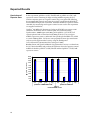
1
BLOCK-iT™ Pol II miR RNAi Expression Vector Kits Gateway®-adapted expression vector for the expression of microRNA (miRNA) in mammalian cells under control of Pol II promoters Catalog nos. K4935-00, K4936-00, K4937-00, K4938-00 Version F December 29, 2010 25-0857 Corporate Headquarters Invitrogen Corporation 1600 Faraday Avenue Carlsbad, CA 92008 T: 1 760 603 7200 F: 1 760 602 6500 E: [email protected] For country-specific contact information visit our web site at www.invitrogen.com User Manual ii Table of Contents Table of Contents . ................................................................................................................................................ iii Expression Clone Generation for Experienced Users . ..................................................................................... v Kit Contents and Storage . ................................................................................................................................... vi Accessory Products. ...............................................................................................................................................x Introduction . .................................................................................................................. 1 Overview. ................................................................................................................................................................1 Using miRNA for RNAi Analysis . ......................................................................................................................5 BLOCK-iT™ Pol II miR RNAi Expression Vector Kits . .....................................................................................9 Green Fluorescent Protein . .................................................................................................................................11 Experimental Outline . .........................................................................................................................................13 Methods . ...................................................................................................................... 14 Designing the Single-Stranded DNA Oligos. ...................................................................................................14 Generating the Double-Stranded Oligo . ...........................................................................................................21 Performing the Ligation Reaction. .....................................................................................................................26 Transforming One Shot® TOP10 Competent E. coli . .......................................................................................28 Analyzing Transformants. ..................................................................................................................................29 Transfecting Cells . ...............................................................................................................................................32 Detecting Fluorescence . ......................................................................................................................................35 Generating a Stable Cell Line . ............................................................................................................................36 Chaining pre-miRNAs . .......................................................................................................................................38 Removing EmGFP Coding Sequence. ...............................................................................................................40 Transferring the Pre-miRNA Expression Cassette to Destination Vectors. .................................................41 Performing the Rapid BP/LR Recombination Reaction. ................................................................................43 Expected Results . .................................................................................................................................................46 Troubleshooting . ..................................................................................................................................................49 Appendix. ..................................................................................................................... 54 Blasticidin...............................................................................................................................................................54 Recipes....................................................................................................................................................................55 Performing the Rapid BP/LR Recombination Reaction for Multisite Gateway® Vectors .........................56 Map and Features of pcDNA™6.2-GW/miR . ...................................................................................................59 Map and Features of pcDNA™6.2-GW/ EmGFP-miR . ...................................................................................60 Map and Features of pcDNA™6.2-GW/miR-neg control plasmid. ...............................................................61 Map and Features of pcDNA™6.2-GW/EmGFP-miR-neg control plasmid.................................................62 Explanation of Features of pcDNA™6.2-GW/± EmGFP-miR. .......................................................................63 Map of pcDNA™1.2/V5-GW/lacZ . ...................................................................................................................64 Technical Service. .................................................................................................................................................65 Purchaser Notification . .......................................................................................................................................66 Gateway® Clone Distribution Policy . ................................................................................................................69 References ..............................................................................................................................................................70 iii iv Expression Clone Generation for Experienced Users Introduction This quick reference sheet is provided for experienced users of the BLOCK-iT™ Pol II miR RNAi Expression Vector Kits. If you are performing the annealing, cloning, or transformation procedures for the first time, follow the detailed protocols provided in the manual. Step Action Design single-stranded DNA oligos Follow the guidelines on pages 14-18 to design single-stranded DNA oligos encoding the pre-miRNA of interest. Anneal the single-stranded oligos to generate a ds oligo 1. 2. 3. 4. 5. Clone the ds oligo into pcDNA™6.2-GW/miR or pcDNA™6.2-GW/EmGFPmiR 1. 2. 3. Transform One Shot® TOP10 1. Chemically Competent E. coli 2. 3. 4. 5. 6. Set up the following annealing reaction. 200 µM top strand oligo 5 µl 200 µM bottom strand oligo 5 µl 10X Oligo Annealing Buffer 2 µl DNase/RNase-free water 8 µl Total volume 20 µl Heat the reaction mixture at 95°C for 4 minutes. Remove the sample and set on the laboratory bench. Allow the reaction to cool to room temperature for 5-10 minutes. Spin down the sample in a microcentrifuge for 5 seconds. Mix gently. Dilute the ds oligo mixture 5,000-fold by performing serial 100-fold and 50-fold dilutions: the first into DNase/RNase-free water and the second into 1X Oligo Annealing Buffer. Final concentration is 10 nM. Set up the following ligation reaction. 5X Ligation Buffer 4 µl ™ pcDNA 6.2-GW/± EmGFP-miR (5 ng/µl), linearized 2 µl ds oligo (10 nM; 1:5,000 dilution) 4 µl DNase/RNase-Free water 9 µl T4 DNA Ligase (1 U/µl) 1 µl Total volume 20 µl Mix reaction well and incubate for 5 minutes at room temperature. Place reaction on ice and proceed to transform E. coli, below. Add 2 µl of the ligation reaction into a vial of One Shot® TOP10 chemically competent E. coli and mix gently. Incubate on ice for 5 to 30 minutes. Heat-shock the cells for 30 seconds at 42°C without shaking. Immediately transfer the tube to ice. Add 250 µl of room temperature S.O.C. Medium. Incubate at 37°C for 1 hour with shaking. Spread 20-100 µl of bacterial culture on a pre-warmed LB agar plate containing 50 µg/ml spectinomycin and incubate overnight at 37°C. v Kit Contents and Storage Types of Kits This manual is supplied with the products listed below. Product Catalog no. BLOCK-iT™ Pol II miR RNAi Expression Vector Kit K4935-00 BLOCK-iT™ Pol II miR RNAi Expression Vector Kit with EmGFP K4936-00 BLOCK-iT™ Lentiviral Pol II miR RNAi Expression System K4937-00 ™ BLOCK-iT Lentiviral Pol II miR RNAi Expression System with EmGFP Kit Components K4938-00 The BLOCK-iT™ Pol II miR RNAi Expression Vector Kits and BLOCK-iT™ Lentiviral Pol II miR RNAi Expression Systems include the following components. For a detailed description of the contents of the BLOCK-iT™ miRNA Expression Vector Kits, see pages vii-ix. For a detailed description of the contents of the BLOCK-iT™ Lentiviral Pol II miR RNAi Expression reagents, see the BLOCK-iT™ Lentiviral Pol II miR RNAi Expression System manual. Component Catalog no. K4935-00 K4936-00 K4937-00 K4938-00 ™ BLOCK-iT Pol II miR RNAi Expression Vector Kit √ BLOCK-iT™ Pol II miR RNAi Expression Vector Kit with EmGFP BLOCK-iT™ Lentiviral Pol II miR RNAi Expression Reagents Shipping/Storage √ √ √ √ √ The BLOCK-iT™ miRNA Expression Vector Kits are shipped as described below. Upon receipt, store each item as detailed below. Note: For information about the BLOCK-iT™ Lentiviral Pol II miR RNAi Expression Reagents (Box 3-11) supplied with the BLOCK-iT™ Lentiviral Pol II miR RNAi Expression Systems, refer to the BLOCK-iT™ Lentiviral Pol II miR RNAi Expression System manual. Box Component ™ Shipping Storage 1 BLOCK-iT Pol II miR RNAi Expression Vector Reagents or BLOCK-iT™ Pol II miR RNAi Expression Vector Reagents with EmGFP Dry ice -20°C 2 One Shot® TOP10 Chemically Competent E. coli Dry ice -80°C Continued on next page vi Kit Contents and Storage, continued BLOCK-iT™ Pol II miR RNAi Expression Vector Reagents The following reagents are included with the BLOCK-iT™ Pol II miR RNAi Expression Vector Reagents or the BLOCK-iT™ Pol II miR RNAi Expression Vector Reagents with EmGFP (Box 1). Store the reagents at -20°C. Reagent Composition Amount pcDNA 6.2-GW/miR, linearized or pcDNA™6.2-GW/EmGFPmiR, linearized 5 ng/µl in: 10 mM Tris-HCl, pH 8.0 1 mM EDTA, pH 8.0 4 x 10 µl 10X Oligo Annealing Buffer 100 mM Tris-HCl, pH 8.0 10 mM EDTA, pH 8.0 1 M NaCl 250 µl DNase/RNase-Free Water -- 3 x 1.5 ml 5X Ligation Buffer 250 mM Tris-HCl, pH 7.6 50 mM MgCl2 5 mM ATP 5 mM DTT 25% (w/v) polyethylene glycol-8000 80 µl T4 DNA Ligase 1 (Weiss) U/µl in 10 mM Tris-HCl, pH 7.5 50 mM KCl 1 mM DTT 50% (v/v) glycerol 20 µl miRNA forward sequencing primer or EmGFP forward sequencing primer 100 ng/µl in TE Buffer, pH 8.0 20 µl miRNA reverse sequencing primer 100 ng/µl in TE Buffer, pH 8.0 20 µl miR-lacZ positive doublestranded (ds) control oligo 50 µM in 1X Oligo Annealing Buffer 4 µl pcDNA™1.2/V5-GW/lacZ control plasmid 500 ng/µl in TE Buffer, pH 8.0 20 µl pcDNA™6.2-GW/miR-neg control plasmid or pcDNA™6.2-GW/EmGFPmiR-neg control plasmid 500 ng/µl in TE Buffer, pH 8.0 20 µl ™ Continued on next page vii Kit Contents and Storage, continued Unit Definition of T4 DNA Ligase One (Weiss) unit of T4 DNA Ligase catalyzes the exchange of 1 nmol 32P-labeled pyrophosphate into [γ/β-32P]ATP in 20 minutes at 37°C (Weiss et al., 1968). One unit is equal to approximately 300 cohesive-end ligation units. Primer Sequences The table below provides the sequence and the amount of the primers included in the kit. Primer Sequence Amount miRNA forward sequencing primer or EmGFP forward sequencing primer 5′- TCCCAAGCTGGCTAGTTAAG –3’ miRNA reverse sequencing primer 5′- CTCTAGATCAACCACTTTGT -3′ or 5′- GGCATGGACGAGCTGTACAA -3′ 2 µg (327 pmol) or 2 µg (323 pmol) 2 µg (332 pmol) LacZ Control Oligo The sequences of the miR-lacZ positive ds control oligo are listed below. The miRlacZ positive ds control oligo are annealed and are supplied in the kit as a 50 µM Sequences double-stranded oligo. The miR-lacZ positive ds control oligo needs to be reannealed and diluted 5000-fold to 10 nM (see page 22) before use in the ligation reaction (page 26). LacZ DNA Oligo Sequence Top strand 5’-TGCTGAAATCGCTGATTTGTGTAGTCGTTTTGGCCACTGACTGACGACTACACATCAGCGATTT-3’ Bottom strand 5’-CCTGAAATCGCTGATGTGTAGTCGTCAGTCAGTGGCCAAAACGACTACACAAATCAGCGATTTC-3’ Continued on next page viii Kit Contents and Storage, continued One Shot® TOP10 Reagents The following reagents are included in the One Shot® TOP10 Chemically Competent E. coli kit (Box 2). Transformation efficiency is ≥ 1 x 10⁹ cfu/µg plasmid DNA. Store Box 2 at -80°C. Reagent Composition Amount S.O.C. Medium (may be stored at +4°C or room temperature) 2% Tryptone 0.5% Yeast Extract 10 mM NaCl 2.5 mM KCl 10 mM MgCl2 10 mM MgSO4 20 mM glucose 6 ml TOP10 cells -- 21 x 50 µl pUC19 Control DNA 10 pg/µl in 5 mM Tris-HCl, 0.5 mM 50 µl EDTA, pH 8 Genotype of TOP10 Cells F- mcrA ∆(mrr-hsdRMS-mcrBC) φ80lacZ∆M15 ∆lacX74 recA1 araD139 ∆(ara-leu)7697 galU galK rpsL (StrR) endA1 nupG BLOCK-iT™ Lentiviral RNAi Expression Reagents In addition to the BLOCK-iT™ Pol II miR RNAi Expression Vector Kits and the BLOCK-iT™ Lentiviral Pol II miR RNAi Expression Vector Systems also include the following components to facilitate production of a replication-incompetent lentivirus that expresses your microRNA (miRNA) of interest. • pLenti6/V5-DEST Gateway® Vector • Gateway® LR Clonase™ II Enzyme Mix • Gateway® BP Clonase™ II Enzyme Mix • One Shot® Stbl3™ Chemically Competent E. coli • ViraPower™ Bsd Lentiviral Support Kit • 293FT Cell Line • pDONR™221 Refer to the BLOCK-iT™ Lentiviral Pol II miR RNAi Expression System manual supplied with Catalog nos. K4937-00 and K4938-00 for a detailed description of the lentiviral expression reagents provided with the kit and instructions to produce lentivirus. For instructions to grow and maintain the 293FT Cell Line, refer to the 293FT Cell Line manual, available for downloading from our Web site (www.invitrogen.com) or by contacting Technical Service (see page 65). ix Accessory Products Introduction The products listed in this section may be used with the BLOCK-iT™ Pol II miR RNAi Expression Vector Kits. For more information, refer to our Web site (www.invitrogen.com) or call Technical Service (see page 65). Accessory Products Some of the reagents supplied in the BLOCK-iT™ Pol II miR RNAi Expression Vector Kits as well as other products suitable for use with the kit are available separately from Invitrogen. Ordering information is provided below. Item ™ Amount Catalog no. BLOCK-iT Lentiviral Pol II miR RNAi Expression Vector System 20 reactions K4937-00 BLOCK-iT™ Lentiviral Pol II miR RNAi Expression System with EmGFP 20 reactions K4938-00 BLOCK-iT™ Pol II miR-XXXX Validated miRNA DuoPak (XXXX=gene symbol) 10 µg V49300-01 through V49300-53 BLOCK-iT™ miR RNAi Select 50 nmol scale See page xi T4 DNA Ligase 100 units 500 units 15224-017 15224-025 One Shot® TOP10 Chemically Competent E. coli 10 reactions 20 reactions 40 reactions C4040-10 C4040-03 C4040-06 PureLink™ HQ Mini Plasmid Purification Kit 100 preps K2100-01 PureLink™ HiPure Plamid Midiprep Kit 25 preps K2100-04 0.75 ml 1.5 ml 11668-027 11668-019 ™ Lipofectamine 2000 Transfection Reagent Lipofectamine™ LTX Reagent 1.0 ml 15338-100 Opti-MEM® I Reduced Serum Medium 100 ml 500 ml 31985-062 31985-070 Phosphate-Buffered Saline (PBS), pH 7.4 500 ml 10010-023 ® 9 gels and Base G6000-04 ® 2% E-Gel Starter Pak 9 gels and Base G6000-02 10 bp DNA Ladder 50 µg 10821-015 293FT Cell Line 3 x 10⁶ cells, frozen R700-07 Blasticidin 50 mg R210-01 50 preps K2100-12 Gateway LR Clonase II Enzyme Mix 20 reactions 100 reactions 11791-020 11791-100 Gateway® BP Clonase™ II Enzyme Mix 20 reactions 100 reactions 11789-020 11789-100 pDONR™221 6 µg 12536-017 20 reactions 12538-013 4% E-Gel Starter Pak ™ Purelink Quick Gel Extraction Kit ® ™ ™ LR Clonase Plus Enzyme Mix Continued on next page x Accessory Products, Continued Spectinomycin For selection of pcDNA™6.2-GW/± EmGFP-miR transformants in E. coli, you will need to obtain spectinomycin. Spectinomycin Dihydrochloride is available from Sigma (Catalog no. S4014). For a recipe to prepare spectinomycin for use, see page 55. RNAi Designer and RNAi Express The BLOCK-iT™ RNAi Designer is an online tool to help you design and order microRNA sequences for any target gene of interest. The RNAi Designer incorporates the guidelines provided in this manual as well as other design rules into a proprietary algorithm to design microRNA sequences that are compatible for use in cloning into the BLOCK-iT™ Pol II miR RNAi Expression Vectors. BLOCK-iT™ miR RNAi Select Invitrogen has predesigned miR RNAi sequences, called BLOCK-iT™ miR RNAi Select, targeting >70% of the human, mouse and rat RefSeq genes. BLOCK-iT™ miR RNAi Select provides up to 4 miR sequences per gene that are supplied as 8 tubes containing 4 top oligos and 4 bottom DNA oligos. Upon annealing and cloning into one of the BLOCK-iT™ Pol II miR RNAi Expression vectors, pcDNA™6.2-GW/miR or pcDNA™6.2-GW/EmGFP-miR, these oligos generate up to four different miR RNAi expression vectors directed against your gene of interest. The resulting miR RNAi expression vectors can be transfected into cells to knock down the gene of interest, or the hairpins can be transferred into lenti vectors to knock down the gene of interest in hard-to-transfect or primary cells. We guarantee that at least two out of the four miR RNAi expression vectors will result in >70% knockdown of the target gene (provided that the transfection efficiency in your experiment is at least 80%). Order BLOCK-iT™ miR RNAi Select online using the BLOCK-iT™ RNAi Express search engine (www.invitrogen.com/rnaiexpress). Just enter the gene name, accession number, or keyword, and choose your desired BLOCK-iT™ miR RNAi Select. BLOCK-iT™ RNAi Products A large variety of BLOCK-iT™ RNAi products are available from Invitrogen to facilitate RNAi analysis including Stealth™ RNAi, Validated Stealth™ RNAi Collection, Validated miRNA Vector Collection, and a large selection of RNAi vectors. For details, visit the RNAi Central portal or contact Technical Service (page 65). Continued on next page xi Accessory Products, Continued Gateway® Destination Vectors A large selection of Gateway® destination vectors are available from Invitrogen to facilitate the transfer of the pre-miRNA sequence into a suitable destination vector to allow the miRNA expression in multiple systems including viral expression systems and tissue-specific expression. See below for a list of compatible destination vectors. Destination Vector Catalog No. ™ V496-10 pLenti6/V5-DEST ™ pLenti6/UbC/V5-DEST V499-10 pEF-DEST51 12285-011 ™ 12301-016 pT-REx -DEST30 ™ ™ pEF5/FRT/V5-DEST (Flp-In ) ™ pDEST /R4-R3 V6020-20 12567-023 ™ pLenti6/R4R2/V5-DEST K591-10 N-terminal reporter tag vectors , e.g.: pcDNA™6.2/nGeneBLAzer™-DEST ™ pcDNA 6.2/N-YFP-DEST 12578-068, 12578-050 V358-20 Note: Transferring the pre-miRNA expression cassette from pcDNA™6.2-GW/± EmGFPmiR to the pLenti6/BLOCK-iT™-DEST destination vector will not yield a functional miRNA expression vector. Expression of the pre-miRNA requires the destination vector to supply a Pol II promoter. xii Introduction Overview Introduction The BLOCK-iT™ Pol II miR RNAi Expression Vector Kits facilitate the expression of microRNA (miRNA) for use in RNA interference (RNAi) analysis of a target gene in mammalian cells. The kits provide a Gateway®-adapted expression vector designed to allow efficient transient or stable expression of miRNA. If more specialized expression is required, the vector allows easy recombination with other suitable destination vectors allowing tissue-specific, regulated, or lentiviral expression of the miRNA in mammalian cells. Note: The BLOCK-iT™ Lentiviral Pol II miR RNAi Expression Systems include the BLOCKiT™ Pol II miR RNAi Expression Vector Kits as well as the pLenti6/V5-DEST destination vector and other reagents required to generate a lentiviral RNAi construct. For more information about the pLenti6/V5-DEST vector and how to generate lentivirus, refer to the BLOCK-iT™ Lentiviral Pol II miR RNAi Expression System manual. This manual is supplied with the BLOCK-iT™ Lentiviral RNAi Expression System, but is also available for downloading from our Web site (www.invitrogen.com) or by contacting Technical Service (see page 65). System Components The BLOCK-iT™ Pol II miR RNAi Expression Vector Kits include: • pcDNA™6.2-GW/miR or pcDNA™6.2-GW/EmGFP-miR (collectively referred to as pcDNA™6.2-GW/± EmGFP-miR) • Reagents for production of an expression clone containing a doublestranded oligonucleotide (ds oligo) encoding a pre-miRNA (oligos have to be ordered separately). • Positive and negative controls for the generation and use of the expression clone. Note: The BLOCK-iT™ Lentiviral Pol II miR RNAi Expression Systems additionally include components for Gateway® recombination and lentiviral production. Refer to the BLOCK-iT™ Lentiviral Pol II miR RNAi Expression System manual, supplied with the BLOCK-iT™ Lentiviral Pol II miR RNAi Expression System, available for downloading from our Web site (www.invitrogen.com), or available from Technical Service (see page 65). Continued on next page 1 Overview, continued Advantages of the BLOCK-iT™ Pol II miR RNAi Expression Vector Kits The BLOCK-iT™ Pol II miR RNAi Technology Using the BLOCK-iT™ Pol II miR RNAi Expression Vector Kits for vector-based expression of miRNA provides the following advantages: • Offers a rapid and efficient way to clone ds oligo duplexes encoding a desired miRNA target sequence into a vector containing a Pol II promoter for use in RNAi analysis. • Allows transient or stable expression of miRNA into mammalian cells. • Enables targeting multiple genes or increasing knockdown of a single target gene with one construct. • Permits visual or automated selection of cells expressing the pre-miRNA through co-cistronic expression of EmGFP( in the BLOCK-iT™ Pol II miR RNAi Expression Vector with EmGFP) • Offers easy transfer of the pre-miRNA expression cassette into Gateway®adapted viral expression systems or vectors driven by a variety of promoters, including tissue-specific and regulated promoters for in vivo experiments • Permits design of predictable RNAi constructs with a high rate of success • In conjunction with the pre-designed BLOCK-iT™ miR RNAi Select oligos, covers >70% of the human, mouse and rat RefSeq genes with a guaranteed rate of success The BLOCK-iT™ Pol II miR RNAi Technology is a next generation RNAi technology employing miRNA expression vectors that allow flexible expression of knockdown cassettes driven by RNA Polymerase II (Pol II) promoters in mammalian cells. See page 5 for more details. The BLOCK-iT™ Pol II miR RNAi Expression Vectors are specifically designed to allow expression of miRNA sequences and contain specific miR flanking sequences that allow proper processing of the miRNA. The expression vector design is based on the miRNA vector system developed in the laboratory of David Turner (U.S. Patent Publication No. 2004/0053876) and includes the use of endogenous murine miR-155 flanking sequences (see page 7 for details). A variety of BLOCK-iT™ RNAi products are available from Invitrogen to facilitate RNAi analysis in mammalian and invertebrate systems. For more information about any of the BLOCK-iT™ RNAi products, see the RNAi Central application portal at www.invitrogen.com/rnai or contact Technical Service (see page 65). Continued on next page 2 Overview, Continued Alternative Expression Systems The pcDNA™6.2-GW/± EmGFP-miR vectors express the pre-miRNA in most mammalian cells at a high, constitutive level using the human cytomegalovirus (CMV) immediate early promoter. If different expression of the pre-miRNA is required, such as tissue-specific, regulated or lentiviral expression, the vector allows easy recombination with other suitable destination vectors using Gateway® Technology. Gateway® Technology Gateway® Technology is a universal cloning method that takes advantage of the site-specific recombination properties of bacteriophage lambda (Landy, 1989) to provide a rapid and highly efficient way to move your DNA sequence of interest (the miRNA sequence) into multiple vector systems. To transfer your pre-miRNA expression cassette (see page 8) into the destination vector, first generate an entry clone by performing a BP recombination reaction between the pcDNA™6.2-GW/± EmGFP-miR expression clone and a suitable donor vector (such as pDONR™221), then perform an LR recombination reaction between the resulting entry clone and a destination vector of choice. See page 40 for more details. For more in depth information about the Gateway® Technology, refer to the Gateway® Technology with Clonase™ II manual which is available from our web site (www.invitrogen.com) or by contacting Technical Service (see page 65). Purpose of this Manual This manual provides the following information: • An overview of the pathway by which miRNA facilitates gene knockdown in mammalian cells. • Rules to design the appropriate single-stranded oligonucleotides representing the target gene. • Instructions to anneal the single-stranded oligonucleotides to generate a double-stranded oligonucleotide (ds oligo). • Instructions to clone the ds oligo into the pcDNA™6.2-GW/± EmGFP-miR vector, and transform the ligation reaction into competent E. coli. • Guidelines to transfect your pcDNA™6.2-GW/± EmGFP-miR construct into mammalian cells for transient analysis or to generate stable cell lines. • Guidelines to perform the chaining reaction to enable co-cistronic expression of multiple pre-miRNAs from one construct. • Guidelines for detection of the EmGFP expressed from pcDNA™6.2-GW/ EmGFP-miR • Information to perform a Rapid BP/LR recombination reaction with a suitable Gateway® destination vector to generate an expression clone. Continued on next page 3 Overview, continued Important The BLOCK-iT™ Pol II miR RNAi Expression Vector Kits are designed to help you generate a CMV promoter-based vector to express miRNA in mammalian cell lines for RNAi analysis. Although the kit has been designed to help you express miRNA representing a particular target sequence in the simplest, most direct fashion, use of the kit for RNAi analysis assumes that users are familiar with the principles of gene silencing, vector-based production of miRNA, transfection in mammalian systems, and cloning. We highly recommend that users possess a working knowledge of the RNAi pathway and lipid-mediated transfection. For more information about miRNA and the RNAi pathways and expression of miRNA in mammalian cells, refer to published references (Ambros, 2004; Bartel, 2004; Boden et al., 2004; Cullen, 2004; Kim, 2005; McManus & Sharp, 2002; Zeng et al., 2002). Refer to Molecular Biology handbooks, such as Current Protocols in Molecular Biology (Ausubel et al., 1994), if you are not familiar with the cloning steps involved 4 Using miRNA for RNAi Analysis Introduction RNA interference (RNAi) describes the phenomenon by which short, homologous RNA duplexes induce potent and specific inhibition of eukaryotic gene expression via the degradation of complementary messenger RNA (mRNA), and is functionally similar to the processes of post-transcriptional gene silencing (PTGS) or cosuppression in plants (Cogoni et al., 1994; Napoli et al., 1990; Smith et al., 1990; van der Krol et al., 1990) and quelling in fungi (Cogoni & Macino, 1997; Cogoni & Macino, 1999; Romano & Macino, 1992). In plants, the PTGS response is thought to occur as a natural defense against viral infection or transposon insertion (Anandalakshmi et al., 1998; Jones et al., 1998; Li & Ding, 2001; Voinnet et al., 1999). In experimental settings, RNAi is widely used to silence genes through transfection of RNA duplexes or introduction of vectorexpressed short hairpin RNA (shRNA). The RNAi Pathway In eukaryotic organisms, dsRNA produced in vivo, introduced by pathogens, or through research, is processed into 21-23 nucleotide double-stranded short interfering RNA duplexes (siRNA) by an enzyme called Dicer, a member of the RNase III family of double-stranded RNA-specific endonucleases (Bernstein et al., 2001; Ketting et al., 2001). Each siRNA then incorporates into an RNA-induced silencing complex (RISC), an enzyme complex that serves to target cellular transcripts complementary to the siRNA for specific cleavage and degradation, or translational repression (Hammond et al., 2000; Nykanen et al., 2001). MicroRNAs (miRNAs) are endogenous RNAs that trigger gene silencing (Ambros, 2001; Carrington & Ambros, 2003). miRNA Pathway MicroRNAs (miRNAs) are endogenously expressed small ssRNA sequences of ~22 nucleotides in length which naturally direct gene silencing through components shared with the RNAi pathway (Bartel, 2004). Unlike shRNAs, however, the miRNAs are found embedded, sometimes in clusters, in long primary transcripts (pri-miRNAs) of several kilobases in length containing a hairpin structure and driven by RNA Polymerase II (Lee et al., 2004), the polymerase also responsible for mRNA expression. Drosha, a nuclear RNase III, cleaves the stem-loop structure of the pri-miRNA to generate small hairpin precursor miRNAs (pre-miRNAs) which are ~70 nucleotides in length (Zeng et al., 2005). The pre-miRNAs are exported from the nucleus to the cytoplasm by exportin-5, a nuclear transport receptor(Bohnsack et al., 2004; Yi et al., 2003). Following the nuclear export, the pre-miRNAs are processed by Dicer into a ~22 nucleotides miRNA (mature miRNA) molecule, and incorporated into an miRNA-containing RNA-induced silencing complex (miRISC)(Cullen, 2004). Continued on next page 5 Using miRNA for RNAi Analysis, Continued The mature miRNAs regulate gene expression by mRNA cleavage (mRNA is Translational Repression versus nearly complementary to the miRNA) or translational repression (mRNA is not sufficiently complementary to the miRNA). Target cleavage can be induced Target Cleavage artificially by altering the target or the miRNA sequence to obtain complete hybridization (Zeng et al., 2002). In animals, most miRNAs imperfectly complement their targets and interfere with protein production without directly inducing mRNA degradation (Ambros, 2004). Nonetheless, these miRNAs are found associated with the RNAi nuclease AGO2 (Liu et al., 2004; Meister et al., 2004), and at least two miRNAs with close matches to their target sequences, particularly in their 5’ regions, have been shown to cleave cognate mRNAs (Yekta et al., 2004; Yu et al., 2005). The engineered miRNAs produced by the BLOCK-iT™ Pol II miR RNAi Expression Vector Kits (see below) fully complement their target site and cleave the target mRNA. Sequence analysis showed that the primary cleavage site at the phosphodiester bond in the mRNA found opposite the tenth and eleventh bases of the engineered miRNA as predicted for RNAi-mediated cleavage (Elbashir et al., 2001) similar to siRNA mediated cleavage. Using a VectorBased System to Express Engineered miRNA Use of siRNA (diced siRNA or synthetic siRNA) for RNAi analysis in mammalian cells is limited by their transient nature. To address this limitations, a number of groups have developed vector-based systems to facilitate expression of engineered short hairpin RNA (shRNA) sequences in mammalian cells using Pol III promoters (Brummelkamp et al., 2002; Paddison et al., 2002; Paul et al., 2002; Sui et al., 2002; Yu et al., 2002). However, the use of shRNA vectors for RNAi analysis requires the screening of large number of sequences to identify active sequences and the use of Pol III promoters limits applications such as tissue-specific expression. To overcome limitations with siRNA and shRNA, we have developed Gateway®adapted expression vectors that enable the expression of engineered miRNA sequences from Pol II promoters. The pcDNA™6.2-GW/± EmGFP-miR expression vectors facilitate the generation of an expression clone containing a ds oligo encoding a pre-miRNA sequence (see page 8). The resulting expression construct may be introduced into mammalian cells for transient expression of the miRNA sequence, or stable transfectants can be generated. If desired, the pre-miRNA sequence may be easily and efficiently transferred into the pLenti6/V5-DEST vector or other suitable destination vector by Gateway® recombination reactions (see page 40). Continued on next page 6 Using miRNA for RNAi Analysis, Continued Types of miRNA Vectors The BLOCK-iT™ Pol II miR RNAi Expression Vector Kits are supplied with one of the following expression vectors that allow the expression of your engineered premiRNA: • pcDNA™6.2-GW/miR Allows expression of the engineered pre-miRNA under the control of the strong, Pol II human CMV (cytomegalovirus) promoter and Herpes Simplex virus (HSV) thymidine kinase (TK) polyadenylation signal • pcDNA™6.2-GW/EmGFP-miR This vector is similar to pcDNA6.2™-GW/miR, except the coding sequence of EmGFP (Emerald Green Fluorescent Protein) is incorporated into the vector such that the pre-miRNA insertion site is in the 3’ untranslated (3’UTR) region of the fluorescent protein mRNA. Addition of EmGFP allows tracking of the miRNA expression and provides strong correlation of EmGFP expression with the knockdown of the target gene by your miRNA. Human CMV Promoter The BLOCK-iT™ Pol II miR RNAi Expression Vectors contain the human cytomegalovirus (CMV) immediate early promoter to allow high-level, constitutive miRNA expression in mammalian cells (Andersson et al., 1989; Boshart et al., 1985; Nelson et al., 1987). We have chosen the human CMV promoter to control vector-based expression of miRNA molecules in mammalian cells for the following reasons: • The promoter is recognized by RNA Polymerase II and controls high-level, constitutive expression of miRNA and co-cistronic reporter genes • The promoter is active in most mammalian cell types Note: Although highly active in most mammalian cell lines, activity of the viral CMV promoter can be down-regulated in some cell lines due to methylation (Curradi et al., 2002), histone deacetylation (Rietveld et al., 2002), or both. Design of the Engineered PremiRNA The engineered pre-miRNA sequence structure is based on the murine miR-155 sequence (Lagos-Quintana et al., 2002). The 5’ and 3’ flanking regions derived from the miR-155 transcript were inserted in the vector to preserve as much as possible of the miR-155 structure. We optimized the stem-loop structure and a 2 nucleotide internal loop results in higher knockdown rate than the 5 nucleotide / 3 nucleotide internal loop found in native miR-155 molecule. An Msc I site was incorporated in the terminal loop to aid in sequence analysis. Below the changes are shown made to the native miR-155 to form an engineered premiRNAs directed against lacZ (targeting sequence in bold). native miR-155 5’-UG| UGUGA UUGGCC CUGUUAAUGCUAAU UAGGGGUU \ |||||||||||||: ||||:||: U GACAAUUACGAUUG AUCCUCAG / 3’-G^ UCC-UCAGUC internal loop terminal loop optimized miR-lacZ MscI UG| UU UUGGCC CUGAAAUCGCUGAU GUGUAGUCGUU \ |||||||||||||| ||||||||||: A GACUUUAGCGACUA--CACAUCAGCAG / AG^ UCAGUC internal loop terminal loop Continued on next page 7 Using miRNA for RNAi Analysis, Continued Structure of the Engineered PremiRNA The pcDNA™6.2-GW/± EmGFP-miR vectors are designed to accept engineered pre-miRNA sequences targeting your gene of interest. The engineered premiRNA sequence structure is based on the murine miR-155 sequence and the stem-loop structure was optimized to obtain a high knockdown rate as described on the previous page. For optimized knockdown results, we recommend that the ds oligo encoding the engineered pre-miRNA have the following structural features: • A 4 nucleotide, 5’ overhang (TGCT) complementary to the vector (required for directional cloning) • A 5’G + short 21 nucleotide antisense sequence (mature miRNA) derived from the target gene, followed by • A short spacer of 19 nucleotides to form the terminal loop and • A short sense target sequence with 2 nucleotides removed (∆2) to create an internal loop • A 4 nucleotide, 5’ overhang (CAGG) complementary to the vector (required for directional cloning) The structural features are depicted in the figure below. TGCT overhang 5G + antisense Loop Loop sequence Sense D2 nt CAGG overhang target sequence target sequence For guidelines to design the oligonucleotides, refer page 14. We recommend using Invitrogen’s RNAi Designer at www.invitrogen.com/rnai, an online tool to help you design and order pre-miRNA sequences for any target gene of interest. Pre-miRNA Expression Cassette The engineered pre-miRNA sequence is cloned into the cloning site of BLOCK-iT™ Pol II miR RNAi Expression Vectors that is flanked on either side with sequences from murine miR-155 to allow proper processing of the engineered pre-miRNA sequence (see page 18-19 for the flanking region sequences). The pre-miRNA sequence and adjacent miR-155 flanking regions are denoted as the pre-miRNA expression cassette and are shown below. This expression cassette is transferred between vectors during Gateway® recombination reactions. EmGFP 5 miR flanking region 3 miR flanking 5G + antisense Loop Loop sequence Sense D2 nt target sequence target sequence region 5 miR flanking region 3 miR flanking 5G + antisense Loop Loop sequence Sense D2 nt target sequence target sequence region attB2 Once the engineered pre-miRNA expression cassette is introduced into the mammalian cells for expression, the pre-miRNA forms an intramolecular stemloop structure similar to the structure of endogenous pre-miRNA that is then processed by the endogenous Dicer enzyme into a 22 nucleotide mature miRNA. Note: The 21 nucleotides are derived from the target sequence while the 3’ most nucleotide is derived from the native miR-155 sequence (see figure on page 18-19). Chaining of miRNAs 8 miRNAs are sometimes expressed in clusters in long primary transcripts driven by RNA Pol II (Lee et al., 2004). Our vectors support chaining of miRNAs to express them in one primary transcript, thus ensuring co-cistronic expression of multiple miRNAs. See page 38 for details. BLOCK-iT™ Pol II miR RNAi Expression Vector Kits Description of the System The BLOCK-iT™ Pol II miR RNAi Expression Vector Kits facilitate the generation of an expression construct that permits high-level expression of a pre-miRNA in mammalian cells for RNAi analysis of a target gene. The kit contains the following major components: • • • Controls The pcDNA™6.2-GW/miR or pcDNA™6.2-GW/EmGFP-miR linearized plasmids into which a ds oligo encoding the pre-miRNA will be cloned to generate an expression clone that contains the elements required for expression of the miRNA in mammalian cells. The pcDNA™6.2-GW/± EmGFP-miR vector is supplied linearized with 4-nucleotide 5′ overhangs on each strand to facilitate directional cloning of the ds oligo insert. The resulting expression clone containing the pre-miRNA expression cassette (see page 8) may be transfected into mammalian cells for transient or stable RNAi analysis, or used to transfer the pre-miRNA expression cassette into a suitable destination vector using Gateway® Technology. T4 DNA Ligase and an optimized ligation buffer to allow 5-minute room temperature ligation of the ds oligo insert into the pcDNA™6.2-GW/± EmGFP-miR vector. One Shot® TOP10 Chemically Competent E. coli for high efficiency transformation of the ligation reaction. The BLOCK-iT™ Pol II miR RNAi Expression Vector Kits also includes a negative control plasmid and a ds positive control oligo. • The pcDNA™6.2-GW/± EmGFP-miR-neg control plasmid contains an insert that can form a hairpin structure that is processed into mature miRNA, but is predicted not to target any known vertebrate gene. Thus, this plasmid serves as a suitable negative control for pre-miRNA experiments with pcDNA™6.2GW/± EmGFP-miR expression vectors. The neg control sequence without 5’ overhangs is shown below (for map, see page 61-62): 5’-GAAATGTACTGCGCGTGGAGACGTTTTGGCCACTGACTGACGTCTCCACGCAGTACATTT-3’ • The miR-lacZ positive double-stranded (ds) control oligo serves as a positive control during the miRNA expression vector generation. Use this oligo to generate a pcDNA™6.2-GW/± EmGFP-miR-lacZ expression clone. • Co-transfecting the resulting pcDNA™6.2-GW/± EmGFP-miR-lacZ expression clone targeting the lacZ gene and the pcDNA™1.2/V5-GW/lacZ reporter plasmid supplied with the kit into mammalian cells provide a means to assess the RNAi response in your cell line by assaying for knockdown of βgalactosidase. Continued on next page 9 BLOCK-iT™ Pol II miR RNAi Expression Vector Kits, continued Generating an miRNA Expression Vector Using the Kit Features of the pcDNA™6.2-GW/± EmGFP-miR Vectors Using the reagents supplied in the BLOCK-iT™ Pol II miR RNAi Expression Vector Kits, you will perform the following steps to generate an expression clone in pcDNA™6.2-GW/± EmGFP-miR: 1. Design and synthesize two complementary single-stranded DNA oligonucleotides, with one encoding the miRNA of interest. Alternatively, order BLOCK-iT™ miR RNAi Select oligos targeting your gene(s) (see page xi). 2. Anneal the single-stranded oligonucleotides to generate a double-stranded oligo (ds oligo). 3. Clone the ds oligo into the linearized pcDNA™6.2-GW/± EmGFP-miR vector. 4. Transform the ligation reaction into One Shot® TOP10 chemically competent E. coli and select for spectinomycin-resistant transformants. 5. Use the pcDNA™6.2-GW/± EmGFP-miR expression construct for transient RNAi analysis in mammalian cells, isolate stable cell lines expressing the miRNA, or perform a Gateway® recombination reaction with a suitable Gateway® destination vector to generate a different expression clone. The pcDNA™6.2-GW/± EmGFP-miR Vectors contain the following features: • Human CMV promoter for high-level, constitutive expression of the miRNA from a RNA Polymerase II-dependent promoter • 5’ and 3’ miR flanking regions for formation of an engineered pre-miRNA • Cloning site containing 4-nucleotide 5′ overhangs on each DNA strand for directional cloning of the ds oligo encoding the pre-miRNA of interest • Two recombination sites, attB1 and attB2 sites, flanking the pre-miRNA expression cassette for recombinational cloning of the pre-miRNA expression cassette into a Gateway® destination vector • Herpes Simplex virus (HSV) thymidine kinase (TK) polyadenylation signal for termination and polyadenylation of the transcript • Spectinomycin resistance gene for selection in E. coli • pUC origin for high-copy maintenance of the plasmid in E. coli • Blasticidin resistance gene for selection in E. coli and mammalian cells to generate cell lines stably expressing the miRNA Additionally, the vector pcDNA™6.2-GW/EmGFP-miR also contains an EmGFP coding sequence for co-cistronic expression with the pre-miRNA. 10 Green Fluorescent Protein Description The BLOCK-iT™ Pol II miR RNAi Expression Vector with EmGFP contains the Emerald Green Fluorescent Protein (EmGFP) derived from Aequorea victoria GFP within the pre-miRNA expression cassette. After transferring the pre-miRNA expression cassette into pLenti6/V5-DEST, you may produce lentiviruses that simultaneously express the EmGFP protein and miRNA, allowing you to visually track the cells in which knockdown is occurring or sort the cells using a flow cytometer. Green Fluorescent Green Fluorescent Protein (GFP) is a naturally occurring bioluminescent protein derived from the jellyfish Aequorea victoria (Shimomura et al., 1962). GFP emits Protein (GFP) fluorescence upon excitation, and the gene encoding GFP contains all of the necessary information for posttranslational synthesis of the luminescent protein. GFP is often used as a molecular beacon because it requires no species-specific cofactors for function, and the fluorescence is easily detected using fluorescence microscopy and standard filter sets. GFP can function as a reporter gene downstream of a promoter of interest and upstream of one or more pre-miRNAs. GFP and Spectral Variants Modifications have been made to the wild-type GFP to enhance its expression in mammalian systems. These modifications include amino acid substitutions that correspond to the codon preference for mammalian use, and mutations that increase the brightness of the fluorescence signal, resulting in “enhanced” GFP (Zhang et al., 1996). Mutations have also arisen or have been introduced into GFP that further enhance and shift the spectral properties of GFP such that these proteins will emit fluorescent color variations (reviewed in Tsien, 1998). The Emerald GFP (EmGFP) is such a variant of enhanced GFP. We have observed reduced EmGFP expression from miRNA-containing vectors due to processing of the transcripts. In most cases, EmGFP expression should remain detectable. Continued on next page 11 Green Fluorescent Protein, Continued EmGFP The EmGFP variant has been described in a published review (Tsien, 1998) and is summarized below. The amino acid mutations are represented by the single letter abbreviation for the amino acid in the consensus GFP sequence, followed by the codon number and the single letter amino acid abbreviation for the substituted amino acid. Fluorescent Protein GFP Mutations* EmGFP S65T, S72A, N149K, M153T, I167T *Mutations listed are as described in the literature. When examining the actual sequence, the vector codon numbering starts at the first amino acid after the initiation methionine of the fluorescent protein, so that mutations appear to be increased by one position. For example, the S65T mutation actually occurs in codon 66 of EmGFP. EmGFP Fluorescence The EmGFP from the pcDNA™6.2-GW/EmGFP-miR expression vector has the following excitation and emission wavelengths, as published in the literature (Tsien, 1998): Excitation (nm) Emission (nm) 487 Filter Sets for Detecting EmGFP Fluorescence 509 The EmGFP can be detected with standard FITC filter sets. However, for optimal detection of the fluorescence signal, you may use a filter set which is optimized for detection within the excitation and emission ranges for the fluorescent protein. The filter set for fluorescence microscopy and the manufacturer are listed below: Filter Set Manufacturer Omega XF100 Omega (www.omegafilters.com) 12 Experimental Outline The figure below illustrates the major steps necessary to produce a pcDNA™6.2GW/miR or pcDNA™6.2-GW/EmGFP-miR expression clone using the BLOCKiT™ Pol II miR RNAi Expression Vector Kits. Flow Chart TGCTG CCTG + 1. Design and synthesize complementary DNA oligos, with each containing 4 nucleotide overhangs necessary for directional cloning. Alternatively, order miR Select oligos directed against your gene of interest C Annealing TGCTG C 2. Anneal DNA oligos to generate a ds oligo. GTCC Ligation V P CM CAGG 3 miR flanking attB2 region TK pA f1 attB1 V P CM or i or i n di SV40 p A i Bla sti ci C or EM7 5699 bp pU C i f1 ori 40 SV Bla sti ci pU or TK pA pcDNA6.2-GW/ EmGFP-miR c t i n o m y ci n or EM7 c t i n o m y ci n 4944 bp Sp e ori 40 SV Sp e pcDNA6.2-GW/miR CAGG 3 miR flanking attB2 region 5 miR flanking region ACGA EmGFP n di attB1 5 miR flanking region ACGA 3. Clone the ds oligo into pcDNA6.2-GW/miR or pcDNA6.2-GW/EmGFP-miR expression vector using T4 DNA Ligase. S V40 p A Transformation V P CM TK pA f1 V P CM TK pA f1 or i ori 40 SV C or i Bla sti ci 5759 bp SV40 p A Recombination with destination vector 4. Transform E. coli and analyze colonies for the desired expression clone. EM7 pcDNA6.2-GW/ EmGFP-miR-neg control plasmid pU n di Transfection c t i n o my ci n C SV40 p A or Sp e Bla sti ci pU i EM7 c t i n o my ci n ori 40 SV Sp e or 5 miR flanking ds miRNA 3 miR flanking attB2 region oligo region EmGFP or i pcDNA6.2-GW/ miR-neg control plasmid 5004bp attB1 n di attB1 5 miR flanking ds miRNA 3 miR flanking attB2 region oligo region 5. Transfect the expression clone for transient or stable RNAi analysis or recombine with a suitable destination vector to generate a different expression clone (through a donor vector intermediate) 13 Methods Designing the Single-Stranded DNA Oligos Introduction To use the BLOCK-iT™ Pol II miR RNAi Expression Vector Kits, you will first need to design two single-stranded DNA oligonucleotides; one encoding the target pre-miRNA (“top strand” oligo) and the other its complement (“bottom strand” oligo). You will then anneal the top and bottom strand oligos to generate a double-stranded oligonucleotide (ds oligo) suitable for cloning into the pcDNA™6.2-GW/± EmGFP-miR vector. MEND ION AT RECOM The design of the single-stranded oligonucleotides (ss oligos) is critical to the success of both the cloning procedure and ultimately, the RNAi analysis. General guidelines are provided in this section to help you choose the target sequence and to design the ss oligos. Note, however, that simply following these guidelines does not guarantee that the pre-miRNA will be effective in knocking down the target gene. For a given target gene, you may need to generate and screen multiple pre-miRNA sequences to identify one that is active in gene knockdown studies. We strongly recommend using Invitrogen’s RNAi Designer, an online tool to help you design and order pre-miRNA sequences for any target gene of interest. The RNAi Designer incorporates the guidelines provided in this manual as well as other design rules into a proprietary algorithm to design pre-miRNA sequences that are compatible for use in cloning into pcDNA™6.2-GW/± EmGFP-miR. Success rates exceeding 70% have been achieved with the RNAi Designer (i.e. more than 70% of designed miRNAs reduce target gene expression by at least 70%). To use the RNAi Designer, see www.invitrogen.com/rnai. Note: Invitrogen’s RNAi Designer is the only online tool for miRNA design at the moment this manual is printed. Other online RNAi designers not intended for miRNA will not necessarily design good pre-miRNA sequences. BLOCK-iT™ miR RNAi Select Invitrogen has pre-designed miR RNAi sequences, called BLOCK-iT™ miR RNAi Select, targeting >70% of the human, mouse and rat RefSeq genes with a guaranteed rate of success. See page xi for more details. Continued on next page 14 Designing the Single-Stranded DNA Oligos, continued Features of PremiRNA Insert When designing the oligos encoding the pre-miRNA, consider that a pre-miRNA insert contains the following features (from 5’ to 3’ end): • 5 nucleotides (TGCTG) derived from the endogenous miR-155, an endogenous murine miRNA that is the basis of the miRNA vector system developed in the laboratory of David Turner (Chung et al., 2006). This also provides a four nucleotide 5’ overhang, compatible with a 4 nucleotide overhang in the provided linearized pcDNA™6.2-GW/± EmGFP-miR to clone the double-stranded oligo. • Reverse complement of the 21-nucleotide target sequence (mature miRNA sequence). When transcribed, this is the core sequence that will target your gene of interest, and therefore needs to be antisense to the targeted messenger RNA. • 19 nucleotides derived from miR-155 to form a terminal loop with an engineered Msc I site to aid in sequence analysis. • Nucleotides 1-8 and 11-21 of the sense target sequence. Note that nucleotides 9 and 10 are removed to form a short internal loop in the mature miRNA, which results in more efficient knockdown. • 4 nucleotides derived from endogenous miR-155. This also constitutes the four nucleotide 5’ overhang, compatible with a 4 nucleotide overhang in the provided linearized pcDNA™6.2-GW/± EmGFP-miR to clone the doublestranded oligo. Upon transcription, the mature miRNA sequence and its complement form a stem of the pre-miRNA with a short internal loop, separated by a larger terminal loop. The folded pre-miRNA structure of miR-lacZ is shown below (lacZ targeting sequence in bold) UG| UU UUGGCC CUGAAAUCGCUGAU GUGUAGUCGUU \ |||||||||||||| ||||||||||: A GACUUUAGCGACUA CACAUCAGCAG / AG^ -UCAGUC Continued on next page 15 Designing the Single-Stranded DNA Oligos, continued Choosing the Target Sequence When performing RNAi analysis on a particular gene, your choice of target sequence can significantly affect the degree of gene knockdown observed. We recommend following the guidelines below when choosing your target sequence. These are general recommendations only; exceptions may occur. Length: The target sequence should be 21 nucleotides in length. Complexity: • Make sure that the target sequence does not contain runs of more than three of the same nucleotide. • Choose a sequence with low to moderate GC content (~30-50% GC content is suggested). • Do not choose a target sequence that is a known site for RNA-protein interaction. • Avoid the following restriction sites, which may be used for optional, advanced features later. Restriction site Sequence Advanced Feature Page Msc I TGGCCA Alternate sequencing protocol 30 BamH I GGATCC miRNA chaining 38 Bgl II AGATCT miRNA chaining 38 Sal I GTCGAC miRNA chaining 38 Xho I CTCGAG miRNA chaining 38 Dra I TTTAAA Removal EmGFP 40 Homology: Make sure that the target sequence does not contain significant homology to other genes as this can increase off-target RNAi effects. Orientation: Choose a target sequence encoding the sense sequence of the target mRNA. Generating the Top Oligo Sequence To generate the top oligo sequence, combine these elements (from 5’ end to 3’ end): 1. 5’ TGCTG 2. Reverse complement of the 21-nucleotide sense target sequence. This is the Mature miRNA Sequence. 3. GTTTTGGCCACTGACTGAC (terminal loop). 4. Nucleotides 1-8 (5’-3’) of sense target sequence. 5. Nucleotides 11-21 (5’-3’) of sense target sequence. Continued on next page 16 Designing the Single-Stranded DNA Oligos, continued Generating the Bottom Oligo Sequence Example of ss Oligo Design To generate the bottom oligo sequence, perform the following steps: 1. Remove 5’ TGCT from top oligo sequence (new sequence starts with G). 2. Take the reverse complement of the sequence from step 1. 3. Add CCTG to the 5’ end of the sequence from step 2. • We recommend using Invitrogen’s RNAi Designer at www.invitrogen.com/rnai, which automatically applies the design rules, and produces a high rate of knockdown success. • It is not necessary to add 5′ phosphates to your single stranded oligos during synthesis. The phosphate groups necessary for ligation are present in the linearized pcDNA™6.2-GW/± EmGFP-miR. The diagram below illustrates the required features of the top strand and bottom strand single-stranded oligos as discussed in this section. This particular example lists the sequences of top and bottom strand oligos encoding an miRNA targeting the lacZ gene. These ss oligos were annealed to generate the miR-lacZ positive ds control oligo supplied in the kit. derived from miR-155 Top strand oligo antisense target sequence (Mature miRNA Sequence) derived from miR-155 sense target sequence (nucleotides 1-8 and 11-21) 5-TGCTGAAATCGCTGATTTGTGTAGTCGTTTTGGCCACTGACTGACGACTACACATCAGCGATTT-3 sequence for 5' overhang derived from miR-155 + loop reverse complement of top strand oligo sequence (minus 5 overhang) Bottom strand oligo 5-CCTGAAATCGCTGATGTGTAGTCGTCAGTCAGTGGCCAAAACGACTACACAAATCAGCGATTTC-3 sequence for 5 overhang MEND ION AT RECOM ds oligo Annealing 5-TGCTGAAATCGCTGATTTGTGTAGTCGTTTTGGCCACTGACTGACGACTACACATCAGCGATTT-3 3-CTTTAGCGACTAAACACATCAGCAAAACCGGTGACTGACTGCTGATGTGTAGTCGCTAAAGTCC-5 We generally order unpurified, desalted single-stranded oligos using Invitrogen’s custom primer synthesis service (see www.invitrogen.com for more information) The ss oligos obtained anneal efficiently and provide optimal cloning results. Note however, that depending on which supplier you use, the purity and quality of the ss oligos may vary. If you obtain variable annealing and cloning results using unpurified, desalted oligos, you may want to order oligos that are HPLC or PAGE-purified. Continued on next page 17 Designing the Single-Stranded DNA Oligos, continued Cloning Site and Recombination Region of pcDNA™6.2GW/miR Use the diagram below to help you design suitable DNA oligonucleotides to clone into pcDNA™6.2-GW/miR after annealing. Note the following features in the diagram below: • The pcDNA™6.2-GW/miR vector is supplied linearized between nucleotides 763 and 764. The linearized vector contains 4 nucleotide overhangs derived from miR-155 sequences. Note that the annealed double-stranded (ds) oligo must contain specific 4 nucleotide 5′ overhangs on each strand as indicated. • The light shaded region corresponds to those DNA sequences that will be transferred from the initial pre-miRNA expression vector into the Gateway® destination vector (e.g. pLenti6/V5-DEST Gateway® Vector) following recombination. Note: Following recombination with a Gateway® destination vector, the resulting expression clone will contain a pre-miRNA expression cassette consisting of the 5’ miR flanking region, miRNA sequence, and the 3’ miR flanking region. The complete sequence of pcDNA™6.2-GW/miR is available for downloading from our Web site (www.invitrogen.com) or by contacting Technical Service (see page 65). For a map of pcDNA™6.2-GW/miR, see the Appendix, page 56. C A AT 501 GACTTTCCAA AATGTCGTAA CAACTCCGCC CCATTGACGC AAATGGGCGG TAGGCGTGTA CTGAAAGGTT TTACAGCATT GTTGAGGCGG GGTAACTGCG TTTACCCGCC ATCCGCACAT TATA 3 end of CMV promoter P u ta t i v e t r a n s c r i p t i o n a l s ta r t 561 CGGTGGGAGG TCTATATAAG CAGAGCTCTC TGGCTAACTA GAGAACCCAC TGCTTACTGG GCCACCCTCC AGATATATTC GTCTCGAGAG ACCGATTGAT CTCTTGGGTG ACGAATGACC miRNA forward sequencing primer site BamH I attB1 Sal I Dra I 621 CTTATCGAAA TTAATACGAC TCACTATAGG GAGTCCCAAG CTGGCTAGTT AAGCTATCAA GAATAGCTTT AATTATGCTG AGTGATATCC CTCAGGGTTC GACCGATCAA TTCGATAGTT 681 CAAGTTTGTA CAAAAAAGCA GGCTTTAAAG GGAGGTAGTG AGTCGACCAG TGGATCCTGG GTTCAAACAT GTTTTTTCGT CCGAAATTTC CCTCCATCAC TCAGCTGGTC ACCTAGGACC 5 miR flanking region 3 miR flanking region pre-miRNA C A G G A C A C A A G G C C T G T T A C T A G C A C T ds oligo GTCCTGT GTTCCGGACA ATGATCGTGA Xho I Bgl II 7 4 1 A G G C T T G C T G A A G G C T G T AT G C T G T C C G A A C G A C T T C C G A C A T A C G AC attB2 791 CACATGGAAC AAATGGCCCA GATCTGGCCG CACTCGAGAT ATCTAGACCC AGCTTTCTTG GTGTACCTTG TTTACCGGGT CTAGACCGGC GTGAGCTCTA TAGATCTGGG TCGAAAGAAC miRNA reverse sequencing primer site s ta r t T K p o l y a d e n y l a t i o n s i g n a l 851 TACAAAGTGG TTGATCTAGA GGGCCCGCGG TTCGCTGATG GGGGAGGCTA ACTGAAACAC ATGTTTCACC AACTAGATCT CCCGGGCGCC AAGCGACTAC CCCCTCCGAT TGACTTTGTG Continued on next page 18 Designing the Single-Stranded DNA Oligos, continued Cloning Site and Recombination Region of pcDNA™6.2GW/EmGFP-miR Use the diagram below to help you design suitable DNA oligonucleotides to clone into pcDNA™6.2-GW/EmGFP-miR after annealing. Note the following features in the diagram below: • The pcDNA™6.2-GW/EmGFP-miR vector is supplied linearized between nucleotides 1518 and 1519. The linearized vector contains 4 nucleotide overhangs derived from miR-155 sequences. Note that the annealed doublestranded (ds) oligo must contain specific 4 nucleotide 5′ overhangs on each strand as indicated. • The light shaded region corresponds to those DNA sequences that will be transferred from the initial pre-miRNA expression vector into the Gateway® destination vector (e.g. pLenti6/V5-DEST Gateway® Vector) following recombination. The dark shaded region represents the EmGFP coding sequence. Note: Following recombination with a Gateway® destination vector, the resulting expression clone will contain a pre-miRNA expression cassette consisting of the EmGFP coding sequence, 5’ miR flanking region, miRNA sequence, and the 3’ miR flanking region. The complete sequence of pcDNA™6.2-GW/EmGFP-miR is available for downloading from our Web site (www.invitrogen.com) or by contacting Technical Service (see page 65). For a map of pcDNA™6.2-GW/EmGFP-miR, see the Appendix, page 60. Continued on next page 19 Designing the Single-Stranded DNA Oligos, continued TATA CAAT 531 3 end of CMV promoter CCATTGACGC AAATGGGCGG TAGGCGTGTA CGGTGGGAGG TCTATATAAG CAGAGCTCTC GGTAACTGCG TTTACCCGCC ATCCGCACAT GCCACCCTCC AGATATATTC GTCTCGAGAG Putative transcriptional start TGGCTAACTA GAGAACCCAC TGCTTACTGG CTTATCGAAA TTAATACGAC TCACTATAGG ACCGATTGAT CTCTTGGGTG ACGAATGACC GAATAGCTTT AATTATGCTG AGTGATATCC 651 GAGTCCCAAG CTGGCTAGTT AAGCTATCAA CAAGTTTGTA CAAAAAAGCA GGCTTTAAAA CTCAGGGTTC GACCGATCAA TTCGATAGTT GTTCAAACAT GTTTTTTCGT CCGAAATTTT Dra I 591 attB1 EmGFP coding sequence EmGFP forward sequencing primer site 1433 Sal I GCTAAGCA CTTCGTGGCC GTCGATCGTT TAAAGGGAGG TAGTGAGTCG ACCAGTGGAT CGATTCGT GAAGCACCGG CAGCTAGCAA ATTTCCCTCC ATCACTCAGC TGGTCACCTA 5 miR flanking region 3 miR flanking region CCTGGAGGCT TGCTGAAGGC TGTATGCTG pre-miRNA GGACCTCCGA ACGACTTCCG ACATACGAC ds oligo 1541 GCACTCACAT GGAACAAATG GCCCAGATCT GGCCGCACTC GAGATATCTA GACCCAGCTT CGTGAGTGTA CCTTGTTTAC CGGGTCTAGA CCGGCGTGAG CTCTATAGAT CTGGGTCGAA 1601 miRNA reverse sequencing primer site CA GGACACAAGG CCTGTTACTA GT CCTGTGTTCC GGACAATGAT Xho I Bgl II 1491 attB2 20 BamH I CC ATG GTG AGC AAG GGC --- --- --- GGC ATG GAC GAG CTG TAC AAG TAA EmGFP GG TAC CAC TCG TTC CCG CCG TAC CTG CTC GAC ATG TTC ATT Met Val Ser Lys Gly --- --- --- Gly Met Asp Glu Leu Tyr Lys *** Dra I 711 start TK polyadenylation signal TCTTGTACAA AGTGGTTGAT CTAGAGGGCC CGCGGTTCGC TGATGGGGGA GGCTAACTGA AGAACATGTT TCACCAACTA GATCTCCCGG GCGCCAAGCG ACTACCCCCT CCGATTGACT Generating the Double-Stranded Oligo Introduction Once you have acquired the appropriate complementary single-stranded DNA oligos, you will anneal equal amounts of each single-stranded oligo to generate a double-stranded oligo (ds oligo). Guidelines and instructions are provided in this section. Single-Stranded Oligos Before beginning, make sure that you have synthesized the single-stranded oligos with the appropriate sequences required for cloning into the pcDNA™6.2GW/± EmGFP-miR vector and for annealing. See the figure below for an illustration. • Top strand oligo: Make sure that this oligo contains the sequence TGCTG at the 5′ end. • Bottom strand oligo: Make sure that this oligo contains the sequence CCTG at the 5′ end, has a C at the 3’ end, and is complementary to the top strand. Top strand oligo Bottom strand oligo 5-TGCTG 5-CCTG + 3 C-3 Annealing ds oligo 3 GTCC-5 5-TGCTG 3-C Note: BLOCK-iT™ miR RNAi Select oligos have been designed to contain all these sequence elements. Annealing BLOCK-iT™ miR RNAi Select Oligos If you have ordered BLOCK-iT™ miR RNAi Select, you will receive up to 8 tubes containing 4 top oligos and 4 bottom DNA oligos per gene, enough to clone up to four different miR RNAi expression vectors. Each tube is marked with a unique code; an example is shown below: Hmi123456_top_SYMB The code consists of three parts (separated by dashes). • A unique miR RNAi identifier, starting with Hmi for human, Mmi for mouse, and Rmi for rat RNAi oligos, followed by six digits. • A strand indicator: “top” for the top strand, “bot” for the bottom strand • The gene symbol (may be partly abbreviated due to space constraints) Make sure you anneal the two matching single-stranded oligos. Anneal the two oligos marked “top” and “bot” with the same miR RNAi identifier (Hmi, Mmi or Rmi number) according to the instructions on the next page. The two matching BLOCK-iT™ miR RNAi Select DNA oligos may arrive in separate shipments; if only one strand is present in a shipment, please wait for the remaining strand before proceeding. Note: BLOCK-iT™ miR RNAi Select oligos come lyophilized; store at -20°C. Resuspending the Oligos If your single-stranded oligos are supplied lyophilized, resuspend them in water or TE Buffer to a final concentration of 200 µM before use. Continued on next page 21 Generating the Double-Stranded Oligo, continued Amount of DNA Oligo to Anneal You will anneal equal amounts of the top and bottom strand oligos to generate the ds oligos. We perform the annealing reaction at a final single-stranded oligo concentration of 50 µM. Annealing at concentrations below 5 µM significantly reduce the efficiency. Note that the annealing step is not 100% efficient. Re-annealing LacZ2.1 Control Oligo If you plan to use the miR-lacZ positive ds control oligo in the ligation reaction, make sure to re-anneal it along with the other oligos as described on the next page. Since the miR-lacZ positive ds control oligo already comes at a concentration of 50 µM in 1 x Oligo Annealing Buffer, re-anneal the miR-lacZ positive ds control oligo without further dilution. Materials Needed Have the following materials on hand before beginning: Setting up the Annealing Reaction • Your “top strand” single-stranded oligo (200 µM in water or TE Buffer) • Your “bottom strand” single-stranded oligo (200 µM in water or TE Buffer) • 50 µM stock of miR-lacZ positive ds control oligo (thaw on ice) • 10X Oligo Annealing Buffer (supplied with the kit, Box 1) • DNase/RNase-Free Water (supplied with the kit, Box 1) • 0.5 ml sterile microcentrifuge tubes • 95°C water bath or heat block Follow this procedure to set up the annealing reaction. Note that the final concentration of the oligo mixture is 50 µM. 1. In a 0.5 ml sterile microcentrifuge tube, set up the following annealing reaction at room temperature. Reagent 2. Amount Top strand DNA oligo (200 µM) 5 µl Bottom strand DNA oligo (200 µM) 5 µl 10X Oligo Annealing Buffer 2 µl DNase/RNase-Free Water 8 µl Total volume 20 µl If re-annealing the miR-lacZ positive ds control oligo, centrifuge its tube briefly (~5 seconds), and transfer contents to a separate 0.5 ml sterile microcentrifuge tube. Continued on next page 22 Generating the Double-Stranded Oligo, continued Annealing Procedure Follow this procedure to anneal your single-stranded oligos to generate the ds oligo. 1. Incubate the tubes from the previous section (Setting up the Annealing Reaction) at 95°C for 4 minutes. 2. Remove the tube containing the annealing reaction from the water bath or the heat block and set on your laboratory bench. 3. Allow the reaction mixture to cool to room temperature for 5-10 minutes. The single-stranded oligos will anneal during this time. 4. Place the sample in a microcentrifuge and centrifuge briefly (~5 seconds). Mix gently. 5. Remove 1 µl of the annealing mixture and dilute the ds oligo as directed in Diluting the ds Oligo, next page. 6. Store the remainder of the 50 µM ds oligo mixture at -20°C (stable for at least a year). Diluting the ds Oligo To clone your ds oligo or miR-lacZ positive ds control oligo into pcDNA™6.2GW/± EmGFP-miR, you must dilute the 50 µM stock to a final concentration of 10 nM (i.e. 5,000-fold dilution). We generally perform 100-fold and 50-fold serial dilutions, the first into DNase/RNase-free water and the second into the Oligo Annealing Buffer supplied with the kit. Follow the procedure below to dilute the ds oligo. 1. Dilute the 50 µM ds oligo mixture (from Annealing Procedure, Step 5, previous page) 100-fold into DNase/RNase-free water to obtain a final concentration of 500 nM. Vortex to mix thoroughly. 50 µM ds oligo DNase/RNase-free water Total volume 2. 1 µl 99 µl 100 µl Dilute the 500 nM ds oligo mixture (from Step 1) 50-fold into Oligo Annealing Buffer as follows to obtain a final concentration of 10 nM. 500 nM ds oligo 1 µl 10X Oligo Annealing Buffer 5 µl DNase/RNase-free water 44 µl Total volume 50 µl 3. Vortex to mix thoroughly. Store the remaining 500 nM ds oligo stock at -20°C. 4. Aliquot the 10 nM ds oligo stock and store at -20°C. Continued on next page 23 Generating the Double-Stranded Oligo, continued Important Storing the ds Oligo The undiluted ds oligos are 5,000-fold more concentrated than the working concentration. When performing the dilutions, be careful not to crosscontaminate the different ds oligo stocks. Remember to wear gloves and change pipette tips after every manipulation. Once you have diluted your ds oligo, you should have three stocks of annealed ds oligo. Use each stock as follows: • 50 µM ds oligo (undiluted): Use this stock for long-term storage, and to prepare new diluted ds oligo stocks if existing stocks become denatured or cross-contaminated. • 500 nM ds oligo (100-fold dilution): Use this stock for gel analysis (see Checking the Integrity of the ds Oligo, next page). • 10 nM ds oligo (5,000-fold dilution): Use this stock for cloning (see Ligation Procedure, page 27). This stock is not suitable for long-term storage. Store the three ds oligo stocks at -20°C. Important When using the diluted ds oligo stock solutions (i.e. 100-fold or 5,000-fold diluted stocks), thaw the solutions on ice. Do not heat or allow the ds oligo solutions to reach greater than room temperature as this causes the ds oligos to melt. The concentration of the oligos in the diluted solutions is not high enough to permit re-annealing and instead favors the formation of intramolecular hairpin structures. These intramolecular hairpin structures will not clone into pcDNA™6.2-GW/± EmGFP-miR. If your diluted ds oligo stock solution is heated, discard the ds oligo solution and prepare new diluted stocks using the procedure on the previous page. Note: If the 50 µM ds oligo solution (undiluted stock) becomes heated, the oligos are sufficiently concentrated and may be re-annealed following the annealing procedure on page 22. Checking the Integrity of the ds Oligo You may verify the integrity of your annealed ds oligo using agarose gel electrophoresis, if desired. We suggest running an aliquot of the annealed ds oligo (5 µl of the 500 nM stock) and comparing it to an aliquot of each starting singlestranded oligo (dilute the 200 µM stock 400-fold to 500 nM; use 5 µl for gel analysis). Be sure to include an appropriate molecular weight standard. We generally use the following gel and molecular weight standard: • Agarose gel: 4% E-Gel® (Invitrogen, Catalog no. G5000-04) • Molecular weight standard: 10 bp DNA Ladder (Invitrogen, Catalog no. 10821-015) Note: 4% E-Gel® resolves these fragments much better than regular 4% agarose gels. Continued on next page 24 Generating the Double-Stranded Oligo, continued What You Should See When analyzing an aliquot of the annealed ds oligo reaction by agarose gel electrophoresis, we generally see the following: • A detectable higher molecular weight band representing annealed ds oligo running around 60 - 70 bp. • A faintly detectable lower molecular weight band representing unannealed oligos that form hairpins, running at around 30-35 bp. For an example of expected results obtained from agarose gel analysis, see the next page. If the band representing ds oligo is weak or if you do not see a band, see Troubleshooting, page 49 for tips to troubleshoot your annealing reaction. Example of Expected Results In this experiment, lacZ control oligos (see page viii for the sequence of each DNA oligo) were annealed (50 µM final concentration) using the reagents supplied in the kit and following the procedure on page 22 to generate the lacZ ds control oligo. The annealing reaction was diluted 100-fold in water to a concentration of 500 nM. Aliquots of the diluted ds oligo (5 µl; 2.5 pmol) and the corresponding single-stranded oligos (mixed but not annealed; 2.5 pmol per oligo) were analyzed on a 4% E-Gel®. Results: The lacZ oligo annealing reaction shows a clearly detectable, higher molecular weight band that differs in size from each component single-stranded oligo. Remaining unannealed ss oligos are also weakly detectable. Note: The agarose gel is non-denaturing; therefore, the single-stranded oligos do not resolve at the expected size due to formation of secondary structure. 1 2 Lane 1. single stranded lacZ oligos Lane 2. annealed lacZ oligos bp 100 60 50 40 30 20 - ds oligo ss oligo 25 Performing the Ligation Reaction Introduction Once you have generated your ds oligo and have diluted it to the appropriate concentration, you will clone the ds oligo into the pcDNA™6.2-GW/± EmGFPmiR vector and transform your ligation reaction into competent TOP10 E. coli. It is important to have everything you need set up and ready to use to ensure that you obtain the best results. We suggest that you read the sections entitled Performing the Ligation Reaction (pages 26-27) and Transforming One Shot® TOP10 Competent E. coli (page 28) before beginning. Note: If you want to perform miRNA chaining, refer to page 38. Important You will use T4 DNA Ligase and a 5X Ligation Buffer supplied with the kit to facilitate ligation of your ds oligo with the linearized pcDNA™6.2-GW/± EmGFPmiR vector. When performing the ligation reaction, note the following: • The T4 DNA Ligase and the 5X Ligation Buffer supplied with the kit have been optimized to permit ligation of the ds oligo into the pcDNA™6.2-GW/± EmGFP-miR vector in 5 minutes at room temperature. T4 DNA Ligase preparations and reaction buffers available from other manufacturers may not be appropriate for use in this application. Note: The T4 DNA Ligase and reaction buffer supplied in the BLOCK-iT™ Pol II miR RNAi Expression Vector Kits are available separately from Invitrogen (Catalog no. 15224-017). • Traditional ligation reactions are performed at 16°C overnight. This is not recommended for this application. Follow the ligation procedure on page 27. Amount of ds Oligo to Use For optimal results, use approximately a 15:1 molar ratio of ds oligo insert: vector for ligation. Ligation Controls We recommend a ligation with DNase/RNase-Free Water instead of oligo as negative control for the ligation reaction. We recommend including the miR-lacZ positive double-stranded (ds) control oligo supplied with the kit as a positive control in your ligation experiment. The miR-lacZ positive ds control oligo is supplied as a 50 µM stock in 1X Oligo Annealing Buffer, and needs to be re-annealed and diluted 5000-fold before use in a ligation reaction (see page 22). See page viii for the sequence of each strand of the lacZ ds control oligo. Note: Once you have cloned the lacZ ds control oligo into pcDNA™6.2-GW/± EmGFPmiR, you may use the resulting expression clone as a positive control for the RNAi response in your mammalian cell line. Simply co-transfect the expression clone and the pcDNA™1.2/V5-GW/lacZ reporter plasmid supplied with the kit into your mammalian cell line and assay for knockdown of β-galactosidase expression. Important Reminder: When using the 10 nM ds oligo stock solution for cloning, thaw the solution on ice. Do not thaw the ds oligo by heating or the ds oligo duplexes may melt and form intramolecular hairpin structures. After use, return the tube to -20°C storage. Continued on next page 26 Performing the Ligation Reaction, continued Materials Needed Ligation Procedure Have the following reagents on hand before beginning: • Double-stranded oligo of interest (10 nM in 1X Oligo Annealing Buffer; thaw on ice before use) • pcDNA™6.2-GW/miR, linearized or pcDNA™6.2-GW/EmGFP-miR, linearized (5 ng/µl, supplied with the kit, Box 1; thaw on ice before use) • 5X Ligation Buffer (supplied with the kit, Box 1) • DNase/RNase-Free Water (supplied with the kit, Box 1) • T4 DNA Ligase (1 U/µl, supplied with the kit, Box 1) Follow the procedure below to perform the ligation reaction. If you wish to include a negative control, set up a separate ligation reaction but omit the ds oligo. 1. Set up a 20 µl ligation reaction at room temperature using the following reagents in the order shown. Sample Positive Negative control control 5X Ligation Buffer 4 µl 4 µl 4 µl pcDNA™6.2-GW/miR, linearized (5 ng/µl) or pcDNA™6.2-GW/EmGFP-miR, linearized (5 ng/µl) 2 µl 2 µl 2 µl miR-ds oligo (10 nM; i.e. 1:5,000 dilution; page 23) 4 µl -- -- miR-lacZ positive ds control oligo (10 nM; i.e. 1:5,000 dilution; page 23) -- 4 µl -- DNase/RNase-Free Water 9 µl 9 µl 13 µl T4 DNA Ligase (1 U/µl) 1 µl 1 µl 1 µl Total volume 20 µl 20 µl 20 µl Reagent 2. Mix reaction well by pipetting up and down. Note: The presence of PEG and glycerol (supplied by the Ligation Buffer and the T4 DNA Ligase) will make the reaction mixture viscous. Be sure to mix the reaction thoroughly by pipetting up and down. Do not vortex. 3. Incubate for 5 minutes at room temperature. Note: Extending the incubation time may result in a higher yield of colonies. Do not exceed 2 hours. 4. Place the reaction on ice and proceed to Transforming One Shot® TOP10 Competent E. coli, next page. Note: You may store the ligation reaction at -20°C overnight. 27 Transforming One Shot® TOP10 Competent E. coli Introduction Once you have performed the ligation reaction, you will transform your ligation mixture into competent E. coli. One Shot® TOP10 Chemically Competent E. coli (Box 2) are included with the kit to facilitate transformation. Follow the guidelines and instructions provided in this section. Note: One Shot® TOP10 E. coli possess a transformation efficiency of 1 x 10⁹ cfu/µg DNA. Materials to Have on Hand You will need the following materials on hand before beginning: • Ligation reaction (from Step 3, previous page) • One Shot® TOP10 Chemically Competent E. coli (supplied with the kit, Box 2; one vial per transformation; thaw on ice immediately before use) • S.O.C. Medium (supplied with the kit, Box 2; warm to room temperature) • pUC19 positive control (supplied with the kit, Box 2; if desired) • 42°C water bath • LB plates containing 50 µg/ml spectinomycin (two for each transformation; warm at 37°C for 30 minutes before use). See page 55 for recipe. • LB plates containing 100 µg/ml ampicillin (if transforming pUC19 control) • 37°C shaking and non-shaking incubator Note: low salt LB agar plates containing 100 µg/ml Blasticidin can also be used to select for transformants. Be sure to use low salt agar plates and check pH carefully for Blasticidin to work efficiently. For more information on Blasticidin and recipes, see page 54 and 55. One Shot® TOP10 Transformation Procedure Use this procedure to transform your ligation reaction into One Shot® TOP10 Chemically Competent E. coli. For a positive control, transform 10 pg (1 µl) of pUC19 plasmid into a vial of One Shot® TOP10 chemically competent E. coli. 1. Add 2 µl of the ligation reaction (from Step 3, previous page) into a vial of One Shot® TOP10 chemically competent E. coli and mix gently. Do not mix by pipetting up and down. 2. Incubate on ice for 5 to 30 minutes. Note: Longer incubations seem to have a minimal effect on transformation efficiency. The length of the incubation is at the user’s discretion. 28 3. Heat-shock the cells for 30 seconds at 42°C without shaking. 4. Immediately transfer the tubes to ice. 5. Add 250 µl of room temperature S.O.C. Medium. 6. Cap the tube tightly and shake horizontally (200 rpm) at 37°C for 1 hour. 7. Spread 50-200 µl from each transformation on a pre-warmed LB agar plate containing 50 µg/ml spectinomycin and incubate overnight at 37°C. We recommend plating two different volumes to ensure that at least one plate will have well-spaced colonies. For the pUC19 control, plate 20-100 µl of the transformation reaction on LB plates containing 100 µg/ml ampicillin. 8. An efficient ligation reaction may produce several hundred colonies. Analyzing Transformants Analyzing Transformants To analyze positive clones, we recommend that you: 1. Pick 5-10 spectinomycin-resistant colonies and culture them overnight in LB or SOB medium containing 50 µg/ml spectinomycin. Note: Low salt LB containing 100 µg/ml Blasticidin can also be used to grow transformants. See page 54 and 55. 2. Isolate plasmid DNA using your method of choice. To obtain pure plasmid DNA for automated or manual sequencing, we recommend using the PureLink™ HQ Mini Plasmid Purification Kit. Sequence each pcDNA™6.2-GW/± EmGFP-miR expression construct (see below) to confirm the following: a. The presence and correct orientation of the ds oligo insert. b. The sequence of the ds oligo insert. Note: Because of the small size of the ds oligo insert, we do not recommend using restriction enzyme analysis to screen transformants. Important We highly recommend sequencing positive transformants to confirm the sequence of the ds oligo insert. When screening transformants, we find that up to 20% of the clones contain mutated inserts (generally 1 or 2 bp deletions within the ds oligo). The reason for this is not known, but may be due to triggering of repair mechanisms within E. coli as a result of the inverted repeat sequence within the ds oligo insert. Note: Expression clones containing mutated ds oligo inserts generally elicit a poor RNAi response in mammalian cells. Identify expression clones with the correct ds oligo sequence and use these clones for your RNAi analysis. Sequencing To facilitate sequencing of your pcDNA™6.2-GW/miR expression clones, use the miRNA forward sequencing primer and miRNA reverse sequencing primer supplied with the kit (Box 1). For pcDNA™6.2-GW/EmGFP-miR expression clones, use EmGFP forward sequencing primer and miRNA reverse sequencing primer. See the diagram on page 18-19 for the location of the priming sites. If you download the sequence for pcDNA™6.2-GW/miR or pcDNA™6.2GW/EmGFP-miR from our Web site, note that the overhang sequences will be shown already hybridized to their complementary sequences (e.g. TGCT will be shown hybridized to ACGA and CAGG will be shown hybridized to GTCC). Continued on next page 29 MEND ION AT RECOM Analyzing Transformants, continued Alternative Sequencing Protocol In some cases, you may have difficulty sequencing the ds oligo insert in your expression construct. This is because the hairpin sequence is an inverted repeat that can form secondary structure during sequencing, resulting in a drop in the sequencing signal when entering the hairpin. If you have difficulty sequencing your expression constructs, we suggest trying the following to improve your sequencing results: • Use high-quality, purified plasmid DNA for sequencing. We recommend preparing DNA using Invitrogen’s PureLink HQ Mini Plasmid Purification Kit. • Add DMSO to the sequencing reaction to a final concentration of 5%. • Increase the amount of template used in the reaction (up to twice the normal concentration). • Standard sequencing kits typically use dITP in place of dGTP to reduce G:C compression. Other kits containing dGTP are available for sequencing G-rich and GT-rich templates. If you are using a standard commercial sequencing kit containing dITP, obtain a sequencing kit containing dGTP (e.g. dGTP BigDye® Terminator v3.0 Cycle Sequencing Ready Reaction Kit, Applied Biosystems, Catalog no. 4390229) and use a 7:1 molar ratio of dITP:dGTP in your sequencing reaction. If sequencing problems persist, use an alternative sequencing protocol that employs Msc I-digestion: 1. Before sequencing, digest the vector with Msc I, which cuts once within the miRNA loop (and once elsewhere in the backbone). 2. Purify the digested product by cleanup on a miniprep column or extraction and precipitation (it is not necessary to gel purify). 3. Sequence the digested vector in independent runs with the forward and reverse primers. The reads will terminate in the middle of the Msc I site but should be very strong and clear to that point. For an example of the sequence of the pre-miRNA insert of the positive control pcDNA™6.2-GW/ miR-lacZ after Msc I digestion see below. BigDye® is a registered trademark of Applied Biosystems Continued on next page 30 Analyzing Transformants, continued Long-Term Storage Once you have identified the correct expression clone, be sure to purify the colony and make a glycerol stock for long-term storage. We recommend that you store a stock of plasmid DNA at -20°C. 1. Streak the original colony out for a single colony on an LB plate containing 50 µg/ml spectinomycin. 2. Isolate a single colony and inoculate into 1-2 ml of LB containing 50 µg/ml spectinomycin. 3. Grow until the culture reaches stationary phase. 4. Mix 0.85 ml of culture with 0.15 ml of sterile glycerol and transfer to a cryovial. 5. Store the glycerol stock at -80°C. Note: Low salt LB containing 100 µg/ml Blasticidin can also be used to grow transformants. What to Do Next Once you have obtained your pcDNA™6.2-GW/± EmGFP-miR expression clone, you have the following options: • Transfect the expression clone directly into the mammalian cell line of interest to perform transient RNAi analysis (see Transfecting Cells, next page). • Transfect the expression clone directly into the mammalian cell line of interest and isolate stable transfectants that knock-down the gene of interest constitutively (see Generating a Stable Cell Line, page 36). • Perform miRNA chaining to express multiple pre-miRNAs from one single construct (see Chaining multiple pre-miRNAs, page 38). • Remove the EmGFP coding sequence from your pcDNA™6.2-GW/EmGFPmiR expression clone (see Removing EmGFP Coding Sequence, page 36). • Perform an LR recombination reaction with your expression construct and a suitable Gateway® destination vector to generate an expression clone in an alternative backbone (see Performing the Rapid BP/LR Recombination Reaction, page 40). 31 Transfecting Cells Introduction This section provides general guidelines to transfect your pcDNA™6.2-GW/± EmGFP-miR expression construct into the mammalian cell line of interest to perform transient RNAi analysis. Performing transient RNAi analysis is useful to: • Quickly test multiple miRNA sequences to a particular target gene • Quickly screen for an RNAi response in your mammalian cell line • Test the effect of gene knock-down on your particular transient assay Once you have tested various miRNA target sequences using transient transfection, you may use the most efficient miRNA expression clone for further transient assays, generate stable transfectants, or transfer the optimal miRNA expression cassettes into suitable destination vectors for use in other RNAi applications (e.g. use of alternative promoters and/or viral transduction). Factors Affecting Gene Knockdown Levels A number of factors can influence the degree to which expression of your gene of interest is reduced (i.e. gene knockdown) in an RNAi experiment including: • Transfection efficiency • Transcription rate of the target gene of interest • Stability of the target protein • Growth characteristics of your mammalian cell line • Efficacy of the miRNA of interest • Activity of the promoter driving the miRNA expression cassette Take these factors into account when designing your RNAi experiments. Plasmid Preparation Once you have obtained your expression clone, you must isolate plasmid DNA for transfection. Plasmid DNA for transfection into eukaryotic cells must be very clean and free from contamination with phenol or sodium chloride. Contaminants will kill the cells, and salt will interfere with lipid complexing, decreasing transfection efficiency. We recommend isolating plasmid DNA using the PureLink™ HQ Mini Plasmid Purification Kit, PureLink™ HiPure Plamid Midiprep Kit, or CsCl gradient centrifugation. Continued on next page 32 Transfecting Cells, continued Methods of Transfection For established cell lines (e.g. COS, HEK-293), consult original references or the supplier of your cell line for the optimal method of transfection. Pay particular attention to media requirements, when to pass the cells, and at what dilution to split the cells. Further information is provided in Current Protocols in Molecular Biology (Ausubel et al., 1994). MEND ION AT RECOM Methods for transfection include calcium phosphate (Chen & Okayama, 1987; Wigler et al., 1977), lipid-mediated (Felgner et al., 1989; Felgner & Ringold, 1989), and electroporation (Chu et al., 1987; Shigekawa & Dower, 1988). Choose the method and reagent that provides the highest efficiency transfection in your mammalian cell line. For a recommendation, see below. For high-efficiency transfection in a broad range of mammalian cell lines, we recommend using the cationic lipid-based Lipofectamine™ 2000 Reagent (Catalog no. 11668-027) available from Invitrogen (Ciccarone et al., 1999). Using Lipofectamine™ 2000 to transfect plasmid DNA into eukaryotic cells offers the following advantages: • Provides the highest transfection efficiency in many mammalian cell types. • DNA-Lipofectamine™ 2000 complexes can be added directly to cells in culture medium in the presence of serum. • Removal of complexes, medium change, or medium addition following transfection are not required, although complexes can be removed after 4-6 hours without loss of activity. For more information on Lipofectamine™ 2000 Reagent, refer to our Web site (www.invitrogen.com) or call Technical Service (see page 65) Positive and Negative Controls If you have performed the positive control reaction and have cloned the miR-lacZ positive ds control oligo supplied with the kit into pcDNA™6.2-GW/± EmGFPmiR, we recommend using the resulting pcDNA™6.2-GW/± EmGFP-miR-lacZ expression construct as a positive control to assess the RNAi response in your cell line. Simply co-transfect the pcDNA™6.2-GW/± EmGFP-miR-lacZ expression construct and the pcDNA™1.2/V5-GW/lacZ reporter plasmid supplied with the kit into your mammalian cells and assay for knockdown of β-galactosidase expression 24-48 hours post-transfection using Western blot analysis or activity assay. For more information about the pcDNA™1.2/V5-GW/lacZ reporter plasmid, recommendations for transfection, and methods to assay for βgalactosidase activity, see the next page. As negative control, perform parallel transfections with the pcDNA™6.2-GW/± EmGFP-miR-neg control plasmid. Continued on next page 33 Transfecting Cells, continued pcDNA™1.2/V5GW/lacZ Reporter Plasmid The pcDNA™1.2/V5-GW/lacZ reporter plasmid is supplied with the kit for use as a positive control to assay for the RNAi response in your mammalian cell line. In this vector, β-galactosidase is expressed as a C-terminally tagged fusion protein under the control of the human cytomegalovirus (CMV) promoter (Andersson et al., 1989; Boshart et al., 1985; Nelson et al., 1987). See page 64 for more information. The pcDNA™1.2/V5-GW/lacZ vector is supplied as 500 ng/µl of plasmid DNA in TE Buffer, pH 8.0. Dilute the stock as necessary for use in transfection (see below). If you wish to propagate the plasmid, transform a recA, endA E. coli strain such as TOP10. Use 10 ng of plasmid for transformation and select on LB agar plates containing 100 µg/ml ampicillin. Transfecting the LacZ-Containing Reagents To perform RNAi analysis using the lacZ control reagents, you will co-transfect the pcDNA™1.2/V5-GW/lacZ reporter plasmid and the pcDNA™6.2-GW/± EmGFP-miR-lacZ expression construct that you have generated into your mammalian cell line. For optimal results, we recommend using 6-fold more expression construct DNA than reporter plasmid DNA in the co-transfection. For example, use 600 ng of pcDNA™6.2-GW/miR-lacZ DNA and 100 ng of pcDNA™1.2/V5-GW/lacZ DNA when transfecting cells plated in a 24-well format. Assaying for β-galactosidase Expression If you perform RNAi analysis using the control expression clone containing the lacZ ds oligo (i.e. pcDNA™6.2-GW/miR-lacZ or pcDNA™6.2-GW/EmGFP-miRlacZ), you may assay for β-galactosidase expression by western blot analysis using β-gal Antiserum (Catalog no. R901-25), by activity assay using cell-free lysates (Miller, 1972) and FluoReporter® lacZ/Galactosidase Quantitation Kit (Catalog no. F-2905), or by staining the cells for activity using the β-Gal Staining Kit (Catalog no. K1465-01) for fast and easy detection of β-galactosidase expression. For an example of results obtained from a β-galactosidase knockdown experiment, see page 46. The β-galactosidase protein expressed from the pcDNA™1.2/V5-GW/lacZ control plasmid is fused to a V5 epitope and is approximately 119 kDa in size. If you are performing Western blot analysis, you may also use the Anti V5 Antibodies available from Invitrogen (e.g. Anti-V5-HRP Antibody; Catalog no. R961-25 or Anti-V5-AP Antibody, Catalog no. R962-25) for detection. For more information, refer to our Web site (www.invitrogen.com) or call Technical Support (see page 65). 34 Detecting Fluorescence Introduction You can perform analysis of the EmGFP fluorescent protein from the expression clone in either transiently transfected cells or stable cell lines. Once you have transfected your expression clone into mammalian cells, you may detect EmGFP protein expression directly in cells by fluorescence microscopy or other methods that use light excitation and detection of emission. See below for recommended fluorescence microscopy filter sets. Filters for Use with EmGFP The EmGFP can be detected with standard FITC filter sets. However, for optimal detection of the fluorescence signal, you may use a filter set which is optimized for detection within the excitation and emission ranges for the fluorescent protein such as the Omega XF100 filter set for fluorescence microscopy. The spectral characteristics of EmGFP are listed in the table below: Fluorescent Protein EmGFP Excitation (nm) 487 Emission (nm) 509 For information on obtaining these filter sets, contact Omega Optical, Inc. (www.omegafilters.com) or Chroma Technology Corporation (www.chroma.com). Fluorescence Microscope You may view the fluorescence signal of EmGFP in cells using an inverted fluorescence microscope with FITC filter or Omega XF100 filter (available from www.omegafilters.com ) for viewing cells in culture or a flow cytometry system. Color Camera If desired, you may use a color camera that is compatible with the microscope to photograph the cells. We recommend using a digital camera or high sensitivity film, such as 400 ASA or greater. Detecting Transfected Cells After transfection, allow the cells to recover for 24 to 48 hours before assaying for fluorescence. Medium can be removed and replaced with PBS during viewing to avoid any fluorescence due to the medium. Be sure to replace PBS with fresh medium if you wish to continue growing the cells. Note: Cells can be incubated further to optimize expression of EmGFP. What You Should See See the Expected Results Section, page 47 We have observed reduced EmGFP expression from miRNA-containing vectors due to processing of the transcripts. In most cases, EmGFP expression should remain detectable. 35 Generating a Stable Cell Line Introduction Once you have determined that the miRNA in your pcDNA™6.2-GW/± EmGFPmiR expression clone is functional, you may wish to establish stable cell lines that constitutively express your miRNA. As negative control, establish cell lines expressing pcDNA™6.2-GW/± EmGFP-miR-neg control plasmid. Blasticidin Selection The pcDNA™6.2-GW/± EmGFP-miR expression construct contains the Blasticidin resistance gene (bsd) (Kimura et al., 1994) to allow for Blasticidin selection (Takeuchi et al., 1958; Yamaguchi et al., 1965) of mammalian cells that are stably transfected with the pcDNA™6.2-GW/± EmGFP-miR construct. Blasticidin is available separately from Invitrogen (see page x for ordering information). For more information about how to prepare and handle Blasticidin, and determine the Blasticidin sensitivity, refer to the Appendix, page 54. Determining Antibiotic Sensitivity Since you will be selecting for stably transduced cells using Blasticidin, you must first determine the minimum concentration of Blasticidin required to kill your untransfected mammalian cell line (i.e. perform a kill curve experiment). Typically, concentrations ranging from 2-10 µg/ml Blasticidin are sufficient to kill most untransfected mammalian cell lines. We recommend that you test a range of concentrations (see protocol below) to ensure that you determine the minimum concentration necessary for your cell line. 1. Plate cells at approximately 25% confluence. Prepare a set of 6 plates. Allow cells to adhere overnight. 2. The next day, substitute culture medium with medium containing varying concentrations of Blasticidin (e.g. 0, 2, 4, 6, 8, 10 µg/ml Blasticidin). 3. Replenish the selective media every 3-4 days, and observe the percentage of surviving cells. 4. Determine the appropriate concentration of Blasticidin that kills the cells within 10-14 days after addition of antibiotic. Continued on next page 36 Generating a Stable Cell Line, continued Materials Needed Guidelines for Transfection and Selection Have the following materials on hand before beginning: • Mammalian cell line of interest (make sure that cells are healthy and > 90% viable before beginning) • pcDNA™6.2-GW/± EmGFP-miR expression clone • pcDNA™6.2-GW/± EmGFP-miR-neg control plasmid • Transfection reagent of choice (e.g. Lipofectamine™ 2000) • Blasticidin (5 to 10 mg/ml) • Appropriate tissue culture dishes and supplies Guidelines are provided below to transfect your pcDNA™6.2-GW/± EmGFP-miR expression clone and pcDNA™6.2-GW/± EmGFP-miR-neg control plasmid into the mammalian cell line of choice and to select for stable cell lines using Blasticidin. 1. One day before transfection, plate cells at a density recommended by the manufacturer of the transfection reagent you are using. 2. On the day of transfection (Day 1), transfect your pcDNA™6.2-GW/± EmGFPmiR expression construct and pcDNA™6.2-GW/± EmGFP-miR-neg control plasmid into cells following the recommendations of the manufacturer of your transfection reagent. Use separate wells for separate constructs. 3. Four to six hours after transfection, remove the medium and replace with fresh growth medium. Incubate the cells overnight at 37°C. 4. The following day (Day 2), trypsinize and replate cells into a larger-sized tissue culture format in fresh complete medium containing the appropriate concentrations of Blasticidin. Example: If transfecting cells in a 6-well format, trypsinize and replate cells into 10 cm tissue culture plates in medium containing Blasticidin. 5. Replace medium with fresh medium containing Blasticidin every 3-4 days until Blasticidin- resistant colonies can be identified (generally 10-14 days after selection). 6. Pick at least 10 Blasticidin-resistant colonies per construct and expand each clone. 7. Assay for target gene knockdown, compare to uninduced cells and cells stably transfected with pcDNA™6.2-GW/± EmGFP-miR-neg control plasmid 37 Chaining pre-miRNAs Introduction miRNAs are sometimes expressed in clusters in long primary transcripts driven by RNA Pol II (Lee et al., 2004). Our vectors support chaining of miRNAs to express them in one primary transcript, thus ensuring co-cistronic expression of multiple miRNAs. In the final construct, the original pattern of restriction sites is regenerated, making the construct amenable to multiples rounds of chaining. The figure below shows the principle of chaining two miRNAs, derived from two different miRNA vectors, into one miRNA expression vector. Note: Chaining together miRNAs targeting different genes usually results in slightly reduced knockdown of each gene. Chaining different miRNAs targeting the same gene or repeating one miRNA can enhance knock-down. Due to increased processing, EmGFP expression is attenuated by miRNA chaining. 1 pre-miRNA Expression Cassette 1 Sal I BamHI pre-miRNA Expression Cassette 2 Sal I BamHI 2 2 Dual pre-miRNA Expression Cassette 1 & 2 Restriction Strategy Sal I BamHI 1 Xho I Bgl II Xho I Bgl II Xho I Bgl II Two strategies of restriction digestions are possible: • A combination of BamH I and Xho I to excise the pre-miRNA insert, and Bgl II and Xho I to digest the pre-miRNA expression vector used as backbone • A combination of Sal I and Bgl II to excise the pre-miRNA insert, and Sal I and BamH I to digest the pre-miRNA expression vector used as backbone Below the procedure for the first strategy is described. For the second strategy, change the restriction enzymes used in the procedure. Continued on next page 38 Chaining pre-miRNAs, continued Procedure for Chaining Below is a protocol for chaining of miRNAs. 1. Insert: Digest 2 µg pcDNA™6.2-GW/± EmGFP-miR-1 with 10 units BamH I and 10 units Xho I for 2 hours at 37º C. 2. Backbone: Digest 1 µg pcDNA™6.2-GW/± EmGFP-miR-2 with 10 units Bgl II and 10 units Xho I for 2 hours at 37º C. 3. Run fragments on 2% E-Gel® or other high percentage agarose gels. 4. Excise the backbone and insert fragments from the gel. Purify the fragments using the Purelink™ Quick Gel Extraction Kit from Invitrogen or equivalent. 5. Ligate the purified backbone and insert fragment at a 1:4 molar ratio, using T4 DNA ligase from Invitrogen or equivalent. 6. Transform competent cells, such as E. coli. One Shot® TOP10 as described on page 28. 7. Analyze resulting clones as described on pages 29-31 8. Test construct for both miRNAs by transfecting cells as described on page 32 and page 46 For an example of results obtained from knockdown by a chained miRNA vector experiment, see page 48. 39 Removing EmGFP Coding Sequence Introduction Depending on your experiment, it may not wish to express EmGFP from the premiRNA expression construct. If you have previously established a pcDNA™6.2GW/EmGFP-miR clone that works well, you can remove the EmGFP coding sequence by Dra I digestion and self-ligation of the vector, forming a pcDNA™6.2GW/ miR clone expressing the same pre-miRNA. This section describes a procedure for removing the EmGFP coding sequence from pcDNA™6.2GW/EmGFP-miR. The EmGFP coding sequence is not present on the pcDNA™6.2-GW/miR vector and therefore does not have to be removed. Procedure for Removing EmGFP 40 Below is a protocol for removing the EmGFP Coding Sequence from pcDNA™6.2GW/ EmGFP-miR. 1. Digest 1 µg pcDNA™6.2-GW/EmGFP-miR with 10 units Dra I for 2 hours at 37ºC. 2. Run fragments on 0.8% E-Gel® or other low percentage agarose gels. 3. Excise the vector fragment from the gel, purify the fragment. Purify the fragment using the Purelink™ Quick Gel Extraction Kit from Invitrogen or equivalent. 4. Ligate the purified backbone and insert fragment at a 1:4 molar ratio, using T4 DNA ligase from Invitrogen or equivalent. 5. Transform competent cells, such as E. coli. One Shot® TOP10 as described on page 28. 6. Analyze resulting clones by restriction analysis with Dra I; no 750 bp fragment should be visible. Transferring the Pre-miRNA Expression Cassette to Destination Vectors Introduction pcDNA™6.2-GW/± EmGFP-miR expression vectors are Gateway® compatible. The pre-miRNA is transcribed by RNA Pol II; the pre-miRNA expression cassette can be transferred to other Gateway® adapted destination vectors utilizing Pol II promoters, which allows expression of the pre-miRNA. Compatible Destination Vectors The various Gateway® vectors have widely different transcriptional and technical properties, which can be used to express the pre-miRNA. They offer custom promoter cloning, tissue-specific expression, regulated expression, and lentiviral transduction of the pre-miRNA. In addition, destination vectors providing N-terminal reporter genes can be used after removal of EmGFP. Below is a list of destination vectors that are compatible with the BLOCK-iT™ Pol II miR RNAi Expression Vector Kits. For more information or to order the destination vectors, refer to our Web site (www.invitrogen.com) or contact Technical Service (see page 65). Destination Vector Catalog No. ™ V496-10 pLenti6/V5-DEST ™ pLenti6/UbC/V5-DEST V499-10 pEF-DEST51 12285-011 ™ 12301-016 pT-REx -DEST30 ™ ™ pEF5/FRT/V5-DEST (Flp-In ) ™ pDEST /R4-R3 V6020-20 12567-023 ™ pLenti6/R4R2/V5-DEST K591-10 N-terminal reporter tag vectors , e.g.: pcDNA™6.2/nGeneBLAzer™-DEST ™ pcDNA 6.2/N-YFP-DEST 12578-068, 12578-050 V358-20 Note : the pLenti6/V5-DEST vector is also provided in the BLOCK-iT™ Lentiviral Pol II miR RNAi Expression Systems Important Transferring the pre-miRNA expression cassette from pcDNA™6.2-GW/± EmGFP-miR to the pLenti6/BLOCK-iT™-DEST destination vector will not yield a functional miRNA expression vector because these vectors do not carry a Pol II promoter upstream of the attR1 site. Transfer to pLenti6/V5-DEST as described in the BLOCK-iT™ Lentiviral Pol II miR RNAi Expression System manual, available for downloading from our Web site (www.invitrogen.com) or by contacting Technical Service (see page 65). Continued on next page 41 Transferring the Pre-miRNA Expression Cassette to Destination Vectors, continued Recombination Reactions Two recombination reactions constitute the basis of the Gateway® Technology: BP Reaction Facilitates recombination of an attB substrate (like a linearized attB expression clone) with an attP substrate (donor vector) to create an attL-containing entry clone. This reaction is catalyzed by BP Clonase™ II enzyme mix. LR Reaction Facilitates recombination of an attL substrate (entry clone) with an attR substrate (destination vector) to create an attB-containing expression clone. This reaction is catalyzed by LR Clonase™ II enzyme mix. Generation of new miRNA expression clones The two recombination reactions are both needed to transfer the pre-miRNA expression cassette from pcDNA™6.2-GW/± EmGFP-miR to a new destination vector. BP Reaction pcDNA™6.2-GW/± EmGFP-miR is an expression clone that contains attB-sites and thus needs to be recombined with a attP substrate (such as pDONR™221) first to form an entry clone. LR Reaction The destination vectors mentioned in this section all contain attR substrates, therefore an entry clone formed by recombination of pcDNA™6.2-GW/± EmGFP-miR and a donor vector like pDONR™221 can be recombined with these destination vectors to form a new miRNA expression clone. 42 Performing the Rapid BP/LR Recombination Reaction Introduction The Rapid BP/LR protocol is used to transfer a gene from one expression clone into another destination vector in two consecutive steps - a BP reaction using a donor vector followed by an LR recombination reaction using a destination vectors without purification of the intermediate entry clone. Using this protocol allows you to generate expression clones more rapidly than the standard BP and LR protocols. Fewer expression clones are obtained (at least 10-20% of the total number of expression clones) using the Rapid BP/LR protocol. If you wish to maximize the number of expression clones generated, do not use this protocol. Use the standard BP and LR as described in the Gateway® Technology with Clonase™ II manual which is available from our web site (www.invitrogen.com) or by contacting Technical Service (see page 65). Note: For Multisite Gateway® destination vectors, follow the alternate protocol at page 56. Important Positive Control This protocol is for experienced Gateway® users. If you are unfamiliar with the Gateway® system, refer to the Gateway® Technology with Clonase™ II manual. We recommend using the pcDNA™6.2-GW/± EmGFP miR-neg Control Plasmid supplied with the BLOCK-iT™ Pol II miR RNAi Expression Kits as a positive control for the Rapid BP/LR protocol. Dilute the supplied negative control plasmid 1:10 in sterile water to obtain a final concentration of 50 ng/µl. Do not use the pEXP7-tet supplied with the BP Clonase™ II Enzyme Mix or pENTR™-gus supplied with the LR Clonase™ II Enzyme Mix as a positive control for the rapid protocol. Materials Needed You will need the following materials: • Expression clone (see page 29) • pDONR™221 vector, or other suitable donor vector (resuspend to 150 ng/µl in sterile water). • Appropriate destination vector (150 ng/µl in TE Buffer, pH 8.0) • pcDNA™6.2-GW/± EmGFP miR-neg control, if desired (supplied with the kit) • BP Clonase™ II enzyme mix • LR Clonase™ II enzyme mix • 2 µg/µl Proteinase K solution (supplied with Clonase™ enzymes; thaw and keep on ice until use) • TE Buffer, pH 8.0 (10 mM Tris-HCl, pH 8.0, 1 mM EDTA) • Sterile 0.5 ml microcentrifuge tubes Continued on next page 43 Performing the Rapid BP/LR Recombination Reaction, Continued Linearizing Expression Clones Setting Up the Rapid BP/LR Recombination Reaction We recommend that you linearize the expression clone using Eag I or BsrD I. 1. Linearize 1-2 µg of the expression clone with a restriction enzyme (such as Eag I or BsrD I) that does not digest within the region of interest and is located outside the attB region. 2. Ethanol precipitate the DNA after digestion by adding 0.1 volume of 3 M sodium acetate followed by 2.5 volumes of 100% ethanol. 3. Pellet the DNA by centrifugation. Wash the pellet twice with 70% ethanol. 4. Dissolve the DNA in TE Buffer, pH 8.0 to a final concentration of 50-150 ng/µl. Follow this procedure to perform the Rapid BP/LR reaction between your linearized expression clone, suitable donor vector, and regular Gateway® destination vector. 1. Add the following components to sterile 0.5 ml microcentrifuge tubes at room temperature and mix. Component Sample Positive Control 1-7 µl -- pcDNA 6.2-GW/miR-neg control (50 ng/µl) -- 2 µl Donor vector (150 ng/µl) 1 µl 1 µl TE Buffer, pH 8.0 to 8 µl 5 µl Linearized attB expression clone, (60-150 ng) ™ 2. Remove the BP Clonase™ II enzyme mix from -20°C and thaw on ice (~ 2 minutes). 3. Vortex the BP Clonase™ II enzyme mix briefly twice (2 seconds each time). 4. To the sample above, add 2 µl of BP Clonase™ II enzyme mix. Mix well by pipetting up and down. Reminder: Return BP Clonase™ II enzyme mix to -20°C immediately after use. 5. Incubate the reaction at 25°C for 1 hour. Important: Unlike the standard BP reaction, do not add Proteinase K but proceed immediately to the next step. 6. Transfer 3 µl from each of the BP reaction from Step 5 to clean, sterile 0.5 ml microcentrifuge tubes. Note: Save the remaining BP reaction mix at -20°C. You can transform the reaction mix into One Shot® TOP10 Chemically Competent E. coli as described on page 28 to check the efficiency of the BP reaction and will also allow you to isolate entry clones for future use. 7. Add the following components to the microcentrifuge tubes containing the 3 µl BP-reaction at room temperature and mix. Component Sample Positive Control Destination vector (150 ng/µl) 1 µl 1 µl TE Buffer, pH 8.0 4 µl 4 µl Continued on next page 44 Performing the Rapid BP/LR Recombination Reaction, Continued Setting Up the Rapid BP/LR Recombination Reaction Continued Protocol continued from the previous page. 8. Remove the LR Clonase™ II enzyme mix from -20°C and thaw on ice (~ 2 minutes). 9. Vortex the LR Clonase™ II enzyme mix briefly twice (2 seconds each time). 10. To the samples above, add 2 µl of LR Clonase™ II enzyme mix. Mix well by pipetting up and down. Reminder: Return LR Clonase™ II enzyme mix to -20°C immediately after use. 11. Incubate the reaction at 25°C for 2-4 hours. Note: The incubation time may be extended from 4 hours to overnight, if more colonies are required. 12. Add 1 µl of the Proteinase K solution to each reaction. Incubate for 10 minutes at 37ºC. Note: You may store the reaction at -20°C for up to 1 week before transformation, if desired 13. Transform an appropriate E. coli strain as recommended for your destination vector. Important: do not transform the LR recombination reaction into E. coli strains that contain the F′ episome (e.g. TOP10F′). These strains contain the ccdA gene and will prevent negative selection with the ccdB gene. 14. Isolate DNA using PureLink™ HQ Mini Plasmid Purification Kit or equivlent and perform restriction analysis to find a clone that has incorporated the pre-miRNA expression cassette (sequence analysis is not necessary). What to Do Next Once you have obtained your new expression clone, we recommend you test it by transfecting an appropriate mammalian cell line to perform transient RNAi analysis if applicable (see Transfecting Cells, page 32). After that, refer to the manual provided with the destination vector to take advantage of the features of the new expression clone. 45 Expected Results Knockdown of Reporter Gene In this experiment, pcDNA™6.2-GW/ EmGFP-miR or pcDNA™6.2-GW/ miR expression vectors containing ds oligo encoding miRNA targeting the lacZ, luciferase reporter genes or a negative control (neg) were generated following the recommended protocols and using the reagents supplied in the BLOCK-iT™ Pol II miR RNAi Expression Vector Kits. Note that the miR-lacZ positive doublestranded (ds) control oligo and negative control vectors used in this experiment are supplied with the kit. GripTite™ 293 MSR cells (Invitrogen, Catalog no. R795-07) were grown to 90% confluence. Individual wells in a 24-well plate were transfected using Lipofectamine™ 2000 Reagent with 100 ng of the pcDNA™1.2/V5-GW/lacZ reporter plasmid and co-transfected with 300 ng of the lacZ, luc or neg premiRNA expression vectors as indicated. Non-specific plasmid DNA was added to a total of 500 ng DNA. Cell lysates were prepared 48 hours after transfection and assayed for β-galactosidase activity using the FluoReporter® lacZ/Galactosidase Quantitation Kit (Catalog no. F-2905),. Results: Potent and specific inhibition of β-galactosidase activity is evident from the lacZ-derived miRNA and not from the luciferase-derived or negative control miRNA for both the pcDNA™6.2-GW/EmGFP-miR and pcDNA™6.2-GW/miR expression vectors. 110% 100% β-gal (% neg control) 90% 80% 70% 60% 50% 40% 30% 20% 10% 0% ctrl lacZ ™ luc neg pcDNA 6.2-GW/EmGFP-miR pcDNA6.2-GW/EmGFP-miR ctrl lacZ luc neg ™ pcDNA 6.2-GW/miR pcDNA6.2-GW/miR Pol II miR RNAi vector Pol II miR RNAi vector Continued on next page 46 Expected Results, Continued Knockdown of Endogenous Lamin A/C HeLa cells transfected with pcDNA™6.2-GW/EmGFP-miR vectors containing lacZ- (top panel) or lamin A/C-directed (bottom panel) miRNA inserts were fixed and stained four days after transfection. A single field of cells is shown in each row to reveal nuclei (Hoechst dye), EmGFP (auto-fluorescence), lamin A/C (immunofluorescence using mouse monoclonal sc-7292, Santa Cruz Biotechnology), and the overlay of EmGFP and lamin A/C signal. Results: The lamin A/C-miRNA transfected cells in which EmGFP fluorescence is detectable show markedly reduced lamin A/C staining, indicating tight correlation between EmGFP expression and lamin knockdown. In the control lacZ-miRNA transfected wells, lamin signals in non-EmGFP and EmGFP expressing cells are similar, indicating no effect of the lacZ-miRNA on lamin A/C expression. Nuclei Detection: Hoechst dye EmGFP Lamin A/C auto-fluorescence immunofluorescence EmGFP + Lamin A/C overlay lacZ miRNA Lamin A/C miRNA Continued on next page 47 Expected Results, Continued Knockdown by Chained miRNA Vector Results of experiment co-transfecting luciferase and lacZ reporter plasmids with single- or dual-miRNA vectors with the indicated inserts. GripTite™ 293 MSR cells (Invitrogen, Catalog no. R795-07) were grown to 90% confluence. Individual wells in a 24-well plate were transfected using Lipofectamine™ 2000 Reagent with 100 ng each of the pcDNA™1.2/V5-GW/lacZ and pcDNA™5/FRT/luc reporter plasmid and co-transfected with 300 ng of the indicated pre-miRNA expression vectors as indicated. Cell lysates were prepared 48 hours after transfection and assayed for β-galactosidase and luciferase activity. Luciferase and β-galactosidase activities are normalized to the single (neg) or dual (neg/neg) miRNA negative control inserts. Results: Both for single and dual miRNA expressing vectors, vectors expressing miRNA-lacZ inhibit β-galactosidase activity, while vectors expressing miRNAluc inhibit luciferase activity. 140% B-galactosidase β-galactosidase luciferase luciferase Reporter activities (% ctrls) 120% 100% 80% 60% 40% 20% 3% 7% 4% 4% 10% 6% 0% miR #1 miR#1 miR#2 miR #2 neg neg -- luc lacZ -- lacZ luc -- neg neg neg neg miRNA combination 48 neg neg lacZ lacZ Luc luc neg neg Luc luc lacZ lacZ Troubleshooting Introduction Use the information in this section to troubleshoot the annealing, cloning, transformation, and transfection procedures. Annealing Reaction The table below lists some potential problems and possible solutions that may help you troubleshoot the annealing reaction. Problem Weak band representing ds oligo observed on an agarose gel Reason Solution Single-stranded oligos designed incorrectly Verify that the sequence of the bottom strand oligo is complementary to the sequence of the top strand oligo. If not, re-synthesize the bottom strand oligo. Allowed oligos to cool at +4°C instead of room temperature during annealing procedure After heating to 95°C, anneal the oligos by setting the microcentrifuge tube at room temperature for 5-10 minutes (see the procedure on page 22). Did not anneal equal amounts of Anneal equal amounts of the top and bottom top and bottom strand oligo strand oligo using the procedure on page 22. No band representing ds oligo observed on an agarose gel Single-stranded oligos designed incorrectly Verify that the sequence of the bottom strand oligo is complementary to the sequence of the top strand oligo. If not, re-synthesize the bottom strand oligo. Used the wrong single-stranded oligos Make sure that you mix single-stranded oligos with complementary sequence. Did not anneal or annealed Anneal the two oligos marked “top” and “bot” incorrect BLOCK-iT™ miR RNAi with the same miR RNAi identifier (see page Select oligos 21) Ligation and Transformation Reactions Problem Few spectinomycinresistant colonies obtained on the selective plate The table below lists some potential problems and possible solutions that may help you troubleshoot the ligation and transformation procedures. Reason Solution Single-stranded oligos designed incorrectly Make sure that each single-stranded oligo contains the 4 nucleotides on the 5′ end required for cloning into pcDNA™6.2-GW/± EmGFP-miR: Top strand oligo: include TGCT on the 5′ end. Bottom strand oligo: include CCTG on the 5′ end. ds oligos were degraded Store the 10 nM ds oligo stock in 1X Oligo Annealing Buffer. Avoid repeated freeze/thaw cycles. Aliquot the 10 nM ds oligo stock and store at -20°C. Continued on next page 49 Troubleshooting, continued Ligation and Transformation Reactions, continued Problem Few spectinomycinresistant colonies obtained on the selective plate, continued Reason Solution ds oligos stored incorrectly Store the ds oligo stocks at -20°C. 500 nM ds oligo stock solution diluted into water instead of 1X Oligo Annealing Buffer To dilute the 50 µM ds oligo reaction: Dilute the 50 µM stock 100-fold into DNase/RNase-free water to generate a 500 nM stock. Dilute the 500 nM stock 50-fold into 1X Oligo Annealing Buffer to generate a 10 nM stock. Use the 10 nM stock for cloning. 10 nM ds oligo stock solution heated above room temperature prior to use Thaw ds oligo stock solution on ice or at +4°C prior to use. Important: Dilute ds oligos will melt and form intramolecular hairpins if heated above room temperature. These hairpins will not clone into pcDNA™6.2-GW/± EmGFP-miR. Incorrect vector: insert ratio used in ligation reaction Forgot to dilute annealed target ds oligo or miR-lacZ positive ds control oligo 1:5,000 before use Annealed ds oligo diluted incorrectly Dilute the 50 µM ds oligo mixture as instructed on page 23 to generate a 10 nM stock. Use the 10 nM ds oligo stock for cloning. Ligation reaction not adequately Mix the ligation reaction well by pipetting up mixed or incorrectly mixed prior and down. to incubation Note: Flicking the tube is not adequate to mix the reagents. Do not vortex the ligation reaction. Did not use the 5X Ligation Buffer supplied with the kit Use the T4 DNA Ligase and 5X Ligation Buffer supplied with the kit for ligation as these reagents have been optimized to facilitate 5-minute ligation at room temperature. Important: Other T4 DNA Ligase and ligation buffers may not support 5-minute, room temperature ligation. Ligation reaction not incubated for long enough Extend the incubation time of the ligation reaction up to 2 hours at room temperature. Ligation reaction incubated overnight at 16°C The ligation conditions used to clone the ds oligo into pcDNA™6.2-GW/± EmGFP-miR differ from traditional ligation conditions. Incubate the ligation reaction at room temperature for 5 minutes. Continued on next page 50 Troubleshooting, continued Ligation and Transformation Reactions, continued Problem Few spectinomycinresistant colonies obtained on the selective plate, continued Many clones contain inserts with sequence mutations Reason Solution Not enough transformation mixture plated Increase the amount of the transformation mixture plated. Selective plates contained too much spectinomycin Use LB agar plates containing 50 µg/ml spectinomycin for selection. Did not use the competent cells supplied with the kit Use the One Shot® TOP10 Chemically Competent E. coli supplied with the kit; transformation efficiency is > 1 x 10⁹ cfu/µg DNA. Not enough of the ligation reaction transformed Increase the amount of ligation reaction transformed. Did not perform the 1 hour grow-out period before plating the transformation mixture After the heat-shock step, add S.O.C. Medium and incubate the bacterial culture for 1 hour at 37°C with shaking before plating. Poor quality single-stranded oligos used Oligo preparation contains mutated sequences Oligo preparation contains contaminants Use mass spectrometry to check for peaks of the wrong mass. Order HPLC or polyacrylamide gel (PAGE)purified oligos. Order oligos from Invitrogen’s custom primer synthesis service (see our Web site for more information). Did not use the competent cells supplied with the kit Use the One Shot® TOP10 Chemically Competent E. coli supplied with the kit; transformation efficiency is > 1 x 10⁹ cfu/µg DNA. Poor sequencing results Loss of sequencing signal in the hairpin region due to secondary structure formation Use high-quality, purified plasmid DNA for sequencing. Add DMSO to the sequencing reaction to a final concentration of 5%. Increase the amount of template used for sequencing (up to twice the normal amount). Use a 7:1 molar ratio of dITP:dGTP in your sequencing reaction. Cut your construct with Msc I and purify before sequencing (see page 30) No colonies obtained on the selective plate Used the wrong antibiotic for selection Select for transformants on LB agar plates containing 50 µg/ml spectinomycin. Continued on next page 51 Troubleshooting, continued Transfection and RNAi Analysis Problem The table below lists some potential problems and possible solutions that may help you troubleshoot your transfection and knockdown experiment. Reason Solution Low levels of gene knockdown observed due to low transfection efficiency Antibiotics added to the media during transfection if using Lipofectamine™ 2000 Reagent Cells too sparse at the time of transfection Not enough plasmid DNA transfected Not enough Lipofectamine™ 2000 used Do not add antibiotics to the media during transfection. Low levels of gene knockdown observed (other causes) Didn’t wait long enough after transfection before assaying for gene knockdown Repeat the transfection and wait for a longer period of time after transfection before assaying for gene knockdown. Perform a time course of expression to determine the point at which the highest degree of gene knockdown occurs. Plate cells such that they will be 90-95% confluent at the time of transfection. Increase the amount of plasmid DNA transfected. Optimize the transfection conditions for your cell line by varying the amount of Lipofectamine™ 2000 used. When analyzing spectinomycin-resistant ds oligo insert in your pcDNA™6.2-GW/± EmGFP-miR transformants, sequence the ds oligo insert to verify its sequence. Select constructs construct contains mutations containing the correct ds oligo insert for use in RNAi analysis. miRNA sequence not optimal due to selected target region. Select a different target region. Order BLOCK-iT™ miR RNAi Select for your target gene (see page xi), which allows you to generate four different miR RNAi expression vectors. We guarantee that at least two out of the four BLOCK-iT™ miR RNAi Select expression vectors will result in >70% knockdown of the target gene (provided that the transfection efficiency in your experiment is at least 80%). Continued on next page 52 Troubleshooting, continued Transfection and RNAi Analysis, continued Problem Cytotoxic effects observed after transfection No gene knockdown observed Reason Solution ™ Too much Lipofectamine 2000 Reagent used Optimize the transfection conditions for your cell line by varying the amount of Lipofectamine™ 2000 Reagent used. Plasmid DNA not pure Prepare purified plasmid DNA for transfection. We recommend using the PureLink™ HQ Mini Plasmid Purification Kit or PureLink™ HiPure Plamid Midiprep Kit to prepare purified plasmid DNA. Targeted an essential gene Make sure that your target gene is not essential for cell viability or growth. miRNA with no activity chosen Select a different target region. Order BLOCK-iT™ miR RNAi Select for your target gene (see page xi), which allows you to generate four different miR RNAi expression vectors. We guarantee that at least two out of the four BLOCK-iT™ miR RNAi Select expression vectors will result in >70% knockdown of the target gene (provided that the transfection efficiency in your experiment is at least 80%). pre-miRNA designed incorrectly Follow the guidelines on pages 14-18 to select the target sequence and design the singlestranded oligos. Non-specific off-target Target sequence contains strong gene knockdown homology to other genes observed No fluorescence signal detected with expression clone containing EmGFP Incorrect filters used to detect fluorescence Select a different target region. Order BLOCK-iT™ miR RNAi Select for your target gene (see page xi), which are designed to limit off-target effects. Be sure to use the recommended filter sets for detection of fluorescence (see page 35). Be sure to use an inverted fluorescence microscope for analysis. If desired, allow the protein expression to continue for additional days before assaying for fluorescence. Note: We have observed reduced EmGFP expression from miRNA-containing vectors due to processing of the transcripts. In most cases, EmGFP expression should remain detectable. 53 Appendix Blasticidin Blasticidin Blasticidin S HCl is a nucleoside antibiotic isolated from Streptomyces griseochromogenes which inhibits protein synthesis in both prokaryotic and eukaryotic cells (Takeuchi et al., 1958; Yamaguchi et al., 1965). Resistance is conferred by expression of either one of two Blasticidin S deaminase genes: bsd from Aspergillus terreus (Kimura et al., 1994) or bsr from Bacillus cereus (Izumi et al., 1991). These deaminases convert Blasticidin S to a non-toxic deaminohydroxy derivative (Izumi et al., 1991). Molecular Weight, Formula, and Structure The formula for Blasticidin S is C17H26N8O5-HCl, and the molecular weight is 458.9. The diagram below shows the structure of Blasticidin. NH2 N N HOOC NH N NH O -HCl CH3 H2N O NH2 O Handling Blasticidin Always wear gloves, mask, goggles, and protective clothing (e.g. a laboratory coat) when handling Blasticidin. Weigh out Blasticidin and prepare solutions in a hood. Preparing and Storing Stock Solutions Blasticidin may be obtained separately from Invitrogen (Catalog no. R210-01) in 50 mg aliquots. Blasticidin is soluble in water. Sterile water is generally used to prepare stock solutions of 5 to 10 mg/ml. 54 • Dissolve Blasticidin in sterile water and filter-sterilize the solution. • Aliquot in small volumes suitable for one time use (see next to last point below) and freeze at -20°C for long-term storage or store at +4°C for shortterm storage. • Aqueous stock solutions are stable for 1-2 weeks at +4°C and 6-8 weeks at -20°C. • pH of the aqueous solution should be 7.0 to prevent inactivation of Blasticidin. • Do not subject stock solutions to freeze/thaw cycles (do not store in a frostfree freezer). • Upon thawing, use what you need and store the thawed stock solution at +4°C for up to 2 weeks. • Medium containing Blasticidin may be stored at +4°C for up to 2 weeks. Recipes Spectinomycin Use this procedure to prepare a 10 mg/ml stock solution of spectinomycin. Materials Needed • Spectinomycin Dihydrochloride (Sigma, Catalog no. S4014) • Sterile, deionized water Procedure 1. Weigh out 50 mg of spectinomycin and transfer to a sterile centrifuge tube. 2. Resuspend the spectinomycin in 5 ml of sterile, deionized water to produce a 10 mg/ml stock solution. 3. Filter-sterilize. 4. Store the stock solution at +4°C for up to 2 weeks. For long-term storage, store at -20°C. LB (Luria-Bertani) Medium and Plates Composition: 1.0% Tryptone 0.5% Yeast Extract 1.0% NaCl pH 7.0 1. For 1 liter, dissolve 10 g tryptone, 5 g yeast extract, and 10 g NaCl in 950 ml deionized water. 2. Adjust the pH of the solution to 7.0 with NaOH and bring the volume up to 1 liter. 3. Autoclave on liquid cycle for 20 minutes at 15 psi. Allow solution to cool to 55°C and add antibiotic if needed. 4. Store at room temperature or at +4°C. 5. For LB agar plates: 6. Prepare LB medium as above, but add 15 g/L agar before autoclaving. 7. Autoclave on liquid cycle for 20 minutes at 15 psi. 8. After autoclaving, cool to ~55°C, add antibiotic if needed, and pour into 10 cm plates. 9. Let harden, then invert and store at +4°C. Low Salt LB Plates Composition: with Blasticidin 10 g Tryptone 5 g NaCl 5 g Yeast Extract 1. Combine the dry reagents above and add deionized, distilled water to 950 ml. Adjust pH to 7.0 with 1 N NaOH and bring the volume up to 1 liter. For plates, add 15 g/L agar before autoclaving. 2. Autoclave on liquid cycle at 15 psi and 121°C for 20 minutes. 3. Allow the medium to cool to at least 55°C before adding the Blasticidin to 100 g/ml final concentration. 4. Let harden, then invert and store at +4°C. Store plates at +4°C in the dark. Plates containing Blasticidin S HCl are stable for up to 2 weeks. 55 Performing the Rapid BP/LR Recombination Reaction for Multisite Gateway® Vectors Introduction The Rapid BP/LR protocol is used to transfer a gene from one expression clone into another destination vector in two consecutive steps - a BP reaction using a donor vector followed by an LR recombination reaction using a destination vectors without purification of the intermediate entry clone. Using this protocol allows you to generate expression clones more rapidly than the standard BP and LR protocols for Multisite Gateway® destination vectors. Fewer expression clones are obtained (at least 10-20% of the total number of expression clones) using the Rapid BP/LR protocol. If you wish to maximize the number of expression clones generated, do not use this protocol. Use the standard BP and LR as described in the Gateway® Technology with Clonase™ II manual which is available from our web site (www.invitrogen.com) or by contacting Technical Service (see page 65). Important Positive Control This protocol is for experienced Gateway® users. If you are unfamiliar with the Gateway® system, refer to the Gateway® Technology with Clonase™ II manual. We recommend using the pcDNA™6.2-GW/± EmGFP miR-neg Control Plasmid supplied with the BLOCK-iT™ Pol II miR RNAi Expression Kits as a positive control for the Rapid BP/LR protocol. Dilute the supplied negative control plasmid 1:10 in sterile water to obtain a final concentration of 50 ng/µl. Do not use the pEXP7-tet supplied with the BP Clonase™ II Enzyme Mix or pENTR™-gus supplied with the LR Clonase™ II Enzyme Mix as a positive control for the rapid protocol. Materials Needed You will need the following materials: • Expression clone (see page 29) • pDONR™221 vector, or other suitable donor vector (resuspend to 150 ng/µl in sterile water). • Appropriate Multisite Gateway® vectors (150 ng/µl in TE Buffer, pH 8.0) • pcDNA™6.2-GW/± EmGFP miR-neg control, if desired (supplied with the kit) • BP Clonase™ II enzyme mix • LR Clonase™ Plus enzyme mix • 2 µg/µl Proteinase K solution (supplied with Clonase™ enzymes; thaw and keep on ice until use) • TE Buffer, pH 8.0 (10 mM Tris-HCl, pH 8.0, 1 mM EDTA) • Sterile 0.5 ml microcentrifuge tubes Continued on next page 56 Performing the Rapid BP/LR Recombination Reaction for Multisite Gateway® Vectors Continued Linearizing Expression Clones Setting Up the Rapid BP/LR Recombination Reaction: Multisite Gateway® We recommend that you linearize the expression clone using Eag I or BsrD I. 1. Linearize 1-2 µg of the expression clone with a restriction enzyme (such as Eag I or BsrD I) that does not digest within the region of interest and is located outside the attB region. 2. Ethanol precipitate the DNA after digestion by adding 0.1 volume of 3 M sodium acetate followed by 2.5 volumes of 100% ethanol. 3. Pellet the DNA by centrifugation. Wash the pellet twice with 70% ethanol. 4. Dissolve the DNA in TE Buffer, pH 8.0 to a final concentration of 50-150 ng/µl. Follow this procedure to perform the Rapid BP/LR reaction between your linearized expression clone, pDONR™221 vector, and Multisite Gateway® destination vector. 1. Add the following components to sterile 0.5 ml microcentrifuge tubes at room temperature and mix. Component Sample Positive Control Linearized attB expression clone, (60-150 ng) 1-7 µl -- pcDNA™6.2-GW/miR-neg control (50 ng/µl) -- 2 µl pDONR 221 vector (150 ng/µl) 1 µl 1 µl TE Buffer, pH 8.0 to 8 µl 5 µl ™ 2. Remove the BP Clonase™ II enzyme mix from -20°C and thaw on ice (~ 2 minutes). 3. Vortex the BP Clonase™ II enzyme mix briefly twice (2 seconds each time). 4. To the sample above, add 2 µl of BP Clonase™ II enzyme mix. Mix well by pipetting up and down. Reminder: Return BP Clonase™ II enzyme mix to -20°C immediately after use. 5. Incubate the reaction at 25°C for 1 hour. Important: Unlike the standard BP reaction, do not add Proteinase K but proceed immediately to the next step. 6. Transfer 6 µl from each of the BP reaction from Step 5 to clean, sterile 0.5 ml microcentrifuge tubes. Note: Save the remaining BP reaction mix at -20°C. You can transform the reaction mix into One Shot® TOP10 Chemically Competent E. coli as described on page 28 to check the efficiency of the BP reaction and will also allow you to isolate entry clones for future use. Continued on next page 57 Performing the Rapid BP/LR Recombination Reaction for Multisite Gateway® vectors, Continued Protocol continued from the previous page. Setting Up the Rapid BP/LR 7. Add the following components to the microcentrifuge tubes containing the 6 µl Recombination Component Sample Positive Control Reaction: Multisite ® Gateway® Multisite Gateway Destination vector 1 µl 1 µl continued (60ng/µl) 5’ pENTR vector (60ng/µl) 1 µl 1 µl 3’ pENTR vector (60ng/µl) 1 µl 1 µl 5X LR Clonase Plus Buffer 3 µl 3 µl TE Buffer, pH 8.0 4 µl 4 µl BP-reaction at room temperature and mix. 8. Remove the LR Clonase™ Plus enzyme mix from -20°C and thaw on ice (~ 2 minutes). 9. Vortex the LR Clonase™ Plus enzyme mix briefly twice (2 seconds each time). 10. To the samples above, add 4 µl of LR Clonase™ Plus enzyme mix. Mix well by pipetting up and down. Reminder: Return LR Clonase™ Plus enzyme mix to -20°C immediately after use. 11. Incubate the reaction at 25°C overnight. 12. Add 1 µl of the Proteinase K solution to each reaction. Incubate for 10 minutes at 37ºC. 13. Transform an appropriate E. coli strain as recommended for your destination vector. Isolate DNA using PureLink™ HQ Mini Plasmid Purification Kit or equivlent and perform restriction analysis to find a clone that has incorporated the pre-miRNA expression cassette (sequence analysis is not necessary). Note: You may store the reaction at -20°C for up to 1 week before transformation, if desired. What to Do Next 58 Once you have obtained your new expression clone, we recommend you test it by transfecting an appropriate mammalian cell line to perform transient RNAi analysis if applicable (see Transfecting Cells, page 32). After that, refer to the manual provided with the destination vector to take advantage of the features of the new expression clone. Map and Features of pcDNA™6.2-GW/miR The figure below shows the features of the pcDNA™6.2-GW/miR vector. The vector is supplied linearized between nucleotides 763 and 764 with 4 base pair 5′ overhangs on each strand as indicated. The complete sequence of pcDNA™6.2GW/miR is available for downloading from our Web site (www.invitrogen.com) or by contacting Technical Service (see page 65). EmGFP attB1 5 miR flanking region ACGA CAGG 3 miR flanking attB2 region TK pA V P CM or i pcDNA6.2-GW/miR 4944 bp EM7 pU Bla sti ci c t i n o m y ci n or C TM f1 ori 40 SV Sp e Comments for pcDNA 4944 nucleotides Xho I Bgl II Dra I Sal I BamH I Note: For an explanation of the features see page 63. i n di pcDNA™6.2GW/miR SV40 p A 6.2-GW/miR CMV promoter: bases 1-588 miRNA forward sequencing primer site: bases 654-673 attB1 site: bases 680-704 5 miR flanking region: bases 737-763 5 overhang (C): bases 760-763 5overhang: bases 764-767 3 miR flanking region: bases 764-808 attB2 site (C): bases 837-861 miRNA reverse sequencing primer site (C): bases 852-871 TK polyadenylation signal: bases 890-1161 f1 origin: bases 1273-1701 SV40 early promoter and origin: bases 1728-2036 EM7 promoter: bases 2091-2157 Blasticidin resistance gene: bases 2158-2556 SV40 polyadenylation signal: bases 2714-2844 pUC origin (C): bases 2982-3655 Spectinomycin resistance gene (C): bases 3725-4735 Spectinomycin promoter (C): bases 4736-4869 (C) = Complementary strand 59 Map and Features of pcDNA™6.2-GW/ EmGFP-miR pcDNA™6.2GW/EmGFP-miR The figure below shows the features of the pcDNA™6.2-GW/EmGFP-miR vector. The vector is supplied linearized between nucleotides 1518 and 1519 with 4 base pair 5′ overhangs on each strand as indicated. The complete sequence of pcDNA™6.2-GW/ EmGFP-miR is available for downloading from our Web site (www.invitrogen.com) or by contacting Technical Service (see page 65). attB1 V P CM f1 or i pU C i Bla sti ci 5699 bp EM7 c t i n o m y ci n TM TK pA ori 40 SV Sp e Comments for pcDNA 5699 nucleotides CAGG 3 miR flanking attB2 region pcDNA6.2-GW/ EmGFP-miR or SV40 p A 6.2-GW/EmGFP-miR CMV promoter: bases 1-588 attB1 site: bases 680 - 704 EmGFP: bases 713-1432 EmGFP forward sequencing primer site: bases 1409-1428 5 miR flanking region: bases 1492-1518 5 overhang (C): bases 1515-1518 5overhang: bases 1519-1522 3 miR flanking region: bases 1519-1563 attB2 site (C): bases 1592-1616 miRNA reverse sequencing primer site (C): bases 1607-1626 TK polyadenylation signal: bases 1645-1916 f1 origin: bases 2028-2456 SV40 early promoter and origin: bases 2483-2791 EM7 promoter: bases 2846-2912 Blasticidin resistance gene: bases 2913-3311 SV40 polyadenylation signal: bases 3469-3599 pUC origin (C): bases 3737-4410 Spectinomycin resistance gene (C): bases 4480-5490 Spectinomycin promoter (C): bases 5491-5624 (C) = Complementary strand 60 Xho I Bgl II 5 miR flanking region ACGA EmGFP n di Dra I Dra I Sal I BamH I Note: For an explanation of the features see page 63. Map and Features of pcDNA™6.2-GW/miR-neg control plasmid The figure below shows the features of the pcDNA™6.2-GW/miR-neg control plasmid. The vector contains an insert between bases 764 and 823 that can form a hairpin structure just as a regular pre-miRNA, but is predicted not to target any known vertebrate gene. The insert has been cloned according to the instructions in this manual (see page 14). Thus, this plasmid serves as a suitable negative control for pre-miRNA experiments with pcDNA™6.2-GW/miR expression vectors. The complete sequence of pcDNA™6.2-GW/miR-neg control plasmid is available for downloading from our Web site (www.invitrogen.com) or by contacting Technical Service (see page 65). attB1 5 miR flanking region V P CM TK pA f1 or i pU C or i Bla sti ci 5004bp EM7 pcDNA6.2-GW/ miR-neg control plasmid c t i n o m y ci n TM 3 miR flanking attB2 region ori 40 SV Sp e Comments for pcDNA 5759 nucleotides miR-neg control Xho I Bgl II Dra I Sal I BamH I Note: For an explanation of the features see page 63. n di pcDNA™6.2GW/miR-neg control plasmid SV40 p A 6.2-GW/-miR-neg control plasmid CMV promoter: bases 1-588 miRNA forward sequencing primer site: bases 654-673 attB1 site: bases 680-704 5 miR flanking region: bases 737-763 miR-neg control: bases 764-823 3 miR flanking region: bases 824-868 attB2 site (C): bases 897-921 miRNA reverse sequencing primer site (C): bases 912-931 TK polyadenylation signal: bases 950-1221 f1 origin: bases 1333-1761 SV40 early promoter and origin: bases 1788-2096 EM7 promoter: bases 2151-2217 Blasticidin resistance gene: bases 2218-2616 SV40 polyadenylation signal: bases 2774-2904 pUC origin (C): bases 3042-3715 Spectinomycin resistance gene (C): bases 3785-4795 Spectinomycin promoter (C): bases 4796-4929 (C) = Complementary strand 61 Map and Features of pcDNA™6.2-GW/EmGFP-miR-neg control plasmid pcDNA™6.2GW/EmGFP-miRneg control plasmid The figure below shows the features of the pcDNA™6.2-GW/EmGFP-miR-neg control plasmid. The vector contains an insert between bases 1519 and 1578 that can form a hairpin structure just as a regular pre-miRNA, but is predicted not to target any known vertebrate gene. The insert has been cloned according to the instructions in this manual (see page 14). Thus, this plasmid serves as a suitable negative control for pre-miRNA experiments with pcDNA™6.2-GW/EmGFP-miR expression vectors. The complete sequence of pcDNA™6.2-GW/EmGFP-miR-neg control plasmid is available for downloading from our Web site (www.invitrogen.com) or by contacting Technical Service (see page 65). attB1 V P CM miR-neg control TK pA f1 3 miR flanking attB2 region or i ori 40 SV Sp e pU C or i Bla sti ci 5759 bp EM7 c t i n o m y ci n pcDNA6.2-GW/ EmGFP-miR-neg control plasmid SV40 p A Comments for pcDNA TM 6.2-GW/EmGFP-miR-neg control plasmid 5759 nucleotides CMV promoter: bases 1-588 attB1 site: bases 680-704 EmGFP: bases 713-1432 EmGFP forward sequencing primer site: bases 1409-1428 5 miR flanking region: bases 1492-1518 miR-neg control: bases 1519-1578 3 miR flanking region: bases 1579-1623 attB2 site (C): bases 1652-1676 miRNA reverse sequencing primer site (C): bases 1667-1686 TK polyadenylation signal: bases 1705-1976 f1 origin: bases 2088-2516 SV40 early promoter and origin: bases 2543-2851 EM7 promoter: bases 2906-2972 Blasticidin resistance gene: bases 2973-3371 SV40 polyadenylation signal: bases 3529-3659 pUC origin (C): bases 3797-4470 Spectinomycin resistance gene (C): bases 4540-5550 Spectinomycin promoter (C): bases 5551-5684 (C) = Complementary strand 62 Xho I Bgl II 5 miR flanking region EmGFP n di Dra I Dra I Sal I BamH I Note: For an explanation of the features see page 63. Explanation of Features of pcDNA™6.2-GW/± EmGFP-miR Explanation of Features pcDNA™6.2-GW/± EmGFP-miR vectors contain the following elements. All features have been functionally tested and the vectors have been fully sequenced. Feature Benefit CMV promoter Permits high-level, constitutive expression of the gene of interest (Andersson et al., 1989; Boshart et al., 1985; Nelson et al., 1987). miRNA forward sequencing primer Allows sequencing of the insert (for pcDNA™6.2-GW/miR vectors) attB1 and attB2 sites Bacteriophage λ-derived recombination sequences that allow recombinational cloning of a gene of interest in the expression construct with a Gateway® destination vector (Landy, 1989). EmGFP coding sequence Allows visual detection of transfected mammalian cells using fluorescence microscopy (for pcDNA™6.2-GW/EmGFP-miR vector) EmGFP forward sequencing primer Allows sequencing of the insert (for pcDNA™6.2-GW/EmGFP-miR vector) 5′ miR flanking region Allows formation of functional engineered pre-miRNA 5′ overhangs Allows ligase-mediated directional cloning of the double-stranded oligonucleotide of interest. miR-neg control Allows formation of a pre-miRNA hairpin sequence predicted not to target any known vertebrate gene (only for pcDNA™6.2-GW/± EmGFPmiR-neg control ). Sequence without 5’ overhangs is shown below: 5’-GAAATGTACTGCGCGTGGAGACGTTTTGGCCACTGACTGACGTCTCCACGCAGTACATTT-3’ 3′ miR flanking region Allows formation of functional engineered pre-miRNA miRNA reverse sequencing primer Allows sequencing of the insert TK polyadenylation signal Allows transcription termination and polyadenylation of mRNA. f1 origin Allows rescue of single-stranded DNA SV40 early promoter and origin Allows high-level expression of the selection marker and episomal replication in cells expressing the SV40 large T antigen. EM7 promoter Synthetic prokaryotic promoter for expression of the selection marker in E. coli. Blasticidin (bsd) resistance gene Permits selection of stably transfected mammalian cell lines (Kimura et al., 1994). SV40 polyadenylation signal Allows transcription termination and polyadenylation of mRNA. pUC origin Permits high-copy replication and maintenance in E. coli. Spectinomycin resistance gene (aadA1) Allows selection of the plasmid in E. coli (Liebert et al., 1999). Spectinomycin promoter Allows expression of the spectinomycin resistance gene in E. coli. 63 Map of pcDNA™1.2/V5-GW/lacZ Description pcDNA™1.2/V5-GW/lacZ (6498 bp) is a control vector expressing a C-terminallytagged β-galactosidase fusion protein under the control of the human cytomegalovirus (CMV) promoter (Andersson et al., 1989; Boshart et al., 1985; Nelson et al., 1987), and was generated using the MultiSite Gateway® ThreeFragment Vector Construction Kit available from Invitrogen (Catalog no. 12537023). Briefly, a MultiSite Gateway® LR recombination reaction was performed with pDEST™R4-R3 and entry clones containing the CMV promoter, lacZ gene, and V5 epitope and TK polyadenylation signal (Cole & Stacy, 1985) to generate the pcDNA™1.2/V5-GW/lacZ vector. β-galactosidase is expressed as a C-terminal V5 fusion protein with a molecular weight of approximately 119 kDa. The complete sequence of pcDNA™1.2/V5-GW/lacZ is available for downloading from our Web site (www.invitrogen.com) or by contacting Technical Service (see page 65). lacZ V5 epitope Stop TK pA V P CM pcDNA1.2/V5-GW/ lacZ C o ri in ill pU Am pi c 6498 bp Comments for pcDNA 1.2/V5-GW/lacZ 6498 nucleotides attB4: bases 5-25 CMV promoter: bases 137-724 attB1: bases 614-637 LacZ fusion protein: bases 643-3798 LacZ ORF: bases 643-3714 attB2: bases 3716-3739 V5 epitope: bases 3739-3780 lacZ forward 2 priming site: 840-859 lacZ reverse 2 priming site: 1820-1839 (C) TK polyadenylation signal: bases 3807-4078 attB3: bases 4079-4099 bla promoter: bases 4603-4701 Ampicillin (bla) resistance gene: bases 4702-5562 pUC origin: bases 5707-6380 (C) = complementary strand 64 Technical Service Web Resources Visit the Invitrogen Web site at www.invitrogen.com for: • Technical resources, including manuals, vector maps and sequences, application notes, MSDSs, FAQs, formulations, citations, handbooks, etc. • Complete technical service contact information • Access to the Invitrogen Online Catalog • Additional product information and special offers Contact Us For more information or technical assistance, call, write, fax, or email. Additional international offices are listed on our Web page (www.invitrogen.com). Corporate Headquarters: Invitrogen Corporation 1600 Faraday Avenue Carlsbad, CA 92008 USA Tel: 1 760 603 7200 Tel (Toll Free): 1 800 955 6288 Fax: 1 760 602 6500 E-mail: [email protected] Japanese Headquarters: Invitrogen Japan LOOP-X Bldg. 6F 3-9-15, Kaigan Minato-ku, Tokyo 108-0022 Tel: 81 3 5730 6509 Fax: 81 3 5730 6519 E-mail: [email protected] European Headquarters: Invitrogen Ltd Inchinnan Business Park 3 Fountain Drive Paisley PA4 9RF, UK Tel: +44 (0) 141 814 6100 Tech Fax: +44 (0) 141 814 6117 E-mail: [email protected] Certificate of Analysis Product qualification is described in the Certificate of Analysis (CofA), available on our website by product lot number at www.invitrogen.com/cofa. Material Data Safety Sheets (MSDSs) MSDSs are available on our Web site at www.invitrogen.com. On the home page, click on Technical Resources and follow instructions on the page to download the MSDS for your product. Limited Warranty Invitrogen is committed to providing our customers with high-quality goods and services. Our goal is to ensure that every customer is 100% satisfied with our products and our service. If you should have any questions or concerns about an Invitrogen product or service, contact our Technical Service Representatives. Invitrogen warrants that all of its products will perform according to specifications stated on the certificate of analysis. The company will replace, free of charge, any product that does not meet those specifications. This warranty limits Invitrogen Corporation’s liability only to the cost of the product. No warranty is granted for products beyond their listed expiration date. No warranty is applicable unless all product components are stored in accordance with instructions. Invitrogen reserves the right to select the method(s) used to analyze a product unless Invitrogen agrees to a specified method in writing prior to acceptance of the order. Invitrogen makes every effort to ensure the accuracy of its publications, but realizes that the occasional typographical or other error is inevitable. Therefore Invitrogen makes no warranty of any kind regarding the contents of any publications or documentation. If you discover an error in any of our publications, please report it to our Technical Service Representatives. Invitrogen assumes no responsibility or liability for any special, incidental, indirect or consequential loss or damage whatsoever. The above limited warranty is sole and exclusive. No other warranty is made, whether expressed or implied, including any warranty of merchantability or fitness for a particular purpose. 65 Purchaser Notification Introduction Use of the BLOCK-iT™ miRNA Expression Vector Kits are covered under the licenses detailed below. Limited Use Label License No. 19: Gateway® Cloning Products This product and its use is the subject of U.S. patents and/or other pending U.S. and foreign patent applications owned by Invitrogen Corporation. The purchase of this product conveys to the buyer the non-transferable right to use the purchased amount of the product and components of the product in research conducted by the buyer (whether the buyer is an academic or for profit entity). The purchase of this product does not convey a license under any method claims in the foregoing patents or patent applications, or to use this product with any recombination sites other than those purchased from Invitrogen Corporation or its authorized distributor. The right to use methods claimed in the foregoing patents or patent applications with this product for research purposes only can only be acquired by the use of ClonaseTM purchased from Invitrogen Corporation or its authorized distributors. The buyer cannot modify the recombination sequence(s) contained in this product for any purpose. The buyer cannot sell or otherwise transfer (a) this product, (b) its components, or (c) materials made by the employment of this product or its components to a third party or otherwise use this product or its components or materials made by the employment of this product or its components for Commercial Purposes. The buyer may transfer information or materials made through the employment of this product to a scientific collaborator, provided that such transfer is not for any Commercial Purpose, and that such collaborator agrees in writing (a) not to transfer such materials to any third party, and (b) to use such transferred materials and/or information solely for research and not for Commercial Purposes. Notwithstanding the preceding, any buyer who is employed in an academic or government institution may transfer materials made with this product to a third party who has a license from Invitrogen under the patents identified above to distribute such materials. Transfer of such materials and/or information to collaborators does not convey rights to practice any methods claimed in the foregoing patents or patent applications. Commercial Purposes means any activity by a party for consideration and may include, but is not limited to: (1) use of the product or its components in manufacturing; (2) use of the product or its components to provide a service, information, or data; (3) use of the product or its components for therapeutic, diagnostic or prophylactic purposes; or (4) resale of the product or its components, whether or not such product or its components are resold for use in research. Invitrogen Corporation will not assert a claim against the buyer of infringement of the above patents based upon the manufacture, use or sale of a therapeutic, clinical diagnostic, vaccine or prophylactic product developed in research by the buyer in which this product or its components was employed, provided that none of (i) this product, (ii) any of its components, or (iii) a method claim of the foregoing patents, was used in the manufacture of such product. Invitrogen Corporation will not assert a claim against the buyer of infringement of the above patents based upon the use of this product to manufacture a protein for sale, provided that no method claim in the above patents was used in the manufacture of such protein. If the purchaser is not willing to accept the limitations of this limited use statement, Invitrogen is willing to accept return of the product with a full refund. For information on purchasing a license to use this product for purposes other than those permitted above, contact Licensing Department, Invitrogen Corporation, 1600 Faraday Avenue, Carlsbad, California 92008. Phone (760) 603-7200. For additional information about Invitrogen’s policy for the use and distribution of Gateway® Clone ® ® Distribution Policy Gateway clones, see the section entitled Gateway Clone Distribution Policy, page 69. Continued on next page 66 Purchaser Notification, continued Limited Use Label License 51: Blasticidin and the Blasticidin Selection Marker Blasticidin and the blasticidin resistance gene (bsd) are the subject of U.S. patents sold under patent license for research purposes only. For information on purchasing a license to this product for purposes other than research, contact Licensing Department, Invitrogen Corporation, 1600 Faraday Avenue, Carlsbad, California 92008. Phone (760) 603-7200. Fax (760) 602-6500. Limited Use Label License No 127: GFP with Heterologous Promoter This product and its use is the subject of U.S. and foreign patents. This product is sold under license from Columbia University. Rights to use this product are limited to research use only, and expressly exclude the right to manufacture, use, sell or lease this product for use for measuring the level of toxicity for chemical agents and environmental samples in cells and transgenic animals. No other rights are conveyed. Not for human use or use in diagnostic or therapeutic procedures. Inquiry into the availability of a license to broader rights or the use of this product for commercial purposes should be directed to Columbia Innovation Enterprise, Columbia University, Engineering Terrace-Suite 363, New York, New York 10027. Limited Use Label License No. 198: Fluorescent Protein Products This product and its use is the subject of U.S. and foreign patents. Any use of this product by a commercial entity requires a separate license from either GE Healthcare or Invitrogen Corporation. For information on obtaining a commercial license to use this product, please refer to the contact information located at the bottom of this statement. No rights are conveyed to modify or clone the gene encoding GFP contained in this product. For information on licensing, contact Licensing Department, Invitrogen Corporation, 1600 Faraday Avenue, Carlsbad, California 92008. Phone (760) 603-7200. Email: [email protected]. Limited Use Label This product and its use is the subject of U.S. and foreign patents. License No 267: Mutant Green Fluorescent Products Limited Use Label License No. 270: miRNA Vectors This product is produced and sold under license from the University of Michigan. Use of this product is permitted for research purposes only. Any other use requires a license from the University of Michigan, Office of Technology Transfer, 3003 S. State St., Suite 2071, Ann Arbor, MI 48190-1280. Continued on next page 67 Purchaser Notification, continued Limited Use Label This product is produced and sold under license from Galapagos Genomics N.V, for research use only and not for therapeutic or diagnostic use in humans. This product is not sold with License No. 271: miRNA Constructs license to use this product in conjunction with adenoviral vectors. Limited Use Label License No. 272: Humanized GFP 68 This product is the subject of U.S. and foreign patents licensed by Invitrogen Corporation. This product is sold for research use only. Not for therapeutic or diagnostic use in humans. Gateway® Clone Distribution Policy Introduction The information supplied in this section is intended to provide clarity concerning Invitrogen’s policy for the use and distribution of cloned nucleic acid fragments, including open reading frames, created using Invitrogen’s commercially available Gateway® Technology. Gateway® Entry Clones Invitrogen understands that Gateway® entry clones, containing attL1 and attL2 sites, may be generated by academic and government researchers for the purpose of scientific research. Invitrogen agrees that such clones may be distributed for scientific research by non-profit organizations and by for-profit organizations without royalty payment to Invitrogen. Gateway® Expression Clones Invitrogen also understands that Gateway® expression clones, containing attB1 and attB2 sites, may be generated by academic and government researchers for the purpose of scientific research. Invitrogen agrees that such clones may be distributed for scientific research by academic and government organizations without royalty payment to Invitrogen. Organizations other than academia and government may also distribute such Gateway® expression clones for a nominal fee ($10 per clone) payable to Invitrogen. Additional Terms and Conditions We would ask that such distributors of Gateway® entry and expression clones indicate that such clones may be used only for research purposes, that such clones incorporate the Gateway® Technology, and that the purchase of Gateway® Clonase™ from Invitrogen is required for carrying out the Gateway® recombinational cloning reaction. This should allow researchers to readily identify Gateway® containing clones and facilitate their use of this powerful technology in their research. Use of Invitrogen’s Gateway® Technology, including Gateway® clones, for purposes other than scientific research may require a license, and questions concerning such commercial use should be directed to Invitrogen’s licensing department at 760-603-7200. 69 References Ambros, V. (2001) MicroRNAs: Tiny Regulators with Great Potential. Cell 107, 823-826 Ambros, V. (2004) The functions of animal microRNAs. Nature 431, 350-355 Anandalakshmi, R., Pruss, G. J., Ge, X., Marathe, R., Mallory, A. C., Smith, T. H., and Vance, V. B. (1998) A Viral Suppressor of Gene Silencing in Plants. Proc. Natl. Acad. Sci. USA 95, 13079-13084 Andersson, S., Davis, D. L., Dahlbäck, H., Jörnvall, H., and Russell, D. W. (1989) Cloning, Structure, and Expression of the Mitochondrial Cytochrome P-450 Sterol 26-Hydroxylase, a Bile Acid Biosynthetic Enzyme. J. Biol. Chem. 264, 8222-8229 Ausubel, F. M., Brent, R., Kingston, R. E., Moore, D. D., Seidman, J. G., Smith, J. A., and Struhl, K. (1994) Current Protocols in Molecular Biology, Greene Publishing Associates and Wiley-Interscience, New York Bartel, D. P. (2004) MicroRNAs: genomics, biogenesis, mechanism, and function. Cell 116, 281-297 Bernstein, E., Caudy, A. A., Hammond, S. M., and Hannon, G. J. (2001) Role for a Bidentate Ribonuclease in the Initiation Step of RNA Interference. Nature 409, 363-366 Boden, D., Pusch, O., Silbermann, R., Lee, F., Tucker, L., and Ramratnam, B. (2004) Enhanced gene silencing of HIV-1 specific siRNA using microRNA designed hairpins. Nucleic Acids Res 32, 1154-1158 Bohnsack, M. T., Czaplinski, K., and Gorlich, D. (2004) Exportin 5 is a RanGTP-dependent dsRNAbinding protein that mediates nuclear export of pre-miRNAs. RNA 10, 185-191. Boshart, M., Weber, F., Jahn, G., Dorsch-Häsler, K., Fleckenstein, B., and Schaffner, W. (1985) A Very Strong Enhancer is Located Upstream of an Immediate Early Gene of Human Cytomegalovirus. Cell 41, 521-530 Brummelkamp, T. R., Bernards, R., and Agami, R. (2002) A System for Stable Expression of Short Interfering RNAs in Mammalian Cells. Science 296, 550-553 Carrington, J. C., and Ambros, V. (2003) Role of MicroRNAs in Plant and Animal Development. Science 301, 336-338 Chen, C., and Okayama, H. (1987) High-Efficiency Transformation of Mammalian Cells by Plasmid DNA. Mol. Cell. Biol. 7, 2745-2752 Chu, G., Hayakawa, H., and Berg, P. (1987) Electroporation for the Efficient Transfection of Mammalian Cells with DNA. Nucleic Acids Res. 15, 1311-1326 Chung, K. H., Hart, C. C., Al-Bassam, S., Avery, A., Taylor, J., Patel, P. D., Vojtek, A. B., and Turner, D. L. (2006) Polycistronic RNA polymerase II expression vectors for RNA interference based on BIC/miR-155. Nucleic Acids Res 34, e53. Ciccarone, V., Chu, Y., Schifferli, K., Pichet, J.-P., Hawley-Nelson, P., Evans, K., Roy, L., and Bennett, S. (1999) LipofectamineTM 2000 Reagent for Rapid, Efficient Transfection of Eukaryotic Cells. Focus 21, 54-55 Cogoni, C., and Macino, G. (1997) Isolation of Quelling-Defective (qde) Mutants Impaired in Posttranscriptional Transgene-Induced Gene Silencing in Neurospora crassa. Proc. Natl. Acad. Sci. USA 94, 10233-10238 Cogoni, C., and Macino, G. (1999) Gene Silencing in Neurospora crassa Requires a Protein Homologous to RNA-Dependent RNA Polymerase. Nature 399, 166-169 Cogoni, C., Romano, N., and Macino, G. (1994) Suppression of Gene Expression by Homologous Transgenes. Antonie Van Leeuwenhoek 65, 205-209 Cole, C. N., and Stacy, T. P. (1985) Identification of Sequences in the Herpes Simplex Virus Thymidine Kinase Gene Required for Efficient Processing and Polyadenylation. Mol. Cell. Biol. 5, 2104-2113 Cullen, B. R. (2004) Derivation and function of small interfering RNAs and microRNAs. Virus Res 102, 39. Cullen, B. R. (2004) Transcription and processing of human microRNA precursors. Mol Cell 16, 861-865 Curradi, M., Izzo, A., Badaracco, G., and Landsberger, N. (2002) Molecular Mechanisms of Gene Silencing Mediated by DNA Methylation. Mol. Cell. Biol. 22, 3157-3173 Continued on next page 70 References, continued Elbashir, S. M., Harborth, J., Lendeckel, W., Yalcin, A., Weber, K., and Tuschl, T. (2001) Duplexes of 21Nucleotide RNAs Mediate RNA Interference in Cultured Mammalian Cells. Nature 411, 494-498 Felgner, P. L., Holm, M., and Chan, H. (1989) Cationic Liposome Mediated Transfection. Proc. West. Pharmacol. Soc. 32, 115-121 Felgner, P. L. a., and Ringold, G. M. (1989) Cationic Liposome-Mediated Transfection. Nature 337, 387-388 Hammond, S. M., Bernstein, E., Beach, D., and Hannon, G. J. (2000) An RNA-Directed Nuclease Mediates Genetic Interference in Caenorhabditis elegans. Nature 404, 293-296 Izumi, M., Miyazawa, H., Kamakura, T., Yamaguchi, I., Endo, T., and Hanaoka, F. (1991) Blasticidin SResistance Gene (bsr): A Novel Selectable Marker for Mammalian Cells. Exp. Cell Res. 197, 229233 Jones, A. L., Thomas, C. L., and Maule, A. J. (1998) De novo Methylation and Co-Suppression Induced by a Cytoplasmically Replicating Plant RNA Virus. EMBO J. 17, 6385-6393 Ketting, R. F., Fischer, S. E., Bernstein, E., Sijen, T., Hannon, G. J., and Plasterk, R. H. (2001) Dicer Functions in RNA Interference and in Synthesis of Small RNA Involved in Developmental Timing in C. elegans. Genes Dev. 15, 2654-2659 Kim, V. N. (2005) MicroRNA biogenesis: coordinated cropping and dicing. Nat Rev Mol Cell Biol 6, 376385. Kimura, M., Takatsuki, A., and Yamaguchi, I. (1994) Blasticidin S Deaminase Gene from Aspergillus terreus (BSD): A New Drug Resistance Gene for Transfection of Mammalian Cells. Biochim. Biophys. ACTA 1219, 653-659 Lagos-Quintana, M., Rauhut, R., Yalcin, A., Meyer, J., Lendeckel, W., and Tuschl, T. (2002) Identification of tissue-specific microRNAs from mouse. Curr Biol 12, 735-739. Landy, A. (1989) Dynamic, Structural, and Regulatory Aspects of Lambda Site-specific Recombination. Ann. Rev. Biochem. 58, 913-949 Lee, Y., Kim, M., Han, J., Yeom, K. H., Lee, S., Baek, S. H., and Kim, V. N. (2004) MicroRNA genes are transcribed by RNA polymerase II. Embo J 23, 4051-4060 Li, W. X., and Ding, S. W. (2001) Viral Suppressors of RNA Silencing. Curr. Opin. Biotechnol. 12, 150-154 Liebert, C. A., Watson, A. L., and Summers, A. O. (1999) Transposon Tn21, Flagship of the Floating Genome. Microbiol. Mol. Biol. Rev. 63, 507-522 Liu, J., Carmell, M. A., Rivas, F. V., Marsden, C. G., Thomson, J. M., Song, J. J., Hammond, S. M., JoshuaTor, L., and Hannon, G. J. (2004) Argonaute2 is the catalytic engine of mammalian RNAi. Science 305, 1437-1441 McManus, M. T., and Sharp, P. A. (2002) Gene Silencing in Mammals by Small Interfering RNAs. Nature Rev. Genet. 3, 737-747 Meister, G., Landthaler, M., Patkaniowska, A., Dorsett, Y., Teng, G., and Tuschl, T. (2004) Human Argonaute2 mediates RNA cleavage targeted by miRNAs and siRNAs. Mol Cell 15, 185-197 Miller, J. H. (1972) Experiments in Molecular Genetics, Cold Spring Harbor Laboratory, Cold Spring Harbor, New York Napoli, C., Lemieux, C., and Jorgensen, R. (1990) Introduction of a Chalcone Synthase Gene into Petunia Results in Reversible Co-Suppression of Homologous Genes in trans. Plant Cell 2, 279-289 Nelson, J. A., Reynolds-Kohler, C., and Smith, B. A. (1987) Negative and Positive Regulation by a Short Segment in the 5´-Flanking Region of the Human Cytomegalovirus Major Immediate-Early Gene. Molec. Cell. Biol. 7, 4125-4129 Nykanen, A., Haley, B., and Zamore, P. D. (2001) ATP Requirements and Small Interfering RNA Structure in the RNA Interference Pathway. Cell 107, 309-321 Paddison, P. J., Caudy, A. A., Bernstein, E., Hannon, G. J., and Conklin, D. S. (2002) Short Hairpin RNAs (shRNAs) Induce Sequence-Specific Silencing in Mammalian Cells. Genes Dev. 16, 948-958 Paul, C. P., Good, P. D., Winer, I., and Engelke, D. R. (2002) Effective Expression of Small Interfering RNA in Human Cells. Nat. Biotechnol. 20, 505-508 Continued on next page 71 References, continued Rietveld, L. E., Caldenhoven, E., and Stunnenberg, H. G. (2002) In vivo Repression of an ErythroidSpecific Gene by Distinct Corepressor Complexes. EMBO J. 21, 1389-1397 Romano, N., and Macino, G. (1992) Quelling: Transient Inactivation of Gene Expression in Neurospora crassa by Transformation with Homologous Sequences. Mol. Microbiol. 6, 3343-3353 Shigekawa, K., and Dower, W. J. (1988) Electroporation of Eukaryotes and Prokaryotes: A General Approach to the Introduction of Macromolecules into Cells. BioTechniques 6, 742-751 Shimomura, O., Johnson, F. H., and Saiga, Y. (1962) Extraction, Purification and Properties of Aequorin, a Bioluminescent Protein from the Luminous hHydromedusan, Aequorea. Journal of Cellular and Comparative Physiology 59, 223-239 Smith, C. J., Watson, C. F., Bird, C. R., Ray, J., Schuch, W., and Grierson, D. (1990) Expression of a Truncated Tomato Polygalacturonase Gene Inhibits Expression of the Endogenous Gene in Transgenic Plants. Mol. Gen. Genet. 224, 477-481 Sui, G., Soohoo, C., Affar, E. B., Gay, F., Shi, Y., Forrester, W. C., and Shi, Y. (2002) A DNA Vector-Based RNAi Technology to Suppress Gene Expression in Mammalian Cells. Proc. Natl. Acad. Sci. USA 99, 5515-5520 Takeuchi, S., Hirayama, K., Ueda, K., Sakai, H., and Yonehara, H. (1958) Blasticidin S, A New Antibiotic. The Journal of Antibiotics, Series A 11, 1-5 Tsien, R. Y. (1998) The Green Fluorescent Protein. Annu. Rev. Biochem. 67, 509-544 van der Krol, A. R., Mur, L. A., Beld, M., Mol, J. N., and Stuitje, A. R. (1990) Flavonoid Genes in Petunia: Addition of a Limited Number of Gene Copies May Lead to a Suppression of Gene Expression. Plant Cell 2, 291-299 Voinnet, O., Pinto, Y. M., and Baulcombe, D. C. (1999) Suppression of Gene Silencing: A General Strategy Used by Diverse DNA and RNA Viruses of Plants. Proc. Natl. Acad. Sci. USA 96, 14147-14152 Weiss, B., Jacquemin-Sablon, A., Live, T. R., Fareed, G. C., and Richardson, C. C. (1968) Enzymatic Breakage and Joining of Deoxyribonucleic Acid. VI. Further Purification and Properties of Polynucleotide Ligase from Escherichia coli Infected with Bacteriophage T4. J. Biol. Chem. 243, 4543-4555 Wigler, M., Silverstein, S., Lee, L.-S., Pellicer, A., Cheng, Y.-C., and Axel, R. (1977) Transfer of Purified Herpes Virus Thymidine Kinase Gene to Cultured Mouse Cells. Cell 11, 223-232 Yamaguchi, H., Yamamoto, C., and Tanaka, N. (1965) Inhibition of Protein Synthesis by Blasticidin S. I. Studies with Cell-free Systems from Bacterial and Mammalian Cells. J. Biochem (Tokyo) 57, 667677 Yekta, S., Shih, I. H., and Bartel, D. P. (2004) MicroRNA-directed cleavage of HOXB8 mRNA. Science 304, 594-596 Yi, R., Qin, Y., Macara, I. G., and Cullen, B. R. (2003) Exportin-5 mediates the nuclear export of premicroRNAs and short hairpin RNAs. Genes Dev 17, 3011-3016 Yu, J. Y., DeRuiter, S. L., and Turner, D. L. (2002) RNA Interference by Expression of Short-interfering RNAs and Hairpin RNAs in Mammalian Cells. Proc. Natl. Acad. Sci. USA 99, 6047-6052 Yu, Z., Raabe, T., and Hecht, N. B. (2005) MicroRNA122a Reduces Expression of the PostTranscriptionally Regulated Germ Cell Transition Protein 2 (Tnp2) Messenger RNA (mRNA) by mRNA Cleavage. Biol Reprod 18 Zeng, Y., Wagner, E. J., and Cullen, B. R. (2002) Both Natural and Designed MicroRNAs Can Inhibit the Expression of Cognate mRNAs When Expressed in Human Cells. Mol Cell 9, 1327-1333 Zeng, Y., Yi, R., and Cullen, B. R. (2005) Recognition and cleavage of primary microRNA precursors by the nuclear processing enzyme Drosha. Embo J 24, 138-148 Zhang, G., Gurtu, V., and Kain, S. (1996) An Enhanced Green Fluorescent Protein Allows Sensitive Detection of Gene Transfer in Mammalian Cells. Biochem. Biophys. Res. Comm. 227, 707-711 ©2005-2007, 2010 Invitrogen Corporation. All rights reserved. For research use only. Not intended for any animal or human therapeutic or diagnostic use. 72 Corporate Headquarters Invitrogen Corporation 1600 Faraday Avenue Carlsbad, CA 92008 T: 1 760 603 7200 F: 1 760 602 6500 E: [email protected] For country-specific contact information visit our web site at www.invitrogen.com User Manual