1
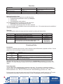
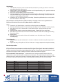
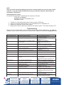
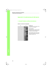
Mega Northern mRNA Array
Introduction
Mega Northern mRNA Array is a novel tool for analysis of gene expression, combining the unique features of
tissue mRNA arrays and mRNA Northern Blots. Using Mega Northern mRNA Arrays, not only gene
expression but also gene size can be determined in many different tissues. The dimensions of this array are
only 1x2 inches, and the detection method is the same as is commonly employed for Northern Blot analysis.
1. Corner that has been cut is upper
__
__
__
__
__
Band
1
5
10
15
20
lane 1
lane 32
2. The two marked dots are 2 mm under lane 1 and 32
Actual Size: 1 x 2”
Signal Locator
Mega Northern mRNA Array. Left: A Human adult normal Mega Northern mRNA Array Composed of 32 Lanes (Cat# N8234480) was
hybridized with non-radioactive labeled glyceraldehyde -3-phosphate dehydrogenase (GAPDH) DNA probe at 65 °C overnight, washed,
detected by non-radioactive method, and exposed to X-film for 30 seconds. The array was hybridized in FastHyb hybridization solution
(Cat# L1021250). Right: A transparent Signal Locator sheet is included for easy identification of the lanes and sizes of the signals.
Features
!" Reproducible results - The product comes in an array format. Any two arrays are nearly identical.
!" Higher sensitivity - mRNA species are highly concentrated per unit area on the membrane and are
readily accessible to probes.
!" Versatile - Suitable for both radioactive and non-radioactive probes.
!" Easy of use - The array can be treated/handled the same as a conventional Northern blot.
!" Economical - One hybridization analysis reveals gene expression in many different tissues, and the
smaller size of the membrane saves reagent costs.
Applications
!" Identification of tissue-specific genes in a wide variety of tissues
!" Gene expression pattern analysis
!" Comparison of expression levels of novel genes
!" Determination of size and relative abundance of genes in different tissues
!" Examination of alternative splicing and premature termination of specific gene transcripts
Description
BioChain’s Mega Northern mRNA Array was developed using a patent-pending proprietary technology. The
Mega Northern mRNA Arrays are manufactured using high quality mRNA from documented tissue sources.
A mRNA sample from each tissue is run 10 cm along with a RNA molecular weight marker on an agarose gel
by electrophoresis and recovered from 20 fractions on the gel. Each mRNA fraction covers a defined
molecular weight range. The recovered mRNA fractions are arrayed on positively charged nylon membranes
and UV cross-linked. Each array can contain many more lanes than traditional RNA Northern Blots, and
each lane is composed of 20 fractions. These arrays are suitable for use with both radioactive and nonradioactive probes. A user's manual and GAPDH control DNA probe are included. The Mega Northern
mRNA Array is supplied ready to use, so that you can immediately screen genes of interest without the
hassle of obtaining hard to find tissues and processing the RNA yourself.
Overview
Component
Item
Mega Northern mRNA Array
GAPDH control probe
Signal locator
Amount
1 array
100 ng (10 µl)
1
Storage
Room temperature, away from
light and moisture
-20ºC
Room Temperature
Storage Conditions: Store arrays at room temperature, away from light. Store control probe at -20°C.
Materials Supplied by User
The following reagents are required for use with these blots:
!" Prehybridization/hybridization solution (see Recipes)
!" Labeled probe
!" Post-hybridization wash solutions
The following equipment is required for use with these blots:
!" Hybridization tube and hybridization oven or
!" Heat-sealable plastic bags and a 30ºC-80ºC incubator
Whenever possible, use hybridization tubes and a hybridization oven. This method reduces the possibility of
radioactive contamination.
Time Line
The following table outlines the steps and times required for probing the Northern Blot.
Step
1
2
3
4
5
Action
Incubate the array in prehybridization solution.
Hybridize the array with radioactive or non-radioactive labeled probe.
Remove hybridization solution. Wash the array.
Wash the array 2 more times.
Expose the array to X-ray film
A longer exposure is recommended for Mega Northern mRNA
Array compared with the conventional Northern Blot
Time
3h
16-24 h
30-45 min.
2 x 20 min.
Minutes for nonradioactive probe,
hours to days for
radioactive probe
Choosing a Probe
Introduction
A probe is a nucleotide sequence that is used to identify a specific RNA within a population of RNA. For this
purpose there are several types of probe to be considered.
Type of Probe
Used when…………..
Analyzing RNA population for the presence of a gene similar to the gene of interest.
DNA
RNA
Oligonucleotide
Analyzing RNA population for the presence of a particular gene using the genetic
transcript
Searching RNA population for the presence of a particular sequence that may be present
in one or more gene transcripts
Remember that the probe you choose will be binding the complementary sequence found in the RNA
population. For more information, please refer to Current Protocols in Molecular Biology (Ausubel, et al.,
1994)
Probe Preparation
You will need to have a labeled probe to hybridize to the array. For procedure on radioactive labeling
probes, please refer to Probe Preparation or one of the following guides: Molecular cloning: A Laboratory
Manual (Sambrook, et al., 2001) or Current Protocols in Molecular Biology (Ausubel, et al., 1994).
For best results, the probe should be made from 25-50 ng DNA and have a specific activity of ! 108 cpm/ml.
Unincorporated nucleotides should be removed by column chromatography or gel electrophoresis.
2
Positive Control Probe
The cDNA fragment of the GAPDH gene is provided as a positive control for Mega Northern mRNA Array.
Please note that GAPDH expression levels vary depending on the cell or tissue types and level of
differentiation. These probes indicate lack of RNA degradation on the array. Please see Probe
Preparation.
Reprobing Mega Northern mRNA Array
The Mega Northern mRNA Array may be stripped and reprobed several times. With successive uses, there
might be an increased background level and a decreased hybridization signal. Please note that the quality of
the signal obtained once the blot has been stripped and reprobed cannot be guaranteed.
Caution
Be aware that probes used with these arrays may be radioactively labeled. Wear gloves and a laboratory
coat when working in a radioactivity designated area. Practice safe laboratory measures when using any
radioactive isotope. Dispose of radioactive waste properly.
Non-radioactive probes
It is ideally to use non-radioactive probes with these arrays. There are several commercial products
available for use in creating non-radioactive probes. Please follow the manufacture’s instructions for
probe preparation and blot hybridization.
Hybridization with Labeled Probes
Important!
It is very important that these arrays are NOT allowed to become completely dry at any time during the
following procedure. It is possible to process multiple arrays at one time. It is not recommended that more
than two be done together. Be sure that both arrays are fully exposed to the prehybridization/hybridization
solutions.
Diagram of Mega Northern mRNA Array
Mega Northern mRNA Array is about 1 x 2 inches in size. Each spots within one lane represents a particular
range of mRNA species.
The upper left corner is cut to indicate correct orientation.
Before Starting
1. Prepare approximately 12 ml of the prehybridization/hybridization solution (see Recipes).
2. If there is undissolved SDS present in the prehybridization solution, heat solution to 50ºC and mix
gently to dissolve.
3. Prepare probe for hybridization. Please see Probe Preparation.
Prehybridization
1. Place the array RNA side up in a hybridization tube (or a heat-sealable bag).
2. Add approximately 6 ml of the prehybridization solution (or enough to cover the array completely).
3. Incubate at 42ºC for 3 hours mixing gently for the entire incubation time (Rotate the hybridization
tube or slowly shake the bag in an incubator or water bath).
3
Hybridization
1. If the labeled probe being used is double-stranded, denature it by heating at 100ºC for 5 minutes.
Immediately transfer the probe to ice.
2. Add the probe to fresh prehybridization solution to a final specific activity of 10 cpm/ml to make the
hybridization solution (see Recipes).
3. Remove the prehybridization solution from the array.
4. Add approximately 6 ml hybridization solution to the membrane. Remove all air bubbles. If you are
using a resealable bag, it is a good idea to enclose this bag in a second bag in order to reduce the
chances of contaminating the water bath.
5. Incubate at 42ºC for 16-24 hours with gentle mixing. (Rotate the hybridization tube or slowly shake
the in an incubator or water bath)
Note: BioChain’s FastHyb Solution (Cat# L1031250) is an ideally solution to do hybridization of
BioChain’s RNA blot products including Mega Northern mRNA Array.
Wash
1. Prepare 30 ml of Wash Solution 1 and 200 ml of Wash Solution (see Recipes).
2. Remove hybridization solution from the membrane and dispose of the radioactive solution properly.
3. Wash the array for 10-15 minutes with 10 ml of Wash Solution 1 with gentle mixing at room
temperature.
4. Decant the wash solution and treat as radioactive waste. Repeat Step 3 two more times.
5. Wash the array for 20 minutes with 100 ml of Wash Solution 2 at 50ºC with constant gentle mixing.
Decant the solution.
6. Repeat Step 5.
7. Remove the array from wash solution and immediately cover with plastic wrap. Proceed to
Autoradiography.
Autoradiography
1. Mark the plastic wrap with radioactive ink in order to be able to orient the array after exposure to Xray film.
2. Expose the array to X-ray film at -70ºC for 16-24 hours. A longer exposure time may be necessary.
Results Interpretation
A. Put the Signal Locator transparency sheet on top of the X-ray film to align the signals to the locations of
the spots on the array. First make sure that the lanes are aligned to the signals. Since the second lane is
negative control, there should be no signal obtained for that lane. Second, align the marked dots to the dots
indicated on the Signal Locator to identify the fraction numbers that contain the signals.
B. Size estimation of the transcripts: To estimate the size of the transcript after Northern Blot, first
determine the numbers of which spots contain the most transcripts. There are twenty fractions from equal
distances of gels for each tissue is arrayed. Each spot contain a range of different sizes of transcripts. When
the number of the spot is determined, check the following table to estimate the size of the transcript.
Spot Number
1
2
3
4
5
6
7
8
9
10
Sizes
#26 kb
20 to 26 kb
15.5 to 20 kb
12 to 15.5 kb
9.2 to 12 kb
7.1 to 9.2 kb
5.5 to 7.1 kb
4.2 to 5.5 kb
3.2 to 4.2 kb
2.5 to 3.2 kb
Spot Number
11
12
13
14
15
16
17
18
19
20
4
Sizes
1.9 to 2.5 kb
1.5 to 1.9 kb
1.1 to 1.5 kb
0.88 to 1.1 kb
0.68 to 0.88 kb
0.52 to 0.68 kb
0.40 to 0.52 kb
0.31 to 0.40 kb
0.24 to 0.31 kb
0.18 to 0.24 kb
C. Multiple Spots for a transcript: In conventional Northern blot analysis, hybridization signals are not
always shown as a thin narrow band. Instead, the band can occupy quite some length of the lane. For the
production of Mega Northern mRNA Array, we used a special gel electrophoresis device to obtain highresolution mRNA fractions. Still it is common that signals for a transcript are found in two or more spots. It is
because the transcript is at the edge of predefined two fractions. Determine the maximum signal intensity
fraction by using the Signal Locator as described above.
Probe Preparation
Introduction
The following protocol is for radioactive labeling a DNA probe using random primers. For additional protocols
on probe preparation, please consult Molecular Cloning: A Laboratory Manual (Sambrook, et al., 2001) or
Current Protocols in Molecular Biology (Ausubel, et al., 1994).
Materials Supplied by User
To prepare a labeled probe with a DNA restriction fragment you will need the following:
25-50 ng of DNA fragment in 23 µl distilled water
random hexamer primers, 0.5 µg/ml
10X labeling reaction buffer
5 mM dATP
5 mM dGTP
5 mM dTTP
[ -³²P] dCTP at 10 mCi/ml
T7 DNA polymerase (2.5 units/µl)
0.2 M EDTA, pH 8.0
Sephadex G50 (Sigma) resuspended in TE buffer (1:1)
1.5 ml microcentrifuge tubes
Labeling the Probe
1. Boil the DNA fragment for 5 minutes. Immediately place on ice.
2. Add the following:
random hexamer primers
10 µl
10X labeling reaction buffer
5 µl
dATP
2 µl
dGTP
2 µl
dTTP
2 µl
[ -32P] dCTP
5 µl
T7 DNA Polymerase
1 µl
3. The reaction stays at room temperature for 2 hours.
4. Add 5 µl EDTA to stop the reaction
Removed Unincorporated Nucleotides
1. Poke a hole in the bottom of a 0.5 ml microcentrifuge tube using a pin or needle.
2. Add glass beads or wool to cover the bottom of the tube.
3. Fill the tube to the top with Sephadex G50 slurry in TE buffer
4. Place this tube within a second microcentrifuge tube and centrifuge at 1,500 x g for 4 minutes at
room temperature.
5. Discard the elute. Fill the tube containing the Sephadex to the top with TE buffer and repeat
centrifuge.
6. Repeat Step 5.
7. Place the tube containing the Sephadex into a clean microcentrifuge tube.
8. Add the labeling reaction to the column and centrifuge at 1500 x g for 4 minutes at room
temperature.
9. The elute will contain the labeled probe. The unincorporated nucleotide should remain in the
column. Discard the column as radioactive waste proceed to Estimating Specific Activity below.
5
NOTE
There are several commercial labeling kits and spin columns available to be used in place of these
protocols. If you plan to label the DNA probe using non-radioactive methods, please follow the
manufacture’s instructions.
Estimating Specific Activity
To measure the incorporation of radioactivity in the probe you will need:
STE buffer (see Recipes)
Scintillation fluid and 5 ml scintillation vials
Scintillation counter
1.
2.
3.
4.
Transfer 2 µl of the labeled probe (Step 9, above) to 98 µl of STE buffer.
Add 50 µl of the above diluted probe to 2 ml of scintillation fluid in a scintillation vial.
Measure the radioactive counts in each sample.
Multiply this number by 1000 µl/ml to determine the cpm/ml of probe. This number should be !108.
Troubleshooting
Please refer to the following table if you have difficulty with the Mega Northern mRNA Array. For additional
assistance, please contact Technical Services using the details supplied or e-mail us at [email protected]
Problem
Poor hybridization signal
Possible Cause
Exposure time may be too short
Probe specific activity may be too low
Probe concentration in hybridization solution
may be too low
Probe is not freshly labeled
High background
Double-stranded probe is not denatured
Use freshly labeled probes,
<1 week old
Denature double-strand probes
Hybridization temperature is too low
Increase hybridization temperature to 42ºC
Hybridization time is too short
Increase hybridization time to 24 hours
Blot has been stripped and
reprobed
Decrease in hybridization signal is expected.
Increase exposure
time of array to X-ray film or try using a new
array
Remove unincorporated nucleotides from
probe by chromatography or electrophoresis
Ensure that the correct amount of DNA was
used in the labeling reaction
Ensure that there are no air bubbles present in
hybridization tube or bag
Increase volume of prehybridization,
hybridization, and or wash solution
Make sure that the prehybridization and
hybridization solutions have been prepared
properly deionized formamide immediately
before use
Optimal size range of probes is
200-800 bp nucleotides
Use the shortest fragment possible that
contains the sequence of interest
Unincorporated nucleotides present
Concentration of probe is too high
Bubbles present during
prehybridization/hybridization
Membrane allowed to dry out during
hybridization procedure
Hybridization mixture is not correct
Probe is too long
Extra bands
Solution
Increase exposure time of blot to
X-ray film up to 48 hours
Check the probe to ensure that the initial
8
specific activity is !10
cpm/µg
Ensure that the hybridization
6
Solution has the DNA probe at 10
cpm/µg
Check amount of probe recovered after
removal of unincorporated nucleotides
Use a single-stranded probe
Probe contains non-specific sequences
6
High background
Unincorporated nucleotides present
Remove unincorporated nucleotides from
probe by chromatography or electrophoresis
Recipes
10 X Labeling Reaction Buffer
0.4 M Tris-HCl (pH 7.5), 0.1 M MgCl2, 50 mM DTT, and 1 mg/ml BSA (Bovine Serum Albumin)
1. For 10 ml, dissolve 630 mg Tris-HCl, 95 mg MgCl2 , 7.7 mg DTT, and 10 mg BSA in 8 ml deionized
water.
2. Bring final volume to 10 ml.
3. Store at -20ºC in 1 ml aliquots.
20 X SSC
3 M NaCl, 0.3 M C6H5Na3O7!2H2O (sodium citrate)
1. For 1 liter, dissolve 175 g NaCl and 88 g sodium citrate in 900 ml deionized water.
2. Adjust pH to 7.0 with 1 M HCl. Bring final volume to 1 liter with deionized water.
3. Store at room temperature.
50 X Denhardt’s Reagent
1% Ficoll (Type 400, pharmacia)
1% polyvinylpyrolidone (Sigma)
1% bovine serum albumin (Fraction V, Sigma)
1. For 500 ml, dissolve 5 g ficoll, 5g polyvinylpyrolidone, and 5 g BSA in 400 ml deionized water.
2. Bring to a final volume of 500 ml with deionized water.
3. Store at –20ºC.
20% SDS
1. For 100 ml, mix 20 g SDS with 80 ml sterile water, stir gently. It may be necessary to heat the
solution to 50ºC in order to dissolve the SDS.
2. Add sterile water to a final volume of 100 ml with deionized water.
3. Store at 4ºC.
Denatured, fragmented salmon sperm DNA
1. Dissolve 100 mg salmon sperm DNA in 10 ml deionized water. It may be necessary to stir the
solution for several hours at room temperature.
2. Add NaCl and adjust the concentration to 0.1 M.
3. Perform a phenol extraction, followed by a phenol: chloroform extraction. Transfer the upper
aqueous phase to a new tube.
4. Shear DNA with a 17-gauge needle.
5. Precipitate DNA with 2 volumes of ice-cold ethanol.
6. Wash pellet with 80% ethanol and air dry.
7. Redissolve DNA in water at 10 mg/ml
8. Measure OD260 and calculate concentration. 1OD260=0.05 µg/µl DNA.
9. Boil the solution for 10 minutes.
10. Store at –20ºC in 1 ml aliquots.
11. Before using , heat solution in boiling water for 5 minutes.
Prehybridization Solution (Per 100 ml)
20 X SSC
50 X Denhardt’s reagent
10 mg/ml denatured, fragmented salmon sperm DNA
Freshly deionized formamide
20% SDS
Sterilized water
30
10
1
50
2.5
6.5
ml
ml
ml
ml
ml
ml
1. Mix together the reagents listed above, 20 X SSC, 50 X Denhardt’s reagent and Salmon sperm DNA
are made up as stock solution.
7
2. It may be necessary to heat the solution to 50ºC in order to dissolve the SDS.
3. You will use 6 ml of this solution for prehybridization and 6 ml to make the hybridization solution.
4. The solution may be stored at 4ºC for 2 weeks. For long-storage, place the solution at –20ºC.
Hybridization Solution (per 6ml)
6 ml prehybridization solution (see above)
Radioactive labeled probe (specific activity > 108 cpm/ml)
1. The final specific activity of the hybridization solution should be 106 cpm/µl. To determine the
amount of probe needed, use the following equation:
Y ml (1 x 10⁶ cpm/ml)
X µl =
Z cpm/µl
X = the amount of probe to add
Y = the volume of hybridization solution
Z = the specific activity of the probe
2. Thoroughly mix the probe with the prehybridization solution.
Where
Note: Using BioChain’s FastHyb solution (Cat# L1031250) can save all the above material and time in
making hybridization solution. Following the simple protocol of FastHyb will give you the best
hybridization results from BioChain’s RNA blot products.
Wash Solution 1
2X SSC
0.05% SDS
1. For 500 ml, add 50 ml 20X SSC and 1.25 ml 20% SDS to 449 ml sterilized water. Mix.
2. Store at room temperature.
Wash Solution 2
0.1X SSC
0.1% SDS
1. For 500 ml, add 2.5 ml 20 X SSC and 2.5 ml 20% SDS to 495 ml sterilized water. Mix.
2. Store at room temperature.
STE Buffer
For a 50 ml volume:
1. Add 0.438 g NaCl to 40 ml of TE buffer.
2. Adjust pH to 8.0 with HCl.
3. Bring volume up to 50 ml with TE buffer.
4. Store at 4ºC.
References
1. Ausubel, F. M. , Brent, R., Kingston, R. E, Moore, D. D., Seidman, J. G., Smith, J. A. and Struhl, K.,
eds (1994). Current protocols in Molecular Biology.Greene Publishing associates and WileyInterscience. New York
2. Hames, B. D. and Higgins, S. J., eds (1993) Gene Transcription: A Practical Approach. Oxford
University Press. Oxford
3. Sambrook, J., Fritsch, E. F. and Maniatis, T. (2001). Molecular Cloning: A Laboratory Manual, 3rd
Edition. Cold Spring Harbor Laboratory Press.Plainview,New York.