1
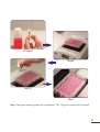
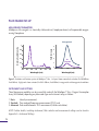
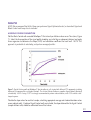
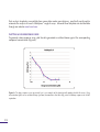
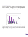
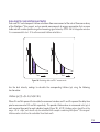
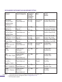
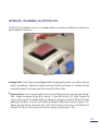
MitoXpress® Xtra
Oxygen Consumption Assay
(HS Method)
For the measurement of
Extracellular Oxygen Consumption
ILLUMINATING DISCOVERY ®
MitoXpress® Xtra
Oxygen Consumption Assay
O2
O2
(HS Method)
O2
For the measurement of
Extracellular Oxygen Consumption
For use with:
●
Adherent cells;
●
Suspension cells;
●
Permeabilised cells;
●
Isolated mitochondria;
●
3D cultures: tissues, spheroids,
●
Raft™ and scaffolds;
●
Isolated enzymes;
●
Bacteria, yeasts and moulds.
O2
TABLE OF CONTENTS
GENERAL INFORMATION .................................................................................................. 03
MATERIALS SUPPLIED ............................................................................................................. 03
STORAGE AND STABILITY ................................................................................................... 03
ADDITIONAL ITEMS REQUIRED .................................................................................... 03
OPTIONAL ITEMS NOT SUPPLIED ............................................................................... 03
DESCRIPTION ................................................................................................................................... 04
FLOW DIAGRAM ......................................................................................................................... 05
PLATE READER SET UP ........................................................................................................... 06
MEASUREMENT PARAMETERS ....................................................................................... 06
INSTRUMENTS AND SETTINGS ....................................................................................... 06
SIGNAL OPTIMISATION.......................................................................................................... 07
PERFORMING THE OXYGEN CONSUMPTION ASSAY ................... 08
CELL CULTURE AND PLATING ......................................................................................... 08
PRE-ASSAY PREPARATION .................................................................................................. 08
TYPICAL ASSAY ............................................................................................................................. 09
ANALYSIS ............................................................................................................................................... 11
ASSESSING OXYGEN CONSUMPTION ..................................................................... 11
PLOTTING A DOSE RESPONSE CURVE .................................................................... 12
CELLULAR ENERGY FLUX ANALYSIS ......................................................................... 13
APPENDIX A - INSTRUMENT SETTINGS ........................................................... 14
APPENDIX B - HS MINERAL OIL PIPETTING TIPS ................................. 17
APPENDIX C - TROUBLE SHOOTING .................................................................. 18
REFERENCES ....................................................................................................................................... 20
RELATED PRODUCTS .............................................................................................................. 21
GENERAL INFORMATION
MATERIALS SUPPLIED
Assay kit will arrive at room temperature. For best results store as indicated below.
Cat No.
Item
96 well1. Quantity / Size
Storage
MX-400
MitoXpress® Xtra reagent
1 vial
+4°C
HS-100D
HS Mineral Oil
1 dropper bottle / 15ml
Room Temp / dark
STORAGE AND STABILITY
The MitoXpress® Xtra reagent should be stored as follows:
● Dry material between +2 to +8°C (see Use Before date on vial).
● Reconstituted product can be aliquoted at -20°C. Use within one month (avoid freeze thaw).
ADDITIONAL ITEMS REQUIRED
● Fluorescence plate reader, with suitable filter and plate temperature control.
● 96-well (black wall) clear bottom TC+ plates or standard PS plates for cell culture.
OPTIONAL ITEMS NOT SUPPLIED
● Repeater pipette (recommended)
● Plate block heater for plate preparation
SUPPORT
● Visit our website www.luxcel.com.
1.
May also be used in a 384-well format, with one vial of probe sufficient for ~ 200 wells.
P3
DESCRIPTION
Luxcel's MitoXpress® Xtra - Oxygen Consumption Assay (HS Method) is a highly flexible 96 or 384-well
fluorescence plate reader-based approach, for the direct, real-time analysis of cellular respiration and
mitochondrial function. The easy-to-use MitoXpress® Xtra assay allows measurement of extracellular oxygen
consumption rates (OCR) with whole cell populations (both adherent and suspension cells), isolated
mitochondria, permeabilised cells and a wide range of 3D cultures including: tissues, small organisms,
spheroids, scaffolds and matrixes. The assay is also suitable for measurement of isolated enzymes, bacteria,
yeasts and moulds.
Scientists at Luxcel Biosciences developed the oxygen-sensing fluorophore known as MitoXpress® Xtra, to
overcome the limitations of specialised low throughput instrumentation that were historically used to measure
oxygen (eg Clark electrode). The MitoXpress® Xtra reagent is chemically stable and inert, water-soluble and
cell impermeable, making it the ideal and scalable mix-and measure reagent for use in a wide range of cell
culture conditions - all measured using a fluorescence plate-reader.
In this assay, MitoXpress® Xtra is quenched by 02, through molecular collision, and thus the amount of
fluorescence signal is inversely proportional to the amount of extracellular 02 in the sample. Rates of oxygen
consumption are calculated from the changes in fluorescence signal over time. The reaction is nondestructive and fully reversible (neither MitoXpress® Xtra nor O2 are consumed), facilitating measurement
of time courses and drug treatments.
Luxcel's flexible plate reader format, allows multiparametric or multiplex combination with Luxcel's other
products, as well as combination with commonly available reagents to measure glycolysis, LDH, JC-1, MMP
(Ѱ), ROS, and cellular ATP. For example, MitoXpress® Xtra in combination with Luxcel's pH-Xtra® - Glycolysis
Assay (Cat No. PH-100) allows the simultaneous real-time measurement of mitochondrial respiration and
glycolysis and analysis of the metabolic phenotype of cells and the shift (flux) between the two pathways
under pathological states.
P4
Resuspend
Aliquot
Add Oil
Read
Figure 1: Flow diagram showing preparation and use of MitoXpress® Xtra - Oxygen Consumption Assay (HS Method)
P5
PLATE READER SET-UP
MEASUREMENT PARAMETERS
MitoXpress® Xtra reagent is a chemically stable and inert, biopolymer-based, cell impermeable oxygensensing fluorophore.
Deoxygenated
5
0.8
Fold Increase
Normalised Intensity
1
0.6
0.4
0.2
4
3
2
Airsaturated
1
0
0
300
350
400
450
500
Wavelength (nm)
550
600
600
625
650
675
700
Wavelength (nm)
Figure 2: Excitation and Emission spectra of MitoXpress® Xtra. Left panel shows normalised excitation (Ex 360-400nm;
Peak 380nm). Right panel shows emission (Em 630 - 680nm; Peak 650nm) in oxygenated and deoxygenated conditions.
INSTRUMENTS AND SETTINGS
Three fluorescence modalities can be successfully used with the MitoXpress® Xtra – Oxygen Consumption
Assay (HS Method), depending on plate reader type and instrument setup, as follows:
1 Basic:
Intensity measurement,
2 Standard: Time-resolved fluorescence measurement (TR-F), and
3 Advanced: Dual-read Ratiometric TR-F measurement (Lifetime calculation).
NOTE: Further details, including instrument, filter selection and measurement settings can be found in
Appendix A - Instrument Settings.
P6
SIGNAL OPTIMISATION - recommended for first time users
NOTE: Use a plate block heater for plate preparation and pre-warm plate reader to measurement
temperature.
STEP 1: Reconstitute contents of the MitoXpress® Xtra vial in 1ml of water, PBS or culture media, gently
aspirating 3-4 times. NOTE: Reconstituted probe stock can be stored in the dark between +2 to +8°C for
several days or stored as aliquots in water at -20°C for use within one month (avoid freeze thaw).
STEP 2: Prepare 8 replicate wells of a 96-well plate, by adding 150µl pre-warmed culture medium to each
well (A1-A4, B1-B4).
STEP 3: Add 10µl reconstituted MitoXpress® Xtra reagent to 4 of the replicate wells (A1-A4) and 10µl water,
PBS or media to the remaining replicates wells (B1-B4).
STEP 4: Promptly add two drops (or 100µl) pre-warmed HS Mineral Oil to all eight replicate wells, taking
care to avoid air bubbles. NOTE: See Appendix B - HS Mineral Oil Pipetting Tips
STEP 5: Read plate immediately in a fluorescence plate reader over 30 minutes (read every 2-3 minutes).
STEP 6: Examine Signal Control well (A1-A4) and Blank Control well (B1-B4) readings (linear phase) and
calculate S:B ratio. NOTE: For dual read TR-F, calculate S:B for each measurement window.
For most fluorescence plate readers, set up according to Appendix A - Instrument Settings, MitoXpress®
Xtra should return a S:B ≥ 3. Higher readings are expected with TR-F and dual read TR-F measurement.
NOTE: See also Appendix C – Trouble Shooting.
A
B
1
Media +
MitoXpress® Xtra +
Oil
Media +
Oil
2
Media +
MitoXpress® Xtra +
Oil
Media +
Oil
3
Media +
MitoXpress® Xtra +
Oil
Media +
Oil
4
Media +
MitoXpress® Xtra +
Oil
Media +
Oil
P7
PERFORMING THE OXYGEN CONSUMPTION ASSAY
CELL CULTURE AND PLATING
NOTE: Always leave two wells (H11 and H12) free from the addition of MitoXpress® Xtra reagent, as Blank
Controls.
● For Adherent cells, seed cells in a 96-well plate at a density (typically 40,000 – 80,000 cells/well) in
200µl culture medium. Incubate overnight in a CO2 incubator at 37°C.
● For Suspension cells, seed on the day of assay in 150µl culture medium at a density of ~ 4 x 10⁶/ml.
Visit our website www.luxcel.com for more information on the use of MitoXpress® Xtra with permeabilised
cells, 3D cultures, tissues, spheroids, Raft™ and scaffolds, isolated enzymes, bacteria, yeasts and moulds.
PRE-ASSAY PREPARATION
● Reconstitute the contents of the MitoXpress® Xtra vial in 1ml of water, PBS or culture media, gently
aspirating 3-4 times (Figure 3). NOTE: Reconstituted probe stock can be stored in the dark between +2 to
+8°C for several days or stored as aliquots in water at -20°C for use within one month (avoid freeze thaw).
● Prepare test compounds, controls and dilutions as
desired. Typical controls are Antimycin A (Complex III
inhibitor), FCCP (ETC uncoupler) and Glucose Oxidase
(GOx; positive signal control).
NOTE: We recommend that all culture media and stock
solutions to be used in the assay are pre-warmed at 37°C
prior to use. Use a plate block heater for plate preparation
and pre-warm the fluorescence plate reader to
measurement temperature.
P8
Figure3: Reconstitution of MitoXpress® Xtra vial
TYPICAL ASSAY
To assess Oxygen Consumption or to investigate the effect of a compound on electron transport chain
function (ETC; oxidative phosphorylation), cells are treated immediately prior to measurement. NOTE: We
recommend the use of triplicate wells for each treatment.
STEP 1: Remove spent culture medium from all assay wells and replace with 150µl of fresh culture media
(Figure 4). NOTE: We recommend always leaving two wells (H11 and H12) free from the addition of
MitoXpress® Xtra reagent, for use as Blank Controls. Add 150µl of fresh culture media to these Blank Control
wells also.
STEP 2: Add 10µl reconstituted MitoXpress® reagent to each well, except those wells for use as Blank
Controls. Add 10µl of fresh culture media to these Blank Control wells. NOTE: If plating a full 96-well
plate of assays, we recommend combining Step 1 and Step 2 by adding the 1ml of reconstituted
MitoXpress® Xtra reagent to 15ml pre-warmed fresh culture media and using a multi-channel pipette to add
150µl of MitoXpress® Xtra in media stock to each well (Figure 4). Add 150µl of fresh culture media only
(no MitoXpress® Xtra) to the Blank Control wells.
STEP 3: Test compound stock or vehicle (typically 1-10µl) may be added at this point if desired. NOTE: We
recommend keeping the volume of added compound low to minimise any potential effects of solvent
vehicle.
Figure 4: Aliquoting fresh media (+/- MitoXpress® Xtra)
P9
STEP 4: Promptly seal each well by adding two drops (or 100µl) pre-warmed HS Mineral Oil, taking care to
avoid air bubbles (Figure 5).
NOTE: Small variations in the volume
of oil (between 90-110µl) should not
adversely affect the readings using
MitoXpress® Xtra.
See also Appendix B - HS Mineral Oil
Pipetting Tips.
STEP 5: Read the plate immediately in
a fluorescence plate reader, with the
set-up as described in Appendix A Instrument Settings (Figure 6). The
plate should be measured kinetically
for >90 minutes. When measurement
is completed, remove the plate and
save measured data to file.
Figure 5: Adding pre-warmed HS Mineral Oil
Optional Controls:
● Signal Controls: Leave 2 or 3 wells free
from the addition of cells for use as Signal
Controls. Add 150µl of fresh culture media
+10µl of reconstituted MitoXpress® Xtra
reagent to each well.
● Positive Controls: Leave 2 or 3 wells free
from the addition of cells for use as Positive
Controls. Add 150µl of fresh culture media
+ 10µl of (1mg/ml) Glucose Oxidase stock
solution (in water) + 10µl reconstituted
MitoXpress® Xtra reagent to each well.
P10
● Negative Controls: To 2 or 3 wells
containing cells, add 1µl of (150 µM)
Antimycin A stock solution (in DMSO) +
10µl reconstituted MitoXpress® Xtra
reagent.
Figure 6: Reading the assay plate
ANALYSIS
NOTE: We recommend that all first time users perform a Signal Optimisation test, as described. Signal and
Blank Control wells may also be included.
ASSESSING OXYGEN CONSUMPTION
Plot the Blank Control well-corrected MitoXpress® Xtra Intensity or Lifetime values versus Time (mins; Figure
7). Select the linear portion of the signal profile (avoiding any initial lag or subsequent plateau) and apply
linear regression to determine the slope (OCR) and correlation coefficient for each well. NOTE: This
approach is preferable to calculating a slope from averaged profiles.
Figure 7: Typical Lifetime profile of MitoXpress® Xtra for adherent cells, treated with different ETC compounds, including
Antimycin A (recommended as a Negative Control). The effect of Glucose Oxidase as a positive Signal Control is illustrated
schematically. NOTE: If using FCCP it is strongly recommended to perform a dose titration, since FCCP exhibits a bellshaped response.
Tabulate the slope values for each test sample, calculating appropriate average and standard deviation values
across replicate wells. If optional Signal Control wells are included, the slope obtained for the Signal Control
(sample without cells) should be subtracted from all test values.
P11
Data analysis templates are available from some plate reader manufacturers, specifically configured to
automate the analysis of Luxcel's MitoXpress® range of assays. Microsoft Excel templates are also available
through our website www.luxcel.com.
PLOTTING A DOSE RESPONSE CURVE
To generate a dose response curve, plot the data generated as outlined above against the corresponding
compound concentration (Figure 8).
Figure 8: The dose response curve presented here is an example of the data typically produced with this assay. Drug
concentration (µM) versus calculated slope (µs/hour) demonstrates that this drug causes inhibitory response on cellular
respiration.
P12
CELLULAR ENERGY FLUX ANALYSIS
Multiparametric (or multiplex) combination of MitoXpress® Xtra - Oxygen Consumption Assay (HS Method)
together with Luxcel's pH-Xtra® - Glycolysis Assay (Cat No. PH-100) allows the simultaneous real-time
measurement of mitochondrial respiration and glycolysis and analysis of the metabolic phenotype of cells and
the shift (flux) between the two pathways under pathological states (Figure 9).
Figure 9: Cellular Energy Flux for HepG2 cells, treated with a combination of drug compounds modulating the ETC or
inhibiting lactate production, shown as a percentage relative to untreated control cells. Comparative measurements with
MitoXpress® Xtra and pH-Xtra®, show the shift between mitochondrial respiration and glycolysis and the cellular control of
energy (ATP; measured 1h post-treatment using Promega Cell Titer-Glo®).
P13
APPENDIX A - INSTRUMENT SETTINGS
Three fluorescence modalities can be successfully used depending on plate reader type and instrument setup.
NOTE: We strongly recommend only using fluorescence plate readers equipped with temperature control.
Basic: Intensity Measurement
Measurement of signal Intensity (sometimes referred to as Prompt) provides flexibility to use a very wide
range of commonly available fluorescence, monochromator or filter-based plate readers. Optimal
wavelengths are 380nm excitation and 650nm for emission, with detection Gain parameters (PMT) typically
set at medium or high. NOTE: MitoXpress® Xtra should return a S:B ≥ 3
Standard: TR-F Measurement
Increased levels of performance can be achieved by using time-resolved fluorescence (TR-F). TR-F
measurement reduces non-specific background and increases probe sensitivity. Optimal delay time is ~30µs
and gate (integration) time is 100µs. NOTE: MitoXpress® Xtra should return a S:B > 3 S:B ~10 are typical.
Advanced: Dual-Read TR-F (Lifetime)
Optimal performance can be achieved using dual-read TR-F in combination with subsequent ratiometric
Lifetime calculation, to maximise dynamic range (Figure 10). NOTE: MitoXpress® Xtra should return a S:B
≥ 3 and S:B up to 60 are possible.
Optimal dual-delay and gate (integration) times:
● Integration window 1: 30µs delay (D1), 30µs measurement time (W1)
● Integration window 2: 70µs delay (D2), 30µs measurement time (W2)
P14
Intensity
Intensity
DUAL-READ TR-F AND LIFETIME ILLUSTRATED
Dual-read TR-F and subsequent Lifetime calculation allows measurement of the rate of fluorescence decay
of the MitoXpress® Xtra reagent, and can provide measurements of oxygen consumption that are more
stable and with a wider dynamic range than measuring signal Intensity. NOTE: S:B for Integration window
2 is recommended to be ≥ 10 to allow accurate Lifetime calculation.
W1
W2
30µs 30µs
D1
70µs
Time
D2
30µs
Time
Figure 10: Illustrating dual-read TR-F measurement.
Use the dual intensity readings to calculate the corresponding Lifetime (µs) using the following
transformation:
Lifetime (µs)[τ] =(D2-D1)/ln(W1/W2)
Where W1 and W2 represent the two (dual) measurement windows and D1 and D2 represent the delay time
prior to measurement of W1 and W2 respectively. This provides Lifetime values in microsecond units (µs) at
each measured time point for each individual sample (Figure 10). NOTE: Lifetime values should be in the
range ~22 to ~68µs, and should only be calculated from samples containing MitoXpress® Xtra reagent.
Lifetime values should not be calculated from blank wells.
P15
RECOMMENDED INSTRUMENT AND MEASUREMENT SETTINGS
Instrument
Optical Configuration
BMG Labtech:*
FLUOStar Omega /
POLARstar Omega
(CLARIOstar)**
BMG Labtech:*
PHERAstar FS
BMG Labtech:*
FLUOStar Optima /
POLARstar Optima
Perkin Elmer:
VICTOR series / X4, X5
Perkin Elmer:
EnVision, EnSpire
BioTek:*
Synergy H1, H4, HT, 2
(Cytation 3)**
BioTek:
Mx, H1m
Filter-based
Top or bottom read
Integration 1
(D1 / W1)
Integration 2
(D2 / W2)
30 / 30µs
70 / 30µs
Filter-based
Top read
Filter-based
Top or bottom read
Tecan:
Infinite / Safire / Genios
Pro
Mol. Devices:
SpectraMax /
Flexstation / Gemini
Hidek:
SENSE / CHAMELEON
Thermo:
Varioskan / Fluoroscan
Ascent
Mode
Ex (nm)
Em (nm)
Dual-read TR-F
(Lifetime)***
Ex 340 ± 50nm (TR-EX L)
Em 650 ± 50nm (BP-655)
40 / 100µs
n/a
30 / 100µs
n/a
TR-F
Ex 337nm (HTRF Module)
Em 665nm (HTRF Module)
Ex 340 ± 50nm (TR-EX L)
Em 655 ± 50nm (BP-655)
Filter-based
Top read
Filter-based
Top read
Filter-based
Top or bottom read
30 / 30µs
70 / 30µs
40 / 100µs
n/a
30 / 30µs
70 / 30µs
Dual read TR-F
(Lifetime)
TR-F
Monochromator /
Filter-based
Top or bottom read
Monochromator /
Filter-based
Top or bottom read
Monochromator-based
Top or bottom read
30 / 100µs
n/a
TR-F
Ex 380 ± 20nm
Em 650±15nm
30 / 100µs
n/a
TR-F
Ex 380 ± 20nm
Em 650 ± 20nm
n/a
n/a
Intensity
(Prompt)
Ex 380nm
Em 650nm
Filter-based
Top or bottom read
Monochromator /
Filter-based
Top or bottom read
30 / 100µs
n/a
30 / 100µs
n/a
TR-F
Ex 390 ± 20nm
Em 660 ± 10nm
Ex 380nm
Em 650nm
TR-F
Dual read TR-F
(Lifetime)
TR-F
®
P16 Notes: * Assay-specific protocols and notes are available from manufacturer for MitoXpress Xtra.
** Assay-specific protocols in development (contact [email protected])
*** TR-F head must be installed
Ex 340 ± 40nm (D340)
Em 642 ±10nm (D642)
Ex 340nm ± 60nm (X340)
Em 650nm ± 8nm (M650)
Ex 380 ±20nm
Em 645 ± 15nm
APPENDIX B - HS MINERIAL OIL PIPETTING TIPS
HS Mineral Oil is provided in an easy to use dropper bottle for convenience, although we recommend a
repeater pipette for routine use.
Figure 4: Add Oil
opper
bottle:
Invert
thethe
pre-warmed
dropper
bottle
andand
apply
gentle
pressure,
just just
sufficient
to prime
the
Dropper
Bottle:
Invert
pre-warmed
dropper
bottle
apply
gentle
pressure,
sufficient
to prime
the oil in the bottle tip. Apply two (2) drops to each well, touching each drop as it is formed to the side
oilof
in the
the well
bottle
Apply
drops
to each
touching
drop asmedia.
it is formed to the side of the well to
totip.
allow
it to2run
down
ontowell,
the surface
of each
the culture
allow it to run down onto the surface of the culture media.
●eRepeater Pipette: Use of a repeater pipette saves time and helps to maintain more precise incubation
times. Prepare the repeater syringe tip by trimming ~ 3-4mm off the tip at a 45° angle. Remove the
internal nozzle cap from the dropper bottle and slowly pick up the pre-warmed HS Mineral Oil (avoid
pipetting up and down, as this can cause bubbles) and dispense 100µl to each well at an angle of ~45°,
allowing the oil to flow the side of each well. NOTE: Small variations in the volume of HS Minerial Oil
(between 90-110µl) should not adversely effect the readings using MitoXpress® Xtra.
●
P17
APPENDIX C – TROUBLE SHOOTING
Extensive literature, including Protocols, Application Notes, Videos, Publications and email technical support
is also available through our website www.luxcel.com
GENERAL NOTES AND RECOMMENDATIONS
Storage and Stability: On receipt the MitoXpress® Xtra reagent should be stored between +2 to +8°C (see
Use Before date on vial). Reconstituted probe stock can be stored in the dark between +2 to +8°C for
several days or stored as aliquots in water at -20°C for use within one month (avoid freeze thaw).
Plate Reader: A fluorescence plate reader capable of measuring excitation at 380nm and emission at 650nm,
and having plate temperature control is required.
Plates: We recommend 96 or 384-well black wall / clear bottom TC+ plates, although standard clear wall
PS plates for cell culture may also be used.
Temperature: We recommend the use of a plate block heater for plate preparation, to maintain a temperature
of 37°C. Pre-warm the fluorescence plate reader to measurement temperature and ensure that all culture
media and stock solutions to be used in the assay are pre-warmed at 37°C prior to use.
Signal Optimisation and Use of Controls: We recommend performing a signal optimisation check, especially
for first time users, and inclusion of blank and optional additional control wells as described.
Pipetting HS Oil: Take care when dispensing the HS Mineral Oil to avoid bubbles. Apply HS Mineral Oil
allowing it to run down the inside surface of each well. Do not shake or rapidly aspirate the HS Mineral Oil.
General Assay Set-Up, Pipetting and Aspirating: Prepare your assay, materials and work space in advance.
Take care not to disrupt the cell monolayer (adherent cells) during pipetting and aspirating. Work rapidly
once the MitoXpress® Xtra reagent has been added, to reduce the potential for assay variability.
Cell Type and Cell Density: Since the MitoXpress® Xtra reagent measures extracellular Oxygen Consumption,
the amount of signal change will be directly dependent on the rate of cellular respiration of the cell type being
measured. We recommend using as high a cell density per well as practical as a starting point, and reducing
cell numbers as required. Not all cell types may consume sufficient oxygen for detection.
P18
SIGNAL TO BLANK (S:B) OPTIMISATION
For most fluorescence plate readers, set up according to Appendix A - Instrument Settings, MitoXpress®
Xtra should return a signal to blank ratio ≥ 3. Higher readings are expected with TR-F and dual read TR-F
measurement. The following options may be helpful to improve S:B if the determine ratio is not as high as
expected:
1
2
3
4
5
6
Increase Gain (PMT) setting or flash energy
Adjust TR-F focal height
Repeat without phenol red or serum.
Repeat as top or bottom-read, respectively.
Increase volume of MitoXpress® Xtra (15µl).
Contact Instrument Supplier for further options.
FREQUENTLY ASKED QUESTIONS:
Q: What do I do if I cannot detect any signal in wells containing cells and MitoXpress® Xtra (or I can detect
a signal but the slope (rate) appears very low)?
A: Check correct Instrument Settings (Appendix A) - Perform Signal Optimisation - Include GOx control
(max signal) - Increase cell density. If tested and not resolved, contact [email protected]
Q: What do I do if I can detect a signal in wells containing cells and MitoXpress® Xtra, but the slope (rate)
falls initially or is variable from well to well?
A: Check cell seeding and pipetting consistency - Increase cell density - Ensure plate, instrument and all
culture media and stock solutions are pre-warmed at 37°C prior to use - Reduce plate preparation times.
NOTE: Some plate readers have inconsistent temperature control. If you suspect this to be the case,
consider: – Reduce assay (and equilibration) temperatures to 30°C and avoid outer wells. If tested and not
resolved, contact [email protected].
P19
REFERENCES
Prediction of liver injury induced by chemicals in human with a multiparametric assay on isolated mouse
liver mitochondria. Porceddu M et al, Toxicol Sci., 2012 Oct; 129(2): 332-45
A high-throughput dual parameter assay for assessing drug-induced mitochondrial dysfunction provides
additional predictivity over two established mitochondrial toxicity assays. Hynes J et al, Toxicol Jn Vitro.,
2012 Mar; 27(2): 560-569
Comparative bioenergetic assessment of transformed cells using a cell energy budget platform. Zhdanov AV
et al, lntegr. Biol., 2011; 3: 1135-1142
High-throughput assay to measure oxygen consumption in digitonin-permeabilized cells of patients with
mitochondrial disorders. Jonckheere AI et al, Clin Chem., 2010; 56:(3): 424-431
Fluorescent pH and oxygen probes of the assessment of mitochondrial toxicity in isolated mitochondria and
whole cells. Hynes J et al, Curr Protoc Toxicol., 2009 May; Chapter 2: Unit 2.16
Analysis of mitochondrial function using phosphorescence oxygen sensitive probes. Will Y et al,
Nature Protocols. 2007; 1(6): 2563-2572.
Circumventing the Crabtree effect: replacing media glucose with galactose increases susceptibility of HepG2
cells to mitochondrial toxicants. Marroquin LD et al, Toxicol Sci., 2007; 97(2): 539-547
Investigation of drug-induced mitochondrial toxicity using fluorescence-based oxygen-sensitive probes.
Hynes J et al, Toxicol Sci., 2006; 92(1): 186-200
P20
RELATED PRODUCTS
●
●
●
pH-Xtra® – Glycolysis Assay (Cat No. PH-100)
MitoXpress® Intra – Intracellular O2 Assay (Cat No. MX-300)
GreenLight® 960 – Microbial Detection Assay (Cat No. GL-960)
P21
Luxcel Biosciences Limited
Suite 2.04
Western Gateway Building
Western Road
Cork
Ireland
t +353 (0)21 420 5348
e [email protected]
w www.luxcel.com