1
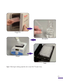
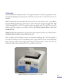
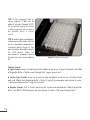
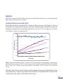
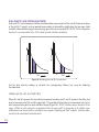
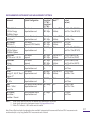
H+
pH-Xtra™
Glycolysis Assay
H
For the measurement of
Extracellular Acidification
ILLUMINATING DISCOVERY ®
+
H+
H+
H+
pH-Xtra™
Glycolysis Assay
H
For the measurement of
Extracellular Acidification
For use with:
●
Adherent cells;
●
Suspension cells;
●
Permeabilised cells;
●
3D cultures: tissues, spheroids,
●
RAFT™ and scaffolds.
+
H+
H+
TABLE OF CONTENTS
GENERAL INFORMATION ............................................................................................................................................ 03
MATERIALS SUPPLIED ........................................................................................................................................................ 03
STORAGE AND STABILITY .............................................................................................................................................. 03
ADDITIONAL ITEMS REQUIRED ............................................................................................................................... 03
OPTIONAL ITEMS NOT SUPPLIED ......................................................................................................................... 03
DESCRIPTION ............................................................................................................................................................................. 04
FLOW DIAGRAM ................................................................................................................................................................... 05
PLATE READER SET UP ..................................................................................................................................................... 06
MEASUREMENT PARAMETERS .................................................................................................................................. 06
INSTRUMENTS AND SETTINGS ................................................................................................................................. 06
SIGNAL OPTIMISATION..................................................................................................................................................... 07
PERFORMING THE OXYGEN CONSUMPTION ASSAY .............................................................. 08
CELL CULTURE AND PLATING .................................................................................................................................... 08
PRE-ASSAY PREPARATION ............................................................................................................................................. 08
TYPICAL ASSAY ........................................................................................................................................................................09
ANALYSIS ......................................................................................................................................................................................... 11
ASSESSING OXYGEN CONSUMPTION ............................................................................................................... 11
TITRATION OF CELL SEEDING DENSITY ........................................................................................................... 12
CELLULAR ENERGY FLUX ANALYSIS .................................................................................................................... 13
CALIBRATION OF PH-XTRATM GLYCOLYSIS ASSAY TO A PH [H+] SCALE .......................... 14
APPENDIX A - INSTRUMENT SETTINGS ...................................................................................................... 15
APPENDIX B - TROUBLE SHOOTING ........................................................................................................... 18
REFERENCES ................................................................................................................................................................................. 20
RELATED PRODUCTS .........................................................................................................................................................21
GENERAL INFORMATION
MATERIALS SUPPLIED
Assay kit will arrive at room temperature. For best results store as indicated below.
Cat No.
Item
96 well1. Quantity / Size
Storage
PH-100
pH-Xtra™ Reagent
1 vial
+4°C
RB-100
Respiration Buffer2.
1 tablet
Room Temp
STORAGE AND STABILITY
The pH-Xtra™ reagent should be stored as follows:
● Dry material between +2 to +8°C (see Use Before date on vial).
● Reconstituted pH-Xtra™ reagent can be stored in the dark between +2 to +8°C for several days or
reconstituted in water and stored as aliquots at -20°C for use within one month (avoid freeze thaw).
The Respiration Buffer tablet should be stored as follows:
Dry material at room temperature (see Use Before date on packaging)
● Reconstituted and filter sterilised product can be stored between +2 to +8°C.
●
ADDITIONAL ITEMS REQUIRED
Fluorescence plate reader, with suitable filter and plate temperature control.
● 96-well (black wall) clear bottom TC+ plates or standard PS plates for cell culture.
●
OPTIONAL ITEMS NOT SUPPLIED
Plate block heater for plate preparation
● 0.22μm sterilization filter, pH meter and acid / base for adjustment
●
SUPPORT
● Visit our website www.luxcel.com.
1.
2.
May also be used in a 384-well format, with one vial of reagent sufficient for ~ 200 wells.
1mM K-phosphate, 20mM Glucose, 70mM NaCl, 50mM KCl, 0.8mM MgSO4, 2.4mM CaCl2.
P3
DESCRIPTION
The pH-Xtra™ Glycolysis Assay from Luxcel Biosciences is an easy to use, highly flexible 96 or 384-well
fluorescence-based approach for the direct, real-time, kinetic analysis of extracellular acidification rates
(ECA/ECAR). As lactate production is the main contributor to this acidification, ECA measurements are a
convenient and informative measure of cellular glycolytic flux. Such measurements offer an important insight
into the central role played by altered glycolytic activity in a wide array of physiological and
pathophysiological processes, including cellular adaptation to hypoxia and ischemia, and the development
and progression of tumorigensis.
The pH-Xtra™ reagent is chemically stable and inert, water-soluble and cell impermeable. It exhibits a positive
signal response (increased signal with increased acidification) across the biological range (pH6-7.5), which,
coupled with its spectral and response characteristics, make pH-Xtra™ the ideal choice for flexible, highthroughput assessment of ECA. This performance facilitates sensitive robust microtitre-plate based
measurements, thereby overcoming many of the problems associated with the more cumbersome
potentiometric pH approach. Rates of extracellular acidification are calculated from changes in fluorescence
signal over time and, as the measurement is non-destructive and fully reversible (pH-Xtra™ reagent is not
consumed), measurement of time courses and multiple drug treatments are possible.
Luxcel's flexible plate reader format also allows multiparametric or multiplex combinations with Luxcel's
other products and with other commonly available reagents, thereby facilitating parallel kinetic measurements
of parameters such as ECA, mitochondria membrane potential ( Ѱm), O2 consumption or ROS generation,
followed by end-point measure of parameters such as ATP content or cell membrane integrity, all on the same
test cells. For example, the combination of Luxcel's MitoXpress® Xtra – Oxygen Consumption Assay (HS
Method; Cat No. MX-200) and pH-Xtra™ Glycolysis Assay allows the simultaneous real-time measurement
of the interplay between of mitochondrial respiration and glycolysis. This facilitates the determination of a
cell’s metabolic phenotype and the quantification of perturbations in the balance between glycolysis and
oxidative phosphorylation under various stimuli or pathological states.
P4
Purge CO2
Resuspend
Aliquot
Read
Figure 1: Flow diagram showing preparation and use of pH-Xtra™ Glycolysis Assay
P5
PLATE READER SET-UP
MEASUREMENT PARAMETERS
pH-Xtra™ reagent is a chemically stable and inert, cell impermeable H+-sensing fluorophore.
7
6
0.8
Fold Increase
Normalised Intensity
1
0.6
0.4
0.2
5
pH6.0
4
pH7.5
3
2
1
0
300 320 340 360 380 400 420 440 460 480 500
Wavelength (nm)
0
570
590
610
630
650
670
690
710
Wavelength (nm)
Figure 2: Excitation and Emision spectra of pH-Xtra™. Left panel shows normalized excitation (Ex 340410nm; Peak 360-380nm). Right panel shows emission maxima (Em 590, 615 and 690nm) fold increase
between pH6.0 and pH7.5
INSTRUMENTS AND SETTINGS
Two fluorescence modalities1. can be optimally used with the pH-Xtra™ Glycolysis Assay, depending on plate
reader type and instrument setup, as follows:
1 Standard: Time-resolved fluorescence measurement (TR-F), and
2 Advanced: Dual-read Ratiometric TR-F measurement (Lifetime calculation).
1.
pH-Xtra™ Glycolysis Assay may also be used in non - TR-F intensity mode on some plate readers, although
we recommend running the Signal Optimisation protocol and optimising cell seeding density.
NOTE: Further details, including instrument, filter selection and measurement settings can be found in
P6 Appendix A - Instrument Settings.
SIGNAL OPTIMISATION - recommended for first time users
NOTE: Use a plate block heater for plate preparation and pre-warm plate reader to measurement
temperature.
STEP 1: Reconstitute Respiration Buffer tablet in 50ml of water, warm to assay temperature (37°C), pH
adjust to approx. pH7.4 and filter sterilise using a 0.22μm filter. Reconstitute (transparent) contents of the
pH-Xtra™ vial in 1ml of Respiration Buffer, gently aspirating 3-4 times. NOTE: Reconstituted pH-Xtra™
reagent can be stored in the dark between +2 to +8°C for several days or stored as aliquots in water at 20°C for use within one month (avoid freeze thaw).
STEP 2: Prepare 8 replicate wells of a 96-well plate, by adding 150μl pre-warmed Respiration Buffer to each
well (A1-A4, B1-B4).
STEP 3: Add 10μl reconstituted pH-Xtra™ reagent to 4 of the replicate wells (A1-A4) and 10μl Respiration
Buffer to the remaining replicate wells (B1-B4).
STEP 4: Read plate immediately in a fluorescence plate reader over 30 minutes (read every 2-3 minutes).
STEP 5: Examine Signal Control well (A1-A4) and Blank Control well (B1-B4) readings (linear phase) and
calculate S:B ratio. NOTE: For dual-read TR-F, calculate S:B for each measurement window.
For most fluorescence TR-F plate readers, set up according to Appendix A - Instrument Settings, pH-Xtra™
should return a S:B ≥3. NOTE: See also Appendix B – Trouble Shooting.
A
1
Respiration Buffer +
pH-Xtra™
2
Respiration Buffer +
pH-Xtra™
3
Respiration Buffer +
pH-Xtra™
4
Respiration Buffer +
pH-Xtra™
B
Respiration Buffer
Respiration Buffer
Respiration Buffer
Respiration Buffer
P7
PERFORMING THE GLYCOLYSIS ASSAY
CELL CULTURE AND PLATING
NOTE: Always leave two wells (H11 and H12) free from the addition of pH-Xtra™ reagent, as Blank
Controls.
●
●
For Adherent cells, seed cells in a 96-well plate at a density (typically 30,000 – 80,000 cells/well) in 200μl
culture medium. NOTE: For new cell types, we recommend setting up a titration to select the optimum
cell seeding density (see Figure 7).
For Suspension cells, seed on the day of assay in 150μl culture medium at a density of approx. 250,000500,000 cells/well.
Visit our website www.luxcel.com for more information on the use of pH-Xtra™ with a range of cell systems.
PRE-ASSAY PREPARATION
NOTE: Where cells are cultured in a CO2 incubator overnight, it is important to purge the media and
plasticware of CO2 prior to conducting the pH-Xtra™ Glycolysis Assay as residual CO2 may contribute to
acidification. Perform a CO2 purge, by incubating cells in a CO2 -free incubator at 37°C with 95% humidity,
approx. 3 hours prior to performing the Glycolysis Assay measurement.
● Reconstitute Respiration Buffer tablet in 50ml of water, pH adjust to
approx. pH7.4 and filter sterilise using a 0.22μm filter. Reconstitute
transparent contents of the pH-Xtra™ vial in 1ml of Respiration Buffer,
gently aspirating 3-4 times. NOTE: Reconstituted pH-Xtra™ reagent
can be stored in the dark between +2 to +8°C for several days or
stored as aliquots in water at -20°C for use within one month (avoid
freeze thaw).
● Prepare test compounds, controls and dilutions as desired. Typical
controls are oxamic acid (inhibitor; decrease ECA), FCCP (ETC
uncoupler; increases ECA) and glucose oxidase (GOx; signal control).
P8
NOTE: We recommend that all culture media and stock solutions to
be used in the assay are pre-warmed at 37°C prior to use. Use a plate
block heater for plate preparation and pre-warm the fluorescence
plate reader to measurement temperature.
Figure 3: Reconstitution of pH-Xtra™ vial
TYPICAL ASSAY
To assess Extracellular Acidification (ECA) or to investigate the effect of a treatment on glycolytic flux, cells
are treated immediately prior to measurement. NOTE: We recommend the use of triplicate wells for each
treatment.
STEP 1: Remove spent culture medium from all assay wells and wash cells twice (2x), using 100µl of
Respiration Buffer per well for each wash (Figure 4). After removing the second wash, replace with 150µl
of fresh Respiration Buffer. NOTE: We recommend always leaving two wells (H11 and H12) free from the
addition of pH-Xtra™ reagent, for use as Blank Controls. Add 150µl of Respiration Buffer to these Blank
Control wells also.
STEP 2: Add 10µl reconstituted pH-Xtra™ reagent to each well, except those wells for use as Blank Controls.
Add 10µl of Respiration Buffer to these Blank Control wells.
NOTE: If plating a full 96-well plate of assays, we recommend simplifying Step 1 and 2 by preparing a
stock solution containing the 1ml of reconstituted pH-Xtra™ reagent added to 15ml pre-warmed Respiration
Buffer, and using a multi-channel pipette to add 150µl of this diluted pH-Xtra™ stock to each well. Add
150µl of Respiration Buffer only (no pH-Xtra™) to each Blank Control well.
Figure 4: Aliquoting fresh Respiration Buffer (+/- pH-Xtra™)
P9
STEP 3: Test compound stock or
vehicle (typically 1-10µl) may be
added at this point if desired. NOTE:
We recommend keeping the volume
of added compound low to minimise
any potential effects of solvent
vehicle.
STEP 4: Read the plate immediately in
a fluorescence plate reader, with the
set-up as described in Appendix A Instrument Settings (Figure 5). The
plate should be measured kinetically
for >120 minutes. When the
measurement is completed, remove
the plate and save measured data to
file.
Figure 5: Reading the assay plate
Optional Controls
Signal Controls: Leave 2 or 3 wells free from the addition of cells for use as Signal Control wells. Add 150µl
of Respiration Buffer + 10µl of reconstituted pH-Xtra™ reagent to each well.
●
Positive Signal Controls: Leave 2 or 3 wells free from the addition of cells for use as Positive Control
wells. Add 150µl of fresh Respiration Buffer + 10µl of [1 mg/ml] glucose oxidase stock solution [in water]
+ 10µl reconstituted pH-Xtra™ reagent to each well.
●
● Negative Controls: To 2 or 3 wells containing cells, washed and refreshed with 150µl of Respiration
Buffer, add 10µl of [750 mM] oxamic acid stock solution [in water] + 10µl reconstituted pH-Xtra™.
P10
ANALYSIS
NOTE: We recommend that all first time users perform a Signal Optimisation test, as described. Signal and
Blank Control wells may also be included.
ASSESSING EXTRACELLULAR ACIDIFICATION
Plot the Blank Control well-corrected pH-Xtra™ Intensity or Lifetime values versus Time (Figure 6). Select the
linear portion of the signal profile (avoiding any initial lag or subsequent plateau) and apply linear regression
to determine the slope (ECA) and correlation coefficient for each well. NOTE: This approach is preferable
to calculating a slope from averaged profiles.
Lifetime Signal (µs)
550
500
GOx
450
Antimycin A
400
FCCP
350
Untreated
300
Oxamic Acid
250
200
0
30
60
90
120
150
180
Time (mins)
Figure 6: Typical Lifetime profile of pH-Xtra™ for adherant cells, treated with typical control compounds, including oxamic
acid recommended as a negative control. The effect of glucose oxidase as a positive signal control is illustrated schematically.
NOTE: If using FCCP it is strongly recommended to perform a dose titration, since FCCP exhibits a bell-shaped dose
response.
Tabulate the slope values for each test sample, calculating appropriate average and standard deviation values
across replicate wells. If optional Signal Control wells are included, the slope obtained for the Signal Control
(sample without cells) should be subtracted from all test values.
P11
Data analysis templates are available from some plate reader manufacturers, specifically configured to
automate the analysis of Luxcel’s pH-Xtra™ Glycolysis Assay. Microsoft Excel templates are also available
through our website www.luxcel.com.
TITRATION OF CELL SEEDING DENSITY
To determine an optimal cell seeding density for performing the pH-Xtra™ Glycolysis Assay, for new cell
types, seed replicate wells with a range of seeding densities (typically 0, 10,000, 20,000, 40,000, 60,000 and
80,000 cells/well). Plot the data generated as a function of intensity or Lifetime values versus time, as
illustrated (Figure 7).
350
80,000
330
Lifetime (µs)
310
60,000
290
270
40,000
250
230
210
20,000
190
10,000
0
170
150
0
30
60
90
120
150
180
Time (mins)
Figure 7: Extracellular Acidification rate profiles (ECA) are shown for A549 cells seeded at 0, 10,000, 20,000, 40,000, 60,000
and 80,000 cells/well. In this experimental example, a seeding density of 40,000 cells/well was chosen for study as this
provided a suitable balance between ECA response and cell availability.
P12
CELLULAR ENERGY FLUX ANALYSIS
Multiparametric (or multiplex) combination of pH-Xtra™ Glycolysis Assay together with Luxcel's MitoXpress®
Xtra - Oxygen Consumption Assay [HS Method] (Cat No: MX-200) allows the simultaneous real-time
measurement of glycolysis and mitochondrial respiration, and analysis of the metabolic phenotype of cells
and the shift (flux) between the two pathways under pathological states (Figure 8).
600
500
ATP
Oxygen
% Effect
400
Consumption
300
Glycolysis
200
100
0
Rotenone
Antimycin
FCCP
Oxamate
Untreated
Figure 8: Cellular Energy Flux for HepG2 cells, treated with a combination of drug compounds modulating the ETC or
inhibiting lactate production, shown as a percentage relative to untreated control cells. Comparative measurements with pHXtra™ (glycolysis) and MitoXpress® Xtra (oxygen consumption), show the shift between glycolysis and mitochondrial
respiration and the cellular control of energy (ATP; measured 1h post-treatment using Promega Cell Titer-Glo®).
P13
CALIBRATION OF pH-XTRA™ GLYCOLYSIS ASSAY TO A pH [H+] SCALE
It is possible to express Extracellular Acidification (ECA) as a function of pH [H+] versus time. This is achieved
by first creating a calibration standard curve, by measuring TR-F intensity or preferably Lifetime values
(selecting stabilised readings over a 30 minute read), from a range of pH-buffered standards at the
appropriate assay temperature (see Figure 9). Select the linear portion of the standard curve and apply linear
regression to determine the calibration function (See Hynes et al., 2009).
5550
00
4500
50
450
Lifetime (µs)
400
400
350
350
300
300
250
250
2200
00
30°C
37°C
1150
50
1100
00
5.5
6
6.5
7
7.5
pH
Figure 9: pH-XTRA™ reagent calibration in Lifetime scale, at 30°C and 37°C using pH-buffered PBS, at increments of 0.2
across a pH range 6.0 - 7.5.
P14
APPENDIX A - INSTRUMENT SETTINGS
Two fluorescence modalities1. can be optimally used with the pH-Xtra™ Glycolysis Assay, depending on plate
reader type and instrument setup. NOTE: We strongly recommend only using fluorescence plate readers
equipped with temperature control.
Standard: TR-F Measurement
Measurement using time-resolved fluorescence (TR-F) provides flexibility to use a wide range of commonly
available plate readers2.. TR-F measurement reduces non-specific background and increases probe sensitivity.
Optimal delay time is ~100µs and gate (integration) time is 100µs. NOTE: pH-Xtra™ should return a S:B ≥3.
Advanced: Dual-Read TR-F (Lifetime)
Optimal performance can be achieved using dual-read TR-F in combination with subsequent ratiometric
Lifetime calculation, to maximise dynamic range (Figure 10) and to express ECA as a function of [H+]. NOTE:
pH-Xtra™ should return a S:B ≥10.
Optimal dual-delay and gate (integration) times:
● Integration window 1: 100µs delay (D1), 30µs measurement time (W1)
● Integration window 2: 300µs delay (D2), 30µs measurement time (W2)
1.
pH-Xtra™ Glycolysis Assay may also be used in non - TR-F Intensity mode on some plate readers, although
we recommend running the Signal Optimisation protocol to confirm an acceptable S:B, and optimising
cells seeding density (Figure 7).
2.
Users may see better performance using filter-based plate readers.
P15
Intensity
Intensity
DUAL-READ TR-F AND LIFETIME ILLUSTRATED
Dual-read TR-F and subsequent Lifetime calculation allows measurement of the rate of fluorescence decay
of the pH-Xtra™ reagent, and can provide measurements of extracellular acidification that are more stable
and with a wider dynamic range than measuring signal Intensity or standard TR-F. NOTE: S:B for Integration
window 2 is recommended to be ≥10 to allow accurate Lifetime calculation.
W1
W2
100μs 30μs
D1
300μs
Time
D2
30μs
Time
Figure 10: Illustrating dual-read TR-F measurement.
Use the dual intensity readings to calculate the corresponding Lifetime (μs) using the following
transformation:
Lifetime (μs)[τ] =(D2-D1)/ln(W1/W2)
Where W1 and W2 represent the two (dual) measurement windows and D1 and D2 represent the delay time
prior to measurement of W1 and W2 respectively. This provides Lifetime values in microsecond units (µs) at
each measured time point for each individual sample (Figure 10). NOTE: Lifetime values should be in the
range ~200µs for cells assayed in respiration buffer at approx pH7.4, increasing up to >400μs upon
acidification, and should only be calculated from samples containing pH-Xtra™ reagent. Lifetime values
should not be calculated from blank wells.
P16
RECOMMENDED INSTRUMENT AND MEASUREMENT SETTINGS
Instrument
Optical Configuration
Filter-based
Top or bottom read
Integration 1 Mode***
(D1 / W1)
Integration 2
(D2 / W2)
100 / 30μs Dual-read TR-F
300 / 30μs (Lifetime)
BMG Labtech:*
FLUOstar Omega
POLARstar Omega
BMG Labtech:*
CLARIOstar**
BMG Labtech:*
PHERAstar FS
BMG Labtech:*
FLUOstar Optima /
POLARstar Optima
Perkin Elmer:
VICTOR series / X4, X5
Perkin Elmer:
EnVision
Perkin Elmer:
EnSpire
BioTek:*
Synergy H1, H4, HT, Neo, 2
Cytation 3**
BioTek:
MX, H1m
Tecan:
Infinite / Safire /
Genios Pro
Mol. Devices:
SpectraMax /
Flexstation / Gemini
Ex 340 ± 50nm (TR-EX L)
Em 615 ± 10nm (BP-615)
Hybrid - filter based
Top or bottom read
Filter-based
Top read (HTRF Module)
Filter-based
Top or bottom read
100 / 30μs
300 / 30μs
100 / 30μs
300 / 30μs
100 /100μs
n/a
Dual-read TR-F
(Lifetime)
Dual-read TR-F
(Lifetime)
TR-F
Ex 340 ± 40nm
Em 620 ± 10nm
Ex 337nm
Em 620nm
Ex 340 ± 50nm (TR-EX L)
Em 615 ± 10nm (BP-615)
Filter-based
Top read
Filter-based
Top read
Monochromator
Top or bottom read
Filter-based
Top or bottom read
100 / 30μs
300 / 30μs
100 / 50μs
300 / 50μs
100 /100μs
n/a
100 /30μs
300 / 30μs
Dual read TR-F
(Lifetime)
Dual-read TR-F
(Lifetime)
TR-F
Ex 340 ± 40nm (D340)
Em 615 ±8.5nm (D642)
Ex 340 ± 60nm (X340)
Em 615 ±8.5nm (M615)
Ex 380nm
Em 615nm
Ex 360 ±40nm
Em 620 ± 10nm
Monochromator
Top or bottom read
Filter-based/Monochromator
Top or bottom read
100 /100μs
n/a
100 /100μs
n/a
TR-F
Monochromator-based
Top or bottom read
50 / 250μs
n/a
Dual read TR-F
(Lifetime)
TR-F
TR-F
Ex (nm)
Em (nm)
Ex 380nm
Em 615nm
Ex 380 ± 20nm
Em 615 ± 10nm
Ex 380nm
Em 615nm
Notes: * Assay-specific protocols and notes are available from manufacturer for pH-XtraTM
** Assay-specific protocols in development (contact [email protected])
*** Where TR-F indicated, a TR-F module must be installed
Note: Choose filter based optical configuration where available. Instruments with recommended Dual read TR-F measurement mode
can alternatively be set up using Standard TR-F measurement mode if desired.
P17
APPENDIX B – TROUBLE SHOOTING
Extensive literature, including application notes, videos, publications and email technical support is also
available through our website www.luxcel.com.
GENERAL NOTES AND RECOMMENDATIONS
Storage and Stability: On receipt the pH-Xtra™ reagent should be stored between +2 to +8°C (see Use
Before date on vial). Reconstituted pH-Xtra™ reagent can be stored in the dark between +2 to +8°C for
several days or stored as aliquots in water at -20°C for use within one month (avoid freeze thaw). Note:
pH-XtraTM reagent diluted in Respiration Buffer / media should be used on the same day.
Respiration Buffer: Kit contains a single Respiration Buffer tablet sufficient for 50ml 1x stock, containing
1mM K-phosphate, 20mM Glucose, 70mM NaCl, 50mM KCl, 0.8mM MgSO4, 2.4mM CaCl2. Alternative
media and supplements may be used as required (such as unbuffered DMEM), so long as care is taken to
ensure a minimal buffering capacity.
Plate Reader: A fluorescence plate reader capable of measuring excitation between 360nm and 390nm (see
Figure 2) and emission at 615nm, and having plate temperature control is required. We strongly recommend
using TR-F measurement.
Temperature: We recommend the use of a plate block heater for plate preparation, to maintain a temperature
of 37°C. Pre-warm the fluorescence plate reader to measurement temperature and ensure that all culture
media and stock solutions to be used in the assay are pre-warmed at 37°C prior to use.
Signal Optimisation and Use of Controls: We recommend performing a signal optimisation check, especially
for first time users, and inclusion of blank and optional additional control wells as described.
General Assay Set-Up, Pipetting and Aspirating: Prepare your assay, materials and work space in advance.
Take care not to disrupt the cell monolayer (adherent cells) during pipetting and aspirating. Work rapidly
once the pH-Xtra™ reagent has been added, to reduce the potential for assay variability. Re-check pH of
Respiration Buffer prior to use.
Cell Type and Cell Density: Since the pH-Xtra™ reagent measures Extracellular Acidification, the amount of
signal change will be directly dependent on the rate of glycolytic flux of the cell type being measured. We
recommend using a medium to high cell density per well as a starting point, and reducing cell numbers as
P18 required. (See Figure 7).
SIGNAL TO BLANK (S:B) OPTIMISATION
For most fluorescence plate readers, set up according to Appendix A - Instrument Settings, pH-Xtra™ should
return a S:B ratio ≥3. The following options may be helpful to improve S:B if the ratio is not as high as
expected:
1
2
3
4
5
6
Increase Gain (PMT) setting or flash energy
Adjust TR-F focal height
Increase length of integration time, the same for both delay windows.
Repeat as top or bottom-read, respectively.
Increase volume of pH-Xtra™ (15µl).
Contact Instrument Supplier for further options.
FREQUENTLY ASKED QUESTIONS:
Q: What do I do if I cannot detect
detect any
any signal
signal in
in wells
wells containing
containingcells
cellsand
andpH-Xtra
pH-Xtra™™(or
(orI Ican
candetect
detectaasignal
signal
but the slope (rate) appears very low)?
A: Check correct
GOx
control
correct Instrument
Instrument Settings
Settings(Appendix
(AppendixA)
A)- -Perform
PerformSignal
SignalOptimisation
Optimisation- Include
- Include
GOx
control
(max signal) - Increase cell
cell density.
density. Check
CheckpH
pHof
ofpre-warmed
pre-warmedRespiration
RespirationBuffer
Bufferand
andcorrect
correctasasnecessary,
necessary,
as pH can drift over time. If tested and not resolved, contact [email protected]
™ ™
, but
thethe
slope
(rate)
fallsfalls
Q: What do
containing
cells
andand
pH-Xtra
do II do
doififI Ican
candetect
detecta asignal
signalininwells
wells
containing
cells
pH-Xtra
, but
slope
(rate)
initially or is variable from well to well?
A: Check cell
plate,
instrument
andand
all all
cell seeding
seeding and
andpipetting
pipettingconsistency
consistency- -Increase
Increasecell
celldensity
density- Ensure
- Ensure
plate,
instrument
culture media and stock solutions are pre-warmed at 37°C prior to use - Reduce plate preparation times.
NOTE: Some plate readers have inconsistent temperature control. If you suspect this to be the case, consider:
– Reduce assay (and equilibration) temperatures to 30°C and avoid outer wells. If tested and not resolved,
contact [email protected].
P19
REFERENCES
A high-throughput dual parameter assay for assessing drug-induced mitochondrial dysfunction provides
additional predictivity over two established mitochondrial toxicity assays. Hynes J et al, Toxicol In Vitro.,
2012 Mar; 27(2): 560-569
Comparative bioenergetic assessment of transformed cells using a cell energy budget platform. Zhdanov AV
et al, lntegr. Biol., 2011; 3: 1135-1142
Fluorescent pH and oxygen probes of the assessment of mitochondrial toxicity in isolated mitochondria and
whole cells. Hynes J et al, Curr Protoc Toxicol., 2009 May; Chapter 2: Unit 2.16
In vitro analysis of cell metabolism using a long-decay pH-sensitive lanthanide probe and extracellular
acidification assay. Hynes J et al, Analytical Biochemistry., 2009; 390: 21-28
P20
RELATED PRODUCTS
●
●
●
®
MitoXpress Xtra - Oxygen Consumption Assay [HS Method] (Cat No. MX-200)
MitoXpress® Intra – Intracellular O2 Assay (Cat No. MX-300)
GreenLight® 960 – Microbial Detection Assay (Cat No. GL-960)
P21
Luxcel Biosciences Limited
Suite 2.04
Western Gateway Building
Western Road
Cork
Ireland
t +353 (0)21 420 5348
e [email protected]
w www.luxcel.com