1
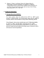
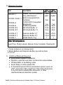
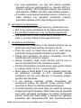
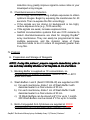
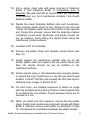
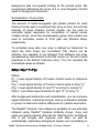
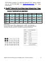
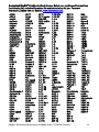
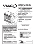
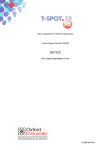
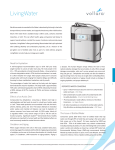
RayBio® Rat Acute Kidney Injury
Antibody Array 1 (C-Series)
Patent Pending Technology
User Manual
RayBio® Rat Acute Kidney Injury Antibody Array C-Series
Cat# AAR-AKI-1-4
RayBio® Rat Acute Kidney Injury Antibody Array C-Series
Cat# AAR-AKI-1-8
RayBio® Rat Cytokine Antibody Array Service
Cat# AAR-SERV
Please read manual carefully
before starting experiment
We provide you with excellent Protein Array systems and services
Tel: (Toll Free) 1-888-494-8555 or +1-770-729-2992; Fax: +1-770-206-2393;
Website: www.raybiotech.com Email: [email protected]
RayBiotech, Inc., the Protein Array Pioneer Company, strives to
research and develop new products to meet demands of the
biomedical community. RayBiotech’s patent-pending technology
allows detection of up to 1000 cytokines, chemokines and other
proteins in a single experiment. Our format is simple, sensitive,
reliable and cost effective.
Our product offerings include:
1. Protein (antigen) Arrays
2. Cytokine Antibody Arrays (Human, Mouse, Rat and Porcine)
o C-Series (Membrane, chemiluminescence detection)
o G-Series (Glass chip, fluorescence detection)
3. Pathway- and Disease-focused antibody arrays
o Angiogenesis Antibody Arrays
o Apoptosis Antibody Arrays
o Atherosclerosis Antibody Arrays
o Chemokine Antibody Arrays
o Growth Factor Antibody Arrays
o Inflammation Antibody Arrays
o MMP Antibody Arrays
o Obesity Antibody Arrays
4. Quantibody® Multiplex ELISA Arrays
5. L-Series Biotin Label-based Antibody Arrays
6. Phosphorylation Antibody Arrays
o Receptor Tyrosine Kinases
o EGFR and ErbB family (site-specific phosphorylation)
7. Over 700 different ELISA kits
8. EIA kits
9. Cell-based phosphorylation assay
10. Over 10,000 different Antibodies
11. Recombinant proteins
12. Peptide
13. Recombinant antibodies
Protocol for RayBio® Rat Acute Kidney Injury Antibody
Array 1 (C Series)
TABLE OF CONTENTS
I.
Introduction···················································································1
II.
Product Information································································3
A. Storage Recommendations·····································3
B. Materials Provided··························································4
C. Additional Materials Required································4
D. How It Works·······································································5
III.
Helpful Tips and General Considerations·············5
A. Preparation and Storage of Samples···············5
B. Handling Array Membranes·····································7
C. Incubations and Washes···········································7
D. Chemiluminescence Detection·····························8
IV.
Protocol···························································································8
A. Preparation and Storage of Reagent···············8
B. Blocking and Incubations··········································9
C. Chemiluminescence Detection··························10
V.
Interpretation of Results··················································12
VI.
Antibody Array Maps·························································15
VII.
Troubleshooting Guide····················································16
VIII.
Selected References·························································17
RayBio® Cytokine Antibody Arrays are patent-pending technology.
RayBio® is the trademark of RayBiotech, Inc.
RayBio® Rat Acute Kidney Injury Antibody Array 1 (C Series) Protocol
0
I. Introduction
New techniques such as cDNA microarrays have enabled us to
analyze global gene expression1-3. However, almost all cell
functions are executed by proteins, which cannot be studied
simply through DNA and RNA techniques. Experimental analysis
clearly shows disparity can exist between the relative expression
levels of mRNA and their corresponding proteins4. Therefore,
analysis of the proteomic profile is critical.
RayBiotech, The Protein Array Pioneer Company, introduced the
first protein arrays to the market in 2001 and continues to lead in
the development of innovative protein array technologies, such
as the RayBio Rat Acute Kidney Injury Antibody Array.
Acute kidney injury is a common complication among ambulatory
and hospitalized patients. It is a rapidly progressive illness that
independently predicts excess morbidity and mortality. It is
critical to early detect acute kidney injury and distinguish it from
prerenal azotemia and chronic kidney disease at the time of
patient presentation to rapidly manage associated illness.
However, serum creatinine, a standard marker of kidney
function, does not distinguish acute kidney injury from prerenal
azotemia5 or chronic kidney disease. In addition, the initial
measurement of serum creatinine cannot reflect the extent of
injury because its accumulation always lags behind the insult6.
The kidney is the primary organ responsible for the excretion of
medications and their biotransformation products from the body.
Rats are widely used for probing pharmacokineticpharmacodynamic (PK-PD) relationships for medications; in
addition, rats have been demonstrated to be a useful model for
evaluating mechanisms of kidney toxicity. In recent years,
numerous molecules have been described and investigated as
candidate biomarkers of kidney injury. The United States Food
and Drug Administration (FDA) has taken an active role in
developing a process for qualification of biomarkers7, 8 that would
potentially improve the drug development and regulatory review
process. In the gentamicin-induced rat model of acute kidney
RayBio® Rat Acute Kidney Injury Antibody Array 1 (C Series) Protocol
1
injury, based on histopathology, necrosis, or apoptosis scoring,
kidney injury molecule-1 (KIM-1) was the best biomarker of
overall renal injury9.
Traditionally, urine proteins or cytokines are detected by using
ELISA. However, RayBio® Rat Acute Kidney Injury Antibody
Array C-Series can detect 7 protein biomarkers simultaneously
with a small amount of sample. It is a great tool in the acute
kidney injury research areas and drug discovery area to hasten
drug development.
1.
Tang X, Marciano DL, Leeman SE, Amar S. LPS induces the
interaction of a transcription factor, LPS-induced TNF-a factor, and
STAT6(B) with effects on multiple cytokines. PNAS. 2005; 102(14):
5132-5137.
2.
Xu Y, Kulkosky J, Acheampong E, Nunnari G, Sullivan J, Pomerantz
RJ. HIV-1-mediated apoptosis of neuronal cells: Proximal molecular
mechanisms of HIV-1-induced encephalopathy. PNAS. 2004; 101(18):
7070-7075.
3.
El-Hage N, Gurwell JA, Singh IN, Knapp PE, Nath A, Hauser KF.
Synergistic increases in intracellular Ca(2+), and the release of MCP-1,
RANTES, and IL-6 by astrocytes treated with opiates and HIV-1 Tat.
Glia. 2005; 50(2):91-106.
4.
Oh HS, Moharita A, Potian JG, Whitehead IP, et al. Bone Marrow
Stroma Influences Transforming Growth Factor-β Production in Breast
Cancer Cells to Regulate c-myc Activation of the Preprotachykinin-I
Gene in Breast Cancer Cells. Cancer Res. 2004; 64: 6327-6336.
5.
Bonventre JV, Weinberg JM. Recent advances in the pathophysiology
of ischemic acute renal failure. J Am Soc Nephrol. 2003; 14:2199–210.
6.
Lameire N, Hoste E. Reflections on the definition, classification, and
diagnostic evaluation of acute renal failure [Editorial]. Curr Opin Crit
Care. 2004; 10:468–75.
7.
Goodsaid F, Frueh F. Biomarker qualification pilot process at the US
Food and Drug Administration. AAPS J 2007; 9: E105–E108.
8.
Goodsaid FM, Frueh FW, Mattes W. Strategic paths for biomarker
qualification. Toxicology 2008; 245: 219–223.
RayBio® Rat Acute Kidney Injury Antibody Array 1 (C Series) Protocol
2
9.
Rodney L. Rouse, Jun Zhang, Sharron R. Stewart, Barry A,
Rosenzweig, Parvaneh Espandiari, and Nakissa K. Sadrieh3.
Comparative profile of commercially available urinary biomarkers in
preclinical drug-induced kidney injury and recovery in rats. Kidney
International (2011) 79, 1186–1197
II. Product Information
A. Storage Recommendations:
For best results, store the entire kit at –20°C or –80°C upon
arrival. If stored frozen, we recommend using the kit within 6
months, which is the duration of the product warranty period.
Once thawed, store array membranes and 1X Blocking Buffer
at –20°C or –80°C, and all other component at 4°C. After
thawing, the entire kit should be used within 3 months. Array
kits are robust and will retain full activity even if stored for up to
24 hours at room temperature.
RayBio® Rat Acute Kidney Injury Antibody Array 1 (C Series) Protocol
3
B. Materials Provided
AARAKI-1-4
AARAKI-1-8
®
RayBio
Rat AKI Antibody
AAR-AKI1-Y
Array Membranes*
0103002- R-AKI -1 Biotin-Conjugated AntiCytokines
1,000X HRP-Conjugated
0103004-H
Streptavidin
0103004-B
1X Blocking Buffer
0103004-W †
20X Wash Buffer I †
0103004-W †
20X Wash Buffer II †
1 or 2
paks*
2 paks*
2 ea
4 ea
1 ea
25 ml
10 ml
10 ml
1 ea
50 ml
20 ml
20 ml
0103004-D †
0103004-D †
0103004-T
1.5 ml
1.5 ml
1
2.5 ml
2.5 ml
1
Item
Description
Detection Buffer C †
Detection Buffer D †
8-Well Plastic Tray
Other Kit Components:
8-well Tray, Plastic sheets, Manual, Array Template, Packing list
* Packs contains 2 or 4 arrays each
† Wash Buffers and Detection Buffers are sold as Sets
Y = 4 or 8
C. Additional Materials Required
• Small plastic boxes or containers
• Pipettors, pipet tips and other common lab consumables
• Orbital shaker or oscillating rocker
• Saran Wrap or similar plastic film
• A chemiluminescent blot documentation system (such as
UVP’s ChemiDoc-It® or EpiChem II Benchtop Darkroom),
X-ray Film and a suitable film processor, or other
chemiluminescent detection system.
RayBio® Rat Acute Kidney Injury Antibody Array 1 (C Series) Protocol
4
D. How It Works
III. Helpful Tips and General Considerations
A. Preparation and Storage of Samples
1. General Considerations:
• Freeze samples as soon as possible after collection.
• Avoid multiple freeze-thaw cycles. If possible, sub-aliquot
your samples prior to initial storage.
• Spin samples hard (5-10 minutes at 10K to 15K RPM)
immediately prior to incubation of samples with array.
• Optimal sample concentrations may need to be
determined empirically based on the signal intensities of
spots and background signals obtained with each sample.
• If spot intensities are weak, increase sample concentration
in subsequent experiments.
• If background or spot intensities are too strong, decrease
sample concentration in subsequent experiments.
RayBio® Rat Acute Kidney Injury Antibody Array 1 (C Series) Protocol
5
• Most samples will not need to be concentrated. If
concentration is required, we recommend using a spincolumn concentrator with a chilled centrifuge.
2. Recommended Sample Volumes and Dilution Factors
NOTE: All sample dilutions should be made using the 1X
Blocking Buffer provided in this kit. For all sample types, final
sample volume = 1.0-1.2 mL per membrane
• Urine 2-fold to 5-fold dilution
Note: If the sample volume is less than 200µl, the membrane
and sample may be sealed in a small plastic bag to increase
sample-membrane coverage. Expel all air bubbles prior to
sealing bag.
Note: The RayBio® Rat Acute Kidney Injury Antibody Array is
intended for use with Rat Urine samples. However, if you
wish, you may test other sample types as follows:
• Serum & Plasma: 2-fold to 5-fold dilution
3. Preparing Urine:
• Prepare 200µl aliquots and store at –20°C or
–80°C as soon as possible after collecting urine samples.
• Addition of protease inhibitors is not required.
• Immediately prior to sample incubation (Step 3 of
protocol), spin samples at 1000 rpm for 10 minutes to
remove particulates and precipitates.
4. Preparing Serum/Plasma:
• Prepare samples according to established protocols or
collection tube manufacturer’s instructions. Sub-aliquot
into plastic tubes. Store at –20°C or –80°C.
• We do not recommend comparing results between serum
and plasma samples or between plasma prepared using
different anticoagulants.
RayBio® Rat Acute Kidney Injury Antibody Array 1 (C Series) Protocol
6
• For most applications, you may test plasma samples
prepared using any anticoagulant (i.e., Heparin, EDTA or
Citrate). However, EDTA-prepared plasma may interfere
with detection of MMPs and other metal-binding proteins.
• If possible, avoid testing hemolyzed serum or plasma, as
these samples may generate anomalous cytokine
expression patterns and/or high background signals.
B. Handling Array Membranes
• Array membranes are fragile when dry. Handle with care.
• Wet or dry, grasp membranes by the edges using forceps.
• Do not allow membranes to dry out during experiments.
• The printed side of each membrane is denoted by a dash
mark (-) or array number in the upper left corner.
C. Incubations and Washes
• All washes and incubations in the standard protocol can be
performed using the 8-well tray provided in the kit.
• Place the cover on 8-well trays with lid to avoid drying,
particularly during extended incubation or wash steps.
• During each incubation, be sure to completely cover the
membranes with sample or reagent.
• During incubation steps, avoid foaming and be sure to
remove all bubbles from the membrane surface.
• Perform all incubation and wash steps under gentle rotation
or rocking motion (≈0.5 to 1 cycles/second).
• Wash steps in Wash Buffer II and all incubation steps may
be performed overnight at 4°C. Overnight sample
incubations are the most effective at increasing antigenspecific spot intensities.
• If you perform overnight sample incubations, we
recommend adding the optional “Large Volume Wash”
described in Step 4 to minimize background signals.
• Overnight blocking and wash steps are useful for reducing
background signal intensities. Wash steps may be repeated
even with completed membranes to reduce background
signals. Wash with Wash Buffer II, followed by repeating
incubation with Streptavidin-HRP and chemiluminescent
RayBio® Rat Acute Kidney Injury Antibody Array 1 (C Series) Protocol
7
detection may greatly improve signal-to-noise ratios in your
developed array images.
D. Chemiluminescence Detection
• We strongly recommend using multiple exposures to obtain
optimum images. Begin by exposing the membranes for 40
seconds. Then re-expose the film accordingly.
• If the signals are too strong (or background is too high),
reduce exposure time (e.g., 5-30 seconds).
• If the signals are weak, increase exposure time.
• Gel/blot documentation systems that use CCD cameras to
detect chemiluminescence are ideal for imaging RayBio®
array membranes. They can easily be programmed to take
multiple exposures, and the dynamic range of these
detectors tends to be 2-3 orders of magnitude greater than
X-ray film.
IV. Protocol
A. Preparation and Storage of Reagents
NOTE: During this protocol, prepare reagents immediately prior to
use and keep working dilutions of all reagents on ice at all times.
1. Blocking Buffer is supplied as 1X concentration, no
reconstitution or dilution is required. Store at –20°C or –80°C
when not in use.
2. Wash Buffers I and II (Item# 0103004-W) are supplied as 20X.
a). For each membrane, dilute 1 mL of Wash Buffer I with
deionized water to a final volume of 20 mL.
b). For each membrane, dilute 1 mL of Wash Buffer II with
deionized water to a final volume of 20 mL.
c). 1X Wash Buffers can be stored at 4°C for up to 1 month.
20X Wash Buffers can be stored 4°C for up to 3 months.
3. Biotin-Conjugated Anti-Cytokines are supplied at 2000X
concentration as a small liquid bead (typically ≈2-5 μL).
RayBio® Rat Acute Kidney Injury Antibody Array 1 (C Series) Protocol
8
Note: Spin down the tube prior to reconstitution, as the
concentrated liquid bead may have moved to the top of the
tube during handling.
a). Add 100 μL 1X Blocking Buffer to the tube containing
2000X Biotin-Conjugated Anti-Cytokines.
b). Mix well and quantitatively transfer stock reagent to
larger tube containing 1900 μL of 1X Blocking Buffer.
c). 1X Biotin-Conjugated Anti-Cytokines may be stored for
2-3 days at 4°C.
4.
Streptavidin-HRP is supplied as 1000X concentration.
a). Mix the tube containing 1,000X Streptavidin-HRP well
before use, as precipitates may form during storage.
b). Add 2 μL of 1000X Streptavidin-HRP to 1998 μL of 1X
Blocking Buffer.
c). This working dilution can be stored for 3-5 days at 4°C.
5.
Detection Buffers C & D are supplied as 1X solutions that are
intended to be mixed in a 1:1 ratio immediately prior use.
Detection reagents may be stored at 4°C for up to 3 months.
B. Blocking and Incubations
NOTE: Please prepare all reagents immediately prior to use as
described above (Section IV.A) and carefully read tips on Sample
Preparation (Section III.A) and Incubations and Washes (Section
III.C) before proceeding.
1)
Place each membrane printed side up (see Section III.B) into
the 8-well tray provided in the kit.
2)
Block membranes by incubating with 2 mL 1X Blocking
Buffer at room temperature (RT) for 30 min.
3)
Decant Blocking Buffer, and incubate membranes with 1 mL
of sample at RT for 1-2 h.
RayBio® Rat Acute Kidney Injury Antibody Array 1 (C Series) Protocol
9
4)
Aspirate samples from membranes. Wash 3 times, 5 min per
wash, with 2 mL Wash Buffer I at RT. Use fresh buffer for
each wash.
OPTIONAL Large Volume Wash: After Step 4, place
membranes into clean container(s). Add 20-30 mL of Wash
Buffer I per membrane, and wash at RT with gentle shaking
or rocking for 30-45 min. Return membranes to the 8-well
tray. Then proceed to Step 5.
5)
Wash 2 times, 5 min per wash, with 2 mL of 1X Wash Buffer
II each at RT. Use fresh wash buffer each time.
6)
Add 1 mL of 1X Biotin-conjugated Anti-Cytokines to each
membrane. Incubate at RT for 1-2 hours.
7)
Decant or aspirate Anti-Cytokine reagent and repeat washes
as described in steps 4 and 5 above.
8)
Incubate at RT for 2 hours with 1 mL of 1X Streptavidin-HRP.
9)
Wash membranes as directed in steps 4 and 5.
10)
Proceed with Detection protocol (below) or store membranes
between plastic sheets (provided in kit) as directed in Steps
20 & 21 below.
C. Chemiluminescence Detection
NOTE: Do not allow membranes to dry out during detection.
Detection of chemiluminescence should be started within 5
minutes after removing Detection Buffers and must be
completed within 20 minutes.
11) Place a plastic sheet (provided in the kit) on your benchtop.
12) Place one or more array membranes protein side up (see
Section III.B) on the plastic sheet. Drain excess liquid by
touching one edge to blotting paper or tissue paper.
RayBio® Rat Acute Kidney Injury Antibody Array 1 (C Series) Protocol
10
13) Into a single, clean tube add equal volumes of Detection
Buffer C and Detection Buffer D immediately prior to
detection. Mix well. Add 250 μL of each buffer per membrane
to be detected, e.g., for 4 membranes, combine 1 mL of each
detection buffer.
14) Pipette the mixed Detection Buffers onto each membrane.
Place another plastic sheet on top, starting at one end and
“rolling” the flexible plastic across the surface to the opposite
end. During this process, ensure that the detection mixture
completely covers each membrane, and gently smooth out
any air bubbles. Avoid sliding the plastic sheet along the
membranes’ printed surfaces.
15) Incubate at RT for 2 minutes.
16) Remove top plastic sheet and aspirate excess liquid (see
Step 12).
17) Gently replace the membranes (protein side up) on the
bottom plastic sheet and replace the top plastic sheet (see
Step 14). Gently smooth out any air bubbles on the
membrane surfaces.
18) Detect signals using a chemiluminescence imaging system
or expose the array membranes to x-ray film (we recommend
Kodak’s X-Omat™ AR film) and develop the film (See tips for
obtaining array images in Section III.D).
19) For each array, use multiple exposures to obtain an image
with low background and strong Positive Control signals that
do not bleed into one another. Typical exposure times are 10
seconds to 2 min.
20) When you finish your last exposure, remove the top plastic
sheet. Gently rinse membranes and plastic sheets with Wash
Buffer II. Remove excess wash buffer as described in Step
14, and replace the membranes between the plastic sheets.
RayBio® Rat Acute Kidney Injury Antibody Array 1 (C Series) Protocol
11
21) Wrap the sheets in Saran Wrap, and store the membranes at
–20°C to –80°C. (Or store membranes for up to 5 days at 4°C
in Wash Buffer II. Cover the container to avoid evaporation.)
V. Interpretation of Results:
Typical results obtained with RayBio® C Series Antibody Arrays
Sample-1
Sample-2
Control
The preceding figure presents typical images obtained with
RayBio® Human Cytokine Antibody Array. These membranes
were probed with conditioned media from two different cell lines.
Membranes were exposed to Kodak X-Omat® film at RT for 1 min.
Note the strong signals of the Positive Control spots, provided by
biotin-conjugated IgG printed directly onto the array membrane in
the upper-left and lower-right corners. These Positive Control
spots are useful for proper orientation of the array image.
The signal intensity for each antigen-specific antibody spot is
proportional to the relative concentration of the antigen in that
sample. Comparison of signal intensities for individual antigenspecific antibody spots between and among array images can be
used to determine relative differences in expression levels of each
analyte sample-to-sample or group-to-group.
Obtaining Densitometry Data:
Visual comparison of array images may be sufficient to see
differences in relative protein expression. However, most
researchers will want to perform numerical comparisons of the
signal intensities (or more precisely, signal densities), using 2-D
RayBio® Rat Acute Kidney Injury Antibody Array 1 (C Series) Protocol
12
densitometry. Gel/Blot documentation systems and other
chemiluminescent or phosphorescent detection systems are
usually sold as a package with compatible densitometry software.
To obtain densitometry data from an X-ray film, one must first
scan the film to obtain a digitized image using an ordinary office
scanner with resolution of 300 dpi or greater. Any densitometry
software should be sufficient to obtain spot signal densities from
your scanned images. One such software program, ImageJ, is
available for free from the NIH (for more info, visit
http://rsbweb.nih.gov/ij/).
We suggest using the following guidelines when extracting
densitometry data from our array images:
•
•
•
For each array membrane, identify a single exposure that the
exhibits low background signal intensity and strong Positive
Control signals that do not “bleed” into one another.
Exposure times do not need to be identical for each array,
but Positive Control signals on each image should have
similar intensities.
Measure the density of each spot using a circle that is
roughly the size of one of the largest spots. Be sure to use
the same circle (area and shape) for measuring the signal
densities on every array for which you wish to compare the
results.
For each spot, use the summed signal density across the
entire circle (i.e., total signal density per unit area)
Before analysis, subtract the background from raw densitometry
data and normalize the signal intensities to the Positive Controls.
Background Subtraction:
On each array, several “Negative Control” and/or “Blank” spots
will be included. Blank spots are literally blank; nothing has been
printed there. Negative Control spots are printed with the same
buffer used to dilute antibodies printed on the array. Thus, the
signal intensities of the Negative Controls represent the
RayBio® Rat Acute Kidney Injury Antibody Array 1 (C Series) Protocol
13
background plus non-specific binding to the printed spots. We
recommend subtracting the mean of 4 or more Negative Control
spots for background correction.
Normalization of Array Data:
The amount of biotin-conjugated IgG protein printed for each
Positive Control spot is consistent from array to array. As such the
intensity of these Positive Control signals can be used to
normalize signal responses for comparison of results across
multiple arrays, much like housekeeping genes and proteins are
used to normalize results of PCR gels and Western Blots,
respectively.
To normalize array data, one array is defined as "reference" to
which the other arrays are normalized. This choice can be
arbitrary. For example, in our Analysis Tool Software, the array
represented by data entered in the first column on the left of each
worksheet is the default “reference array.” You can calculate the
normalized values as follows:
X(Ny) = X(y) * P1/P(y)
Where:
P1 = mean signal density of Positive Control spots on reference
array
P(y) = mean signal density of Positive Control spots on Array "y"
X(y) = mean signal density for spot "X" on Array for sample "y"
X(Ny) = normalized signal intensity for spot "X" on Array "y"
After background subtraction and normalization, you can compare
signal intensities, analyte-by-analyte, among or between samples
or groups to determine relative differences in cytokine expression.
The RayBio® Analysis Tool software is available for use with data
obtained using RayBio® Cytokine Antibody Arrays. Copy and
paste your signal intensity data into the “Aligning Data” worksheet,
and it will compile and organize your data, as well as
automatically subtracting background signals and normalizing to
RayBio® Rat Acute Kidney Injury Antibody Array 1 (C Series) Protocol
14
the Positive Controls. To order the Analysis Tool, please contact
us at +1-770-729-2992 or [email protected] for more
information.
VI. RayBio® C Series Rat Acute Kidney Injury Antibody Array 1 Maps:
Detects 7 cytokines in one experiment
A
B
C
D
E
F
G
H
1
POS
POS
NEG
NEG
Cystatin C
FABP1
KIM-1
MCP-1
2
POS
POS
NEG
NEG
Cystatin C
FABP1
KIM-1
MCP-1
3
NGAL
TIMP-1
VEGF
NEG
NEG
NEG
NEG
POS
4
NGAL
TIMP-1
VEGF
NEG
NEG
NEG
NEG
POS
Abbreviations:
POS = Positive Control, NEG = Negative Control, L-FABP = Liver Fatty-Acid
Binding Protein, KIM-1 = Kidney Injury Molecule-1, NGAL= Neutrophil GelatinaseAssociated Lipocalin (Lipocalin 2). All others use standard abbreviations.
RayBio® Rat Acute Kidney Injury Antibody Array 1 (C Series) Protocol
15
VII. Troubleshooting guide
Problem
Cause
No signal for any
spots, including
Positive Controls
Repeat incubation with
Global detection failure
HRP-Streptavidin and
Detection Buffers
Repeat experiment using
Sample is too dilute
higher sample concentration
Tube may contain
precipitants. Repeat
Improper dilution of HRPdetection, mix 1000X HRPStreptavidin
Streptavidin well before
diluting reagent
Repeat detection, making
Waiting too long to detect
sure to complete this
chemiluminescent signals
process within 20 min.
Weak or no signals
antigen-specific
spots
Incubate with sample O/N at
4°C
Other Tips
Uneven signal or
background
High background
signals
Recommendation
Increase concentration of
HRP-Streptavidin
Overexposure
Increase concentration of
Biotin-conjugated AntiCytokine
Extend exposure time (may
go overnight)
Be sure to completely
remove all bubbles from
membrane surface
Completely cover
membranes with solution,
use a rocker or shaker
during washes and
incubations
Decrease exposure time
Sample is too
concentrated
Repeat experiment using
more dilute sample
Bubbles present on
membrane during
incubations
Membranes were not
evenly covered during
washes/incubations or
allowed to dry out
NOTE: To reduce background on completed membrane,
wash O/N @ 4°C in Wash Buffer II, then re-incubate
with HRP-Streptavidin and repeat detection.
RayBio® Rat Acute Kidney Injury Antibody Array 1 (C Series) Protocol
16
VIII. Selected References Featuring RayBio® C-Series Arrays
1. Ray S, Britschgi M, Herbert C, et al. Classification and prediction of
clinical Alzheimer's diagnosis based on plasma signaling proteins.
Nature Med. 2007; 13(11):1359-1362.
2. McAllister SS, Gifford AM, Greiner AL , et al. Systemic Endocrine
Instigation of Indolent Tumor Growth Requires Osteopontin. Cell. 2008;
133: 994-1005.
3. Acosta JC, O'Loghlen A, Banito A, et al. Chemokine Signaling via the
CXCR2 Receptor Reinforces Senescence. Cell. 2008; 133: 1006-1018.
4. Brown JM, Boysen MS, Chung S, et al. Conjugated Linoleic Acid
Induces Human Adipocyte Delipidation: Autocrine/Paracrine Signaling
by Adipokines. J Biol Chem. 2004; 279(25): 26735-26747.
5. Celis JE, Moreira JMA, Gromova I, et al. Towards discovery-driven
translational research in breast cancer. FEBS J. 2004;272: 2-15.
6. Lin PW, Simon PO, Gewirtz AT, et al. Paneth Cell Cryptdins Act in
Vitro as Apical Paracrine Regulators of the Innate Inflammatory
Response. J Biol Chem. 2004; 279(19): 19902-19907.
7. Xu Y, Kulkosky J, Acheampong E, et al. HIV-1-mediated apoptosis of
neuronal cells: Proximal molecular mechanisms of HIV-1-induced
encephalopathy. PNAS. 2004; 101(18):7070-7075.
8. Tang X, Marciano DL, Leeman SE, Amar S. LPS induces the
interaction of a transcription factor, LPS-induced TNF-alpha factor, and
STAT6(B) with effects on multiple cytokines. PNAS. 2005; 102(14):
5132-5137.
9. Wang F-X, Xu Y, Sullivan J, et al. IL-7 is a potent and proviral strain–
specific inducer of latent HIV-1 cellular reservoirs of infected individuals
on virally suppressive HAART. J Clin Invest. 2005; 155: 128–137.
10. De Ceuninck F, Marcheteau E, Berger S, et al. Assessment of Some
Tools for the Characterization of the Human Osteo-arthritic Cartilage
Proteome. J Biomol Tech. 2005; 16: 256–265.
RayBio® Rat Acute Kidney Injury Antibody Array 1 (C Series) Protocol
17
11. Boucharaba A, Serre C-M, Guglielmi J, et al. The type 1
lysophosphatidic acid receptor is a target for therapy in bone
metastases. PNAS. 2006; 103(25):9643-9648.
12. Matsunaga K, Yanagisawa S, Ichikawa T, et al. Airway cytokine
expression measured by means of protein array in exhaled breath
condensate: Correlation with physiologic properties in asthmatic
patients. J Allergy Clin Immunol. 2006; 188: 84-90.
13. Vargas DL, Nascimbene C, Chitra Krishnan C, et al. Neuroglial
activation and neuroinflammation in the brain of patients with autism.
Ann Neurol. 2005; 57: 67-81.
14. Miyoshi N, Oubrahim H, Chock PB, Stadtman ER. Age-dependent cell
death and the role of ATP in hydrogen peroxide-induced apoptosis and
necrosis. PNAS. 2006; 103(6): 1727–1731.
15. Coppinger JA, O'Connor R, Wynne K, et al. Moderation of the platelet
releasate response by aspirin. Blood. 2007; 109: 4786-4792.
16. Cortez DM, Feldman MD, Mummidi S, et al. IL-17 stimulates MMP-1
expression in primary human cardiac fibroblasts via p38 MAPK- and
ERK1/2-dependent C/EBP-beta, NF-kB, and AP-1 activation. Am J
Physiol Heart Circ Physiol. 2007; 293: H3356-H3365.
17. Walt DR, Blicharz TM, Hayman RB, Rissin DM, et al. Microsensor
Arrays for Saliva Diagnostics. Ann NY Acad Sci. 2007; 1098:389-400.
RayBio® Rat Acute Kidney Injury Antibody Array 1 (C Series) Protocol
18
Customized RayBio® Cytokine Antibody Arrays. Select your cytokines of interest from
the following list, and we will produce the customized array for you. For more
information, please visit our website, www.raybiotech.com.
4-1BB
ACE-2
Acrp30
Activin A
Adiposin
Adipsin
AgRP
ALCAM
α-Fetoprotein
Amphiregulin
Angiogenin
Angiopoietin-1
Angiopoietin-2
Angiostatin
ANGPTL4
Axl
B7-1
BCAM
BCMA
BDNF
β2M
β IG-H3
bFGF
BLC
BMP-4
BMP-5
BMP-6
BMP-7
β-NGF
BTC
CA125
CA15-3
CA19-9
CA IX
Cardiotrophin-1
Cathepsin S
CCL14a
CCL21
CCL-28
CD14
CD23
CD30
CD40
CD40 Ligand
CD80
CEA
CEACAM-1
CK β8-1
CNTF
Cripto
CRP
CTACK
CXCL16
DAN
Decorin
Dkk-1
Dkk-3
Dkk-4
DPPIV
DR6
Dtk
E-Cadherin
EDA-A2
EGF
EGFR
EG-VEGF
ENA-78
Endoglin
Eotaxin
Eotaxin-2
Eotaxin-3
Ep CAM
ErbB2
ErbB3
EPO R
E-Selectin
Fas
Fas Ligand
Fcr RIIB/C
Ferritin
FGF-4
FGF-6
FGF-6
FGF-7
FGF-9
Fit-3 Ligand
FLRG
Follistatin
Fractalkine
FSH
Furin
Galectin-7
GCP-2
G-CSF
GDF-15
GDNF
GITR
GITR Ligand
GM-CSF
GRO (α/β/γ)
GROα
GH
HB-EGF
HCC-4
hCG (intact)
HGF
HVEM
I-309
ICAM-1
ICAM-2
ICAM-3
IFNγ
IGF-1 SR
IGFBG-1
IGFBP-2
IGFBP-3
IGFBP-4
IGFBP-6
IGF-I
IGF-I SR
IGF-II
IL-1α
IL-1β
IL-1 R II
IL-1 R4/ST2
IL-1 RI
IL-1 sRI
IL-10
IL-10 Rα
IL-10 Rβ
IL-11
IL-12
IL-12 p40
IL-12 p70
IL-13
IL-13 Rα-2
IL-13 RI
IL-15
IL-16
IL-17
IL-17B
IL-17C
IL-17F
IL-17R
IL-18 BPα
IL-18 Rβ
IL-1ra
IL-2
IL-2 Rβ
IL-2 Rγ
IL-2 Ra
IL-21R
IL-22
IL-28A
IL29
IL-3
IL-31
IL-4
IL-5
IL-5 Rα
IL-6
IL-6 sR
IL-7
IL-8
IL-9
Insulin
IP-10
I-TAC
LAP
Leptin
Leptin R
LIF
LIGHT
LIMPII
L-Selectin
LH
Lymphotactin
LYVE-1
Marapsin
MCP-1
MCP-2
MCP-3
MCP-4
M-CSF
M-CSF R
MDC
MICA
MICB
MIF
MIG
MIP-1α
MIP-1β
MIP-1δ
MIP-3α
MIP-3β
MMP-1
MMP-10
MMP-13
MMP-2
MMP-3
MMP-7
MMP-8
MMP-9
MPIF-1
MSPα
NAP-2
NCAM-1
NGF R
Nidogen-1
NrCAM
NRG1-β1
NT-3
NT-4
Oncostatin M
Osteopontin
OPG
PAI-I
PARC
PDGF Rα
PDGF Rβ
PDGF-AA
PDGF-AB
PDGF-BB
PECAM-1
PIGF
PF4
Procalcitonin
Prolactin
PSA-free
PSA-total
RAGE
RANK
RANTES
Resistin
S-100b
SAA
SCF
SCF R
SDF-1
SDF-1β
RayBio® Rat Acute Kidney Injury Antibody Array 1 (C Series) Protocol
SAA
sgp130
Shh N
Siglec-5
Siglec-9
ST2
sTNF RI
sTNF RII
TACE
TARC
TECK
TGFα
TGFβ1
TGFβ2
TGFβ3
TPO
Thyroglobulin
Tie-1
Tie-2
TIM-1
TIMP-1
TIMP-2
TIMP-4
TNFα
TNFβ
TNFRSF21
TNFRSF6
TRAIL R2
TRAIL R3
TRAIL R4
Trappin-2
TREM-1
TSH
TSLP
Ubiquitin
uPAR
VCAM-1
VE-Cadherin
VEGF
VEGF R2
VEGF R3
VEGF-C
VEGF-D
XEDAR
19
RayBio® Rat Acute Kidney Injury Antibody Array 1 (C Series) Protocol
20
Testing Services:
RayBiotech offers full testing services using any of our Array,
ELISA or EIA products, including customized products.
Just send your samples, and we will send you the results.
Custom Services:
1.
2.
3.
4.
5.
6.
7.
8.
Customized Antibody and Protein Arrays
Customized Phosphorylation Arrays
Peptide synthesis
Peptide arrays
Recombinant protein and antibody production
ELISA
EIA
Assay development
Technology Transfer Program:
Have you developed technologies or reagents of interest to
the scientific and research community? RayBiotech can help
you commercialize your technologies, reagents and dream.
RayBio® Rat Acute Kidney Injury Antibody Array 1 (C Series) Protocol
21
RayBio® is the registered trademark of RayBiotech, Inc.
The RayBio® C-Series Cytokine Antibody Array is a patentpending technology developed by RayBiotech.
This product is intended for research only and is not to be
used for clinical diagnosis. Our produces may not be resold,
modified for resale, or used to manufacture commercial
products without written approval by RayBiotech, Inc.
Under no circumstances shall RayBiotech be liable for any
damages arising out of the use of the materials.
Products are guaranteed for 6 months from the date of
purchase when handled and stored properly. In the event of
any defect in quality or merchantability, RayBiotech’s liability
to buyer for any claim relating to products shall be limited to
replacement or refund of the purchase price.
X-OmatTM is the trademark of The Kodak Company.
ChemiDoc-It® is the registered trademark of UVP.
This product is for research use only.
©2011 RayBiotech, Inc.
RayBio® Rat Acute Kidney Injury Antibody Array 1 (C Series) Protocol
22