1
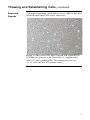
GIBCO® Rat (SD) Mesenchymal Stem Cells Catalog no. S1601-100 Revision date: 9 December 2009 Manual part no. A11561 MAN0001768 Corporate Headquarters Invitrogen Corporation 1600 Faraday Avenue Carlsbad, CA 92008 T: 1 760 603 7200 F: 1 760 602 6500 E: [email protected] For country-specific contact information visit our web site at www.invitrogen.com User Manual Table of Contents Contents and Storage........................................................................................... iv GIBCO® Rat (SD) Mesenchymal Stem Cells ......................................................1 Methods............................................................................................... 3 Handling GIBCO® Rat (SD) MSCs ......................................................................3 Thawing and Establishing Cells ..........................................................................5 Subculturing Cells..................................................................................................8 Freezing Cells........................................................................................................10 Differentiation Media ..........................................................................................12 Differentiating GIBCO® Rat (SD) MSCs ...........................................................14 Osteogenic Differentiation..................................................................................15 Adipogenic Differentiation.................................................................................17 Chondrogenic Differentiation ............................................................................19 Appendix ........................................................................................... 21 Troubleshooting ...................................................................................................21 Additional Products.............................................................................................23 Technical Support ................................................................................................24 Purchaser Notification.........................................................................................25 References..............................................................................................................27 iii Contents and Storage Contents Type of cells: GIBCO® Rat (SD) Mesenchymal Stem Cells Amount supplied: One vial containing ≥1 × 10⁶ viable cells. Composition: 1 mL of cells in freezing medium.* *Freezing medium: 60% D-MEM, 30% MSC-Qualified FBS, and 10% DMSO. Shipping and Storage GIBCO® Rat (SD) Mesenchymal Stem Cells are shipped on dry ice. Upon receipt, store the cells in liquid nitrogen. Handle the cells as potentially biohazardous material under at least Biosafety Level 1 (BL-1) containment. This product contains Dimethyl Sulfoxide (DMSO), a hazardous material. Review the Safety Data Sheet (SDS) before handling. Safety Data Sheets (SDSs) are available at www.invitrogen.com/sds. Intended Use iv GIBCO® Rat (SD) Mesenchymal Stem Cells are for research use only. They are not intended for any animal or human therapeutic or diagnostic use. GIBCO® Rat (SD) Mesenchymal Stem Cells Mesenchymal Stem Cells (MSCs) Mesenchymal Stem Cells (MSCs) are multipotent stem cells that have a large capacity for self-renewal while maintaining their multipotency. They can differentiate into multiple mature cell phenotypes in vitro, including adipocytes, osteocytes, and chondrocytes (De Ugarte et al., 2003; Meirelles Lda & Nardi, 2003; Pittenger et al., 1999; Wu et al., 2002). In vitro differentiation into non-mesenchymal cell types, such as neuronal and myogenic cells have also been described (AnjosAfonso et al., 2004; Deng et al., 2001; Han et al., 2002; Han et al., 2004; Moscoso et al., 2005; Phinney et al., 1999; Wakitani et al., 1995). In addition, MSCs are shown to be involved in certain types of cancers (Houghton et al., 2004; Singh et al., 2004), and are known to secrete immunomodulatory, anti-angiogenic, anti-inflammatory, pro-cardiovasculogenic, and proarteriogenic factors (Djouad et al., 2003; Gojo et al., 2003; Houghton et al., 2004; Kinnaird et al., 2004; Krampera et al., 2003; Oh et al., 2008; Olivares et al., 2004; Orlic et al., 2001). Source of GIBCO® Rat (SD) MSCs GIBCO® Rat (SD) Mesenchymal Stem Cells (MSCs) are produced from bone marrow isolated from Sprague Dawley (SD) rats at ≤ 8 weeks of gestation through mechanical and enzymatic digestion. The cells were isolated under sterile conditions, expanded in D-MEM medium (low glucose) containing 10% MSC-Qualified FBS, and cryopreserved at passage 4 (P4) in cryopreservation medium consisting of 60% D-MEM, 30% FBS, and 10% DMSO. Uses of GIBCO® Rat (SD) MSCs GIBCO® Rat (SD) MSCs can be used for studies of adult stem cell differentiation, tissue engineering, cell and gene therapy, and potential future clinical applications. Rat is a preferred animal model for transplantation studies, and GIBCO® Rat (SD) MSCs can be used in testing and evaluating MSCs in the host animal as the cells differentiate into mature phenotypes. We recommend that you use D-MEM (low glucose) with GlutaMAX™-I and MSC-Qualified FBS (see page 23) for optimal growth and expansion. Note: For some applications, such as chondrogenic differentiation, -MEM may be a better basal medium choice. Continued on next page 1 GIBCO® Rat (SD) MSCs, continued In vitro Growth Capacity The in vitro growth capacity of MSCs has not been definitely established and can vary greatly depending on the culture conditions such as seeding density and growth factors used, but the cells can be expected to expand for at least 30 population doublings before their growth rate decreases significantly (Bruder et al., 1997; Meirelles Lda & Nardi, 2003). GIBCO® Rat (SD) MSCs exhibit a population doubling time of ~20 to 30 hours when cultured in D-MEM (low glucose) with GlutaMAX™-I and MSC-Qualified FBS. Differentiation Potential Multiple investigators have demonstrated that MSCs can be differentiated towards multiple mature cell phenotypes. In addition to traditional mesenchymal lineages, MSCs have been differentiated towards cardiomyocytic and neuronal phenotypes using specialized media. The in vitro differentiation potential of MSCs has not been definitely established, but long-term culture and high cell density are implicated in the loss of differentiation potential (Meirelles Lda & Nardi, 2003). Characteristics of GIBCO® Rat (SD) MSCs 2 Prepared from low-passage (passage 4) adherent rat primary cell cultures Express a flow-cytometry cell-surface protein profile positive for CD29, CD44, CD90, and C106 (> 70%), and negative for CD11b, CD34, and CD45 (< 5%) Exhibit a population doubling time of ~20 to 30 hours Demonstrate at least tri-potential differentiation (i.e., can differentiate into osteogenic, adipogenic, and chondrogenic lineages) Methods Handling GIBCO® Rat (SD) MSCs As with other mammalian cell lines, handle GIBCO® Rat (SD) MSCs as potentially biohazardous material under at least Biosafety Level 1 (BL-1) containment. For more information on BL-1 guidelines, refer to Biosafety in Microbiological and Biomedical Laboratories, 5th ed., published by the Centers for Disease Control, or see the following website: www.cdc.gov/od/ohs/biosfty/bmbl5/bmbl5toc.htm Guidelines for GIBCO® Rat (SD) MSC Culture Important Follow the general guidelines below to grow and maintain GIBCO® Rat (SD) Mesenchymal Stem Cells. All solutions and equipment that come in contact with the cells must be sterile. Always use proper aseptic technique and work in a laminar flow hood. Before starting experiments, ensure cells have been established (at least 1 passage post-thaw), and also have some frozen stocks on hand. For differentiation studies and other experiments, we recommend using cells below passage 5 post-thaw. For general maintenance of cells, cell confluency should be 60–80%, cell viability should be at least 90%, and the growth rate should be in mid-logarithmic phase prior to subculturing. When thawing or subculturing cells, transfer cells into pre-warmed medium. Antibiotic-antimycotic containing penicillin, streptomycin, and amphotericin B may be used if required (see page 23 for ordering information). It is very important to strictly follow the guidelines for culturing GIBCO® Rat (SD) Mesenchymal Stem Cells in this manual to keep them undifferentiated. Continued on next page 3 Handling GIBCO® Rat (SD) MSCs, continued Media Requirements Important 4 We recommend culturing and expanding GIBCO® Rat (SD) MSCs in Dulbecco’s Modified Eagle Medium (D-MEM) (low glucose) with GlutaMAX™-I and supplemented with 10% MSC-Qualified Fetal Bovine Serum (FBS) for optimal growth performance, and to keep the MSCs undifferentiated (see page 23 for ordering information). Prepare your growth medium prior to use. When thawing or subculturing MSCs, transfer them into pre-warmed medium at 37°C. You may store the complete growth medium in the dark at 4°C for up to four weeks. Avoid repeated freeze-thaw cycles of MSC-Qualified FBS. We have observed that a small percentage of GIBCO® Rat (SD) MSCs adhere poorly after their initial thaw; however, the cells recover and adhere well after their first passage. We recommend that you treat your cells gently (i.e., do not vortex, bang the flasks to dislodge the cells, or centrifuge the cells at high speeds). Thawing and Establishing Cells Materials Needed Rat MSC Growth Medium GIBCO® Rat (SD) MSCs, stored in liquid nitrogen Ethanol or 70% isopropanol Rat MSC growth medium (see below); pre-warmed to 37°C Disposable, sterile 15-mL and 50-mL tubes 37°C water bath 37°C incubator with a humidified atmosphere of 5% CO2 Microcentrifuge Tissue-culture treated flasks, plates or dishes Hemacytometer, cell counter and Trypan Blue, LIVE/DEAD® Cell Vitality Assay Kit, or the Countess™ Automated Cell Counter Rat MSC growth medium consists of D-MEM (low glucose) medium with GlutaMAX™-I, 10% MSC-Qualified FBS, and 5 μg/mL gentamycin reagent solution. To prepare 500 mL of Rat MSC growth medium, aseptically mix the following (see page 23 for ordering information): Component D-MEM medium (low glucose) with GlutaMAX™-I FBS, MSC-Qualified Gentamicin (10 mg/mL) Final Conc. For 500 mL 1X 450 mL 10% 50 mL 5 μg/mL 250 μL Note: GIBCO® Rat (SD) MSCs show comparable morphology and performance when -MEM with GlutaMAX™-I is substituted for D-MEM medium (low glucose) with GlutaMAX™-I. Invitrogen’s Countess™ Automated Cell Counter is a benchtop counter designed to measure cell count and viability (live, dead, and total cells) accurately and precisely in less than a minute per sample, using the standard Trypan Blue technique (see page 23 for ordering information). Continued on next page 5 Thawing and Establishing Cells, continued Thawing Procedure 1. Pre-warm the prepared rat MSC growth medium (page 5) to 37°C. 2. Remove the cells from liquid nitrogen storage, and wipe the cryovial with ethanol or 70% isopropanol before opening. In an aseptic field, briefly twist the cap a quarter turn to relieve pressure and then re-tighten. Do not expose cells to air before thawing. 3. Quickly thaw the vial of cells by swirling it in a 37°C water bath, and remove it when the last bit of ice has melted, typically < 2 minutes. Do not submerge the vial completely. Do not thaw the cells for longer than 2 minutes. 4. When thawed, wipe the vial with ethanol or 70% isopropanol to sterilize it, and immediately transfer the cells into a 50-mL sterile tube. Slowly add pre-warmed rat MSC growth medium to the cells dropwise up to 10 mL while swirling the tube to mix. 5. Centrifuge the cells for 5 minutes at 300 g. 6. Aspirate the supernatant and resuspend the cells in 2 mL of rat MSC growth medium. 7. Take a 50 μL aliquot and determine the viable cell count using your method of choice. 8. Calculate the total number of viable cells, and add enough rat MSC growth medium to the cells to generate a cell solution at 1 106 cells/mL. Plate the resuspended cells at a seeding density of 3,000 cells per cm2. 9. Note: A seeding density of 3 103 viable cells/cm2 is equivalent to 2.25 105 cells for a T75 flask and 6.75 105 cells for a T225 flask. Each vial contains roughly 1 106 cells. 10. Following inoculation, swirl the medium in the flasks to evenly distribute the cells. 11. Incubate the cells at 37°C, 5% CO2 and 90% humidity, and allow the cells to adhere for at least 24 hours. 12. The next day, replace the medium with an equal volume of fresh, pre-warmed rat MSC growth medium. 13. Change the medium every 2–3 days. Continued on next page 6 Thawing and Establishing Cells, continued Expected Results The bright field image (100X) below shows GIBCO® Rat (SD) Mesenchymal Stem Cells 4 days after thaw. Figure 1. GIBCO® Rat (SD) MSCs thawed and expanded on D-MEM (low glucose) with GlutaMAX™-I supplemented with 10% MSC-qualified FBS. The seeding density was 3 × 10³ cells/cm2 in a T75 culture vessel. 7 Subculturing Cells When to Subculture Subculture GIBCO® Rat (SD) MSCs when they are near confluency, typically every 3–5 days. Materials Needed Culture vessels containing GIBCO® Rat (SD) MSCs Tissue-culture treated flasks, plates or dishes Rat MSC growth medium (page 5), pre-warmed to 37°C Disposable, sterile 15-mL and 50-mL tubes 37°C incubator with humidified atmosphere of 5% CO2 Dulbecco’s Phosphate Buffered Saline (D-PBS), containing no calcium, magnesium, or phenol red TrypLE™ Express Dissociation Reagent, pre-warmed to 37°C Hemacytometer, cell counter and Trypan Blue, LIVE/DEAD® Cell Vitality Assay Kit, or the Countess™ Automated Cell Counter 1. Aspirate the spent rat MSC growth medium from the culture vessel. 2. Rinse the surface of the cell layer with D-PBS without Ca2+ and Mg2+ (approximately 1–2 mL of D-PBS per 10 cm2 culture surface area) by adding the D-PBS to the side of the vessel opposite the attached cell layer, and rocking the vessel back and forth several times. 3. Aspirate and discard the D-PBS. 4. Add a sufficient volume of pre-warmed TrypLE™ Express to cover the cell layer (5 mL for T75 or 10 mL for T225). 5. Incubate the cells at 37˚C for 3–6 minutes. Passaging Cells Procedure continued on next page Continued on next page 8 Subculturing Cells, continued Passaging Cells, continued Procedure continued from previous page 6. Observe the cells under a microscope. If the cells are less than 90% detached, continue incubating the cells and observe within 2 minutes for complete detachment of the cells. You may tap the vessel gently to expedite cell detachment. 7. Once detached, pipet the cell solution up and down a few times to generate a homogenous suspension. Transfer the cell suspension to 15-mL tube. 8. Take a 50 μL aliquot and determine the total number of cells and percent viability using your method of choice. 9. During the cell count, centrifuge the rest of the cells at 300 g for 5 minutes at room temperature. Aspirate and discard the medium without disturbing the cell pellet. 10. Calculate the total number of vessels to inoculate by using the following equation: Number of vessels = Number of viable cells ÷ (growth area of vessel in cm2 × 3,000 cells per cm2 recommended seeding density) 11. Add rat MSC growth medium to each vessel so that the final culture volume is 0.2–0.5 mL per cm2. 12. Add the appropriate volume of cells to each vessel and incubate the vessels at 37°C, 5% CO2 and 90% humidity. 13. 2–3 days after seeding, completely remove the spent medium from the culture vessels and replace it with an equal volume of pre-warmed rat MSC growth medium. 9 Freezing Cells Materials Needed Guidelines Preparing Freezing Media Culture vessels of GIBCO® Rat (SD) MSCs Rat MSC growth medium Fetal Bovine Serum, MSC-Qualified DMSO (use a bottle set aside for cell culture; open only in a laminar flow hood) Disposable, sterile 15-mL conical tubes. D-PBS, containing no calcium, magnesium, or phenol red TrypLE™ Express Hemacytometer, cell counter and Trypan Blue, LIVE/DEAD® Cell Vitality Assay Kit, or the Countess™ Automated Cell Counter Sterile freezing vials When freezing MSCs, we recommend the following: Freeze cells at a density of 1–2 × 10⁶ viable cells/mL. Use a freezing medium composed of final concentrations of 30% MSC-Qualified FBS and 10% DMSO. Bring the cells into freezing medium in two steps. Prepare Freezing Medium A and B immediately before use. You will need enough of each freezing medium to resuspend cells at a density of 1–2 × 10⁶ cells/mL (see the freezing procedure below). 1. In a sterile 15-mL tube, mix together the following reagents for every 1 mL of Freezing Medium A needed: D-MEM medium (low glucose) with GlutaMAX™-I FBS, MSC-Qualified 2. 0.6 mL In another sterile 15-mL tube, mix together the following reagents for every 1 mL of Freezing Medium B needed: D-MEM medium (low glucose) with GlutaMAX™-I DMSO 3. 0.4 mL 0.8 mL 0.2 mL Place tube with Freezing Medium B on ice until use (leave Freezing Medium A at room temperature). Note: Discard any remaining freezing medium after use. Continued on next page 10 Freezing Cells, continued Procedure for Freezing Cells 1. Aspirate the rat MSC growth medium from the culture flask. 2. Follow the Passaging Cells protocol, steps 2–9 (pages 8–9). 3. Gently aspirate the medium from the vessel and resuspend the cells to a concentration of 2–4 × 10⁶ cells/mL in Freezing Medium A. 4. Add the same volume of Freezing Medium B to the cells in a dropwise manner. 5. Aliquot 1 mL of the cell suspension to each freezing vial, and store at –80°C overnight in an isopropanol chamber. 6. The next day, transfer the frozen vials to a liquid nitrogen tank (vapor phase) for long-term storage. Note: You may check the viability and recovery of frozen cells 24 hours after storing cryovials in liquid nitrogen by following the procedure outlined in Thawing and Establishing Cells, page 6. 11 Differentiation Media Introduction One critical hallmark of MSCs is their ability to differentiate into three or more mature cell types. Traditional and modern bioassays are used to demonstrate the multipotency of MSCs to differentiate along the osteogenic, adipogenic, and chondrogenic lineages. This section provides guidelines for preparing media that are used for inducing GIBCO® Rat (SD) MSCs to differentiate into osteogenic, adipogenic and chondrogenic cell types. Mesenchymal Stem Cell Basal Medium MSC basal medium is used as a cell attachment medium and as a negative control during differentiation experiments. It consists of -MEM medium with GlutaMAX™-I containing 10% MSCQualified FBS and 5 μL/mL gentamicin (see page 23). Component -MEM medium with GlutaMAX™-I FBS, MSC-Qualified Gentamicin (10 mg/mL) Osteogenic Differentiation Medium Final Conc. For 500 mL 1X 450 mL 10% 50 mL 5 μg/mL 250 μL To prepare osteogenic differentiation (OD) medium, combine the following in a sterile flask. You may use the StemPro® Osteocyte/Chondrocyte Differentiation Basal Medium or the -MEM as the basal medium. Store the OD medium at 4°C in the dark up to four weeks. Note: We recommend testing both basal media to find the optimal induction media for your osteogenic cultures. Component StemPro® Osteocyte/Chondrocyte Differentiation Basal Medium or Final Conc. For 100 mL 1X 90 mL 1X 10 mL 5 μg/mL 50 μL -MEM medium with GlutaMAX™-I StemPro® Osteogenesis Supplement Gentamicin (10 mg/mL) Continued on next page 12 Differentiation Media, continued Adipogenic Differentiation Medium To prepare adipogenic differentiation (AD) medium, combine the following in a sterile flask. You may use the StemPro® Adipocyte Differentiation Basal Medium or the -MEM as the basal medium. Store the AD medium at 4°C in the dark up to four weeks. Note: We recommend testing both basal media to find the optimal induction media for your adipogenic cultures. Component Final Conc. For 100 mL StemPro Adipocyte Differentiation Basal Medium or -MEM medium with GlutaMAX™-I 1X 90 mL StemPro® Adipogenesis Supplement 1X 10 mL Gentamicin (10 mg/mL) 5 μg/mL 50 μL ® Chondrogenic Differentiation Medium To prepare chondrogenic differentiation (CD) medium, combine the following in a sterile flask. You may use the StemPro® Osteocyte/Chondrocyte Differentiation Basal Medium, or the -MEM as the basal medium. Store the CD medium at 4°C in the dark up to four weeks. Note: We recommend testing both basal media to find the optimal induction media for your chondrogenic cultures. Component Final Conc. For 100 mL StemPro Osteocyte/Chondrocyte Differentiation Basal Medium or -MEM medium with GlutaMAX™-I 1X 90 mL StemPro® Chondrogenesis Supplement 1X 10 mL Gentamicin (10 mg/mL) 5 μg/mL 50 μL ® 13 Differentiating GIBCO® Rat (SD) MSCs Materials Needed Harvesting MSCs Culture vessels containing GIBCO® Rat (SD) MSCs Tissue-culture treated flasks, plates, or dishes MSC Basal Medium, prewarmed to 37°C (see page 12) Appropriate Differentiation Medium, pre-warmed to 37°C (see pages 12–13) Dulbecco’s Phosphate Buffered Saline (D-PBS), containing no calcium, magnesium, or phenol red Disposable, sterile 50-mL tubes 37°C incubator with humidified atmosphere of 5% CO2 TrypLE™ Express, pre-warmed to 37°C Hemacytometer, cell counter and Trypan Blue, LIVE/DEAD® Cell Vitality Assay Kit, or the Countess™ Automated Cell Counter Follow the protocol below to harvest GIBCO® Rat (SD) MSCs for differentiation experiments. We recommend that you expand your cells to 70% confluency in a tissue-culture treated T-225 flask, and prepare the appropriate differentiation medium ahead of time. 1. Aspirate the spent growth from the flask. 2. Follow the Passaging Cells protocol, steps 2–9 (pages 8–9). 3. Calculate required amount of MSC basal medium to obtain the appropriate seeding concentration (see differentiation protocols, pages 15–19). Resuspend the cells in the appropriate amount of MSC basal medium. 4. 5. 14 Dispense the cell solution according to the differentiation condition being tested (see the differentiation protocols, pages 15–19). Osteogenic Differentiation Osteogenic Differentiation Protocol 1. Seed the MSCs into culture vessels at 0.5 104 cells/cm2. For classical stain differentiation assays, seed the cells into a 12-well plate. For gene-expression profile studies, seed the cells into a T-75 flask. For immunocytochemistry studies, seed the cells into a 16-well CultureWell™ chambered coverglass or 96-well plate. 2. To six wells of a 12-well plate, add 1 mL of the cell solution per well and let the cells attach to the plate in the 37°C, 5% CO2 incubator for a minimum of two hours. Note: Culturing the cells up to four days in MSC basal medium before switching to OD medium has been shown to enhance osteogenic differentiation. 3. Replace three of the wells with MSC basal medium as negative controls, and the other three wells with fresh OD medium. Incubate the cultures at 37°C with 5% CO2. 4. Refeed the cultures every 2–3 days with the media prepared at initiation of differentiation. The MSCs will continue to expand as they differentiate under the osteogenic conditions. 5. After specific periods of cultivation, osteogenic cultures can be processed for alkaline phosphatase staining (7–14 days) or Alizarin Red S staining (>21 days), gene expression analysis, or protein detection. For long term culture (>21 days), we recommend that you reduce the seeding density by half to prevent overgrowth. Continued on next page 15 Osteogenic Differentiation, continued Expected Results The bright field images below show GIBCO® Rat (SD) MSCs at P3 post-thaw induced to differentiate along the osteogenic lineage. Figure 2. GIBCO® Rat (SD) MSCs at P3 post-thaw were differentiated on -MEM basal medium containing StemPro® Osteogenesis Supplement for 28 days, and stained with Alizarin Red S. The images were obtained using 4X (top) and 10X (bottom) objectives. 16 Adipogenic Differentiation Adipogenic Differentiation Protocol 1. Seed the MSCs into culture vessels at 2.0 104 cells/cm2. For classical stain differentiation assays, seed the cells into a 12-well plate. For gene-expression profile studies, seed the cells into a T-75 flask. For immunocytochemistry studies, seed the cells into a 16-well CultureWell™ chambered coverglass or 96-well plate. 2. To six wells of a 12-well plate, add 1 mL of cell solution per well, and let the cells attach to the plate in the 37°C, 5% CO2 incubator for a minimum of two hours. Note: Culturing the cells up to four days in MSC basal medium before switching to OD medium has been shown to enhance osteogenic differentiation. 3. Replace three of the wells with MSC basal medium as negative controls, and the other three wells with fresh AD medium. Incubate the cultures at 37°C and 5% CO2. 4. Refeed the cultures every 3–4 days with the media prepared at initiation of differentiation. The MSCs will continue to undergo limited expansion as they differentiate under adipogenic conditions. 5. After specific periods of cultivation, adipogenic cultures can be processed for Oil Red O or LipidTOX™ staining (beginning at 7–14 days), gene expression analysis, or protein detection. Continued on next page 17 Adipogenic Differentiation, continued Expected Results The bright field images below show GIBCO® Rat (SD) MSCs at P3 post-thaw induced to differentiate along the adipogenic lineage. Figure 3. GIBCO® Rat (SD) MSCs were differentiated into adipocytes on -MEM basal medium containing StemPro® Adipogenesis Supplement for 16 days, and stained with Oil Red O. The images were obtained using 200X (top) and 400X (bottom) objectives. 18 Chondrogenic Differentiation Chondrogenic Differentiation Protocol 1. Detach the cells using TrypLE™ Express and perform a cell count as described in Harvesting MSCs, page 14 (through Step 3). 2. Resuspend the cells in MSC basal medium to a concentration of 8 × 10⁶ cells/mL. 3. To each of the six wells of a 12-well tissue-culture dish, spot 10 μL of cells. 4. Incubate the plate for two hours at 37°C, 5% CO2 and 90% humidity. Note: If this step is not performed under high humidity conditions, the spots may dehydrate, inhibiting the formation of chondrogenic pellets. 5. To three of the spotted wells, add 1 mL of MSC basal medium as a negative control. To the other three wells, add 1 mL of CD medium. 6. Incubate the plate at 37°C, 5% CO2, and 90% humidity. Refeed the cultures every 2–3 days with the same media, prepared at the initiation of differentiation. 7. Check for chondrogenesis after a set period of cultivation. You may perform alcian blue staining on the pellets (to detect glycosaminoglycans) after 14 days, or paraffin section of pellets for collagen 2a immunohistological staining after ~21 days. Continued on next page 19 Chondrogenic Differentiation, continued Expected Results The bright field image below shows GIBCO® Rat (SD) MSCs at P3 post-thaw induced to differentiate into chondrocytes. Figure 4. GIBCO® Rat (SD) MSCs were differentiated on -MEM basal medium containing StemPro® Chondrogenesis Supplement for 28 days, and stained with Alcian Blue. The images were obtained using 4X (top) and 10X (bottom) objectives. 20 Appendix Troubleshooting Culturing Cells The table below lists some potential problems and solutions that may help you troubleshoot your cell culture problems. Problem Cause Solution No viable cells after thawing stock Stock not stored correctly Order new stock and store in liquid nitrogen. Keep in liquid nitrogen until thawing. Freeze cells at a density of 1–2 × 10⁶ viable cells per mL. Use low-passage cells to make your own stocks. Follow procedures in Freezing Cells (pages 10–11) exactly. Slow freezing and fast thawing is the key. Add Freezing Medium B drop wise manner (slowly). At time of thawing, thaw quickly and do not expose vial to the air but quickly change from nitrogen tank to 37°C water bath. Obtain new GIBCO® Rat (SD) MSCs. Use pre-warmed rat MSC growth medium, prepared as described on page 5. Be sure to use MSC-Qualified FBS. Generally we recommend seeding the culture vessels at a density of 3,000 cells per cm2. GIBCO® Rat (SD) MSCs are fragile; treat your cells gently, do not vortex, bang the flasks to dislodge the cells, or centrifuge the cells at high speeds. Use prewarmed rat MSC growth medium. Home-made stock not viable Thawing medium not correct Cells too diluted Cell not handled gently Cells grow slowly Growth medium not correct Cells too old Use healthy MSCs, under passage 5 postthaw; do not overgrow or passage more than 5 times. Continued on next page 21 Troubleshooting, continued Culturing Cells, The table below lists some potential problems and solutions that may help you troubleshoot your cell culture problems. continued Problem Cause Solution Cells differentiated Culture conditions not correct Thaw and culture a fresh vial of GIBCO® Rat (SD) MSCs. Follow the thawing instructions (page 6) and the subculture procedures (pages 8–9) exactly. MSCs above passage 7 post-thaw may lose their multipotency and become more differentiated. Be sure to prepare your culture medium using MSC-Qualified FBS (see page 23 for ordering information). Cells too old Cells not adherent after initial thaw Used serum other than MSCQualified FBS Differentiating Cells The table below lists some potential problems and solutions that may help you troubleshoot your cell culture problems. Problem Cause Solution Cells fail to differentiate Used StemPro® Osteocyte/Chondrocyte or Adipocyte Differentiation Basal Media Although you may use the StemPro® Osteocyte/Chondrocyte or Adipocyte Differentiation Basal Media for your differentiation studies, we have observed that differentiation can be more efficient with -MEM as the basal media. Repeat your differentiation studies using -MEM as the basal media If this step is not performed under high humidity conditions, the spots may dehydrate and the formation of chondrogenic plates inhibited. Repeat the initial spotting step at 37°C, 5% CO2, and 90% humidity, and incubate the culture in a humidified box with loose-fitting cover or aluminum foil perforated with small holes. For long term culture (>21 days), we recommend that you seed at a lower cell density to prevent overgrowth and cell detachment. Initial spotting step not performed under high humidity (if differentiating into chondrocytes) Cells have overgrown the culture plates and have detached 22 Initial seeding density too high (if differentiating into osteocytes) Additional Products Additional Products The products listed in this section may be used with GIBCO® Rat (SD) Mesenchymal Stem Cells. For more information, refer to our website (www.invitrogen.com) or contact Technical Support (see page 24). Quantity Cat. no. StemPro® Alk Phos-expressing Rat Mesenchymal Stem Cells Item 1 × 10⁶ cells R7789-120 Dulbecco’s Modified Eagle Medium (D-MEM) (1X), low glucose with GlutaMAX™-I 500 mL 10567-014 Minimum Essential Medium (MEM) Medium (1X) with GlutaMAX™-I, ribonucleosides and deoxyribonucleosides 500 mL 32571-036 GlutaMAX™-I Supplement 100 mL 35050-061 Fetal Bovine Serum (FBS), MSC-Qualified 100 mL 500 mL 12662-011 12662-029 StemPro® Adipogenesis Differentiation Kit 100 mL A10070-01 StemPro® Chondrogenesis Differentiation Kit 100 mL A10071-01 StemPro® Osteogenesis Differentiation Kit 100 mL A10072-01 Gentamicin (10 mg/mL) 10 mL 15710-064 Dulbecco’s Phosphate Buffered Saline (D-PBS), containing no calcium, magnesium, or phenol red 500 mL 14190-144 TrypLE™ Express Dissociation Enzyme 100 mL 12604-013 Antibiotic-Antimycotic (100X), liquid 100 mL 15240-062 Gentamycin Reagent Solution (10 mg/mL), liquid 10 mL 15710-064 Gentamycin Reagent Solution (50 mg/mL), liquid 10 mL 15750-060 Trypan Blue Stain 100 mL 15250-061 HCS LipidTOX™ Green neutral lipid stain 1 each H34475 LIVE/DEAD® Cell Vitality Assay Kit Countess™ Automated Cell Counter (includes 50 Countess™ cell counting chamber slides and 2 mL of Trypan Blue Stain) CultureWell™ chambered coverglass (16 wells per coverglass, set of 8) 1000 assays L34951 1 unit C10227 1 set C37000 23 Technical Support Web Resources Contact Us Visit the Invitrogen website at www.invitrogen.com for: Technical resources, including manuals, vector maps and sequences, application notes, SDSs, FAQs, formulations, citations, handbooks, etc. Complete Technical Support contact information Access to the Invitrogen Online Catalog Additional product information and special offers For more information or technical assistance, call, write, fax, or email. Additional international offices are listed on our website (www.invitrogen.com). Corporate Headquarters: 5791 Van Allen Way Carlsbad, CA 92008 USA Tel: 1 760 603 7200 Tel (Toll Free): 1 800 955 6288 Fax: 1 760 602 6500 E-mail: [email protected] Japanese Headquarters: LOOP-X Bldg. 6F 3-9-15, Kaigan Minato-ku, Tokyo 108-0022 Tel: 81 3 5730 6509 Fax: 81 3 5730 6519 E-mail: [email protected] European Headquarters: Inchinnan Business Park 3 Fountain Drive Paisley PA4 9RF, UK Tel: 44 (0) 141 814 6100 Tech Fax: 44 (0) 141 814 6117 E-mail: [email protected] Material Safety Data Sheets (SDSs) Safety Data Sheets (SDSs) are available on our website at www.invitrogen.com/sds. Certificate of Analysis The Certificate of Analysis provides detailed quality control information for each product. Certificates of Analysis are available on our website. Go to www.invitrogen.com/support and search for the Certificate of Analysis by product lot number, which is printed on the box. 24 Purchaser Notification Limited Warranty Invitrogen (a part of Life Technologies Corporation) is committed to providing our customers with high-quality goods and services. Our goal is to ensure that every customer is 100% satisfied with our products and our service. If you should have any questions or concerns about an Invitrogen product or service, contact our Technical Support Representatives. All Invitrogen products are warranted to perform according to specifications stated on the certificate of analysis. The Company will replace, free of charge, any product that does not meet those specifications. This warranty limits the Company’s liability to only the price of the product. No warranty is granted for products beyond their listed expiration date. No warranty is applicable unless all product components are stored in accordance with instructions. The Company reserves the right to select the method(s) used to analyze a product unless the Company agrees to a specified method in writing prior to acceptance of the order. Invitrogen makes every effort to ensure the accuracy of its publications, but realizes that the occasional typographical or other error is inevitable. Therefore the Company makes no warranty of any kind regarding the contents of any publications or documentation. If you discover an error in any of our publications, please report it to our Technical Support Representatives. Life Technologies Corporation shall have no responsibility or liability for any special, incidental, indirect or consequential loss or damage whatsoever. The above limited warranty is sole and exclusive. No other warranty is made, whether expressed or implied, including any warranty of merchantability or fitness for a particular purpose. Continued on next page 25 Purchaser Notification, continued Limited Use Label License No. 5: Invitrogen Technology 26 The purchase of this product conveys to the buyer the nontransferable right to use the purchased amount of the product and components of the product in research conducted by the buyer (whether the buyer is an academic or for-profit entity). The buyer cannot sell or otherwise transfer (a) this product (b) its components or (c) materials made using this product or its components to a third party or otherwise use this product or its components or materials made using this product or its components for Commercial Purposes. The buyer may transfer information or materials made through the use of this product to a scientific collaborator, provided that such transfer is not for any Commercial Purpose, and that such collaborator agrees in writing (a) not to transfer such materials to any third party, and (b) to use such transferred materials and/or information solely for research and not for Commercial Purposes. Commercial Purposes means any activity by a party for consideration and may include, but is not limited to: (1) use of the product or its components in manufacturing; (2) use of the product or its components to provide a service, information, or data; (3) use of the product or its components for therapeutic, diagnostic or prophylactic purposes; or (4) resale of the product or its components, whether or not such product or its components are resold for use in research. For products that are subject to multiple limited use label licenses, the terms of the most restrictive limited use label license shall control. Life Technologies Corporation will not assert a claim against the buyer of infringement of patents owned or controlled by Life Technologies Corporation which cover this product based upon the manufacture, use or sale of a therapeutic, clinical diagnostic, vaccine or prophylactic product developed in research by the buyer in which this product or its components was employed, provided that neither this product nor any of its components was used in the manufacture of such product. If the purchaser is not willing to accept the limitations of this limited use statement, Life Technologies is willing to accept return of the product with a full refund. For information about purchasing a license to use this product or the technology embedded in it for any use other than for research use please contact Out Licensing, Life Technologies, 5791 Van Allen Way, Carlsbad, California 92008 or [email protected]. References Anjos-Afonso, F., Siapati, E. K., and Bonnet, D. (2004) In vivo contribution of murine mesenchymal stem cells into multiple cell-types under minimal damage conditions. J Cell Sci 117, 5655-5664 Bruder, S. P., Jaiswal, N., and Haynesworth, S. E. (1997) Growth kinetics, selfrenewal, and the osteogenic potential of purified human mesenchymal stem cells during extensive subcultivation and following cryopreservation. J Cell Biochem 64, 278-294 De Ugarte, D. A., Morizono, K., Elbarbary, A., Alfonso, Z., Zuk, P. A., Zhu, M., Dragoo, J. L., Ashjian, P., Thomas, B., Benhaim, P., Chen, I., Fraser, J., and Hedrick, M. H. (2003) Comparison of multi-lineage cells from human adipose tissue and bone marrow. Cells Tissues Organs 174, 101109 Deng, W., Obrocka, M., Fischer, I., and Prockop, D. J. (2001) In vitro differentiation of human marrow stromal cells into early progenitors of neural cells by conditions that increase intracellular cyclic AMP. Biochem Biophys Res Commun 282, 148-152 Djouad, F., Plence, P., Bony, C., Tropel, P., Apparailly, F., Sany, J., Noel, D., and Jorgensen, C. (2003) Immunosuppressive effect of mesenchymal stem cells favors tumor growth in allogeneic animals. Blood 102, 3837-3844 Gojo, S., Gojo, N., Takeda, Y., Mori, T., Abe, H., Kyo, S., Hata, J., and Umezawa, A. (2003) In vivo cardiovasculogenesis by direct injection of isolated adult mesenchymal stem cells. Exp Cell Res 288, 51-59 Han, S. S., Kang, D. Y., Mujtaba, T., Rao, M. S., and Fischer, I. (2002) Grafted lineage-restricted precursors differentiate exclusively into neurons in the adult spinal cord. Exp Neurol 177, 360-375 Han, S. S., Liu, Y., Tyler-Polsz, C., Rao, M. S., and Fischer, I. (2004) Transplantation of glial-restricted precursor cells into the adult spinal cord: survival, glial-specific differentiation, and preferential migration in white matter. Glia 45, 1-16 Houghton, J., Stoicov, C., Nomura, S., Rogers, A. B., Carlson, J., Li, H., Cai, X., Fox, J. G., Goldenring, J. R., and Wang, T. C. (2004) Gastric cancer originating from bone marrow-derived cells. Science 306, 1568-1571 Kinnaird, T., Stabile, E., Burnett, M. S., Shou, M., Lee, C. W., Barr, S., Fuchs, S., and Epstein, S. E. (2004) Local delivery of marrow-derived stromal cells augments collateral perfusion through paracrine mechanisms. Circulation 109, 1543-1549 Kisseberth, W. C., Brettingen, N. T., Lohse, J. K., and Sandgren, E. P. (1999) Ubiquitous expression of marker transgenes in mice and rats. Dev Biol 214, 128-138 Continued on next page 27 References, continued Krampera, M., Glennie, S., Dyson, J., Scott, D., Laylor, R., Simpson, E., and Dazzi, F. (2003) Bone marrow mesenchymal stem cells inhibit the response of naive and memory antigen-specific T cells to their cognate peptide. Blood 101, 3722-3729 Meirelles Lda, S., and Nardi, N. B. (2003) Murine marrow-derived mesenchymal stem cell: isolation, in vitro expansion, and characterization. Br J Haematol 123, 702-711 Moscoso, I., Centeno, A., Lopez, E., Rodriguez-Barbosa, J. I., Santamarina, I., Filgueira, P., Sanchez, M. J., Dominguez-Perles, R., Penuelas-Rivas, G., and Domenech, N. (2005) Differentiation "in vitro" of primary and immortalized porcine mesenchymal stem cells into cardiomyocytes for cell transplantation. Transplant Proc 37, 481-482 Mujtaba, T., Han, S. S., Fischer, I., Sandgren, E. P., and Rao, M. S. (2002) Stable expression of the alkaline phosphatase marker gene by neural cells in culture and after transplantation into the CNS using cells derived from a transgenic rat. Exp Neurol 174, 48-57 Oh, J. Y., Kim, M. K., Shin, M. S., Lee, H. J., Ko, J. H., Wee, W. R., and Lee, J. H. (2008) The anti-inflammatory and anti-angiogenic role of mesenchymal stem cells in corneal wound healing following chemical injury. Stem Cells 26, 1047-1055 Olivares, E. L., Ribeiro, V. P., Werneck de Castro, J. P., Ribeiro, K. C., Mattos, E. C., Goldenberg, R. C., Mill, J. G., Dohmann, H. F., dos Santos, R. R., de Carvalho, A. C., and Masuda, M. O. (2004) Bone marrow stromal cells improve cardiac performance in healed infarcted rat hearts. Am J Physiol Heart Circ Physiol 287, H464-470 Orlic, D., Kajstura, J., Chimenti, S., Jakoniuk, I., Anderson, S. M., Li, B., Pickel, J., McKay, R., Nadal-Ginard, B., Bodine, D. M., Leri, A., and Anversa, P. (2001) Bone marrow cells regenerate infarcted myocardium. Nature 410, 701-705 Phinney, D. G., Kopen, G., Isaacson, R. L., and Prockop, D. J. (1999) Plastic adherent stromal cells from the bone marrow of commonly used strains of inbred mice: variations in yield, growth, and differentiation. J Cell Biochem 72, 570-585 Pittenger, M. F., Mackay, A. M., Beck, S. C., Jaiswal, R. K., Douglas, R., Mosca, J. D., Moorman, M. A., Simonetti, D. W., Craig, S., and Marshak, D. R. (1999) Multilineage potential of adult human mesenchymal stem cells. Science 284, 143-147 Singh, S. K., Hawkins, C., Clarke, I. D., Squire, J. A., Bayani, J., Hide, T., Henkelman, R. M., Cusimano, M. D., and Dirks, P. B. (2004) Identification of human brain tumour initiating cells. Nature 432, 396401 Continued on next page 28 References, continued Spees, J. L., Olson, S. D., Ylostalo, J., Lynch, P. J., Smith, J., Perry, A., Peister, A., Wang, M. Y., and Prockop, D. J. (2003) Differentiation, cell fusion, and nuclear fusion during ex vivo repair of epithelium by human adult stem cells from bone marrow stroma. Proc Natl Acad Sci U S A 100, 23972402 Wakitani, S., Saito, T., and Caplan, A. I. (1995) Myogenic cells derived from rat bone marrow mesenchymal stem cells exposed to 5-azacytidine. Muscle Nerve 18, 1417-1426 Wu, Y. Y., Mujtaba, T., Han, S. S., Fischer, I., and Rao, M. S. (2002) Isolation of a glial-restricted tripotential cell line from embryonic spinal cord cultures. Glia 38, 65-79 ©2009 Life Technologies Corporation. All rights reserved. For research use only. Not intended for any animal or human therapeutic or diagnostic use. 29 Corporate Headquarters 5791 Van Allen Way Carlsbad, CA 92008 T: 1 760 603 7200 F: 1 760 602 6500 E: [email protected] For country-specific contact information, visit our web site at www.invitrogen.com User Manual