1
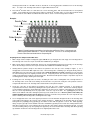
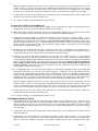
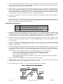
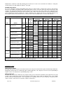
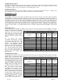
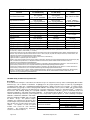
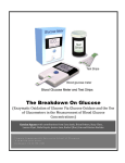
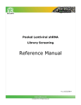
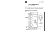
®
BacTx® Bacterial Detection Kit for Platelets
Immunetics Catalog Number: DK-B502-032
32 Tests
INTENDED USE:
®
The Immunetics BacTx Bacterial Detection Kit for detection of bacteria in platelets is a rapid, qualitative, colorimetric, quality
control test for the detection of aerobic and anaerobic, Gram-positive and Gram-negative bacteria in:
-leukocyte reduced apheresis platelet units (LRAP) (Apheresis Platelets Leukocytes Reduced) as a quality control test following
testing with a growth based bacterial detection device cleared by the FDA for quality control testing of leukocyte reduced
apheresis platelets and;
-pools of up to six (6) units of leukocyte reduced whole blood-derived platelets (Platelets, Leukocytes Reduced) that are pooled
within four (4) hours of transfusion.
INTRODUCTION:
Bacteria are the most common contaminating infectious agents found in platelet units. It has been estimated that the rate of
bacterial contamination for apheresis platelet donations is 1 in 5000, making platelets the most frequent source of transfusion1
º
related infection . Since platelets must be stored at 20 - 24 C in order to maintain function, small numbers of bacteria that are
2
mostly introduced from a donor’s skin flora into platelet units can multiply to very high numbers in a matter of days . Skin flora
are not the only source of bacteria that contaminate platelet units; many strains of Gram-positive and Gram-negative bacteria
have been identified in contaminated platelet units, including Staphylococcus, Pseudomonas, Bacillus, and Streptococcus
3
species . To increase transfusion safety, the AABB issued directive 5.1.5.1 in March 2004 requiring blood banks and
4
transfusion services to implement bacterial testing methods on all platelet units . Further safeguards were made effective in
January 2011 with the implementation of AABB Interim directive 5.1.5.1.1, which specified that such bacterial testing methods
5
must have prior FDA clearance or must demonstrate equivalent sensitivity to FDA-cleared methods .
®
The BacTx Kit is a qualitative colorimetric assay which detects bacteria in platelet samples through recognition of
peptidoglycan, a ubiquitous component of bacterial cell walls. The kit consists of lyophilized peptidoglycan detection reagents,
sample preparation reagents, controls and microfuge tubes. In the presence of peptidoglycan, the assay generates a redcolored product indicating the presence of bacteria in the platelet sample. The reaction product is detected by a photometer and
results interpreted automatically by PC-based software.
PRINCIPLE:
®
The BacTx Assay detects bacteria in both Apheresis Platelets Leukocytes Reduced (LRAP), and pools of up to six (6) units of
leukocyte reduced whole blood-derived platelets (LR-WBDP) that are pooled within four (4) hours of transfusion. Gram positive,
®
Gram negative, aerobic and anaerobic bacteria are expected to be recognized by the BacTx Assay. This test detects the
presence of peptidoglycan (PG), which is a component of bacterial cell walls in both Gram-positive and Gram-negative bacteria.
These instructions for use contain the necessary protocols for analyzing LRAP or LR-WBDP. To test platelets for the presence
of bacterial PG, a volume of platelets is sterilely sampled from the platelet bag and added to a microfuge tube containing Lysis
Reagent and mixed. The microfuge tube is then briefly centrifuged to pellet insoluble platelet debris and bacterial cell wall
fragments, if present. The peptidoglycan present at the bottom of the tube is then homogenized in Extraction Reagent. The
alkaline Extraction Reagent effectively releases PG from bacterial cell walls for optimal detection. Lastly, the suspension is
added to a clean microfuge tube containing Neutralization Reagent and the tube is mixed by inversion.
From the resulting sample, an aliquot is added to a tube containing lyophilized detection reagents, the tube is vortexed, and the
®
®
tube is then placed in the BacTx Reader. The BacTx Reader is a photometer which automatically monitors the detection
reaction and interprets the result using software installed on the provided laptop PC. If bacteria are detected within the 30minute reading time, a “Fail” result accompanied by an optional audible alarm is generated; otherwise, a “Pass” result will be
recorded.
®
The BacTx Kit and associated protocols are covered under US Patent 7,598,054, and other pending patents.
MATERIALS SUPPLIED:
1.
2.
3.
4.
5.
6.
7.
8.
®
BacTx Reaction Tubes (Part # CB-B005-032). 32 sealed glass tubes each containing lyophilized detection reagent for 1
test.
Lysis Reagent (Part # CC-L001-060). Aqueous solution containing sodium hydroxide, surfactant, and n-butanol, 1 x 60
mL.
Extraction Reagent (Part # CC-E001-060). Aqueous sodium hydroxide solution, 1 x 60 mL.
Neutralization Reagent (Part # CC-N001-060). 4-morpholinopropanesulfonate (MOPS) solution with gentamycin added
as a preservative, 1 x 60 mL.
Positive Control (Part # CB-P012-000). Purified peptidoglycan from Bacillus subtilis suspended in MOPS buffer with
gentamycin added as a preservative, 2 x 0.9 mL.
Negative Control (Part # CB-N031-000). MOPS buffer with gentamycin added as a preservative, 1 x 1.8 mL.
Sterile 2 mL Microfuge tubes (microcentrifuge tubes) (Part # AA-T029-000), qty 50.
Sterile 1.5 mL Microfuge tubes (microcentrifuge tubes) (Part # AA-T032-000), qty 50.
EQUIPMENT REQUIRED BUT NOT PROVIDED:
1.
2.
3.
4.
5.
6.
7.
8.
9.
10.
11.
12.
13.
Bench top microcentrifuge (with capacity to spin at 14,000 – 20,000 x g) (examples include Eppendorf Models 5418 and
5424, Beckman Coulter Microfuge 18, and Thermo Scientific Sorvall Legend Micro 21).
Rack for 1.5 – 2 mL microfuge tubes.
Vortexer.
A calibrated single channel 1000µL micropipettor.
Sterile disposable 1000µL pipette tips (preferably with filters).
Clean, disposable container for liquid waste (examples include a 50 mL conical tube or a sterile specimen cup).
Sterile containers with lids to receive the initial platelet samples prior to sample preparation (microfuge or centrifuge
tubes).
Sterile sampling device or tube stripper, tube sealer, clean cutting edge (scissors, scalpel, or segment sampling device),
and single-use alcohol prep pads.
®
BacTx Reader (Immunetics Part # BTX-001).
®
Laptop PC with BacTx Assay Software (Immunetics Part # AA-C018-000).
Personal protective equipment (including safety glasses).
®
®
Canned compressed air for cleaning tube slots on BacTx Reader, when necessary. Refer to BacTx Reader User
Manual (Immunetics Doc# GN-U0001) for guidance.
®
Clean plastic forceps for removing debris from tube slots, when necessary. Refer to BacTx Reader User Manual
(Immunetics Doc# GN-U0001) for guidance.
WARNINGS:
1.
2.
3.
4.
5.
6.
7.
8.
9.
10.
11.
12.
13.
For in vitro diagnostic use.
Use nitrile gloves (preferably powder-free) at all times when handling kit components. Latex gloves should not be
worn, as latex particulates may cause erroneous results.
For optimal, reproducible, and accurate performance of the test, technicians should be trained on the procedure and must
follow all instructions in this package insert. Deviations from instructions may lead to false results.
®
All samples and materials used in the BacTx Assay should be disposed of in a biohazard waste container. Handle all
platelet containing solutions in accordance with the OSHA Standards on Bloodborne Pathogens (29 CFR 1910.1930).
Wear proper personal protection, including safety glasses, while running the assay.
Cap bottles tightly after use to avoid contamination.
Room temperature should be between 19 – 26°C for all steps.
®
®
Keep lid of BacTx Reader on during an assay. Do not attempt to remove tubes from BacTx Reader during an assay.
REMOVAL OF THE LID DURING AN ASSAY WILL ABORT THE ASSAY RUN.
®
Do not use reagents past their expiration date. Once opened, the BacTx Bacterial Detection Kit Reagent Pack can be
used for 21 days.
®
The BacTx Reaction Tubes contain human serum albumin. No known test method can offer complete assurance that
products derived from human sources will not transmit infection. Therefore, all human sourced materials should be
®
considered potentially infectious. It is recommended that the BacTx Reaction Tubes be handled in accordance with
6
OSHA Standards on Bloodborne Pathogens using Universal Precautions . The human serum albumin contained within
®
the BacTx Reaction Tubes were found non-reactive for antibodies to human immunodeficiency virus types 1 and 2 (HIV1/HIV-2), HIV-1 antigen (HIV-1 AG), hepatitis C virus (HCV) and hepatitis B surface Antigen (HBsAg) at FDA approved
centers.
The Positive Control contains components sourced from inactivated microorganisms. No known test method can offer
complete assurance that products derive from inactivated microorganisms will not transmit infection. Biosafety level 2 or
other appropriate biosafety practices should be used for materials that contain or are suspected of containing infectious
agents.
®
Performance characteristics of the BacTx Bacterial Detection Kit were established using LRAPs with ACD-A
anticoagulant and LR-WBDPs with CP2D anticoagulant.
®
Users should validate the BacTx Assay System according to a scientifically sound sampling method.
LIMITATIONS:
1.
2.
3.
4.
5.
6.
LRAP and LR-WBDP platelet samples should be tested within 2 hours after sampling.
LR-WBDPs to be tested should be prepared within 8 hours of collection and suspended in plasma.
LRAPs to be tested should be suspended in plasma.
For optimal performance, leukocyte reduction of LR-WBDP should take place at the time of processing (pre-storage).
®
Pooling with subsequent filtration prior to BacTx sampling may reduce the bacterial load in the leukocyte-reduced
®
platelet pool to a level that is lower than the limit of detection of the BacTx Assay.
For optimal performance, LRAPs should be leukoreduced at the time of the apheresis collection process. Filtration of
®
apheresis products prior to BacTx sampling may cause false negative results.
The recommended optimal sampling time of 72 hours was based only on the detection of eight aerobic organisms. The
anaerobic organisms tested did not grow in the Time to Detection Study. Consult the section of Performance
Characteristic, below, for details.
Page 2 of 16
Effective Date: August 16, 2013
CF-B502-706
REAGENT PRECAUTIONS:
Reagents were classified according to OSHA 29 CFR 1910.1200 and applicable European Community (EC) Directives.
Applicable Classification, Risk (R) and Safety (S) phrases are listed below. Material Safety Data Sheets are available upon
request.
Lysis Reagent contains n-butanol which is classified as Harmful (Xn).
Xn
R10 – Flammable.
R22 – Harmful if swallowed.
R37/38 – Irritating to respiratory system and skin.
R41– Risk of serious damage to eyes.
S16 – Keep away from sources of ignition – No smoking.
S26 – In case of contact with eyes, rinse immediately with plenty of water and seek medical advice.
S35 – This material and its container must be disposed of in a safe way.
S36/37/39– Wear suitable protective clothing, gloves and eye/face protection.
S46 – If swallowed, seek medical advice immediately and show this container or label.
Lysis Reagent and Extraction Reagent contain sodium hydroxide which is classified as Irritant (Xi).
Xi
R41 – Risk of serious damage to eyes.
S26 – In case of contact with eyes, rinse immediately with plenty of water and seek medical advice.
S35 – This material and its container must be disposed of in a safe way.
S36/37/39– Wear suitable protective clothing, gloves and eye/face protection.
S46 – If swallowed, seek medical advice immediately and show this container or label.
Neutralization Reagent contains 4-morpholinopropanesulfonate which is classified as Irritant (Xi).
Xi
R 36/37/38 – Irritating to eyes, respiratory system and skin.
S26 – In case of contact with eyes, rinse immediately with plenty of water and seek medical advice.
S35 – This material and its container must be disposed of in a safe way.
S37/39 – Wear suitable gloves and eye/face protection.
S46 – If swallowed, seek medical advice immediately and show this container or label.
STORAGE AND STABILITY:
®
Store BacTx Reaction Tubes, Extraction Reagent, Controls, and Neutralization Reagent at 2 – 8°C.
Store Lysis Reagent and microfuge tubes at 2 – 26°C.
®
Use BacTx Reaction Tubes, Lysis Reagent, Extraction Reagent, Positive Control, Negative Control and Neutralization
®
Reagent prior to expiration date on kit label. Once opened, the BacTx Bacterial Detection Kit Reagent Pack can be
used for 21 days.
•
•
•
RECOMMENDED TECHNIQUES:
PROPER CLEANING OF WORK AREA:
This test can be performed on the bench top as long as careful laboratory technique is used and regular cleaning is performed
to prevent the introduction of contaminants into test reagents:
•
Clean surfaces where the test will be performed with disinfectant (70% alcohol spray or similar) and clean paper towel
on a routine basis as good laboratory practice. Ideally, disinfection should be performed before performing each test.
•
The micropipettor, microfuge tube rack, vortexer, and centrifuge required for sample preparation should be routinely
cleaned to avoid contamination of samples.
•
Workspace should be a draft-free area with low foot traffic.
PROPER HANDLING OF SAMPLES:
•
•
•
•
•
Use nitrile gloves (preferably powder-free) at all times when handling kit components. Latex gloves should not be
worn, as latex particulates may cause erroneous results.
Avoid touching tops of tubes and caps as this could potentially introduce contaminants.
Gloves should be changed frequently to avoid contamination of samples.
Keep all tubes closed, opening only briefly when necessary. This will prevent contamination from the air or other
surfaces.
®
During sample preparation, use only 1.5 mL and 2.0 mL microfuge tubes that are supplied with the BacTx Kit.
Page 3 of 16
Effective Date: August 16, 2013
CF-B502-706
SAMPLE TYPE:
1.
2.
For optimal performance:
a. Sample LRAPs from 72 hours after collection of the unit through the expiration date.
b. Sample LR-WBDP pools 72 hours after collection of the freshest unit in the LR-WBDP pool through the expiration
date of the oldest unit in the pool.
º
Platelets should be sampled from units stored on a platform shaker at 20 - 24 C.
SAMPLE COLLECTION:
Sampling volume is 1.0 mL for LRAP and 0.5 mL for LR-WBDP pools.
For each LRAP unit to be tested:
1. Write Unit ID# with fine-tip permanent ink lab marker on a sterile collection tube (microfuge or capped centrifuge tube).
2. Verify that ID# written on collection tube is identical to ID# on LRAP Unit.
3. Transfer at least 1 mL of platelets to the sterile collection tube using one of two methods:
•
To use an approved sterile connecting device, refer to the device manufacturer’s instructions.
To sample from a freshly created segment:
a. Use a tube stripping device to express the platelets present in the segment back into the platelet unit.
b. Without releasing the stripping device, invert the platelet unit several times.
c. Release the stripping device and allow platelets to flow into the empty segment. Strip the segment back into
the platelet unit. Repeat the segment stripping and mixing step several times to obtain a well mixed sample.
d. Use a tube sealing device to seal off a segment of tubing sufficiently long to contain 1 mL of platelets. For
tubing with an internal diameter of 3 mm, use a 7 – 9” length (follow the manufacturer’s instructions on proper
use of the tube sealing device).
e. Detach the segment.
f. To transfer the platelets into the sterile collection tube:
f1. Clean one end of the segment using a sterile alcohol prep pad.
f2. Place the clean end of the segment directly over the sample collection container.
f3. Cut open the end of the segment using a clean cutting edge (scissors, scalpel, or segment sampling
device) and drain the platelets into the collection tube.
f4. To increase the flow rate of platelets from the segment, either squeeze the segment from the closed end
and push the sample toward the opening or cut open the closed end of the segment.
f5. Place the lid on the collection tube.
f6. Discard the emptied segment and segment sampling device (if using) as biohazardous waste.
f7. Clean scissors or scalpel with a sterile alcohol prep pad.
LRAP samples should be processed within 2 hours of removal from the platelet unit.
Repeat steps 1 – 3 with up to eight (8) LRAP units.
•
4.
5.
For each LR-WBDP pool to be tested:
1. Write Pool ID# with fine-tip permanent ink lab marker on a sterile collection tube (microfuge or capped centrifuge tube).
2. Verify that ID# written on collection tube is identical to ID# on Pool.
3. Transfer at least 0.5 mL of platelets to the sterile collection tube using one of two methods:
•
To use an approved sterile connecting device, refer to the device manufacturer’s instructions.
To sample from a freshly created segment:
a. Use a tube stripping device to express the platelets present in the segment back into the platelet unit.
b. Without releasing the stripping device, invert the platelet unit several times.
c. Release the stripping device and allow platelets to flow into the empty segment. Strip the segment back into
the platelet unit. Repeat the segment stripping and mixing step several times to obtain a well mixed sample.
d. Use a tube sealing device to seal off a segment of tubing sufficiently long to contain 0.5 mL of platelets. For
tubing with an internal diameter of 3 mm, use a 4 – 6” length (follow the manufacturer’s instructions on proper
use of the tube sealing device).
e. Detach the segment.
f. To transfer the platelets into the sterile collection tube:
f1. Clean one end of the segment using a sterile alcohol prep pad.
f2. Place the clean end of the segment directly over the sample collection container.
f3. Cut open the end of the segment using a clean cutting edge (scissors, scalpel, or segment sampling
device) and drain the platelets into the collection tube.
f4. To increase the flow rate of platelets from the segment, either squeeze the segment from the closed end
and push the sample toward the opening or cut open the closed end of the segment.
f5. Place the lid on the collection tube.
f6. Discard the emptied segment and segment sampling device (if using) as biohazardous waste.
f7. Clean scissors or scalpel with a sterile alcohol prep pad.
Pooled platelet samples should be processed within 2 hours after removal from the platelet pool.
Repeat steps 1 – 4 with up to eight (8) platelet pools.
•
4.
5.
Page 4 of 16
Effective Date: August 16, 2013
CF-B502-706
TEST PROCEDURE:
PERFORM THE FOLLOWING STEPS BEFORE BEGINNING TEST:
•
•
®
Remove the BacTx Reaction Tubes, Lysis Reagent, Extraction Reagent, and Neutralization Reagent from the
refrigerator. Keep these reagents at room temperature (19 – 26°C) for 45 minutes prior to use.
®
®
Follow instructions provided in the BacTx Reader User Manual (document GN-U0001) to turn on the BacTx Reader
®
®
and open the BacTx Assay software program. In the BacTx program window, all eight channels should display
“Ready”. The ID of the technologist (2 – 40 characters) should be entered into the “Technician ID” field at the top of the
®
BacTx software window.
CONTROLS:
®
Controls should be run once per shift on each BacTx Reader that is intended for use, or in accordance with the
®
laboratory’s quality control policies. Controls should also be run after each calibration of the BacTx Reader. This
®
calibration procedure is described in the BacTx Reader User Manual (Document GN-U0001). Each laboratory is
®
responsible for using BacTx controls to establish an acceptable quality assurance program to monitor the performance of
®
the test under its specific laboratory environment and conditions of use. Control solutions are added directly to BacTx
Reaction Tubes. The positive control must be vortexed thoroughly before use in order to thoroughly resuspend the
peptidoglycan solution.
1.
To run controls, first vortex the positive control tube at maximum speed for 30 seconds. The negative control tube
requires only pulse vortexing before use.
2.
Using a fine-tip permanent lab marker, designate two BacTx Reaction Tubes as positive and negative by writing on
®
the “ID# _______” blank printed on the BacTx Reaction Tube label.
3.
Remove the green caps from each of the BacTx Reaction Tubes, by inserting the barbed end of the decapper under
the inner lip of the cap. Gently twist the decapper to remove the cap. Discard the green caps in the appropriate waste
container.
4.
Using a calibrated single channel 1000µL micropipettor, transfer 0.3 mL of positive and negative control to the
®
respective BacTx Reaction Tubes.
5.
Vortex each BacTx Reaction Tube for 3 seconds at maximum speed to mix.
6.
Immediately place the BacTx Reaction Tubes in the BacTx Reader. Make sure that the bottoms of the Reaction
®
®
Tubes contact the bottom of the wells in the BacTx Reader. Replace the cover on the BacTx Reader.
7.
In the BacTx Assay window on the laptop PC, click on the “Start All Samples” button. The test will run for 30 minutes.
®
Save the assay results by following the instructions in the BacTx Reader User Manual.
8.
At the end of the 30 minute assay, the BacTx Assay Software should report a result of “PASS” for the negative control
tube and “FAIL” for the positive control tube. If control results do not meet quality assurance acceptance criteria,
contact Immunetics. Do not initiate testing of platelet pools in the absence of satisfactory performance of controls.
®
®
®
®
®
®
®
PREPARATION OF BACTX® SAMPLES:
®
Labeling Microfuge Tubes and BacTx Reaction Tubes:
This section describes the proper labeling of both microfuge tubes and Reaction Tubes, in order to minimize the risk of
sample mix-up and misidentification. An example of the labeling procedure is illustrated in Figure 1.
1.
Place the platelet samples (from the “Sample Collection” section above) in a single row in a microfuge tube rack.
2.
Place 2 mL microfuge tubes in a row above the platelet samples, with one 2 mL microfuge tube for each platelet
sample. Using a fine-tip permanent ink lab marker, label each 2 mL microfuge tube with the same ID# of the platelet
sample beneath it. Verify that the ID# written on the 2 mL microfuge tube is identical to the platelet sample. If the ID#
is incorrect, discard the 2 mL microfuge tube and label a new 2 mL microfuge tube. Use only 2 mL microfuge tubes
®
that are supplied with the BacTx Kit.
3.
Place 1.5 mL microfuge tubes in a row above the 2 mL microfuge tubes, with one 1.5 mL microfuge tube for each
platelet sample. Using a fine-tip permanent ink lab marker, label each 1.5 mL microfuge tube with the same ID# on the
2 mL microfuge tube directly beneath it. Verify that the ID# written on the 1.5 mL microfuge tube is identical to the 2 mL
Page 5 of 16
Effective Date: August 16, 2013
CF-B502-706
microfuge tube beneath it. If the ID# is incorrect, discard the 1.5 mL microfuge tube and label a new 1.5 mL microfuge
®
tube. Use only 1.5 mL microfuge tubes that are supplied with the BacTx Kit.
4.
®
Place BacTx Reaction Tubes in a row above the 1.5 mL microfuge tubes, with one Reaction Tube for each platelet
®
sample. In the Sample ID space printed on the label on each BacTx Reaction Tube, use a fine-tip permanent ink lab
marker to write the same ID# on each BacTx Reaction Tube as on the 1.5 mL microfuge tube beneath it.
Example:
Reaction Tubes
1.5 mL tubes
2 mL tubes
Platelet Samples
Figure 1. Example illustrating proper labeling of microfuge tubes and Reaction Tubes. In this example, eight
platelet samples (labeled with ID#s P1 – P8) have been collected. 2 mL and 1.5 mL microfuge tubes and
Reaction Tubes have been labeled with the corresponding ID#.
®
Preparing BacTx Samples from LRAP units:
1. With a single channel 1000µL micropipettor, pipet 0.75 mL of Lysis Reagent into each empty 2 mL microfuge tube in
the microfuge rack. Close the cap on each tube after addition of Lysis Reagent.
2.
With a single channel 1000µL micropipettor, pipet 0.5 mL of Neutralization Reagent into each empty 1.5 mL microfuge
tube in the microfuge rack. Close the cap on each tube after addition of Neutralization Reagent.
3.
Starting with the platelet sample in the farthest left position in the rack (P1 in the example in Figure 1), use a
micropipettor to transfer 1.0 mL of platelets to the 2 mL microfuge tube containing Lysis Reagent directly above it.
Immediately close the cap on the 2 mL microfuge tube, vortex the tube for 5 seconds at maximum speed, and return
the 2 mL microfuge tube to its original position in the rack. Verify that platelets were transferred only from the platelet
sample located directly beneath the 2 mL microfuge tube. Do not discard the platelet samples. Repeat this step for the
remaining platelet samples, working from left to right.
4.
Centrifuge the 2 mL microfuge tubes at 14,000 – 20,000xg for 3 min. Once centrifugation ends, carefully return the 2
mL microfuge tubes to the tube rack, making sure that the tubes are placed in exactly the same position as before.
Verify that the IDs for the 2 mL microfuge tubes match the IDs on the platelet samples.
5.
Uncap the 2 mL tube on the farthest left position in the rack (P1 in the example in Figure 1), and decant the
supernatant into the waste container by holding the tube upside-down for 3 seconds. Remove the remaining droplets
of supernatant by tapping the upside-down tube against the rim of the waste container or by tapping the upside-down
tube on a clean, dry laboratory tissue or paper towel. Close the cap on the tube. The peptidoglycan present will
adhere to the bottom of the tube, although a pellet may not be visible to the user. Even if a pellet is not visible, the
sample should be processed as per the protocol. Return the 2 mL tube to its position in the rack. Repeat this step
for the remaining 2 mL microfuge tubes, working from left to right.
6.
Use a micropipettor and a clean sterile pipet tip to transfer 0.5 mL of Extraction Reagent into the 2 mL microfuge tube
in the farthest left position in the rack (P1 in the example in Figure 1). Holding the 2 mL microfuge tube in one hand,
homogenize the peptidoglycan by pipetting up and down 10 times with the pipettor set at 0.5 mL. While pipetting,
always keep the pipet tip near the bottom of the tube and allow the pipettor piston to return to its initial position before
fully depressing the piston. This technique will ensure that the entire volume will pass through the pipet tip. To avoid
excessive foaming, always keep the opening of the pipette tip close to the bottom of the microfuge tube when pipetting
up and down. Return the 2 mL microfuge tube to its original position in the rack.
Page 6 of 16
Effective Date: August 16, 2013
CF-B502-706
Without changing the pipette tip, transfer 0.5 mL of the suspension in the farthest left 2 mL microfuge tube (P1 in the
example in Figure 1) to the 1.5 mL microfuge tube containing Neutralization Reagent directly above it. Close the cap
on the 1.5 mL microfuge tube and invert the tube 3 times to mix. Return the 1.5 mL microfuge tube to its original
position in the rack. Repeat this step for the remaining 2 mL and 1.5 mL microfuge tubes, working from left to right.
7.
Verify that the IDs for the 1.5 mL microfuge tubes match the IDs on the 2 mL microfuge tubes, platelet samples, and
Reaction Tubes in the same column on the tube rack.
8.
The samples should be tested immediately in the BacTx Assay.
®
®
Preparing BacTx Samples from LR-WBDP pools:
9. With a single channel 1000µL micropipettor, pipet 1.0 mL of Lysis Reagent into each empty 2 mL microfuge tube in the
microfuge rack. Close the cap on each tube after addition of Lysis Reagent.
10. With a single channel 1000µL micropipettor, pipet 0.5 mL of Neutralization Reagent into each empty 1.5 mL microfuge
tube in the microfuge rack. Close the cap on each tube after addition of Neutralization Reagent.
11. Starting with the platelet sample in the farthest left position in the rack (P1 in the example in Figure 1), use a
micropipettor to transfer 0.5 mL of platelets to the 2 mL microfuge tube containing Lysis Reagent directly above it.
Immediately close the cap on the 2 mL microfuge tube, invert the tube 3 times to mix, and return the 2 mL microfuge
tube to its original position in the rack. Verify that platelets were transferred only from the platelet sample located
directly beneath the 2 mL microfuge tube. Do not discard the platelet samples. Repeat this step for the remaining
platelet samples, working from left to right.
12. Centrifuge the 2 mL microfuge tubes at 14,000 – 20,000xg for 3 min. Once centrifugation ends, carefully return the 2
mL microfuge tubes to the tube rack, making sure that the tubes are placed in exactly the same position as before.
Verify that the IDs for the 2 mL microfuge tubes match the IDs on the platelet samples.
13. Uncap the 2 mL tube on the farthest left position in the rack (P1 in the example in Figure 1), and decant the
supernatant into the waste container by holding the tube upside-down for 3 seconds. Remove the remaining droplets
of supernatant by tapping the upside-down tube against the rim of the waste container or by tapping the upside-down
tube on a clean, dry laboratory tissue or paper towel. Close the cap on the tube. Inspect the tube to verify that the
white, gelatinous pellet is still present. If the pellet is missing, a new sample must be prepared using another
0.5 mL of platelets. Return the 2 mL tube to its position in the rack. Repeat this step for the remaining 2 mL
microfuge tubes, working from left to right.
14. Use a micropipettor and a clean sterile pipet tip to transfer 0.5 mL of Extraction Reagent into the 2 mL microfuge tube
in the farthest left position in the rack (P1 in the example in Figure 1). Holding the 2 mL microfuge tube in one hand,
homogenize the pellet by pipetting up and down 10 times with the pipettor set at 0.5 mL, allowing the pipettor piston to
return to its initial position before fully depressing the piston. This technique will ensure that the entire volume will pass
through the pipet tip. To avoid excessive foaming, always keep the opening of the pipette tip close to the bottom of the
microfuge tube when pipetting up and down. Return the 2 mL microfuge tube to its original position in the rack.
Without changing the pipette tip, transfer 0.5 mL of the suspension in the farthest left 2 mL microfuge tube (P1 in the
example in Figure 1) to the 1.5 mL microfuge tube containing Neutralization Reagent directly above it. Close the cap
on the 1.5 mL microfuge tube and invert the tube 3 times to mix. Return the 1.5 mL microfuge tube to its original
position in the rack. Repeat this step for the remaining 2 mL and 1.5 mL microfuge tubes, working from left to right.
15. Verify that the IDs for the 1.5 mL microfuge tubes match the IDs on the 2 mL microfuge tubes, platelet samples, and
Reaction Tubes in the same column on the tube rack.
®
16. The samples should be tested immediately in the BacTx Assay.
PERFORMING THE BACTX® ASSAY:
®
1.
Starting with the BacTx Reaction Tube in the farthest left position (P1 in the example in Figure 1), remove the green
®
cap from the BacTx Reaction Tube, by inserting the barbed end of the decapper under the inner lip of the cap. Gently
®
twist the decapper to remove the cap. Discard the green cap in the appropriate waste container. Return the BacTx
®
Reaction Tube to its original position in the rack. Repeat this step with the remaining BacTx Reaction Tubes, working
from left to right.
2.
Verify that the IDs for the BacTx Reaction Tubes match the IDs on the 1.5 mL microfuge tubes located in the row
directly beneath them
3.
Starting with the 1.5 mL microfuge tube in the farthest left position (P1 in the example in Figure 1), use a clean sterile
®
pipette tip to transfer 0.3 mL from the 1.5 mL microfuge tube into the BacTx Reaction Tube in the position directly
above it. Repeat this step with the remaining 1.5 mL microfuge and BacTx Reaction Tubes, working from left to right.
®
Page 7 of 16
Effective Date: August 16, 2013
CF-B502-706
®
®
4.
Vortex each of the BacTx Reaction Tubes for 3 seconds at maximum speed to mix and return each BacTx Reaction
®
Tube to its original position in the rack. Verify that the IDs on the BacTx Reaction Tubes match the IDs on the 1.5 mL
microfuge tubes located in the row directly beneath them.
5.
Place the BacTx Reaction Tube located in the farthest left position in the tube rack into tube slot #1 of the BacTx
®
Reader. Make sure that the bottom of the Reaction Tube contacts the bottom of the tube slot in the BacTx Reader.
®
Enter the platelet ID# into the Sample ID field #1 in the BacTx software window. Verify that the sample ID entered into
the Sample ID field matches the ID# on the 1.5 mL, 2mL, and platelet sample tubes in the same column on the tube
rack. Repeat this step with the remaining Reaction Tubes, working from left to right.
6.
The ID of the technologist (2 – 40 characters) should be entered into the “Technician ID” field at the top of the BacTx
software window.
7.
Once all the Reaction Tubes have been placed in the BacTx Reader, click on the “Start All Samples” button. The test
®
®
will run for 30 minutes. Save the BacTx Assay results by following the instructions in the BacTx Reader User
Manual. Follow institution guidelines for handling data files.
®
®
®
®
INTERPRETATION OF RESULTS:
Result
Interpretation
PASS
No bacteria detected above assay threshold
FAIL
Bacteria detected above assay threshold.
The result should be confirmed by retesting in duplicate.
®
1.
The BacTx Assay software will interpret the result for each tube as Pass or Fail automatically. For samples which
have not generated a “FAIL” result during the 30 minute test period, the result will be interpreted as “PASS”. A FAIL
result will be displayed as soon as it is detected, accompanied by an audible alarm. A result log will be created and
®
saved for each run. See User Manual for further information on operation of the BacTx Reader and software.
2.
A “PASS” result means that no bacteria were detected in the sample above the assay threshold. A “PASS” result is
®
valid for up to 24 hours post-sampling for LRAP. For pools of LR-WBDP the BacTx Assay is performed within 4 hours
prior to transfusion.
3.
A “FAIL” result should be confirmed by re-testing in duplicate. If either of the retests also “FAIL,” this means that
®
bacteria were detected at a concentration above the assay threshold. A flowchart of the BacTx Assay Testing
algorithm is shown in Chart 1.
4.
An immediate ABORT displayed by the BacTx Reader within 30 seconds of initiating an assay (i.e. within 30 seconds
of pressing the “Start All Samples” button) may indicate a highly contaminated platelet unit that is too turbid to be
®
read by the BacTx Reader. In the case of an immediate ABORT, dilution and retesting of the sample is recommended
as follows:
● Using a clean, sterile pipette, place 0.5 mL of Extraction Buffer into a 1.5 mL microfuge tube.
● Add 0.5 mL of Neutralization Buffer to the tube and pipette up and down 5 times to mix.
● Add 0.1 mL of the remaining platelet extract from the platelet sample that ABORTED.
● Close the lid of the tube and vortex for 3 seconds.
®
®
● Add 0.3 mL of the diluted sample to a new BacTx Reaction Tube and perform the BacTx Assay following
the standard procedure
Please report any immediate ABORT of an assay to Immunetics Technical Service.
5.
Deviations from the procedure may lead to aberrant results. Results from assays with protocol deviations should be
invalidated and the assay repeated. Follow the assay instructions carefully.
6.
For potentially interfering substances, see the “Performance Characteristics” section below.
®
Chart 1. BacTx® Assay Testing Algorithm
Assay #1
BacTx®
Result =
PASS
BacTx®
Result =
FAIL
Retest in
Duplicate
Both BacTx®
Retest
Results =
PASS
Bacteria
Detected
No Bacteria
Detected
Page 8 of 16
One or Both
BacTx® Retest
Results =
FAIL
Effective Date: August 16, 2013
CF-B502-706
PERFORMANCE CHARACTERISTICS:
ANALYTICAL SENSITIVITY STUDY
LRAP Study Description:
®
The limit of detection of the BacTx Assay was determined for 10 species of bacteria (four Gram-positive aerobes, four Gram®
negative aerobes, and 2 anaerobes). Spiking studies were performed at two external sites. Three lots of BacTx Kits were
used during the analytical sensitivity testing. Bacterial concentrations in LRAPs were estimated by optical density to be between
3
5
1 x 10 CFU/mL and 1 x 10 CFU/mL. The actual titer was confirmed by quantitative plate culture. The lowest bacterial
®
concentration at which 10 out of 10 replicates of the BacTx Assay were positive for bacterial contamination (i.e. 10 out of 10
“FAIL” results) was recorded, and the higher value between the two clinical sites was taken to be the limit of detection (see
Table 1).
LR-WBDP Study Description:
®
The limit of detection of the BacTx Assay was determined for 10 species of bacteria (four Gram-positive aerobes, four Gram®
negative aerobes, and 2 anaerobes). Spiking studies were performed at two external sites. Four lots of BacTx Kits were used
during the analytical sensitivity testing. Bacterial concentrations in the pooled platelets were estimated by optical density to be
3
5
between 1 x 10 CFU/mL and 1 x 10 CFU/mL. The actual titer was confirmed by quantitative plate culture. The lowest
®
bacterial concentration at which 10 out of 10 replicates of the BacTx Assay were positive for bacterial contamination (i.e. 10 out
of 10 “FAIL” results) was recorded, and the higher value between the two clinical sites was taken to be the limit of detection (see
Table 1).
LRAP and LR-WBDP Study Results:
The limit of detection for each of the 10 bacterial strains is listed in Table 1.
Species
Escherichia coli
Table 1. Analytical Sensitivity of the BacTx® Assay
GramPositive
Limit of
Aerobe
(GP) or
ATCC
Detection with
or
GramNumber
LR-WBDP
Anaerobe
Negative
(CFU/mL)
(GN)
GN
Aerobe
25922
8.7 x 10³
Limit of
Detection with
LRAP
(CFU/mL)
7.6 x 10⁴
Pseudomonas aeruginosa
GN
Aerobe
27853
5.0 x 10⁴
2.7 x 10⁴
Klebsiella oxytoca
GN
Aerobe
43863
9.9 x 10³
1.6 x 10⁴
Serratia marcescens
GN
Aerobe
43862
5.8 x 10⁴
5.3 x 10³
Bacillus cereus
GP
Aerobe
11778
1.7 x 10³
1.9 x10³
Staphylococcus aureus
GP
Aerobe
27217
4.0 x 10³
2.2 x 10³
Staphylococcus epidermidis
GP
Aerobe
49134
2.4 x 10³
1.3 x 10³
Streptococcus agalactiae
GP
Aerobe
12386
2.7 x 10⁴
4.5 x 10³
Clostridium perfringens
GP
Anaerobe
3629
4.5 x 10³
4.8 x10³
Propionibacterium acnes
GP
Anaerobe
11827
7.2 x 10³
8.5 x 10³
TIME TO DETECTION (BACTERIAL GROWTH) STUDY
LRAP Study Description:
To determine the time to detection of bacteria growing in LRAP units, low titers (1.3 - 5.3 CFU/mL) of bacteria were spiked into
LRAP units and allowed to proliferate for 7 days. The same bacterial strains used in the analytical sensitivity study above (See
Table 1) were used for the Time to Detection Study. In order to ensure growth of bacteria in LRAPs, for each strain tested, four
LRAP units were spiked, the LRAP that best supported growth was used for testing, and the other units were discarded. An
LRAP spiked with sterile PBS were used as a negative control and also incubated for the 7 days. At approximately 48 hours
after inoculation, a small volume of platelets was withdrawn from the contaminated and uncontaminated LRAPs. Ten samples
®
from the contaminated LRAP and three samples from the uncontaminated LRAP were blinded and tested with the BacTx
Assay. If less than 10 of the contaminated samples were detected at the 48 hour time point, this testing was repeated at
®
approximately 72 hours after inoculation. All LRAPs were also tested at approximately 7 days after inoculation. When BacTx
testing was performed, quantitative plate culture (QPC) was carried out to determine the bacterial titer in the contaminated
LRAP unit at that time point. Culture plates made at 24 hours and 7 days after inoculation from the spiked units were submitted
for bacterial identification to confirm the strain that proliferated in the LRAP was the same as the strain that was inoculated.
Page 9 of 16
Effective Date: August 16, 2013
CF-B502-706
®
BacT/ALERT testing was performed at day 0 to confirm sterility of the LRAP unit. Testing was conducted at two sites with
®
multiple lots of BacTx Assay Kits.
LRAP Study Results:
®
The results of the BacTx testing and quantitative plate culture are shown in Table 2. Of the 8 aerobes tested, six species were
detected at 48 hours at both sites. S. agalactiae, was detected at 48 hours at one site and at 72 hours at the second site. P.
aeruginosa was detected at 72 hours at both sites. As expected, Clostridium perfringens did not grow during the Time to
®
Detection study. Propionibacterium acnes was not detected by the BacTx Assay or by Quantitative Plate Culture during the
Time to Detection study. Based on these results, the optimal time to detection for all of the bacterial strains that proliferate in
platelet units is 72 hours.
Table2. Summary of BacTx® Testing and Quantitative Plate Culture Results for LRAP Time-To-Detection Study
48 hours after
72 hours after
168 hours after
Inoculation
Inoculation
Inoculation
Bacterial
Type
Species
ATCC#
Escherichia coli
25922
Pseudomonas aeruginosa
27853
Klebsiella oxytoca
43863
Serratia marcescens
43862
Bacillus cereus
11778
Aerobe
Staphylococcus aureus
27217
Staphylococcus epidermidis
49134
Streptococcus agalactiae
12386
Propionibacterium acnes
11827
Clostridium perfringens
3629
Anaerobe
Titer in unit
at
inoculation
(CFU/mL)
# Detected
by BacTx®
Assay
(out of 10)
Bacterial
Titer in
LRAP
(CFU/mL)
# Detected
by BacTx®
Assay
(out of 10)
Bacterial
Titer in
LRAP
(CFU/mL)
# Detected
by BacTx®
Assay
(out of 10)
Bacterial
Titer in
LRAP
(CFU/mL)
1.8
10
3.0x10⁸
--
--
10
5.2x10⁸
2
3.8
10
6.4x10⁸
--
--
10
4.6x10⁸
1
3.1
0
7.7x101
10
2.6x10⁷
10
1.1x10⁹
2
2.6
0
9.2x10³
10
1.7x10⁶
10
5.1x10⁸
1
2.7
10
1.9x10⁸
--
--
10
3.9x10⁸
--
--
10c
5.4x10⁸
Site
1
2
3.0
10
3.4x10⁸
1
1.3
10
1.8x10⁸
--
--
10c
8.1x10⁸
2
2.1
10
4.1x10⁸
--
--
10c
2.0x10⁹
1
5.3
10
8.4x10⁶
--
--
10c
2.3x10⁶
2
3.0
10
6.2x10⁶
--
--
10c
8.3x10⁶
--
--
10c
1.7x10⁸
1
3.9
10
1.5x10⁷
2
4.4
10
TNTCa
--
--
10c
6.6x10⁸
1
1.6
10
5.0x10³
--
--
10
1.9x10⁷
2
1.9
10
6.0x10⁴
--
--
9c
5.2x10⁸
1
3.7
10
4.9x10⁶
--
--
9
5.0x10⁶
2
2.4
0
5.4x10²
10
1.4x10⁴
10
3.1x10⁷
1
3.7
0
0
0
0
0
0
2
2.5
0
0
0
0
0
0
1
3.4
0
0
0
0
0
0
2
2.1
0
0
0
0
0
0
aTNTC
= Too numerous to count after dilution plating.
bClostridium perfringens did not grow during the Time to Detection study. Propionibacterium acnes was not detected by the BacTx® Assay or by Quantitative Plate Culture during the
Time to Detection study.
cOne or more processed samples gave an ABORT result, due to high sample turbidity. In these instances, the results represented are for an 11-fold dilution of the processed sample,
as described in the section on INTERPRETATION OF RESULTS.
The first time point at which 10 out of 10 samples were detected by the BacTx® Assay is shaded in grey.
LR-WBDP Study Description:
To determine the time to detection of bacteria growing in LR-WBDP units, low titers (0.6 - 5.0 CFU/mL) of bacteria were spiked
into individual LR-WBDP units and incubated on a platelet shaker for 7 days. The same bacterial strains used in the analytical
sensitivity study above (See Table 1) were used for the Time to Detection Study. Platelet units spiked with sterile PBS were
used as a negative control and also incubated for the 7 days. At approximately 48 hours after inoculation, a small volume of
platelets was withdrawn from the contaminated and uncontaminated units. These volumes were each combined with volumes
from 5 other sterile LR-WBDP units in order to create contaminated and uncontaminated platelet pools, respectively. Ten
®
samples from the contaminated pool and three samples from the uncontaminated pool were blinded and tested with the BacTx
Assay. If less than 10 of the contaminated samples were detected at the 48 hour time point, this testing was repeated at
®
approximately 72 hours after inoculation. All units were also tested at approximately 7 days after inoculation. When BacTx
testing was performed, quantitative plate culture was carried out to determine the bacterial titer in the contaminated pool at that
time point. Culture plates made at 24 hours and 7 days after inoculation from the spiked units were submitted for bacterial
Page 10 of 16
Effective Date: August 16, 2013
CF-B502-706
identification to confirm the strain that proliferated in the unit was the same as the strain that was inoculated. Testing was
®
conducted at two sites with multiple lots of BacTx Assay Kits.
LR-WBDP Study Results:
®
The results of the BacTx testing and quantitative plate culture are shown in Table 3. All titer values at 48, 72, and 168 hours
reflect the concentration after pooling. Of the eight aerobic strains tested, seven of the strains were detected at 48 hours, with
®
159 of the 160 contaminated platelet detected by the BacTx Assay at this time point. All 10 samples of the contaminated pool
containing S. epidermidis were detected at the 72 hour time point. Neither of the two anaerobes tested (C. perfringens and P.
acnes) exhibited any detectable growth in the aerobic environment of the platelet unit over the 7 day study, and were not
®
detected by the BacTx Assay. Based on these results, the time to detect all eight aerobes is 72 hours after collection.
Table 3. Summary of BacTx® Testing and Quantitative Plate Culture Results for LR-WBDP Time-to-Detection Study
48 hours after
72 hours after
168 hours after
Inoculation
Inoculation
Inoculation
Bacterial
Type
Species
ATCC#
Escherichia coli
25922
Pseudomonas aeruginosa
27853
Klebsiella oxytoca
43863
Serratia marcescens
43862
Bacillus cereus
11778
Aerobe
Staphylococcus aureus
27217
Staphylococcus epidermidis
49134
Streptococcus agalactiae
12386
Propionibacterium acnes
11827
Clostridium perfringens
3629
Anaerobe
Titer in unit
at
inoculation
(CFU/mL)
# Detected
by BacTx®
Assay
(out of 10)
Bacterial
Titer in
Pool
(CFU/mL)
# Detected
by BacTx®
Assay
(out of 10)
Bacterial
Titer in
Pool
(CFU/mL)
# Detected
by BacTx®
Assay
(out of 10)
Bacterial
Titer in
Pool
(CFU/mL)
1.9
10
2.2x10⁷
--
--
10
2.6x10⁷
2
5.0
10
6.5x10⁷
--
--
10
6.0x10⁷
1
2.2
10
5.2x10⁶
--
--
10
4.0x10⁷
2
2.0
10
1.2x10⁶
--
--
10
1.9x10⁸
1
2.0
10
2.8x10⁷
--
--
10
1.5x10⁷
--
--
10
2.4x10⁷
Site
1
2
3.5
10
9.9x10⁷
1
0.7
10
3.5x10⁸
--
--
10
3.0x10⁸
2
1.5
10
7.5x10⁷
--
--
10
2.6x10⁸
1
1.8
10
3.6x10⁵
--
--
10
5.4x10⁵
2
3.7
10
5.4x10⁶
--
--
10
8.9x10⁵
--
--
10
5.6x10⁷
1
3.4
10
1.0x10⁷
2
2.0
10
8.7x10⁶
--
--
10
7.0x10⁷
1
2.7
9
6.6x10²
10
2.9x10⁴
10
1.5x10⁷
2
3.4
10
7.0x10⁴
--
--
10
5.5x10⁷
1*
0.8
10
8.7x10²
--
--
10
6.2x10⁶
2
2.1
10
3.6x10⁶
--
--
10
4.9x10⁶
1
0.6
0
0
0
0
0
0
2
2.3
0
0
0
0
0
0
1
4.7
0
0
0
0
0
0
2
3.7
0
0
0
0
0
0
*Second of two TTD studies performed at Site 1 with S. agalactiae. In the first attempt, S. agalactiae did not readily proliferate in the platelet unit, with measured titers of 60 and 660
CFU/mL at 48 and 72 hours after inoculation, respectively. A Time-to-Detection within the 5 day shelf-life of the platelet unit could not be determined and this study was repeated.
The first time point at which 10 out of 10 samples were detected by the BacTx® Assay is shaded in grey.
SPECIFICITY STUDY
LRAP Study Description:
®
®
Specificity of the BacTx Assay was tested at two external sites using six lots of BacTx Assay Kits and 505 unique LRAP units.
Sterility of the platelet units were confirmed by culture on blood agar plates. This study also served as a test of reproducibility of
negative assays, as described in the “reproducibility study” section.
LRAP Study Results:
®
®
505 LRAP units were tested, of which 501 were negative for the presence of bacteria in the BacTx Assay (BacTx Assay result
= PASS.) Of the 4 LRAP units (0.79%) that were positive in initial testing, 3 were negative in duplicate retests. Thus 1 LRAP
unit out of 505 was Repeat Reactive. This corresponds to a specificity, defined as (1-the frequency of Repeat Reactive
samples) of 99.8%, with a lower one-sided 95% confidence limit of 99.1%.
Page 11 of 16
Effective Date: August 16, 2013
CF-B502-706
LR-WBDP Study Description:
®
®
Specificity of the BacTx Assay was tested at two external sites using three lots of BacTx Assay Kits and 432 unique, 6-unit
platelet pools. Sterility of the platelet pools was confirmed sterile by plate culture. This study also served as a test of
reproducibility of negative assays, as described in the “reproducibility study” section.
LR-WBDP Study Results:
®
®
Out of the 432 BacTx Assays, 431 were negative for the presence of bacteria in the BacTx Assay (i.e. a “PASS” result),
corresponding to a specificity of 99.8% (with a lower one-sided 95% confidence limit of 99.0%).
REPRODUCIBILITY STUDY
LRAP Study Description:
®
Reproducibility of the BacTx Assay Kit was determined inter-assay, inter-lot, and inter-site for both negative and positive
(spiked) LRAPs. The analytical sensitivity study above served as an inter-assay reproducibility study for spiked LRAPs, as
®
10/10 positive BacTx Assay results were required to be positive for the presence of bacteria at a given titer before the assay
®
was validated as positive. For the inter-assay reproducibility of negative BacTx assays, 21 unique, sterile LRAP units were
®
tested with 3 kit lots at a single site with 10 replicates of the BacTx Assay for a total of 210 assays. For the inter-lot and inter®
site reproducibility, a test panel was used for testing at three sites using three lots of BacTx Assay Kits on three different days.
The test panel consisted of 10 bacterial members and one negative member, and the composition of the positive panel
®
members is listed in Table 4. The sterility of LRAPs used in the study was confirmed by either BacT/ALERT culture or plate
culture.
LRAP Study Results:
For the inter-assay reproducibility of negative assays, 210 out of 210 assays gave the expected negative result (100%
concordance with a lower one-sided 95% confidence limit of 98.7%). For the inter-lot and inter-site reproducibility of negative
samples, as described in the Specificity
Table 4. Inter-lot/Inter-site Reproducibility Results for the LRAP Study
Study above, 501 assays of 505 sterile
Logs
LRAP units gave the expected negative
Expected
Above
# Detected
Detection
result (reproducibility of 99.2%, with a
Sample
ID
BacTx®
lower one-sided 95% confidence limit of
Limit of
(out of 36)
Rate
Result
98.2%).
Detection
For
the
inter-lot
and
inter-site
reproducibility testing with the 11-member
®
test panel, the expected BacTx result
was observed with 396 out of 396
samples.
No statistically significant
difference in reproducibility was observed
between the three sites or between the
three lots (p=1.0, Fisher-Freeman-Halton
test). All 360 bacterial panel members
were successfully detected with the
®
BacTx Assay, as shown in Table 4, and
all sterile samples were negative in the
®
BacTx Assay.
Escherichia coli
Staphylococcus aureus
Bacillus cereus
Staphylococcus epidermidis
Klebsiella oxytoca
Pseudomonas aeruginosa
Streptococcus agalactiae
Serratia marcescens
Clostridium perfringens
Propionibacterium acnes
0.5
0.8
1.4
1.0
0.5
0.8
1.3
1.1
0.7
1.2
FAIL
FAIL
FAIL
FAIL
FAIL
FAIL
FAIL
FAIL
FAIL
FAIL
36
36
36
36
36
36
36
36
36
36
100%
100%
100%
100%
100%
100%
100%
100%
100%
100%
LR-WBDP Study Description:
®
Reproducibility of the BacTx Assay Kit was determined inter-assay, inter-lot, and inter-site for both negative and positive
(spiked) assays. The analytical sensitivity study above served as an inter-assay reproducibility study for spiked pools, as 10/10
®
positive BacTx
Assay results were
Table 5. Inter-lot and Inter-site Reproducibility Test Panel for LR-WBDP Study
required to be positive for the presence of
bacteria at a given titer before the assay
Logs
Expected
was validated as positive. For the interAbove
# Detected
Detection
®
Sample
ID
BacTx®
assay reproducibility of negative BacTx
Limit of
(out of 36)
Rate
Result
assays, 21 unique, sterile 6-unit LRDetection
WBDP pools were tested with 3 kit lots at
Escherichia coli
1.0
FAIL
36
100%
a single site with 10 replicates of the
®
Staphylococcus aureus
1.2
FAIL
36
100%
BacTx Assay for a total of 210 assays.
For
the
inter-lot
and
inter-site
Bacillus cereus
1.4
FAIL
36
100%
reproducibility, a test panel was used for
Staphylococcus epidermidis
1.0
FAIL
36
100%
testing at three sites using three lots of
Klebsiella oxytoca
1.2
FAIL
36
100%
®
BacTx Assay Kits on three different days.
Pseudomonas
aeruginosa
0.8
FAIL
36
100%
The test panel consisted of 10 bacterial
Streptococcus
agalactiae
1.2
FAIL
36
100%
members and one negative member, and
the composition of the positive panel
Serratia marcescens
1.2
FAIL
36
100%
members is listed in Table 5. Sterile 6Clostridium perfringens
0.8
FAIL
36
100%
unit LR-WBDP pools were used for
Propionibacterium
acnes
1.2
FAIL
36
100%
testing, verified sterile by plate culture.
Page 12 of 16
Effective Date: August 16, 2013
CF-B502-706
LR-WBDP Study Results:
For the inter-assay reproducibility of negative assays, 209 out of 210 assays gave the expected negative result (99.5%
concordance with a lower one-sided 95% confidence limit of 97.9%). For the inter-lot and inter-site reproducibility of negative
samples, as described in the Specificity Study above, 431 assays of 432 sterile, 6-unit platelet pools gave the expected negative
result (a specificity of 99.8% with a lower one-sided 95% confidence limit of 99.0%). No statistically significant difference in
reproducibility was observed among the three lots (p=1.0, Fisher-Freeman-Halton test) used for specificity testing.
®
For the inter-lot and inter-site reproducibility testing with the 11-member test panel, the expected BacTx result was observed
with 395 out of 396 samples. No statistically significant difference in reproducibility was observed between the three sites or
between the three lots (p=1.0, Fisher-Freeman-Halton test). All 360 bacterial panel members were successfully detected with
®
the BacTx Assay, as shown in Table 5.
POTENTIALLY INTERFERING SUBSTANCES STUDY
Turbidity-Causing Substances
LRAP Study Description:
®
In the BacTx Assay System, a dedicated photometer is used to monitor the change in absorbance of green colored light that
®
passes through the BacTx Reaction Tube during the 30 minute assay. Since the determination of a “Fail” or “Pass” result in the
®
BacTx assay is based on whether the absorbance exceeds 0.5 during the assay, endogenous substances or specific platelet
®
conditions that contribute to sample turbidity may potentially interfere with the BacTx assay. These include hyperproteinemia,
hypergammaglobulinemia, hemolysis, hypercholesterolemia and lipemia. In addition, specific platelet conditions may also
interfere with the proper functioning of the Lysis, Extraction, or Neutralization Reagents used during sample preparation. These
conditions include high and low pH, platelet concentration, and red blood cell concentration. The concentrations of interfering
substance were tested at pathological levels compared to normal (or reference) levels.
To test each of the substances or conditions described above, 100 positive samples (10 samples for each of the 10 bacterial
strains, in which the concentration of bacteria was 0.5-1.5 logs above the limit of detection (LOD) determined during the
analytical sensitivity study for each strain) and 10 negative samples were prepared using LRAP containing the interfering
substance or condition. All of the LRAP units used for this study were 5 days old or less. The concentrations of the ten bacterial
strains used for interference testing are the same as for the reproducibility study. They are listed in Table 4. Three lots of
®
BacTx Bacterial Detection Kits were used to prepare and test 10 samples for each bacterial strain listed in Table 4, and three
lots were also used to test 10 negative samples. For each set of 10 samples, 3 samples were prepared with one lot, another 3
samples with a second lot, and the remaining 4 samples with a third lot. A summary of the sample conditions tested can be
found in Table 7. The concentrations of interfering substances were tested at pathological levels compared to normal (or
reference) levels.
LR-WBDP Study Description:
To test each of the interfering substances or conditions, a six-unit pool
of LR-WBDPs containing the interfering substance was tested using
the test panel of bacteria described in Table 6. Three kit lots were
used to prepare and test the samples by three different users. 10
replicates of each test panel member were tested for each interfering
substance. The concentration of the interfering substance was
measured in the platelet pool and, if sufficient volume was available,
prior to pooling. A summary of the sample conditions tested can be
found in Table 7.
LRAP Study Results:
Out of the 1300 positive samples tested with potential interferents,
®
100% were detected with the BacTx Assay. Out of the 130 negative
samples tested, no false positives were observed. Based on these
results, the following substances and platelet conditions do not
®
interfere with the BacTx
Assay: 50-200% normal platelet
concentration, low, normal and high pH, 0.7% hematocrit, hemolysis,
hyperproteinemia, hypoproteinemia, lipemia, hypercholesterolemia,
and hypergammaglobinemia (IgA, IgG, and IgM).
Table 6. Test Panel for LR-WBDP Interfering
Substances Study
Logs Above
Sample
Limit of Detection
Escherichia coli
1.0 – 1.3
Staphylococcus aureus
1.1 – 1.2
Bacillus cereus
1.4
Staphylococcus epidermidis
0.6 – 1.0
Klebsiella oxytoca
0.9 – 1.2
Pseudomonas aeruginosa
0.8 – 1.1
Streptococcus agalactiae
1.2 – 1.5
Serratia marcescens
0.8 – 1.2
Clostridium perfringens
0.5 – 0.8
Propionibacterium acnes
1.2 – 1.3
LR-WBDP Study Results:
®
Out of the 1300 positive samples tested, 100% were detected with the BacTx Assay. Out of the 427 negative samples tested,
no false positives were observed. Based on these results, the following substances and platelet conditions do not interfere with
®
the BacTx Assay: 50-200% normal platelet concentration, low and high pH, 0.7% hematocrit, hemolysis, hyperproteinemia,
hypoproteinemia, lipemia, hypercholesterolemia, and hypergammaglobinemia (IgA, IgG, and IgM).
Page 13 of 16
Effective Date: August 16, 2013
CF-B502-706
Table 7. Summary of Interfering Substances Conditions and Normal (Reference) Values
Test Concentration
Test Concentration
Normal (Reference)
Condition
for LR-WBDP Study
for LRAP Study
Values
3.0x10¹¹ platelets /
b
a
50% of normal
50% of normal
Low Platelet Concentration
250 – 300 mL for LRAPa
(4.2 – 6.1)x108 platelets/mL
(5.0 – 6.0)x108 platelets/mL
High Platelet Concentration
Low pH
Normal pH
High pH
Red Blood Cells
Hemolysis
Hyperproteinemia
Hypoproteinemia
Lipemia
Hypercholesterolemia
Hypergammaglobulinemia (IgG)
Hypergammaglobulinemia (IgM)
Hypergammaglobulinemia (IgA)
200% of normalb
(1.7 – 2.4)x109 platelets/mL
5.5 – 5.68
7.15 – 7.36
8.4 – 8.8
0.7% hematocrit
0.25 g/dL hemoglobin
10.1 - 10.7 g/dL
2.3 - 3.3 g/dL
206 - 484 mg/dL
212 - 436 mg/dL
3600 – 4000 mg/dL
730 mg/dL
1675 mg/dL
200% of normala
(2.0 – 2.4)x109 platelets/mL
5.5
7.16 – 7.21
8.5
0.7% hematocrit
0.25 g/dL hemoglobin
10.4 g/dL
3.2 g/dL
1350 mg/dL
234 mg/dL
3500 mg/dL
1000 mg/dL
2205 mg/dL
5.5x10¹⁰ platelets /
45 – 65 mL for LR-WBDPb
6.2b
7.38 – 7.42c
0 – 0.4%d
0 – 0.13 g/dLe
6.0 – 8.3 g/dLf
<150 mg/dLg
<200 mg/dLh
560 – 1800 mg/dLi
45 – 250 mg/dLi
100 – 400 mg/dLi
aDecember
2007. “Guidance for Industry and FDA Review Staff: Collection of Platelets by Automated Methods” Rockville, MD: City, State: U.S. Department of
Health and Human Services, Food and Drug Administration, Center for Biologics Evaluation and Research. (Downloaded on June 22, 2011).
b21 CFR, Chap 1, Subpart C, section 640.24 (4–1–12 Edition).
cNormal range of blood serum from "Merck Manual of Diagnosis and Therapy, Section: Endocrine and Metabolic Disorders, Subject: Acid Base Regulation and
Disorders. Topic: Introduction" http://www.merckmanuals.com/. James L. Lewis, III, MD (ed). July 2008. Merck Sharp & Dohme Corp. Accessed June 22, 2011
<http://www.merckmanuals.com/professional/sec12/ch157/ch157b.html>.
dUpper limit based on 2 mL of packed red blood cells in 500 mL plasma volume, as described in (b).
eUpper limit based on complete hemolysis of 0.4% hematocrit.
fBased on normal serum total protein level from: David C. Dugdale. "Total Protein”. http://www.nlm.nih.gov/medlineplus/medlineplus.html. David Zieve (Rev). May
2009. U.S. National Library of Medicine, U.S. Department of Health and Human Services, National Institutes of Health. Accessed June 22, 2011
<http://www.nlm.nih.gov/medlineplus/ency/article/003483.htm>.
gBased on serum triglyceride reference range classified as desirable from "Triglycerides”. http://www.labtestsonline.org. March 2011. American Association for
Clinical Chemistry. Accessed June 22, 2011 <http://www.labtestsonline.org/understanding/analytes/triglycerides/sample.html>.
hBased on serum cholesterol reference range classified as desirable from "Cholesterol”. http://www.labtestsonline.org. March 2011. American Association for
Clinical Chemistry. Accessed June 22, 2011 <http://www.labtestsonline.org/understanding/analytes/cholesterol/test.html>.
iBased on normal serum levels from: David C. Dugdale. "Quantitative Nephelometry”. http://www.nlm.nih.gov/medlineplus/medlineplus.html. David Zieve (Rev).
June 2010. U.S. National Library of Medicine, U.S. Department of Health and Human Services, National Institutes of Health. Accessed June 22, 2011
<http://www.nlm.nih.gov/medlineplus/ency/article/003545.htm>.
LR-WBDP Study of Immunoassay Interferents:
Description:
®
As described in the “Principle” section above, the BacTx Assay is an enzyme-based assay and is technologically different than
immunoassays such as ELISA or lateral-flow. Peptidoglycan in the prepared platelet sample is bound by a peptidoglycan
recognition protein, which has a peptidoglycan binding domain that is similar in structure to T7 lysozyme. In contrast, lateralflow immunoassays typically use mono- and polyclonal antibodies from multiple sources (mouse, rabbit, goat) for both analyte
7,8
capture and detection. Interference of immunoassays caused by endogenous substances is well-documented . Substances
that interfere with antibody binding are heterophilic antibodies, autoimmune antibodies (such as rheumatoid factor and
®
antinuclear antibodies), and human anti-animal antibodies. To demonstrate that the BacTx Assay is not affected by these
common immunoassay interferents, pooled platelets were resuspended in patient plasma or serum containing heterophilic
antibodies, autoimmune antibodies, and human
Table 8. Samples for Immunoassay Interference Testing in LR-WBDP Study
anti-mouse antibody (HAMA) and tested with
®
Sample
Concentration
the BacTx Assay.
The platelet samples
containing the immunoassay interferent were
Heterophilic plasma
Positive heterophile antibody test
tested using the test panel described in Table 6.
ANA (Positive, qualitative test)
Three kit lots were used to prepare and test the
dsDNA (10.4 – 123 IU/mL)
Autoimmune Antibodies
samples. 10 replicates of each test panel
RF (67 – 1075 IU/mL)
member were tested for each interfering
Human anti-mouse antibody (HAMA)
37 – 329 ng/mL
substance.
The immunoassay interferents
tested are described in Table 8.
Page 14 of 16
Effective Date: August 16, 2013
CF-B502-706
LR-WBDP Study Results:
®
Out of the 370 positive samples tested, all 370 were successfully detected with the BacTx Assay. Out of the 91 negative
®
samples tested, all 91 tested negative in the BacTx Assay. Based on these results, the Immunoassay interference substances
®
tested in this study do not interfere with the BacTx Assay.
LR-WBDP STUDY OF PROZONE (HOOK EFFECT)
Description:
The hook effect is a type of assay interference most commonly associated with immunoassays, in which the concentrations of
antigen are in such excess that the capture antibodies and labeled antibodies do not simultaneously bind the same analyte unit
®
and leads to falsely negative test results. While the BacTx Assay is not an immunoassay, testing was performed to determine
if a hook effect is present at high concentrations of bacteria present in platelet samples.
3
5
Single LR-WBDP units (3-days-old) were inoculated with moderate concentrations (10 -10 CFU/mL) of each of the eight
aerobic bacterial strains tested in the analytical sensitivity study. The platelet units were incubated for five days to allow the
bacteria to proliferate and reach stationary growth phase. Aliquots were withdrawn at 3, 4, and 5 days after inoculation for
dilution plating on 5% sheep blood agar plates to determine if the bacteria in the unit had reached stationary phase. On the fifth
day after inoculation, a platelet pool was made using the inoculated unit and 5 in-date, sterile (as determined by agar plating),
BacTx-unreactive, LR-WBDP units. For each of the three BacTx Test lots, ten samples from the platelet pool were tested in the
BacTx Test to determine if any falsely negative test results occur. Dilution plating on 5% sheep blood agar plates was
performed on the platelet pool to determine final bacterial concentration in the pool.
Since the two anaerobes (Clostridium perfringens and Propionibacterium acnes) do not grow to high concentrations in platelet
concentrates, 4 mL volumes of high titer cultures of each strain were centrifuged and the bacteria pellets were resuspended in
separate 4 mL volumes of platelets from single in-date, sterile, WBDP units. For each strain, a six-unit platelet pool was made
by combining the bacterially contaminated platelets with platelets from 5 in-date, sterile (as determined by agar plating), BacTxunreactive, LR-WBDP units (a 1:5 volume ratio). For each of the three kit lots, ten replicates from the platelet pool were tested
®
in the BacTx Assay to determine if any falsely negative test results occur. Dilution plating on anaerobic culture plates was
performed on the platelet pool to determine the final bacterial concentration in the pool.
Results:
®
Table 9 shows the growth of bacteria in LR-WBDPs, and the results of the Prozone testing with the BacTx Assay. The bacteria
titers recorded over the 5 day period for the aerobes indicated that the eight aerobes had either attained stationary phase growth
or a very high titer (>1E9 CFU/mL) by day 5. A greater than expected decrease in the bacterial concentration was observed
upon pooling of the bacteria-containing unit with the 5, in-date LR-WBDP units for almost all of the aerobes tested. This loss of
viability was attributed to the susceptibility of the stationary phase bacteria to the bactericidal properties of in-date LR-WBDP
®
platelets upon pooling. For each of the ten strains, none of the 30 samples tested with the BacTx Assay were falsely negative.
®
These results indicate that the BacTx Assay is not affected by Prozone effects caused by high bacterial concentrations
attainable in LR-WBDP pools.
Table 9. Prozone Testing Results for the LR-WBDP Study
Bacterial Concentration (CFU/mL)
Individual Inoculated WBDP Unit
DAY 5
BACTERIA
DAY 0
DAY 3
DAY 4
DAY 5
(6-unit pool)
Bacillus cereus
2.1 x 10⁵
n.d.
2.6 x 10⁷
3.7 x 10⁷
9.0 x 10⁵
Escherichia coli
1.3 x 10⁵
4.7 x 10⁸
4.4 x 10⁸
4.5 x 10⁸
1.1 x 10⁸
4
8
9
9
Klebsiella oxytoca
7.3 x 10
3.9 x 10
1.0 x 10
1.3 x 10
1.6 x 10⁸
Pseudomonas aeruginosa
3.0 x 10⁵
3.0 x 10⁹
3.0 x 10¹⁰
3.3 x 10⁹
2.2 x 10⁸
Aerobe
5
7
8
Streptococcus agalactiae
4.0 x 10
n.d.
2.4 x 10
1.2 x 10
3.9 x 10⁶
Staphylococcus aureus
2.6 x 10⁵
n.d.
5.6 x 10⁸
5.0 x 10⁹
2.0 x 10⁸
5
9
9
9
Staphylococcus epidermidis
5.0 x 10
1.7 x 10
2.6 x 10
1.0 x 10
3.7 x 10⁷
Serratia marcescens
3.1 x 10⁴
8.0 x 10⁸
4.6 x 10⁹
3.7 x 10⁹
4.2 x 10⁸
Clostridium perfringens
1.1 x 10⁷
Anaerobe
Propionibacterium acnes
3.4 x 10⁸
n.d. = not determined.
Page 15 of 16
Effective Date: August 16, 2013
Samples Detected
by BacTx®
(out of 30)
30
30
30
30
30
30
30
30
30
30
CF-B502-706
REFERENCES:
1.
2.
3.
4.
5.
6.
7.
8.
Eder AF, Kennedy JM, Dy BA, Notari EP, Weiss JW, Fang CT, Wagner S, Dodd RY, Benjamin RJ; American Red Cross
Regional Blood Centers. “Bacterial screening of apheresis platelets and the residual risk of septic transfusion reactions: the
American Red Cross experience (2004-2006).” Transfusion. 47:1134-1142, 2007.
Brecher ME, Holland PV, Pineda AA, Tegtmeier GE, Yomtovian R. “Growth of bacteria in inoculated platelets: implications
for bacteria detection and the extension of platelet storage.” Transfusion. 40:1308-1312, 2000.
Jacobs MR, Good CE, Lazarus HM, Yomtovian RA. “Relationship between bacterial load, species virulence, and
transfusion reaction with transfusion of bacterially contaminated platelets.” Clin Infect Dis. 46:1214-1220, 2008.
Standards for blood banks and transfusion services. Bethesda: American Association of Blood Banks; 2004.
Interim standard 5.1.5.1.1. Association bulletin 10-02 (May 3, 2010). Bethesda, MD: AABB, 2010.
29 CFR 1929.1030; Occupational Safety and Health Standards: Bloodborne Pathogens
Tate J and Ward G.” Interferences in immunoassay”. Clin Biochem Rev. 25(2):105-120, 2004.
Dimeski G.” Interference testing.” Clin Biochem Rev. 29 Suppl 1:S43-48, 2008.
CONTACT INFORMATION:
Immunetics, Inc.
27 Drydock Avenue
Boston, MA 02210-2377 USA
Tel.: 800-227-4765 or 617-896-9100
Fax: 617-896-9110
Email: [email protected]
Internet: http://www.immunetics.com
Page 16 of 16
© 2006-2013 Immunetics, Inc. All rights reserved.
®
®
BacT/ALERT and bioMérieux are registered trademarks of bioMérieux SA.
®
®
“BacTx " and “Immunetics ” are registered trademarks of Immunetics, Inc.
Effective Date: August 16, 2013
CF-B502-706