1
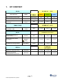
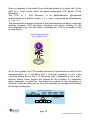
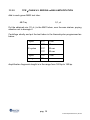
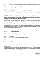
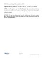
USER MANUAL
γTCR
Kit for the analysis of clonality of V-J regions
of γ-chain of T-cell receptor (TCR)
REF. 04-60A
04-60R-50(8033622780731)-EN.doc
1. KIT CONTENT
3
2. STORAGE AND STABILITY OF THE REAGENTS
4
3. PRECAUTIONS FOR USE
4
4. SAFETY RULES
5
4.1. General safety rules
5
4.2. Safety rules about the kit
6
5. MATERIALS REQUIRED, BUT NOT PROVIDED
7
5.1
Reagents
7
5.2
Instruments
7
5.3
Materials
7
6. PREPARATION OF THE REAGENTS
8
7. INTRODUCTION
9
8. TEST PRINCIPLE
12
10. COLLECTION, MANIPULATION AND PRE-TREATMENT OF THE
SAMPLES
14
10.1. Peripheral or bone marrow blood
10.1.1. Pre-treatment of peripheral or bone marrow blood
14
14
10.2. Histological samples
10.2.1. Pre-treatment of histological samples
14
15
11.
16
11.1.
PROTOCOL
DNA EXTRACTION
16
11.2. γ CHAIN V-J REGION DNA AMPLIFICATION
11.2.1. Synthesis of a single strand DNA
11.2.2. TCR γ CHAIN V-J REGION ssDNA AMPLIFICATION
pag. 1
04-60R-50(8033622780731)-EN.doc
17
17
18
11.3. VISUALIZATION OF THE AMPLIFICATION PRODUCTS
11.3.1. Agarose gel electrophoresis
11.3.2. Sample loading
19
19
19
11.4.
21
INTERPRETATION OF THE RESULTS
12.
TROUBLESHOOTING
23
13.
DEVICE LIMITS
25
14.
DEVICE PERFORMANCES
25
14.1.
Specificity
25
14.2.
Sensitivity
25
15.
BIBLIOGRAPHIC REFERENCES
26
16.
INFORMATION FOR ORDERS
27
16.1.
RELATED PRODUCTS
28
pag. 2
04-60R-50(8033622780731)-EN.doc
1.
KIT CONTENT
BOX P
STORE AT – 20°C
COLOUR OF
TUBE (T)
OR LID
25 test
50 test
8 test
MM1 monodose premix tubes
Colourless(T)
25
50
10
MM2 monodose premix tubes
Green(T)
25
50
10
Red
1 x 25 μL
1x 50 μL
1 x 10 μL
DESCRIPTION
Thermostable Taq DNA
polymerase
LABEL
AB TAQ
5 U/μL
SMALL BAG
Plasmid DNA obtained from
monoclonal T cells
STORE AT – 20°C
Positive control
γTCR
Blue
BOX F
1 x 30 μL
1 x 50 μL
1 x 10 μL
STORE AT +2°/+8°C
Electrophoresis Buffer for
sample loading
Blu 6X
Blue
1 x 150 μL
1 x 300 μL
1 x 100 μL
Ethidium Bromide solution
(2,5 mg/mL)
Ethidium
Bromide
Red
1 x 80 μL
1 x 150 μL
1 x 50 μL
Yellow
1 x 80 μL
1 x 150 μL
1 x 50 μL
DNA Molecular Weight Marker
Toxic
R 23 68
S 36/37 45
MW Marker
STORE AT
+15°/+25°C
BOX A
Agarose for molecular biology
Electrophoresis Buffer
TRIS-Borate-EDTA pH: 8,00
AGAROSE
1 x 10 g
1 x 20 g
1x5g
TBE 50X
1 x 250 mL
1 x 500 mL
1 x 100 mL
pag. 3
04-60R-50(8033622780731)-EN.doc
2.
STORAGE AND STABILITY OF THE REAGENTS
Each component of the kit should be stored according to the directions
indicated on the label of the single boxes.
In particular:
Box P
Small bag
Box F
Box A
store at -20°C
store at -20°C
store at a +2/+8°C
store at +15 / +25°C
When stored at the recommended temperature, all test reagents are stable
until their expiration date.
3.
PRECAUTIONS FOR USE
• The kit should be handled by investigator qualified through education and
training in molecular biology techniques applied to diagnostics;
• Before starting the kit procedure, read carefully and completely the
instruction manual;
• Keep the product out of heating sources;
• Do not use any part of the kit if over the expiration date;
• In case of any doubt about the storage conditions, box integrity or method
application,
contact
AB
ANALITICA
technical
support
at:
[email protected] .
In the amplification of nucleic acids, the investigator has to take the
following special precautions:
• Use filter-tips;
• Store the biological samples, the extracted DNA, positive control included
in the kit and all the amplification products in different places from where
amplification reagents are stored.
pag. 4
04-60R-50(8033622780731)-EN.doc
• Organise the space in different pre- and post-PCR units; do not share
consumables (pipets, tips, tubes, etc) between them.
• Change the gloves frequently;
• Wash the bench surfaces with 5% sodium hypochloride;
• Thaw the PCR premixes at room temperature before use.
Add the Taq DNA polymerase and purified DNA very quickly at room
temperature or in an ice-bath.
4.
SAFETY RULES
4.1.
General safety rules
• Wear disposable gloves to handle the reagents and the clinical samples
and wash the hands at the end of work.
• Do not pipet with mouth.
• Since no known diagnostic method can assure the absence of infective
agents, it is a good rule to consider every clinical sample as potentially
infectious and handle it as such.
• All the devices that get directly in touch with clinical samples should be
considered as contaminated and disposed as such. In case of accidental
spilling of the samples, clean up with 10% Sodium Hypochloride. The
materials used to clean up should be disposed in special containers for
contaminated products.
• Clinical samples, materials and contaminated products should be disposed
after decontamination by:
immersion in a solution of 5% Sodium Hypochloride (1 volume of 5% Sodium
Hypochloride solution every 10 volumes of contaminated fluid) for 30 minutes
OR
autoclaving at 121°C at least for 2 hours (NOTE: do not autoclave solutions
containing Sodium Hypochloride!!).
pag. 5
04-60R-50(8033622780731)-EN.doc
4.2.
Safety rules about the kit
The risks for the use of this kit are related to the single components:
Dangerous components:
3,8-diamino-1-ethyl-6-phenylphenantridiumbromide (Ethidium Bromide) <2%
Description of risk:
T (Toxic)
RISK SENTENCES AND S SENTENCES
ETHIDIUM BROMIDE
R 23 and R 68
S 36/37 45
Toxic for inhalation.
Risk of irreversible effects.
Wear laboratory coat and disposable gloves.
In case of accident or discomfort, seek for medical
assistance and show the package label.
R and S sentences refer to the concentrated product, as provided in the kit.
In particular, for Ethidium Bromide, until the dilution in the agarose gel.
In manipulating concentrated Ethidium Bromide, use a chemical dispensing
fume cabinet. Always wear disposable gloves and laboratory coat in
manipulating the diluted Ethidium solution as well.
The product can not be disposed with the common waste. It must not reach
the drainer system. For the disposal, follow the local law.
In case of accidental spilling of Ethidium Bromide, clean with Sodium
hypochloride and water.
Safety data sheet (MSDS) of Ethidium Bromide is available upon request.
pag. 6
04-60R-50(8033622780731)-EN.doc
5.
MATERIALS REQUIRED, BUT NOT PROVIDED
5.1 Reagents
• Sterile DNase and RNase free water;
• Distilled water.
5.2 Instruments
• Laminar flow cabinet (use is recommended while adding TAQ polymerase
to the amplification premix to avoid contamination; it would be
recommended to use another laminar flow cabinet to add the extracted
DNA);
• Micropipettes (range: 0,2-2 µL; 0,5-10 µL; 2-20 µL);
• Thermalcycler;
• Microcentrifuge (max 12.000-14.000 rpm);
• Balance;
• Magnetic heating stirrer or microwave;
• Chemical cabinet (use is recommended in handling Ethidium Bromide);
• Horizontal electrophoresis chamber for agarose minigel;
• Power supply (50-150 V);
• UV Transilluminator;
• Photo camera or image analyzer.
5.3 Materials
•
•
•
•
•
Disposable gloves;
Disposable sterile filter-tips (range: 0,2-2 µL; 0,5-10 µL; 2-20 µL;);
Graduate cilinders (1 L) for of TAE dilution;
Pyrex bottle or Becker for agarose gel preparation;
Parafilm.
pag. 7
04-60R-50(8033622780731)-EN.doc
6.
PREPARATION OF THE REAGENTS
Preparation of 1 L of 1X TAE buffer:
• Mix 200 mL of 5X TBE with 800 mL of distilled water.
pag. 8
04-60R-50(8033622780731)-EN.doc
7.
INTRODUCTION
The lymphoid diseases include malignant lymphoma (non-Hodgkin lymphoma
and Hodgkin disease) and the chronic lymphoid leukaemia (Harris et al.,
1994).
In the diagnosis of these pathologies the molecular techniques (Southern blot
and Polymerase Chain Reaction) are becoming more popular.
Their applications are in the diagnosis of complete or partial disease
remission and in monitoring the minimal residual disease (MRD).
A correct diagnosis of haematological diseases needs a multidisciplinary
approach, that allows to correlate the traditional morphological evaluations
(Hematoxylin-eosin stain or Wright) with a variety of special analysis, like
immuno-histochemical staining, molecular and cytogenetic techniques.
Lymphoid diseases are an heterogeneous group of diseases that result as
the consequence of a B and T lymphocytes neoplastic transformation in
different phases of their development. Their big variety is related to
lymphocytes diversity during the different phases of their development and to
the huge complexity of the immune system.
Since the DNA rearrangements take place in great numbers during T cells
development, T cells tumours are very frequent. As for B cells tumours, T
cells tumours are related to different development phases.
While B cells tumours are referred to the entire phase of B cells development,
T cells tumours correspond to the precocious and latest stadiums of it, the
lack of involvement of the intermediate stadiums of development could be
due to the fact that immature T cells, if not rescued in a short time after the
successive positive maturation, are programmed for cellular death.
In these circumstances, there is not sufficient time for tymocites to
accumulate enough mutations for malignant transformation (Zucchini A. et al.
2002).
The availability of molecular techniques has allowed not only the increase of
the capacity to diagnose the diseases, but also the possibility of a correct
classification of them (Cossman et al., 1998).
The most important application of molecular methods in lymphoid diseases
evaluation is the determination of cell clonality. These techniques are
considered as the “gold standard” for the clonality determination and are used
when this information can not be obtained by the immuno-pathologic
techniques, mostly for lymphoid diseases of T-origin.
The molecular method allows to evaluate if the lymphoid abnormality is due to
a neoplastic process, therefore to a monoclonal proliferation, or to a reactive
benignant process, to which polyclonal proliferation is associated.
pag. 9
04-60R-50(8033622780731)-EN.doc
Since a neoplasy is the result of an unlimited growth of a single cell, all the
cells of a T-cell cancer have the same rearranged TCR genes (T-Cell
Receptor).
The TCR, or T -Cell Receptor, is an hetherodimeric glycoprotein
characterised by 4 distinct chains, α, β, γ and δ, expressed as heterodimers
α / β or γ / δ.
The aminoacidic sequence analysis of the heterodimers revealed a surprising
analogy between TCR structural dominium and those relating to the
Immunoglobulin (Ig); this fact leads to consider the TCR as a member of the
Ig superfamily.
ANTIGEN RECOGNITION
BY T LYMPHOCYTES
T
TCR
α/β
γ/δ
Ag
MHC
APC
As for the Ig genes, the TCR variable dominium is produced as a result of the
rearrangements of V (variable) and J (Joining) segments in the α and
γ families chains and of the V, D (diversity) and J segments in the β and γ
families chains; these regions are flanked by the constant (C) respective
genic segments. In particular, the dominium nearest to the membrane is
constant , while the dominium far from the membrane is variable and contains
the antigen binding site.
γ -chain rearrangement
V genes
V1
V2
J1 genes
V3
C1genes
Vn
J2 genes
C2 genes
C
C
germline DNA
rearrangement
V1
V2
V3 J2 C2
pag. 10
04-60R-50(8033622780731)-EN.doc
Most of T lymphocytes expresses the α / β heterodimers: the T lymphocytes
percentage with α / β or γ / δ varies in the different organs and from specie to
specie; in man, the most of mature T cells located in the peripheral blood
expresses the α / β TCR, while T cells expressing γ / δ TCR are in a little
amount: from 1 % to 5 %.
The complete and functional TCR is joined to CD3, a signal transduction
molecular complex. This structure is involved in the antigenic
acknowledgment, only after the antigen has been processed by an antigen
presenting cell (APC).
Before being recognised by T lymphocytes, the antigen has to be fagocitated
or endocytated by the APC and processed in little peptides.
Antigen fragments are then presented to TCR in the context of the products
of I and II class Major Histocompatibility Complex (MHC), that are
constitutively expressed on the accessory cells membrane.
The contact between TCR and its specific antigenic determinant leads to the
internalisation of the CD3 molecule.
The analysis of the TCR gene rearrangements by molecular biology
techniques allow to detect the neoplastic nature of T cells expansions.
It is not possible instead to demonstrate the clonal nature of NK cells
leukaemia, whose diagnosis is done only on the basis of morphological,
immuno-phenotypic and clinical analysis.
pag. 11
04-60R-50(8033622780731)-EN.doc
8.
TEST PRINCIPLE
PCR method (Polymerase Chain Reaction) has been the first method of DNA
amplification described in literature (Saiki RK et al., 1985). It can be defined
as an in vitro amplification reaction of a specific part of DNA (target
sequence) by a thermostable DNA polymerase.
Three nucleic acid segments are involved in the reaction: double stranded
DNA template to be amplified (target DNA) and two single-stranded
oligonucleotides “primers” that are designed in order to anneal specifically to
the template DNA.
The DNA polymerase begins the synthesis process at the region marked by
the primers and synthesizes new double stranded DNA molecules, identical
to the original double stranded target DNA region, by facilitating the binding
and joining of the complementary nucleotides that are free in solution
(dNTPs). After several cycles, one can get millions of DNA molecules which
correspond to the target sequence.
The sensitivity of this test makes it particularly suitable for the application in
laboratory diagnostics.
Moreover, the amplification reaction can be executed from a wide range of
biological samples and since it allows to amplify very small DNA fragments,
the starting DNA can be also partially degraded.
pag. 12
04-60R-50(8033622780731)-EN.doc
9.
PRODUCT DESCRIPTION
The molecular strategy adopted in this kit for detecting T cells monoclonality
is characterised by the use of consensus primers for the V and J regions of
the TCR γ chain.
TCR γ chain analysis is the simplest and most efficient method to study TCR
gene rearrangements. In effect, with reference to β chain, the number of gene
segments of γ chain coding locus is reduced and moreover, γ genes
rearrangements are present in most of neoplastic and normal T lymphocytes.
The TCR gene rearrangement takes place in a precocious stadium of T
differentiation and, particularly, γ / δ rearrangement precedes that of α / β
chains.
The ssDNA (single strand DNA) synthesis followed by γ chain V-J regions
amplification guarantees to the method the necessary sensitivity for its
application in the T-cells monoclonality study.
The revelation of the amplified fragment is performed by a high resolution
agarose gel electrophoresis. The different aspects of the amplified products
allow to discriminate a monoclonal sample from an oligoclonal or polyclonal
sample.
The kit also provides with positive control of amplification. When the
amplification of the positive control is successful, it is guaranteed that the
reaction is correct. These controls are not dangerous for the operator
because they are plasmid DNA containing only the V-J region of the γ chain,
obtained from monoclonal T cells.
The kit is in premix format: all the reagents for the amplification are pre-mixed
and aliquoted in monodose test tubes (0,2 or 0,5 mL) to which Taq
polymerase and the extracted DNA will be added. This premix format allows
the reduction of the manipulation steps in pre-amplification, with considerable
time saving for the operator; the repeated freezing/thawing of reagents (that
could alter the product performances) is avoided and, above all, this form
minimizes the risk of contamination, so the risk to get false positive results.
Nevertheless, it’s always recommended to use all the proper amplification
controls.
The MM1 premixes include a wax bead; this bead will allow to perform a hot
start amplification during ssDNA amplification: the Taq DNA polymerase
becomes active only after the initial denaturation step, when the wax melt will
allow its descending in the mix. The hot start amplification reduces the
possibility of an eventual presence of unspecific amplified products.
pag. 13
04-60R-50(8033622780731)-EN.doc
10. COLLECTION, MANIPULATION AND PRETREATMENT OF THE SAMPLES
10.1.
Peripheral or bone marrow blood
During the sample collection, follow all the usual sterility precautions.
Blood should be treated with EDTA. Other anticoagulating agents, as heparin,
are strong inhibitors of TAQ polymerase and so they could alter the efficiency
of the amplification reaction.
Fresh blood can be stored at +2/+8°C and processed in a short time; if DNA
extraction is not performed in a short time, the sample must be frozen.
10.1.1.
Pre-treatment of peripheral or bone marrow blood
In order to obtain a more appreciable yield in the DNA extraction, it is
recommended to prepare a buffy coat by centrifuging the whole blood at 3300
x g, for 10 min at room temperature.
Then, three different fractions will be distinguishable: the plasma is the clear
upper fraction; the buffy coat is the intermediate fraction, containing the
concentrated leucocytes, and the remaining fraction contains the
erythrocytes.
Alternatively, it is also possible to perform a FICOLL protocol in order to
isolate a pellet of white cells.
10.2.
Histological samples
Histological samples include fresh, frozen or formalin-fixed and paraffinembedded biopsies.
The fresh biopsy could be treated, within few minutes from sampling, or
quickly frozen with liquid nitrogen and successively stored at -80°C until
mechanic disruption by using sterile cutter, followed by enzymatic digestion
with Proteinase K.
In case that the biopsy is fixed and paraffin-embedded, it is suggested the
use of formalin buffer at pH 7 with sodium and potassium salts at 10%, as
Lilie formula. Tissue fixation with not-buffered formalin in Bouin, Holland or
other acidic fixatives (osmic acid, for instance) is not suitable for subsequent
DNA extraction because those substances produce cross-links in the tissue,
making it not-digestible.
pag. 14
04-60R-50(8033622780731)-EN.doc
10.2.1.
Pre-treatment of histological samples
In case of fresh or frozen histological sample (up to 50 mg), proceed quickly
with mechanic disruption of the sample by a sterile cutter. Do this operation
on a glass slide, transfer the minced tissue in a tube, then proceed with
digestion with Proteinase K.
In case that the biopsy is fixed and paraffin-embedded proceed first with
paraffin removal and then with sample digestion.
pag. 15
04-60R-50(8033622780731)-EN.doc
11. PROTOCOL
11.1.
DNA EXTRACTION
For DNA extraction, any extraction method can be used, provided that it
allow to isolate an integral and pure DNA.
The obtained DNA should be quantified by photometric measures.
The DNA amount to use in the amplification is 500 ng and, if necessary, the
obtained solution should be diluted.
Please note that the required volume of extracted DNA to be added in each
premix tube is 10 μL.
For any further information contact AB ANALITICA technical support
e-mail: [email protected]
pag. 16
04-60R-50(8033622780731)-EN.doc
11.2.
11.2.1.
γ CHAIN V-J REGION DNA AMPLIFICATION
Synthesis of a single strand DNA
Add into each MM1 colourless test tube (containing 9.9 μL of mix and a wax
bead):
0,1 μL
5* μL
5 μL
AB Taq
Extracted DNA
Sterile water
It is important to include in each experiment a negative polyclonal control to
monitor the contamination (any genomic DNA of a healthy patient) and 10 μL
of a monoclonal positive control (included in the kit).
Moreover, the introduction of a negative amplification control (water instead of
DNA) will allow to monitor the contaminations.
*The ideal amount of DNA to amplify is 500 ng. It is suggested to verify the
extracted DNA concentration by photometric measures.
In particular, for DNA extracted from paraffin-included specimen, it could be
necessary to amplify a DNA sample bigger that 5 μL: in this case, the volume
of sterile water in the reaction mix can be diminished.
Centrifuge briefly, then incubate the tubes in the termalcycler programmed as
below:
1 cycle
94°C
5 min
94°C
30 sec
55°C
45 sec
73°C
45 sec
1 cycle
73°C
5 min
storage
4°C
25 cycles
Proceed directly with the ssDNA amplification.
pag. 17
04-60R-50(8033622780731)-EN.doc
11.2.2.
TCR γ CHAIN V-J REGION ssDNA AMPLIFICATION
Add to each green MM2 test tube:
0,1 μL
AB Taq
Put the obtained mix (21 μL) in the MM1 tubes, over the wax stratum, paying
attention not to damage it.
Centrifuge shortly and put the test tubes in the thermalcycler programmed as
below:
1 cycle
94°C
5 min
94°C
30 sec
55°C
45 sec
73°C
45 sec
1 cycle
73°C
7 min
storage
4°C
30 cycles
Amplification fragments length is in the range from 160 bp to 190 bp.
pag. 18
04-60R-50(8033622780731)-EN.doc
11.3.
11.3.1.
VISUALIZATION OF THE AMPLIFICATION PRODUCTS
Agarose gel electrophoresis
Preparation of a 3% agarose gel:
Weight 1,5 g of HR Agarose and pour it into 50 mL of 1X TAE.
Leave the solution on a magnetic stirring heater or in a microwave until the
solution becomes clear. Allow the gel to cool to “hand warm” and then add 10
µL of Ethidium Bromide solution.
NOTICE: Ethidium Bromide is a strong mutagenic agent; always wear
gloves and preferably work under a chemical safety cabinet during the
handling of this reagent or gels containing it.
Place the gel into the appropriate gel casting tray, with the comb placed in
and allow the gel to cool at room temperature or in a fridge until the gel
becomes solid.
When the gel is solidified, remove carefully the comb (pay attention not to
damage the gel wells) transfer the tray into the electrophoresis chamber and
pour the appropriate amount of TAE buffer so that it covers completely the gel
(about 1-2 mm over the gel surface).
11.3.2.
Sample loading
Mix into a tube or directly on a parafilm layer:
4 μL
20 μL
of 6X Blue
of γ-TCR V-J region amplification product
OR
2 μL
10 μL
of 6X Blue
of DNA molecular weight marker (MW Marker)
Load the mixture on the gel wells; switch on the power supply and set the
voltage at 50 V.
Run the gel for about 5-6 hours, then place the gel on an UV transilluminator
and analyze the results by comparing the size of the amplification products
with the reference Molecular Weight Marker.
pag. 19
04-60R-50(8033622780731)-EN.doc
*DNA Molecular Weight Marker (Marker MW):
fragment sizes: 501-489, 404, 353, 242, 190, 147, 110, 89, 67, 34, 26 bp.
NOTE: In a 3% agarose gel the 501-489 bp bands are usually not clearly
resolved and appear as an unique band; the 26 and 34 bp bands are
sometimes too small to be visible in a 3% agarose gel (because of their low
molecular weight).
NOTICE: UV rays are dangerous for skin and, above all, eyes: always
wear gloves and safety glass or make use of the protection screen of
the UV transilluminator.
pag. 20
04-60R-50(8033622780731)-EN.doc
11.4.
INTERPRETATION OF THE RESULTS
In order to discriminate a polyclonal sample from a monoclonal sample it is
necessary to evaluate the clearness and the resolution of the bands of the
amplification products. This is possible only if the agarose gel preparation is
accurate and if the electrophoretic run lasted for at least 5 hours.
For these reasons, the correct amplification and visualisation of the
monoclonal positive control included in the kit and of the negative polyclonal
control (any genomic DNA of a healthy patient) are essential.
After agarose gel electrophoresis, the controls should show the following
results:
CONTROL
RESULT
INTERPRETATION
Negative Control
absent
(water)
Well defined band
Monoclonal
in the range
positive Control
160 bp -190 bp
Not well defined
Polyclonal
band or smears
negative control in the range
160 bp -190 bp.
Absence of contaminations
during the first amplification.
Amplification and visualisation
were correct.
Amplification and visualisation
were correct.
Then, for the interpretation of the band pattern, follow the table below:
RESULT
INTERPRETATION
Presence of two well Presence of T cells MONO- or
defined bands
BI- CLONAL populations.
in the range
160 bp -190 bp
Presence of a smear
Presence of T cells POLYCLONAL
in the range
populations.
160 bp -190 bp
pag. 21
04-60R-50(8033622780731)-EN.doc
1 2
3 4 5 6
7
Fig. 1. :
3% HR agarose gel electrophoresis after a 5 hours run
1.
2.
3.
4.
5.
6.
7.
MW DNA Marker
Positive sample for T cells receptor monoclonality (Jurkatt cell line)
PATIENT 1, monoclonal sample
PATIENT 2, biclonal sample
Negative polyclonal control
Negative control of amplification (water)
DNA MW Marker
pag. 22
04-60R-50(8033622780731)-EN.doc
12. TROUBLESHOOTING
1. Neither amplification products, nor positive control DNA band
• TAQ polymerase was not correctly added to the premix
-Use pipets and tips with suitable volumes (pipet range 0,2 - 2 μL);
-Check visually that TAQ polymerase diffuses in the premix: this is
easy because the enzyme is dissolved in glycerol that has a higher
density;
-alternatively, check visually the drop of TAQ polymerase put on the
tube wall, then centrifuge briefly.
• The thermalcycler was not correctly programmed
-Check the conformity of the thermalcycler program and the
temperature profile in the instruction manual; then repeat the
amplification with the correct program.
• The kit doesn’t work properly
-Store the premix, TAQ polymerase and positive control at -20°C;
-Avoid repeated freezing/thawing of the premix and the reagents.
2. Presence of unspecific products out of the respective range after
visualisation in agarose gel
• The thermalcycler makes temperature changes too slowly
– Do a thermalcycler revision.
• The preparation of the amplification reaction has been executed in a long
time at room temperature.
– Accelerate the work time at room temperature;
– Work on ice.
• The extracted has not been purified
– Use an extraction system which allows a good sample purification.
pag. 23
04-60R-50(8033622780731)-EN.doc
• The starting sample contained degraded DNA
– Repeat the extraction step using another clinical starting sample;
– Be sure that the sample had been collected and stored in appropriate
way.
3. The positive monoclonal control doesn’t appear as a clear and well
resolved band
• The gel overheated
- decrease the voltage applied.
• The gel was not correctly prepared
-
be sure to completely melt the agarose powder, by mixing it with TBE
1X.
For any further problem contact AB ANALITICA technical support (e-mail:
[email protected].
pag. 24
04-60R-50(8033622780731)-EN.doc
13. DEVICE LIMITS
The kit can have reduced performances if:
the blood sample is not suitable for this analysis (not correct sample
storing, using of heparin as anticoagulant ,);
the histological sample is not suitable for this analysis (using of
alternative fixatives instead of neutral formalin buffer, not correct sample
storing);
14. DEVICE PERFORMANCES
14.1.
Specificity
Primer sequence alignment in the most important databanks shows the
absence of unspecific alignment. Cross reactions with genomic DNA have not
been revealed.
14.2.
Sensitivity
The sensitivity of the method was tested with samples in which monoclonal,
polyclonal Tcells and B cells were mixed.
The system is able to detect until 3% of monoclonal T cells in a population of
polyclonal T cells and until 1% in a population of B cells.
pag. 25
04-60R-50(8033622780731)-EN.doc
15. BIBLIOGRAPHIC REFERENCES
Cossman J, Uppenkamp M, sundeen J, Coupland R, Raffeld M. Arch Pathol
Lab Med.(112): 117-27. 1988.
Greer J. P., Kinney M. C., Loughran TP., T cell and NK Cell
Lymphoproliferative disorders. Haematology (1) 259-286. 2001
Harris HL, Jaffe ES, Stein H, Banks PM, Chan JKC, Clearly ML et al. Blood
(84): 1361-92. 1994.
Saiki RK, S Scharf, F Faloona, KB Mullis, GT Horn, HA Erlich and N Arnheim,
Science. (230): 1350-1354. 1985.
Zucchini A. Bagli L., Fattori P.P., Ravaioli A., Zamai L., Papa S., Biologi
Italiani. (9):36-42. 2002.
pag. 26
04-60R-50(8033622780731)-EN.doc
16. INFORMATION FOR ORDERS
γTCR:
Kit for the analysis of clonality of V-J regions of the γ-chain of T-cell
receptor (TCR):
(cod. 04-60A)
The kit contains the reagents for DNA amplification, for visualization by
agarose gel electrophoresis and positive control.
Target region: T-cell receptor gene (TCR).
Cod.
Prod
Pkg
04-60A-25
γTCR
25 tests
03-60A-50
γTCR
50 tests
pag. 27
04-60R-50(8033622780731)-EN.doc
16.1.
RELATED PRODUCTS
γTCR:
Kit for the analysis of clonality of V-J regions of the γ-chain of T-cell
receptor (TCR):
(cod. 04-60R)
The kit contains the reagents for DNA amplification and positive control
Target region: T-cell receptor gene (TCR).
Cod.
Prod
Pkg
04-60R-25
γTCR
25 tests
03-60R-50
γTCR
50 tests
pag. 28
04-60R-50(8033622780731)-EN.doc
04-60R-50(8033622780731)-EN.doc
AB ANALITICA srl - Via Svizzera 16 - 35127 PADOVA, (ITALY)
Tel +39 049 761698 - Fax +39 049 8709510
e-mail: [email protected]