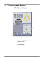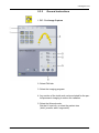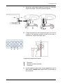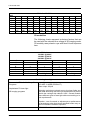
1
ENGLISH ORTHOPANTOMOGRAPH® OP300 Digital Panoramic X-Ray System User Manual 203887 rev. 5 ORTHOPANTOMOGRAPH® OP300 Copyright Code: 203887 rev 5 Date: March 28, 2013 Copyright © 3/28/13 by Instrumentarium Dental, PaloDEx Group Oy. All rights reserved. ORTHOPANTOMOGRAPH® is a registered trademark of Instrumentarium Dental, PaloDEx Group Oy. U.S. patents US6731717, US6829326 and USRE41197. Finnish patents 114383. Documentation, trademark and the software are copyrighted with all rights reserved. Under the copyright laws the documentation may not be copied, photocopied, reproduced, translated, or reduced to any electronic medium or machine readable form in whole or part, without the prior written permission of Instrumentarium Dental. The original language of this manual is English. Instrumentarium Dental reserves the right to make changes in specification and features shown herein, or discontinue the product described at any time without notice or obligation. Contact your Instrumentarium Dental representative for the most current information. Manufacturer Instrumentarium Dental, PaloDEx Group Oy Nahkelantie 160 (P.O. Box 20) FI-04300 Tuusula FINLAND Tel. +358 10 270 2000 Fax. +358 10 270 2230 For service, contact your local distributor. ORTHOPANTOMOGRAPH® OP300 Table of Contents 1 Introduction.................................................................................................................. 1 1.1 ORTHOPANTOMOGRAPH® OP300 .................................................................... 1 1.2 References............................................................................................................ 2 1.3 Intended use ......................................................................................................... 2 1.4 Associated documentation .................................................................................... 3 1.5 Abbreviations used in this manual ........................................................................ 3 1.6 Warnings and precautions .................................................................................... 4 1.6.1 Warnings to be observed during use ......................................................... 4 1.6.2 Warnings for cross infection....................................................................... 5 1.6.3 General warnings ....................................................................................... 7 1.7 Disclaimer ............................................................................................................. 9 1.8 Disposal ................................................................................................................ 9 2 Unit description ......................................................................................................... 11 2.1 Main parts and controls....................................................................................... 11 2.2 Patient positioning lights ..................................................................................... 13 2.3 Patient positioning panel ..................................................................................... 16 2.3.1 Cephalometric unit ................................................................................... 17 2.4 Emergency stop switch ....................................................................................... 19 3 Imaging programs ..................................................................................................... 21 3.1 Panoramic programs........................................................................................... 21 3.1.1 Cephalometric programs.......................................................................... 29 3.1.2 3D programs ............................................................................................ 31 3.1.3 MAR, Metal Artifact Reduction ................................................................. 32 3.1.4 Exposure settings for 3D imaging ............................................................ 33 4 Touch screen display ................................................................................................ 35 4.1 Main control panel............................................................................................... 35 4.2 Modality section .................................................................................................. 36 4.2.1 Exposure indicators and settings ............................................................. 36 4.3 Status section...................................................................................................... 37 4.4 Other sections ..................................................................................................... 37 5 Using the unit............................................................................................................. 39 5.1 Attaching and removing the sensor..................................................................... 39 5.1.1 Attaching the sensor ................................................................................ 40 5.1.2 Removing the sensor ............................................................................... 41 5.2 Preparing the system .......................................................................................... 41 5.3 Panoramic exposures ......................................................................................... 42 5.3.1 Positioning devices .................................................................................. 42 5.3.2 General instructions ................................................................................. 43 5.3.3 Patient positioning.................................................................................... 45 5.3.3.1 Panoramic exposure.................................................................. 45 5.3.3.2 TMJ exposure ............................................................................ 48 5.3.3.3 Maxillary Sinus exposure........................................................... 51 5.3.4 Taking the exposure................................................................................. 52 5.3.5 Multilayer Selection .................................................................................. 55 rev i 5.4 Cephalometric exposures ................................................................................... 56 5.4.1 General instructions ................................................................................. 57 5.4.2 Patient positioning.................................................................................... 59 5.4.2.1 Pediatric lateral and Lateral projection ...................................... 59 5.4.2.2 PA projection ............................................................................. 60 5.4.2.3 Reverse towne projection .......................................................... 61 5.4.2.4 Waters view ............................................................................... 62 5.4.2.5 Carpus view (Not available in USA and Canada) ...................... 63 5.4.2.6 Taking the exposure .................................................................. 63 5.5 3D exposures ...................................................................................................... 64 5.5.1 Positioning devices .................................................................................. 64 5.5.2 General instructions ................................................................................. 64 5.5.3 Scout image ............................................................................................. 69 5.5.4 3D image.................................................................................................. 70 5.5.4.1 Stone model and radiographic guide scan ................................ 72 5.7 Warnings and error messages ............................................................................ 74 5.7.1 Acknowledging errors............................................................................... 74 5.7.2 Image transfer errors................................................................................ 74 6 Troubleshooting ........................................................................................................ 75 6.1 Patient positioning............................................................................................... 75 6.2 Image appearance .............................................................................................. 78 6.3 Artefacts .............................................................................................................. 79 6.4 Unit operation...................................................................................................... 81 7 Maintenance ............................................................................................................... 83 7.1 Maintenance procedure ..................................................................................... 83 7.1.1 Annual maintenance ................................................................................ 83 7.1.2 Calibration intervals.................................................................................. 83 7.2 Changing the fuses ............................................................................................. 84 7.3 Cleaning and decontaminating the unit............................................................... 84 8 Calibration and adjustment ...................................................................................... 87 8.1 Introduction ......................................................................................................... 87 8.2 Preparing for calibration ...................................................................................... 88 8.3 Panoramic calibration.......................................................................................... 89 8.3.1 Panoramic geometry calibration............................................................... 89 8.3.2 Panoramic pixel calibration ...................................................................... 90 8.3.3 Panoramic Quality Check (optional)......................................................... 91 8.4 3D calibration ...................................................................................................... 92 8.4.1 3D geometry calibration ........................................................................... 92 8.4.2 3D pixel calibration................................................................................... 93 8.4.3 3D Quality Check program....................................................................... 94 8.5 Cephalometric calibration.................................................................................... 95 8.5.1 Ceph pixel calibration............................................................................... 95 8.5.2 Ceph Quality check program (Optional)................................................... 95 9 Technical data............................................................................................................ 97 9.1 Technical specifications ...................................................................................... 97 9.2 Unit dimensions................................................................................................. 109 9.3 Symbols that appear in the unit......................................................................... 111 ii rev 9.4 Labels on the unit.............................................................................................. 113 9.5 Electromagnetic Compatibility (EMC) tables..................................................... 113 9.6 X-ray tube assemblies....................................................................................... 119 rev iii iv rev 1 Introduction 1.1 ORTHOPANTOMOGRAPH® OP300 INSTRUMENTARIUM DENTAL® ORTHOPANTOMOGRAPH® OP300 x-ray unit (hereafter called “OP300”) is a dental x-ray system for producing high quality digital images of dentition, TM-joints and skull. In order to take images with OP300 you need a suitable PC hardware connected to the OP300 unit and CLINIVIEW™ software (or suitable third party software via TWAIN driver) to capture and manage images. OP300 performs the following procedures: Panoramic Standard panoramic Pediatric panoramic Wide arch panoramic Bitewing TMJ, PA projection Ortho TMJ, axial corrected lateral projection Maxillary sinus Ortho Zone enhanced panoramic Orthogonal panoramic Cephalometric (optional) Cephalometric lateral projection Cephalometric pediatric lateral projection Cephalometric postero-anterior (PA) projection Reverse Towne projection Waters view Carpus program (optional) (Not available in USA and Canada) 3D (optional) 203887 rev 5 61x41 mm Field of View 61x78 mm Field of View Endo 61 x 41 mm Field of View Instrumentarium Dental 1 1 Introduction 1.2 References The following instructions are delivered with in the OP300 installation manual: • Firmware update instructions • Calibration instructions • Cephalostat upgrade instructions • Cephalostat side changing instructions The following instructions are separate and can be ordered from customer service: • 3D upgrade instructions are delivered with the 3D upgrade kit 1.3 Intended use OP300 must only be used and operated by dentist and other qualified professionals. OP300 must only be used to take panoramic, cephalometric and 3D images of the dento-maxillofacial complex of the human skull. It must not be used to take images of any other part of the human body. Panoramic and 3D exposures should not be used if conventional intraoral radiographic images (bitewing exposures) would be sufficient. Cone beam computed tomography images are not adequate for the analysis of soft tissue. CAUTION! USA only: Federal law restricts this device to sale by or on the order of a dentist or other qualified professional. 2 Instrumentarium Dental 203887 rev 5 1 Introduction 1.4 Associated documentation • OP300 user manual • OP300 installation manual • The CLINIVIEW™ software user manual • The CLINIVIEW™ software installation manual • The user manual supplied with the dental imaging software • The installation manual supplied with the dental imaging software • The user manual supplied with the 3D imaging software • The installation manual supplied with the 3D imaging software 1.5 Abbreviations used in this manual FOV = Field Of View. The cylindrical 3D volume that is reconstructed by the system. ROI = Region Of Interest. The anatomical area or region of the patient that you are interested to examine. FH = Frankfort-Horizontal H = Horizontal 203887 rev 5 Instrumentarium Dental 3 1 Introduction 1.6 Warnings and precautions 1.6.1 Warnings to be observed during use The unit may be dangerous to the user and the patient, if the safety regulations in this manual are ignored, if the unit is not used in the way described in this manual and/or if the user does not know how to use the unit. The unit must only be used to take the dental x-ray exposures described in this manual. The unit must NOT be used to take any other x-ray exposures. It is not safe to use the unit to take x-ray exposures, that it is not designed for. Only professionally qualified dental and/or medical personnel are allowed to operate the unit and carry out any diagnoses based on output from the unit. Because the x-ray limitations and safety regulations change from time to time, it is the responsibility of the user to make sure that all the valid safety regulations are fulfilled. When taking an x-ray exposure of a patient with exceptional anatomy (typically very tall or large) use the Test-mode (no x-rays) first to make sure that patient can be positioned correctly to the unit and for checking that the unit doesn't hit the patient. Operator should maintain visible contact with the patient and technique factors. This allows immediate termination of radiation by the release of the exposure button in the event of a malfunction or disturbance. It is the responsibility of the doctor to decide whether x-ray exposure or any additional exposures are justified and necessary. The minimum height of patient that can be x-rayed is 120 cm (3.9ft / 47.2in) and the maximum is 200 cm (6ft /78in). These heights only apply to patients with normal anatomy. Always use the lowest suitable x-ray dose to obtain the desired level of image quality. Avoid taking x-ray exposures of pregnant women. When taking an x-ray exposure of a child always use the lowest possible x-ray dose, the smallest possible image area and the lowest possible resolution that allows you to perform the required diagnostic task. 4 Instrumentarium Dental 203887 rev 5 1 Introduction If the patient is using a pacemaker, consult the manufacturer of the pacemaker before taking an exposure to confirm that the x-ray unit will not interfere with the operation of the pacemaker. Decontaminate all the surfaces that the patient is in contact with after every patient to prevent cross infection. Decontaminate all device accessories that contact the patient during a radiographic examination. Do not open or remove any of the unit’s enclosures. No user serviceable parts inside. The customer must ensure that the siting environment fulfills the requirements listed in the Installation manual. Special attention must be paid to the strength of the floor and wall materials, electrical mains and radiation protection. It is the responsibility of the customer to ensure that the site is large enough for the patients. The unit contains toxic materials that need to be handled properly when disposing the unit. Return the unit to the dealer in the end of its life cycle. Excessive dust should be cleaned from the unit for free airflow and cooling. Switch of the unit before cleaning. Always follow the instructions for patient positioning and imaging procedures instructed in the User Manual. 1.6.2 Warnings for cross infection Always use available disposable protective covers with the patient positioning accessories: Bite fork cover Chin support cover Head support cover Nose support cover Ear holder cover 203887 rev 5 Instrumentarium Dental 5 1 Introduction 1.6.3 General warnings Personnel operating the device must be adequately trained with respect to the technological principles of operation and radiation protection when using cone beam computed tomography (CBCT) imaging. This unit complies with the EMC (Electromagnetic Compatibility) according to IEC 60601-1-2. Radio transmitting equipment, cellular phones etc. shall not be used in close proximity of the unit as they could influence the performance of the unit. The correct software and settings in the workstation are essential to the performance of the unit. Consult technical support to ensure correct setup. Danger: Explosion hazard - do not use in the presence of flammable anesthetics, gases or vapors. The unit is factory set to operate using a 230-240 ±10 VAC power supply. Never connect the unit to a power supply different to the voltage marked on the unit. The site must fulfill the environmental requirements in the installation manual chapter technical specifications. There should be free space around the unit for safe operation. To maintain patient safety it is mandatory to unshielded CAT6 Ethernet cable between the unit network or workstation, so that multiple chassis connected. Non-medical grade PC should not be patient environment. use an and the are not used in This product itself complies with IEC 60601-1 medical safety standard but in order to the system incorporating also a PC to comply the standard, EITHER the PC has to be a medical PC OR the PC has to be located over 1,5 meters apart from the unit. The installer and the user of the system shall confirm that at least one of the above requirements is fulfilled. A PC is a medical one if it complies IEC 60601-1 standard and that is indicated in the accompanying documents of the PC. See chapter Technical specifications, Minimum PC Requirements, in user manual. All service operations must be made by authorized service personnel only. 6 Instrumentarium Dental 203887 rev 5 1 Introduction The annual service as described in manual is mandatory for the correct and safe operation of the unit. When taking exposures, operators and service personnel must protect themselves from radiation and remain at least two meters (six feet) away from the unit during exposure. Protect the patient from scattered radiation by placing a protective lead apron over the patient. The unit must be installed and serviced according to the unit Installation & adjustments manual by a qualified technician. Only personnel trained and approved by the manufacturer of the unit are allowed to service the unit. 3D should not be used for routine or screening examinations in which a radiograph is taken regardless of the presence or absence of clinical signs and symptoms. 3D imaging examinations must be justified for each patient to demonstrate that the benefits outweigh the risks. Where it is likely that evaluation of soft tissues will be required as part of the patient’s radiological assessment, the imaging should be done using conventional medical CT or MR, rather than 3D imaging using Cone Beam technology. Make sure that patient’s thyroid glands are protected by a lead apron during the exposure. The place where the unit is to be installed and the position from where the user will take exposures must be correctly shielded from the radiation that is generated when the unit is operated. Ensure to fulfill or exceed the requirements of your local regulations. The unit or its parts must not be changed or modified in any way without approval and instructions from the manufacturer. When servicing use only approved replacement parts supplied by the manufacturer. The use of accessories not complying with the equivalent safety requirements of this equipment may lead to a reduced level of safety of the resulting system. If this device is used with 3rd party imaging application software not supplied by the manufacturer, the 3rd party imaging application software must comply with all local 203887 rev 5 Instrumentarium Dental 7 1 Introduction laws on patient information software. This includes the Medical Device Directive 93/42/EEC and/or relevant legal requirements in the USA. Do not connect any equipment to the unit that has not been supplied with the unit or that is not recommended by the manufacturer. The use of accessory equipment not complying with the equivalent safety requirements of this equipment may lead to a reduced level of safety of the resulting system. Correct sharp layer should be chosen when using multilayer PAN images. See user manual chapter Multilayer PAN images for correct procedure. 1.7 Disclaimer The manufacturer shall have no liability for consequential damages, personal injury, loss, damage or expense directly or indirectly arising from the use of its products. No agent, distributor or other party is authorized to make any warranty or other liability on behalf of the manufacturer with respect to its products. 1.8 Disposal The device, its spare parts, its replacement parts and its accessories may include parts that are made of or include materials that are non-environmentally friendly or hazardous. These parts must be disposed of in accordance with all local, national and international regulations regarding the disposal of non-environmentally friendly or hazardous materials. Unit has at least the following parts that should be regarded as nonenvironmental friendly waste products: ■ ■ ■ ■ 8 Tubehead (Pb, oil) Collimator (Pb) All electronic circuits, electronic boards inside Sensor covers (EMC painted) Instrumentarium Dental 203887 rev 5 2 Unit description 2.1 Main parts and controls 1. Column 2. Carriage 3. Main support 4. Rotating unit 5. On/off switch (rear of carriage) and main fuses 6. Tubehead assembly 7. Touch screen display 8. Positioning panel 9. Sensor head 10.Head support 11.Chin rest 12.Handles 13.Cephalostat unit Fig 1.1. 14.Cephalostat sensor 15.Secondary collimator 16.Positioning panel Fig 1.1. On/off switch and main fuses 203887 rev 5 Instrumentarium Dental 9 2 Unit description PC with MDD approved dental imaging software and 3D viewing software (not included). All software must conform to the MDD and the relevant legal requirements in the USA. The PC must conform to all the unit and dental imaging software requirements. Fig 1.2. 1. Sensor holder (units without 3D option) 2. Panoramic sensor 10 Instrumentarium Dental 203887 rev 5 2 Unit description Fig 1.3. 1. 3D sensor (units with 3D option) 2. Panoramic sensor 203887 rev 5 Instrumentarium Dental 11 2 Unit description 2.2 Patient positioning lights Fig 1.4. 12 1. Midsagittal light 2. Frankfort horizontal (FH) light 3. Image layer light 4. Cephalometric FH light 5. TMJ light 6. Horizontal light, top of FOV (3D option only) 7. Horizontal light, bottom of FOV (3D option only) Instrumentarium Dental 203887 rev 5 2 Unit description Panoramic lights Fig 1.5. 1. Midsagittal light 2. FH light Fig 1.6. 1. Image layer light 2. TMJ light 203887 rev 5 Instrumentarium Dental 13 2 Unit description Cephalometric lights Fig 1.7. 1. FH light 3D lights Fig 1.8. 1. Midsagittal light 2. Horizontal light, top of FOV 3. Horizontal light, bottom of FOV 14 Instrumentarium Dental 203887 rev 5 2 Unit description 2.3 Patient positioning panel Fig 1.9. 1. Carriage UP 2. Carriage DOWN 3. Positioning lights ON/OFF 4. Patient positioning 5. Start positioning 6. Chin support UP 7. Chin support DOWN 8. Move the image layer anterior before exposure 3 mm, with sinus program 10 mm 9. Normal occlusion/ reset position 10.Move the image layer posterior before exposure 3 mm, with sinus program 10 mm 203887 rev 5 Instrumentarium Dental 15 2 Unit description 2.3.1 Cephalometric unit 1. Carriage UP 2. Carriage DOWN 3. Carriage UP 4. Carriage DOWN 5. Positioning lights ON/OFF 16 Instrumentarium Dental 203887 rev 5 2 Unit description 2.4 Emergency stop switch In case of malfunction of the exposure button or other protective devices of the unit, an emergency stop switch is provided near the handles and on the roof of the cephalostat head so that the patient can reach it. If the emergency stop switch is pressed during an exposure, the exposure is terminated immediately and the x-ray unit is completely stopped. An interrupted exposure cannot be continued later, but has to be retaken from the beginning. Fig 1.10. Fig 1.11. Press to stop the unit, rotate to release. 203887 rev 5 Instrumentarium Dental 17 2 Unit description 18 Instrumentarium Dental 203887 rev 5 3 Imaging programs 3.1 Panoramic programs Standard: Magnification 1.3 Exposure settings for panoramic program 100 VAC 230 VAC 66 kV/5 mA 66 kV/5 mA 66 kV/8 mA 66 kV/8 mA 66 kV/10 mA 66 kV/10 mA 70 kV/13 mA 70 kV/13 mA Note! Patient size adult (third from the left) is the default exposure setting. 203887 rev 5 Instrumentarium Dental 19 3 Imaging programs Pediatric: Magnification 1.3 Exposure settings for pediatric program 100 VAC 230 VAC 66 kV/4 mA 66 kV/4 mA 66 kV/6.3 mA 66 kV/6.3 mA 66 kV/8 mA 66 kV/8 mA 70 kV/10 mA 70 kV/10 mA Note! Patient size adult (third from the left) is the default exposure setting. Pediatric patients can be imaged with less radiation dosage and shorter exposure time. Patients with jaw more narrow than average jaw can be exposed with this procedure too. 20 Instrumentarium Dental 203887 rev 5 3 Imaging programs Ortho Zone: Magnification 1.25 The Ortho Zone program produces two different scanning geometries combined in the same image. The first geometry (#1 and #3 in the figure) gives a standard panoramic view of the molar region. The result of this scanning location will allow for views of the TM joint and molar area without redundant shadows from the opposite side ramus obscuring the image. Patients with prosthetic condyles or other posterior radio opaque objects can have the opposite side successfully imaged. The second view (#2 in the figure) produces an image of the anterior region with a very wide layer of focus (approx. 35 mm). This view may be helpful when diagnosing trauma, wired shut, severe class III malocclusion and uncooperative patients. Exposure settings for Ortho Zone program 100 VAC 230 VAC 66 kV/5 mA 66 kV/5 mA 66 kV/8 mA 66 kV/8 mA 66 kV/10 mA 66 kV/10 mA 70 kV/13 mA 70 kV/13 mA Note! Patient size adult (third from the left) is the default exposure setting. 203887 rev 5 Instrumentarium Dental 21 3 Imaging programs Orthogonal: Magnification 1.3 An optimized view of the dentition only with optimized angulation and reduced radiation. Orthogonal program produces a panoramic view with modified projection geometry. The Y axis of the rotation path is changed to improved the beam angle to be closer to 90° to the interproximal surfaces. With this improvement, other trade off's must be made. The ascending rami may be lost and in adult patients and redundant shadows will be increased. Y Exposure settings for Orthogonal program 100 VAC 230 VAC 66 kV/5 mA 66 kV/5 mA 66 kV/8 mA 66 kV/8 mA 66 kV/10 mA 66 kV/10 mA 70 kV/13 mA 70 kV/13 mA Note! Patient size adult (third from the left) is the default exposure setting. 22 Instrumentarium Dental 203887 rev 5 3 Imaging programs Wide arch: Magnification 1.3 Used when the patient has a wider than normal dental arch. Exposure settings for Wide arch program 100 VAC 230 VAC 66 kV/5 mA 66 kV/5 mA 66 kV/8 mA 66 kV/8 mA 66 kV/10 mA 66 kV/10 mA 70 kV/13 mA 70 kV/13 mA Note! Patient size adult (third from the left) is the default exposure setting. Ortho Lateral TMJ: Magnification 1.23 a b Ortho TMJ program provides a wide layer axially corrected views for the patient's left and right temporomandibular joints. Exposure settings for Ortho Lateral TMJ program 100 VAC 230 VAC 73 kV/6.3 mA 73 kV/6.3 mA 73 kV/10 mA 73 kV/10 mA 73 kV/13 mA 73 kV/13 mA 73 kV/16 mA 73 kV/16 mA Note! Patient size adult (third from the left) is the default exposure setting. 203887 rev 5 Instrumentarium Dental 23 3 Imaging programs PA TMJ: Magnification 1.55 Exposure settings for PA TMJ program 100 VAC 230 VAC 73 kV/6.3 mA 73 kV/6.3 mA 73 kV/10 mA 73 kV/10 mA 73 kV/13 mA 73 kV/13 mA 73 kV/16 mA 73 kV/16 mA Note! Patient size adult (third from the left) is the default exposure setting. 24 Instrumentarium Dental 203887 rev 5 3 Imaging programs Maxillary Sinus: Magnification 1.3 Mesial 10 mm Start Distal 10 mm Maxillary Sinus program produces a pan - tomographic layer through the posterior maxillary sinus. The layer is flatter than the standard panoramic programs and is moved 18 mm backward. These images are helpful in visualizing the mid and posterior maxillary sinus. Exposure settings for Maxillary Sinus program 100 VAC 230 VAC 66 kV/6.3 mA 66 kV/6.3 mA 66 kV/10 mA 66 kV/10 mA 66 kV/13 mA 66 kV/13 mA 73 kV/13 mA 73 kV/13 mA Note! Patient size adult (third from the left) is the default exposure setting. 203887 rev 5 Instrumentarium Dental 25 3 Imaging programs Bitewing: Magnification 1.3 An orthogonal view of the dentition from the canine and posterior. Exposure settings for Bitewing program 100 VAC 230 VAC 66 kV/5 mA 66 kV/5 mA 66 kV/8 mA 66 kV/8 mA 66 kV/10 mA 66 kV/10 mA 70 kV/13 mA 70 kV/13 mA Note! Patient size adult (third from the left) is the default exposure setting. 26 Instrumentarium Dental 203887 rev 5 3 Imaging programs 3.1.1 Cephalometric programs Cephalometric pediatric lateral projection Pediatric Lateral Cephalostat has an optimized image height (180 mm) that is used e.g. for pediatric patients but also adult patients to reduce the radiation dose. The pediatric lateral projection covers all the typical cephalostat landmarks from Nasion down to the spine and the starting point of the lateral scan is adjustable with both Standard and pediatric lateral cephalostat programs. Exposure settings for cephalometric pediatric lateral program 100 VAC 230 VAC 81 kV/8 mA/10 s 85 kV/8 mA/12 s 90 kV/8 mA/16 s 90 kV/8 mA/20 s 85 kV/8 mA/10 s 90 kV/8 mA/12 s 90 kV/10 mA/ 90 kV/13 mA/ 16 s 20 s Note! Patient size adult (third from the left) is the default exposure setting. Cephalometric lateral projection Lateral Cephalostat uses a full height image field (223 mm). Exposure settings for cephalometric lateral program 100 VAC 230 VAC 81 kV/8 mA/10 s 85 kV/8 mA/12 s 90 kV/8 mA/16 s 90 kV/8 mA/20 s 85 kV/8 mA/10 s 90 kV/8 mA/12 s 90 kV/10 mA/ 90 kV/13 mA/ 16 s 20 s Note! Patient size adult (third from the left) is the default exposure setting. 203887 rev 5 Instrumentarium Dental 27 3 Imaging programs Cephalo posterior-anterior (PA) projection Reverse towne projection Waters view Exposure settings for cephalometric PA program 100 VAC 230 VAC 81 kV/8 mA/10 s 85 kV/8 mA/12 s 90 kV/8 mA/16 s 90 kV/8 mA/20 s 85 kV/8 mA/10 s 90 kV/8 mA/12 s 90 kV/10 mA/ 90 kV/13 mA/ 16 s 20 s Note! Patient size adult (third from the left) is the default exposure setting. Carpus view (Not available in USA and Canada) Exposure settings for Carpus view program 100 VAC 230 VAC 66 kV/3,2 mA/ 8s 66 kV/3,2 mA/ 8s 70 kV/3,2 mA/ 8s 70 kV/3,2 mA/ 8s 73 kV/3,2 mA/ 8s 73 kV/3,2 mA/ 8s 73 kV/6,3 mA/ 8s 73 kV/6,3 mA/ 8s Note! Patient size adult (third from the left) is the default exposure setting. 28 Instrumentarium Dental 203887 rev 5 3 Imaging programs 3.1.2 3D programs 61 x 41 mm FOV High resolution (133μm voxel size) Standard resolution (200μm voxel size) Endo FOV 61 x 41 mm (height x diameter) Program for optimized endodontic imaging. Endo program (85µm voxel size) 61 x 78 mm FOV Standard resolution or high resolution. High resolution (200μm voxel size) Standard resolution (300μm voxel size) Exposure settings for 3D imaging (default values) Resolution FOV kV mA Exposure (h x w) time Endo program 61 x 41 mm 90 10 6,1 s High Res 61 x 41 mm 90 8 6,1 s Std Res 61 x 41 mm 90 10 2,3 s High Res 61 x 78 mm 90 6,3 13 s Std Res 61 x 78 mm 90 10 4,9 203887 rev 5 Instrumentarium Dental 29 3 Imaging programs 3.1.3 MAR, Metal Artifact Reduction MAR, Metal Artifact Reduction software can be used to reduce the effect of metals and other dense radiopaque objects on the 3D image. These create artifacts that are seen typically as stripes and shadows from the abovementioned objects. To utilize MAR on a 3D image may have affect to image reconstruction time MAR-button is ON. MAR-button becomes visible on the 3D modality. MAR-button is OFF. 30 Instrumentarium Dental 203887 rev 5 3 Imaging programs 3.1.4 Exposure settings for 3D imaging Exposure settings for 3D imaging Note! Voltage settings is always 90kV with the unit 3D modality. Resolution FOV (h x w) Exposure time Scanning time Amount of projections mA Low dose (DAP mGycm2) mA High-quality (DAP mGycm2) mA High-quality (DAP mGycm2) High Res 61 x 41 mm 6,1 s 10 s 608 Std Res 61 x 41 mm 2,3 s 10 s 230 High Res 61 x 78 mm 12,6 s 20 s 1260 Std Res 4,9 s 20 s 460 6.3 mA (298) 8 mA (145) 5 mA (491) 8 mA (302) 8 mA (379) 10 mA (181) 6.3 mA (619) 10 mA (372) 13 mA (593) 13 mA (227) 10 mA (983) 13 mA (472) 61 x 78 mm DAP values vary from unit to unit in relation to the x-ray tube output. Thus above values indicate average DAP values. In addition to these recommended values, there is a possibility to use the whole mA range if the user prefers. Available mA ranges for each field of view sizes and resolution settings Resolution FOV (h x w) 4 mA 5 mA 6.3 mA 8 mA 10 mA 13 mA Endo program High Res Std Res High Res Std Res 61 x 41 mm 61 x 41 mm 61 x 41 mm 61 x 78 mm 61 x 78 mm x x x x x x x x x x x x x x x x x x x x x x x x x Exposure settings for scout imaging (default values) 203887 rev 5 Resolution FOV (h x w) kV mA Scanning time Scout Scout 61 x 41 mm 61 x 78 mm 90 90 13 13 0,02 s 0,04 s Instrumentarium Dental 31 3 Imaging programs 32 Instrumentarium Dental 203887 rev 5 4 Touch screen display 4.1 Main control panel 1. Modality / imaging program section 2. Status of the unit 3. Settings 4. End examination 5. Exposure settings 203887 rev 5 Instrumentarium Dental 33 4 Touch screen display 4.2 Modality section Select the modality tab PAN, CEPH or 3D. When panoramic modality is selected, a program specific dental arch is shown. This can be used for partial panoramic imaging. Cephalometric programs have their own, program specific model heads and setting buttons for the start position of lateral scanning. 3D programs (two FOV sizes) have buttons for selecting low resolution, high resolution or scout image mode. The FOV for 3D imaging can be positioned on the XY-plane by selecting the center point of the FOV on the dental arch of the touch screen display. The FOV is positioned in the Zdirection by using the chin rest movement and positioning lights. 4.2.1 Exposure indicators and settings kV value mA value Exposure time Dose value Patient size settings (Child, juvenile, small adult, large adult) Manual dose rate control Test mode Exposure Indicator 34 Instrumentarium Dental 203887 rev 5 4 Touch screen display 4.3 Status section Status field shows when the unit is ready for capturing or any trouble occurs. Green, yellow and blue color indicate the status in question. Ready for imaging 4.4 Other sections General settings ■ 203887 rev 5 Retrieve Last Image - Use this to retrieve the last image from the device memory e.g. after a system crash Instrumentarium Dental 35 4 Touch screen display ■ Retrieve With MAR - After taking a 3D image with MAR either on or off, image can be re-reconstructed with different setting by selecting either “Retrieve last image” or “Retrieve With MAR™” from the general settings on the touch screen. If reconstruction with MAR is unavailable for the last captured image, e.g. last taken image is cephalometric, “Retrieve With MAR”-button will not be visible. NOTE! Only the last taken x-ray image is saved in the unit until the power is switched off. This image data is used in the retrieve procedure. 36 ■ Languages - Use this to select language on the touchscreen ■ Service - Use this to reach the programs for periodical maintenance Instrumentarium Dental 203887 rev 5 5 Using the unit 5.1 Attaching and removing the sensor Note! The pixel calibration results are sensor specific. If the x-ray unit is equipped with separate panoramic and cephalometric sensors, the cephalometric sensor cannot be used for panoramic imaging without re-calibration (and vice versa). Re-do panoramic pixel calibration, if cephalostat sensor ismoved to panoramic side or the sensor is changed. WARNING! Handle the sensor with care as instructed in this manual. The sensor must not be dropped or exposed to impacts. A shock indicator inside the sensor shows if the sensor has been exposed to excess impact. 203887 rev 5 Instrumentarium Dental 37 5 Using the unit 5.1.1 Attaching the sensor 1. Insert the four slots on the rear of the sensor, into the four hooks in the sensor holder. 2. Slide the sensor downwards until it stops and then slide the locking knob down on the side of the sensor to lock the sensor in position. 38 Instrumentarium Dental 203887 rev 5 5 Using the unit 5.1.2 Removing the sensor 1. Slide the locking knob upwards on the side of the sensor to unlock the sensor. 2. Slide the sensor up and remove it. 5.2 Preparing the system 1. Switch on the unit and the PC. 2. PC: Start CLINIVIEW™ software (or 3rd party application). 3. PC: Open a new or existing patient or select a patient from the worklist. See the user’s guide supplied with the dental imaging program. 203887 rev 5 Instrumentarium Dental 39 5 Using the unit 5.3 Panoramic exposures ■ ■ ■ ■ ■ ■ ■ ■ ■ 5.3.1 40 Standard Pediatric Ortho Zone Orthogonal Wide arch Bitewing Ortho TMJ axially corrected lateral projection TMJ PA projection Maxillary sinus view Positioning devices Bite fork with the bite block Bite fork with the edentulous bite positioner Chin support for edentulous patients Sinus rest Chin rest TMJ nose support Instrumentarium Dental 203887 rev 5 5 Using the unit 5.3.2 General instructions 1. PC: Click Image Capture 2. Select PAN tab. 3. Select the imaging program. 4. Any section of the tooth arch can be selected for the partial panoramic imaging to reduce the radiation. 5. Select the Manual mode. Set the kV and mA, or select the patient size (child, juvenile, adult, large adult). 203887 rev 5 Instrumentarium Dental 41 5 Using the unit 6. Press the patient positioning button to rotate the unit to ‘patient in’ position. 7. Open the temple supports. 8. Ask the patient to remove any spectacles, hearing aids, removable dentures, jewellery and hair clips and pins. Place a protective lead apron on the patient. 42 Instrumentarium Dental 203887 rev 5 5 Using the unit 203887 rev 5 5.3.3 Patient positioning 5.3.3.1 Panoramic exposure 1. Insert the sinus rest, chin rest and bite fork with the bite block. Place the disposable covers. Note! Use a new disposable cover for every patient. 2. Adjust the unit height 3. Guide the patient to the unit and instruct to stand as straight and tall as possible. Exposure can be taken also in sitting position. Ask the patient to take grip on the handles and bite on the bite block. Use the edentulous bite positioner or the chin support for an edentulous patient. 4. Ask the patient to take one step forward to straighten the spinal column. Patient is slightly leaning backwards during the imaging. Instrumentarium Dental 43 5 Using the unit 44 5. Adjust the height of the Frankfort-Horizontal plane (FH) laser to get the laser light over the orbita porion. Straighten the patient's head if needed. 6. Check the position of the midsagittal light. If it is not on the midsagittal plane of the patient, adjust the patient’s head. Make sure the patient’s head is not turned or tilted. Instrumentarium Dental 203887 rev 5 5 Using the unit 7. Move the head support against the patient’s forehead. Adjust the height. Close the temple supports. 8. Check the position of the image layer light. If it is not on middle of the maxillary canine (or base of the nose, if edentulous), adjust the image layer. Image layer adjustment buttons: A) B) C) 9. 203887 rev 5 Retrusion Normal occlusion (default) Protrusion Ask the patient to press their tongue against the roof of their mouth, swallow and remain still for the duration of the exposure. Instrumentarium Dental 45 5 Using the unit 5.3.3.2 TMJ exposure Nose support 46 1. Insert the required positioning devices, including the TMJ nose support. Place the disposable covers. Note! Use a new disposable cover for every patient. 2. Adjust the unit height 3. Guide the patient to the unit and instruct to stand as straight and tall as possible. Ask the patient to take grip on the handles and set the nose against the TMJ nose support. Instrumentarium Dental 203887 rev 5 5 Using the unit 203887 rev 5 4. Adjust the height of the Frankfort-Horizontal plane (FH) laser to get the laser light over the orbita porion. Straighten the patient's head if needed. 5. Check the position of the midsagittal light. If it is not on the midsagittal plane of the patient, adjust the patient’s head. 6. Move the head support against the patient’s forehead. Adjust the height. Close the temple supports. Instrumentarium Dental 47 5 Using the unit 7. Adjust the position of the TMJ light until it aligns in the middle of condyle. Note! The condyle moves forward by approximately 10 mm when the mouth is opened. TMJ light adjustment buttons: A) Forward (towards the mirror) B) Reset C) Backward (away from the mirror) 48 Instrumentarium Dental 203887 rev 5 5 Using the unit 5.3.3.3 1. Maxillary Sinus exposure Insert the required positioning devices, bite fork with the bite block on the sinus rest. Place the disposable covers. Note! Use a new disposable cover for every patient. 203887 rev 5 2. Adjust the unit height 3. Guide the patient to the unit and instruct to stand as straight and tall as possible. Ask the patient to take grip on the handles and bite on the bite block. 4. Adjust the height of the Frankfort-Horizontal plane (FH) laser to get the laser light over the orbita porion. Straighten the patient's head if needed. 5. Check the position of the midsagittal light. If it is not on the midsagittal plane of the patient, adjust the patient’s head. Instrumentarium Dental 49 5 Using the unit 6. Move the head support against the patient’s forehead. Adjust the height. Close the temple supports. 7. Adjust the position of the image layer as necessary. The image layer is 18 mm posterior compared to standard panoramic procedure. Image layer adjustment buttons: A 10 mm anterior B Center C 10 mm posterior Ask the patient to press their tongue against the roof of their mouth and remain still for the duration of the exposure. 8. 50 Instrumentarium Dental 203887 rev 5 5 Using the unit 5.3.4 1. Taking the exposure Press Start position. Check the patient positioning. Protect yourself from radiation by standing behind a suitable x-ray radiation shield. Make sure that you can see and hear the patient during the exposure. Note! In all examinations the user of the x-ray equipment should wear protective clothing. The operator does not need to be close to the patient during normal use. The protection against stray radiation can be achieved by using the hand switch not less than 2 m (7 ft) from the focal spot and the xray beam. Operator should maintain visible contact with the patient and technique factors. This allows immediate termination of radiation by the release of the exposure button in the event of a malfunction or disturbance. Note! If the patient is nervous, or a child, you can demonstrate how the unit works to reassure them. Press the T (Test mode) button and then press and hold the exposure button. The unit will complete an exposure cycle without generating x-rays. 2. Press and hold down the exposure button. During the exposure you hear an audible signal and the exposure warning symbol on the touch screen display appears. The unit rotates around the patient’s head and stops. When the rotating unit stops, the exposure has been taken. 203887 rev 5 Instrumentarium Dental 51 5 Using the unit 3. After the exposure the rotating unit is in ‘patient out’ position, if the exposure switch has been pressed until all movements have stopped. Release temple supports. Guide the patient out. Remove disposable covers and disinfect the unit. 5.3.5 Multilayer Selection For maximum image clarity and sharpness, the position of the focal trough may be adjusted after exposure. Five preadjusted images are calculated and displayed on the touch screen. In the midmost image no adjustment is applied, i.e. the focal through is located exactly at the layer laser position. To the left of this are images where the focal through is adjusted towards the patients neck (posterior). To the right are images where the focal through is adjusted towards the patients lips (anterior). The difference in adjustment between the images is 3 mm. 1. The touch screen shows one of the thumbnail images as magnified. To select which image is shown, press the corresponding thumbnail image in the lower part of the screen. Once shown as magnified, an image may be marked for saving by again pressing the thumbnail. An save indication icon is shown in the upper right of the thumbnail image. Repeat this process for all images of interest. 2. Press the save button in the upper right of the screen. The image(s) marked for saving will be sent to the workstation. 3. PC: The image(s) can be examined using the Cliview software. See CLINIVIEW™ user manual for details. Note! The X-ray unit may be configured to automatically select either the unadjusted (midmost) image or send all images to the workstation. In such cases, the selection process described above is bypassed. 52 Instrumentarium Dental 203887 rev 5 5 Using the unit 2 1 1. Preview selection 2. Save button ■ 203887 rev 5 Instrumentarium Dental 53 5 Using the unit 5.4 Cephalometric exposures ■ ■ ■ ■ ■ ■ Pediatric lateral projection Lateral projection PA projection Reverse towne projection Waters view Carpus view (Not available in USA and Canada) WARNING! Remove all calibration tools, Pan and 3D patient positioning accessories before taking any cephalomateric exposures! 5.4.1 54 General instructions 1. Move the ceph sensor to the ceph sensor holder. 2. PC: Click Image Capture. 3. Select CEPH tab. Instrumentarium Dental 203887 rev 5 5 Using the unit 203887 rev 5 4. Select the imaging program. 5. Select the Manual mode (Default). Set the kV and mA or select the patient size (child, juvenile, adult, large adult). 6. Press the patient positioning button to drive the unit to ‘patient in’ position. 7. Ask the patient to remove any spectacles, hearing aids, removable dentures, jewellery and hair clips and pins. Place a protective lead apron on the patient. Instrumentarium Dental 55 5 Using the unit 5.4.2 5.4.2.1 1. Patient positioning Pediatric lateral and Lateral projection Unlock the lever and turn the ear rods to the lateral projection position. Lock the position. Tilt the nasion support aside. Place the disposable covers. Unlock first the lever, turn the ear rods and lock the lever again. Figure 1.1 Locking lever (1), ear holder brake (2) Note! Use a new disposable cover for every patient. 2. 56 Adjust the unit height. Instrumentarium Dental 203887 rev 5 5 Using the unit 3. Guide the patient to the unit. Instruct the patient to stand as straight and tall as possible under the cephalostat head. Slide the ear rods towards to patient’s ears. Tall patients can also sit on a chair. 4. Adjust the unit height to get the Frankfort-Horizontal plane (FH) laser light passing over the orbita porion. Note! The shown laser line is a horizontal reference line. 5. 203887 rev 5 Tilt the nasion support down and slide it towards patient’s nasion. Instrumentarium Dental 57 5 Using the unit 5.4.2.2 1. PA projection Unlock the lever and turn the ear rods to the PA projection position. Lock the position. Tilt the nasion support aside. Place the disposable covers. Note! Use a new disposable cover for every patient. 2. Adjust the unit height. 3. Guide the patient to the unit facing the sensor. Instruct the patient to stand as straight and tall as possible under the cephalostat head. Slide the ear rods towards patient’s ears. Tall patients can also sit on a chair. Figure 1.2 Unlock first the lever, turn the ear rods and lock the lever again. Figure 1.3 Locking lever (1), ear holder brake (2) Note! Use a new disposable cover for every patient. 58 Instrumentarium Dental 203887 rev 5 5 Using the unit 5.4.2.3 1. Reverse towne projection Unlock the lever and turn the ear rods to the PA projection position. Lock the position. Tilt the nasion support aside. Place the disposable covers. Note! Use a new disposable cover for every patient. 203887 rev 5 2. Adjust the unit height. 3. Guide the patient to the unit. Instruct the patient to stand as straight and tall as possible under the cephalostat head. 4. Turn the head ventral as reference to the canthomeatal line about 30° below the horizontal plane. 5. Slide the ear rods towards patient’s ears. 6. Ask the patient open mouth maximally. Instrumentarium Dental 59 5 Using the unit 5.4.2.4 1. Waters view Unlock the lever and turn the ear rods to the PA projection position. Lock the position. Tilt the nasion support aside. Place the disposable covers. Note! Use a new disposable cover for every patient. 60 2. Adjust the unit height. 3. Guide the patient to the unit. Instruct the patient to stand as straight and tall as possible under the cephalostat head. 4. Turn the head dorsal as reference to the canthomeatal line about 35-40° above the horizontal plane. 5. Slide the ear rods towards patient’s ears. 6. Ask the patient open or close mouth. Instrumentarium Dental 203887 rev 5 5 Using the unit 5.4.2.5 Carpus view (Not available in USA and Canada) CAUTION! Before taking Carpus image make sure this imaging method is approved by local authorities of your country. 203887 rev 5 1. Unlock the lever and turn the ear rods to the PA projection position. Lock the position. Tilt the nasion support aside. Place the carpus holder to the nasion support holder. 2. Adjust the unit height if needed. 3. Ask the patient to remove rings and metal objects and to place hand on the carpus holder. Instrumentarium Dental 61 5 Using the unit 5.4.2.6 Taking the exposure 1. Protect yourself from radiation by standing behind a suitable x-ray radiation shield. Make sure that you can see and hear the patient during the exposure. 2. Press and hold down the exposure button. During the exposure you hear an audible signal and the exposure warning symbol on the touch screen display appears. 3. Release the ear rods and guide the patient out. Remove disposable covers and disinfect the unit. 4. PC: The image can be examined using the CLINIVIEW™ software. See CLINIVIEW™ user manual. Note! In all examinations the user of the x-ray equipment should wear protective clothing. The operator does not need to be close to the patient during normal use. The protection against stray radiation can be achieved by using the hand switch not less than 2 m (7 ft) from the focal spot and the xray beam. Operator should maintain visible contact with the patient and technique factors. This allows immediate termination of radiation by the release of the exposure button in the event of a malfunction or disturbance. 62 Instrumentarium Dental 203887 rev 5 5 Using the unit 5.5 3D exposures 5.5.1 Positioning devices Chin support 5.5.2 General instructions 1. PC: Click Image Capture. 2. Select the 3D modality tab. 2 3 4 203887 rev 5 Instrumentarium Dental 63 5 Using the unit 64 3. Select the Field Of View (FOV): 61 x 41 mm FOV (Program 1) 61 x 78 mm FOV (Program 2) 4. Move the FOV cursor on the area of interest. For precise adjustment the arrow keys can be used. The 3D FOV is positioned more accurately by using the scout image mode. The area of interest can be adjusted on the touch screen display after the scout image has been taken. 5. Lock the position by pressing the lock key. Use the same key for unlocking if the position needs to be changed. 6. Press the patient positioning button to rotate the unit to ‘patient in’ position 7. Adjust the unit height. 8. Ask the patient to remove any spectacles, hearing aids, removable dentures, jewellery and hair clips and pins. 9. Guide patient to the unit. Instruct patient to stand as straight and tall as possible next to the unit. Patient can also be imaged in sitting position. Ask the patient to take grip on the handles and place chin on the chin rest. Instrumentarium Dental 203887 rev 5 5 Using the unit 10. Check the position of the midsagittal light. If it is not on the midsagittal plane of the patient, adjust the patient’s head. 11. Adjust the unit height and chin rest height to get the area of interest between the top and bottom FOV lights. Position the patient so that the occlusal plane is horizontal. 1. H light, top of FOV 2. H light, bottom of FOV 203887 rev 5 Instrumentarium Dental 65 5 Using the unit 12. Move the head support against the patient’s forehead. Adjust the height. Close the temple supports. 13. Select A the Scout image or B the Standard or high resolution 3D image B A 14. Move the FOV cursor on the area of interest. For precise adjustment the arrow keys can be used. Lock the position by pressing the lock key. Use the same key for unlocking if the position needs to be changed. 66 Instrumentarium Dental 203887 rev 5 5 Using the unit 5.5.3 203887 rev 5 Scout image 1. Press and hold down the exposure button. During the exposure you hear an audible signal and the exposure warning symbol on the touch screen display appears. 2. Scout preview image appears to the touch screen display. 3. Fine adjust scout position using the side arrow keys. Press the right upper corner icon to continue. 4. Take a new scout or save current and continue to 3D image. Instrumentarium Dental 67 5 Using the unit 5.5.4 1. 3D image Select the Standard (A), high resolution (B) or endo program (C). Endo program is only for 61 x 41 mm FOV. A B C 1 2 68 2. Select mA. 3. Press and hold down the exposure button. During the exposure you hear an audible signal and the exposure warning symbol on the touch screen display appears. 4. Select MAR ON or OFF based on your estimation of the need. See chapter 3.4.1. Instructions for using MAR. Instrumentarium Dental 203887 rev 5 5 Using the unit 5.5.4.1 Stone model and radiographic guide scan For scanning of stone models and radiographic guides, a positioning plate is available for the system. 1. Scan the patient with an open bite by securing the bite with cotton pads. 2. Install positioning plate. Position stone model. Note! It is recommended to use a sponge or a foam under radiographic guide during the scan. 3. Take scout image with default values. Correct position if needed. 4. Select same resolution and parameters as in patient scan. Note! More detailed instructions in OP300 Quick Guide Stone model and radiographic guide scan protocol. 203887 rev 5 Instrumentarium Dental 69 5 Using the unit 5.6 Warnings and error messages The unit responds to error situations by showing a dialog box containing an error code and descriptive text on the touch screen. When an error code appears on the display the unit will stop working and cannot be operated while the error code is on the display. In less severe cases a warning message will be displayed, leaving the unit operable. 5.6.1 Acknowledging errors Most errors may be acknowledged by closing the dialog box the error is reported in. Some errors require the unit to be rebooted. If such an error occurs, or if the unit fails to operate as described in the user's manual, switch the unit off, wait a few seconds and switch the unit on again. 5.6.2 Image transfer errors If an image is not transferred successfully to the PC, close and then reopen the dental imaging software and/or restart the PC. DO NOT restart the unit as this will erase any image that is stored in the unit memory and this retrievable image will be lost. If restarting the PC and/or restarting the dental imaging software does not allow you to retrieve the images, contact technical support without restarting the unit. 70 Instrumentarium Dental 203887 rev 5 6 Troubleshooting High quality images with sharp contrast and good detail provide optimum diagnostic information. Images with less quality are usually the result of one or more common problems. 6.1 Patient positioning Problem Incisors and canines narrow and unsharp. Overshadow in molar and premolar areas. Rows of teeth are compressed. Possible cause Remedy 1. Occlusal correction of focal trough set too far posterior 2. Image layer laser light not obeyed 3. Bite block was not used 1. Check patient positioning with laser light lines and occlusion correction buttons 2. Check patient positioning with laser light lines and occlusion correction buttons 3. Insert bite block AUP Problem Possible cause Incisors and canines wide and unsharp. Rows of teeth widened. 1. Occlusal correction of focal trough set too far anterior 2. Image layer laser light not obeyed 3. Bite block was not used ABACK 203887 rev 5 Instrumentarium Dental Remedy 1. Check patient positioning with laser light lines and occlusion correction buttons 2. Check patient positioning with laser light lines and occlusion correction buttons 3. Insert bite block 71 6 Troubleshooting Problem Possible cause Remedy Teeth appear wider on one side and narrower on the opposite. Ramus widths are different on opposite sides. 1. Midsagittal line not obeyed 2. Patient's head not in center position 1. Check patient's mid sagittal plane with laser light line 2. Check that patient's head is centered, and that the head support side clamps where closed to keep the head straight. Problem Possible cause Remedy The shadow of hard palate is exposed over maxillary molars. Row of teeth has a wavy appearance. TM joints are exposed outward. Image is not "smiling". Mandible is imaged sharper than maxilla. Patient head tilted back Check FH plane AUP Problem Rows of teeth curved upwards. Mandibular incisors are unsharp. TMJ joints exposed high and are often cut off from the image. Image is "smiling" too much. 72 Possible cause Patient head tilted forward Instrumentarium Dental Remedy Check FH plane 203887 rev 5 6 Troubleshooting Problem Possible cause Middle area of the image too bright and unsharp. Spine shadow. Patient's neck was not stretched Remedy Stretch patient's neck ANECK Problem Possible cause Remedy Black shadow over maxillary teeth apex area. Tongue was not against the roof of palate. Ask patient to swallow and place tongue against the roof of palate during the exposure. Problem Possible cause Remedy TMJ's exposed on different heights on image. Bilateral distortion in molar and premolar regions. 1. Patient tilted to one side 2. Midsagittal laser light line not obeyed. 1. Check midsagittal plane and center patient's head. 2. Check midsagittal plane and center patient's head. Problem Possible cause Remedy Rows of teeth exposed too high. TMJ's cut off. 1. Chin was not resting on chin support 2. Patient positioned too high 1. Check patient positioning and type of bite fork rod. 2. Check patient positioning and type of bite fork rod. Problem Possible cause Rows of teeth exposed too low. Mandible not exposed completely to the image. Chin rest was not used with bite fork. 203887 rev 5 Instrumentarium Dental Remedy Install chin rest. 73 6 Troubleshooting 6.2 Image appearance Problem Images are too light Problem Images are too dark Problem Lack of image contrast 74 Possible cause Remedy 1. CLINIVIEW™: Contrast and brightness not optimum 2. CLINIVIEW™: Gamma not set correctly 1. Adjust contrast and brightness. 2. Select a more fitting histogram type and check gamma setting. Possible cause Remedy 1. CLINIVIEW™: Contrast and brightness not optimum. 2. Manual technique factors used too high. 1. Adjust contrast and density. 2. Decrease technique factors. Possible cause Remedy 1. CLINIVIEW™: Contrast and brightness not optimum. 2. kV used is too high. 3. Gamma value is not correct for the monitor being used. 1. Adjust contrast and brightness. 2. Lower the kV setting. 3. Adjust Gamma value Instrumentarium Dental 203887 rev 5 6 Troubleshooting 6.3 Artefacts Problem Possible cause Remedy Irregular, bright shadows or artefacts Patient is wearing metal objects, such as earrings, necklace etc. Ask patient to remove objects. Possible cause Remedy AJEWEL Problem An unexposed area is shown down in the lower middle section of the image. Lead apron misplaced. Check the lead apron positioning. AAPRO Problem Possible cause Partial lack of detail and motion artefacts. Irregular vertical bright lines on image. Patient has moved during the exposure. Remedy Problem Possible cause Remedy Vertical dark lines on image. Patient's shoulder in touch with machine parts. Check patient positioning. Retake the image. Problem Possible cause Patient's right side tooth are not exposured. Exposure button released prematurely. Problem Possible cause Remedy Right and left image sides are uncomplete. TMJ's are not shown. Orthogonal procedure was mistakenly used. Select correct panoramic procedure. 203887 rev 5 Instrumentarium Dental Remedy Retake the image. 75 6 Troubleshooting Problem Possible cause Remedy A light horizontal line on QA image. Bite block was left on place. Remove the bite block and retake QA image. Problem Possible cause Remedy Horizontal lines on image. Sensor problem. Problem Possible cause CEPH: Lateral view has 2 ear holder pins. 1. Cephalostat lock not locked 2. Ear holders misaligned 76 Instrumentarium Dental Consult the dealer. Remedy 1. Lock it 2. Call service 203887 rev 5 6 Troubleshooting 6.4 Unit operation Problem Possible cause Remedy Back of the patient's head is touching the x-ray tube during the exposure. 1. Patient's head inclination not correct 2. Patient is too big for the unit. 3. Patient has slumped. If the image is not acceptable then 1. Check the head position and retake the image. 2. Check the patient positioning. Make the exposure even though the head may touch the tube head. 3. Check the patient positioning. Make the exposure even though the head may touch the tube head. Problem Possible cause Remedy Patient's shoulders are touching the x-ray tube or sensor. Patient is too big for the unit. Wide and high shoulders. Reverse patient's hands on handles: left to right side handle and vice versa. 203887 rev 5 Instrumentarium Dental 77 6 Troubleshooting 78 Instrumentarium Dental 203887 rev 5 7 Maintenance 7.1 Maintenance procedure The maintenance procedure described below shall be seen as a minimum requirement and can be made more stringent to comply with regulations regarding the use and maintenance of dental x-ray devices that are in force in the country in which the unit is installed. 7.1.1 Annual maintenance An annual maintenance procedure must be carried out at least once a year by qualified service personnel. Contact your local distributor for details. 7.1.2 Calibration intervals To keep the image quality at best possible level, calibrations and quality checks shall be carried out at regular intervals according to the table below. Modality Minimum requirement Recommendation 3D Two (2) times annually Four (4) times annually Panoramic Annually during normal maintenance Two (2) times annually Cephalometric Annually during normal maintenance Two (2) times annually Note! The calibrations mentioned in this manual can be done by the user or qualified service personnel. 203887 rev 5 Instrumentarium Dental 79 7 Maintenance 7.2 Changing the fuses Main fuses are located next to the on/off power switch. Push inward on the fuse base and twist it counterclockwise with a screwdriver. The fuse with the base comes out. Remove the fuse from the base and replace it with the new one. Repeat this with each blown fuse. Fasten both fuses by pushing the base in and twisting it clockwise with a screwdriver. Use only appropriate fuses: • Line voltage 220-240 Vac: 326 Littelfuse 10A (slow blow) or Cooper Bussman MDA-10 (time delay) • Line voltage 100-120 Vac: 326 Littelfuse 15A (slow blow) or Cooper Bussman MDA-15 (time delay) 7.3 Cleaning and decontaminating the unit CAUTION! Switch the unit off or disconnect it from mains before cleaning the unit. If you use a spray cleaner do not spray into any ventilation grills. Do not allow water or other cleaning liquids to enter the unit interior since these may cause short-circuits or corrosion. The unit should be cleaned after every usage. CAUTION! Clean the dust off the unit regularly. The unit might overheat if excess dust is gathered on the cooling grilles. Unit surfaces All surfaces can be wiped clean with a soft cloth dampened with a mild detergent, e.g. soapy water. DO NOT use abrasive cleaning agents or polishes on this equipment. 80 Instrumentarium Dental 203887 rev 5 7 Maintenance Positioning light covers The positioning light covers are made of clear plastic. Use a soft cloth dampened with a mild detergent, e.g. soapy water. NEVER use abrasive cleaning agents or polishes to clean the covers. Surfaces that the patient touches All surfaces and parts that the patient touches or comes into contact with must be decontaminated after each patient. Use a disinfectant that is formulated specifically for decontaminating dental equipment and use the disinfectant in accordance with the instructions supplied with the disinfectant. All items and surfaces should be dried before next usage. Note! Wear gloves and other protective equipment during decontamination process. In accordance with the instructions supplied with the cleaner. WARNING! Do not use any disinfecting sprays since the vapor could ignite causing injury. Decontamination techniques for both the unit and the room must comply with all laws and regulations within the local jurisdiction. Examples of cleaning agents that can be found in disinfectant products which are allowed or prohibited when cleaning the unit: Allowed: Methanol (metyl alcohol), Soap, Isopropyl alcohol, distilled water. Not allowed: Bentzene, Chlorine bentzene, Acetone, Acetic ether, agents containing phenol, paracetic acid, peroxide and other oxygen-cleaving agents, sodium hypochlorite and iodine-cleaving agents. Autoclave Some removable parts in contact with the patient may be autoclaved. These parts are: bite rods, bite guides and chin supports. 203887 rev 5 Instrumentarium Dental 81 7 Maintenance If autoclaving is performed for these items, disinfection by alternate methods is not needed. Steam sterilization Recommended parameters for sterilizable parts are: Gravity-displacement steam sterilization "Flash" sterilization: Temperature: 270 F (132°C) Exposure time: 3 minutes Prevacuum steam sterilization "Flash" sterilization: Temperature: 270 F (132°C) Exposure time: 3 minutes Steam-flush pressure-pulse steam sterilization Temperature: 270 F to 275 F (132°C to 135°C) Exposure time: 3 to 4 minutes 82 Instrumentarium Dental 203887 rev 5 8 Calibration and adjustment 8.1 Introduction Calibrations and quality checks are performed by taking exposures of calibration tools. The system does needed adjustments according to the image data captured. For panoramic and cephalometric quality checks the quality is visually evaluated by the operator. Resulting from the each calibration is an image containing calibration results, telling the operator how to proceed with the calibration and adjustment procedure. In addition to the calibration name (e.g. Adjustment panCol) the images contain image data sampled during the calibration, adjustment instructions and a "Passed / Not Passed / Failed" calibration status. 203887 rev 5 - Passed means that the calibration program is successfully done. Move on to next calibration. - Not passed means that adjustment is still needed. Follow the instructions the image (if any) and take another exposure. Some calibration programs are iterative and demand a few repetitions. - Failed means that the system could not decide what adjustment should be done in order for the calibration to succeed. This calibration status is always the result of some error condition. Taking another exposure will not help. The image may give a hint on what the problem is (e.g. no radiation, collimator severely tilted, image data corrupted…). Contact service if the problem persists after restarting the unit and PC. Instrumentarium Dental 83 8 Calibration and adjustment 8.2 Preparing for calibration 1. Close the head support and lock it in its upmost position. 2. Switch the PC and unit on. 3. PC: Open the dental imaging software and then open a patient (card) and give it and identifiable name, for example: calibration (refer to the user’s manual supplied with the dental imaging software for more information). 4. PC: Click the image acquisition button to activate image capture. 5. Touch the settings button on the touch screen display. 6. Select the Quality assurance button. The calibration display appears. 84 Instrumentarium Dental 203887 rev 5 8 Calibration and adjustment 8.3 Panoramic calibration 8.3.1 Panoramic geometry calibration 1. Select the program. 2. Press Patient Positioning. 3. Install the double cone calibration tool. 4. Take an exposure. 5. Repeat the calibration until calibration result “passed” is achieved. 203887 rev 5 Instrumentarium Dental 85 8 Calibration and adjustment 8.3.2 Panoramic pixel calibration Note! The pixel calibration results are sensor specific. If the x-ray unit is equipped with separate panoramic and cephalometric sensors, the cephalometric sensor cannot be used for panoramic imaging without re-calibration (and vice versa). Note! Re-do panoramic pixel calibration, if cephalostat sensor is moved to panoramic side or the sensor is changed. 1. Remove the double cone calibration tool. 2. Select the program. 3. Press Patient Positioning. 4. Take an exposure. 86 Instrumentarium Dental 203887 rev 5 8 Calibration and adjustment 8.3.3 Panoramic Quality Check (optional) Note! Use the same tool for cephalostat Quality Check. 1. Attach a panoramic Quality Check Tool (optional) to the chin support. 2. Select the Pan QC program. 3. Press Patient Positioning. 4. Take an exposure. 5. Visually evaluate the result using the installed imaging software. 203887 rev 5 Instrumentarium Dental 87 8 Calibration and adjustment 2 1 3 4 5 Subjects to be evaluated: 1. Smoothness of the exposed area. Non-exposed area surrounds the whole image. 2. High contrast resolution; minimum 3.1LP/mm must be distinguishable. 3. All four low contrast holes must be visible. 4. Roundness of the ball. The ball should be placed symmetrically between the two pins (5). Other pin is longer than the other. Note! The panoramic QC collimator is equipped with a 0.8 mm copper filter. If more filtration is required, additional filtration may be attached to the tubehead cover. The unit may be configured to use higher exposure values to compensate for an additional 1 mm copper filter. Ask Technical Support to adjust the copperthickness setting as required. 88 Instrumentarium Dental 203887 rev 5 8 Calibration and adjustment 8.4 3D calibration 8.4.1 3D geometry calibration 1. Attach the base of the phantom to the lower shelf. Level it with the bubble. 2. Select the program.There is a calibration procedure for both 3D imaging modes, standard and high resolution. Standard geometry calibration has to be done first. 3. Press Patient Positioning. 4. Install the 3D calibration phantom. 5. Take an exposure. 6. Repeat the calibration until calibration result “passed” is achieved. This calibration is only needed with 3D units. 203887 rev 5 Instrumentarium Dental 89 8 Calibration and adjustment 8.4.2 3D pixel calibration 1. Remove the 3D calibration phantom. 2. Select the program. 3. Press Patient Positioning. 4. Take an exposure. The result image informs when the calibration is passed. 90 Instrumentarium Dental 203887 rev 5 8 Calibration and adjustment 8.4.3 3D Quality Check program 1. Attach the QC phantom to the unit. 2. Select the 3D QC program. 3. Press Patient Positioning. 4. Take an exposure. 5. The resulting image contains information on whether the quality check was passed. 203887 rev 5 Instrumentarium Dental 91 8 Calibration and adjustment 8.5 Cephalometric calibration 8.5.1 Ceph pixel calibration Note! The pixel calibration results are sensor specific. If the x-ray unit is equipped with separate panoramic and cephalometric sensors, the cephalometric sensor cannot be used for panoramic imaging without re-calibration (and vice versa). Note! Re-do panoramic pixel calibration, if cephalostat sensor is moved to panoramic side or the sensor is changed. 1. Rotate the ear holders into PA view position and move them completely apart. Turn nasion support up out of the way. 2. Select Ceph Pix program. 3. Press Patient Positioning. 4. Take an exposure. 5. This calibration should always be a pass. 92 Instrumentarium Dental 203887 rev 5 8 Calibration and adjustment 8.5.2 Ceph Quality check program (Optional) Note! Rotate the ear holders into PA view position. Turn nasion support up out of the way. 1. Attach the QC phantom to the ceph unit and ensure that it’s leveled from the spirit level. 2. Select the Ceph QC program. 3. Press Patient Positioning. 4. Take an exposure. 5. Visually evaluate the result using the installed imaging software. 203887 rev 5 Instrumentarium Dental 93 8 Calibration and adjustment 1 3 2 Subjects to be evaluated: 1. Smoothness of the exposed area. Non-exposed area surrounds the whole image. 2. High contrast resolution; minimum 3.1LP/mm must be distinguishable. 3. All four low contrast holes must be visible. 94 Instrumentarium Dental 203887 rev 5 9 Technical data 9.1 Technical specifications Manufacturer Instrumentarium Dental, Nahkelantie 160 (P.O. Box 20) FIN-04300 Tuusula, FINLAND Quality system In accordance with ISO13485 and ISO9001 standard Environmental management system In accordance with ISO14001 standard Conformity to standards: IEC 60601-1: 1988 and A1+A2 IEC 60601-1-1: 2000 IEC 60601-1-4: 1996 and A1 IEC 60601-2-7: 1998 IEC 60601-2-28: 1993 IEC 60601-2-32: 1994 IEC 60601-1-2: 2001 and A1 IEC 60601-1-3: 1994 UL 60601-1: 2003 CAN/CSA –C22.2 No. 601-1-M90 and S1+A2 standards This product complies with DHHS 21 CFR Chapter I, Subchapter J at the date of manufacture. OP300 is in conformity with the provisions of Council Directive 93/42/EEC as amended by the Directive 2007/47/ EC concerning medical devices. Performance Standards and European Union Directive 93/42/EEC (Medical Devices Directive). ORTHOPANTOMOGRAPH® OP300 Product name Model: OP300 Product type: Digital dental imaging system with panoramic cephalometric and Cone Beam 3D imaging programs. 203887 rev 5 Instrumentarium Dental 95 9 Technical data Unit data Protection against electric shock Class I Degree of protection Type B applied with no conductive connection to the patient Protection against the ingress of liquids IP20 Disinfection methods - mild soapy water (non-abrasive) - non-alcohol based disinfectant for the chin rest - disposable plastic covers for bite block, chin rest and chin support For use In environments where no flammable anaesthics nor flammable cleaning agents are present Mode of operation continuous operation/intermittent loading Safety IEC 60601-1 EMC Classification Class B Tube head assembly Tube head assembly type THA 300 Tube type Toshiba D-052SB, D-054SB-C Stationary anode Tube voltage 57 - 90 kV Max. tube current 16 mA Max. electric output 1,44 kW Target angle 5 degrees Focal spot 0,5 x 0,5 mm (IEC 336/1982) Nominal anode input 1750 W Reference axis In the middle of the panoramic sensor´s active area Max. anode heat content 35 kJ Max. X-ray tube assembly heat content 385 kJ Max. continuous heat dissipation of the X-ray tube assembly 38 W Total filtration >3,2 mm Al Leakage Technique Factors 90 kV /4 mA 96 Instrumentarium Dental 203887 rev 5 9 Technical data Electrical connections Nominal mains voltage 100 - 240 Vac (Selectable) Input power frequency 50 / 60 Hz Nominal current 10A @ 230 VAC, 15A @ 110 VAC Fuses 230 Vac: Littelfuse 326 (slow blow) 10A Cooper Bussman (time delay) MDA-10 110 Vac: Littelfuse 326 (slow blow) 15A Cooper Bussman (time delay) MDA-15 Power consumption 2.3 kVA @ 230 VAC, 1.65 kVA @ 110 VAC Maximum impedance of main 0,2 2D modalities The following charts represent technique factors that can be used with the selected line voltage and continuous radiation. One of the three technique factors is always fixed. Table 1: 100 VAC mA 16 13 10 8 6 4 x x x x x 57 x x x x x 60 x x x x x 63 x x x x 66 x x x x 70 x x x x 73 x x x x 77 x x x 81 x x x 85 x x x 90 kV x x x x x 77 x x x x x 81 x x x x 85 x x x x 90 kV Table 2: 120 VAC mA 16 13 10 8 6 4 203887 rev 5 x x x x x x 57 x x x x x x 60 x x x x x x 63 x x x x x 66 x x x x x 70 x x x x x 73 Instrumentarium Dental 97 9 Technical data Table 3: 240 VAC mA 16 13 10 8 6 4 x x x x x x 57 x x x x x x 60 x x x x x x 63 x x x x x x 66 x x x x x x 70 x x x x x x 73 x x x x x x 77 x x x x x 81 x x x x x 85 x x x x x 90 kV 3D modalities The following charts represent technique factors that can be used with the selected line voltage in 3D imaging mode. 3D modality uses pulsed x-rays with fixed kV and exposure time. Table 4: 100 VAC @ 90 kV 120 VAC @ 90 kV 240 VAC @ 90 kV mA 13 10 8 6 4 x x x x x x x x x 6 x 4 high res 6 x 4 std res x x x x 6 x 8 std res x x x x 6 x 8 high res Positioning laser lights Panoramic, TMJ & Maxillary Sinus Programs laser light (CLASS 1 LASER PRODUCT) max output 100µW Cephalostat FH laser light 3D imaging programs Warning symbols are placed next to the laser lights and the label describing the laser light classification is placed inside the carriage side cabinet. USA / Canada models have different types of laser light stickers according to local requirements. Caution - use of controls or adjustments or performance of procedures other than those specified herein may result in hazardous radiation exposure. IEC 60825-1:1993+A1:1997+A2:2001 98 Instrumentarium Dental 203887 rev 5 9 Technical data X-ray generator Nominal power 1750 W nominal at 90 kV, 12 mA Tube voltage 57 - 90 kV (+/- 5 kV) Tube current 3,2 - 16 mA (+/- 1 mA) Supply frequency 75 - 150 kHz Spine compensation kV / mA compensated Spine compensation mode Pre-programmed User interface Program and technique factors selection, exposure control Touch screen panel, optional remote exposure switch Patient positioning Positioning panel, integrated Connection cable (OP300 - PC) CAT6 Ethernet cable Panoramic programs & technique factors & magnification: Standard Adult Panoramic 57-90 kV/ 3.2-16 mA/16.4 s 30% Pediatric Panoramic 57-90 kV/ 3.2-16 mA/14.4 s 30% Ortho Zone 57-90 kV/ 3.2-16 mA/17.9 s 25% Orthogonal Panoramic 57-90 kV/ 3.2-16 mA/12.9 s 30% Wide Arch Panoramic 57-90 kV/ 3.2-16 mA/16.2 s 30% Ortho TMJ 57-90 kV/ 3.2-16 mA/10.6 s 23% PA TMJ View 57-90 kV/ 3.2-16 mA/10.6 s 55% Maxillary Sinus 57-90 kV/ 3.2-16 mA/12.5 s 30% Bitewing 57-90 kV/ 3.2-16 mA/11.9 s 30% Panoramic QC 57-90 kV/ 3.2-12.6 mA/16.4 s 30% Exposure Control Pre-programmed icons for all programs Automatic Spine Compensation Cephalometric programs & technique factors: Pediatric lateral view 85-90 kV / 8-12.6 mA / 6.9-14 s Lateral view 85-90 kV / 8-12.6 mA / 10-20 s 203887 rev 5 Instrumentarium Dental 99 9 Technical data Cephalometric programs & technique factors: PA/AP, facial and oblique views 85-90 kV / 8-12.6 mA / 10-20 s Carpus View (Not available in USA and Canada) 60-90 kV / 3.2-12.6 mA / 8-20 s Exposure Control Automatic Facial Contour (AFC), Pre-programmed icons for all programs. Magnification factor 1.15 (15%) 3D imaging programs: Small FOV dimensions 61 x 41 mm (HxD) Large FOV dimensions 61 x 78 mm (HxD) Small FOV standard resolution 90 kV / 6.3 - 12.5 mA / 2.3 s Large FOV standard resolution 90 kV / 6.3 - 12.5 mA / 4.9 s Small FOV high resolution 90 kV / 4 - 12.5 mA / 6.1 s Large FOV high resolution 90 kV / 4 - 10 mA / 12.6 s Small FOV scout 90 kV / 4 - 12.5 mA / 0.02 s Large FOV scout 90 kV / 4 - 12.5 mA / 0.04 s Image storing and retrieving: File formats PNG (16-bit), JPG (12-bit) File compression PNG (lossless), JPG (100%-60% quality) Typical panoramic file size About 2-4 MB (PNG 16 bits) Typical cephalometric file size 3-5 MB (PNG 16 bits) Typical 3D file size 150-250 MB (DICOM) Patient database Standalone workstation Server on local area network (LAN) Panoramic patient positioning Operation Left or right side of unit Motorised carriage movement Positioning aids Chin rest, bite block, 3-point headrest Curved mirror, 3 positioning laser lights, Occlusion correction buttons 100 Instrumentarium Dental 203887 rev 5 9 Technical data Cephalostat patient positioning Operation Arm mounts on left or right side of the unit Interlocked pan/ceph sensor Motorised carriage buttons at cephalostat head assembly. Positioning aids Ear holders, Nasion support with vertical mm scale, Frankfort horizontal plane laser light, Contact plate (Carpus program). 3D imaging patient positioning Operation Left or right side of unit Motorised carriage movement Positioning aids Chin rest, chin support, 3-point headrest, Curved mirror, 3 positioning laser lights Cephalostat scanning Scanning method Horizontal scan, synchronized sensor and secondary slot motion Scanning time 10 - 20 s. Panoramic image receptor Sensor unit Pan sensor or interchangeable Ceph sensor Technology CMOS Image pixel size 100 x 100 m Active area height 5.8 inches / 148 mm / 1480 pixels Resolution Pan: 5 LP/mm 203887 rev 5 Instrumentarium Dental 101 9 Technical data Cephalometric image receptor Sensor unit interchangeable Ceph sensor Technology CMOS Image pixel size 100 x 100 m Active area height 223,2 mm / 2232 pixels Image field width in lateral view 10.2 inches / 260 mm, maximum 6.7 inches / 170 mm, minimum Image field width in PA view 7.9 inches / 200 mm Resolution 4 LP/mm (cephalometric) 3D image receptor Sensor unit 3D sensor Technology CMOS Image pixel size 200 x 200 m Photodiode area: 100 x 68.2 mm Unit physical measures: source-image distance (SID) 500 mm (Panoramic) 570 mm (3D) Installation Standard wall mount with ±45° angled joint. Optional base for free standing unit (unit height is increased 25 mm). Height x Width x Depth (inches/mm) 2410x830x1126mm (standard column) 94.9 x 32.7 x 44.3 inches -Max. Weight 200 kg / 441 lbs. (Panoramic) 102 Instrumentarium Dental 203887 rev 5 9 Technical data OP300 ceph physical measures: source-image distance (SID) 68.7 inches / 1745 mm source-object distance (SOD) 60 inches / 1520 mm Installation Standard wall mount with 45° angled joint. Optional base for free standing unit (unit height is increased 25 mm) Height x Width x Depth (inches/mm) 2410 x 1931 x 1193 mm 94.9 x 76 x 47 inches Weight 240 kg / 529 lbs. (Cephalometric) Ambient temperatures: Transportation and Storage -10°…+60°C Operation Temperature +10°...+35°C, RH max. 85% Minimum PC requirements for 3D acquisition workstation Processor 2.5 GHz dual core, or better Memory 3 Gigabytes RAM, or more Hard disk 500 GB, or more Power supply 500 watt minimum Network Gigabit Ethernet 1000Base-T Operating system Windows 7 or Windows Vista (32 or 64-bit) Display 20" LCD display, 1600 x 1200 or 22" LCD widescreen display, 1680 x 1050, or better Standard The PC must meet the IEC 60950 standard (minimum requirements) Graphics board The system is highly dependent on the used graphics board and graphics driver of the reconstruction workstation. Consult technical support for information on the supported boards and drivers. Examples of supported boards: NVidia Quadro 4000 NVidia GeForce GTX 560 The compatible graphics driver versions are found on the driver update DVD. PCI board connection Full-length PCIe x16 slot (for GPU board) 203887 rev 5 Instrumentarium Dental 103 9 Technical data Minimum PC requirements for 3D acquisition workstation USB USB ports (for HASP Dongle keys) • 1 for reconstruction system • 1 for 3D viewing SW (if needed) Mouse Mouse with scroll wheel Minimum PC requirements for 2D acquisition workstation * Processor 2.5 GHz dual core, or better Memory 3 Gigabytes RAM, or more Hard disk 500 GB, or more Graphics board NVidia GeForce 6600 or ATI Radeon X700 or better; 256MB or more memory (integrated graphics are not supported) Power supply 500 watt minimum Network Gigabit Ethernet 1000Base-T Operating system Windows 7 or Windows Vista (32 or 64-bit) Display 20" LCD display, 1600 x 1200 or 22" LCD wide screen display, 1680 x 1050, or better Standard The PC must meet the IEC 60950 standard (minimum requirements) Mouse Mouse with scroll wheel OpenCL support OpenCL 1.1 supported by either processor or graphics adapter * These requirements are valid only if driver update 11.4 or later is used. For earlier versions see table Minimum PC requirements for 3D acquisition workstation. Note! This is an abbreviated list of requirements. Please refer to the software installation manual or contact your local dealer for detailed installation requirements. 104 Instrumentarium Dental 203887 rev 5 9 Technical data Minimum PC requirements for 2D/3D Viewing workstation * Processor 2.0 GHz dual core, or better Memory 2 Gigabytes RAM, or more Graphics board NVIDIA GeForce 6600 or ATI Radeon X700 or better; 256MB or more memory (integrated graphics are not supported) Hard disk 3 GB free space, or more Network Gigabit Ethernet 1000Base-T (recommended) or Fast Ethernet 100Base-TX Operating system Windows 7 or Windows Vista (32 or 64-bit) Display 19" LCD display, 1280 x 1024, or better * For the PC requirements of the 3D viewing software consult the 3D software manuals. System requirements and connections 203887 rev 5 - The PC and any other external device(s) connected to the system must meet the IEC 60950 standard (minimum requirements). Devices that do not meet the IEC 60950 standard must not be connected to the system as they may pose a threat to operational safety. - The PC and any other external devices must be connected in accordance with IEC 60601-1-1. - The x-ray unit must be connected to it’s own separate power supply. The PC and any other external devices must NOT be connected to the same power supply as the x-ray unit. - Position the PC and any other external device at least 1.5 m (60”) from the xray unit so that the patient cannot touch the PC or any other external device while being x-rayed. - The PC and any other external devices shall not be connected to an extension cable. - Multiple extension cables shall not be used. - Do not position the PC where it could be splashed with liquids. - Clean the PC in accordance with the manufacturer’s instructions. Instrumentarium Dental 105 9 Technical data Ceph ready option (Ordered separately) Options Description Interchangeable Ceph sensor with OP300 pan unit Unit has the same sensor as ceph unit. Cost saving with future digital ceph upgrade. Field upgrades for model OP300 Upgrade Description Digital Cephalostat Kit Add digital ceph imaging to OP300 digital pan 3D Upgrade Kit Add 3D option to OP300 106 Instrumentarium Dental 203887 rev 5 9 Technical data 9.2 Unit dimensions 203887 rev 5 Instrumentarium Dental 107 9 Technical data 108 Instrumentarium Dental 203887 rev 5 9 Technical data 9.3 Symbols that appear in the unit Radiation warning Dangerous voltage On or enabled Off or disabled Exposure switch Connector for exposure switch CLASS II equipment (Double insulated electrical appliance) Ethernet connector Operating instruction (More details in operation instructions) Laser radiation Attention, consult accompanying documents 203887 rev 5 Instrumentarium Dental 109 9 Technical data Ground (Functional) Protective ground Laser class label (Patient positioning lights) This symbol indicates that the waste of electrical and electronic equipment must not be disposed as unsorted municipal waste and must be collected separately. Please contact an authorized representative of the manufacturer for information concerning the decommissioning of your equipment. Type B equipment CE (0537) symbol MDD 93/42/EEC ETL Classified conforms to UL STD 60601-1. Certified to CSA. 110 Instrumentarium Dental 203887 rev 5 9 Technical data 9.4 Labels on the unit The main label of the unit is located on the vertical carriage next to the on/off power switch. The unit is class I, type B and with IP20 protection. Type: OP300-1 Ser. No: Manufactured: 10 AType: 220 - 240 V~ 50 / 60 Hz (1A continuous) OP300-1 No:/ 120 V~ 50 / 60 Hz (1A continuous) 15 ASer. 100 Manufactured: 10 A 220 - 240 V~ 50 / 60 Hz (1A continuous) 15 A 100 / 120 V~ 50 / 60 Hz (1A continuous) This product complies with DHHS 21 CFR Chapter I, Subchapter J at the date of manufacture. This product complies with DHHS 21 CFR Chapter I, Rx only. Subchapter J at the date of manufacture. Rx only. Warning: ThisWarning: X-ray unit may be dangerous to patient and This X-ray unit safe may be dangerousfactors, to patient and operator unless exposure operating operator unless safe exposure factors, operating instructions and maintenance schedules are instructions and maintenance schedules are observed. observed. FI-04300 TUUSULA, Finland 203887 rev 5 Instrumentarium Dental D502470 / 13 3174420 Manufactured by Instrumentarium Dental Manufactured by Instrumentarium Dental Nahkelantie 160 Nahkelantie 160 FI-04300 TUUSULA, Finland D502470 / 13 3174420 ETL ETLCLASSIFIED CLASSIFIED CONFORMS TO STD 60601-1. CONFORMS TOULUL STD 60601-1. CERTIFIED TO CERTIFIED TOCSA CSA STD C22.2 NO 601.1. STD C22.2 NO 601.1. 111 9 Technical data 9.5 Electromagnetic Compatibility (EMC) tables Note! Medical electrical equipment needs special precautions regarding EMC and needs to be installed according to EMC information. Table 1.1 Electromagnetic emissions IEC 60601-1-2 Ed2 ORTHOPANTOMOGRAPH® OP300 is suitable for use in the specified electromagnetic environment. The purchaser or user of ORTHOPANTOMOGRAPH® OP300 should assure that it is used in an electromagnetic environment as described below: Emissions Test Compliance Electromagnetic Environment Radio-Frequency Emissions CISPR11 Group 1 ORTHOPANTOMOGRAPH® OP300 uses RF energy only for its internal function. Therefore, the RF emission is very low and not likely to cause any interference in nearby electronic equipment. Radio-Frequency Emissions CISPR11 Class B ORTHOPANTOMOGRAPH® OP300 is suitable for use in all establishments, including domestic establishments and those directly connected to the public low-voltage power supply network that supplies buildings used for domestic purposes. Harmonic emissions IEC 61000-3-2 Class A ORTHOPANTOMOGRAPH® OP300 is suitable for use in all establishments, including domestic establishments and those directly connected to the public low-voltage power supply network that supplies buildings used for domestic purposes. Complies ORTHOPANTOMOGRAPH® OP300 is suitable for use in all establishments, including domestic establishments and those directly connected to the public low-voltage power supply network that supplies buildings used for domestic purposes. IEC 61000-3-2 Voltage fluctuations/ flicker emissions IEC 61000-3-3 112 Instrumentarium Dental 203887 rev 5 9 Technical data Table 1.2 Electromagnetic immunity IEC 60601-1-2 Ed2 ORTHOPANTOMOGRAPH® OP300 is suitable for use in the specified electromagnetic environment. The purchaser or user of ORTHOPANTOMOGRAPH® OP300 should assure that it is used in an electromagnetic environment as described below: Immunity Test IEC 60601-1-2 Test Level Compliance Level Electromagnetic Environment Electrostatic discharge (ESD) IEC 61000-4-2 ± 2, 4, 6 kV for contact discharge ± 2, 4, 8 kV for air discharge ± 2, 4, 6 kV for contact discharge ± 2, 4, 8 kV for air discharge Floors are wood, concrete, or ceramic tile, or floors are covered with synthetic material and the relative humidity is at least 30 percent. Electrical fast transient/burst IEC 61000-4-4 ± 2 kV for power supply lines ± 2 kV for power supply lines Mains power quality is that of a typical commercial and/or hospital environment ± 1 kV for input/output lines ± 1 kV for input/output lines Surge IEC 61000-4-5 ± 1 kV differential mode ± 2 kV common mode ± 1 kV differential mode ± 2 kV common mode Mains power quality is that of a typical commercial and/or hospital environment. Voltage dips, short interruptions and voltage variations on power supply input lines IEC 61000-4-11 < 5 % UT (> 95 % dip in UT) for 0,5 cycle 40 % UT (60 % dip in UT) for 5 cycles 70 % UT (30 % dip in UT) for 25 cycles < 5 % UT (> 95 % dip in UT) < 5 % UT (> 95 % dip in UT) for 0,5 cycle 40 % UT (60 % dip in UT) for 5 cycles 70 % UT (30 % dip in UT) for 25 cycles < 5 % UT (> 95 % dip in UT) Mains power quality is that of a typical commercial and/or hospital environment. If the user of ® ORTHOPANTOMOGRAPH OP300 requires continued operation during power mains interruptions, it is recommended that ORTHOPANTOMOGRAPH® OP300 be powered from an uninterruptible power supply or a battery. Power frequency (50/60 Hz) magnetic field IEC 61000-4-8 3 A/m 3 A/m Power frequency magnetic fields are at levels characteristic of a typical location in a typical commercial and/or hospital environment. NOTE: UT is the a.c. mains voltage prior to application of the test level. 203887 rev 5 Instrumentarium Dental 113 9 Technical data Table 1.3 RF immunity of non-life-support equipment or system IEC 60601-1-2 ORTHOPANTOMOGRAPH® OP300 is suitable for use in the specified electromagnetic environment. The purchaser or user of ORTHOPANTOMOGRAPH® OP300 should assure that it is used in an electromagnetic environment as described below: Immunity Test IEC 60601-1-2 Test Level Compliance Level Electromagnetic Environment Portable and mobile RF communications equipment are used no closer to any part of ORTHOPANTOMOGRAPH® OP300, including cables, than the recommended separation distance calculated from the equation appropriate for the frequency of the transmitter. Recommended Separation Distance: Conducted RF IEC 61000-4-6 3 V150 kHz to80 MHz Radiated RFIEC 61000-4-3 3 V/m80 MHz to2,5 GHz [ V1 ] 3 V d =[ 3,5 ] P V1 [ E1 ] 3 V/m d =[ 3,5 ] P E1 d =[ 7 ] P E1 80 MHz to 800 MHz 800 MHz to 2,5 GHz Where P is the maximum output power rating of the transmitter in watts (W) according to the transmitter manufacturer and d is the recommended separation distance in meters (m).Field strengths from fixed RF transmitters, as determined by an electromagnetic site survey,* are less than the compliance level in each frequency range.** Interference may occur in the vicinity of equipment marked with the following symbol: *Field strengths from fixed transmitters, such as base stations for cellular telephones and land mobile radios, amateur radio, AM and FM radio broadcast, and TV broadcast cannot be estimated accurately. To assess the electromagnetic environment due to fixed RF transmitters, an electromagnetic site survey should be performed. If the measured field strength exceeds the RF compliance level above, observe ORTHOPANTOMOGRAPH® OP300 to verify normal operation in each use location. If abnormal performance is observed, additional measures may be necessary, such as re-orienting or relocating ORTHOPANTOMOGRAPH® OP300. **Over the frequency range 150 kHz to 80 MHz, field strengths are less than [V1] V/m.The Recommended Separation Distances are listed in the next table.Note: These guidelines may not apply in all situations. Electromagnetic propagation is affected by absorption and reflection from structures, objects, and people. 114 Instrumentarium Dental 203887 rev 5 9 Technical data Note! RF communications equipment can effect medical electrical equipment. Note! This equipment generates, uses and can radiate radio frequency energy. If not installed and used in accordance with this manual, it may cause harmful interference to radio communications. Portable and mobile RF communications equipment can also affect the performance of OP300. Table 1.4 Table 4 Recommended Separation Distances for Portable and Mobile RF Communications Equipment IEC 60601-1-2 Frequency of Transmitter 150KHz to 80 MHz d =[ 3,5 ] P 80 MHz to 800 MHz d =[ 3,5 ] P 800 MHz to 2,5 GHz d =[ 7 ] P V1 E1 E1 Separation Distance (meters) Separation Distance (meters) Separation Distance (meters) 0,01 0,12 0,12 0,23 0,1 0,37 0,37 0,74 1 1,17 1,17 2,34 10 3,69 3,69 7,38 100 11,67 11,67 23,34 Equation Rated Maximum Output Power of Transmitter (watts) USE LIMITATION: External components The use of accessories, transducers, and cables other than those specified may result in degraded ELECTROMAGNETIC COMPATIBILITY of the EQUIPMENT and/or SYSTEM 203887 rev 5 Instrumentarium Dental 115 9 Technical data INSTALLATIONS REQUIREMENTS & ENVIRONMENT CONTROL: In order to minimize interference risks, the following requirements shall apply. Cables shielding & grounding All interconnect cables to peripheral devices must be shielded and properly grounded. Use of cables not properly shielded and grounded may result in the equipment causing radio frequency interference. Electrostatic discharges environment & recommendations In order to reduce electrostatic discharge interference, a charge dissipative floor should be installed to prevent charge accumulation. ■ ■ The dissipative floor material must be connected to the system reference ground, if applicable. Relative humidity must be maintained above 30 percent. Stacked components & equipment The ORTHOPANTOMOGRAPH® OP300 should not be used adjacent to or stacked with other equipment; if adjacent or stacked use is necessary, the ® ORTHOPANTOMOGRAPH OP300 should be observed to verify normal operation in the configuration in which it will be used. Interference may occur in the vicinity of equipment marked with the following symbol: No portable or mobile RF communications equipment may be used closer to any part of the ® ORTHOPANTOMOGRAPH OP300, including cables, than the recommended separation distance calculated from the equation appropriate to the frequency of the transmitter. See Table 4. 116 Instrumentarium Dental 203887 rev 5 9 Technical data 9.6 X-ray tube assemblies Duty cycle 1:8 Rectification type: Constant pot ential x-ray generator Generator rating: Generator nominal power 1750W Figure 1.4 Anode Thermal Characteristics 35 225W HEAT STORAGE [kJ] 30 175W 25 20 15 10 COOLING 5 HEATING 0 0 2 4 6 8 10 TIME [min] Figure 1.5 Maximum Rating Charts (Absolute maximum rating charts) DC (Center-Grounded) Focal Spot : 0.5 mm 25 70kV TUBE CURRENT [mA] 80kV 60kV 90kV 20 50kV 100kV 15 10 5 1 2 3 5 7 10 20 EXPOSURE TIME [s] 203887 rev 5 Instrumentarium Dental 117 9 Technical data Figure 1.6 118 Instrumentarium Dental 203887 rev 5