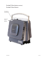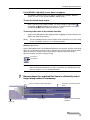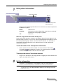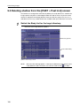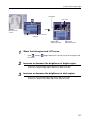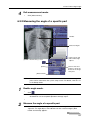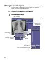
1
1114cw
User Manual
1210c
®
®
YOUR IMAGE IS OUR BUSINESS.
Table of Contents
1114cw
User Manual
1210c
®
®
Important Safety Information................................................................................ i
Note.......................................................................................................... i
Warning...................................................................................................... i
Caution...................................................................................................... i
Specifications .............................................................................................iv
Disposal.....................................................................................................iv
Guidance and Manufacturer’s Declaration for Electromagnetic Compatibility — EMC........... v
Electromagnetic Emissions.............................................................................. v
Electromagnetic Immunity.............................................................................. v
The Mark®1114cw System .................................................................................. vii
The Mark®1114cw Labels.............................................................................. viii
The Mark®1114cw Features.............................................................................ix
The Mark®1114cw Important Part Numbers......................................................... x
The Mark®1114cw Key Connections Overview.....................................................xii
Connecting the Generator............................................................................ xiii
Powering Up the Mark®1114cw System............................................................. xiv
CXDI Control Software NE Operation Manual ............................................................ 1
Safety Summary............................................................................................... 3
Contents......................................................................................................... 4
1 Getting Started............................................................................................ 9
2 Starting and Shutting Down the Software.......................................................... 27
3 Preparing for Examinations............................................................................39
4 Conducting Examinations.............................................................................. 67
5 Reviewing Images........................................................................................ 93
6 Post-Processing Images............................................................................... 109
7 Other Functions........................................................................................ 139
8 Stitching images (Long-length imaging)........................................................... 165
9 Troubleshooting Remedies........................................................................... 181
Index.......................................................................................................... 204
03282013
Sound-Eklin®
Important Safety Information
Note
Read all the instructions before connecting or operating the component.
Keep this manual so you can refer to these safety instructions.
This product is intended as a portable, digital, x-ray image
capturing system for Veterinary use only (not for human use).
The Mark®1114cw needs special precautions regarding EMC
and needs to be installed and put in to service according
to the EMC information provided in this manual.
Portable and mobile RF communication equipment
can affect the Mark®1114cw system.
The use of accessories, transducers and cables other than those specified
by Sound-Eklin®, may result in increased EMISSIONS or decreased IMMUNITY
of the EQUIPMENT. The accessories defined on page xi define the
approved components for this system. The cable lengths are considered
to be the maximum length for acceptable EMMISSIONS and IMMUNITY.
The Mark®1114cw should not be used adjacent to or stacked
with other equipment and that if adjacent or stacked use is
necessary, the EQUIPMENT should be observed to verify normal
operation in the configuration in which it will be used.
Warning
There are no user serviceable parts inside.
There are no preventative maintenance requirements.
Refer all servicing to qualified service personnel.
To avoid risk of electric shock, this equipment must be
connected to a supply mains with protective earth.
To reduce the risk of fire or electric shock, do not expose the unit to
moisture or water. Do not allow foreign objects to get into the enclosure.
If the unit is exposed to moisture, or a foreign object gets into the
enclosure, immediately disconnect the power cord from the wall.
Take the unit to a qualified service person for
inspection and necessary repairs.
Sound-Eklin®
Page i
Important Safety Information (continued)
Caution
Heed all warnings and safety information in these instructions
and on the product itself. Follow all operating instructions.
Clean the enclosure only with a dry cloth or a vacuum cleaner.
Clean the DR plate with a damp cloth. Use no chemicals or alcohol.
The detector may be placed in a protective bag
to avoid contamination during use.
IPXO per IEC 60529
Type B per IEC 60601-1 (detector is applied part)
You must allow 10 cm or 4 inches of unobstructed clearance around the
unit. Do not place the unit on a bed, sofa, rug, or similar surface that could
block the ventilation slots. If the component is placed in a bookcase or
cabinet, there must be ventilation of the cabinet to allow proper cooling.
Keep the component away from radiators, heat registers,
stoves, or any other appliance that produces heat.
Keep the component away from flammable materials.
The unit must be connected to a power supply only of the
type and voltage specified on the side panel of the unit.
Connect the component to the power outlet only with the supplied
power supply cable or an exact equivalent. Do not modify the
supplied cable in any way. Do not attempt to defeat grounding
and/or polarization provisions. Do not use extension cords.
Do not route the power cord where it will be crushed, pinched, bent at severe
angles, exposed to heat, or damaged in any way. Pay particular attention
to the power cord at the plug and where it exits the back of the unit.
The power cord should be unplugged from the wall outlet if the unit is to be
left unused for a long period of time. Immediately stop using the component
and have it inspected and/or serviced by a qualified service agency if:
· The power supply cord or plug has been damaged.
· Objects have fallen or liquid has been spilled into the unit.
· The unit has been exposed to rain.
· The unit shows signs of improper operation
· The unit has been dropped or damaged in any way
Place the unit on a fixed, level surface strong enough to support its
weight. Do not place it on a moveable cart that could tip over.
Do not place on the floor or in an area where
the system may be exposed to liquid.
Sound-Eklin®
Page ii
Important Safety Information
Caution (continued)
System will become unresponsive when exposed to Electrostatic
Discharge (ESD) or Electrical Fast Transient (EFT) conditions.
This is the normal response to these environmental conditions.
Please restart the system to return to normal operation.
Do not install detector close to strong electromagnetic fields
or devices which may interfere with wireless communication
(see detector service manual for details).
USB ports are for keyboard and mouse only. User is
responsible for evaluating the system with any devices
connected to this port for compliance to IEC 60601-1-1.
Ethernet port is not to be used during acquisition. User
is responsible for evaluating the system with any devices
connected to this port for compliance to IEC60601-1-1.
This system is not intended for use in an oxygen-rich environment.
Sound-Eklin®
Page iii
Specifications
Operation Environment: +15°C to 35°C,
30% to 75% RH, 70 to 106kPa (pressure)
Storage / Transport : –10° C to 55° C, 15% to 80% RH,
70 to 106kPa (pressure)
Power Requirements:
100 - 240 VAC 5.6A 50/60Hz
Weight (net) 47 lbs
CAUTION
Risk of electric shock.
Do not open.
Caution: To reduce the risk of electric shock, do
not remove cover. No user-serviceable parts inside.
Refer servicing to qualified service personnel.
This symbol is to alert the user to
important operating and maintenance
(service) instructions in this manual and
literature accompanying the product.
Applicable for USA, Canada or
where approved for the usage.
Caution: To prevent electric shock, match wide
blade of plug to wide slot. Insert fully.
This symbol is to alert the user to the
presence of uninsulated dangerous voltages
inside the product’s enclosure that may
constitute a risk of electric shock.
Disposal
The customer is responsible for the disposal of the medical device. Disposal activities must follow
the regulations (especially controlled industrial waste) of the country where the device is used.
Sound-Eklin®
Page iv
Guidance and Manufacturer’s Declaration for
Electromagnetic Compatibility — EMC
Electromagnetic Emissions
The CDXI-80C Wireless is intended for use in the electromagnetic environment specified below.
The user of the CDXI-80C Wireless should assure that it is used in such an environment.
Emission Test
Compliance
Electromagnetic Environment — Guidance
RF emissions CISPR11
Group 1
The CDXI-80C Wireless uses RF energy only for its internal function.
Therefore, its RF emission are very low and are not likely to
cause any interference in nearby electromagnetic equipment.
RF emissions CISPR11
Class A
Harmonic emissions
IEC 61000-3-2
Class A
The CDXI-80C Wireless is suitable for use in all establishments
other than domestic and those directly connected
to the public low-voltage power supply network that
supplies buildings used for domestic purposes.
Voltage fluctuations
/ flicker emissions
IEC 61000-3-3
1*
1: Applies to regions where rated voltage is 220 V or higher.
Not applicable to regions where the rated voltage is less than 220 V.
Electromagnetic Immunity
The CDXI-80C Wireless is intended for use in the electromagnetic environment specified below.
The user of the CDXI-80C Wireless should assure that it is used in such an environment.
Immunity Test
IEC 60601
Test Level
Compliance
Level
Electromagnetic
Environment — Guidance
Electrostatic
Discharge (ESD)
IEC 61000-4-2
±(2, 4, 6) kV contact
±(2, 4, 8) kV air
±(2, 4, 6) kV contact
±(2, 4, 8) kV air
Floors should be wood, concrete or
ceramic tile. If floors are covered
with synthetic material, the relative
humidity should be at least 30%
Electrical Fast
Transient / Burst
IEC 61000-4-4
± 2 kV for power
supply lines
± 1 kV for input
/ output lines
± 2 kV for power
supply lines
± 1 kV for input
/ output lines
Mains power quality should be that of a
typical commercial or hospital environment.
Surge
IEC 61000-4-5
± 1 kV
differential mode
± 2 kV common mode
± 1 kV
differential mode
± 2 kV common mode
Mains power quality should be that of a
typical commercial or hospital environment.
Voltage dips,
short interruptions
and voltage variations
on power supply
input lines
IEC 61000-4-11
<5% UT
(>95% dip in UT)
for 0.5 cycle.
40% UT
(60% dip in UT)
for 5 cycles.
70% UT
(30% dip in UT)
for 25 cycles.
<5% UT
(>95% dip in UT)
for 5 seconds.
<5% UT
(>95% dip in UT)
for 0.5 cycle.
40% UT
(60% dip in UT)
for 5 cycles.
70% UT
(30% dip in UT)
for 25 cycles.
<5% UT
(>95% dip in UT)
for 5 seconds.
Mains power quality should be that of a typical
commercial or hospital environment.
If the user of the CDXI-80C Wireless requires
continued operation during power mains
interruptions, it is recommended that the
CDXI-80C Wireless be powered from an
uninterruptible power supply or a battery.
Power Frequency
(50 / 60 Hz)
Magnetic Field
IEC 61000-4-8
3 A/m
3 A/m
Power frequency magnetic fields should be at
levels characteristic of a typical location in a
typical commercial or hospital environment.
Note: UT is the a.c. mains voltage prior to application of the test level.
Sound-Eklin®
Page v
Guidance and Manufacturer’s Declaration for
Electromagnetic Compatibility — EMC
Electromagnetic Immunity (continued)
Immunity Test
IEC 60601
Test Level
Compliance
Level
Electromagnetic
Environment — Guidance
Conducted RF
IEC 61000-4-6
Conducted RF
IEC 61000-4-3
3 Vrms 150kHz
to 80 MHz
3 V/m 80 MHz
to 2.5 GHz
3 Vrms
Portable and mobile RF communications
equipment should be used no closer to any part
of the CDXI-80C Wireless, including cables,
than the recommended separation distance
calculated from the equation applicable
to the frequency of the transmitter.
Recommended separations distance
d=1.2√P
d=1.2√P 80 MHz to 800 MHz
d=2.3√P 800 MHz to 2.5 GHz
where “P” is the maximum output
power rating of the transmitter in
watts (W) according to the transmitter
manufacturer and “d” is the recommended
separation distance in meters (m).
Field strengths from fixed RF transmitters,
as determined by an electromagnetic site
survey a, should be less than the compliance
level in each frequency range b.
Interference may occur in the vicinity of
equipment marked with the following symbol:
3 V/m
Note: At 80 MHz and 800 MHz, the higher frequency range applies.
These guidelines may not apply in all situations. Electromagnetic propagation is affected by absorption
and reflections from structures, object and people.
a Field strengths from fixed transmitters, such as base stations for radio (cellular/cordless) telephones and
land mobile radios, amateur radio, AM and FM radio broadcast and TV broadcast cannot be predicted
theoretically with accuracy. To assess the electromagnetic environment due to fixed RF transmitters, an
electromagnetic site survey should be considered. If the measured field strength in the location in which
the CXDI-80C Wireless is used exceeds the applicable RF compliance level above, the CXDI-80C Wireless
should be observed to verify normal operation. If abnormal performance is observed, additional
measures may be necessary, such as reorienting or relocating the CXDI-80C Wireless.
b Over the frequency range 150 kHz to 80 MHz, field strengths should be less than 3 V/m.
Sound-Eklin®
Page vi
The Mark®1114cw System
The Mark®1114cw system links seamlessly with the X-ray generator to provide the best image quality
in a small, lightweight, self-contained system. The Mark®1114cw portable Acquisition Stations are
intended to be used by Veterinarians for intermittent mobile diagnostic applications. They will
connect the Mark®1114cw Device to a CXDI Canon 80C Wireless Digital X-ray Detector.
Sound-Eklin®
Page vii
The Mark®1114cw System (continued)
The Mark®1114cw Labels
Mark®1114cw
Electronics
Box Serial
number
Windows License
Key is located
on bottom of
Electronics Box
Mark®1114cw A–frame Serial Number,
CE Markings, and Manufacture Date
MODEL: MARK 1114cw™
Sound-Eklin©
6359 Paseo Del Lago
Carlsbad . CA 92011
760.918.9626
soundeklin.com
INPUT POWER 120 VAC ~ 5.6A 60Hz
Sound-Eklin®
EN60601-1
EN60601-1-4
Page viii
The Mark®1114cw System (continued)
The Mark®1114cw Features
Power Button
with LED Light
Hard Drive
Activity
Indicator
Sound-Eklin®
Page ix
The Mark®1114cw System (continued)
The Mark®1114cw Important Part Numbers
94-859 Mark®1114cw
34-851 Mark 70/80C,
Electronics Assembly
Sound-Eklin®
34-852 Mark,
A-Frame Assembly, v2
Page x
The Mark®1114cw System
The Mark®1114cw Important Part Numbers (continued)
10-867 Min-X-Ray
25’ Sync Cable for
Mark®1114cw
Syncs the
generator’s prep
and expose cycle
with the computer.
10-137 Battery
charger for Canon
Wireless Series
90-403 10’
Ethernet
Patch Cable Connects the
computer to the
network to allow
for easy image
transfers from the
Mark®1114cw to
PACS.
10-135 Extra
Battery Pack for
Canon Wireless
94-124 CXDI-80C
Canon Wireless
Detector
(includes two
batteries)
Captures digital
radiographs.
10-171 Equine 20’
IEC-NEMA Power
Extension Cord
Connects the
Mark®1114cw to any
standard U.S. 110V
power outlet (Nema
5-15).
34-671 Detector
Handle Unit for
Canon 80C
Sound-Eklin®
Page xi
The Mark®1114cw System (continued)
The Mark®1114cw Key Connections Overview
10–121 Min–X–ray Sync
Cable for Mark®1114cw
24–122 Keyboard with Touch–Pad.
Interruption to the USB
keyboard or Mouse can be
cleared by re-mating the
connection to the system.
10’ Ethernet Patch Cable
10–171 Equine 20’
IEC–NEMA Power
Extension Cord
Sound-Eklin®
Page xii
The Mark®1114cw System (continued)
Connecting the Generator
The sync cable should stay connected to the generator and should be stored in the generator box.
1. Plug the Min–X–ray Sync Cable (10-867) into the Mark®1114cw.
Align by red dots on each end of the connector.
2. Plug the hand switch end of the Min–X–ray Sync Cable
(10-867) into the generator hand switch port.
Min–X–ray Sync
Cable (10-867)
Sound-Eklin®
Page xiii
The Mark®1114cw System (continued)
Powering Up the Mark®1114cw System
1. Plug the female end of the power cable into the power cable slot on the Mark®1114cw
box. Plug the other end into a grounded power outlet (see page iii Power Requirements).
2. With the cables connected, turn on the Mark®1114cw by
pressing the power button on the top of the unit.
Power Button
with LED Light
10-171 Equine 20’ IEC-NEMA
Power Extension Cord
Standard
US 110v
Power Outlet
(NEMA 5-15)
Cord
Sound-Eklin®
Page xiv
The Mark®1114cw CXDI Control Software NE
Sound-Eklin®
Page 1
To the Customers
Thank you for purchasing the Canon CXDI Control Software NE (hereinafter called this product).
Operating instructions are divided into two volumes: the Operation Manual and the Setup Guide.
Before using this product, be sure to read these manuals thoroughly in order to utilize this product more
effectively.
Disclaimer
1. In no event shall Canon be liable for any damage or loss arising from fire, earthquake, any action by a third
party or other accidents, any intentional or negligent action by the users, any trial usage, or other usage
under abnormal conditions.
2. In no event will Canon be liable for direct or indirect consequential damage arising out of the use or
unavailability of this product.
Canon will not be liable for loss of image data due to any reason.
3. In no event shall Canon be liable for personal physical harm or property damage that is sustained when
the instructions are not followed or the product is misused.
4. It is the responsibility of the attending physicians to provide medical care services. Canon will not be liable
for the faulty diagnosis.
5. Roentgenography, image processing, image reading, and image data storage must be performed in
accordance with the laws of the country or region in which the product is being used.
The user is responsible for maintaining the privacy of image data.
6. Information in this manual may change without prior notice.
7. Screen images in this manual may differ from those displayed on your image capture computer due to
customization, updates and improvements in the software.
8. We’ve taken all possible means to ensure the content accuracy of this manual. If you have any questions
or problems, contact your sales representative or local Canon dealer.
Trademarks
• The Canon name and Canon logo are registered trademarks of Canon Inc.
• Ethernet is a trademark of Xerox Corporation.
• Microsoft and Windows are either registered trademarks or trademarks of Microsoft Corporation in the
United States and/or other countries or regions.
• TrueType is a registered trademark of Apple Inc.
• Intel and Intel Core are trademarks of Intel Corporation in the U.S. and other countries.
• Other systems and product names in this manual are the trademarks of the manufacturers that have
developed them.
Copyright
• All rights reserved.
• Under copyright laws, this manual may not be reproduced, in whole or in part, without the written
permission of Canon.
Third Party Software
Third-party software will be installed on the image-capture computer when the control software is being
installed by the service engineer. For details on the third-party software and its license agreements, consult
your service engineer.
2
Safety Summary
Before using this product, read this safety summary thoroughly. This information will prevent the users and
persons involved from sustaining physical harm and/or property damage.
Read the separate Setup Guide and the Digital Radiography CXDI series User’s Manual as well.
Safety Notices
The following safety notices are used to emphasize certain safety instructions. This manual uses the caution
symbol along with a caution message.
CAUTION
This notice is used to identify conditions under which improper use of the product
may cause minor personal injury.
CAUTION
This notice is used to identify conditions under which improper use of the product
may cause property damage.
Safety Precautions
Follow these safeguards and properly use the application software to prevent injury and damage to any
equipment/data.
While preparing for examinations
CAUTION
• Be sure to confirm that the entered information (patient name, ID number, birth date, and sex)
matches that of the patient. If the information is incorrect, the resulting patient mix-up and a
misdiagnosis may cause harm to the patient.
• Be sure to use the [Emergency] button only for an emergency examination. If not heeded, the
resulting patient mix-up and a misdiagnosis may cause harm to the patient.
While shutting down
CAUTION
Do not turn the image-capture computer off before it automatically shuts down. Doing so may result
in damage to the hardware and/or destruction of data.
3
Contents
Safety Summary ............................................................................................................ 3
1 Getting Started
1.1 Introduction ........................................................................................................... 10
1.2 Notations................................................................................................................ 10
1.2.1 Screen names ......................................................................................................................... 10
1.2.2 Buttons .................................................................................................................................... 10
1.2.3 Mouse wheel operations........................................................................................................ 11
1.2.4 Touch-screen display operations ......................................................................................... 11
1.2.5 References .............................................................................................................................. 11
1.3 Workflow diagram for a general radiographic examination.............................. 12
1.4 Part names of screens (Quick reference) ........................................................... 14
1.4.1 [EXAM > Worklist] screen ...................................................................................................... 14
1.4.2 Study information pane and protocols ................................................................................. 15
1.4.3 Edit Exam screen .................................................................................................................... 16
1.4.4 [EXAM > Manual] screen........................................................................................................ 18
1.4.5 [EXAM > Examination] screen ............................................................................................... 19
1.4.6 [PAST > Past List] screen ...................................................................................................... 20
1.4.7 [PAST > View] screen ............................................................................................................. 21
1.4.8 Toolbar..................................................................................................................................... 22
1.4.9 Image Processing panels....................................................................................................... 23
1.4.10 Measurement panel.............................................................................................................. 24
1.4.11 Annotation panel................................................................................................................... 24
1.4.12 Stitch Screen......................................................................................................................... 25
2 Starting and Shutting Down the Software
2.1 Starting the Software ............................................................................................ 30
2.2 Shutting down the Software................................................................................. 32
2.3 Conducting offline examinations ......................................................................... 36
2.4 Utilizing peripheral devices .................................................................................. 37
4
Contents
3 Preparing for Examinations
3.1 Selecting study orders (Worklist tab) .................................................................. 42
3.1.1 Acquiring study orders from the HIS/RIS database ............................................................ 42
3.1.2 Checking patient/study information in detail....................................................................... 46
3.1.3 Modifying study information.................................................................................................. 47
3.1.4 Changing a workspace .......................................................................................................... 49
3.1.5 Configuring protocols ............................................................................................................ 51
3.2 Selecting protocols ............................................................................................... 53
3.2.1 Searching for a protocol by a body part............................................................................... 53
3.2.2 Searching for a protocol by category ................................................................................... 56
3.3 Creating study orders (Manual tab)..................................................................... 58
3.3.1 Registering patient/study information.................................................................................. 58
3.3.2 Modifying patient information ............................................................................................... 62
3.3.3 Modifying study information.................................................................................................. 63
3.3.4 Changing a workspace .......................................................................................................... 63
3.4 Creating emergency study orders (Emergency button) .................................... 64
4 Conducting Examinations
4.1 Conducting radiographic examinations.............................................................. 70
4.2 Replacing an incomplete protocol with another protocol................................. 78
4.3 Reprocessing an image of a complete protocol ................................................ 80
4.4 Recapturing images .............................................................................................. 83
4.5 Rejecting images................................................................................................... 84
4.6 Suspending an examination ................................................................................. 85
4.7 Rebinding other study information with that of the suspended study order... 86
4.8 Binding study information with the images of an emergency study ................ 89
4.9 Adding information to a complete protocol........................................................ 91
5 Reviewing Images
5.1 Reviewing images on the [EXAM > Examination] screen.................................. 96
5.2 Reviewing images on the [PAST > View] screen................................................ 98
5.2.1 Reviewing images saved in the image-capture computer ............................................... 100
5.2.2 Conducting additional examinations using a complete study ......................................... 101
5.2.3 Creating images using different image processing parameters of a copied study ....... 102
5.2.4 Reviewing images saved in storage media ........................................................................ 104
5.3 Deleting studies from the [PAST > Past List] screen....................................... 106
5
Contents
6 Post-Processing Images
6.1 Using the toolbar (Basic processing) ................................................................ 112
6.1.1 Rotating/flipping images...................................................................................................... 114
6.1.2 Inverting a negative image to a positive image and vice versa........................................ 114
6.1.3 Panning images .................................................................................................................... 115
6.1.4 Zooming images ................................................................................................................... 116
6.1.5 Embedding laterality markers in images ............................................................................ 117
6.1.6 Adjusting the overall brightness on the ROI basis ............................................................ 118
6.1.7 Cropping images .................................................................................................................. 120
6.1.8 Masking the peripheral area................................................................................................ 123
6.2 Using the Image Processing panel (Level 1)..................................................... 126
6.2.1 Adjusting the brightness of bright region and dark region .............................................. 126
6.2.2 Adjusting the overall brightness and contrast................................................................... 128
6.3 Using the Measurement panel ........................................................................... 129
6.3.1 Measuring the distance between two specific points ...................................................... 129
6.3.2 Measuring the angle of a specific part ............................................................................... 131
6.3.3 Measuring the Cobb angle................................................................................................... 132
6.3.4 Measuring the height difference ......................................................................................... 134
6.4 Using the Annotation panel ................................................................................ 136
6.4.1 Creating/editing custom annotations ................................................................................. 136
7 Other Functions
7.1 Printing and transferring images ....................................................................... 142
7.1.1 Checking and changing the image transfer settings ........................................................ 143
7.1.2 Arranging and printing images automatically .................................................................... 144
7.1.3 Arranging and printing images manually............................................................................ 149
7.1.4 Manually printing images in the [PAST > View] screen..................................................... 151
7.1.5 Manually transferring images to a storage server or storage media
in the [PAST > View] screen ................................................................................................ 152
7.2 Using the Image Processing panel (Levels 2 and 3) ........................................ 154
7.2.1 Switching access levels ....................................................................................................... 154
7.2.2 Automatically processing images by specific anatomical part
(Anatomical Part control)..................................................................................................... 155
7.2.3 Adjusting the overall image tone (LUT control) ................................................................. 156
7.2.4 Enhancing contrast for anatomical part shape definition (Enhancement control) ........ 158
7.2.5 Expanding or narrowing the dynamic range (Dynamic Range Adjustment control)...... 159
7.2.6 Reducing noise in images (Noise Reduction control) ....................................................... 161
7.2.7 Reducing grid lines (Grid Suppression control)................................................................. 162
7.2.8 Adjusting the image sharpness (Sharpness Adjustment control).................................... 163
7.2.9 Masking the area outside of the irradiation field (Peripheral Mask control)................... 164
6
Contents
8 Stitching images (Long-length imaging)
8.1 What is long-length imaging? ............................................................................ 168
8.2 Capturing and stitching images......................................................................... 168
8.3 Realigning, modifying, and stitching partial images........................................ 175
8.3.1 Aligning partial images......................................................................................................... 175
8.3.2 Modifying long-length images in the [PAST > View] screen............................................. 179
9 Troubleshooting Remedies
9.1 Confirming details of the problem ..................................................................... 182
9.1.1 Fatal error list ........................................................................................................................ 183
9.1.2 Error list ................................................................................................................................. 186
9.1.3 Warning list............................................................................................................................ 201
Index ....................................................................................................... 204
7
Contents
8
1
Getting Started
1.1 Introduction
1.2 Notations
1.3 Workflow diagram for a general radiographic
examination
1.4 Part names of screens (Quick reference)
1 Getting Started
1.1 Introduction
The CXDI Control Software NE (hereinafter referred to as the “Software”) is designed
to provide a steady and efficient workflow in the field of digital radiography when
linked to an RIS/HIS network. Up to four CXDI detectors including wireless detectors
can be connected simultaneously to the Software. Images captured using the CXDI
detector are automatically processed by the Software to achieve diagnostic image
quality. Furthermore, the Software offers advanced processing features with intuitive
operability.
1.2 Notations
1.2.1 Screen names
In this manual, screens are identified by the titles of active tabs.
For example, “[EXAM > Worklist] screen” indicates that the [EXAM] and [Worklist] tabs
are selected in sequence.
Active tabs
1.2.2 Buttons
In this manual, the key top labels of buttons on the Software screen are written in
square brackets, such as [OK] and [Cancel], or the icon is shown.
Button appearance on the Software screen
Enabled buttons: Appear with a navy-blue background. (A)
Enabled buttons (prompted):
Appear with a navy-blue background surrounded by a blinking
line.
Disabled buttons: Appear with a grayed-out key-top label. (B)
Selected buttons: Appear highlighted in pale-blue surrounded by a white line. (C)
Toggle buttons:
Key-top label and color alternately change (e.g.,
/
) with
each click. (D)
NOTE:
10
Six types of screen background color are prepared for the Software: Warm
Dark, Cool Dark, Monochrome Dark, Warm Light, Cool Light, and
Monochrome Light. Refer to 2.3.1 in the Setup Guide for details on setting
the screen background color.
In this manual, the Cool Light screen, that is the default setting, is used for
explanation.
1 Getting Started
Example
A
C
D
A
B
B
C
A
A
B
1.2.3 Mouse wheel operations
The mouse pointer may change to
while pointing on a list table, at a numeric entry
field, or on a control slider. In such cases, rotating the mouse wheel scrolls through the
list, enters values, or moves the slider. For a control slider, dragging the slider is an
alternative operation.
1.2.4 Touch-screen display operations
This manual assumes that users are using a mouse and keyboard. However, touchscreen displays are also available. Unless otherwise stated, the operating procedures
using a touch-screen display is the same as those using a mouse and keyboard. When
using a touch-screen display, “Click [OK]” is equivalent to “Touch [OK].”
1.2.5 References
References to this manual:
Associated information in this manual is indicated by the section number or by the
section number followed by the title, as seen below:
(See 3.1.5)
See step 2 in 3.1.5 for details on operation.
Proceed to “3.1.5 Configuring protocols.”
References to other documents:
Associated information in other manuals is indicated by the manual title or the section
number followed by the manual title, as seen below:
Refer to the Operation Guide supplied with the power box.
For details, refer to the Setup Guide*.
(refer to 2.4.1 in the Setup Guide* for details on setting essential information
items)
* “Setup Guide” refers to the CXDI Control Software NE Setup Guide.
11
1 Getting Started
1 Getting Started
1.3 Workflow diagram for a general radiographic
examination
NOTE:
[PAST] tab
[Back To List]
The following example illustrates a case in which the Software is used under
interoperability services with the HIS/RIS database.
[Recall Exam]
[PAST > View] screen
[View]
[EXAM] tab
Edit Exam screen
[Log In]
Start Screen
Post-Processing Images
(see chapter 6)
Manually printing images in
the [PAST > View] screen.
(see 7.1.4)
[Worklist]
[Edit Exam]
[PAST > Past List] screen
Configuring protocols
(see 3.1.5)
Reviewing images on the
[PAST > View] screen (see 5.2)
[Past List]
Local disk of the
image-capture
computer
[EXAM]
[PAST]
[Examination]
[Start Exam]
Starting the Software
(see 2.1)
[Start Exam]
[EXAM > Examination] screen
(Before exposure)
[EXAM > Worklist] screen
[Suspend Exam]
Selecting study orders
(Worklist tab) (see 3.1)
Conducting
Examinations
(see chapter 4)
[Manual]
Captured
images
Exposure switch
[Worklist]
[End Exam]
Screen transition
[EXAM > Examination] screen
(After exposure)
Screen buttons
[Send to Storage]
[Start Exam]
[EXAM > Manual] screen
Screen tabs
Captured image transfer
12
[Manual]
Creating study orders (Manual tab)
(see 3.3)
[Examination]
Reviewing Images (see chapter 5)
Post-Processing Images (see chapter 6)
Printing and transferring images (see 7.1)
PACS server/
Storage media
Printer
13
1 Getting Started
1.4 Part names of screens (Quick reference)
NOTE:
Screen details may differ depending on the settings of the Software.
1.4.1 [EXAM > Worklist] screen
A
B
E
F
G
C
D
H
I
J
A: HDD free space icon (➞3.1.1, page 43)
B: System setup button (➞2.1.1 in the Setup Guide)
C: Search For Study List pane (➞3.1.1, page 42)
D: Study List (➞3.1.1, page 42)
E: Data transfer indicator (➞4.1, page 70)
F: [On Line]/[Off Line] selector/indicator (➞2.3, page 36)
G: Patient information pane (➞3.1.2, page 46)
H: Study information pane (➞1.4.2, page 15) (➞3.1.2, page 46)
I: [Start Exam] (➞4.1, page 70)
J: [Emergency] (➞3.4, page 64)
14
1 Getting Started
1.4.2 Study information pane and protocols
C
A
B
D
E
A: Study title (Accession number) (➞3.1.2, page 46) (➞“Study Information Display” in
2.4.1 in the Setup Guide)
B: Protocol list (➞Chapter 4, page 67)
C: Information button (➞3.1.2, page 46) (➞3.1.3, page 47)
D: Thumbnail
E: [Edit Exam] (➞3.1.5, page 51)
15
1 Getting Started
1.4.3 Edit Exam screen
C
D
A
E
F
B
Body part search mode
A: Body part selector (➞3.2.1, page 53)
B: [Search by Category] (➞3.2.2, page 56)
C: Frequently-used protocols list (➞3.2.1, page 53)
D: Recently-used protocols list (➞3.2.1, page 53)
E: Study information pane (➞1.4.2, page 15) (➞3.1.2, page 46)
F: [Start Exam] (➞4.1, page 70)
G: Examination history list (➞3.1.5, page 51)
16
G
1 Getting Started
H
J
K
I
Category search mode
H: Category tabs (➞3.2.2, page 56)
I: [Search by Body Part] (➞3.2.2, page 56)
J: Protocol list (➞3.2.2, page 56)
K: [Category List] (➞3.2.2, page 56)
17
1 Getting Started
1.4.4 [EXAM > Manual] screen
A
B
D
C
E
A: Patient information entry (➞3.3.1, page 58)
B: Recent Patient List (➞3.3.1, page 58)
C: [Delete] (➞3.3.1, page 58)
D: [Emergency] (➞3.4, page 64)
E: [Start Exam] (➞4.1, page 70)
18
1 Getting Started
1.4.5 [EXAM > Examination] screen
A
F
G
B
H
C
D
I
J
K
L
M
E
N
O
P
A: View mode buttons (➞5.1, page 96)
B: [Info] (➞5.1, page 96)
C: Toolbar (➞1.4.8, page 22) (➞6.1, page 112)
D: Image view pane (➞4.1, page 70)
E: [Retake] (➞4.4, page 83),
[Reject] (➞4.5, page 84)
F: System status bar (➞4.1, page 70)
G: Output setting button (➞7.1, page 142)
H: Patient information pane (➞3.1.2, page 46)
I: Study information pane (➞1.4.2, page 15) (➞4.1, page 70)
J: Image Processing panel (➞1.4.9, page 23) (➞6.2, page 126)
K: [ImageProc] (➞6.2, page 126) (➞7.2, page 154)
L: [Measurement] (➞1.4.10, page 24) (➞6.3, page 129)
M: [Annotation] (➞1.4.11, page 24) (➞6.4, page 136)
N: [End Exam] (➞4.1, page 70)
O: [Send to Storage] (➞4.1, page 70)
P: [Suspend Exam] (➞4.6, page 85)
19
1 Getting Started
1.4.6 [PAST > Past List] screen
A
D
B
E
C
F
G
H
A: HDD free space icon (➞3.1.1, page 43)
B: Search For Study List pane (➞3.1.1, page 42)
C: Study List (➞5.2, page 98)
D: Patient information pane (➞3.3.2, page 62)
E: Study information pane (➞1.4.2, page 15) (➞5.2, page 98)
F: [Restart Exam] (➞5.2.2, page 101)
G: [Recall Copied Exam] (➞5.2.3, page 102)
H: [Recall Exam] (➞5.2.1, page 100)
20
1 Getting Started
1.4.7 [PAST > View] screen
A
E
B
F
C
G
D
H
I
J
A: View mode buttons (➞5.1, page 96)
B: [Info] (➞5.1, page 96)
C: Toolbar (➞1.4.8, page 22) (➞6.1, page 112)
D: Image view pane (➞5.2, page 98)
E: Output setting button (➞7.1, page 142)
F: Patient information pane (➞3.3.2, page 62)
G: Study information pane (➞1.4.2, page 15) (➞5.2, page 98)
H: [Restart Exam] (➞5.2.2, page 101)
I: [Save Image(s)] (➞5.2.1, page 100)
J: [Back To List] (➞5.2.1, page 100)
21
1 Getting Started
1.4.8 Toolbar
A
B
C
D
E
F
G
H
I
J
K
L
M
N
A: Rotation/Flip buttons (➞6.1.1, page 114)
B: Negative/positive button (➞6.1.2, page 114)
C: Panning button (➞6.1.3, page 115)
D: [HQ] (➞6.1.4, page 116)
E: Zoom in/out buttons (➞6.1.4, page 116)
F: Fit button (➞6.1.4, page 116)
G: [L]/[R] (➞6.1.5, page 117)
H: [ROI] (➞6.1.6, page 118)
I: Crop button (➞6.1.7, page 120)
J: [Mask] (➞6.1.8, page 123)
K: [Retake] (➞4.4, page 83)
L: [Reject] (➞4.5, page 84)
M: [Reset] (➞6.1, page 112)
N: Undo button (➞6.1, page 112)
22
1 Getting Started
1.4.9 Image Processing panels
A
B
C
Basic image processing (Level 1)
A: Show/hide selector for histogram and LUT curve (➞6.2.1, page 126)
B: Access level selector/indicator (➞6.2, page 126) (➞7.2, page 154)
C: Brightness and Contrast controls (➞6.2.2, page 128)
D
E
F
G
H
I
J
K
L
Advanced image processing (Level 2/3)
D: Anatomical Part control (➞7.2.2, page 155)
E: LUT control (➞7.2.3, page 156)
F: Enhancement control (➞7.2.4, page 158)
G: Dynamic Range Adjustment control (➞7.2.5, page 159)
H: Noise Reduction control (➞7.2.6, page 161)
I: Grid Suppression control (➞7.2.7, page 162)
J: Sharpness Adjustment control (➞7.2.8, page 163)
K: Peripheral Mask control (➞7.2.9, page 164)
L: [Save as Default] (➞7.2.1, page 154)
23
1 Getting Started
1.4.10 Measurement panel
D
E
A
F
B
G
C
H
A: Measure distance button (➞6.3.1, page 129)
B: Measure Cobb angle button (➞6.3.3, page 132)
C: Measure height difference button (➞6.3.4, page 134)
D: Measure angle button (➞6.3.2, page 131)
E: [Delete] (➞6.3, page 129)
F: Show/hide object selector (➞6.3, page 129)
G: Show/hide scale selector (➞6.3, page 129)
H: Correction of Magnification option (➞6.3.1, page 129)
1.4.11 Annotation panel
A
B
A: [Add/Edit] (➞6.4.1, page 136)
B: [Enlarge], [Reduce], [Delete] (➞6.4.1, page 136)
24
1 Getting Started
1.4.12 Stitch Screen
B
C
A
D
E
F
A: Image view pane (➞8.3.1, page 175)
B: Toolbar (➞8.3.1, page 175)
C: Image overview pane (➞8.2, page 168)
D: [Auto] (➞8.3.1, page 175)
E: Arrow buttons (➞8.3.1, page 175)
F: Edge Enhancement control (➞8.3.1, page 175)
25
1 Getting Started
26
2
Starting and Shutting Down the
Software
2.1 Starting the Software
2.2 Shutting down the Software
2.3 Conducting offline examinations
2.4 Utilizing peripheral devices
This chapter explains how to start and shut down the Software.
Overview
Start screen
[EXAM > Worklist] screen
System setup screen
Selecting study orders (Worklist tab)
(see 3.1)
System setup screen (refer to chapter 2
in the Setup Guide)
2.1 Starting the Software
2.3 Conducting offline examinations
Inspecting/maintaining the system
NOTE:
28
2.2 Shutting down the Software
Regular inspection and maintenance of a medical
imaging system is required to assure the strict image
quality. For details on operation, refer to 3.5 in the
Setup Guide.
The frequency of adjustment work required using the
QC Tool, such as calibration, will differ depending on
the detector type. For details, refer to the Digital
Radiography CXDI series User’s Manual.
29
2 Starting and Shutting Down the Software
2.1 Starting the Software
1
Turn the power box and the image-capture computer on.
2
Log in to the Software.
The start screen appears. For details on operation, refer to the hardware
operation manual.
Select the user name, type the password, and then click [Log In].
Click to shut down
the Software.
User Name selector
[Log In]
Password text box
Start screen
NOTE:
30
Another start option that does not require user authentication is also
available. For details, consult your service engineer.
2 Starting and Shutting Down the Software
To modify the user authentication settings
The system setup screen allows users to change, add or delete user names and
change passwords. To add or delete user names, it is necessary in advance to
log in to the Software under a user name that has the Administrator role.
For details, refer to 2.2 in the Setup Guide.
Automatic logout function
The Software automatically logs out after 10 minutes of inactivity. To change the
setting of the automatic logout function, refer to 2.3.1 in the Setup Guide.
The error button and warning button
An
(error button) or
(warning button) will appear at the top center of the
screen if the Software experiences a problem. If this happens, see 9.1.
31
2 Starting and Shutting Down the Software
2.2 Shutting down the Software
NOTE:
It is necessary to shut down the image-capture computer once a day (at
closing, etc.), to maintain the optimum performance of the Software.
If a study is currently being examined
Be sure to click [End Exam] in the examination screen before shutting down the
Software. See step 6 in 4.1 for details on [End Exam].
1
Show the system setup screen.
Click
.
[EXAM > Worklist] screen
[Logout]
System setup screen
NOTE:
A dialog box related to image arrangement may appear. For details, see
NOTE in step 4 in 7.1.2.
To log in under another user name
Click [Logout], click [OK] in the confirmation dialog box, and then select another
user name in the start screen to log in again.
32
2 Starting and Shutting Down the Software
2
Show the Shutdown Confirmation Dialog Box.
Click
.
Transfer All Data option
Backup Database option
[Shutdown]
[Restart Options]
Shutdown
Confirmation Dialog
Box
Click to cancel shutdown and close the Shutdown
Confirmation Dialog Box.
To complete all image data transfer before exiting the Software
Select the Transfer All Data option.
To back up the Software database completely before exiting the
Software
Select the Backup Database option.
To exit the Software without data transfer
Clear both the Transfer All Data and Backup Database options.
Continued
33
2 Starting and Shutting Down the Software
3
Exit the Software and shut down the image-capture
computer.
Click [Shutdown] in the Shutdown Confirmation Dialog Box.
CAUTION
Do not turn the image-capture computer off before it automatically shuts down.
Doing so may result in damage to the hardware and/or destruction of data.
To restart the Software without shutting down the image-capture
computer
Click [Restart Options], and then click [CXDI Restart] in the recovery mode
screen that appears.
[CXDI Restart]
[PC Sleep]
[PC Hibernate]
[PC Restart]
[Shutdown]
Recovery mode screen
To put the image-capture computer to sleep mode
Click [PC Sleep] in the recovery mode screen. The computer enters low-power
mode and the monitor display enters standby mode. As the memory contents
are preserved during sleep mode, the computer quickly resumes working.
34
2 Starting and Shutting Down the Software
To put the image-capture computer to hibernation mode
Click [PC Hibernate] in the recovery mode screen. The HDD of the computer
and monitor display turn off. As the memory contents in recovery mode are
preserved during hibernation mode, the computer resumes working faster than
restarting the computer.
NOTE:
Do not enable sleep or hibernation mode using the power switch on the
image-capture computer.
NOTE:
The image-capture computer resumes in recovery mode from sleep or
hibernation.
NOTE:
[PC Sleep] and [PC Hibernate] can be hidden. For details, consult your
service engineer.
To exit the Software and restart the image-capture computer
Click [PC Restart] in the recovery mode screen.
NOTE:
When [Shutdown] in the recovery mode screen, as well as [Shutdown] in the
Shutdown Confirmation Dialog Box, is clicked, the Controller is exited.
If the Software is unexpectedly aborted and the recovery mode
screen appears
Click [CXDI Restart] to restart the Software and continue operations.
NOTE:
If an error persists after restarting the Software by clicking [CXDI Restart],
restart the image-capture computer.
35
2 Starting and Shutting Down the Software
2.3 Conducting offline examinations
When the Software is used in a mobile system, the Software can operate without
connection to the HIS/RIS database. In such cases, follow the steps below.
1
Disconnect from the network.
Click [On Line]. Clicking the button toggles between [On Line] and [Off Line].
To resume connection
Click [Off Line].
[On Line]
[EXAM > Worklist] screen
[Off Line]
NOTE:
36
Switching to online mode again transfers the images captured during offline
mode to the PACS server, storage device, and printer, and also exchanges
the study history with the HIS/RIS database.
2 Starting and Shutting Down the Software
2.4 Utilizing peripheral devices
The Software supports the use of a bar-code reader, and a magnetic card reader. For
details on these devices, refer to the operation manuals supplied with these devices.
37
2 Starting and Shutting Down the Software
38
3
Preparing for Examinations
3.1 Selecting study orders (Worklist tab)
3.2 Selecting protocols
3.3 Creating study orders (Manual tab)
3.4 Creating emergency study orders (Emergency
button)
This chapter explains how to prepare for examinations and how to check patient and study*
information before capturing images. It is also possible to modify study information details if
necessary.
* See 3.1 for details.
Workflow
Patient information pane
Start screen
Starting the Software (see 2.1)
[EXAM > Worklist] screen
3.1 Selecting study orders (Worklist tab)
3.4 Creating emergency study orders
(Emergency button)
[EXAM > Worklist] screen
3.1.2 Checking patient/study information
in detail
3.1.3 Modifying study information
3.1.4 Changing a workspace
3.1.5 Configuring protocols
Study information pane
[EXAM > Manual] screen
3.3 Creating study orders (Manual tab)
Edit Exam screens
3.2.1 Searching for a protocol by a body part
3.2.2 Searching for a protocol by category
[EXAM > Examination] screen
Conducting Examinations (see chapter 4)
40
41
3 Preparing for Examinations
3.1 Selecting study orders (Worklist tab)
Data exchange and updating services between the HIS/RIS database and modalities
streamline the examination workflow and maintain steady operation. If the Software
does not have these services, create study orders by manually entering patient/study
information on the [EXAM > Manual] screen (see 3.3).
.
Before starting examinations, learn the following basic terms used in the Software.
Study:
A request (order) for radiographic examination, consisting of patient information,
schedule, and examination details, issued by a referring physician
Exam:
A group of studies for one patient
Protocol:
Details of examination procedures, which include the following information:
Workspace, grid, image processing parameters, and DICOM attributes. In this
Software, a protocol is associated with a specific workspace, and that pair is shown
as a “protocol” button. In other word, according to the requirements of an individual
site, multiple buttons are created for one protocol, and all differ from each other in
the workspace. They are organized in protocol lists. Therefore, users can select a
pair including a protocol and workspace with one click of these buttons (see 3.1.5).
Workspace: Details of a protocol that specify the detector used and the posture of the patient
during the examination
3.1.1 Acquiring study orders from the HIS/RIS database
Study orders are automatically acquired from the HIS/RIS database and listed when
the user logs in.
NOTE:
An HIS/RIS database introduction is required to list study orders on the
worklist. Consult your service engineer for details.
[On Line]/[Off Line]
selector/indicator
[EXAM > Worklist]
tab
HDD free space
icon
/
Column header
Study orders
Click to create an
emergency study
order.
[EXAM > Worklist] screen
42
3 Preparing for Examinations
If the [EXAM > Worklist] screen does not appear
The [EXAM > Manual] screen may appear instead of the [EXAM > Worklist]
screen. In such a case, click the [EXAM > Worklist] tab.
To sort the listed study orders
Click a sort item in the column header. The current order is indicated by
(ascending) or
(descending) in the column. To switch between ascending
and descending sort order, click the same item again.
To arrange the order of the column headers
Drag an item and drop it in the target position. Dragging the column border also
adjusts the width of the column.
NOTE:
The items displayed in the column head can be selected in the system setup
screen. For details, refer to 2.4.1 in the Setup Guide.
HDD free space icon
To get a good performance of the Software during an examination, check the free HDD
space on a regular basis using this icon. If the icon color changes from white to yellow,
delete unnecessary studies in the image-capture computer. (If the free space
decreases further, the color changes to red.)
Free space
Used space
NOTE:
1
Past study data deletion that starts automatically in the background may
affect the Software performance. See 5.3 for details on study deletion and
also see step 2 in 3.1.3 on image protection.
Narrow down the acquired list items to efficiently select
target study orders if necessary.
Click to shrink and expand the pane.
Study Status
[Refresh]
Title boxes and their keyword
boxes
Click to clear all narrowing
conditions.
Continued
43
3 Preparing for Examinations
Enter any narrowing conditions in the Search For Study List pane.
Three of the following information titles can be selected for the narrowing
conditions: Patient ID, Name, Accession No., Referring Physician,
Requesting Physician, Comment, RP ID, RP Description, and SPS
Description. Select an option in each title box, and then enter any text in its
keyword box. Note that these three keywords are used as AND search
conditions.
Study Status:
Specifies the search range from among All Exam, New
Exam, Pending Exam, and Restarted Exam.
NOTE:
The information titles that can be selected for the narrowing conditions are
those currently displayed in the column header. For details on the column
header settings, refer to the Column Headers option under 2.4.1 in the Setup
Guide.
NOTE:
Patient Name text box can be divided into five components for
accommodating family name, given name, middle name, prefix, and suffix
input. For details, refer to 2.4.1 in the Setup Guide.
If the target study orders are not found on the list
Click [Refresh] to restore the available study orders.
If narrowing conditions are predetermined, narrowing down at the point of data
acquisition aids in efficient study order selection.
Click [Refresh Option] to show the Refresh Option dialog box.
NOTE:
When [Refresh] and [Refresh Option] are disabled, click [Off Line] to resume
connection with the HIS/RIS database.
[Refresh Option]
Click to cancel the operation.
[OK]
Refresh Option dialog box
44
3 Preparing for Examinations
Enter any narrowing conditions in the Refresh Option dialog box, and then click
[OK].
ID:
Patient ID
Name:
Patient name
ACC#:
The accession number is automatically given to each study
order at the point of issue. This number can also serve as the
study title. Refer to “Study Information Display” in 2.4.1 in
the Setup Guide.
Requested Procedure ID:
Identifier that identifies the Requested Procedure in the
Imaging Service Request.
CAUTION
2
Range:
Period refers to the period of time in which the study orders
are to be conducted. Relative refers to a period relative to
the current time in which the study orders are to be
conducted, specified by hours in the past/future. To clear the
Range condition, select the All option.
Modality:
DX refers to digital X-ray radiography, and CR to computed
radiography.
Be sure to confirm that the entered information (patient name, ID number, birth
date, and sex) matches that of the patient. If the information is incorrect, the
resulting patient mix-up and a misdiagnosis may cause harm to the patient.
Select the target study order.
Click the target study order to highlight. The selected study orders are listed in
the study information pane.
Multi-selection for ease of operation
With a click of one study order in the list, study orders with the same descriptions for
five categories - Patient ID, Name, Birth (date), Sex, and Input data type* - will
automatically be selected. To deselect unwanted study orders from the selection, click
each order in turn.
* Indicates either HIS/RIS originated or manually created data
3
Start examination.
Proceed to “4 Conducting Examinations.”
If a dialog box appears indicating that the protocol includes an
unknown code value
Consult your service engineer to create the protocol applicable to the unknown
code value.
45
3 Preparing for Examinations
3.1.2 Checking patient/study information in detail
1
Select a study order.
Click a study order in the Study List.
Patient
information pane
Detailed patient
information
Patient name
Date of birth
Pregnancy status
(none): not pregnant
: pregnant
: possibly
pregnant
Study title (ACC#)
Study List
Click to modify study
information (if necessary,
see 3.1.3).
Study order
Study information pane
2
46
Detailed study information
Show the detailed information.
Click
in the patient information pane or
hide the detailed information, click it again.
on the target study order. To
3 Preparing for Examinations
3.1.3 Modifying study information
If necessary, the ordered study information can be modified before examination.
Click to hide or
show the protocols
in the study order.
The selected study
order is
highlighted.
Continued
47
3 Preparing for Examinations
1
Show the Edit Study Information dialog box.
Select the target study order in the Study List, and then click
study in the study information pane, and then click [Edit].
on the target
[Edit]
Referring Physician/
Study Description/
Reading Physician/
Institution Name text
box
Protect Image option
Click to cancel the
operation.
Study information pane
[OK]
2
48
Modify the study information.
Modify information in the Study Description text box (up to 64 characters).
Preset descriptions can be selected from the drop-down list, as well as by
entering a description in the text boxes.
NOTE:
Grayed-out information in the Edit Study Information dialog box cannot be
modified.
NOTE:
Other study information items, such as Requesting Physician and Study
Instance UID, can be configured to be shown in this dialog and modified in
this step. For details, refer to 2.4.1 in the Setup Guide.
NOTE:
Frequently used descriptions for some study information items can be
preset. For details, refer to 2.4.2 in the Setup Guide.
3 Preparing for Examinations
To protect the stored images
To maintain a certain amount of free space in the local storage directory of the
image-capture computer, stored images except for those being reviewed on the
[EXAM > Examination] or [PAST > View] screen will automatically be deleted on
a study basis, beginning with the oldest one, as the amount of data exceedingly
increases. To prevent images from being deleted, select the Protect Image
option. To cancel, clear the option.
3
End modification.
Click [OK].
3.1.4 Changing a workspace
If necessary, the ordered workspace can be changed before examinations. When two
or more detectors are connected to the system, detectors can be switched by
changing the workspace.
Thumbnail
Protocol
The selected study
order is
highlighted.
Continued
49
3 Preparing for Examinations
1
Show the Select Workspace dialog box.
Select a study order, and then double-click the thumbnail in the target protocol
in the study information pane.
Detector name
Patient posture (detector setup)
: Stand
: Table
: Cassette
: Universal
Available workspaces
The connection with the detector is incomplete, or the
power box is turned off.
Workspace name
Click to cancel the operation.
[OK]
NOTE:
A battery indicator and a signal strength indicator are shown on the
workspace for wireless detectors. For the wireless detector, refer to the
Digital Radiography CXDI series User’s Manual.
Example of workspace indicator for wireless detectors
Battery indicator
Signal strength indicator
: 60–100% charge
: Strong
: 9–59%
: Medium
: 5–8%*
: Poor
: 4% or less*
: No signal
* When the indicator changes to or and it starts to flash, a warning or
error dialog box appears to prompt the user to recharge the battery.
50
3 Preparing for Examinations
2
Select the target workspace.
Click the target workspace, and then click [OK].
3.1.5 Configuring protocols
Study orders are modified by including additional protocols, deleting existing
protocols, or arranging the order of existing protocols.
1
Show available protocols.
Select a study order, and then click [Edit Exam] in the study information pane.
[Edit Exam]
Click to navigate
the pages on the
list.
Click to shrink and
expand the protocol
list.
Protocols included
in the selected
study order
Frequently-used
protocols list
Body part selector
Recently-used
protocols list
Examination
history list
[∧]/[∨]
[Delete]
Edit Exam screen (body part search mode)
Click to cancel
the operation.
Continued
51
3 Preparing for Examinations
2
Configure protocols.
To include an additional protocol in the study order
Click a target protocol in a preferred protocol list. The selected protocol appears
at the bottom of the protocol list in the study information pane. To specify an
insertion point, click to highlight a protocol in the study information pane before
clicking the additional target protocol. The additional protocol is inserted just
below the highlighted protocol.
NOTE:
In body part search mode, up to three types of protocol lists (Frequentlyused protocols, Recently-used protocols, and Examination history) are
available for protocol selection. For details on list operation, see 3.2, and on
list configuration, refer to Protocol Settings in 2.4.1 in the Setup Guide. In
addition, category search mode is also available (see 3.2.2).
NOTE:
The Recently-used protocols list and Frequently-used protocols list cannot
be shrunk at the same time.
To delete a protocol
Click a target protocol in the study information pane, and then click [Delete].
To arrange the order of protocols
Click to highlight a target protocol in the study information pane, and then click
[∧] or [∨]. Note that the protocols can be moved up or down within a single
study.
52
3 Preparing for Examinations
3.2 Selecting protocols
This Software provides a variety of useful protocol selection methods so that users
can easily prepare for examinations and configure protocols.
3.2.1 Searching for a protocol by a body part
1
Specify a target body part.
Click in the body part selector to highlight the target body part.
The protocols in the enabled lists are narrowed down into the focused body
part.
NOTE:
Two or more parts can be selected.
Specified body part
Click to clear all
conditions
(including the
Search options
conditions) in the
body part selector.
Edit Exam screen (body part search mode)
To clear the body part conditions
Click the highlighted body part again.
Continued
53
3 Preparing for Examinations
2
Specify detailed narrowing conditions.
If necessary, click [Search Options], and then click target condition buttons to
highlight them in the Search Options panel.
Exposure Type:
[Radiography] and [Stitch] are available (see 4.1).
View Position:
Nine conditions are available.
Protocol Name:
Type part or the entire protocol name in the text box and
click [Search].
[Search Options]
Exposure Type selector
View Position selector
Protocol Name text box
[Search]
Click to clear all
conditions in the Search
Options panel.
54
Click to close the Search Options panel.
Search Options panel
3 Preparing for Examinations
To clear the detailed conditions
Click the highlighted button again.
3
Select a target protocol.
If a target protocol appears in a protocol list
Click the target protocol in the protocol list.
The selected protocol appears at the bottom of the study information pane.
If a target protocol does not appear in a protocol list
(Select by Workspace option)
Select the Select by Workspace option and click the protocol to show the
Select by Workspace dialog box, select the target protocol, and then select the
target workspace.
The selected protocol with the desired workspace appears at the bottom of the
study information pane.
Select by Workspace option
Only the protocol
names appear.
Select by
Workspace dialog
box
Click to cancel workspace
selection and return to the
previous screen.
55
3 Preparing for Examinations
3.2.2 Searching for a protocol by category
NOTE:
1
PrePack protocols (refer to 3.4.3 in the Setup Guide) can be selected only in
category search mode.
Enable category search mode.
Click [Search by Category] in body part search mode.
[Search by Category]
Edit Exam screen
(body part search
mode)
Category tabs
Protocol list
[∧]/[∨]
Click to switch to
body part search
mode.
Edit Exam screen (category search mode)
To select a category tab
If necessary, click [∧] or [∨] to scroll tabs, and then click a target category tab.
56
3 Preparing for Examinations
To select a protocol list using the category list
Click [Category List], and then click a target category in the list.
[Category List]
Category list
Click to close the
category list.
2
Select a target protocol.
Click a target protocol in the protocol list.
The selected protocol appears at the bottom of the study information pane.
57
3 Preparing for Examinations
3.3 Creating study orders (Manual tab)
Additional study orders can be created using the Software in radiology rooms, and the
created studies as well as those acquired from the HIS/RIS database will be
conducted.
3.3.1 Registering patient/study information
[EXAM > Manual]
tab
/
Column head
Patient list
[Delete]
[EXAM > Manual] screen
The [EXAM > Manual] screen appears when the user is logged in to the Software that
does not have data exchange and updating services with the HIS/RIS database.
If the [EXAM > Manual] screen does not appear
The [EXAM > Worklist] screen may appear instead of the [EXAM > Manual]
screen. In such a case, click the [EXAM > Manual] tab.
CAUTION
Be sure to confirm that the entered information (patient name, ID number, birth
date, and sex) matches that of the patient. If the information is incorrect, the
resulting patient mix-up and a misdiagnosis may cause harm to the patient.
To delete patient information in Recent Patient List
Click unused patient information to highlight, click [Delete], and then click [OK]
in the confirmation dialog box.
58
3 Preparing for Examinations
1
Enter patient information.
Patient information
[Set ➞]
Click to clear the patient
information entry.
Enter any of the following information in the text boxes and select an option for
the Sex of the patient.
Name:
Patient name
DoB/Age:
Birth date of a patient (Age will be automatically calculated
based on this entry.)/Age of a patient
*ID:
Patient ID
When the target patient information already exists
Patient information previously created in the [EXAM > Manual] screen appears in the
Recent Patient List pane. In such a case, click the target patient information on the list
instead of performing step 1 above. For more efficient selection, it is advisable to
narrow down the information on the list. Information entry in step 1 above also
narrows the selection.
To sort the order of the listed patient information
Click a sort item in the column header. The current order is indicated by
(ascending) or
(descending) in the column. To switch between ascending
and descending sort order, click the same item again.
To arrange the order of the column headers
Drag an item and drop it in the target position. Dragging the column border also
adjusts the width of the column.
2
Set the information.
Click [Set ➞]. The Edit Exam screen appears and a new study order that
contains no study information is created in the study information pane.
NOTE:
Insufficient entry of ID and items marked with an “*” in step 1 disables [Set
➞] (refer to 2.4.1 in the Setup Guide for details on the setting of essential
information items).
Continued
59
3 Preparing for Examinations
3
Show the Edit Study Information dialog box.
Click
on the newly created study order, and then click [Edit].
[Edit]
Edit Exam screen
Newly created study
order
[Delete]
[Add Study]
See “To protect the stored images” in
3.1.3 for details.
Click to cancel the operation.
[OK]
4
Enter study information for the newly registered/selected
patient.
Select preset descriptions from the drop-down lists or enter descriptions for the
following information boxes, select or clear the Protect Image option if
necessary, and then click [OK].
ACC#:
The accession number is automatically given to each study
order at the point of issue.
Referring Physician/Reading Physician:
Enter the relevant physician’s name.
Study Description:
Users can freely enter study information.
Institution Name:
Enter the name of the institution where the examination is
conducted.
60
3 Preparing for Examinations
NOTE:
5
Frequently used descriptions for some study information items can be
preset. For details, refer to 2.4.2 in the Setup Guide.
Set the detailed study information.
Click [OK].
To add a study order
Click [Add Study].
To delete a study order
Click the target study order to highlight, click [Delete], and then click [OK] in the
confirmation dialog box.
6
Configure protocols.
7
Start examination.
See step 2 in 3.1.5 for details on operation.
Proceed to “4 Conducting Examinations.”
61
3 Preparing for Examinations
3.3.2 Modifying patient information
1
Show the Edit Patient Information dialog box.
Click [Edit] in the patient information pane.
[Edit]
Click to cancel the operation.
[OK]
CAUTION
62
Be sure to confirm that the entered information (patient name, ID number, birth
date, and sex) matches that of the patient. If the information is incorrect, the
resulting patient mix-up and a misdiagnosis may cause harm to the patient.
2
Modify the patient information.
3
Set the information.
See step 1 in 3.3.1 for details on operation.
Click [OK].
3 Preparing for Examinations
3.3.3 Modifying study information
See steps 3 through 5 in 3.3.1 for details on operation.
3.3.4 Changing a workspace
See 3.1.4 for details on operation.
63
3 Preparing for Examinations
3.4 Creating emergency study orders (Emergency button)
In the case of an emergency, examinations can immediately be started for quick
operation. However, patient information details are left unspecified. If necessary, the
patient information acquired from the HIS/RIS database can be bound with the
resulting images at or after the examination.
CAUTION
1
Be sure to use the [Emergency] button only for an emergency examination. If
not heeded, the resulting patient mix-up and a misdiagnosis may cause harm to
the patient.
Create a study order.
Click [Emergency]. A new study order (emergency study) is created in the study
information pane, with a temporary patient name and a patient ID automatically
entered by the Software.
[Emergency]
[EXAM > Worklist] or
[EXAM > Manual] screen
Temporary patient
name
Patient ID
2
64
Select the target protocols.
See step 2 in 3.1.5 for details on operation.
3 Preparing for Examinations
3
Start the examination.
Proceed to “4 Conducting Examinations.”
To bind study information acquired from the HIS/RIS database to
the resulting images
Proceed to “4.8 Binding study information with the images of an emergency
study” after finishing the examination.
65
3 Preparing for Examinations
66
4
Conducting Examinations
4.1 Conducting radiographic examinations
4.2 Replacing an incomplete protocol with another
protocol
4.3 Reprocessing an image of a complete protocol
4.4 Recapturing images
4.5 Rejecting images
4.6 Suspending an examination
4.7 Rebinding other study information with that of
the suspended study order
4.8 Binding study information with the images of an
emergency study
4.9 Adding information to a complete protocol
This chapter explains how to conduct examinations and how to reprocess, retake, or reject images.
Furthermore, data binding feature to associate study information with the images of emergency
study is explained.
Workflow
[EXAM > Worklist] screen
Preparing for Examinations (see chapter 3)
4.7 Rebinding other study
information with that of the
suspended study order
[EXAM > Examination] screen (before capturing an image)
4.1 Conducting radiographic examinations
4.6 Suspending an examination
[EXAM > Examination] screen
(after capturing an image)
Study information pane
4.4 Recapturing images
4.5 Rejecting images
Image arrangement screen
Arranging and printing images
automatically (see 7.1.2)
Rebind Study Information dialog box
(only for emergency studies)
4.8 Binding study information with the
images of an emergency study
Replace Protocol & Select
Workspace dialog box
Replace Protocol & Series/Image Information
dialog box
4.2 Replacing an
incomplete protocol
with another protocol
4.3 Reprocessing an image of a
complete protocol
4.9 Adding information to a
complete protocol
[EXAM > Examination] screen
Reviewing Images (see chapter 5)
Post-Processing Images (see chapter 6)
68
69
4 Conducting Examinations
4.1 Conducting radiographic examinations
Before starting examinations, learn the following basic terms used in the Software.
Exposure mode:
Mode enabled for every single exposure, categorized by the images created in that
mode. Two modes are available and images created in each mode are as follows:
Radiography:
Stitch:
Radiographic images
Combined images
Exposure type:
Type of protocols categorized by the available exposure mode. Two types are available.
Radiography:
Stitch:
Radiography mode
Stitch mode
DX (Digital X-ray) protocol:
A generic name for protocols for which the exposure type is Radiography, or Stitch (see
chapter 8).
Examples of appearance of incomplete protocols and their
features by exposure type
Radiography type
A (ready/preview) thumbnail is displayed to the left of the
protocol name.
Stitch type
Captures two to four radiographic images and combines
them to form a single long length image
CAUTION
70
Be sure to confirm that the entered information (patient name, ID number, birth
date, and sex) matches that of the patient. If the information is incorrect, the
resulting patient mix-up and a misdiagnosis may cause harm to the patient.
4 Conducting Examinations
1
Start an examination.
Click [Start Exam].
NOTE:
If [Start Exam] does not work even if a study is selected, click [Edit Exam]
and add the protocol to the study order.
Protocol name
System status bar
[Start Exam]
Workspace name
Patient posture
(detector setup)
Detector name
Grid attachment status
Status (Waiting.../Ready/
Not Ready/Capturing)
Click to show and
hide preview
annotations.
[Workspace Sort]/
[Study Sort]
[Unselect]
Click to configure
protocols
(see 3.1.5).
Preview annotations
Continued
71
4 Conducting Examinations
NOTE:
A battery indicator and a signal strength indicator are shown on the system
status bar during an examination using a wireless detector. For the wireless
detector, refer to the Digital Radiography CXDI series User’s Manual.
Example of the system status bar indication while using a wireless detector
Battery indicator
Signal strength indicator
: 60–100% charge
: Strong
: 9–59%
: Medium
: 5–8%*
: Poor
: No signal
: 4% or less*
* When the indication changes to or and it starts to flash, a warning or
error dialog box appears to prompt the user to recharge the battery.
NOTE:
When a wide display monitor (not a 4:3 monitor) is used, the image view
pane in the [EXAM > Examination] screen can be expanded fully in the
vertical direction by shifting the tabs and buttons located above the pane to
the right. For details, consult your service engineer.
Protocol sort for efficient workflow
Users can arrange the order of incomplete and complete protocols in the study
information pane.
[Workspace Sort]:
Groups protocols that have the same workspace across the
boundary between study orders.
[Study Sort]:
Restores the original order of protocols on the basis of
study.
Clicking the button toggles it between [Workspace Sort] and [Study Sort].
NOTE:
While image data is being transferred in the background, the data transfer
indicator
or
flashes (see 1.4.1). If the transfer has priority over the
examination, wait until the
Exam].
or
indicator goes off, and then click [Start
: appears during data transfer to the PACS server and printers
: appears during data transfer to the storage media such as an external
HDD
72
4 Conducting Examinations
2
Select the target protocol.
If necessary, click the incomplete target protocol in the study information pane
and confirm that the indicator changes from
to
in the system
status bar.
NOTE:
If the
and
indicators appear in the system status bar, check that:
- the detector is attached to the system.
- the cable is securely connected.
- the power box is turned on.
NOTE:
The user can choose to have the exposure summary displayed on the right
side of the system status bar. For details, consult your service engineer.
Patient posture (detector setup)
: Stand
: Table
: Cassette
: Universal
Ready thumbnail
Workspace name
Protocol name
Ready status protocol
(highlighted)
3
Arrange the patient in the correct posture.
Arrange the patient so that the target body part fits appropriately in the
irradiated field and instruct the patient not to move during the examination.
If necessary, help the patient to maintain the proper posture.
Continued
73
4 Conducting Examinations
4
Exposure
switch
Perform exposure.
Press and release the exposure switch of the X-ray generator. The
indicator changes to
, and then
in the system status bar.
After processing is complete, a preview image appears in the image view pane,
and then the next available protocol is automatically highlighted.
Preview thumbnail
Complete protocol
Series number
Image view pane
[Unselect]
Crop frame (green)
(See 6.1.7)
[Send to Storage]
[End Exam]
NOTE:
The series number that indicates the examination sequence can be changed
to a number that indicates the protocol order in the study information pane in
descending order. For details, consult your service engineer.
Automatic selection of protocols
After each protocol is complete, the next available protocol is automatically
highlighted. If the user prefers to manually select a protocol, automatic selection can
be disengaged and the user can click on an incomplete protocol to select it. For
details, refer to the Automatic Next Protocol Selection option under 2.3.1 in the Setup
Guide.
74
4 Conducting Examinations
If an image transfer has been interrupted
If an image transfer has been interrupted by a wireless detector communication
error, the following dialog boxes appear. Follow the instructions and click [Retry]
to resume image transfer.
NOTE:
If [Cancel] is clicked, the following dialog box appears. Note that the image
being transferred will be deleted if [Yes] is clicked in the following dialog box.
When an image is deleted, a corrupted image icon appears on the protocol’s
thumbnail and a dialog box that prompt the user to retry image transfer
appears.
Corrupted image
icon
Error dialog box
Continued
75
4 Conducting Examinations
5
Continue the examination.
If any incomplete protocols remain, repeat steps 2 through 4.
Before replacing the detector during the examination
Click [Unselect] to disengage the Ready status of the protocol. Confirm that the
indicator appears in the system status bar, and then replace the
detector. To resume the Ready status, click on an incomplete protocol.
To review captured images during examinations
Proceed to “5 Reviewing Images.”
To perform post-processing
Proceed to “6 Post-Processing Images.”
To transfer captured images to PACS before finishing examination
Click [Send to Storage] to transfer all captured images to the designated PACS.
After transfer, the preview thumbnails on the transferred protocols turn sepia in
color.
NOTE:
The transferred images cannot be modified through the post-processing.
To check the current output settings before finishing an
examination
Proceed to “7.1.1 Checking and changing the image transfer settings.”
To enter freely information on examination
See 4.9.
When a detector enters sleep mode
Depending on the detector type, when a detector is left unused for a certain
period of time, the
indicator appears in the
system status bar, and all protocols are deselected. To restart the examination,
select the protocols again.
NOTE:
6
76
To reduce power consumption, the Ready status, shown in the system
status bar, can be manually canceled. Click [Unselect].
Finish the examination.
Click [End Exam]. The image data will be transferred to a preset destination
such as a printer (refer to 2.6 in the Setup Guide). During data transfer, the data
transfer indicator
or
flashes (see 1.4.1).
After finishing an examination, captured images can be reviewed on
[PAST > View] screen (see 5.2).
4 Conducting Examinations
When the image arrangement screen appears
Proceed to “7.1.2 Arranging and printing images automatically.”
NOTE:
Users can finish the examination even if incomplete protocols remain.
77
4 Conducting Examinations
4.2 Replacing an incomplete protocol with another
protocol
Protocol configuration of Exams can be modified in the [EXAM > Examination] screen
using [Edit Exam] as described in 3.1.5. Even after starting an examination, if
incomplete protocols remain, they can be replaced with other protocols easily.
Furthermore, the protocol’s workspace can also be changed.
1
Show the Replace Protocol & Select Workspace dialog
box.
Click to highlight the target incomplete protocol and click it again. The selected
protocol name appears in the Before box.
Incomplete protocol
(highlighted)
[EXAM > Examination]
screen
Protocol list
Before box
Select Workspace list
Click to cancel the
operation.
[OK]
Replace Protocol & Select Workspace
dialog box
To simply change the workspace
Select the target workspace in the Select Workspace list, and then click [OK].
78
4 Conducting Examinations
2
Select the replacement protocol.
Click the replacement protocol in the protocol list. The selected protocol name
appears in the After box.
After box
Replace Protocol &
Select Workspace
dialog box
Select Workspace list
Click to cancel the operation.
[OK]
To change the workspace for the replaced protocol
Select the target workspace in the Select Workspace list.
3
Set the replacement protocol.
Click [OK].
79
4 Conducting Examinations
4.3 Reprocessing an image of a complete protocol
To reduce the integral dose applied to patients when mis-application of protocol
has occurred, etc., an already captured image can be reprocessed using the
parameters of another incomplete protocol with the same workspace as that
used for the target image protocol.
1
Show the Replace Protocol & Series/Image Information
dialog box.
Click to highlight the complete protocol that includes the target image and click
the protocol again. The selected protocol name appears in the Before box.
Complete protocol
(highlighted)
[EXAM > Examination]
screen
Protocol list
Before box
Series/Image
Information option
(see 4.9)
Click to cancel the
operation.
Replace Protocol & Series/Image
Information dialog box
80
4 Conducting Examinations
2
Select the replacement protocol used for reprocessing.
Click the replacement protocol in the protocol list. The selected protocol name
appears in the After box.
NOTE:
Only replaceable protocols (whose workspaces are identical to those of the
original protocol) are displayed in the protocol list. For details on searching
for a protocol, see 3.1.5 and 3.2.
After box
Replace Protocol &
Series/Image
Information dialog
box
Click to cancel the operation.
[OK]
3
Set the replacement protocol.
Click [OK].
Continued
81
4 Conducting Examinations
To perform reprocessing using incomplete protocols already listed
in the study information pane
Drag the preview thumbnail of the original complete protocol and drop it on the
target incomplete protocol, and then click [OK] in the warning dialog box that
appears. The original complete protocol changes to an incomplete protocol.
Preview thumbnail of the complete
protocol
[EXAM > Examination]
screen
82
Incomplete protocol used for
reprocessing
Preview thumbnail after
reprocessing
4 Conducting Examinations
4.4 Recapturing images
If a captured image is not acceptable, further examination using the same
protocol is required. In such cases, follow the procedure below.
1
Select a failed protocol.
2
Start duplication of the protocol.
Click the target complete protocol in the study information pane.
Click [Retake] (Clicking the button changes [Reject] to [Restore]). The Series/
Image Information dialog box appears.
Series Information
tab (see 4.9)
Rejected protocol
Patient Orientation
Rejection Reason
[Retake]
[Reject]/
[Restore]
Click to cancel the
operation.
Rejection Reason
List
Duplication of the original
protocol
[OK]
Series/Image Information dialog box
(Image Information tab)
3
Select a patient orientation and enter the reason for
rejection.
Select an option in the Patient Orientation box and fill out the Rejection Reason
box. The reason can also be selected from the Rejection Reason List.
(For details on Rejection Reason List management, refer to 2.4.2 in the Setup
Guide.)
Continued
83
4 Conducting Examinations
4
Set the duplicated protocol.
Click [OK]. The preview thumbnail of the selected protocol is crossed out and a
duplication of the original protocol appears as an incomplete protocol.
To restore the original image
Click [Restore]. The original failed protocol resumes and the duplicated protocol
is deleted.
5
NOTE:
After the image of the rejected original protocol is reprocessed using another
protocol (see 4.3), the duplicated protocol remains even if the original image
is restored.
NOTE:
After the duplicated protocol is replaced with another protocol (see 4.2), the
duplicated protocol remains even if the original image is restored.
Conduct an examination using the duplicated protocol.
See 4.1 for details on operation.
4.5 Rejecting images
Depending on the Software setup, the images of complete protocols are generally
transferred to PACS, printers and other destinations for diagnosis and other purposes
when [End Exam] is clicked. The transfer feature can be disabled for unnecessary
images resulting from failed exposure and other reasons.
1
Select a failed image.
2
Reject the image.
Click the target complete protocol in the study information pane.
Click [Reject] (Clicking the button changes [Reject] to [Restore]. See the figure in
step 2 under 4.4.). The selected preview thumbnail is crossed out.
To restore the rejected images
Click the target complete protocol to restore, and then click [Restore].
84
4 Conducting Examinations
4.6 Suspending an examination
If necessary, an examination that includes incomplete protocols can be suspended.
The incomplete protocols will remain in that examination and can be restarted later.
A suspended examination, even if it was abnormally terminated, can be restarted later.
1
Suspend an examination currently being conducted.
Click [Suspend Exam].
Listing functionality for unexpectedly terminated examinations
Unexpectedly terminated examinations are listed in the [EXAM > Worklist] or
[EXAM > Pending List] screen with Pending indications in the Study Status column.
NOTE:
Examinations listed in red indicate that they were abnormally terminated
during processes. Indications and operations for the listed items are
identical with those for examinations that are suspended by clicking
[Suspend Exam].
To rebind patient information with that of another study order
Proceed to “4.7 Rebinding other study information with that of the suspended
study order.”
NOTE:
After rebinding, incomplete protocols in the study order will be deleted.
NOTE:
Once images are transferred to PACS by clicking [Send to Storage], the
corresponding images stored in the image-capture computer cannot be
modified or post-processed. However, it will be enabled by patient
information rebinding.
To restart the suspended study order
Select the suspended study order in the [EXAM > Worklist] or [EXAM > Pending
List] screen, and then click [Start Exam].
85
4 Conducting Examinations
4.7 Rebinding other study information with that of the
suspended study order
When an examination was started for a different patient by mistake or another
reasons, suspend the study order (see 4.6), rebind the correct study information with
the suspended study order as explained in the following procedures, and start the
examination again.
NOTE:
Only study information included in a study order, which were listed in the
[EXAM > Worklist] screen, can be added/moved to a suspended study order.
dose not appear for a study order which are manually created.
1
Select a suspended study order in the [EXAM > Worklist]
screen.
Click to highlight a study order for which Pending is displayed in the Study
Status column. Patient and study information of the selected study order
appears in the study information pane.
NOTE:
If two or more study orders are selected using the multi-selection feature,
click unwanted study orders to clear them and leave only one target study
order highlighted.
[On Line]/[Off Line]
selector/indicator
(rebind)
Study Status
Suspended study
[EXAM > Worklist] screen
86
4 Conducting Examinations
2
Show the Rebind Study Information dialog box.
Click
.
NOTE:
In offline mode,
mode.
is disabled. In such cases, click [Off Line] to enable online
NOTE:
If the Controller is disconnected from the MWL server,
is disabled. In
such cases, enable connection in the [Connection > MWL] tab (refer to 2.6.3
in the Setup Guide).
Study Information list
[Refresh Option]
[Refresh]
Click to cancel the operation.
[OK]
Rebind Study Information dialog box
3
Select the target study order to be rebound.
Select a study order in the Study Information list.
To refresh or narrow down the study information
Click [Refresh]. If necessary, change the narrowing conditions using [Refresh
Option] in advance. For details on operation, see step 1 in 3.1.1.
Continued
87
4 Conducting Examinations
4
Bind the study information with the suspended study
order.
Click [OK] and then click [OK] in the Confirm study information to bind dialog
box that appears.
Click to return to step 3.
[OK]
To conduct examinations using the rebound study information
Proceed to step 1 in 4.1, 4.2, or 4.3.
88
4 Conducting Examinations
4.8 Binding study information with the images of an
emergency study
See also 3.4 for details on emergency studies.
When an emergency study is ended with [End Exam], the following dialog box
appears.
Data binding options
[EXAM > Examination]
screen
[Refresh], [Refresh Option]
Click to cancel the operation.
[OK]
NOTE:
1
When the data exchange and updating services with the HIS/RIS database
are not provided with the Software, [EXAM > Manual] screen appears
instead of the dialog box above.
Select a data binding option.
Confirm the Add study information option is selected.
To bind the information later
Select the Do not add study information yet option, and then click [OK] to
suspend the examination.
Later, restart the suspended emergency study (see 4.6 for details), click [End
Exam] to show the above dialog box, and then select the Add study information
option to bind the information.
Continued
89
4 Conducting Examinations
Not to bind the information
Select the Not add study information option, and then click [OK].
CAUTION
2
Be sure to confirm that the entered information (patient name, ID number, birth
date, and sex) matches that of the patient. If the information is incorrect, the
resulting patient mix-up and a misdiagnosis may cause harm to the patient.
Select the target study information.
Click the item in the Study Information list.
To refresh or narrow down the information
Click [Refresh]. If necessary, change the narrowing conditions using [Refresh
Option] in advance. For details on operation, see step 1 in 3.1.1.
3
Bind the study information with the image.
Click [OK], and then click [OK] in the Confirm study information to bind dialog
box.
Click to return to step 2.
[OK]
When the image arrangement screen appears
Proceed to “7.1.2 Arranging and printing images automatically.”
90
4 Conducting Examinations
4.9 Adding information to a complete protocol
For further references in diagnosis, any description on a series of examination,
Operator Name, and Body Part, as well as Patient Orientation, can be added to the
complete protocol.
1
Show the Replace Protocol & Series/Image Information
dialog box.
Click to highlight the complete protocol and click the protocol again in the study
information pane on the [EXAM > Examination] screen. For details on
operations, see step 1 in 4.3.
2
Enter descriptions in the Series Information tab.
Under the Series Information tab, enter information in the Series Description
box. An option can be selected from the drop-down list as well as directly
entering a text.
Series Description box
Replace Protocol &
Series/Image
Information dialog
box
Operator Name box
Body Part option
Patient Orientation box
Click to cancel the
operation.
[OK]
Series Information tab
NOTE:
Image Information tab
Frequently used descriptions for some series information items can be
preset. For details, refer to 2.4.2 in the Setup Guide.
Continued
91
4 Conducting Examinations
92
3
Record an operator name.
4
Record a body part.
5
Record a patient orientation.
6
Set the information.
Enter an operator name in the Operator Name box. An option can be selected
from the drop-down list as well as directly entering a text.
Select a body part from the Body Part drop-down list.
In the Image Information tab, select a patient orientation from the Patient
Orientation drop-down list.
Click [OK].
5
Reviewing Images
5.1 Reviewing images on the [EXAM > Examination]
screen
5.2 Reviewing images on the [PAST > View] screen
5.3 Deleting studies from the [PAST > Past List]
screen
This chapter explains how to review images immediately after image capturing in the
[EXAM > Examination] screen as well as later in the [PAST > View] screen.
Workflow
[EXAM > Examination] screen
[EXAM > Examination] screen
Conducting Examinations
(see chapter 4)
Post-Processing Images
(see chapter 6)
[EXAM > Examination] screen
5.1 Reviewing images on the
[EXAM > Examination] screen
Image arrangement screen
Arranging and printing images
automatically (see 7.1.2)
[PAST > Past List] screen
5.3 Deleting studies from the
[PAST > Past List] screen
94
[PAST > View] screen
5.2 Reviewing images on the
[PAST > View] screen
95
5 Reviewing Images
5.1 Reviewing images on the [EXAM > Examination]
screen
NOTE:
It is best to disengage the Ready status of the next available protocol during
a prolonged review. Click [Unselect] in the study information pane so that
appears in the system status bar.
1
Select a target radiographic image thumbnail.
Click a preview thumbnail of a complete target protocol in the study information
pane. The image appears in the image view pane in single view mode.
(single view)
The selected view
mode button is
highlighted.
Click to show and
hide preview
annotations.
Preview thumbnail
[Unselect]
Preview
annotations
(refer to 2.5.1 in the
Setup Guide)
[EXAM > Examination] screen
96
5 Reviewing Images
To overview images in the current Exam (multi view mode)
Click
. Up to nine images for the current Exam appear in the image view
pane at a time. To resume single view mode, double-click an image in multi view
mode or click
.
(multi view)
The selected view
mode button is
highlighted.
Image view pane
To perform post-processing
Proceed to “6 Post-Processing Images.”
To restart examinations after review
Click an incomplete protocol and continue the examination (see step 2 in 4.1).
97
5 Reviewing Images
5.2 Reviewing images on the [PAST > View] screen
Images previously captured and saved in the directory in the image-capture computer,
storage media, and other storage can be reviewed. In particular, studies saved in the
image-capture computer can be restarted for additional examinations or copied for
further image processing.
1
Show the [PAST > Past List] screen.
Click the [PAST] tab, and then click the [Past List] tab.
To sort the listed studies
Click a sort item in the column header. The current order is indicated by
(ascending) or
(descending) in the column. To switch between ascending
and descending sort order, click the same item again.
To arrange the order of the column headers
Drag an item and drop it in the target position. Dragging the column border also
adjusts the width of the column.
NOTE:
The items displayed in the column head can be selected in the system setup
screen. For details, refer to 2.4.1 in the Setup Guide.
NOTE:
The directory options are displayed when external storage is specified on the
Disk Storage tab (refer to 2.6.5 in the Setup Guide). If the directory options
do not appear, it means that all of the listed studies are stored in the imagecapture computer.
[Past List] tab
Directory options
Column headers
/
[Restart Exam]
The selected study is
highlighted.
[Recall Copied
Exam]
[Recall Exam]
[PAST > Past List] screen
(example of the Local DB selected for the directory option)
98
5 Reviewing Images
Image transfer results can be confirmed
In the Study List, complete studies are listed with indications of image transfer results
to the destination printer or data storage. The indications in the transfer result (Store
Result/Print Result) column are as follows:
(Done),
(Sending),
(Error), and
(no setup). For details on image transfer, see 7.1.
The storage commitment results (
(Committed),
(Committing),
(Commitment
error), or
(no setup)) can also be confirmed in the SC column in the same list. For
details on the storage commitment, refer to 2.6.1 in the Setup Guide.
To narrow down the list items
Enter any narrowing conditions in the Search For Study List pane.
Click to shrink or
expand the pane.
Target object
option
Title boxes and their keyword
boxes
[Refresh]
Study Date option
Target object:
Click to clear the conditions for
the keyword boxes and Study
Date option.
Select a period where the target examination was
conducted, from among Today, Yesterday, Recent week,
Recent month, and All Exam.
Three of the following information titles can be selected for the narrowing
conditions: Patient ID, Name, ACC#, Study Description, Protocol Name,
Series Description, Body Part, and View Position. Select an option in each
title box, and then enter any keyword in its text box. Note that these three
keywords are used as AND search conditions.
NOTE:
Only when All Exam is selected for the Target object option during Local DB
search (see 5.3), [Refresh] appears. In such cases, click [Refresh] to start
search after entering necessary keywords in the text boxes.
NOTE:
The information titles that can be selected for the narrowing conditions are
those currently displayed in the column header. For details on the column
header settings, refer to the Column Headers option under 2.4.1 in the Setup
Guide.
Study Date:
Specify both or either the start and end date of the target
period. This option is enabled only when the Recent week,
Recent month, or All Exam is selected for the Target Object
option.
99
5 Reviewing Images
5.2.1 Reviewing images saved in the image-capture
computer
Images stored in the image-capture computer can be reviewed.
1
Select the volume containing the target study.
2
Select the target study.
3
Show the image.
Select Local DB for the directory option.
Click to highlight a study. The selected study is listed in the study information
pane.
Click [Recall Exam]. The image appears in the image view pane in single view
mode.
[Past List] tab
[Restart Exam]
Click to save
changes for the
current image.
[Back To List]
[PAST > Past List] screen
To perform post-processing
Proceed to “6 Post-Processing Images.”
100
5 Reviewing Images
To review other images
Click [Back To List] and then follow steps 2 and 3. The Study List can also be
displayed by clicking [Past List] tab.
[View] tab
[PAST > Past List] screen
(when an image is displayed in the [PAST > View] screen)
NOTE:
The image of a study listed in the [PAST > Past List] screen with
indicated in the attribute icon column is being reviewed in the [PAST > View]
screen (see NOTE in 5.2.3).
5.2.2 Conducting additional examinations using a
complete study
Using exclusively complete studies that are saved in the image-capture computer,
users can restart studies without creating a new study order.
1
Select the target study.
2
Move the study to the Study List in the [EXAM > Worklist]
screen.
See steps 1 and 2 in 5.2.1 for details on operation.
Click [Restart Exam] and click [OK] in the confirmation dialog box that appears.
The study disappears from the Study List in the [PAST > Past List] screen.
[OK]
[Restart Exam]
Click to return to step 1.
[PAST > Past List] screen
NOTE:
[Restart Exam] is also available in the [PAST > View] screen.
Continued
101
5 Reviewing Images
3
Start examination.
In the [EXAM > Worklist] screen, select the restarted study with the Restarted
indication in the Study Status column, add new protocols (see 3.1.5), and then
click [Start Exam]. For details on conducting examinations, see chapter 4.
Study Status
Selected restarted
study
[Start Exam]
[EXAM > Worklist] screen
NOTE:
Only the images captured with the newly added protocols can be transferred
to destination storages and printers.
NOTE:
Only panning (see 6.1.3) and zoom in/out (see 6.1.4) operations can be
enabled for images that are carried over from the restarted study.
5.2.3 Creating images using different image processing
parameters of a copied study
Using exclusively complete studies that are saved in the image-capture computer,
users can post-process images included in those studies and can transfer the images
to destination storages and printers. The copied studies can also be restarted.
1
102
Select the target study.
See steps 1 and 2 in 5.2.1 for details on operation.
5 Reviewing Images
2
Review the copied study.
Click [Recall Copied Exam] and click [OK] in the confirmation dialog box that appears.
The copied study appears in the [PAST > View] screen.
[OK]
Click to return to step 1.
[Recall Copied Exam]
[PAST > Past List]
screen
[Back To List]
[PAST > View] screen
(copied study)
To perform post-processing
Proceed to “6 Post-Processing Images.”
To transfer the post-processed image
Proceed to “7.1 Printing and transferring images.”
To restart the copied study
For details on operation, see steps 2 and 3 in 5.2.2.
To review other images
Click [Back To List] or the [Past List] tab, and then follow steps 2 and 3 in 5.2.1.
Continued
103
5 Reviewing Images
NOTE:
When the copied study is listed in the [PAST > Past List] screen,
is
displayed in its attribute icon column. Furthermore, when the copied study is
protected (see 3.1.3),
changes to
.
Attribute icon
column
Copied study
[PAST > Past List] screen
[Recall Copied
Exam]
5.2.4 Reviewing images saved in storage media
Images saved in storage media such as an external HDD can be reviewed.
NOTE:
1
Except for reviewing, operations such as post-processing, copying studies,
restarting studies, and zooming in/out are not enabled for images saved in
storage media.
Select the volume containing the target study from the
[PAST > Past List] screen.
Select Disk Storage for the directory option.
2
104
Select the target study.
Click a study to highlight it.
5 Reviewing Images
3
Review the study.
Click [Recall Copied Exam]. The selected study appears in the [PAST > View]
screen.
Protocol
information for each
image
ACC# for the
selected image
appears.
Current/total
studies
Current/total
images
/
,
/
[PAST > View] screen
NOTE:
[Back To List]
Age in the patient information pane remains blank during review.
To navigate the images in the directory
Click
/
to show previous/next image.
Click
/
to show previous/next study.
To review other images
Click [Back To List] or the [Past List] tab, and then follow steps 2 and 3.
105
5 Reviewing Images
5.3 Deleting studies from the [PAST > Past List] screen
This Software is configured to automatically delete past study data that is stored in the
image-capture computer in chronological order from old to recent. To prevent useful
reference study data from being deleted, check the HDD free space icon (see 3.1.1)
and delete unnecessary studies before the available HDD space decreases too much.
1
Switch the Study List for the target directory.
Click one of the target directory options.
Directory options
[Select All Studies]
[PAST > Past List] screen
NOTE:
106
When the Disk Storage option is selected, the Software automatically enters
study deletion mode. Therefore, users do not need to click
in step 2.
5 Reviewing Images
2
Select the target studies.
Click
list.
to enter study deletion mode, and then select the target studies in the
Selected study
(highlighted)
Click to exit study
deletion mode.
Reduction target
text box
[Select All Studies]
[Delete]
[PAST > Past List] screen (study deletion mode)
NOTE:
Studies with
or
displayed in the attribute icon column (no title)
cannot be deleted as the Protect Image option is selected. For details on the
Protect Image option, see step 2 in 3.1.3. Also, refer to 2.4.1 in the Setup
Guide for the Column Headers setting.
To select multiple studies (keyboard and mouse operation)
Click one study and then shift-click the second one. The two clicked studies
and all studies between them are selected. Control-click enables another
multiple selection. All the control-clicked studies are selected.
To select multiple studies (touch-screen display operation)
Touch [Enable additional selection] and then touch the target studies one by
one. All touched studies are selected.
To select all studies at the same time
Click [Select All Studies].
To clear the selection
Click a highlighted study to clear other selections.
Control-click a highlighted study to clear the selection.
Click [Select All Studies] twice to clear all selections.
Continued
107
5 Reviewing Images
To specify the target data volume to be deleted
(for the Local DB option only)
Enter a value in the reduction target text box. If the amount of deleted data does
not reach the target amount, an alert will appear.
NOTE:
3
The reduction target text box and [Delete] are available only when the user
who has the Security Administrations privilege logs in to the Software. For
details, consult your service engineer.
Delete the target studies.
Click [Delete], and then click [OK] in the Warning dialog box that appears.
Select to delete studies during transfer.
Warning dialog box when the target
capacity is not specified
Warning dialog box when the target
capacity is specified
108
6
Post-Processing Images
6.1 Using the toolbar (Basic processing)
6.2 Using the Image Processing panel (Level 1)
6.3 Using the Measurement panel
6.4 Using the Annotation panel
This chapter explains how to modify captured images for more effective image interpretation during
diagnosis. Furthermore, measurement results (distance and angle), including values, units, and line
segments, can be superimposed over images in the image view pane.
Workflow
[EXAM > Examination] screen
[EXAM > Examination] screen
([PAST > View] screen)
Conducting Examinations (see chapter 4)
Reviewing Images (see chapter 5)
[PAST > View] screen
Reviewing images on the [PAST > View] screen
(see 5.2)
6.2.1 Adjusting the brightness of bright
region and dark region
6.2.2 Adjusting the overall brightness
and contrast
6.1.1 Rotating/flipping
images
6.1.2 Inverting a negative
image to a positive
image and vice versa
6.1.3 Panning images
6.1.4 Zooming images
6.1.5 Embedding laterality
markers in images
6.3.1 Measuring the distance between
two specific points
6.3.2 Measuring the angle of a specific
part
Shutting down the Software (see 2.2)
Image arrangement screen
6.3.3 Measuring the Cobb angle
Arranging and printing images automatically
(see 7.1.2)
6.3.4 Measuring the height difference
6.1.6 Adjusting the overall
brightness on the ROI
basis
6.1.7 Cropping images
6.1.8 Masking the peripheral
area
6.4.1 Creating/editing custom annotations
110
111
6 Post-Processing Images
6.1 Using the toolbar (Basic processing)
When reviewing images, the following buttons are available.
Rotation (clockwise/counterclockwise) buttons (see 6.1.1)
Flip (vertical/horizontal) buttons (see 6.1.1)
[EXAM >
Examination]
screen
Inversion (negative/positive) button (see 6.1.2)
Panning button (see 6.1.3)
[HQ]/ Zooming (zoom in/zoom out/fit) buttons (see 6.1.4)
[L]/[R] (see 6.1.5)
[ROI] (see 6.1.6)
Crop button (see 6.1.7)
[Mask] (see 6.1.8)
[Reset]
Toolbar
To undo the previous operation
Click
. Note that some operations, such as panning, cannot be undone.
To restore the default processing settings of the current
protocol
Click [Reset]. All modifications made for the current image are canceled.
112
6 Post-Processing Images
To switch the toolbar
If [Tool 1] or [Tool 2] appears at the bottom of the toolbar, two types of toolbar
have already been configured.
Click [Tool 1] or [Tool 2].
[Tool 2]
NOTE:
[Tool 1]
Refer to 2.4.3 in the Setup Guide for details on toolbar customization.
113
6 Post-Processing Images
6.1.1 Rotating/flipping images
Images can be rotated and flipped.
1
Rotate or flip the image.
: Click to rotate the image clockwise 90 degrees.
: Click to rotate the image counterclockwise 90 degrees.
: Click to flip the image vertically.
: Click to flip the image horizontally.
NOTE:
Generally, the preview image before clicking
or
is a view from
the X-ray generator. The image will be reversed after clicking one of these
buttons.
6.1.2 Inverting a negative image to a positive image and
vice versa
Images can be inverted between negative and positive.
1
114
Invert negative to positive and vice versa.
Click
. To restore the original appearance, click it again.
6 Post-Processing Images
6.1.3 Panning images
To more easily view certain parts of an image, click inside a cropped or magnified
image and drag it to change the viewing boundaries.
NOTE:
1
The panning operation does not affect the images that are transferred to a
storage server, printer, or other destination directories.
Pan the image.
Click
, and then drag the image to any position, keeping the center of the
image within the boundaries of the image view pane.
See 6.1.4.
[OK]
To finish panning
Click [OK].
115
6 Post-Processing Images
6.1.4 Zooming images
Images can be magnified for review in detail and then reduced to the original size (at
the same time, the original position will be resumed). Pixel-to-pixel and high-resolution
display modes are also available.
NOTE:
1
The change in magnification does not affect the size of the images that are
transferred to a storage server, printer, or other destination directories.
Zoom in the image.
Click
repeatedly. The image is enlarged from the center of the cropped
image in four steps.
To enable pixel-to-pixel display mode
Click
repeatedly until
changes to
, and then click
.
[HQ]
/
To review images in high-resolution mode
Click [HQ]. Fine textures will be clearly displayed at the same magnification
when [HQ] is clicked.
NOTE:
Usually, the resolution of a reduced image is automatically adjusted
depending on the image magnification. In such cases, [HQ] enables original
resolution display at the same magnification. On the other hand,
magnifies the image, keeping the automatically adjusted resolution.
To zoom out of the image
Click
is reset.
116
repeatedly. The enlarged image display is reduced until the zooming
6 Post-Processing Images
To resume default magnification and display position
Click
.
6.1.5 Embedding laterality markers in images
In general, laterality markers can automatically be embedded in an image during an
examination by presetting protocols. This section explains manual embedding
procedures after an examination.
Though these markers generally indicate DICOM laterality, they can also be used to
simply indicate left and right of the image (refer to “Protocol Settings” in 3.4.1 in the
Setup Guide). Annotations other than “L” and “R” can also be embedded (see 6.4).
1
Embed a laterality marker in an image.
Click [L] or [R]. The “L” or “R” mark appears at the preset position. (For details
on the position settings, refer to “Protocol Settings” under 3.4.1 in the Setup
Guide.)
To change the position of an embedded laterality marker
Click a laterality marker to show its boundaries in orange, and then click a
destination point on the image view pane. The marker appears at the click
point.
Continued
117
6 Post-Processing Images
Laterality marker
[L]/[R]
6.1.6 Adjusting the overall brightness on the ROI basis
Use this function if details in dark or bright regions are missing in an image even
though brightness was automatically adjusted by the Software. The brightness of the
overall image can be adjusted based on the mean brightness of an ROI.
1
118
Enable overall brightness adjustment mode.
Click [ROI].
6 Post-Processing Images
2
Specify an area to serve as the adjustment criteria.
Drag the pointer on the image view pane to make a yellow rectangular selection
that covers the ROI, and the brightness of the overall image is adjusted.
Clicking two diagonally opposite points also make a rectangle.
[ROI]
[Delete]
[Select]
[OK]
Click to exit
processing mode.
Handles
To adjust the specified area
Click [Select], point any handle to change the pointer to
and then drag the handle.
(resize pointer),
To cancel the selection
Click [Delete].
Continued
119
6 Post-Processing Images
3
Exit adjustment mode.
Click [OK].
6.1.7 Cropping images
A green frame indicating a preset cropping area (this information will be transferred to
a storage server or printer) appears on a preview image immediately after an
exposure. The preset cropping area can be modified using
NOTE:
120
.
Preset cropping area is specified in the Common Cropping Area options in
the System Settings tab (refer to 2.3.1 in the Setup Guide), or the Cropping
Area options in the Protocol Workspace Settings screen (refer to 3.4.1 in the
Setup Guide).
1
Enable cropping mode.
2
Specify the area to be kept.
Click
.
A crop frame and handles (at the corners and the middle of each side) appear
on the preview image.
If handles are not displayed, click [Select], and drag any handle to adjust the
cropping area.
6 Post-Processing Images
[2208x2688px]
(cropping area
preset button)
,
[Reset]
[Select]
Arrow buttons
[Close]
[Renewed]
Handles
Handle and resize
pointer
Handle and resize
pointer
To specify a new area
Click [Renewed] so that it is highlighted and the pointer changes to
(crop
pointer). Click two diagonally opposite points or drag the pointer on the image
to specify a rectangular area.
To adjust the specified area
Point any handle to change the pointer to
handle.
(resize pointer), and then drag the
Continued
121
6 Post-Processing Images
To move the specified area
Click [Renewed] so that the highlighting is cleared, hover on the cropping area
to change the pointer to
(move pointer), and then drag the area. To finely
adjust the position, click the arrow buttons (
/
/
/
).
To rotate the specified area
Click
NOTE:
to rotate the area clockwise 90 degrees.
When Custom Area is selected for the Cropping Area option in Protocol
Workspace Settings (refer to 3.4.1 in the Setup Guide), when
is
clicked, the setting for the Alignment option will be applied to the cropping
area.
To cancel the changes and resume to the area specified in Protocol
Workspace Settings
Click [Reset]. For details, refer to 2.3.1 and 3.4.1 in the Setup Guide.
To specify an area with a single click of a button
Click one of the following buttons to cancel the selection and specify the preset
area. Three preset areas can be specified.
[2208x2688px] (Cropping Area Preset):
For radiographic images, resumes default custom cropping
area specified in the Common Cropping Area options in the
System Settings tab (refer to 2.3.1 in the Setup Guide), or
Cropping Area options in the Protocol Workspace Settings
screen (refer to 3.4.1 in the Setup Guide).
(Detected Irradiated Field):
Crops only the irradiated field.
(Effective Area):
Crops the effective area of the detector.
NOTE:
Custom area buttons other than [2208x2688px] can be preset. For details,
refer to 2.3.3 in the Setup Guide.
NOTE:
When users compare images on the monitor display, the following should be
observed: As the size of Effective Area/Detected Irradiated Field varies
depending on the detector, the magnification may change among images
that are captured using different detectors when images are displayed after
clicking
.
NOTE:
[2208x2688px] and
are not available for partial images captured by
long-length imaging, and [2208x2688px] and
are not available for
combined long-length images as well. For details, see chapter 8.
3
122
Set the area and exit cropping mode.
Click [OK] or
.
6 Post-Processing Images
To mask outside the specified area
Click
. Only when masking is enabled for the image (see 6.1.8),
is available.
6.1.8 Masking the peripheral area
The area outside a specified polygonal area can be masked (see also 7.2.9) on
images.
NOTE:
Unlike its gray appearance on the screen, the masked area appears black on
the images transferred to a storage server or printer.
1
Enable mask edit mode.
2
Show the specified area.
Click [Mask].
Click
to highlight. The currently specified area appears enclosed by a
dotted line. A new area can also be specified while a specified area is shown.
NOTE:
The masked area on a radiographic image immediately after an exposure is
identical to the area outside the detected irradiated field.
To hide the specified area
Click
to clear the highlight.
Continued
123
6 Post-Processing Images
3
Specify the area to be kept.
Click repeatedly on the image view pane to create boundary points for the area
to be kept, and then enclose the area by either clicking [Preview] or the initial
click point. Double-clicking the last boundary point will also enclose the area.
Up to 12 boundary points can be created.
[Mask]
[Reset]
[Preview]
[OK]
Click to exit mask
edit mode.
Initial click point
Boundary points
To specify a rectangular area
Click a point on the image view pane, and then double-click the diagonally
opposite point.
To fit the specified area to the current cropping area
Click
124
. The area outside the current cropping area will be masked.
6 Post-Processing Images
To cancel changes and resume to the area when the mask edit was
started
Click [Reset].
4
Apply the masking effect to the image and exit mask edit
mode.
Click [OK].
To disable the masking effect and exit mask edit mode
Click
so that the highlighting is cleared, and then click [OK]. The masked
area will not be displayed, but the area itself will be saved.
NOTE:
Changes made to the on/off setting of the masking effect do not affect the
state of the Mask check box (see 7.2.9).
125
6 Post-Processing Images
6.2 Using the Image Processing panel (Level 1)
When reviewing images, fine adjustment of the brightness and contrast is available
using the Image Processing panel.
NOTE:
Depending on the image, the adjustable range of the following image
processing parameters may be restricted. In such cases, the preview image
may not change even if the sliders are moved past a certain point.
Imaging parameter adjustment restrictions
Most of the advanced imaging parameters are preset when the Software is installed,
and their access privileges are classified into two levels, 2 and 3. These levels may be
restricted to administrative users. The advanced parameters for level 2 and 3 modes
are explained in 7.2.1 Switching access levels.
If the Image Processing panel does not appear
Click [ImageProc]. To hide the panel, click it again.
6.2.1 Adjusting the brightness of bright region and dark
region
These parameters are available for all access levels. However, at all level modes,
adjustments are not needed for normal use, since appropriate pre-processing is
applied to the images.
NOTE:
For further information on auto brightness adjustment applied to bright and
dark regions, refer to 5.2.3 “Dynamic Range Analysis” and 5.3.2 “Dynamic
Range Adjustment” under Appendix in the Setup Guide.
NOTE:
These adjustments are not available when the REX option is selected for
Brightness Adjustment mode (see 7.2.3).
LUT (look up table) curve
The raw data generated by the detector’s sensor panel is automatically processed
using one of the preset imaging parameters (LUT curves) that suit several anatomical
parts and is shown in the image view pane as a film-like image. The LUT curve
changes, reflecting changes made using the Brightness and Contrast controls. The
stronger the contrast, the steeper the curve becomes, and the weaker the contrast,
the flatter the curve becomes. Note that this curve serves as a rough indicator.
Histogram
This indicates the pixel value distribution of the raw data generated by the detector’s
sensor, and is used as a rough indicator of the X-ray dose. The x-axis of this histogram
is logarithmic. The more the histogram leans toward the right, the higher the dose. The
adjustment of processing controls will not affect the histogram.
126
6 Post-Processing Images
Histogram
LUT curve
[EXAM >
Examination]
screen
[ImageProc] Access level selectors/
indicator
Bright region
adjustment slider
Dark region
adjustment slider
1
Show the histogram and LUT curve.
2
Increase or decrease the brightness of bright region.
3
Increase or decrease the brightness of dark region.
Click
. Clicking
LUT curve.
toggles between the on/off state of the histogram and
To increase, drag the bright region adjustment slider to the right.
To decrease, drag the bright region adjustment slider to the left.
To increase, drag the dark region adjustment slider to the right.
To decrease, drag the dark region adjustment slider to the left.
127
6 Post-Processing Images
6.2.2 Adjusting the overall brightness and contrast
Brightness control
[EXAM >
Examination]
screen
Contrast control
1
2
Adjust the brightness.
Rotate the scroll wheel on the mouse or click the arrows of the Brightness
control.
To increase, move the slider to the right.
To decrease, move the slider to the left.
NOTE:
Fine-adjust the brightness in a range of -10 to +10 relative to the Base
Brightness value.
The supported adjustment range depends on the Base Brightness value. In
some cases, the range may be narrower than -10 to +10.
For details on Base Brightness control, see 7.2.3.
NOTE:
When adjusting images where negative/positive inversion is applied, the
moving directions of the Brightness control are reversed.
Adjust the contrast.
Rotate the scroll wheel on the mouse or click the arrows of the Contrast control.
To increase, move the slider to the right.
To decrease, move the slider to the left.
NOTE:
128
Fine-adjust the contrast in a range of -10 to +10 relative to the Base Contrast
value.
The supported adjustment range depends on the Base Contrast value. In
some cases, the range may be narrower than -10 to +10.
For details on Base Contrast control, see 7.2.3.
6 Post-Processing Images
6.3 Using the Measurement panel
When reviewing images, measurement objects such as line/angle objects, values, and
units are superimposed over the images on the image view pane, and these objects
are embedded in the image data to be transferred to a storage server, printer, or other
destination directories.
To enable measurement mode
Click [Measurement].
6.3.1 Measuring the distance between two specific
points
Handles
Measured distance
[EXAM > Examination] screen
[Delete]
Correction of Magnification
Click to show and
hide all line/angle
objects.
Click to show and
hide the scale at
the bottom of the
image view pane.
[Measurement]
NOTE:
The measurement unit and the scale shown at the bottom of the image view
pane can be selected on the system setup screen. For details, refer to 2.4.1
in the Setup Guide.
Continued
129
6 Post-Processing Images
1
Enable distance mode.
Click
NOTE:
.
Once distance mode is enabled, operations other than step 2 below are
disabled. Be sure to complete operations through step 2.
2
Measure the distance between two specific points.
3
Perform other distance measurements.
Click two points on the image view pane. A line object indicating the measured
distance between the points appears.
Repeat steps 1 and 2.
To change the position and length of a line object
Click on the line object so that handles appear on both ends of the object. Drag
the middle of the object to move, or drag either handle to lengthen or shorten
the line object.
To delete a line object
Click on the line object so that handles appear on both ends of the object, and
then click [Delete].
To correct the measured values and the scale
The measured values can be corrected using the magnification ratio based on
the SOD (source object distance) and SID (source image distance) values. The
values preset for the protocol (refer to 3.4.1 in the Setup Guide) or executed
values received from the X-ray generator are displayed in the SOD and SID
boxes in the Correction of Magnification option. The scale displayed at the
bottom of the image view pane also changes.
[Correct]
SID box
SOD box
Measurement panel
To correct the measured values, enter or modify the values in the SOD and SID
boxes, and then click [Correct].
NOTE:
130
The measured value in height difference mode (see 6.3.4) will also be
corrected.
6 Post-Processing Images
4
Exit measurement mode.
Click [Measurement].
6.3.2 Measuring the angle of a specific part
Handles
Measured angles
[EXAM > Examination] screen
[Delete]
Click to show and
hide all line/angle
objects.
Click to show and
hide the scale at the
bottom of the image
view pane.
[Measurement]
NOTE:
1
Enable angle mode.
Click
NOTE:
2
The measurement unit and the scale shown at the bottom of the image view
pane can be selected on the system setup screen. For details, refer to 2.4.1
in the Setup Guide.
.
Once angle mode is enabled, operations other than step 2 below are
disabled. Be sure to complete operations through step 2.
Measure the angle of a specific part.
Click three points on the image view pane so that an angle is formed by two line
segments. An angle object, with indicators for the measured angles (both
interior and exterior) appears.
Continued
131
6 Post-Processing Images
3
Perform other angle measurements.
Repeat steps 1 and 2.
To change the angle and position of an angle object
Click on the angle object so that handles appear at the angle and the two
endpoints of the object. Drag a line to move it, or drag either handle to change
the angle.
To delete an angle object
Click on the angle object so that handles appear at the angle and the two
endpoints of the object, and then click [Delete].
4
Exit measurement mode.
Click [Measurement].
6.3.3 Measuring the Cobb angle
The Cobb angle is used to evaluate the curvature of the spine in a diagnosis of
scoliosis.
For long-length images that capture the entire spine and/or lower extremities, see
chapter 8.
Handles
Measured Cobb
angle
[EXAM > Examination] screen
[Delete]
[Measurement]
132
Click to show and
hide all line/angle
objects.
Click to show and
hide the scale at the
bottom of the image
view pane.
6 Post-Processing Images
NOTE:
1
Enable Cobb angle mode.
Click
. The on-screen Help for measurement location settings is provided
at the top of the image view pane.
NOTE:
2
The measurement unit and the scale shown at the bottom of the image view
pane can be selected on the system setup screen. For details, refer to 2.4.1
in the Setup Guide.
Once Cobb angle mode is enabled, operations other than step 2 below are
disabled. Be sure to complete operations through step 2.
Measure the Cobb angle of a specific part.
Click six points on the image view pane in the order shown in the figure below
to form a Cobb angle. A Cobb angle object, with indicators for the measured
angles (both interior and exterior) appears.
4)
1), 2):The entire edge of the segment
identifying the inferior surface of the
lower vertebra
3), 4):The entire edge of the segment
identifying the superior surface of the
upper vertebra
5): The center point of Cobb angle
6): A set point to determine the arc size
of Cobb angle indication
6)
3)
Cobb
angle
5)
1)
2)
3
Perform other Cobb angle measurements.
Repeat steps 1 and 2.
To change the angle and position of a Cobb angle object
Click on the Cobb angle object so that handles appear at the angle and the four
end points of the object. Drag a line to move it, or drag either handle to change
the Cobb angle.
To delete a Cobb angle object
Click on the Cobb angle object so that handles appear at the angle and the four
end points of the object, and then click [Delete].
4
Exit measurement mode.
Click [Measurement].
133
6 Post-Processing Images
6.3.4 Measuring the height difference
In preoperative/postoperative diagnosis for knee replacement arthroplasty, the height
differences between the right-and-left sides of the pelvis or kneecaps are measured to
check the alignment of the body.
For long-length images that capture the entire spine and/or lower extremities, see
chapter 8.
Handles
Measured height
difference
[EXAM > Examination] screen
[Delete]
Click to show and
hide all line/angle
objects.
Click to show and
hide the scale at the
bottom of the image
view pane.
[Measurement]
NOTE:
1
Enable height difference mode.
Click
. The on-screen Help for measurement location settings is available
at the top of the image view pane.
NOTE:
134
The measurement unit and the scale shown at the bottom of the image view
pane can be selected on the system setup screen. For details, refer to 2.4.1
in the Setup Guide.
Once height difference mode is enabled, operations other than step 2 below
are disabled. Be sure to complete operations through step 2.
6 Post-Processing Images
2
Measure the height difference between two specific
points.
Click four points on the image view pane to determine a reference line and two
measurement points. A line object indicating the measured height between the
points appears.
1)
Baseline
2)
3)
Height difference
1), 2):The entire edge
of the segment
identifying the
baseline
3): The start point of
the measurement
4): The end point of
the measurement
4)
3
Perform other height difference measurements.
Repeat steps 1 and 2.
To change the position of the height measurement object
Click the height measurement object so that handles appear at the four points
of the object. Drag the baseline or either handle to move the position of the
height measurement object.
To delete the height measurement object
Click the height measurement object so that handles appear at the four points
of the object, and then click [Delete].
To correct the measured values and the scale
See step 3 in 6.3.1 for details.
4
Exit measurement mode.
Click [Measurement].
135
6 Post-Processing Images
6.4 Using the Annotation panel
Custom annotations created following the procedures below can be embedded on
both the screen preview images and on film images.
6.4.1 Creating/editing custom annotations
1
Enable annotation mode.
Click [Annotation].
Custom annotation list,
Scroll arrows, current
page/total pages
Custom annotation
list
[Add/Edit]
Scroll arrows, current page/
total pages
[Annotation]
Text box
[Place & Apply], [Place only]
[Place & Add to List]
Click to cancel the operation and close the
dialog box.
136
6 Post-Processing Images
2
Select a preset custom annotation or input a new
annotation.
Select a preset custom annotation from the custom annotation list in the
Annotation panel.
To input a new annotation, click [Add/Edit] in the Annotation panel to show the
Add/Edit Annotation dialog box, type an annotation in the text box, and then
click [Place only].
The selected custom annotation or a new annotation, highlighted with a dashed
orange border, appears at the preset position on the image view pane.
To add the new annotation to the custom annotation list
If the newly input annotation is expected to be frequently used, click [Place &
Add to List] instead of [Place only].
NOTE:
Custom annotations can also be created on the system setup screen. For
details, refer to 2.5.2 in the Setup Guide.
NOTE:
A maximum of 100 annotations can be registered in the custom annotation
list. If the target annotation cannot be found in the list, click the scroll arrows
to navigate the list.
To modify a preset custom annotation
In the Add/Edit Annotation dialog box, select a preset custom annotation from
the custom annotation list to put the text in the text box, and then modify the
text. When using a modified annotation temporarily, click [Place only]. When
both the original and modified annotations must be saved in the list, click [Place
& Add to List]. When the original annotations must be overwritten with the
modified one, click [Place & Apply].
Continued
137
6 Post-Processing Images
To select/deselect an annotation
Click the target annotation on the image view pane. An annotation highlighted
with dashed orange border is selected and active. Before performing other
operation, be sure to deselect an active annotation by dragging it to the same
position. Clicking an inactive annotation makes it active.
Selected annotation
Unselected
annotation
Click to undo the
previous operation.
[Enlarge]
[Delete]
[Annotation]
[Reduce]
3
Change the position of an annotation.
4
Change the size of an annotation.
Click an annotation to select, and then drag it to any position or click any point
within the image view pane.
Click an annotation to select, and then click [Enlarge] or [Reduce] repeatedly
until the desired size is reached.
To delete an annotation
Click an annotation to select, and then click [Delete].
138
5
Continue to edit other annotations.
6
Exit annotation mode.
Repeat steps 2 through 4.
Click [Annotation].
7
Other Functions
7.1 Printing and transferring images
7.2 Using the Image Processing panel
(Levels 2 and 3)
This chapter explains how to arrange images, and how to print or transfer images. Advanced image
processing features for administrative users are also explained.
Workflow
[EXAM > Examination] screen
Conducting Examinations (see chapter 4)
Reviewing Images (see chapter 5)
7.1 Printing and
transferring images
[PAST > View] screen
Reviewing images on the
[PAST > View] screen (see 5.2)
7.2 Using the Image Processing panel
(Levels 2 and 3)
Shutting down the Software (see 2.2)
Image arrangement screen
7.1.2 Arranging and printing images automatically
140
141
7 Other Functions
7.1 Printing and transferring images
The Software can be configured to automatically print or transfer images to PACS,
storage media, and other destination directories after finishing examinations on the
[EXAM > Examination] screen. For details on configuring the destinations, refer to
2.6.1, 2.6.2, and 2.6.5 in the Setup Guide.
Users can manually perform additional image transfer as required.
NOTE:
Images cannot be printed from the [PAST > Past List] screen.
(Output setting)
Print Output option
[EXAM > Examination]
screen
[PAST > View] screen
([PAST > Past List]
screen)
Show the image
arrangement screen
before printing option
Printer options
Printer options*
Storage options
Disk Storage option
[Save to System]
[Send Exam]
[Send Image]*
Click to cancel the
settings.
[Apply]
Output Settings dialog box
(For [EXAM] screen)
* Not available in the [PAST > Past List] screen.
142
Output Settings dialog box
(For [PAST] screen)
7 Other Functions
Target images for printing or data transfer
The range of images to be printed or transferred will vary depending on conditions.
When starting operations in the [EXAM > Examination] screen, images in the current
Exam will be printed or transferred.
When starting operations in the [PAST > Past List] screen, images in the selected
Exam will be printed or transferred.
When starting operations in the [PAST > View] screen (see 7.1.4), either the current
image or images in the current Exam will be printed or transferred.
7.1.1 Checking and changing the image transfer
settings
Before finishing examinations, users can check and change the destination directories
and printers.
1
Show the Output Settings dialog box.
Click
.
To enable the preset printer for printing images
Select the Print Output option. When users need to check the image
arrangement before printing, select the Show the image arrangement screen
before printing option.
To disable the printer, clear the Print Output option.
To enable the preset storage servers (PACS) or a storage media for
image transfer
Select a storage server name or storage media name option under the Storage
or Disk Storage option. To disable the storage server or storage media, clear the
option.
To save the changes made in the Output Settings dialog box
Click [Save to System]. The changes will be reflected in the default setting that
can be configured in the Connection tab in the system setup screen, and also
applied to subsequent printing and transfer operations.
2
Apply the changes and close the dialog box.
Click [Apply] to apply changes to the current study.
NOTE:
The changes are cleared after the study is ended (and its image printing/
transfer is complete) or suspended, and then the default settings resume.
143
7 Other Functions
7.1.2 Arranging and printing images automatically
The Software can be configured to start printing automatically after finishing an
examination by clicking [End Exam]. Furthermore, depending on the settings, the
image arrangement screen, that manages printer settings and image arrangement
features, appears before printing starts. In this screen, users can check and change
image arrangement on film sheets as well as printer settings.
144
NOTE:
For details on displaying the image arrangement screen and other printer
settings, refer to step 5 and 6 under 2.6.2 in the Setup Guide.
NOTE:
If the Film Size and Direction option in the Protocol Workspace Settings
screen has been left blank for the protocol used for capturing the image
currently selected for printing, automatic arrangement feature does not
function. In such cases, click [OK] in the warning dialog box that appears,
and then arrange images manually (see 7.1.3).
7 Other Functions
[End Exam]
[EXAM > Examination] screen
Study tabs (ACC#)
Printer options
Image List
Arrangement
preview pane
Click to cancel
and exit image
arrangement
mode.
[OK]
Image arrangement screen
Arrangement options
Page arrows,
current page/total pages
[Auto Layout]
[Add Page]
[Insert Page]
Click to delete the current page.
[Delete All Page]
[Printable area adjustment]
Continued
145
7 Other Functions
1
Finish an examination.
Click [Edit Exam] in the [EXAM > Examination] screen.
All images to be printed in the current Exam are arranged in the arrangement
preview pane.
NOTE:
The essential settings for the automatic arrangement feature such as the
Film Size and Direction, Fit option, and other options, are configured in the
Printer tab. For details on operations, refer to step 6 under 2.6.2 in the Setup
Guide.
NOTE:
Automatic arrangement is performed based on the conducted sequence of
the studies. Although blank segments may remain on some pages
depending on the size and shape of images, this is the best result and does
not indicate a malfunction of the Software.
2
Select the target study.
3
Change arrangement and modify the printer/arrangement
options.
Click a study tab. Images in a single study are arranged under a single study
tab. So, there appear just as many study tabs for the studies as there are in the
current Exam.
Operations and settings relevant to image arrangement modification are
following:
To temporarily exclude an image from arrangement preview pane
Drag an image from the arrangement preview pane to the Image List.
To exchange positions of two images
Drag one image to the other.
To specify printer and arrangement options
Click the page arrows to show the target page, and then specify the following
printer and arrangement options. These options can be set for each page.
Size:
Click on the drop-down arrow and select an option from the
list. The next Destination Printer option will be selected
automatically depending on the configuration made in the
Printer tab (refer to 2.6.2 in the Setup Guide).
Fit Style:
Select a scaling option from among Fit (the image is shrunk
or enlarged to fit the image box keeping its aspect ratio) and
Fixed Ratio (the image is placed at the center of the image
box at the specified zoom ratio).
Zoom Ratio:
When Fixed Ratio is selected for the Fit Style option above, it
is necessary to specify the ratio in %.
NOTE:
146
Settings for this feature can be preset for each protocol.
Refer to “Protocol Workspace Settings” in step 2 under 3.4.1 in the Setup
Guide for details on the option settings.
7 Other Functions
To check and adjust the printable area
When an image is larger than the printable area, the area is automatically
aligned to the center, top, or bottom of the image and the outside the area
cannot be printed.
Click [Printable area adjustment] and check the area shown in the orange
rectangle. Adjust the position using the arrow buttons and then click [OK] to set
the adjustment.
NOTE:
The printable area can be preset for each protocol.
Refer to “Protocol Workspace Settings” in step 2 under 3.4.1 in the Setup
Guide for details on position settings.
[Printable area adjustment]
Arrow buttons
Click to reset the area adjustment.
Click to cancel the operation.
Printable area
[OK]
4
NOTE:
The scale of which magnification is corrected (see 6.3.1) can be embedded
at the bottom of an image. For details on the settings, refer to step 5 under
2.6.5 in the Setup Guide.
NOTE:
Even if the size of the ruler is corrected based on the SOD and SID values,
minor errors may occur caused by the margins and other output settings of
the printer.
Print the arranged images.
Click [OK].
NOTE:
If a dialog box indicating “Arrange all images on the Image List.” appeared,
drag all images to the arrangement preview pane, and click [OK].
For details on automatic image selection for printing (the Selected exposure
mode to be printed automatically option), refer to step 5 under 2.6.2 in the
Setup Guide.
Continued
147
7 Other Functions
Example of automatic arrangement
Layout templates are automatically selected according to the size and captured
sequence of images.
image 1
image 2
image 3
image 4
image 5
image 6
image 7
Images in the
selected study
(processed in order
of protocols)
Layout templates
page 1
148
page 2
page 3
page 4
7 Other Functions
7.1.3 Arranging and printing images manually
It is possible to create printouts of specified images arranged side by side on film
sheets.
1
Finish an examination.
2
Enable image arrangement mode.
3
Create a page and select a target layout template.
Click [Edit Exam] in the [EXAM > Examination] screen.
All images to be printed in the current Exam are arranged in the arrangement
preview pane.
Click [Delete All Page] in the image arrangement screen. All images in the
current Exam appear in the Image List.
Click [Add Page], click on a target layout template in the template pallet that
appears, and then click [Next].
[Add Page]
Template pallet
[Next]
Template dialog box
Click to cancel selection.
Continued
149
7 Other Functions
4
Select the film size and scaling options.
Select the following options and click [OK].
Size:
Click on the drop-down arrow and select an option from the
list. The next Destination Printer option will be selected
automatically depending on the configuration made in the
Printer tab (refer to 2.6.2 in the Setup Guide).
Fit Style:
Select a scaling option from among Fit (The image is shrunk
or enlarged to fit the image box keeping its aspect ratio.) and
Fixed Ratio (The image is placed at the center of the image
box at the specified zoom ratio.)
Zoom Ratio:
When Fixed Ratio is selected for the Fit Style option above, it
is necessary to specify the ratio in %.
When pages have already been created, the newly created page is added after
the last page.
Size option
Destination Printer option
Fit Style
option
Zoom Ratio option
[OK]
Click to return to the
template dialog box.
Click to cancel creating a new page.
To insert a page before the current page
Click [Insert Page], and then click on a layout template on the list that appears.
5
Arrange the images.
Drag an image from the Image List to a destination image box in the
arrangement preview pane.
NOTE:
6
150
When all images on the Image List do not blink, add a new page before
dragging the images.
Continue arrangement.
Repeat steps 3 through 5.
7 Other Functions
To automatically arrange the rest of the images
Click [Auto Layout], and then click [OK] in the confirmation dialog box that
appears.
7
Print the arranged images.
Click [OK].
7.1.4 Manually printing images in the [PAST > View]
screen.
1
Show the Output Settings dialog box.
2
Enable the preset printer.
Click
.
Select the Print Output option. When users need to check the image
arrangement before printing, select the Show the image arrangement screen
before printing option.
To disable the printer, clear the Print Output option.
To save the changes made in the Output Settings dialog box
Click [Save to System]. The changes will be reflected in the default setting that
can be configured in the Connection tab in the system setup screen, and also
applied to subsequent printing and transfer operations.
3
Start printing.
4
Confirm or modify the image arrangement.
Click [Send Image] to print the current image or click [Send Exam] to print the
images in the current Exam.
For details, see 7.1.2 and 7.1.3.
151
7 Other Functions
7.1.5 Manually transferring images to a storage server
or storage media in the [PAST > View] screen
In the system setup screen, users can specify storage media and an external HDD in
the image-capture computer where the Software itself is installed, as well as a storage
server (PACS), to save captured images. For details on the setting for the destination
directory, refer to 2.6.1 or 2.6.5 in the Setup Guide.
When starting on the [PAST > Past List] screen
Click the target study in the Study List.
Click to select all
studies.
[PAST > Past List] screen
To select multiple studies
Follow step 2 in 5.3, but do not click
at the start of the operation.
1
Show the Output Settings dialog box.
2
Enable PACS or storage media.
Click
.
Select a storage server name or storage media name option under the Storage
or Disk Storage option. To disable the storage server or storage media, clear the
option.
To save the changes made in the Output Settings dialog box
Click [Save to System]. The changes will be reflected in the default setting that
can be configured in the Connection tab in the system setup screen, and also
applied to subsequent printing and transfer operations.
152
7 Other Functions
3
Transfer images.
In the [PAST > Past List] screen:
Click [Send Exam] to transfer the images in the selected
Exam.
In the [PAST > View] screen:
Click [Send Image] to transfer the current image or click
[Send Exam] to transfer the images in the current Exam.
NOTE:
Rejected images can be transferred to the preset directories using only
[Send Image].
153
7 Other Functions
7.2 Using the Image Processing panel (Levels 2 and 3)
The following selectors and controls available in levels 2 and 3 mode select and adjust
imaging parameters for advanced image processing.
NOTE:
In general, users do not need to use controls in level 3 mode.
If the Image Processing panel does not appear
Click [ImageProc]. To hide the panel, click it again.
7.2.1 Switching access levels
Show/hide selector for histogram and LUT
curve (see 6.2.1)
Access level selector/
indicator
[EXAM >
Examination] screen
Click to shrink and expand
the parameter controls.
[Save as Default]
[ImageProc]
If the Authorization dialog box appears
To perform restricted image processing, enter a user name with image
processing privilege and password, and then click [OK]. For the image
processing privilege, refer to 2.2 in the Setup Guide.
1
Enter level 2 mode.
2
Enter level 3 mode.
Click [2] of the access level selector/indicator.
Click [3] of the access level selector/indicator.
NOTE:
154
To enter level 3 mode from level 1 mode, click [2] first, and then click [3] of
the access level selector/indicator.
7 Other Functions
To save changes in the parameter settings for the current protocol
Click [Save as Default] in level 3 mode. In addition to the parameter changes
made using the Image Processing panel, those of the toolbar will also be saved.
7.2.2 Automatically processing images by specific
anatomical part (Anatomical Part control)
This control is available only in level 3 mode.
A set of processing parameters optimized for certain anatomical parts and their
orientations can be applied to an image for automatic and efficient processing.
[EXAM >
Examination] screen
Category selector
Anatomical Part selector
Direction selector
Anatomical Part control
1
Narrow down the Anatomical Part options.
2
Specify the target Anatomical Part and Direction options.
Click on the Category drop-down arrow, and select an option from the list.
Click on the Anatomical Part and Direction drop-down arrow, and select an
option from each list.
155
7 Other Functions
7.2.3 Adjusting the overall image tone (LUT control)
Selectors and controls other than the Brightness and Contrast control are available
only in level 3 mode.
[EXAM >
Examination]
screen
Curve Shape
selector
Brightness
Adjustment mode
selector
REX control
Base Brightness
control
Base Contrast control
Available in all level
modes (See 6.2.2)
LUT control (REX mode for
radiographic images)
LUT control (Auto mode for
radiographic images)
1
156
Select a preset Curve Shape.
Click on the Curve Shape drop-down arrow, and select an option from the list.
SA:
This s-shaped curve has characteristics of standard radiographic films.
SB:
This s-shaped curve enhances the contrast in bright regions compared to
SA.
SC:
This s-shaped curve enhances the contrast in dark regions compared to
SA.
LN:
This linear shaped LUT applies uniform contrast throughout all
brightnesses.
7 Other Functions
2
Select Brightness Adjustment mode.
Click on the Brightness Adjustment drop-down arrow, and select a mode from
the list.
Auto:
Mode for automatic tone curve adjustment, for uniform
brightness in the region of examination regardless of X-ray
dose
REX:
Mode for tone curve adjustment to change brightness of the
region of examination based on X-ray dose
When the REX option is selected
Click the REX control text box and type a value. [+] and [-] can be used for the
value specification.
The higher the value, the brighter the image.
The lower the value, the darker the image.
3
4
Adjust the Base Brightness.
Rotate the scroll wheel on the mouse or click the arrows of the Base Brightness
control.
To increase, move the slider to the right.
To decrease, move the slider to the left.
NOTE:
These adjustments are not available when the REX option is selected for
Brightness Adjustment mode (see 7.2.3).
NOTE:
When adjusting images where negative/positive inversion is applied, the
moving directions of the Base Brightness control are reversed.
Adjust the Base Contrast.
Rotate the scroll wheel on the mouse or click the arrows of the Base Contrast
control.
To increase, move the slider to the right.
To decrease, move the slider to the left.
NOTE:
The value of Base Brightness and Base Contrast are used as a reference
value for the Brightness control and Contrast control (see 6.2.2).
157
7 Other Functions
7.2.4 Enhancing contrast for anatomical part shape
definition (Enhancement control)
This control is available only in level 2 and 3 modes, and it is necessary that Auto
mode is selected for the LUT control.
[EXAM >
Examination] screen
Edge Enhancement control
Edge Frequency control
Contrast Boost control
Enhancement control
1
Adjust the amount of enhancement.
Select the Edge Enhancement check box to enable this control, and then rotate
the scroll wheel on the mouse or click the arrows.
To increase, move the slider to the right.
To decrease, move the slider to the left.
To disable edge enhancement control mode and cancel the effect
Clear the Edge Enhancement check box. The on/off setting of the Edge
Frequency and the Contrast Boost controls (see the following steps) are subject
to that of the Edge Enhancement control.
158
7 Other Functions
2
Specify the frequency to be enhanced in the section
rendered with higher spatial frequency.
Rotate the scroll wheel on the mouse or click the arrows of the Edge Frequency
control.
To increase spatial frequency, move the slider to the right. (Applicable for vessel
and trabecula imaging)
To decrease spatial frequency, move the slider to the left. (Applicable for organ
and bone imaging)
3
Adjust the amount of enhancement applied to the section
rendered with lower spatial frequency.
Rotate the scroll wheel on the mouse or click the arrows of the Contrast Boost
control.
To increase, move the slider to the right. (Applicable for trunk imaging)
To decrease, move the slider to the left. (Applicable for extremity and cephalic
imaging)
7.2.5 Expanding or narrowing the dynamic range
(Dynamic Range Adjustment control)
This control is available only in level 2 and 3 modes, and it is necessary that Auto
mode is selected for the LUT control.
[EXAM >
Examination] screen
Dark Region control
Bright Region control
Dynamic Range Adjustment control
Continued
159
7 Other Functions
1
Adjust the dynamic range in the dark region.
Select the Dark Region check box to enable this control, and then rotate the
scroll wheel on the mouse or click the arrows.
To narrow, move the slider to the right.
To expand, move the slider to the left.
NOTE:
When adjusting images where negative/positive inversion is applied, the
operation directions of the Dark Region control are reversed. The same goes
for step 2.
To disable dark region control mode and cancel the effect
Clear the Dark Region check box.
2
Adjust the dynamic range in the bright region.
Select the Bright Region check box to enable this control, and then rotate the
scroll wheel on the mouse or click the arrows.
To narrow, move the slider to the right.
To expand, move the slider to the left.
To disable bright region control mode and cancel the effect
Clear the Bright Region check box.
160
7 Other Functions
7.2.6 Reducing noise in images (Noise Reduction
control)
This control is available only in level 2 and 3 modes, and it is necessary that Auto
mode is selected for the LUT control.
[EXAM >
Examination] screen
Effect control
Noise Reduction control
1
Adjust the power of noise reduction.
Select the Effect check box to enable this control, and then rotate the scroll
wheel on the mouse or click the arrows.
To increase, move the slider to the right (Graininess will increase, however, the
afterimage from moving subjects will be emphasized).
To decrease, move the slider to the left.
To disable noise reduction control mode and cancel the effect
Clear the Effect check box.
161
7 Other Functions
7.2.7 Reducing grid lines (Grid Suppression control)
This control is available only in level 3 mode.
[EXAM >
Examination] screen
Grid Name selector
Grid Suppression option
Grid Suppression control
1
Enable grid line suppression mode.
Select the Grid Suppression check box.
Grid Name selector
When the grid in use is different from the one specified for the current protocol,
select the name of the grid in use from the drop-down list. Also refer to 3.4.1 in
the Setup Guide for details on the Protocol Workspace Settings option.
To disable grid line suppression mode and cancel the effect
Clear the Grid Suppression check box. Some details of the image may be
restored.
162
7 Other Functions
7.2.8 Adjusting the image sharpness (Sharpness
Adjustment control)
This control is available only in level 3.
This control increases the sharpness of an entire image for an effective diagnosis.
[EXAM >
Examination] screen
Effect control
Sharpness Adjustment control
1
Adjust the sharpness of the image.
Select the Effect check box, and then rotate the scroll wheel on the mouse or
click the arrows.
To increase sharpness, move the slider to the right.
To disable sharpness adjustment control mode and cancel the
effect
Clear the Effect check box.
163
7 Other Functions
7.2.9 Masking the area outside of the irradiation field
(Peripheral Mask control)
This control is available only in level 3 mode.
The setting of this option will be the default for the current protocol, and will be applied
to images in the [EXAM > Examination] screen immediately after every exposure.
[Mask] in the toolbar can temporarily switch the setting as well (see 6.1.8).
[EXAM >
Examination] screen
Mask option
Peripheral Mask control
1
Enable the mask processing.
Select the Mask check box.
To disable peripheral mask processing
Clear the Mask check box.
NOTE:
164
Even if this setting is changed after exposure, the change does not affect the
captured image.
8
Stitching images
(Long-length imaging)
8.1 What is long-length imaging?
8.2 Capturing and stitching images
8.3 Realigning, modifying, and stitching partial
images
This chapter explains how to create a long-length image that shows the entire spine or the entire
lower extremities (full spine or full leg) in a single image by stitching together two or more original
partial images. Automatic image alignment and other manual adjustment features are also
explained.
Workflow
[EXAM > Worklist] screen
8.2 Capturing and stitching images
[EXAM > Examination] screen
(before capturing an image)
[EXAM > Examination] screen
(after capturing the first image)
Stitch Screen
8.3.1 Aligning partial images
[PAST > View] screen
8.3.2 Modifying long-length images in
the [PAST > View] screen
Image arrangement screen
Arranging and printing images automatically
(see 7.1.2)
166
[EXAM > Examination] screen
(after stitching images)
167
8 Stitching images (Long-length imaging)
8.1 What is long-length imaging?
The long-length imaging feature generates image such as full spine and full leg
images. These images are used to diagnose scoliosis or knee osteoarthritis - genu
varum and genu valgum, and to measure the Cobb angle or the angle that the upper
and lower legs make.
The Software automatically aligns captured partial images to stitch and generate a
long-length image, while fine adjustments can be made using the manual controls.
As an example, a diagnosis of scoliosis is used in the following explanations.
NOTE:
The long-length imaging feature accommodates only X-ray generators
whose X-ray tubes rotate or tilt for serial radiography. If the detector and the
X-ray tube move along parallel paths during an examination, the quality of
the created image may deteriorate, resulting in the necessity of an additional
examination.
NOTE:
Be sure to capture images from top to bottom or bottom to top (keeping the
same direction during an examination), with each image in portrait
orientation. This examination can be performed for a patient in the
recumbent or upright position.
NOTE:
Be sure to use 3 to 5 mm diameter small metal balls as stitching markers.
Markers other than the specified ones cannot be detected in long-length
imaging.
Stitch type protocol
A protocol in which long-length images can be generated by stitching captured partial
images. Up to four partial images can be stitched for each examination.
8.2 Capturing and stitching images
A review of chapters 3 and 4 for details on DX protocol selection and examination
procedures is recommended to better understand the long-length imaging workflow
using the stitch type protocol.
When the system is not linked to a RIS/HIS network, be sure to manually create a
necessary study order (see 3.3) to start an examination.
NOTE:
168
This section explains an examination method that uses stitch markers for
image alignment. For details, refer to 2.3.1 in the Setup Guide.
8 Stitching images (Long-length imaging)
1
Select a target study order that includes a stitch type
protocol.
Stitch type protocol
[Edit Exam]
[Start Exam]
[EXAM > Worklist] screen
To add a stitch type protocol to the current study order
Click [Edit Exam] on the [EXAM > Worklist] screen to show the Edit Exam
screen, and then click the target stitch type protocol. For details on the Edit
Exam screen, see 3.1.5 through 3.2.2.
Stitch type
protocols
[Stitch]
Edit Exam screen
Continued
169
8 Stitching images (Long-length imaging)
2
Start the examination.
3
Select the target stitch type protocol.
Click [Start Exam].
If necessary, click the incomplete target protocol and confirm that the
indicator changes to
in the system status bar, and that a ready
thumbnail appears in the target protocol.
System status bar
Ready thumbnail
(highlighted in green)
4
170
NOTE:
If the protocol does not automatically enter ready mode, click to select the
target protocol.
NOTE:
Depending on the X-ray generator, the number of partial images less than
the preset value can be specified from the X-ray generator side.
Arrange the patient in the correct posture.
Arrange the patient so that the stitch markers are positioned appropriately
within the irradiated field and instruct the patient not to move during the
examination.
8 Stitching images (Long-length imaging)
5
Exposure
switch
Perform exposure.
Press and release the exposure switch on the X-ray generator. The
indicator changes to
, and then to
in the system status bar.
After processing is complete, a preview image appears in the image view pane,
and the next ready thumbnail is automatically highlighted.
NOTE:
The number of partial images required for a long-length image is preset for
each stitch type protocol within a range of two to four.
[Retake]
[Suspend Exam]
[Stitch]
Ready thumbnail for the next exposure
6
Continue image capturing.
Repeat steps 3 and 5 quickly until the required number of images are captured,
instructing the patient to maintain the posture.
When the last partial image is captured (Automatic alignment
feature)
Partial images are automatically aligned by making use of the detected stitch
markers and the stitched image appears in the Stitch Screen (see step 7). If
necessary, this feature can be canceled. For details, refer to 2.3.1 in the Setup
Guide.
Continued
171
8 Stitching images (Long-length imaging)
To stitch partial images manually
Even if some required partial images are left undone, already captured images
can be stitched. Click [Stitch] after two or more images are captured. Images
are aligned by making use of the detected stitch markers and the stitched
image appears in the Stitch Screen.
NOTE:
If [Stitch] is clicked before capturing all the necessary partial images, further
image capturing is not available.
To recapture partial images
Click [Retake]. A duplication of the original protocol appears next to the rejected
protocol, of which the preview thumbnails are crossed out (see 4.4).
NOTE:
The partial images of the rejected protocol are not carried over to the
duplicated protocol.
When [Retake] is clicked after the first capture
Rejected protocol
Preview thumbnail
(first image) of the
rejected protocol
Duplication of the original protocol
To reject partial images
Click [Reject]. The preview thumbnails of the rejected protocol are crossed out
(see 4.5).
NOTE:
Even if the rejected protocol is restored, the partial images of the rejected
protocol are not carried over to the restored protocol.
To suspend an examination
Be sure to capture all the necessary partial images, and then click [Suspend
Exam] (see 4.6). The [EXAM > Examination] screen changes to the
[EXAM > Worklist] screen.
After suspending the examination, partial images can be stitched (proceed to
step 7), if two or more images have already been captured.
172
NOTE:
Further image capturing is not available using the suspended protocol.
NOTE:
Depending on the X-ray generator, if two or more images have already been
captured, the examination can be ended from the X-ray generator.
8 Stitching images (Long-length imaging)
7
Check the automatically aligned images.
Check that two pairs of stitch markers on the overlapped images are fully
matched in the Stitch Screen (i.e. a pair of stitch markers appears). For details,
see 8.3.1.
Aligned images.
The area shown in
the image view
pane is indicated
by the orange
borders.
Image view pane
[OK]
Stitch Screen
NOTE:
If stitch marker detection fails, the misaligned image appears accompanied
by an error message. In such cases, manually align the partial images. For
details on manual alignment, see 8.3.1.
To stitch images after the examination is suspended
Select the target suspended study from the [EXAM > Worklist] screen (or
[EXAM > Pending List] screen if HIS/RIS interoperability services are not used),
and then click [Start Exam]. Click the target stitch type protocol in the
[EXAM > Examination] screen, and then click [Stitch]. For details, see 8.3.1.
Continued
173
8 Stitching images (Long-length imaging)
8
Continue the examination.
Click [OK] in the Stitch Screen. The [EXAM > Examination] screen returns and a
stitched image preview thumbnail appears in the protocol.
If any incomplete protocols remain, repeat steps 3 through 7.
To post-process long-length images
For details, see “6 Post-Processing Images.” See also 6.3.3 for the Cobb angle
measurement and 6.3.4 for the height difference measurement.
9
174
Finish the examination.
Click [End Exam]. The image data will be transferred to a preset destination
such as a printer (refer to 2.6 in the Setup Guide).
8 Stitching images (Long-length imaging)
8.3 Realigning, modifying, and stitching partial images
The following procedures are performed in the dedicated Stitch Screen after both
partial images are captured and they are stitched.
8.3.1 Aligning partial images
When the last partial image is captured, the Stitch Screen automatically appears so
that the created long-length image can be checked (see step 7 in 8.2).
If partial image alignment failed, realign the images manually. Cropping and rotating
features are available for perfect alignment.
(Maximum
magnification)
(Fit in pane)
(Zoom in)
Preview pane
(Zoom out)
Click to cancel the
previous operation.
An example of failed
alignment (two pairs
of markers should
fully overlap)
Click to close the
Stitch Screen
without saving
changes for the
long-length image.
[OK]
Stitch Screen
1
Check the automatically aligned images.
Check that the two pairs of stitch markers on the overlapped images are fully
matched.
To scale the image in the preview pane
Click
or
required. Click
Click
2
, and then click on the area where close observation is
, and then click on the preview pane to zoom out.
to fit the image to the preview pane.
Select a target partial image for an arrangement.
Click a target partial image. Small squares appear at the image corners.
Continued
175
8 Stitching images (Long-length imaging)
To select two or more partial images
Shift-click partial images one by one. To clear a selected image, shift-click it
again.
To clear all image selection
Click on a blank space in the preview pane.
(Cropping)
(Free
rotation)
(Rotate
clockwise)
Selected partial
image
(Rotate
counterclockwise)
Click to cancel the
previous operation.
Arrow buttons
[Auto]
3
Arrange the selected partial image.
Drag a partial image to fully overlap the stitch markers.
To finely adjust the position
While the image is selected, click the arrow buttons. One click of the button
moves the image one pixel in the same direction as the arrow indicated on the
button.
To redo automatic alignment
Click [Auto].
176
8 Stitching images (Long-length imaging)
To crop a partial image
Click the target partial image to select it, and then click
. The image edges
change to green.
Click [Select] to show the handles on the edges, drag the handle to crop the
image, and then click [OK] to set the change.
Handles
Click to cancel the
operation.
[Select]
[OK]
Click to exit
cropping mode
without saving any
changes.
To rotate a partial image 90 degrees
Click the target partial image, and then click
or
. Each time the
buttons are clicked, the image rotates clockwise or counterclockwise 90
degrees.
Continued
177
8 Stitching images (Long-length imaging)
To freely rotate a partial image
Click on the target partial image to select and click
. Then click on the
image to specify a pivot point, and then drag the image to rotate.
Outline for
positioning
Pivot point
Edge Enhancement
control
[OK]
To enhance the edges of the object as an alignment aid
Rotate the mouse wheel on the Edge Enhancement control or click the arrow on
the control.
To increase, move the slider to the right.
To decrease, move the slider to the left.
NOTE:
4
Save the changes to the image.
Click [OK] to close the Stitch Screen.
NOTE:
178
The Edge Enhancement effect on the Stitch Screen is temporal and it is not
saved.
When [Cancel] is clicked to close the Stitch Screen, images cannot be
stitched.
8 Stitching images (Long-length imaging)
8.3.2 Modifying long-length images in the [PAST > View]
screen
Partial images for an already stitched long-length image can be aligned again on the
Stitch Screen to create a new image.
1
Show a long-length image.
See 5.2 for details on operation.
[Recall Exam]
[PAST > Past List] screen
[Stitch]
Click to save
changes for the
long-length image.
Click to return to
the [PAST > Past
List] screen.
[PAST > View] screen
Continued
179
8 Stitching images (Long-length imaging)
2
Modify the target long-length image on the Stitch Screen.
Click [Stitch] in the target protocol.
For details on operations on the screen, see 8.3.1.
Click to close the
Stitch Screen
without saving
changes for the
long-length image.
[OK]
When two or more complete protocols are shown in the study
information pane
Click the preview thumbnail of the target protocol, and then click [Stitch].
3
180
Close the Stitch Screen.
Click [OK] to save the changes for the stitched image.
9
Troubleshooting Remedies
9.1 Confirming details of the problem
9 Troubleshooting Remedies
+
If the error persists
If you encounter an error that is not described in this chapter, or if an
error persists after trying the measures described in this chapter,
contact your service engineer.
Before making your inquiry, make a note of the error type, error
code, and other details referring to the error dialog described below
or the Log Viewer screen (refer to 3.2 in the Setup Guide).
9.1 Confirming details of the problem
If an error occurs,
(error button) or
(warning button) will appear at the
top of the screen.
If
appears, click it to show the error dialog box. Search for a solution in the
error and warning lists by error type, error code, and description.
NOTE:
Usually,
appears accompanied with an error dialog box, except when
an examination is in progress. If only
appears, click it to show the
error dialog box after the examination is ended.
Click to display the
dialog box.
Error code
Error type
Description
Click to show
additional
information.
Error dialog box
Button
Click to close the
dialog box.
Error type
FATAL (see 9.1.1)
ERROR (see 9.1.2)
WARNING (see 9.1.3)
182
9 Troubleshooting Remedies
9.1.1 Fatal error list
F030100005
Required COM+ servers are not installed. Contact a service
engineer.
➞ Inform a service engineer of the error code.
Click [OK] to close the dialog box, and then shut down the image-capture
computer (see 2.2).
F03020200E
Memory allocation failed. Restart the system. If the problem is not
resolved after restarting, contact a service engineer.
F03020201B
Failed to upload calibration data. Restart the system. If the problem
is not resolved after restarting, contact a service engineer.
F03020201C
Failed to transfer the image data. Restart the system. If the problem
is not resolved after restarting, contact a service engineer.
F03020201F
A detector error occurred. Restart the system. If the problem is not
resolved after restarting, contact a service engineer.
➞ Click [OK] to close the dialog box, and then restart the image-capture
computer and the Software (see 2.2).
If this error occurs again, inform a service engineer of the error code.
F030202021
The control software/controller is not compatible with the current
detector firmware. Update the control software/controller firmware.
F030202022
The Jumbo packet settings are incorrect or a network card not
compatible with Jumbo packet is used. Contact a service engineer.
F030202023
The detector firmware may have been updated after registration of
the detector. Register the detector again.
➞ Inform a service engineer of the error code.
Click [OK] to close the dialog box, and then shut down the image-capture
computer (see 2.2).
Continued
183
9 Troubleshooting Remedies
F03020FFFF
An internal error occurred. Restart the system. If the problem is not
resolved after restarting, contact a service engineer.
➞ Click [OK] to close the dialog box, and then restart the image-capture
computer and the Software (see 2.2).
If this error occurs again, inform a service engineer of the error code.
F040100066
The IP addresses for the wireless and wired configurations have
been reversed. Contact a service engineer.
➞ Click [OK] to close the dialog box and inform a service engineer of the error
code.
F040300048
The control software/controller is not compatible with the current
detector firmware. Update the control software/controller firmware.
F040300049
The detector firmware may have been updated after registration of
the detector. Register the detector again.
F040300050
The Jumbo packet settings are incorrect or a network card not
compatible with Jumbo packet is used. Contact a service engineer.
➞ Inform a service engineer of the error code.
Click [OK] to close the dialog box, and then shut down the image-capture
computer (see 2.2).
F040300051
The IP addresses for the wireless and wired configurations have
been reversed. Contact a service engineer.
➞ Click [OK] to close the dialog box and inform a service engineer of the error
code.
F040400003
Failed to acquire the system settings. Restart the system. If the
problem is not resolved after restarting, contact a service engineer.
➞ Click [OK] to close the dialog box, and then restart the image-capture
computer and the Software (see 2.2).
If this error occurs again, inform a service engineer of the error code.
F040700100
The system memory settings are invalid. Check the setting
information.
➞ Inform a service engineer of the error code.
Click [OK] to close the dialog box, and then shut down the image-capture
computer (see 2.2).
184
9 Troubleshooting Remedies
F050502009
A detector communication error occurred. Restart the system. If the
problem is not resolved after restarting, contact a service engineer.
➞ Click [OK] to close the dialog box, and then restart the image-capture
computer and the Software (see 2.2).
If this error occurs again, inform a service engineer of the error code.
185
9 Troubleshooting Remedies
9.1.2 Error list
E020201003
Detector cannot be used because the battery has lost its charge.
Charge the detector battery.
➞ Click [OK] to close the dialog box and charge the battery of the wireless
detector. Or, if the optional wired connection is available, switch to the
connection. For the wireless detector, refer to the Digital Radiography CXDI
series User’s Manual.
E020201004
Unexpected error has occurred. Gamma adjustment is not available.
Restart the system. If the problem is not resolved after restarting,
contact a service engineer.
➞ Click [OK] to close the dialog box, and then restart the image-capture
computer and the Software (see 2.2).
Then perform gamma adjustment (refer to 2.3.2 in the Setup Guide).
If this error occurs again, inform a service engineer of the error code.
E020201005
Failed to connect to the barcode reader. Check that the barcode
reader cable is connected properly and restart the system. If the
problem is not resolved after restarting, contact a service engineer.
➞ Click [OK] to close the dialog box, and check that the barcode reader is
connected properly. Then restart the image-capture computer and the
Software (see 2.2).
If this error occurs again, inform a service engineer of the error code.
E020201006
Cannot start specified examination. Return to the Exam screen and
retry the failed examination.
➞ Click [OK] to close the dialog box and retry the previous operation on the
[EXAM > Worklist] screen or the [EXAM > Manual] screen.
If this error occurs again, inform a service engineer of the error code.
E020201007
A detector communication error occurred. Please retake this
protocol. If the problem is not resolved, contact a service engineer.
➞ Click [OK] to close the dialog box and retry the examination for the same
protocol.
If this error occurs again, inform a service engineer of the error code.
186
9 Troubleshooting Remedies
E020201008
The detector temperature has reached dangerous levels. Images
cannot be captured until the temperature has dropped.
➞ Suspend the examination (see 4.6) and wait until the temperature decreases.
Then restart the suspended study order.
E020201009
Failed to start up the OverwrapSoftware. Restart the system. If the
problem is not resolved after restarting, contact a service engineer.
➞ Inform a service engineer of the error code.
Click [OK] to close the dialog box, and then shut down the image-capture
computer (see 2.2).
E030100001
No calibration data. Perform calibration in the QC Tool.
➞ Click [OK] to close the dialog box, and then calibrate the detector (refer to
3.5.1 in the Setup Guide).
E030201002
Periodic noise detected in acquisition data. The grid must be
removed in calibration.
➞ Click [OK] to close the dialog box. Remove the grid from the detector, and
then calibrate the detector (refer to 3.5.1 in the Setup Guide).
E030201003
The cooling unit has been attached or detached. Processing was
halted.
➞ Click [OK] to close the dialog box and retry the examination.
If this error occurs again, inform a service engineer of the error code.
E030201005
E030201006
Generator is disconnected. Enable the generator connection with
[Connect GEN] on the system setup screen. Restart the system if the
generator connection has failed. If the problem is not resolved after
restarting, contact a service engineer.
➞ Click [Connect GEN] in the system setup screen (refer to 2.1.1 in the Setup
Guide). If the connection with the X-ray generator device continues to be
disabled, restart the image-capture computer and the Software (see 2.2).
Continued
187
9 Troubleshooting Remedies
E030201007
Failed to enter the image capture ready condition. Restart the
system. If the problem is not resolved after restarting, contact a
service engineer.
➞ Click [OK] to close the dialog box. Exit the QC Tool mode and restart the
image-capture computer and the Software (see 2.2). If this error occurs again,
inform a service engineer of the error code.
E030201008
Detector cannot be used because the battery has lost its charge.
Processing was halted.
➞ Click [OK] to close the dialog box and suspend the examination (see 4.6).
Then charge the battery of the wireless detector and restart the suspended
study order. For the wireless detector, refer to the Digital Radiography CXDI
series User’s Manual.
E030201009
The detector temperature has reached dangerous levels. Processing
was halted.
➞ Suspend the examination (see 4.6) and wait until the temperature decreases.
Then restart the suspended study order.
E030201010
No battery is attached to the detector. Attach the battery.
➞ Click [OK] to close the dialog box and attach the battery pack to the detector.
E040100008
An invalid detector has been specified. Check the setting
information. If the problem is not resolved, contact a service
engineer.
➞ Click [OK] to close the dialog box and retry the previous operation.
If this error occurs again, inform a service engineer of the error code.
E040100023
Generator communication failed. Enable the generator connection
with [Connect GEN] on the system setup screen. Restart the system
if the generator connection has failed.
➞ Click [OK] to close the dialog box, and then click [Connect GEN] (refer to 2.1.1
in the Setup Guide). If the generator cannot be connected, restart the imagecapture computer and the Software (see 2.2). If this error occurs again, inform
a service engineer of the error code.
188
9 Troubleshooting Remedies
E040100053
The detector issued an error. Try again. If the problem is not resolved,
contact a service engineer.
➞ When using a wireless detector, show Additional Information to check the
Error Code number (detector error code) to be shown and then click [OK] to
close the dialog box. Refer to the “Troubleshooting” in the “User’s Manual” of
the Digital Radiography and check the operation corresponding to the Error
Code number. Perform the operation and retry the examination.
If this error occurs again, inform a service engineer of the error code.
When using a detector other than wireless detector, click [OK] to close the
dialog box and retry the previous operation. If this error occurs again, show
Additional Information to check the Error Code number (detector error code)
to be shown and then inform a service engineer of the error code.
E040100054
The detector issued a fatal error. Restart the system. If the problem
is not resolved after restarting, contact a service engineer.
➞ Click [OK] to close the dialog box, and then restart the image-capture
computer and the Software (see 2.2). (However, users can continue
examinations without restarting by switching to another available detector.) If
this error occurs again, inform a service engineer of the error code.
E040100056
Generator is disconnected. Enable the generator connection with
[Connect GEN] on the system setup screen. Restart the system if the
generator connection has failed. If the problem is not resolved after
restarting, contact a service engineer.
➞ Click [OK] to close the dialog box, and then click [Connect GEN] (refer to 2.1.1
in the Setup Guide). If the generator cannot be connected, restart the imagecapture computer and the Software (see 2.2). If this error occurs again, inform
a service engineer of the error code.
E040500003
Exceeded the maximum number of the study order acquisitions.
If you want to change the number of maximum study order
acquisitions, contact a service engineer.
➞ Click [OK] to close the dialog box.
When the target study orders have been acquired, start the examination.
Otherwise, acquire the target order by narrowing down the study orders using
[Refresh Option] (see 3.1.1).
To change the maximum number of orders that can be listed, contact a
service engineer.
Continued
189
9 Troubleshooting Remedies
E040500004
Invalid studies contained in the acquired data are not displayed.
Check the data identical to the acquired ones on the RIS database,
etc.
➞ Show Additional Information to check the number of the invalid orders and
then click [OK] to close the dialog box.
Check the study orders that failed to be shown. The Patient ID and Study
Instance UID of the orders may be invalid. In that case, correct the study order
information and retry the acquisition (see 3.1.1). If this error occurs again,
inform a service engineer of the error code.
E040500005
Invalid values in the received tag have been replaced with blanks.
Check the data identical to the acquired ones on the RIS database,
etc.
➞ Show Additional Information to check the study orders that failed to be shown
and then click [OK] to close the dialog box. Correct the study order
information and retry the data acquisition (see 3.1.1). If this error occurs again,
inform a service engineer of the error code.
E040500006
Received data includes characters that do not exist in the CXDI
system's character set. The characters may not be displayed
correctly. If the problem is not resolved, contact a service engineer.
➞ Click [OK] to close the dialog box.
Supplementary Chinese characters contained in the acquired study order
were replaced with “?.”
Do not use the supplementary Chinese characters for study order creation on
RIS database for this control software does not support these characters.
E040500008
A date entry before 1753 is included in the acquired Exam. Check the
date.
➞ Check the data information and retry a data entry.
E04050000C
Failed to read DICOMDIR file. Check the target disk and retry
reading.
➞ Click [OK] to close the dialog box. Check the removable disk and read the
files again. If this error occurs again, files in the DICOMDIR directory may be
corrupted. Check the original files.
190
9 Troubleshooting Remedies
E04050000D
A transmission error occurred. Locate the failed study in the [PAST >
Past List] and retransmit the study.
➞ Delete the data transfer error log from the Process Viewer (refer to 3.3 in the
Setup Guide), and then resend the image from the [PAST > Past List] screen
(see 7.1.5).
E04050000F
Failed to read DICOM file. Check the target disk and retry reading.
➞ Click [OK] to close the dialog box. Check that there is no problem in the
storage media and the target file exists in the destination directory, and then
retry reading. Errors also occur when the file format does not comply with
DICOM standard or log-in user has not been granted read access to the file.
E040500010
Failed to read DICOM file. Check the target disk and retry reading.
➞ Click [OK] to close the dialog box. Ensure enough HDD’s free space and retry
reading.
E040501011
Cannot continue the processing because a file already exists at the
specified storage destination. Check the setting information.
➞ Click [OK] to close the dialog box. Delete all files in the folder designated as
the storage, and then retry the previous operation. If the destination folder
cannot be identified, consult the system administrator. If this error occurs
again, inform a service engineer of the error code.
E040501012
Cannot continue the processing because access to the specified
storage destination was denied. Check the setting information.
➞ Click [OK] to close the dialog box, right-click the folder designated as the
storage, select Property > Security tab, change the access privileges, and
then retry the operation from the start. If the destination folder cannot be
identified, consult the system administrator. If this error occurs again, inform
a service engineer of the error code.
E040501013
Cannot continue the processing because there is not enough space
at the specified storage destination. Check the setting information.
➞ Change the destination folder. Or ensure enough HDD’s free space and retry
the operation.
Continued
191
9 Troubleshooting Remedies
E040501014
DICOM GSPS output was halted because the object image transfer
was incomplete. Try again after object image transfer.
➞ Click [OK] to close the dialog box.
Select the halted transfer task, and then click [Retry] on the process viewer to
retry the task (refer to 3.3 in the Setup Guide).
If the above remedy does not work, try to reconfigure the Storage tab settings
(refer to 2.6.1 in the Setup Guide), and manually try to transfer the image
starting on the [PAST > View] screen (see 7.1.5).
E040501016
Data communication between the hospital network was interrupted
during file transmission. Check the setting information.
➞ Click [OK] to close the dialog box. After confirming that the destination server
and the network are functioning normally, retry the operation. If this error
occurs again, inform a service engineer of the error code.
E040501017
The CXDI system read an unsupported DICOMDIR file. Check the
target disk and retry reading.
➞ Use a disk that was made using the Software.
E040501018
A transmission error occurred. Try again. If the problem is not
resolved, contact a service engineer.
➞ Retry the data transfer from the Process Viewer (refer to 3.3 in the Setup
Guide). Or resend the image from the [PAST > Past List] screen (see 7.1.5). If
this error occurs again, inform a service engineer of the error code.
E040502002-040502004
E04050200D-04050200F
E040502010-040502014
Association negotiation failed. There may be a problem in the
settings of the system or that of the other communication party.
Check the setting information.
E04050201B-04050201F
E040502020
Association was stopped. There may be a problem in the settings of
the system or that of the other communication party. Check the
setting information.
192
9 Troubleshooting Remedies
E040502021
E040502023
E040502058
Association negotiation failed. There may be a problem in the
settings of the system or that of the other communication party.
Check the setting information.
➞ Click [OK] to close the dialog box and check the properties of Host Name,
Port or Called AE Title in the Connection tab screens (refer to 2.6 in the Setup
Guide).
Check that destination server and the network are functioning normally.
If the problem cannot be resolved, contact the system administrator. The
problem may be resolved by checking the destination server or the DICOM
conformance statement. If this error occurs again, inform a service engineer
of the error code.
E040505001
E040505002
E040505004-040505009
E04050500B-040505014
An error occurred in DICOM communication. Try again. If the
problem is not resolved, contact a service engineer.
➞ Click [OK] to close the dialog box. Click [Refresh] to acquire the study orders
(see 3.1.1).
The problem may be resolved by checking the destination server or the
DICOM conformance statement.
If this error occurs again, inform a service engineer of the error code.
E040505015
A response error occurred in DICOM communication. Check the
destination modality or server, etc.
➞ Click [OK] to close the dialog box, and then contact the system administrator.
The problem may be resolved by checking the destination server or the
DICOM conformance statement.
If this error occurs again, inform a service engineer of the error code.
E040505017
An error occurred in DICOM communication. Try again. If the
problem is not resolved, contact a service engineer.
➞ Click [OK] to close the dialog box. Click [Refresh] to acquire the study orders
(see 3.1.1).
The problem may be resolved by checking the destination server or the
DICOM conformance statement.
If this error occurs again, inform a service engineer of the error code.
Continued
193
9 Troubleshooting Remedies
E040505018
A response error occurred in DICOM communication. Check the
destination modality or server, etc.
➞ Click [OK] to close the dialog box, and then contact the system administrator.
The problem may be resolved by checking the destination server or the
DICOM conformance statement.
If this error occurs again, inform a service engineer of the error code.
E04050501B
An error occurred in DICOM communication. Try again. If the
problem is not resolved, contact a service engineer.
➞ Click [OK] to close the dialog box. Click [Refresh] to acquire the study orders
(see 3.1.1).
The problem may be resolved by checking the destination server or the
DICOM conformance statement.
If this error occurs again, inform a service engineer of the error code.
E04050501F
An error occurred in DICOM communication. Contact the system
administrator.
E040505020
A response error occurred in DICOM communication. Check the
destination modality or server, etc.
E040505027
An error occurred in the DICOM printer output. Contact the system
administrator.
➞ Click [OK] to close the dialog box, and then contact the system administrator.
The problem may be resolved by checking the destination server or the
DICOM conformance statement.
If this error occurs again, inform a service engineer of the error code.
E040505028
An error occurred in the DICOM printer output. Try again later.
➞ Click [OK] to close the dialog box, and retry the data transfer from the Process
Viewer (refer to 3.3 in the Setup Guide).
If the problem cannot be resolved, contact the system administrator. The
problem may be resolved by checking the DICOM conformance statement. If
this error occurs again, inform a service engineer of the error code.
194
9 Troubleshooting Remedies
E040505029
E04050502A
E04050502E
An error occurred in the DICOM printer output. Contact the system
administrator.
➞ Click [OK] to close the dialog box, and then contact the system administrator.
The problem may be resolved by checking the destination server or the
DICOM conformance statement.
If this error occurs again, inform a service engineer of the error code.
E04050502C
E04050502F
An error occurred in the DICOM printer output. Try again later.
➞ Click [OK] to close the dialog box, and then retry the data transfer from the
Process Viewer (refer to 3.3 in the Setup Guide).
If the problem cannot be resolved, contact the system administrator. The
problem may be resolved by checking the DICOM conformance statement. If
this error occurs again, inform a service engineer of the error code.
E040506001
The transmission was interrupted as the CXDI system received a
FAILURE status from the destination DICOM printer. Check the
DICOM printer.
➞ Check the DICOM printer. For details of Printer Status, refer to Annex C.13.9.1
in the DICOM standard Part3: Information Object Definitions or the DICOM
conformance statement.
E040507001
DICOM StorageCommitment failed. Try again. If the problem is not
resolved, contact a service engineer.
➞ Click [OK] to close the dialog box, and then retry the data transfer from the
Process Viewer (refer to 3.3 in the Setup Guide). Or resend the image from the
[PAST > View] or [PAST > Past List] screen (see 7.1.5).
If this error occurs again, inform a service engineer of the error code.
E040507002-040507005
DICOM StorageCommitment failed. If the problem is not resolved,
contact a service engineer.
➞ Click [OK] to close the dialog box, and then retry sending the image from the
[PAST > View] or [PAST > Past List] screen (see 7.1.5).
If this error occurs again, inform a service engineer of the error code.
Continued
195
9 Troubleshooting Remedies
E040507006
DICOM StorageCommitment failed. Try again. If the problem is not
resolved, contact a service engineer.
➞ Click [OK] to close the dialog box, and then retry the data transfer from the
Process Viewer (refer to 3.3 in the Setup Guide). Or resend the image from the
[PAST > View] or [PAST > Past List] screen (see 7.1.5).
If this error occurs again, inform a service engineer of the error code.
E040507007
DICOM StorageCommitment failed. If the problem is not resolved,
contact a service engineer.
➞ Click [OK] to close the dialog box, and then retry the data transfer from the
Process Viewer (refer to 3.3 in the Setup Guide). Or resend the image from the
[PAST > View] or [PAST > Past List] screen (see 7.1.5).
If this error occurs again, inform a service engineer of the error code.
E040600008
Failed to access the target disk. Check the disk drive and retry from
the Process Viewer.
➞ Check that the destination storage disk or device is functioning normally.
Then, retry the data transfer from the Process Viewer (refer to 3.3 in the Setup
Guide).
If this error occurs again, inform a service engineer of the error code.
E040600009
The process was interrupted as there was not enough free disk
space. Replace the disk with a new one and retry from the Process
Viewer. If recording is in progress, wait until the process is
completed and replace the disk.
➞ Replace the current disk with one containing sufficient free space. Then, retry
the data transfer from the Process Viewer (refer to 3.3 in the Setup Guide).
E04060000B
Cannot retry the process as an error occurred. Delete the failed
process in the process viewer, locate the process in the [PAST > Past
List], and then retransmit the process.
➞ Delete the data transfer error log from the Process Viewer (refer to 3.3 in the
Setup Guide). Then, resend the image from the [PAST > Past List] screen (see
7.1.5).
196
9 Troubleshooting Remedies
E04060000C
The system is attempting to transfer old study information created
before data rebinding. Delete the corresponding process as it cannot
be executed.
➞ Delete the data transfer error log from the Process Viewer (refer to 3.3 in the
Setup Guide). The image including old study information cannot be
transferred as the rebinded image has been successfully transferred.
E040700006
Failed to load the image file. If the problem is not resolved, contact a
service engineer.
➞ Click [OK] to close the dialog box, and then retry the previous operation.
E040800100
Cannot start the examination as an invalid protocol exists in the
study. Delete the invalid protocol and retry the examination.
➞ Click the suspended examination including the invalid protocol in
[EXAM > Worklist] and then [Edit Exam] to delete it (see 3.1.5).
E040D0001F
Failed to free up the hard disk. If the problem is not resolved after
restarting, contact a service engineer.
➞ Deselect the Protect Image check box of the study order in the [PAST > Past
List] (see 3.1.3). Select the transfer failed studies in the Process Viewer and
click [Delete] or [Retry] (refer to 3.3 in the Setup Guide). If this error occurs
again, inform a service engineer of the error code.
E040E01000
Image analysis processing failed. Adjust the image processing
parameters manually.
➞ If the brightness of selected area is not appropriate, click [ROI] to specify the
target area (see 6.1.6).
If the selected area is not cropped appropriately, click
to specify the
target area (see 6.1.7).
If the masked area is not appropriate, click [Mask] to specify the target area
(see 6.1.8).
E040E01001
Failed to load the extra defect correction file. If the problem is not
resolved, contact a service engineer.
➞ Click [OK] to close the dialog box, and then calibrate the detector (refer to
3.5.1 in the Setup Guide). If this error occurs again, inform a service engineer
of the error code.
Continued
197
9 Troubleshooting Remedies
E041000004
Invalid studies are acquired. Check the examination file.
➞ Check if the study orders are valid. Acquire valid study orders again from the
HIS/RIS database.
E041000005
Entered information has invalid value. Invalid values are replaced
with blanks. Check the examination file.
➞ Check if the study orders are valid. Acquire valid study orders again from the
HIS/RIS database.
E041000006
Specified ID is not found in the examination file. Check the
examination file.
➞ Check if the study orders are valid. Acquire valid study orders again from the
HIS/RIS database.
E041000008
An error was discovered while checking the user input by the script.
If the problem is not resolved, contact a service engineer.
➞ Click [OK] to close the dialog box, and inform a service engineer of the error
code.
E041000010
E041000012
An error has occurred in CCRHIS library. Try again. If the problem is
not resolved, contact a service engineer.
➞ Show Additional Information to check the error details and click [OK] to close
the dialog box. Then retry the previous operation according to the error
details.
If this error occurs again, inform a service engineer of the error code.
E041000020
Examination file loading was halted because the acquired exams
reached the maximum number. End the suspended exam and retry
reading.
➞ Click [OK] to close the dialog box. Restart suspended studies from the
[EXAM > Worklist] screen (see 4.6), finish the examinations, and then retry
reading.
198
9 Troubleshooting Remedies
E041000021
Invalid studies are loaded and cannot be displayed. Check the
examination file.
➞ Click [OK] to close the dialog box. Check the values for essential information,
and then retry reading.
E041000022
Entered information has invalid value. Invalid values are replaced
with blanks. Check the examination file.
➞ Click [OK] to close the dialog box. Check the values for information which are
not essential, and then retry reading.
E041000023
Failed to read the examination file. Check the setting information.
➞ Click [OK] to close the dialog box. Check that the information entered in CSV
format is identical with the actual location and name of a folder which
includes study orders, and retry reading. If the folder is mapped as a network
drive, check the connection status of the folder.
E041000024
An error occurred during automatic examination file updating. Check
the setting information.
➞ Click [OK] to close the dialog box. Check that the information entered in CSV
format is identical with the actual location and name of a folder which
includes study orders, and retry reading. If the folder is mapped as a network
drive, check the connection status of the folder.
E041000025
Failed to read the examination file. Check the setting information.
➞ Click [OK] to close the dialog box. Check that there is no problem in the media
that includes the examination files or whether the file is corrupted or not.
E041100004
Failed to start up CxdiInfo. Check the setting information.
➞ Click [OK] to close the dialog box and inform a service engineer of the error
code.
E050501006
The internal temperature of the detector exceeds the upper limit.
Images cannot be captured until the temperature has dropped.
➞ Suspend the examination (see 4.6) and wait until the temperature decreases.
Then restart the suspended study order.
Continued
199
9 Troubleshooting Remedies
E050501008
E050501009
A drive circuit error occurred. Contact a service engineer.
E05050100A
E05050100B
An abnormality was detected in the detector structure. Contact a
service engineer.
E05050100C
E05050100D
A control line connection error was detected in the detector. Contact
a service engineer.
➞ Inform a service engineer of the error code.
E050501010
Failed to transfer the image data. Try again. If the problem is not
resolved, contact a service engineer.
➞ Click [OK] to close the dialog box, and then retry the image transfer.
If this error occurs again, inform a service engineer of the error code.
E050501012
An abnormality was detected in the analog power supply for the
detector. Contact a service engineer.
E050501014
A grid error was detected. Contact a service engineer.
E050501016
The firmware is different. Contact a service engineer.
➞ Inform a service engineer of the error code.
E050502026
A detector communication error occurred. The cable may be
disconnected or there may be power discontinuity in the detector.
Check that the cable is correctly connected and restart the detector.
If the problem is not resolved after restarting, contact a service
engineer.
➞ Click [OK] to close the dialog box, check the cable connection to the detector,
and restart the detector.
If this error occurs again, inform a service engineer of the error code.
200
9 Troubleshooting Remedies
9.1.3 Warning list
W020202001
The detector temperature has reached warning levels.
W020202002
The detector temperature has reached dangerous levels.
➞ Suspend the examination (see 4.6) and wait until the temperature decreases.
Then restart the suspended study order.
W020202003
The detector is not available. Irradiation has finished.
➞ Click [OK] to close the dialog box and then continue the operation.
W020202004
The detector battery is weakening. Charge the detector battery.
➞ Click [OK] to close the dialog box and charge the battery of the wireless
detector. If the optional wired connection is available, switch to the
connection. For details on the wireless detector, refer to the Digital
Radiography CXDI series User’s Manual.
W020202005
The specified disk space could not be cleaned up.
➞ Check the target data volume to be deleted and then retry deletion (see 5.3).
W030203003
The internal temperature of the detector exceeds the upper limit.
➞ Suspend the examination (see 4.6) and wait until the temperature decreases.
Then restart the suspended study order.
W040100045
Calibration data is invalid. Update the calibration data.
➞ Click [OK] to close the dialog box and then calibrate the detector (refer to
3.5.1 in the Setup Guide).
W040100052
The detector issued a warning.
➞ Click [OK] to close the dialog box and then continue the operation.
Continued
201
9 Troubleshooting Remedies
W040500007
Characters which do not match the current character code are used
in this data. This character code will be properly changed
automatically. Check the setting information.
➞ Click [OK] to close the dialog box.
Characters that do not match the current character code will be replaced with
“?.” Continue the operation when the replacement is acceptable.
To disable the replacement, inform a service engineer of the error code.
W040500009
Failed to copy a tag to C-STORE. Check the tag information and the
C-STORE communication log.
If the problem is not resolved, contact a service engineer.
➞ Click [OK] to close the dialog box, and check the original DICOM tag and the
C-STORE communication. Then correct the original tag and retry the previous
operation. If this error occurs again, inform a service engineer of the error
code.
W04050000A
Failed to copy tag to the DicomFile. Check the original data. If the
problem is not resolved, contact a service engineer.
➞ Click [OK] to close the dialog box, and then check the original DICOM tags of
the DicomFile and the DICOM communication. Correct the original tags. If
this error occurs again, inform a service engineer of the error code.
W040505003
W04050500A
W04050501C-04050501E
An error occurred in DICOM communication.
➞ Contact the system administrator.
The problem may be resolved by checking the destination server or the
DICOM conformance statement. If the problem cannot be resolved, inform a
service engineer of the error code.
W040505030
The transmission was complete normally. However, the CXDI system
received WARNING status from the destination DICOM printer.
Check the DICOM printer.
➞ Check the DICOM printer. For details on Printer Status, refer to Annex
C.13.9.1 in the DICOM standard Part3: Information Object Definitions or the
DICOM conformance statement.
202
9 Troubleshooting Remedies
W050501002-050501004
A Flash ROM error was detected in the detector. Contact a service
engineer.
➞ Click [OK] to close the dialog box, and then restart the image-capture
computer and the Software (see 2.2).
If this error occurs again, inform a service engineer of the error code.
W050501005
A grid error may have caused an image abnormality.
➞ Click [OK] to close the dialog box. There may be a problem in the captured
image. Check the image and retry the examination.
W050501007
The internal temperature of the detector is close to the upper limit.
Do not leave the detector in the Ready condition.
➞ Click [OK] to close the dialog box, click [Unselect], and wait until the
temperature decreases.
W05050100E
W05050100F
An error was detected in the detector.
➞ Inform a service engineer of the error code.
W050501011
The transfer of the image data was stopped.
➞ Click [OK] to close the dialog box. There may be a problem in the captured
image. Check the image and retry the examination.
W050501013
A grid ID loading error was detected.
➞ Check if the grid is properly attached.
If the problem cannot be resolved, inform a service engineer of the error code.
W050501015
A grid error may have caused an image abnormality.
➞ Click [OK] to close the dialog box, and check the captured image.
If there is a problem in the image, inform a service engineer of the error code.
203
Index
A
E
Access level 154
Accession Number 44
Age 59
Anatomical Part 155
Annotation
Creating/editing 136
Preview annotation 71, 96
Showing/hiding 71
Arranging images
Automatically 144
Manually 149
Authentication 31
Auto 157
Edit Exam 51
Edit Patient Information 62
Edit Study Information 48, 60
Emergency 64
Enhancement 158
Error 182
List 186
EXAM 10, 12
Exam 42
Examination
Ending 76
Recalling 100
Restarting 85
Starting 71
Suspending 85
Exposure mode 70
Exposure type 70
B
Background program screen 35
Binding data 89
Birth date 59
Body part search mode 53
Brightness
Base Brightness 157
Gradation 156
Highlights and shadows 126
Overall brightness 128
ROI 118
Brightness Adjustment mode 157
Button 10
F
Fatal error 182
List 183
Fit 116
Flip 114
G
Grid Suppression 162
C
H
Calibration 29
Category search mode 56
Complete protocol
Reprocessing images 80
Contrast
Base Contrast 157
Overall contrast 128
Shape enhancement 158
Crop 120
HDD free space 43
High-resolution mode 116
HIS
Study order 42
Histogram 126
D
Directory options 98
Dynamic Range Adjustment 159
204
Index
I
P
ID
Panning 115
PAST 13, 98
Patient ID
Searching 44, 99
Patient information
Checking 46
Modifying 62
Registering 58
Sorting 59
Peripheral device 37
Peripheral Mask 164
Pixel-to-pixel display mode 116
Posture
Protocol 73
Workspace 50
Power box
Before starting system 30
Not Ready 73
Preview 124
Preview annotation 71, 96
Preview image 74
Preview thumbnail 74
Printable area adjustment 147
Protect image 49
Protocol 42
Configuring 51
Replacing incomplete protocol 78
Patient information 59
Refresh Option 45
Image
Reprocessing 80
Image processing
Level 1 126
Levels 2 and 3 154
Incomplete protocol
Reprocessing 83
Restarting 85
Input New Annotation 137
L
Laterality marker 117
Login 30
Logout 32
Long-length imaging 168
LUT control 156
LUT curve 126, 129
Curve shape 156
M
Manual 58
Mask 123
Peripheral Mask 164
Measure angle 131
Measure Cobb angle 132
Measure distance 129
Measure height difference 134
Measurement 129
Modality 45
Multi view 97
Multiple study selection 107
N
Name
Patient information 59
Refresh Option 45
Searching 44, 99
Negative and positive 114
Noise Reduction 161
O
Off Line/On Line 36
Output setting 142
R
Rebind 86
Recovery mode 34
Recovery mode screen 35
Refresh Option 44
Reject 84
Before retake 83
Rejection Reason 83
Reset 112
Retake 83
Review
[EXAM > Examination] screen 96
[PAST > View] screen 98
REX 157
REX control 156
RIS
Pending indications 85
Study order 42
ROI 118
Rotate 114
205
Index
S
Z
Save as Default 155
Save to System 143, 151, 152
Search for Study List 44, 99
Select Workspace 50
Send 76
Series/Image Information 91
Sex 59
Shutdown 34
Single view mode 96
Stitch screen 173, 175, 180
Stitch type protocol 168
Storage commitment 99
Study deletion 106
Study information
Binding 89
Checking 46
Modifying 47, 63
Pane 15
Rebinding 86
Registering 58
Study list 42
Study order 42
Acquiring 42
Creating 58, 64
Deleting 61
Modifying 51
Multi-selection 45
Selecting 45
Sorting 43, 98
Suspend Exam 85
System status bar 71
Wireless detector 72
Zoom in/Zoom out 116
T
Tab 10
Toolbar 22, 112
Touch-screen 11
V
View mode 96
W
Warning 182
List 201
Wireless detector 50, 72
Worklist
Screen 42
Tab 10
Workspace 42
Change 78
206
Sound-Eklin® Technical Support
6359 Paseo Del Lago
Carlsbad . California 92011 USA
toll free: 800.268.5354 option 3
phone:760.918.9626
fax:76.918.9620
international:+1.760.918.9626
www.soundeklin.com
© 2013 All Rights Reserved
Sound-Eklin® — a VCA ANTECH company