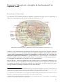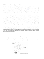
1
AVERTISSEMENT
Ce document est le fruit d'un long travail approuvé par le
jury de soutenance et mis à disposition de l'ensemble de la
communauté universitaire élargie.
Il est soumis à la propriété intellectuelle de l'auteur. Ceci
implique une obligation de citation et de référencement lors
de l’utilisation de ce document.
D’autre part, toute contrefaçon, plagiat, reproduction
illicite encourt une poursuite pénale.
➢
Contact SCD Nancy 1 : [email protected]
LIENS
Code de la Propriété Intellectuelle. articles L 122. 4
Code de la Propriété Intellectuelle. articles L 335.2- L 335.10
http://www.cfcopies.com/V2/leg/leg_droi.php
http://www.culture.gouv.fr/culture/infos-pratiques/droits/protection.htm
Université Henri Poincaré – Nancy I
U.F.R. Sciences et techniques de la matière et des procédés
École doctorale lorraine de chimie et physique moléculaires
Thèse
présentée pour l’obtention du titre de
Docteur de l’Université Henri Poincaré
en Chimie informatique et théorique
par Vincent
LEROUX
Modélisation d’inhibiteurs du domaine SH2 de la protéine Grb2
par dynamique moléculaire, docking et criblage virtuel
sous la direction de Bernard MAIGRET
Équipe de dynamique des assemblages membranaires, UMR UHP / CNRS 7565
Soutenue publiquement le 15 décembre 2006
Rapporteurs
Dr. Nohad GRESH
Université René Descartes – Paris V
Dr. Didier ROGNAN
Université Louis Pasteur – Strasbourg
Examinateurs Prof. Daniel CANET
Invités
Université Henri Poincaré – Nancy I
Prof. Christiane GARBAY
Université René Descartes – Paris V
Dr. Bernard MAIGRET
Université Henri Poincaré – Nancy I
Dr. Peter BLADON
Interprobe Chemical Services, Glasgow, Écosse
Dr. Gilles MOREAU
30 avenue Jean Jaurès, 94220 Charanton
Mes remerciements les plus chaleureux vont à mon directeur de thèse, Bernard, pour son encadrement, sa
patience face à mon fonctionnement irrégulier, mais surtout pour avoir fait en sorte que je termine cette thèse
en ayant une vision de la recherche scientifique qui a dépassé le simple stade de l’intérêt et de la curiosité.
Un grand merci également à Nohad, Christiane, Peter et Gilles pour leur soutien et leur gentillesse. Je tiens
également à saluer l’ensemble des membres du laboratoire eDAM, ainsi qu’à tous ceux avec qui j’ai
travaillé, spécialement mes compagnons d’armes Alexandre, Jean-Paul, Matthieu, Jérôme, Adil, Amel,
Yesmine, Muhannad et Werner. Bon courage à ceux qui n’en ont pas encore fini, et pareillement à ceux qui
sont partis vers d’autres aventures. Enfin, je garde beaucoup de reconnaissance pour ma famille et mes amis,
qui ont su rester présents même lorsque j’étais un peu déconnecté par mon travail et la distance. Merci en
particulier à mes parents, Sophie, Pascal, Estelle et Catherine. Je ne regrette pas une seule seconde d’avoir
fait cette thèse, cette expérience m’a apporté beaucoup, et chacun d’entre vous m’y a aidé.
Note importante
Ce document constitue une version révisée du manuscrit de thèse.
Il répercute les suggestions formulées par le jury lors de la soutenance.
La section publications a été modifiée, car au moment de l'impression du manuscrit
de thèse les articles n'étaient pas publiés – deux d'entre eux étaient toutefois sous
presse. On trouvera ici les publications dans leur version finale. La liste des
changements intervenus pour l'un d'entre eux pourra être consultée en exergue de la
section publications.
Ce document (ainsi que sa révision initiale) est disponible sous forme électronique
sur demande : [email protected]
Table des matières
Présentation
1
Contexte
3
Cancer
3
Avant-propos
Fonctionnement du cycle cellulaire et causes du cancer
Les différentes approches de la lutte contre le cancer
L’étude théorique du vivant à l’échelle moléculaire
Structure des biomolécules
Des acides nucléiques à l’information génétique
Du génome au protéome
Du génome à l’interactome : description du fonctionnement d’un organisme vivant
Du protéome à l’interactome
Constitution moléculaire d’un organisme vivant
L’ère de la post-génomique
Importance des simulations numériques
L’informatique : une révolution scientifique
Les simulations numériques : une nouvelle méthode de recherche
La simulation des systèmes biologiques par modélisation moléculaire
Drug design
3
4
6
8
8
8
9
11
11
12
12
13
13
13
14
15
Étapes de la mise au point d’un médicament
Identification de la cible pharmaceutique
Identification des composés prometteurs (hits)
Mise au point et optimisation de composés actifs spécifiques (leads)
Essais
Bilan financier
La recherche de nouveaux médicaments anti-cancer
15
15
15
16
17
17
18
Références bibliographiques
19
Cible
23
Description
23
La voie de signalisation Ras-MAPK
Les domaines SH2
La protéine Grb2
Description et fonction
Intérêt pharmaceutique
Accessibilité expérimentale
Accessibilité théorique
23
24
26
26
26
28
29
État actuel des connaissances sur l’inhibition de Grb2 SH2
Ligands peptidiques
Détermination de la séquence de référence
Mode de liaison
Spécificité
Ligands pseudo-peptidiques ou non peptidiques optimisés
Substitution de groupes peptidiques (Novartis)
Extension du récepteur ciblé (INSERM/CNRS)
Substituants à pTyr et cyclisation (Affymax, NCI)
Recherches de ligands actifs in vivo : contraintes
Contrainte de spécificité
Contrainte de stabilité
Contrainte d’accessibilité
Recherches de ligands actifs in vivo : stratégies pour Grb2 SH2
Modification de ligands existants
Nouvelles bases structurales
30
30
30
30
31
32
32
32
33
34
34
34
35
36
36
36
Références bibliographiques
39
Principaux résultats
47
Dynamique moléculaire sur deux complexes de référence
Situation initiale
Mise en œuvre
Systèmes modélisés
Techniques d’analyse
Choix du champ de force
Validation du protocole
Nouvelles connaissances concernant l’interaction de ligands sur le récepteur Grb2 SH2
Effets du solvant
Propositions au niveau du design de nouveaux ligands
Docking flexible sur des bases de molécules
Situation initiale
Prise en charge du solvant
Test et validation des paramètres
Limitations
Screening virtuel sur la cible Grb2 SH2
Molécules sélectionnées comme candidates
Résultats
Validation
Situation présente
47
47
48
48
49
49
50
51
51
51
52
52
52
52
53
53
53
54
55
55
Screening virtuel : mise en place d’outils innovants
56
Principales problématiques liées au screening virtuel
56
Le projet VSM-G
57
Présentation
Développements en cours et objectifs
Perspectives à long terme : vers un screening virtuel ”intelligent”
Conclusions
Publications
57
58
58
59
61
Présentation
L’application de simulations numériques reposant sur les bases théoriques de la chimie est une approche
relativement récente dans le monde de la recherche pharmaceutique. Plusieurs facteurs la rendent
particulièrement attrayante.
Tout d’abord, son champ d’application croît parallèlement aux progrès réguliers de la puissance informatique
accessible – une croissance bien plus spectaculaire que celle des avancées technologiques au niveau des
appareillages de la recherche biomédicale classique. Récemment, de nouveaux concepts de calcul distribué,
popularisés par le projet SETI@Home, ont encore accru l’intérêt de la communauté scientifique envers les
approches in silico en général.
Ensuite, l’utilisation de modèles numériques peut permettre, lorsque ceux-ci se basent sur la structure des
assemblages moléculaire (nous utiliserons ici, dans ce domaine, les méthodes de la dynamique moléculaire et
du docking), de ”visualiser” l’action d’une drogue sur sa cible. Loin d’être une abstraction, les méthodes
numériques permettent ainsi l’accès le plus frontal qui soit aux phénomènes biologiques à l’échelle
microscopique.
Enfin, dans la continuité des investissements colossaux effectués dans la génomique, la communauté
scientifique se tourne désormais vers le protéome et l’interactome, qui seuls permettront une bonne
compréhension des mécanismes fondamentaux du vivant. En particulier, les structures expérimentales
disponibles de biomolécules, à travers des bases publiques telles que la Protein Data Bank, croissent
exponentiellement. Dans ce contexte, les méthodes numériques sont de plus en plus intéressantes face aux
techniques purement expérimentales qui ne sont guère en mesure d’appréhender seules le potentiel d’un tel
flux de données.
Parmi les méthodes numériques dont il est question ici, la plupart ont pour origine la chimie théorique. Pour
l’expert dans ce domaine, les recherches en biologie et en pharmacologie constituent des champs
d’application passionnants, de par la nature des progrès scientifiques qu’ils conditionnent et du fait qu’ils se
situent toujours à la limite des progrès technologiques. De nombreux défis, sur les plans théorique aussi bien
que technique, dans ce secteur de plus en plus interdisciplinaire, attendent d’être relevés.
Ce manuscrit constitue une étude théorique de l’inhibition de l’activité du domaine SH2 de la protéine Grb2
par la liaison de ligands.
La première partie décrit le contexte de ce travail, qui se situe plus généralement à travers le ciblage de Grb2
SH2 parmi les multiples approches de lutte contre le cancer, de m’étude du vivant à l’échelle moléculaire
(domaine interdisciplinaire par excellence), et, plus précisément, de la recherche pharmaceutique.
La seconde partie s’emploiera à détailler les connaissances disponibles sur la cible au moment de débuter ce
travail, en incluant les avancées qui sont apparues en parallèle à ce travail.
On trouvera dans la troisième partie un résumé des principaux résultats obtenus au cours de cette thèse.
Ceux-ci se classent selon trois approches méthodologiques distinctes : celle de la dynamique moléculaire, du
docking et enfin du screening virtuel. On pourra constater que de nouvelles connaissances sur la nature
physico-chimique de la liaison de certains ligands sur Grb2 SH2 sont mises en évidence dans le premier cas,
tandis que les deux autres approches ont surtout donné naissance à des avancées méthodologiques.
Les publications auxquelles ce travail a donné lieu sont enfin disponibles.
1
2
Contexte
Cancer
Avant-propos
Le cancer est une famille de maladies caractérisée par une dérégulation des mécanismes de division
cellulaire d’un organisme. [1] La croissance cellulaire incontrôlée qui en résulte parasite le fonctionnement
normal de la machinerie biologique, et peut aboutir à des déficiences graves d’un ou plusieurs organes vitaux
(principalement reins, foie et poumons). Ainsi, la plupart des cancers humains peuvent causer la mort, et il
s’avère que les cancers sont devenus la première cause de mortalité dans les pays développés. [2]
Le nombre de décès en France provoqués par le cancer croît constamment (150000 en 2000, contre 125000
en 1980), parallèlement à l’accroissement de l’espérance de vie (les cancers touchant principalement, et avec
une gravité accrue, les personnes les plus âgées). Cette augmentation est toutefois moins importante que celle
du nombre de cas diagnostiqués (278000 contre 170000) [3], qui traduit aussi bien les progrès de la
médecine anti-cancer * que les efforts de santé publique principalement axés sur la prévention et le dépistage.
Le cancer n’est pas seulement un axe crucial de santé publique, c’est également un problème social. [4, 5]
Cela peut principalement s’expliquer du fait que la vision populaire du cancer se décline et se déforme bien
en dehors de la réalité médicale.
Tout d’abord, au cancer est quasi systématiquement associée l’image de mourants, condamnés pour lesquels
la médecine et la science ne peuvent que s’acharner inhumainement et en vain : le cancer représente la mort
violente précédée de la déchéance physique, celle que personne ne souhaite et qui inspire universellement la
peur. À cela vient s’ajouter le fait que le cancer est bien souvent interprété de façon exclusive soit comme
fatalité, soit sous l’angle causal. Dans le premier cas, le cancer est symbole d’injustice et
d’incompréhension : il peut frapper n’importe qui, n’importe quand. Dans le second cas, sachant que
certaines formes de cancer sont bien connues comme amplifiées par des comportements à risques, une
généralisation peut s’opérer : si on en est malade, c’est ”qu’on l’a bien cherché”. Enfin, le cancer est parfois
considéré comme une maladie contagieuse… Tous ces éléments combinés font du cancer une maladie
honteuse † ainsi qu’un facteur important d’exclusion sociale.
En 2002, la lutte contre le cancer est déclarée chantier prioritaire par l’État. Un "plan Cancer" est alors mis
en place rapidement sur une durée de cinq ans, et doté d’un budget de 1,5 milliard d’euros. Cependant, le
plan Cancer est principalement axé sur des mesures de prévention et de dépistage (effectivement prioritaires
sur le court terme au niveau de la santé publique) ; les actions de recherche, centralisées autour de
”cancéropôles” pilotés au niveau ministériel, sont d’une importance toute relative et comprennent au final
peu de projets théoriques ou orientés vers l’aspect pharmaceutique. Ainsi, la part de financement des
recherches de nouveaux médicaments anti-cancer dans le plan Cancer ne s’élève qu’à 10 millions d’euros fin
2004.
*
Ces progrès sont particulièrement flagrants en ce qui concerne le cancer du sein, qui reste le plus fréquent pour les femmes, et dont
le taux de mortalité associé a baissé plus que de moitié de 1980 à 2000.
†
Maladie honteuse jusque dans la mort ! N’emploie-t-on pas systématiquement la formule pudique "mort des suites d’une longue
maladie" sur les faire-part de décès ?
3
Les organismes de recherche publics tels que l’Inserm * ou le CNRS † , déjà impliqués dans les projets du plan
Cancer, participent également de façon indépendante à la lutte contre le cancer, soit en finançant directement
des projets de recherche, soit en initiant et participant activement à des projets financés de façon externe.
Cette thèse résulte ainsi de la mise au point en 2002 d’une ACI (Action Concertée Incitative) dans la
catégorie ”Molécules et cibles thérapeutiques” nommée ”Nouvelles approches thérapeutiques du cancer :
conception et validation de molécules inhibant des interactions protéiques et ciblant la voie Ras induite par
les protéines à activité tyrosine kinase”.
Fonctionnement du cycle cellulaire et causes du cancer
Les cellules d’un organisme ont une durée de vie limitée. Ainsi, elles ont la capacité de se multiplier afin que
l’organisme puisse se maintenir en vie, et il existe de nombreux mécanismes permettant de réguler la
population des cellules [6, 7], en favorisant leur prolifération (les gènes codant ainsi pour la division
cellulaire sont appelés proto-oncogènes), ou au contraire en la freinant (les gènes correspondants sont
appelés suppresseurs de tumeurs). Une trop faible population de cellules peut provoquer l’activation de
mécanismes favorisant l’expression de proto-oncogènes, et à l’inverse, la détection de mutations pourra
favoriser les suppresseurs de tumeurs, ce qui permettra de procéder à la réparation ou éventuellement à
l’élimination des cellules endommagées avant que celles-ci ne puissent mettre en danger l’organisme en se
multipliant.
En temps normal, l’ensemble de ces mécanismes permet de maintenir un équilibre entre le nombre de
cellules résultantes de la division cellulaire et le nombre de cellules mortes, tout en régulant d’éventuelles
altérations du code génétique. Cela permet aussi également de réparer les pertes cellulaires accidentelles
ponctuelles dues par exemple à des blessures, assurant ainsi le bon fonctionnement des organes et des tissus.
Lorsque cet équilibre est rompu, cela peut avoir pour effet une multiplication anarchique de certaines
cellules, lesquelles peuvent se concentrer, formant des tumeurs, lesquelles caractérisent le cancer lorsque leur
taille ne permet plus leur retrait par des interventions médicales simples. Dans de nombreux cas, ces tumeurs
peuvent menacer directement le bon fonctionnement d’un ou plusieurs organes vitaux ou du système
sanguin, ou sont susceptibles de le faire en migrant dans l’organisme (on parle alors de métastases). Le
cancer met alors gravement en danger la vie de l’individu affecté et aboutit souvent à sa mort en l’absence de
traitement. À un tel stade, le cancer met alors gravement en danger la vie de l’individu affecté et aboutit
souvent à sa mort en l’absence de traitement (90% des cas de mortalité dues au cancer sont consécutives à la
formation de métastases [8]).
Quelle qu’en soit la cause, le développement d’un cancer a toujours pour origine la mutation d’un ou
plusieurs proto-oncogènes, qui sont alors appelés oncogènes [9] : le cancer est une maladie génétique. La
totalité des cancers résultent une série concertée de mutations génétiques et non d’une seule [10-13], ce qui
explique l’augmentation du risque avec l’âge et le long cycle de développement de la maladie que l’on
observe généralement. Le développement du cancer est souvent lié à une surexpression de protéines
exprimées par les proto-oncogènes mutés en oncogènes, donc impliquées - lorsque leur concentration dans
l’organisme est appropriée - dans les processus normaux de croissance cellulaire, et aboutissant à une
multiplication incontrôlée des cellules affectées. Le cancer peut également être favorisé par la mutation d’un
ou plusieurs gènes suppresseurs de tumeurs [14, 15], aboutissant à la diminution ou la disparition de
biomolécules chargées de réparer ou d’éliminer les cellules endommagées. La plupart des cancers combinent
*
http://www.inserm.fr/fr/questionsdesante/dossiers/cancer/
http://www.inserm.fr/fr/recherches/etats_des_lieux/att00002003/CANCER2002_v3.4-num.pdf
†
www.cnrs.fr/SDV/cnrscancer.html
4
ces deux causes, une mutation pouvant en provoquer d’autres par cascade, ceci étant accéléré à chaque fois
qu’un gène suppresseur de tumeurs est touché. Cela aboutit à la formation d’au moins une cellule
cancéreuse, pour laquelle les signaux génétiques qu’elle contient et favorisant sa prolifération prévalent, suite
à un nombre critique de mutations (un nombre trop faible de mutations étant régulé naturellement), sur ceux
codant pour sa régulation.
Les différentes mutations nécessaires à la formation d’une
cellule cancéreuse sont à présent bien caractérisées. [13] Les
mutations favorisant la formation d’une cellule cancéreuse
confèrent à celle-ci une autonomie par rapport à l’organisme
hôte : la cellule cancéreuse génère ses propres facteurs de
croissance [16] (ce qui dérégule les voies de signalisation
correspondantes [17]), et est insensible aux mécanismes de
régulation de la population cellulaire par l’organisme, en
particulier la mort cellulaire programmée [18, 19]. D’autre
part, la cellule cancéreuse acquiert une grande capacité de
multiplication [20, 21] et de prolifération dans l’organisme,
ainsi qu’une meilleure résistance aussi bien au système
immunitaire qu’à un traitement extérieur (cela se traduit, en
particulier, par la mutation du gène suppresseur de tumeur p53
[22]). Une cellule cancéreuse est une cellule pour laquelle
l’ensemble de telles mutations rend irréversible sa mutation
infinie dans l’organisme, à moins qu’un traitement approprié
ne soit effectué sur la personne affectée.
Il est encore impossible de déterminer l’origine d’un cancer donné antérieurement à la série de mutations
génétiques qui le provoque ; on estime même souvent que ces mutations peuvent survenir spontanément.
Toutefois, il s’agit d’évènements anormaux et étant donné que seule une combinaison de plusieurs types de
mutations provoque le cancer, on estime qu’un des préalables à la survenue du cancer est une instabilité du
génome sur un intervalle de temps important. Il a été démontré que certains facteurs extérieurs précis
peuvent en être la cause. On peut citer l’exposition répétée à des substances dites cancérigènes [23, 24], qui
peuvent être des drogues (alcool, tabac...), des produits chimiques (solvants, pesticides...) ou bien des
produits naturels (amiante, matériaux radioactifs...). De nombreux cancérigènes ont été - et sont toujours,
pour certains d’entre eux - massivement employés dans l’industrie (arsenic, amiante, nickel, goudrons...), si
bien que la part des expositions professionnelles parmi les causes du cancer n’est certainement pas
négligeable.
Certains cancérigènes sont caractéristiques d’un type précis
de cancer, par exemple le tabac dont la consommation est
parfaitement corrélée démographiquement au nombre de cas
de cancer du poumon. La combinaison de plusieurs
substances cancérigènes augmente encore les risques ; en
particulier, l’association alcool / tabac est connue pour son
incidence très importante sur les cancers de la bouche, de la
gorge et de la vessie. On estime, plus généralement, qu’une
mauvaise hygiène de vie (un régime alimentaire trop riche
en graisses, par exemple), ou bien un environnement pollué
favorisent l’apparition de cancers. Des mutations génétiques
prédisposant à certaines formes de cancer peuvent également
être héritées [25] (c’est le cas d’un français sur 60), par
exemple les mutations du gène BRCA1 reliées aux cancers
5
du sein et des ovaires. Cet aspect semble cependant avoir moins d’incidence que les facteurs
environnementaux [26]. Enfin, de façon moins fréquente, certains virus, contenant dans leur génome des
oncogènes ou gènes inactivant les suppresseurs de tumeur, ou bien favorisant indirectement des protooncogènes de leur cellule hôte lors de leur réplication, peuvent provoquer des mutations pouvant aboutir à la
formation de tumeurs cancéreuses [27, 28].
Les différentes approches de la lutte contre le cancer
La lutte contre le cancer est constituée de multiples approches souvent complémentaires. Il en va de même
en ce qui concerne les aspects aussi bien médicaux que scientifiques. Dans un tel contexte, il est évident que
la lutte contre les différentes formes de cancer ne pourra pas être le fait d’une seule technique scientifique,
d’un seul axe de recherche. En énumérant brièvement les principales directions qui peuvent être suivies, nous
pouvons donner l’impression qu’il s’agit de domaines distincts ; bien au contraire, dans le cadre le la
recherche anti-cancer l’interdisciplinarité est d’une importance cruciale, tant le sujet d’étude est complexe.
En premier lieu, la prévention est fondamentale car il est établi que plus un cancer est décelé précocement et
les traitements thérapeutiques effectués en conséquence, meilleures sont les chances de survie du patient. La
prévention inclut des actions de dépistage et de sensibilisation du public, mais aussi des études cliniques et
épistémologiques. Ces dernières permettent cerner avec plus de précision les facteurs de risque, ce qui
constitue un préalable indispensable à toute action de prévention visant les populations à risque, mais permet
également de recouper les connaissances issues des recherches en biologie, et de fournir au milieu médical
des données statistiques précieuses pour la prise en charge et le suivi des patients.
Si des examens réguliers sont indispensables pour les formes de cancer les plus courantes, on peut également
isoler les facteurs de risques potentiels chez un individu. Une fois le cancer diagnostiqué, le choix d’une ou
plusieurs approches dépend de la nature du cancer, de son avancement, ainsi que de l’état de santé général du
patient concerné. L’intervention chirurgicale [29] (extraction de tumeur ou amputation de l’organe affecté),
plus ancienne méthode thérapeutique de traitement contre le cancer, est toujours largement pratiquée dès lors
que la tumeur est localisée et solide. Afin d’éviter sa résurgence après l’opération, on utilise la radiothérapie
[30] et la chimiothérapie [31], qui peuvent d’autre part s’avérer suffisantes lorsque la tumeur n’a pas atteint
une taille critique, et peuvent constituer au minimum des traitements palliatifs efficaces dans les cas où le
développement et la nature de la tumeur ne permettent plus un traitement curatif. Toutefois, ces deux
méthodes sont connues pour provoquer nombre d’effets secondaires désagréables car elles ne ciblent pas
exclusivement les cellules cancéreuses. D’autres méthodes thérapeutiques telles que l’hormonothérapie [32,
33] sont accessibles, ne ciblant plus les cellules cancéreuses mais favorisant ou non des biomolécules
(protéines, anticorps, hormones...) qui leurs sont associées, cependant elles n’ont souvent encore qu’un rôle
complémentaire du fait de leur efficacité plus modeste ou de leur domaine d’action plus limité. Ainsi, il est
courant d’employer simultanément plusieurs techniques parmi celles que nous venons d’évoquer. [34]
Parallèlement à cela, de nouvelles thérapies sont en développement. Ainsi, de nombreux travaux de
recherche portent sur la thérapie génique [35], consistant à insérer dans les cellules des gènes à fonctions
thérapeutiques. Une des grandes difficultés de cette approche réside dans la mise au point de vecteurs
efficaces (virus, bactéries...) permettant de transférer l’information génétique dans les cellules, tandis qu’ils
sont ciblés par le système immunitaire de l’individu. Le développement de vaccins anti-cancer ou
immunothérapie [36-40] dont le but est aussi bien de prévenir l’infection de l’organisme par des agents
cancérigènes que de favoriser les réponses immunitaires au développement d’une tumeur ultérieure
potentielle, fait également l’objet d’efforts de recherche poussés.
6
Un autre domaine de recherche prometteur de la lutte anti-cancer, et dans lequel nous nous situons dans le
cadre de cette étude, consiste à mettre au point des inhibiteurs chimiques plus spécifiques [41, 42], avec
potentiellement une efficacité accrue et des effets secondaires [43] moins importants pour le patient. [44] De
tels médicaments s’avèrent déjà utiles en complément de traitements anti-cancer plus conventionnels. [45]
Ces inhibiteurs peuvent être des molécules en partie ou en totalité synthétiques, et ne ciblent ni le système
immunitaire (immunothérapie), ni certaines hormones (hormonothérapie), mais des protéines [46, 47] et des
voies de signalisation [48-51] dont le rôle dans le développement du cancer est bien caractérisé. Cela
nécessite préalablement une connaissance plus poussée des mécanismes biomoléculaires correspondants.
[52] À ce niveau, l’interprétation de résultats de recherches en génétique peut s’avérer précieux. [53]
Quelques liens utiles sur le cancer :
Ligue contre le cancer : http://www.ligue-cancer.asso.fr
Fédération Nationale des Centres de Lutte Contre le Cancer : http://www.fnclcc.fr/
Rapport de la commission d’orientation sur le cancer (2002) : http://www.sante.gouv.fr/htm/dossiers/cancer/
Mission interministérielle pour la lutte contre le cancer : http://www.plancancer.fr/
Cancer Medicine 5th edition (BC Decker) :
http://www.ncbi.nlm.nih.gov/books/bv.fcgi?call=bv.View..ShowTOC&rid=cmed.TOC&depth=10
American Cancer Society : http://www.americancancersociety.org
National Comprehensive Cancer Network : http://www.nccn.org
Résumé de l’ACI qui a permis de financer cette thèse :
Le but de ce projet est de concevoir des agents pharmacologiques ciblés, capables d’inhiber des voies de signalisation
dérégulées par la présence de protéines codées par des oncogènes et conduisant à la formation de cancers.
L’approche choisie est nouvelle mais donne déjà des résultats. Elle consiste à inhiber les interactions entre ces protéines
et leurs effecteurs ou entre des protéines situées en aval dans la voie dérégulée. Les cibles sélectionnées sont situées
dans la voie de signalisation Ras-dépendante impliquée dans des cancers à la fois nombreux et souvent fréquents :
ERBB2 (sein, ovaire), Grb2 (sein, ovaire et certaines leucémies comme la LMC), RasGAP dans les cancers exprimant
Ras oncogénique (30% des tumeurs, 90% pancréatiques).
La réunion de biologistes, pharmacologues, chimistes et physicochimistes académiques et du secteur industriel désireux
de mettre en commun leurs compétences va permettre, dans un premier temps de concevoir des agents peptidiques
compétiteurs des interactions ciblées, et de les valider par des tests cellulaires et vivo en les vectorisant, puis de
procéder à des études structurales des complexes entre protéine et peptide inhibiteur afin de définir les points
d’interaction pour concevoir grâce à l’aide de la modélisation moléculaire « in silico », des molécules non peptidiques
selon une approche de chimie combinatoire « ciblée ».
7
L’étude théorique du vivant à l’échelle
moléculaire
La lutte contre le cancer comme objectif de la recherche scientifique s’inscrit dans le cadre plus général des
sciences du vivant. On entend par là un ensemble de disciplines scientifiques, telles que la biochimie et la
génétique, issues des progrès réalisés au XXème siècle autour de la biologie, et dont les applications fondent
désormais une très grande part des découvertes en pharmacologie et en médecine. Les sciences du vivant
reposent avant tout, au niveau théorique, sur la connaissance du fonctionnement d’un organisme vivant. Une
des approches possibles pour décrire les mécanismes correspondants, et sur laquelle ce travail repose, est
celle de la biochimie, pour laquelle les mécanismes de base sont des interactions entre biomolécules. L’étude
théorique de ces biomolécules est rendue possible par le progrès de certaines techniques expérimentales, et
surtout grâce à la banalisation des calculs informatiques ; il s’agit d’un axe de recherche relativement récent.
Dans cette partie, nous décrirons tout d’abord les deux classes fondamentales de biomolécules : les acides
nucléiques (ADN et ARN) et les protéines. Les premières contiennent le ”mode d’emploi” de l’organisme,
tandis que les secondes œuvrent à son fonctionnement en assurant la communication entre les cellules. Nous
expliquerons ensuite en quoi le champ de vision des sciences du vivant s’est considérablement élargi ces
dernières années, entre autres grâce au poids croissant des méthodes de calcul informatique dans la recherche
scientifique, passant d’une vision centrée sur le génome à une vision plus large qui inclut l’ensemble des
protéines, ou protéome, puis ensuite l’ensemble des interactions entre biomolécules, ou interactome.
Structure des biomolécules
Des acides nucléiques à l’information génétique
Les acides nucléiques s’observent sous deux formes polymériques : l’acide ribonucléique (ARN) et l’acide
désoxyribonucléique (ADN), dans lesquelles des groupes fonctionnels appelés nucléotides sont greffés sur
un squelette polymère pentose-phosphate. Quatre nucléotides différents sont rencontrés dans la structure de
l’ADN* : adénine (A), thymine (T), cytosine (C) et guanine (G).
Le squelette pentose-phosphate de l’ADN et les 4 bases possibles correspondantes
*
Dans l’ARN les thymines sont mutées en uraciles, et les déoxyriboses du squelette en riboses.
8
ADN et ARN ont des fonctions différentes de par leur structure. La structure de l’ADN fut
déterminée par Watson, Crick et Franklin en 1953 [54-56], et a pour particularité le
positionnement des nucléotides à l’intérieur d’une double hélice formée par deux chaînes
pentose-phosphate antisymétriques. Ce positionnement dans un espace si confiné est rendu
possible par un appariement des nucléotides par paires : adénine et thymine d’une part, cytosine
et guanine d’autre part, sont reliées d’une hélice à l’autre par des liaisons hydrogène.
L’information génétique est ainsi ”doublée” et stabilisée : le rôle biologique de conservation
tenu par l’ADN apparaît alors évident. L’ARN a une structure en simple hélice dans laquelle les
nucléotides sont exposés, permettant la réplication et l’expression directe de la séquence.
Appariement des nucléotides au sein de la structure en double hélice de l’ADN
Du génome au protéome
Les protéines sont également des polymères, constitués à
partir des 20 acides aminés naturels. La signification du
codage génétique donne le lien entre les séquences
nucléiques et peptidiques : les séquences génétiques codent
les séquences protéiques [57] ; par séquences de trois
nucléotides de l’ARN ou codon (voir ci-contre) [58, 59]. La
synthèse d’une protéine a plus précisément pour origine la
transcription d’une partie spécifique fonctionnelle de l’ADN
(un gène) sous forme d’ARN * . [60] Le mécanisme de
transcription de l’ARN vers des protéines fait intervenir le
ribosome, ensemble d’une centaine de protéines
enzymatiques qui sont les ouvrières de cette ”chaîne de
production”. [61]
Au niveau du protéome, un niveau de complexité supplémentaire † est présent par rapport au génome : alors
que l’ADN et l’ARN possèdent des structures invariables, l’activité et la fonction des protéines dépendent
intrinsèquement de leur géométries ‡ , qui définissent un large espace conformationnel. §
*
On appelle ce processus transcription de l’ADN ; proche sur de nombreux points du processus de réplication de l’ADN intervenant
lors de la division cellulaire. Certaines protéines dites régulatrices de gènes servent à déterminer quelles parties de l’ADN doivent
être transcrites et quelles parties sont ignorées, définissant ainsi les segments sur les chromosomes correspondant aux gènes.
†
On peut noter qu’alors que dans l’organisme humain la taille moyenne des protéines est de 300 résidus, on est très loin d’observer
20300 soit environ 10390 séquences peptidiques possibles. L’ordre de grandeur des séquences significatives est estimé à 105.
‡
De façon remarquable, l’adoption par une protéine d’une conformation ”anormale” peut changer sa fonction biologique et aboutir à
des pathologies particulièrement graves. C’est le cas de la ”maladie de la vache folle”, dans laquelle une protéine du cerveau (un
prion) subit un changement conformationnel se propageant aux autres prions, formant des aggrégats à la source d’un processus
dégénératif mortel.
§
La conformation d’une protéine peut être caractérisée par l’orientation relative des différentes unités du squelette. On distingue pour
ce faire les hélices α (caractérisées par des liaisons H intra-chaîne, et représentées graphiquement sous la formes de cylindres) des
feuillets ß (correspondent à l’association parallèle ou anti-parallèle de deux chaînes distinctes par des liaisons H inter-chaînes, par des
flèches plates). On appelle tour (ß ou Ω lorsque l’angle est très marqué) le passage d’une forme géométrique à l’autre.
9
Liste des 20 acides aminés classés naturels classés par propriétés,respectivement : acides (D, E), basiques
(K, R, H), structure spécifique (G, C, P), hydrophiles (S, T, N, Q) et hydrophobes (A, V, F, Y, L, I, M, W)
Il a été démontré que la séquence d’une protéine détermine la structure géométrique globulaire adoptée par
un processus de repliement sur elle-même qui survient après sa synthèse. Les principes de base de ce
mécanisme sont connus [62], ainsi que des résultats statistiques * . Sa prédiction, désignée comme le problème
de repliement des protéines [63], a un intérêt évident, mais la tâche s’avère si complexe qu’elle en constitue
une sorte de ”saint Graal” de la biochimie.
Ainsi, alors que l’étude du génome correspond fondamentalement à l’exploration d’un espace séquentiel (sur
quatre bases), celui du protéome, en plus de l’extension d’une partie de cet espace (cette fois codé sur 20
bases), lui ajoute un espace conformationnel † beaucoup plus vaste. [64] L’exploration de ce protéome est
très délicate, et ce même si on ne s’intéresse qu’à une protéine bien définie, à moins qu’une structure
expérimentale de celle-ci soit disponible, ce qui fut le cas pour ce travail. Les techniques de la modélisation
moléculaire peuvent alors être mises en œuvre.
*
Par exemple, les résidus Met et Glu se retrouvent plus fréquemment dans des hélices α, Val et Ile dans des feuillets ß, Gly et Pro
dans les tours. L’Arg est le seul acide aminé qui ne semble pas avoir de ”préférence” particulière.
†
On passe alors de la structure primaire (linéaire) des protéines qui se limite à la séquence à la structure secondaire (globulaire). On
parle de structure tertiaire lorsque l’on considère en plus les interactions ”longue distance” sur les chaînes peptidiques qui entre
autres différencient structuralement les résidus hydrophiles des résidus hydrophobes. Enfin, la structure quaternaire se réfère à
l’organisation de chaînes peptidiques analogues en super-structures.
10
Du génome à l’interactome : description du fonctionnement d’un
organisme vivant
Du protéome à l’interactome
Les interactions entre protéines peuvent être identifiées, regroupées au sein de voies de signalisation, et
représentées schématiquement sous la forme de graphes d’interactions. [65, 66].
Schéma des principales voies de signalisation connues intervenant au niveau cellulaire [13]
L’étude sur le plan moléculaire du fonctionnement d’un organisme vivant peut ainsi s’effectuer à différents
niveaux conceptuels successifs. Le génome repose sur l’espace des séquences de nucléotides, le protéome
y ajoute l’espace géométrique des protéines correspondantes, et l’interactome la liste et la nature des
interactions possibles qui en découlent. Le génome humain est à présent décrypté, et concernant les
protéines le nombre de structures expérimentales résolues et accessibles [67, 68] croit exponentiellement. *
La cartographie de l’interactome n’en est quant à elle qu’à ses balbutiements. †
Dans le cadre de ce travail, nous étudions une cible protéique précise et en particulier l’inhibition de son
activité au sein d’une voie de signalisation correctement caractérisée. Il ne nous apparaît pas important dans
ces conditions de détailler l’état actuel des recherches sur l’interactome, qui peuvent inclure des aspects
autres que purement géométriques et chimiques, tels les effets cinétiques. ‡
*
Au 23 août 2005, la base PDB (Protein Data Bank) recence 29532 structures protéiques. La portion de ces structures qui
correspondent à des complexes, et qui sont donc d’un grand intérêt afin de relier protéome et interactome au niveau de l’espace
géométrique et non plus seulement au niveau de l’espace des interactions, n’est toutefois que de 1316 (4,4%).
†
Si l’interactome est un réseau routier, on n’en connaît pour le moment qu’une partie des autoroutes sous la forme de voies de
signalisation. La plupart des interactions entre biomolécules sont inconnues à l’heure actuelle.
‡
En effet, plusieurs interactions peuvent être ”en concurrence” au sein du même milieu extra- ou intra-cellulaire. Ces considérations
peuvent être cruciales pour des applications d’ordre médical.
11
Constitution moléculaire d’un organisme vivant
La ”composition chimique” d’un organisme vivant ne saurait bien évidemment se réduire aux acides
nucléiques et aux protéines. Toutefois, ces deux classes fondamentales de biomolécules sont à la base du
fonctionnement des organismes et suffisent à caractériser le cadre de cette thèse. De façon générale, si l’on
exclut les biomolécules de taille importante (acides nucléiques, protéines, membranes…), la diversité en
terme chimiques d’un organisme vivant est limitée – les organismes vivants les plus simples sont ainsi
construits autour d’une centaine de petites molécules organiques [69] – en comparaison du nombre de
molécules organiques théoriquement synthétisables (estimé à 1060 [70]). ”L’espace biologique” est de
nombreux ordres de grandeur plus petit que ”l’espace chimique”. [71]
Un aspect important de la nature d’un organisme vivant observée sous l’angle de sa constitution chimique
doit néanmoins être mentionné. Les systèmes en biochimie ne doivent pas être considérés comme des
systèmes chimiques classiques dans lesquels les molécules sont en interaction en milieu dilué. Le fait que le
corps humain soit constitué d’environ 70% d’eau signifie en particulier du point de vue chimique que la
plupart des biomolécules se trouvent en solution à des concentrations particulièrement élevées d’environ
30% [71], formant une ”masse” en mouvement et en interaction. [72]
L’ère de la post-génomique
Conceptuellement, le code génétique d’un organisme vivant correspond à son ”programme de
fonctionnement” ; ainsi, le déchiffrage de ce code constitua pour la communauté scientifique un objectif
fondamental. Étant donné que biologiquement toute fonction découle de ce codage, le but ultime de la
génétique fut l’interprétation de toute fonction biologique à partir d’un code génétique.
Cet enthousiasme est à présent retombé. La génétique s’avéra un outil remarquable sur le plan médical et a
permis d’identifier la source de nombreuses pathologies, tout en fournissant de précieuses indications sur des
traitements pharmaceutiques potentiels. Mais il est à présent évident que l’étude du génome ne peut pas être
la ”recette miracle” qui révolutionnera la compréhension du vivant. En particulier, l’hypothèse réductionniste
”un gène = une fonction biologique” s’avéra fausse. Pour un grand nombre de gènes, la fonction biologique
correspondante est inconnue et il n’est même pas possible de déterminer s’il y en a une.
Pour ces raisons, on estime souvent que l’effort investi dans la génétique doit à présent être étendu en
direction de l’interactome. En effet, si la génétique permet d’identifier de nombreuses pathologies, elle reste
souvent impuissante dès lors qu’il s’agit de les corriger. Si la thérapie génique est un domaine de recherche
plus connu, la mise au point de médicaments ciblant l’interactome est également un axe prometteur en
particulier sur le plan pharmaceutique [73]. Le travail de cette thèse se situe dans ce dernier domaine.
12
Importance des simulations numériques
L’informatique : une révolution scientifique
Dans le passé, les modèles physiques théoriques de la matière ne pouvaient être vérifiés que dans des
circonstances expérimentales particulières, dans lesquelles des variables physiques isolées peuvent être
mesurées. D’une façon générale, la mise au point de modèles théoriques par le scientifique résulte
classiquement d’une approche réductionniste de la nature * . Le point faible d’une telle démarche est que les
modèles validés obtenus seront par nature réducteurs : la complexité n’est pas accessible.
L’informatique a été une révolution scientifique, car elle a permis à la complexité d’être – en partie –
accessible scientifiquement. La possibilité de conduire des calculs étendus et automatisés permet
l’application un modèle ou une combinaison de modèles à des systèmes dont la taille et la complexité ne les
rend pas traitables par d’autres moyens. Les techniques informatiques permettent ainsi de se rapprocher de la
réalité physique, validant ou invalidant les modèles avec plus de rigueur. Si l’informatique n’est pas à
l’origine de la découverte de théories scientifiques, bien souvent elle seule permet d’étendre le champ
d’application des modèles qui en découlent. †
Les simulations numériques : une nouvelle méthode de recherche
L’apparition de moyens de calcul informatique de plus en plus importants (la loi de Moore, jusqu’ici
vérifiée, postule que la puissance de calcul des microprocesseurs double tous les 18 mois) à partir des années
1950 a altéré le binôme théorie-expérience de la recherche scientifique en insérant une méthode
intermédiaire : la simulation numérique. Fondamentalement, aussi bien l’aspect théorique que l’aspect
expérimental de la recherche sont intégrés dans l’outil informatique, le premier à travers des
programmes et des algorithmes, et le second à travers des simulations et des données. Ainsi, la conduite
d’une simulation peut être analogue, selon les circonstances, à la celle d’une expérience ou à la mise en place
d’une théorie, rendant l’une comme l’autre plus accessibles.
Un autre avantage considérable de l’informatique doit également être mentionné. Une simulation
numérique n’est soumise à aucune autre contrainte matérielle que celle du temps de calcul nécessaire.
Le choix entre mesure expérimentale et simulation numérique ‡ n’a ainsi pas lieu d’être lorsque l’expérience
est trop contraignante (par exemple, elle requiert des conditions de température et de pression délicates, elle
peut s’avérer dangereuse…), voire impossible. Les simulations numériques peuvent aller encore plus loin, en
permettant de repousser artificiellement certaines contraintes physiques § , afin de mieux appréhender le
comportement d’un système.
*
L’approche réductionniste consiste à fractionner un système complexe en sous-systèmes plus simples, jusqu’à ce que tous les soussystèmes puissent être définis de façon satisfaisante par des modèles théoriques. Pour valider un sous-système donné pour lequel des
hypothèses théoriques sont avancées il faut mettre en place une expérience dans laquelle interviennent le moins possible de
phénomènes physiques externes au sous-système décrit. Cette contrainte est souvent particulièrement difficile à satisfaire.
†
Les premiers modèles physiques des gaz parfaits ne pouvaient être validés que par des mesures sur des gaz rares, et les modèles de
cristaux devaient l’être avec des échantillons très purs. S’ils permettent bien d’appréhender les lois fondamentales qui leur sont
associées, ils ne permettent aucunement de décrire un gaz ou un cristal quelconque. Et en chimie théorique, les méthodes ab-initio se
limitent à des assemblages simplistes en l’absence de moyens numériques.
‡
Remarquons que de nombreux appareillages modernes incorporent, afin d’affiner ou d’interpréter les mesures (par exemple, des
données spectrométriques), des traitements par simulations numériques. L’outil informatique n’est donc pas seulement indispensable
au niveau conceptuel, mais également au niveau technique, même si ce dernier point n’est pas aussi ”visible”.
§
Dans le cas de systèmes moléculaires, on peut franchir des barrières de potentiel afin d’explorer au mieux un espace
conformationnel. De plus, la mise en place de transformations alchémiques est à la base des méthodes de calcul d’énergie libre.
13
Pour de nombreux systèmes, la mise en œuvre de la simulation correspond à la mise en place d’un modèle et
d’un protocole qui constituent un compromis acceptable entre le réalisme des résultats et l’accessibilité en
termes de temps de calcul. Nous détaillerons à quel niveau des approximations devront être effectuées dans
le cadre de la simulation de systèmes biomoléculaires. *
La simulation de systèmes biologiques par modélisation moléculaire
Les différentes méthodes que nous avons utilisées pour ce travail appartiennent à la classe de la modélisation
moléculaire. Nous les avons appliquées à l’étude de systèmes biomoléculaires, en général à la fois complexes
chimiquement et difficilement accessibles expérimentalement ; les moyens informatiques sont dans un tel
contexte indispensables.
Le champ d’application des techniques de modélisation moléculaire est immense, à l’image du spectre des
connaissances et des domaines scientifiques qui sont susceptibles d’intervenir. Nous nous limiterons aux
systèmes que nous nous apprêtons à étudier, de type biologique.
La modélisation moléculaire peut être précieuse dans l’identification de cibles thérapeutiques, typiquement
des récepteurs protéiques dont le rôle spécifique dans une pathologie est caractérisé. Elle peut s’avérer
ensuite indispensable lors de la mise au point d’inhibiteurs chimiques pour cette cible. Un bon exemple du
rôle positif de telles méthodes dites de structure-based drug design [74] est constitué par la mise au point
d’inhibiteurs de l’HIV protéase [75]. Des techniques telles que la modélisation par homologie peuvent
permettre de pallier à l’absence de structures expérimentales complètes dans le cas de certaines cibles
d’intérêt majeur, telles que les récepteurs de type GPCR. [76, 77] De nombreuses approches sont disponibles
et peuvent être combinées afin de produire des résultats scientifiques de grand intérêt, en particulier sur le
plan médical. [78]
La modélisation peut, plus généralement, profiter directement des progrès dans une grande variété de
domaines. En premier lieu, on peut citer les progrès de l’informatique, aussi bien sur le plan matériel
(puissance de calcul brute, architectures parallèles et distribuées) que logiciel (outils spécialisés, techniques
de programmation). La disponibilité plus importante de structures expérimentales est également très
profitable ; les progrès technologiques des appareillages ont un impact positif pour les modélisateurs. Il en va
de même pour les avancées purement théoriques, ainsi que les nouvelles découvertes en biologie et
biochimie, qui peuvent permettre d’améliorer les modèles au niveau conceptuel. Nous nous situons donc,
avec ce travail, dans un contexte résolument interdisciplinaire.
Relier conceptuellement des domaines si différents constitue bien évidemment un défi scientifique
particulièrement intéressant, mais la médaille a son revers. En effet, nous allons être confrontés rapidement à
une grande hétérogénéité au niveau des moyens mis en œuvre, des appareillages, des concepts, des
données… À notre niveau, on peut supposer que les difficultés qui vont apparaître seront plus d’ordre
technique, au niveau de la mise en œuvre des moyens informatiques, que purement conceptuels.
*
Dans le domaine de la modélisation moléculaire, les conditions permettant d’obtenir un modèle très réaliste sont connues : il suffit
d’appliquer les lois de la mécanique quantique. Mais en contrepartie, les calculs seront si complexes que l’accessibilité du système
sera la plupart du temps impossible à atteindre, limitant les simulations à des modèles n’impliquant qu’un petit nombre d’atomes (<<
1000). La simulation de systèmes de grande taille tels que les biomolécules nécessite de rabaisser le réalisme et la représentativité des
simulations. En particulier, les effets électroniques seront particulièrement difficiles à caractériser.
14
Drug design
Étapes de la mise au point d’un médicament
Identification de la cible pharmaceutique
L’identification d’une cible pharmaceutique peut se faire par différents moyens, parmi lesquels viennent en
premier lieu les méthodes de la biologie cellulaire et moléculaire. Il faut leur ajouter des techniques plus
récentes issues de la génomique et de la bioinformatique. Une fois la cible identifiée, il convient de
rassembler scrupuleusement toutes les informations accessibles à son sujet, à commencer par les
informations bibliographiques. Une cible peut enfin être caractérisée de différentes façons : une structure, un
mécanisme biologique, une séquence nucléique ou protéique, etc. Chacune de ces caractérisations ouvre des
possibilités d’étude dans l’optique de la découverte de médicaments actifs.
On peut énumérer plusieurs exemples d’informations utiles, à différents niveaux. Ainsi, il est essentiel de
disposer d’une structure fiable de la cible afin d’accéder aux techniques dites de structure-based drug design
dont cette thèse se fait l’écho. * D’autres aspects sont particulièrement importants. Quelle est l’accessibilité
expérimentale de la cible, aussi bien in vitro que in vivo † ? Dans les deux cas, il est utile de disposer de
protocoles de mesures biologiques de l’activité d’une molécule donnée, qui soient les plus fiables et
accessibles qu’il se peut : si le coût d’un tel test est important, cela limitera considérablement le nombre de
molécules que l’on pourra envisager de tester expérimentalement par la suite. Et, avant tout, toujours dans
l’optique du drug design, est-ce que la cible peut être accessible dans les conditions physiologiques par une
molécule administrée de façon externe ?
Dans tous les cas, toute information initiale sur la cible est susceptible de fournir des moyens d’adapter
les protocoles de recherche de médicaments qui s’ensuivront. ‡ L’étude de l’interactome (validation d’une
cible et étude sa fonction biologique à travers l’identification du mode d'action des protéines associées) vient
compléter dans ce contexte l'étude du génome (identification d’une cible à partir de l’expression des gènes
codant pour les protéines impliquées).
Identification des composés prometteurs (hits)
Une fois la cible pharmaceutique identifiée, il faut soit tester un ensemble de molécules-candidates sur cette
cible, selon un processus qualifié de screening § (criblage), soit procéder à la mise au point rationnelle de
molécules, soit opter pour une combinaison de ces deux approches de base.
*
Dans l’idéal, des structures expérimentales résolues par RMN ou RX seront disponibles ; si tel n’est pas le cas, il faudra construire
un modèle et être particulièrement vigilant lors de sa validation. En effet, si le modèle de départ n’est pas correct, le temps qui sera
ensuite passé dans les étapes suivantes de recherche le sera en pure perte.
†
On entend par accessibilité in vitro la disponibilité de protocoles de test en laboratoire, de préférence quantitatifs. In vivo, il est utile
de disposer d’un marqueur moléculaire approprié et non toxique, afin de ”visualiser” l’activité de la molécule dans l’organisme.
‡
Il ne s’agit pas toutefois de disposer d’une connaissance complète de la cible, mais d’une connaissance suffisante afin de pouvoir
mettre en place un protocole de recherche de médicaments robuste.
§
Le screening peut être expérimental ou bien virtuel. Dans le premier cas des tests biochimiques expérimentaux mesurent l’affinité
d’une molécule donnée pour la cible ; dans le second, cette affinité est estimée par des calculs numériques. Ces méthodes doivent se
baser sur une référence quelconque, qui peut être la structure de la cible (si celle-ci est disponible, on parle alors de structure-based
drug design), la structure d’un ligand de référence (ligand-based drug design), des jeux de données statistiques (QSAR), etc.
15
La définition que l’on fait d’un composé prometteur ou hit (touche) est arbitraire. Dans le cas d’un screening,
il s’agit d’un composé qui apparaît comme interagissant significativement plus que la moyenne des
composés testés sur la cible visée. Cela ne signifie en rien que ce composé sera compétitif vis-à-vis d’une
référence connue, ni même qu’il aura une quelconque activité in vivo. Dans le cas de molécules mises au
point spécifiquement, c’est la compétence du chercheur qui déterminera en premier lieu la ”qualité” des hits
mis en place.
Il est bien sûr impossible de tester par screening l’ensemble des molécules possibles ; celles qui le sont font
partie de banques de molécules préexistantes * , ou bien sont définies spécifiquement par rapport à la cible,
screening ”aveugle” d’une part, test de structures déterminées rationnellement d’autre part.
Divers facteurs permettent de caractériser l’utilité dans le cadre du drug design d’une base de molécule
utilisée pour le screening. La diversité chimique doit être importante, et surtout les molécules doivent avoir
les caractéristiques physico-chimiques caractérisant un médicament. Des règles élémentaires permettent de
filtrer rapidement bon nombre de molécules ne répondant pas à ce dernier critère hors des banques (avec une
probabilité faible d’exclusion de molécules valables). Les plus connues sont celles de Lipinski (rule of five † ).
[79] La mesure et la prédiction de la toxicité sont des problèmes plus complexes, mais qui doivent être
abordés, en particulier afin d’améliorer la qualité d’une banque de molécules ”générique” ‡ .
Mise au point et optimisation de composés actifs spécifiques (leads)
On définit un lead (tête de série) comme une molécule (ou structure de base d’un ensemble de molécules)
qui non seulement présente une activité significative pour la cible (hit), mais qui en plus est sélective pour
celle-ci lors d’un test expérimental. Cette définition est arbitraire et peut varier d’un protocole de sélection à
un autre. De façon générale, à partir d’un nombre important de molécules de départ, on effectue au moins
trois filtrages successifs : le premier sert à identifier les hits, le second à sélectionner les leads, enfin le
troisième niveau de filtrage correspond à la sélection éventuelle, après optimisations des leads, d’un ou
plusieurs composés candidats pour les tests cliniques. Cette dernière étape correspond en général à l’emploi
de techniques ”manuelles” contrairement aux précédentes pour lesquelles le filtrage peut être plus ou moins
automatisé.
Étapes du développement d’un médicament potentiel avant essais [80]
*
Là encore, on ne présume en rien de la forme que prennent les molécules dans ces banques. Il peut s’agir d’une forme matérielle
(plaques) adaptée à un screening expérimental, ou bien d’une forme immatérielle (base de données informatiques) adaptée à un
screening virtuel.
†
Les règles de Lipinski (nommées rule of 5 à cause des paramètres et non à cause du nombre de règles) sont : (1) masse moléculaire
< 500 Da, (2) donneurs de liaisons H < 5, (3) accepteurs de liaisons H (atomes N et O) < 10, (4) logP (cœfficient de partition
octanol/eau) < 5 (indique que la molécule est capable de franchir les membranes biologiques).
‡
On entend par là une banque de taille raisonnable destinée à être employée de façon routinière sur une cible quelconque. Une telle
banque se doit d’avoir une diversité chimique importante, et d’exclure des composés non drug-like, en premier lieu les toxiques.
16
Essais
La ”vie” d’un médicament s’organise en plusieurs étapes, suivant un protocole rigoureux : mise au point,
essais pré-cliniques, essais cliniques (phase I à III), puis suivi post-commercialisation (phase IV).
Les essais pré-cliniques ont pour objectif d’évaluer, avant l’étude chez l’homme, la sécurité du produit
(toxicité, mutagenèse, cancérogenèse…), son action sur les organes cibles, ainsi que son cycle de vie dans
l’organisme (absorption, propagation, élimination).
La phase I correspond à la première administration à l’homme, sur une cinquantaine de volontaires sains
durant 6 à 18 mois, afin de déterminer plus précisément le dosage du produit. La phase II, effectuée en
général en milieu hospitalier, sur plusieurs centaines de malades durant 2 à 3 ans, a pour but de déterminer
les conditions optimales d’administration (dose et posologie) et de les relier aux concentrations passant
effectivement dans l’organisme. La phase III, pouvant être étendue à plusieurs milliers de patients, mesure
l’efficacité du médicament (rapport traitement de la pathologie / effets secondaires) dans les conditions
d’utilisation préconisées, en concurrence avec un placebo et d’éventuels médicaments de références. Une
autorisation de mise sur le marché est délivrée en cas de succès.
Bilan financier
Les experts évaluent le coût total du développement d’un nouveau médicament à l’heure actuelle (2000) à
800 millions de $. [81] À noter que ce montant correspond aux coûts totaux investis par l’industrie
pharmaceutique divisés par le nombre de médicaments franchissant toutes les étapes menant à leur
commercialisation ; ce chiffre inclut donc toutes les dépenses liées au développement de molécules qui
finalement n’ont pas pu être commercialisées. Le laps de temps entre le démarrage du processus de recherche
et la commercialisation, lorsque la recherche pharmaceutique est couronnée de succès, est d’environ 15 ans.
Dans le cas du Taxol, la découverte de la molécule remonte à 1963, tandis que l’autorisation de mise sur le
marché s’est faite en 1990. [82]
La découverte de médicaments suit clairement un processus en entonnoir au cours duquel, à chaque étape, de
moins en moins de molécules satisfont l’ensemble des critères successifs, et dont le taux d’échec est au final
particulièrement important. Si la phase de recherche de hits peut comprendre de plusieurs milliers à plusieurs
millions de molécules, il n’est même pas certain qu’une seule de ces molécules passe la phase des essais précliniques. Lorsqu’une molécule arrive à ce stade, la probabilité qu’elle doive être abandonnée suite aux
essais cliniques est encore élevée. Une étude statistique mentionne 12 succès, c’est-à-dire 12 molécules dont
l’effet thérapeutique supposé a été prouvé par les tests cliniques, sur 209 molécules candidates. [83]
Il est de plus crucial de noter que les coûts croissent exponentiellement d’une étape à l’autre, si bien que les
progrès conceptuels des phases pré-cliniques, durant lesquelles les méthodes de simulations informatiques
jouent un rôle crucial [84], doivent concentrer une bonne part des efforts des chercheurs et des industriels. *
Enfin, les médicaments actuellement sur le marché ne ciblent qu’une toute petite partie de l’organisme :
moins de 500 biomolécules. [85] Les méthodes de recherche pharmaceutiques classiques ne sont plus
efficaces, les cibles étant de plus en plus complexes et spécifiques. Dans ce contexte, l’apport des techniques
de la bioinformatique, permettant une réduction des coûts et un traitement plus poussé en complément des
approches expérimentales, prend naturellement un poids de plus en plus important.
*
On peut rajouter ici que si l’industrie pharmaceutique milite fréquemment pour une simplification et surtout un racourcissement des
processus de validation cliniques des médicaments, il s’agit plus pour elle d’un problème économique que d’un enjeu scientifique.
L’amélioration des protocoles de sélection de candidats antérieurement aux essais cliniques est, à l’inverse, un problème
méthodologique purement scientifique.
17
La recherche de nouveaux médicaments anti-cancer
Durant de nombreuses années, la communauté scientifique a pensé que le décryptage du génome pourrait
être à l’origine d’une révolution scientifique. Si tel n’a pas été le cas (les efforts sont désormais tournés vers
l’interactome), un défi scientifique de taille est posé : comment interpréter et exploiter de façon utile la
masse des données obtenues ? Le cancer étant la maladie génétique par excellence, il constitue un axe de
recherche idéal pour l’exploitation des connaissances de la génomique.
Un grand nombre d’approches désignées sous le terme d’oncologie moléculaire ont permis l’identification
d’un nombre important de gènes impliqués dans les processus de prolifération cancéreuse. Le cancer étant la
résultante d’un ensemble de mutations génétiques, il ne s’agit pas tant de caractériser de façon
particulièrement détaillée ces anomalies, mais plutôt de détecter celles qui surviennent de façon récurrente.
De nombreuses études basées à l’origine sur une telle approche et visant à la mise au point de molécules anticancer spécifiques sont actuellement conduites, ce qui souligne l’intérêt de la communauté scientifique. [86]
Des méthodes de recherche innovantes sont susceptible d’en résulter.
La faible spécificité des traitements anti-cancer actuels * , tels que la chimiothérapie et la radiothérapie,
massivement employées, pose problème. La mise au point d’une nouvelle classe de médicaments
spécifiquement anti-cancer est, dans ce contexte, particulièrement prometteuse, car porteuse à la fois d’une
plus grande efficacité et d’une plus faible toxicité.
Enfin, au fur et à mesure que l’espérance de vie
augmente dans les sociétés modernes, le cancer
devient de plus en plus fréquent, et ses
implications médicales et sociales sont, comme
nous l’avons vu, un problème majeur. Il
constitue aussi pour l’industrie pharmaceutique
un
marché
émergeant
particulièrement
prometteur. Le cancer est déjà la première
cause de mortalité dans les pays développés (cicontre : évolution des cause de mortalité aux
Etats-Unis [2]). On estime que d’ici à 2008, le
cancer deviendra aussi le premier marché
mondial pour l’industrie pharmaceutique. Celleci semble effectivement consentir à cet axe de
recherche des moyens particulièrement
importants. †
En résumé, les enjeux de la recherche d’une nouvelle classe de médicaments anti-cancer sont aussi bien
scientifiques que médicaux et commerciaux.
*
Chimiothérapie comme radiothérapie s’attaquent à la réplication de l’ADN et à la division cellulaire, et sont efficaces contre le
cancer lorsqu’ils sont dirigés vers les zones de l’organisme hébergeant les tumeurs. Il s’agit de traitements ”anti-prolifération” et non
spécifiquement ”anti-cancer”, qui s’accompagnent d’effets secondaires toxiques non négligeables.
†
On peut toutefois émettre quelques réserves sur une telle réalité. Des annonces concernant les ”médicaments du futur” agissant sur
des pathologies aussi complexes que les cancers avec la même facilité que des traitements antiviraux sont certainement profitables
sous un angle publicitaire, et les acteurs de l’industrie pharmaceutique communiquent volontiers sur le sujet. On peut envisager que
l’effort de recherche réellement consenti soit moindre, étant donné que ces entreprises dépensent désormais plus d’argent dans leurs
départements marketing que dans la R&D.
18
Références bibliographiques
1. Pierce G.B., Shikes R. and Fink L.M. Cancer: A
problem of developmental biology. (1978) PrenticeHall, Englewood Cliffs, N.J.
2. Jemal A., Murray T., Ward E., Samuels A., Tiwari
R.C., Ghafoor A., Feuer E.J. and Thun M.J. Cancer
statistics, 2005. CA: A Cancer Journal for Clinicians 55
(2005) 10-30.
3. Cancer - Pronostics à long terme. Expertise
collective Inserm. (2005) Les éditions Inserm.
4. Pinell P. How do cancer patients express their points
of view? Sociology of Health & Illness 9, issue 1 (1987)
25-44.
5. Moulin P. Imaginaire social et cancer. Revue
Francophone de Psycho-Oncologie 4, issue 4 (2005)
261-267.
6. Pardee A.B., Dubrow R., Hamlin J.L. and Kletzen
R.F. Animal cell cycle. Annual Review of Biochemistry
47 (1978) 715-750.
7. Yanishevsky R.M. and Stein G.H. Regulation of the
cell cycle in eucaryotic cells. International Review of
Cytology - A Survey of Cell Biology 69 (1981) 223-259.
8. Sporn M.B. The war on cancer. Lancet 347 (1996)
1377-1381.
9. Cantley L.C., Auger K.R., Carpenter C., Duckworth
B., Graziani A., Kapeller R. and Soltoff S. Oncogenes
and signal transduction. Cell 65, issue 5 (1991) 914.
10. Foulds L. The experimental study of tumor
progression. I-III (1954) Academic press, London.
11. Nowell P.C. The clonal evolution of tumor cell
populations. Science 194 (1976) 23-28.
12. Land H., Parada L.F. and Weinberg R.A. Cellular
oncogens and multistep carcinogenesis. Science 222
(1983) 771-778.
13. Hanahan D. and Weinberg R.A. The hallmarks of
cancer. Cell 100 (2000) 57-70.
14. Weinberg R.A. Tumor suppressor genes. Science
254 (1991) 1138-1146.
15. Greenblatt M.S., Bennett W.P., Hollstein M. and
Harris C.C. Mutations in the p53 tumor suppressor
gene: clues to cancer etiology and molecular
pathogenesis. Cancer Research 54, issue 18 (1994)
4855-4878.
16. Fedi P., Tronick S.R. and Aaronson S.A. Growth
factors, in Cancer Medicine, eds. J.F. Holland, R.C.
Bast, D.L. Morton, E. Drei, D.W. Kufe, and R.R.
Weichselbaum (1997) Williams and Wilkins, Baltimore,
MD, p. 41-64.
17. Hunter T. Oncoprotein networks. Cell 88 (1997)
333-346.
18. Kerr J.F., Wyllie A.H. and Currie A.R. Apoptosis: a
basic biological phenomenon with wide-ranging
implications in tissue kinetics. 26 (1972) 239-257.
19. Kerr J.F., Winterford C.M. and Harmon B.V.
Apoptosis. Its significance in cancer and cancer therapy.
Cancer 73, issue 8 (1994) 2013-2026.
20. Wright W.E., Pereira-Smith O.M. and Shay J.W.
Reversible cellular senescence: implications for
immortalization of normal human diploid fibroblasts.
Molecular and Cellular Biology 9 (1989) 3088-3092.
21. Shay J.W. and Bacchettti S. A survey of telomerase
activity in human cancer. European Journal of Cancer
33, issue 5 (1997) 787-791.
22. Levine A.J. p53, the cellular gatekeeper for growth
and division. Cell 88 (1997) 323-331.
23. Heidelberger C., Freeman A.E., Pienta R.J., Sivak
A., Bertram J.S., Casto B.C., Dunkel V.C., Francis
M.W., Kakunaga T., Little J.B. and Schechtman L.M.
Cell transformation by chemical agents--a review and
analysis of the literature. A report of the U.S.
Environmental Protection Agency Gene-Tox Program.
Mutation Research 114, issue 3 (1983) 283-385.
24. Ashby J. and Tennant R.W. Definitive relationships
among chemical structure, carcinogenicity and
mutagenicity for 301 chemicals tested by the US NTP.
Mutation Research 257, issue 3 (1991) 229-306.
25. Lynch H.T., Fusaro R.M. and Lynch J. Heredity
cancer in adults. Cancer Detection and Prevention 19,
issue 3 (1995) 219-233.
26. Lichtenstein P., Holm N.V., Verkasalo P.K., Iliadou
A., Kaprio J., Koskenvuo M., Pukkala E., Skytthe A.
and Hemminki K. Environmental and heritable factors
in the causation of cancer - Analyses of cohorts of twins
from Sweden, Denmark and Finland. New England
Journal of Medicine 343, issue 2 (2000) 78-85.
27. Duesberg P.H. and Vogt P.K. Differences between
the ribonucleic acids of transforming and
nontransforming avian tumor viruses. Proceedings of
the National Academy of Sciences USA 67, issue 4
(1970) 1673-1680.
19
28. zur Hausen H. Papillomaviruses and cancer: From
basic studies to clinical application. Nature Reviews
Cancer 2 (2002) 342-350.
29. Påhlman L., Beger H. and Kroon B. The place of
surgical oncology in general surgery. European Journal
of Surgical Oncology 25 (1999) 619-621.
30. Dobbs J., Barrett A. and Ash D. Practical
radiotherapy planning. 3rd ed. (1999) Arnold, London.
31. Cairns J., Boyle D. and Frei E. Cancer
chemotherapy. Science 220, issue 4594 (1983) 252-256.
32. Grady D., Rubin S.M., Petitti D.B., Fox C.S., Black
D., Ettinger B., Ernster V.L. and Cummings S.R.
Hormone therapy to prevent disease and prolong life in
postmenopausal women. Annals of Internal Medicine
117, issue 12 (1992) 1016-1037.
33. Muss H.B. Endocrine therapy for advancer breast
cancer: A review. Breast Cancer Research and
Treatment 21, issue 1 (1992) 15-26.
34. Azria D., Lemanski C., Zouhair A., Gutowski M.,
Belkacémi Y., Dubois J.B., Romieu G. and Ozsahin M.
Hormonoradiothérapie adjuvante concomitante des
cancers du sein: état de l'art. Cancer / Radiothérapie 8
(2004) 188-196.
35. Dachs G.U., Dougherty G.J., Stratford I.J. and
Chaplin D.J. Targeting gene therapy to cancer: a review.
Oncology Research 9 (1997) 313-325.
36. Pardoll D.M. Cancer vaccines. Trends in
Pharmacological Sciences 14, issue 5 (1993) 202-208.
37. Parmiani G., Castelli C., Dalerba P., Mortarini R.,
Rivoltini L., Marincola F.M. and Anichini A. Cancer
immunotherapy with peptide-based vaccines: What
have we achieved? Where are we going? Journal of the
National Cancer Institute 94, issue 11 (2002) 805-818.
38. Adam J.K., Odhav B. and Bhoola K.D. Immune
responses in cancer. Pharmacology & Therapeutics 99
(2003) 113-132.
39. Berzofsky J.A., Terabe M., Oh S., Belyakov I.M.,
Ahlers J.D., Janik J.E. and Morris J.C. Progress on new
vaccine strategies for the immunotherapy and
prevention of cancer. Journal of Clinical Investigation
113, issue 11 (2004) 1515-1525.
40. Rosenberg S.A., Yang J.C. and Restifo N.P. Cancer
immunotherapy: moving beyond current vaccines.
Nature Medicine 10, issue 9 (2004) 909-915.
41. Nam N.-H. and Parang K. Current targets for
anticancer drug discovery. Current Drug Targets 4,
issue 2 (2003) 159-179.
42. Dancey J. and Sausville E.A. Issues and progress
with protein kinase inhibitors for cancer treatment.
Nature Reviews Drug Discovery 2 (2003) 296-313.
20
43. Laurence V. and Espié M. Effets secondaires de la
chimiothérapie. Le Généraliste 2118 (2001).
44. Boutin J.A. Tyrosine proteine kinase inhibition and
cancer. International Journal of Biochemistry 26, issue
10-11 (1994) 1203-1226.
45. Awada A., Cardoso F., Atalay G., Giuliani R., Mano
M. and Piccart M.J. The pipeline of new anticancer
agents for breast cancer treatment in 2003. Critical
Reviews in Oncology / Hematology 48 (2003) 45-63.
46. Sastry L., Cao T. and King C.R. Multiple Grb2protein complexes in human cancer cells. International
Journal of Cancer 70 (1997) 208-213.
47. Buolamwini J.K. Novel anticancer drug discovery.
Current Opinion in Chemical Biology 3 (1999) 500-509.
48. Clark G.J. and Der C.J. Aberrant function of the Ras
signal transduction pathway in human breast cancer.
Breast Cancer Research and Treatment 35, issue 1
(1995) 133-144.
49. Garbay C., Liu W.-Q., Vidal M. and Roques B.P.
Inhibitors of Ras signal transduction as antitumor
agents. Biochemical Pharmacology 60 (2000) 11651169.
50. Sebolt-Leopold J.S. Development of anticancer
drugs targeting the MAP kinase pathway. Oncogene 19,
issue 56 (2000) 6594-6599.
51. Boldt S., Weidle U.H. and Kolch W. The role of
MAPK pathways in the action of chemotherapeutic
drugs. Carcinogenesis 23, issue 11 (2002) 1831-1838.
52. Kaelin Jr. W.G. Choosing anticancer drug targets in
the postgenomic era. Journal of Clinical Investigation
104, issue 11 (1999) 1503-1506.
53. Bange J., Zwick E. and Ullrich A. Molecular targets
for breast cancer therapy and prevention. Nature
Medicine 7, issue 5 (2001) 548-552.
54. Watson J.D. and Crick F.H.C. Genetic implications
of the structure of deoxyribonucleic acid. Nature 171
(1953) 964-967.
55. Watson J.D. and Crick F.H.C. Molecular structure
of nucleic acids. A structure for deoxyribose nucleic
acid. Nature 171 (1953) 737-738.
56. Crick F.H.C. The structure of the hereditary
material. Scientific American 191, issue 4 (1954) 54-61.
57. Yanofsky C. Gene structure and protein structure.
Scientific American 216, issue 5 (1967) 80-94.
58. Crick F.H.C. On protein synthesis. Symposia of the
Society for Experimental Biology 12 (1958) 138-163.
59. Crick F.H.C., Barnett L., Brenner S. and WattsTobin R.J. General nature of the genetic code for
proteins. Nature 192 (1961) 1227-1232.
60. Brenner S., Jacob F. and Meselson M. An unstable
intermediate carrying information from genes to
ribosomes for protein synthesis. Nature 190 (1961) 576581.
61. Lake J.A. The ribosome. Scientific American 245,
issue 2 (1981) 84-97.
62. Anfinsen C.B. Principles that govern the folding of
protein chains. Science 181, issue 4096 (1973) 223-230.
63. Richards F.M. The protein folding problem.
Scientific American 264, issue 1 (1991) 54-57.
76. Lundstrom K. Structural genomics of GPCRs.
Trends in Biotechnology 23, issue 2 (2005) 103-108.
77. Hénin J., Maigret B., Tarek M., Escrieut C., Fourmy
D. and Chipot C. Probing a model of a GPCR/ligand
complex in an explicit membrane environment: The
human cholecystokinin-1 receptor. Biophysical Journal
90 (2006) 1232-1240.
78. Ooms F. Molecular modeling and computer aided
drug design. Examples of their applications in medicinal
chemistry. Current Medicinal Chemistry 7 (2000) 141158.
64. James L.C. and Tawfik D.S. Conformational
diversity and protein evolution - a 60-year-old
hypothesis revisited. Trends in Biochemical Sciences
28, issue 7 (2003) 361-368.
79. Lipinski C.A., Lombardo F., Dominy B.W. and
Feeney P.J. Experimental and computational approaches
to estimate solubility and permeability in drug discovery
and development settings. Advanced Drug Discovery
Reviews 23, issue 1 (1997) 3-25.
65. Figeys D. Combining different 'omics' technologies
to map and validate protein-protein interaction in
humans. Briefings in functional genomics and
proteomics 2, issue 4 (2004) 357-365.
80. Bleicher K.H., Böhm H.-J., Müller K. and Alanine
A.I. Hit and lead generation: beyond high-throughput
screening. Nature Reviews Drug Discovery 2, issue 5
(2003) 369-378.
66. Uetz P. and Finley Jr. R.L. From protein networks to
biological systems. FEBS Letters 579 (2005) 18211827.
81. DiMasi J.A., Hansen R.W. and Grabowski H.G. The
price of innovation: new estimates of drug development
costs. Journal of Health Economics 22 (2003) 151-185.
67. Berman H.M., Westbrook J., Feng Z., Gilliland G.,
Bhat T.N., Weissig H., Shindyalov I.N. and Bourne P.E.
The Protein Data Bank. Nucleic Acids Research 28,
issue 1 (2000) 235-242.
82. Rowinsky E.K. and Donehower R.C. Drug therapy:
paclitaxel (Taxol). New England Journal of Medicine
332 (1995) 1004-1114.
68. Smith Schmidt T. Banking on structures. BioIT
World 1, issue 8 (2002).
69. Goto S., Okuno Y., Hattori M., Nishioka T. and
Kanehisa M. LIGAND: Database of chemical
compounds and reactions in biological pathways.
Nucleic Acids Research 30 (2002) 402-404.
70. Bohacek R.S., McMartin C. and Guida W.C. The art
and practice of structure-based drug design: a molecular
modelling perspective. Medicinal Research Reviews 16
(1996) 3-50.
71. Dobson C.M. Chemical space and biology. Nature
432 (2004) 824-828.
83. Nygren P. and Larsson R. Overview of the clinical
efficacy of investigational anticancer drugs. Journal of
Internal Medicine 253 (2003) 46-75.
84. Jorgensen W.L. The many roles of computation in
drug discovery. Science 303, issue 5665 (2004) 18131818.
85. Drews J. Drug discovery: A historical perspective.
Science 287 (2000) 1960-1964.
86. Garrett M.D. and Workman P. Discovering novel
chemotherapeutic drugs for the third millenium.
European Journal of Cancer 35, issue 14 (1999) 20102030.
72. Ellis R.J. and Minton A.P. Join the crowd. Nature
425 (2003) 27-28.
73. Chanda S.K. and Caldwell J.S. Fulfilling the
promise: drug discovery in the post-genomic era. Drug
Discovery Today 4 (2003) 168-174.
74. Anderson A.C. The process of structure-based drug
design. Chemistry & Biology 10 (2003) 787-797.
75. Wlodawer A. and Vondrasek J. Inhibitors of HIV-1
protease: A major success of structure-assisted drug
design. Annual Review of Biophysics and Biomolecular
Structure 27 (1998) 249-284.
21
22
Cible
Description
La voie de signalisation Ras-MAPK
Dans un organisme vivant, une voie de signalisation correspond à une cascade d’interactions entre protéines
dont la finalité est la propagation d’un signal biologique. De tels processus ont lieu à l’échelle de la
milliseconde, voire de la seconde. Ils sont localisés aussi bien à l’extérieur qu’à l’intérieur des cellules ; ceux
qui sont à la fois extra- et intra-cellulaires permettent aux cellules de communiquer avec leur environnement.
Souvent, un stimulus mineur au début de la voie de signalisation donne lieu à une réponse biologique bien
plus importante. L’ensemble des voies de signalisation constitue un circuit logique définissant en grande
partie le fonctionnement d’un organisme. L’identification, la caractérisation et la classification fonctionnelle
des différentes voies de signalisation d’un organisme constitue donc un aspect crucial dans l’étude de son
interactome.
Fonctionnement simplifié des mécanismes de croissance et différenciation cellulaire incluant la voie de
signalisation Ras-MAPK (partie gauche) [1]
23
La voie de signalisation Ras-MAPK * [2-6] correspond à une cascade d’interactions intra-cellulaires
essentielle dans la croissance, la régulation et la différentiation des cellules [7, 8]. Elle semble également être
impliquée dans des processus biologiques variés tels que le fonctionnement de la mémoire à long terme. [9]
Elle suscite un grand intérêt scientifique, car les mutations du gène Ras † se signalent parmi les mutations les
plus fréquemment observées dans les cas de cancers humains [6, 10, 11]. De plus, certaines de ces mutations
sont en particulier reliées à l’activité de Ras-MAPK, ce qui fait d’elle une cible thérapeutique [12, 13]. Au
sein de Ras-MAPK, on trouve deux formes complexées de Ras : Ras GDP et Ras GTP. De façon générale, la
protéine Ras a besoin d’être modifiée, souvent par complexation, pour acquérir une activité biologique. Dans
Ras-MAPK, seule la forme Ras GTP est active : le passage de Ras GDP à Ras GTP provoque un changement
conformationnel qui expose un site actif de Ras [14] pour Raf [15, 16], ce qui permet de poursuivre la
cascade d’interaction de Ras-MAPK. Cette transformation est favorisée, entre autres, par l’intervention de
complexes de la protéine-adaptatrice Grb2. [17, 18]
Les domaines SH2
On désigne par domaine une famille de séquences d’acides aminés présente sur un nombre important de
protéines ‡ et dont les membres présentent un très fort taux d’homologie. Les domaines correspondent à des
codes génétiques généralement fortement conservés au cours de l’évolution.
Les domaines SH2 (Src homology 2) [19] sont des
séquences d’environ 100 acides aminés d’abord
observées en commun dans les oncoprotéines Src et Fps
[20], puis ensuite dans une grande variété de protéines
intra-cellulaires [21], telles que Fyn, PI3K/p85α, p56Lck, Syk, Sap, Grb2... soit plus de 100 protéines dans le
génome humain. [22] La conformation des domaines
SH2 correspond à deux hélices α encadrant plusieurs
feuillets ß centraux anti-parallèles (voir ci-contre). La
séquence de la plupart § des domaines SH2 suit une
géométrie ßαßßßßßαß ; on décompose alors la séquence
en zones géométriques notées successivement ßA, AA’,
αA, AB, ßB, BC, ßC, CD, ßD... [23]
*
Ras est un oncogene qui a été historiquement isolé sur des rats et est associé au virus Sarcoma, d’où son sigle. MAPK signifie
”mitogen-activated protein kinase”. La voie de signalisation Ras-MAPK est parfois désignée différemment : Ras kinase, Ras-Erk,
Ras-Raf-Mek-Erk-MAPK…
†
Ras peut désigner aussi bien la protéine qui intervient dans la voie de signalisation Ras-MAPK que le gène qui code pour la
production de cette protéine. Ici les mutations du gène dont on parle sont bien entendu corrélées à des mutations sur la protéine
(concernant principalement les résidus 12, 13 et 61).
‡
Ces protéines n’ont pas nécessairement les mêmes fonctions, et le domaine non plus, s’il est exposé.
§
La séquence des domaines SH2 de Cbl et STAT1 a une géométrie αßßßα.
24
La particularité des domaines SH2 réside dans une très haute affinité pour des petites séquences
protéiques débutant par un résidu tyrosine phosphorylé (pTyr). [24, 25] Chaque famille de domaines
SH2 reconnaît préférentiellement des séquences plus spécifiques, selon la nature des résidus à proximité
immédiate de la phosphotyrosine. * [24] Les domaines SH2, en général, se lient aux résidus pTyr présents sur
les récepteurs tyrosine kinase [26] et régulent ainsi leur activité [27], ce qui les relie en particulier à
différentes cascades de réactions (dont Ras-MAPK) contrôlant la division cellulaire. [28, 29]
Les domaines SH2 sont ainsi des cibles intéressantes pour la mise au point de régulateurs de la division
cellulaire, classe de molécules potentiellement anticancéreuses. De plus, la surexpression de protéines
contenant des domaines SH2 (impliquant une dérégulation des voies de signalisation correspondantes)
caractérise souvent le développement de tumeurs cancéreuses, ce qui fait des domaines SH2, tout comme
Ras-MAPK, des cibles thérapeutiques de choix dans la recherche pharmaceutique contre le cancer. [1, 12,
13, 30]
Positionnement des domaines SH2 présents dans différentes classes de protéines. [31]
Les fonctions correspondantes dans l’organisme sont indiquées à gauche.
*
Par exemple, Src SH2 reconnaît préférentiellement la séquence pYEEI, PI3K/p85α pY(M/V)XM (X = acide aminé quelconque),
Crk pYXXP, Grb2 pYXN, Cbl D(N/D)XpYXXX(P/F), STAT pYXXQ, SAP (T/S)IpYXX(I/V), Syk pYEXL, Sh-PTP1
(I/L/V/S)XpYXX(I/L/V), Sh-PTP2 (V/S)XpY(I/V)X(I/L/V)X(W/F), Vav pYMEP, etc.
25
La protéine Grb2
Description et fonction
Grb2 (growth factor receptor bound protein 2) est une petite
protéine globulaire d’environ 200 résidus, présente en milieu
intra-cellulaire, et simplement constituée d’un domaine SH2
compris entre deux domaines SH3. [32-35]
Les fonctions que peuvent prendre les protéines sont déterminées par leur réactivité chimique, qui dépend en
grande partie de leur géométrie, et est définie par les séquences particulières d’acides aminés. La diversité
fonctionnelle des protéines est particulièrement importante. * Grb2 est une protéine adaptateur : elle permet
de mettre en relation des protéines, généralement de taille plus importante. Grb2 possède une activité
significative dans un nombre varié de processus biologiques [36-39]. En particulier, lorsqu’elle est
complexée avec Sos par ses domaines SH3 † [40], Grb2 se lie très efficacement par son domaine SH2 avec
des récepteurs tyrosine kinase phosphorylés tels que le facteur de croissance EGF-R [41], Shc [42, 43] ou
FAK [44]. Le complexe Grb2/Sos active alors la voie de signalisation Ras-MAPK [18] dont nous avons
décrit précédemment les caractéristiques principales, en se positionnant comme intermédiaire entre
l’expression initiale de facteurs de croissance et l’activation de Ras. Il est important de noter que l’inhibition
de l’activité de Grb2, via son domaine SH2 ou ses deux domaines SH3, constituerait un blocage du
mécanisme de la voie de signalisation Ras-MAPK, et plus généralement de différents processus de régulation
cellulaires. [45]
Intérêt pharmaceutique
Le domaine SH2 de Grb2 se situe dans un contexte multiple particulièrement intéressant du point de vue
pharmacologique : les mécanismes de croissance cellulaire en général et la voie de signalisation Ras-MAPK
en particulier, les protéines à activité tyrosine kinase, ainsi que les domaines SH2, constituent tous, à
différents niveaux, des systèmes activement étudiés dans l’optique de la mise au point de médicaments
innovants.
Un grand nombre de voies de signalisation relatives à la croissance cellulaire sont régulées par des réactions
de phosphorylation (grâce aux kinases), de déphosphorylation (via les phosphatases), souvent grâce à un
équilibre entre ces deux processus antagonistes. [46] En effet, le changement de l’état de phosphorylation de
certaines protéines au niveau de résidus tyrosine, sérine et thréonine principalement constitue alors un
”interrupteur” pour le signal propagé par la voie de signalisation. Celle-ci possède dans de nombreux cas une
étape qui implique la reconnaissance moléculaire d’un résidu pTyr, tâche pour laquelle sont spécialisés les
domaines SH2. [26]
*
Certaines protéines extra-cellulaires ont surtout un rôle mécanique, anatomique. Ainsi, la collagène, dont la structure est une triple
hélice extrêmement stable, forme par empilement les tendons, fibres musculaires particulièrement résistantes à la tension. L’élastine,
à l’inverse, est une constituante essentielle des tissus, fournissant l’élasticité indispensable au fonctionnement artériel. De
nombreuses protéines sont des enzymes : leur rôle est de catalyser des réactions biochimiques. Des protéines peuvent également avoir
une spécialisation biologique, par exemple en constituant le système immunitaire (anticorps) ou bien en transcrivant l’information
génétique (ribosome). On qualifie de transporteurs les protéines dont le rôle est la diffusion dans l’organisme de substrats, telle
l’hémoglobine qui transporte l’oxygène dans le sang. De nombreuses autres catégories fonctionnelles de protéines peuvent être
définies.
†
Le domaine SH3 de Grb2 reconnaît spécifiquement des motifs peptidiques riches en proline, tels que ceux présents sur Sos.
26
Une grande variété de pathologies peuvent être
corrélées au niveau de l’interactome à la
dérégulation d’une voie de signalisation bien
précise. En particulier parmi celles qui ont trait à la
régulation de la croissance cellulaire, la
surexpression des signaux protéine kinase est
particulièrement fréquente. Leur inhibition
constitue
ainsi
une
cible
thérapeutique
extrêmement intéressante [47-49] à laquelle
l’industrie pharmaceutique accorde une grande
importance (ci-contre : sélection d’inhibiteurs de
protéines kinase en phase de test clinique [50]). Au
niveau moléculaire, ces recherches visent en
premier lieu à identifier les protéines de la voie de
signalisation visée les plus impliquées dans
l’expression exagérée du signal biologique. Un
nombre important de liens de causalité entre
différentes pathologies et une protéine contenant
un domaine SH2 a ainsi été établi. [51] L’inhibition
de la fonction de ce dernier (la reconnaissance des
séquences présentant un résidu pTyr) est alors
susceptible de constituer un traitement médical
efficace et spécifique. [1, 19, 52]. La détermination
d’inhibiteurs pour les différentes familles de
domaines SH2 constitue donc un sujet de recherche
d’intérêt pharmaceutique particulièrement suivi.
[19, 53]
Dans environ 25% des cas de cancers humains, des protéines exprimant le signal Ras sont présentes sous des
formes altérées dans lesquelles elles ne sont plus régulées normalement, permettant ainsi aux cellules
cancéreuses d’acquérir leur autonomie fonctionnelle. [54] La recherche d’inhibiteurs de Ras-MAPK suscite
pour cette raison un très grand intérêt * . [6] Dans ce contexte, nous avons étudié plus particulièrement
l’inhibition du domaine SH2 de Grb2. Il a été démontré que la prolifération anarchique des cellules
caractérisant certaines formes de leucémie [55], ainsi que les cancers du sein, des ovaires, du poumon et de la
prostate [56-58], est reliée à une surexpression de HER2/ErbB2, un analogue du complexe Grb2/EGF. [59]
Des inhibiteurs de Grb2 SH2 sont également susceptibles de réduire, non seulement la capacité de
multiplication, mais aussi la mobilité et les capacités de propagation de cellules cancéreuses. [45, 60, 61]
Leur mise au point en tant qu’éventuels médicaments anti-cancer est donc pleinement justifiée. [1, 12, 13,
62-64]
*
Actuellement, certains produits de la classe des inhibiteurs de farnesyltransferase, actifs sur Ras-MAPK et pouvant être prescrits par
voie orale, sont testés cliniquement en tant que médicaments anti-cancer spécifiques.
27
Accessibilité expérimentale
Grb2, tout d’abord, est une biomolécule accessible pour l’expérimentateur ; les protocoles de cristallisation
sont bien définis. [65] Des structures pour Grb2 SH2 libre [35, 66-68] et complexé avec différents ligands
[68-74] sont disponibles dans la base PDB [75, 76], aussi bien par RX [35, 68-71, 73, 74] que par RMN [66,
67, 70, 72]. Cela constitue un argument de poids en faveur de l’utilisation de Grb2 comme cible
thérapeutique à travers des études de modélisation moléculaire. En effet, de telles études reposent sur le
modèle de départ employé ; dans le cas de Grb2, un tel modèle sera dérivé directement de structures
expérimentales, dont on peut assurer la fiabilité. *
Cristaux de Grb2 [65]
L’accessibilité expérimentale à Grb2 SH2, déjà garantie sur un plan purement structural, est également
assurée au niveau de la grandeur physique qui quantifie l’intérêt ou non d’un inhibiteur donné : l’affinité. En
effet, un protocole standardisé, ELISA [77] est disponible et permet de mesurer in vitro de façon fiable et
relativement simple le pouvoir d’inhibition d’un produit donné sur le fonctionnement d’un domaine SH2
quelconque dans l’organisme. Au moment de débuter cette thèse, des mesures obtenues par ce protocole pour
un nombre significatif d’inhibiteurs de Grb2 étaient déjà publiées. [69, 78-83] L’étude théorique de
l’inhibition Grb2 SH2 inclut la mise en place d’un protocole permettant d’estimer l’affinité d’un ligand
donné ; toutefois, la valeur d’un tel protocole serait très limitée s’il n’était pas possible de valider
expérimentalement ses résultats. La présence en parallèle d’un protocole expérimental robuste fournissant
directement des valeurs d’affinité correspond à ce niveau à la meilleure des situations. Ajoutons enfin que
parmi les équipes de chercheurs impliquées dans l’ACI ayant permis la conduite de cette thèse sont présentes
différentes équipes d’expérimentateurs aptes à procéder aussi bien à la synthèse chimiques d’inhibiteurs
potentiels qu’à la mesure de leur affinité par le protocole ELISA. C’est un point extrêmement positif aussi
bien dans ce qu’il implique de collaboration entre chercheurs issus d’horizons scientifiques différents qu’au
niveau de la variété des approches qui pourront être entreprises pour améliorer la compréhension de la cible
d’étude.
Différentes techniques expérimentales autres que le protocole ELISA ont également été proposées afin de
caractériser l’inhibition de la cible Grb2 SH2 [84-86]. Bien que leur emploi ne semble pas préférable si l’on
cherche à obtenir des valeurs d’affinité quantitatives, elles peuvent se révéler utiles dans un contexte
purement qualitatif, par exemple pour l’identification d’inhibiteurs par screening.
*
Un tel cas de figure est évidemment bien plus favorable que celui qui requiert la construction par homologie d’un modèle de
structure dont la qualité conditionnera celle de toutes les simulations ultérieures qui l’emploieront.
28
Accessibilité théorique
Nous venons de voir que Grb2 SH2 s’avérait accessible sur le plan expérimental, de par la disponibilité des
structures expérimentales nécessaires à l’emploi de méthodes de la modélisation moléculaire, ainsi qu’au
niveau des expériences qui pourront être à la fois conduites de façon complémentaire aux simulations et afin
de valider celles-ci. Qu’en est-il de l’accessibilité de Grb2 SH2 au niveau des simulations elles-mêmes ?
Tout d’abord, d’une façon générale, nous avons à modéliser des complexes biologiques constitués d’une
protéine liée à un inhibiteur. Un système moléculaire d’une telle taille ne peut pas être étudié par toutes les
méthodes de la chimie théorique ; il peut toutefois être traité par un certain nombre d’entre elles. Dans le
cadre de cette étude, nous avons principalement effectué des calculs par dynamique moléculaire et docking
flexible. On peut remarquer que relativement à la plupart des complexes moléculaires représentatifs d’une
activité biologique * , Grb2 SH2 correspond à une structure de taille modeste. Les ligands peptidiques de Grb2
SH2 sont ainsi des séquences constituées de 4 acides aminés actifs [87], ce qui est raisonnable et permet
d’envisager que la diversité chimique des inhibiteurs possibles soit accessible. À moyens de calcul égaux et
pour un protocole de simulation donné, il est donc à priori possible d’obtenir des résultats plus précis, ou en
plus grand nombre, en comparaison avec la moyenne des cibles biologiques potentielles.
Antérieurement à cette thèse, des travaux de recherche théoriques sur Grb2 SH2 ont révélé de nombreuses
informations intéressantes [70, 78, 79, 88, 89], et constituent une base de travail appréciable. Pour autant, si
on dispose d’une connaissance initiale satisfaisante du domaine SH2 de Grb2, celle-ci n’apparaît pas pour
autant suffisante pour résoudre toutes les interrogations s’y rapportant. On peut souligner que c’est le cas des
domaines SH2 en général. [90] La cible Grb2 SH2 est toujours en cours d’investigation, traduisant son
intérêt de la part de la communauté scientifique.
Notons enfin que les complexes formés par Grb2 SH2 avec des inhibiteurs présentent certains autres
avantages facilitant leur modélisation. Tout d’abord, il a été démontré que le domaine SH2 de Grb2 a une
activité biologique indépendante de ses deux domaines SH3 [91] ; en conséquence, ces derniers n’ont pas à
être inclus dans les modèles. Ensuite, la liaison Grb2 SH2 / ligand fait intervenir une unité Grb2 SH2 et une
unité du ligand ; à l’inverse, la liaison Grb2 SH3 / ligand implique en général les deux domaines SH3, ce qui
complique la modélisation (au vu d’une publication récente [74], il pourrait toutefois être nécessaire de
reconsidérer ce point † ). Enfin, comme allons le voir, la forme et le mode de liaison des ligands de Grb2 SH2
est spécifique et bien déterminée, ce qui devrait simplifier les démarches de mises au point de nouveaux
inhibiteurs.
*
On peut explorer certaines structures issues de la base de données PDB pour se faire une idée. Le site http://www.rcsb.org/ présente
chaque mois une section consacrée aux structures les plus remarquables – cliquer sur le lien ”Molecule of the month” de la page
d’acceuil.
†
Celle-ci indique que certains ligands synthétiques pourraient se lier avec deux unités de Grb2 SH2. Reste à déterminer si une telle
structure, observée par crystallographie RX, caractérise bien le complexe dans les conditions physiologiques. Pour ce faire, la
publication de la structure RMN de ce complexe serait idéale.
29
État actuel des connaissances sur l’inhibition
de Grb2 SH2
Ligands peptidiques
Détermination de la séquence de référence
Tous les domaines SH2 sont des récepteurs naturels pour les séquences peptidiques de type pYXXX, où pY
désigne une tyrosine phosphorylée et X n’importe quel acide aminé. [92] En ce qui concerne Grb2 SH2, les
séquences naturelles connues sont principalement pYINQ (ligand : EGF-R) [41, 93] et pYVNV (Shc) [42].
La première étape dans la mise au point d’inhibiteurs pour Grb2 SH2 consista à identifier la séquence
peptidique optimale. Ce problème fut résolu par Songyang et al. qui effectuèrent un screening par
chromatographie des séquences pYXXX sur un grand nombre de domaines SH2. [24, 87] Il s’avéra que les
trois résidus préférentiels consécutifs à la phosphotyrosine sont spécifiques à chaque domaine SH2, bien que
des similitudes permettent d’effectuer une classification des domaines SH2 en familles. De façon
intéressante, il est constaté que certains domaines SH2 (dont Grb2) sont hautement sélectifs sur la position
pY+2 ; d’autres (par exemple, Src) le sont pour la position pY+3. Des travaux ultérieurs montrèrent que
certains domaines SH2 sont sélectifs bien en-dehors du motif pYXXX. [19] En ce qui concerne le domaine
SH2 de Grb2, les séquences de type pYXNX sont préférées.
Différents screenings effectués plus spécifiquement sur Grb2 SH2 [84, 93-96] soulignèrent ensuite
l’importance d’un résidu asparagine en position pY+2. De façon plus précise, des mesures calorimétriques
ont démontré que sa substitution par une alanine divise l’affinité de la séquence pYVNV 2000 fois [97].
Deux études [93, 98] suggèrent toutefois que des séquences ne disposant pas d’un résidu asparagine en
position pY+2 – en particulier pYQQD – sont favorables à un degré moindre, ce qui semble indiquer que la
présence de l’asparagine constitue plus une contrainte conformationnelle très favorable qu’une obligation
structurale. Les résultats divergent quant à la préférence sur les sites pY+1 et pY+3 ; ceux-ci ne doivent donc
pas être cruciaux en regard de l’interaction sur le récepteur de Grb2 SH2, bien qu’il ait été proposé de placer
un résidu chargé positivement en pY+1 afin d’augmenter la spécificité du ligand sans pour autant handicaper
trop fortement son affinité. [99] À la lumière de ces informations, il est légitime de considérer la séquence
naturelle pYVNV comme une référence en vue de mettre au point des inhibiteurs - peptidiques ou non - plus
performants ciblant le domaine SH2 de Grb2.
Mode de liaison
Comme avec l’ensemble des domaines SH2, le récepteur du domaine SH2 de Grb2 possède deux cavités
bien distinctes : la première reconnaît spécifiquement un résidu pTyr, tandis que la seconde, plus spécifique,
est hydrophobe. La zone de reconnaissance pTyr est caractérisée principalement par la présence de deux
arginines, R67 (Arg αA) et R86 (Arg ßB) * ; elle possède donc une charge +2, ce qui permet d’interpréter la
force de la liaison pTyr / SH2. Le reste de la cavité est composé de trois sérines, S88 (Ser ßB), S90 (Ser BC) et
S96 (Ser ßC). La cavité hydrophobe, quand à elle, est délimitée par K109 (Lys ßD) et constituée de L120 (Leu
*
La numérotation correspond à l’ordre des résidus sur Grb2, tandis que la notation entre parenthèses distingue les résidus suivant la
zone géométrique de SH2 à laquelle ils appartiennent (dans l’ordre, ßA, AA’, αA, AB, ßB, BC…).
30
ßE) et W121 (Trp EF) qui interagissent plus faiblement avec l’asparagine de la séquence de référence
pYXNX. Ce modèle est conforme à la description classique et simpliste [100] des complexes de domaines
SH2, qui comprennent pour la plupart un ligand à deux ”branches” liées dans deux cavités distinctes, l’une
pour pTyr, et l’autre hydrophobe. La séquence du domaine SH2 de Grb2 contient également le motif
”FLIRES” analogue au motif ”FLVRES” souvent cité comme étant d’une importance capitale pour la
reconnaissance de la phosphotyrosine * et présent sur la plupart des domaines SH2.
Spécificité
Grb2 SH2, comme nous l’avons vu, est doublement sélectif sur les séquences peptidiques auquel il se lie : si
la présence d’un résidu pTyr est attendue car systématique avec les domaines SH2, la sélectivité pour un
résidu asparagine en position pY+2 distingue Grb2 des autres protéines contenant un domaine SH2.
L’analyse des structures RX de complexes peptidiques de Grb2 SH2 [68-70] comparées à celles de
complexes d’autres domaines SH2 [23, 77, 101-105] permet de vérifier que cette spécificité se retrouve dans
le mode de liaison correspondant.
L’analyse des structures RX montre en effet que la surface accessible du récepteur de Grb2 SH2 diffère
totalement de celle des autres récepteurs SH2 par le résidu W121 (Trp EF), dont l’importance avait déjà été
mise en évidence par mutagenèse la détermination des structures géométriques [25].
W121 (Trp EF), de par son encombrement, impose une contrainte géométrique spécifique à Grb2 en plus de la
nécessité de la présence d’un résidu pTyr, commune à tous les domaines SH2. Seules les chaînes peptidiques
présentent un tour ß en position pY+1, d’où une conformation ”coudée” que seul un résidu asparagine en
position pY+2 permet de garantir, ne sont pas défavorisés. [106-108]
Ainsi, une séquence ne présentant pas le motif pYXNX sera spécifiquement inactive avec Grb2 SH2. † Ainsi,
la séquence pYINQ a la même activité avec Grb2 SH2 et Lck SH2, tandis que la séquence pYEEI n’est
active qu’avec Lck SH2, et de façon plus poussée que pour pYINQ. [107] L’interaction d’un ligand avec un
domaine SH2 peut donc se faire suivant au moins deux modes de liaison, l’un ”linéaire” et l’autre ”courbé”,
le premier étant à priori plus favorable, sauf dans le cas de Grb2 où seul le second est possible.
Séquences des domaines SH2 de (de haut en bas) Grb2, PI3K/p85α, P56-Lck et C-Src.
En jaune, résidus formant une hélice α ; en bleu, résidus formant un feuillet ß.
En orange, résidus fondamentaux pour la reconnaissance des ligands.
*
L’importance de ce motif nous semble toutefois sur-estimée, car il diverge sur un certains nombre de domaines SH2, par exemple
avec Cbl on a ”YIFRLS”, avec Syk ”FLIRAR”, avec SAP ”YLLRDS”… sans pour autant que la sélectivité de ces domaines SH2
pour les phosphotyrosines soit remise en question. L’analyse des structures RX montre que seul le résidu arginine de ”FLIRES”,
conservé dans tous les domaines SH2, est vraiment crucial, ainsi dans une moindre mesure la sérine et une autre arginine sur l’hélice
αA, conservées dans la plupart des cas. Il est donc sans doute plus juste de définir plus généralement la spécificité-pTyr des domaines
SH2 comme la résultante de la présence de plusieurs résidus arginine et sérine accessibles dans une cavité formée à la jonction de
l’hélice αA et du feuillet ßB.
†
On peut souvent lire que pYXNX est un motif spécifique à Grb2 SH2, ce qui n’est pas tout à fait exact. En effet, ce motif peut être
actif avec n’importe quel domaine SH2 ; toutefois il l’est alors moins que d’autres motifs. Concernant Grb2, ces autres séquences
sont quasiment toutes inactives. C’est bien cette inactivité qui est spécifique, et non l’activité de pYXNX.
31
Ligands pseudo-peptidiques ou non peptidiques optimisés
Substitution de groupes peptidiques (Novartis)
La première approche visant à découvrir des inhibiteurs plus efficaces de Grb2 SH2 consista pour une équipe
de Novartis, après avoir résolu la structure RX d’un complexe de référence Grb2 SH2 / KPFpYVNV [106]
(code PDB : 1ZFP), à se baser sur la petite séquence peptidique Ac-pYIN-NH2, qui conserve une affinité
micromolaire. Les optimisations de cette séquence ont ensuite consisté à tester l’effet de substituants non
peptidiques [89, 109] aux différents groupes Ac-1 [69, 110], pTyr0 [111, 112], Ile+1 [78, 88, 113, 114], Asn+2
[71, 113] et (NH2)+3 [78, 115]. La combinaison de la plupart des substituants les plus efficaces donna
naissance au ligand pseudo-peptidique ”CGP78850” [64], qui s’avéra 200 fois plus actif que Ac-pYIN-NH2
[60]. Le squelette tri-peptidique lui-même fut, dans une démarche séparée, remplacé afin d’aboutir à un petit
ligand phosphaté totalement non peptidique [116], mais il semble que des optimisations sur cette nouvelle
base ne furent pas tentées. *
Extension du récepteur ciblé (INSERM/CNRS)
L’approche d’optimisation initiée par Novartis, consistant à
substituer les différents groupes d’une séquence peptidique de
référence (pYXN dans le cas de Grb2), est reprise par la
plupart des équipes cherchant à mettre au point des inhibiteurs
de domaines SH2. [52, 53, 83, 86, 109, 117-119] Une de ces
équipes a permis, grâce à l’une de ces substitutions, de mettre
en évidence qu’il était possible pour un ligand d’atteindre des
zones du récepteur Grb2 SH2 non ciblées par les ligands
connus jusqu’à présent, étendant ainsi la taille du récepteur.
Après avoir mis au point un ligand inhibant simultanément l’activité des deux domaines SH3 de Grb2 [120],
l’équipe dirigée par Christiane Garbay s’intéressa à l’inhibition du domaine SH2 de Grb2, autre cible
possible pour bloquer la voie de signalisation Ras-MAPK. Pour ce faire, ils prirent comme base un des
pseudo-peptides développés par Novartis, mAz-pTyr-Ac6c-Asn-NH2 [88], dont les substituants aux positions
-1 et +1 sont optimisés par rapport à la référence Ac-pYIN-NH2, et tentèrent de déterminer un substituant
encore plus efficace que Ac6c+1 [79, 121]. D’autres travaux conduits à Novartis avaient indiqué qu’une
séquence pY0pY+1N+2 s’avérait 25 fois plus active sur le domaine SH2 de Src que la séquence pYYN
obéissant au motif pYXN exigé par Grb2 SH2. [107] Le composé mAz-pTyr-pTyr-Asn-NH2 fut donc
synthétisé, mais s’avéra deux fois moins actif que mAz-pTyr-Ac6c-Asn-NH2, indiquant que dans le cas de
Grb2 le résidu pTyr+1 n’était pas dans une conformation optimale. [79] Il fut donc α-méthylé dans le but de le
stabiliser dans la conformation ß-turn spécifique aux ligands de Grb2 SH2. Le ligand résultant, mAz-pTyr(α-Me)pTyr-Asn-NH2 présenta cette fois-ci une activité dix fois plus importante (Kd = 30 ± 5 nM ; IC50 = 11
± 1 nM) [79] ce qui en fait un des inhibiteurs les plus actifs pour Grb2 SH2 connus à ce jour.
*
Ce ligand s’avéra en effet un peu moins actif que la référence Ac-pYIN-NH2 (IC50 = 26 µM et 9 µM respectivement), ce qui n’en
faisait pas à priori une base d’optimisation plus intéressante, d’autant plus que des modifications sur une telle base non peptidique
sont notoirement plus difficiles à réaliser pour les expérimentateurs que des substitutions de groupes peptidiques.
32
De façon intéressante, des études par modélisation moléculaire de mAZ-pTyr-(α-Me)pTyr-Asn-NH2
indiquèrent que si les résidus mAZ-1, pTyr0 et Asn+2 étaient liées dans les deux cavités connues du récepteur
Grb2 SH2, (α-Me)pTyr+1 interagissait dans une troisième cavité jusqu’alors non ciblée, composée des résidus
S141 (Ser BG), R142 (Arg BG) et N143 (Asn BG). [79] Cette observation fut confirmée expérimentalement par
la détermination de la structure RX du complexe correspondant (code PDB : 1JYQ). [68]
Structure expérimentale du ligand mAZ-pTyr-(α-Me)pTyr-Asn-NH2 lié au récepteur de Grb2 SH2.
Les principaux résidus actifs sont mentionnés ; on distingue clairement trois cavités, notées 1, 2 et 3.
Substituants à pTyr et cyclisation (Affymax, NCI)
Du moment qu’un résidu Asn+2 est présent dans la séquence, on peut supposer que la cyclisation du ligand
n’est pas incompatible avec le maintien de la conformation ß-turn requise par Grb2 SH2. Une équipe du
centre de recherche Affymax montra ainsi que la cyclisation d’une séquence peptidique C…pYXN…C par
un pont disulfure, peut augmenter l’affinité pour Grb2 SH2, d’un ordre de grandeur dans certains cas. [96]
Cela souligne le grand intérêt que constitue la stratégie de cyclisation afin de mettre au point des ligands plus
efficaces de Grb2 SH2.
En se basant sur l’inhibiteur ”CGP78850” de Novartis, Burke Jr. et al. (NCI), qui auparavant avaient déjà
étudié les effets d’analogues de pTyr sur des inhibiteurs de SH2 [122, 123], orientèrent les optimisations
ultérieures de l’inhibition de Grb2 SH2 dans deux directions distinctes : d’une part, l’emploi de divers
substituants au groupe principal dérivé de pTyr [124-126], et d’autre part, la cyclisation du squelette
peptidique afin de stabiliser la conformation ß-turn nécessaire à la reconnaissance du ligand [82, 127-131].
La combinaison de ces deux approches [132, 133] donna lieu à une classe de ligands cycliques non
peptidiques efficaces [131, 134]. La structure RX de l’un d’entre eux [74] (PDB : 2AOA, 2AOB) indique
une possibilité d’action simultanée sur deux domaines SH2 * .
*
Il peut s’agir d’artefacts de cristallisation, d’autant plus que pour les besoins de la RX le domaine SH2 a été isolé des deux
domaines SH3 qui l’encadrent dans Grb2, et qui auraient pu constituer une gène stérique interdisant toute dimérisation SH2-ligandSH2 de ce type. Cette dimérisation observée par RX souligne tout de même un caractère versatile du ligand, capable de se lier sous
certaines conditions à Grb2 SH2 selon deux modes de liaison distincts.
33
Recherche de ligands actifs in vivo : contraintes
Une molécule active in vitro obéit à des contraintes de complémentarité géométrique et chimique par rapport
à la cible biologique qu’elle vise. Une telle molécule ne sera pas nécessairement active in vivo, car pour cela
elle doit obéir à trois contraintes supplémentaires qui sont en rapport avec le milieu biologique : (1) elle doit
être spécifique à sa cible, (2) elle doit être stable chimiquement dans l’organisme, et (3) elle doit pouvoir se
diffuser dans l’organisme jusqu’à la localisation du récepteur cible.
Contrainte de spécificité
Au récepteur cible peuvent correspondre dans l’organisme plusieurs homologues dont les fonctions
biologiques sont différentes. Dans pareil cas l’inhibiteur se doit d’être spécifique afin d’être suffisamment
actif sur la cible sans avoir à recourir à des dosages importants qui pourraient engendrer des effets
secondaires indésirables.
Dans le cadre de la recherche de ligands actifs in vivo, la spécificité est ainsi une contrainte qui s’impose
aussi bien à la cible qu’à l’inhibiteur. La spécificité d’un récepteur peut être estimée par des recherches
bibliographiques et des techniques de base de la protéomique tels que les logiciels d’alignement de
séquences. À ce niveau, les spécificités structurales du domaine SH2 de Grb2 contribuent grandement à son
intérêt en tant que cible thérapeutique au sein de la voie de signalisation Ras-MAPK.
Estimer la spécificité d’un inhibiteur quelconque pour une cible donnée est, par contre une tâche bien plus
délicate ; il s’agit d’une des raisons expliquant le taux d’échec élevé des protocoles de recherche de
médicaments. Pour garantir qu’un ligand soit spécifique, il faudrait en effet le tester sur l’ensemble des
biomolécules avec lesquelles il est susceptible d’interagir au sein d’un organisme, ce qui ne peut se faire
qu’expérimentalement, en milieu clinique. Au niveau théorique, non seulement l’ensemble du protéome n’est
pas connu, mais en plus l’espace connu est déjà trop vaste pour envisager son exploration systématique. On
doit donc se limiter à l’emploi de méthodes statistiques sur des bases de molécules actives connues, ce qui
est très limité, tant la portion de l’interactome couverte par ces molécules est restreinte.
Contrainte de stabilité
Dans l’organisme, les réactions de phosphorylation et de déphosphorylation sont courantes, ce qui pose
problème au niveau des inhibiteurs de Grb2 SH2 disposant d’un résidu pTyr ou de tout autre résidu
phosphaté : celui-ci est susceptible d’être déphosphorylé par les phosphatases ; le ligand perdrait alors une
grande partie de son affinité pour Grb2 SH2.
De façon intéressante, l’étude par screening réalisée par Hart et al. [96] sur les séquences non phosphorylées
de type X-5…Y0XN+2…X+10, si elle confirme que la déphosphorylation aboutit à une baisse importante de
l’affinité du ligand (de un à trois ordres de grandeur), semble indiquer également que l’absence du groupe
phosphate en position 0 a pour conséquence de déplacer la sélectivité sur les autres résidus du ligand, avec en
particulier pour un Glu-2, et de façon moins notable aux positions +4, +5 et +6. Cela pourrait traduire un
mode de liaison légèrement différent des séquences peptidiques non phosphatées et souligne l’importance
des groupes ”distants” dans la mise au point de ligands pour Grb2 SH2 dans lesquels le groupe pTyr est
substitué.
34
Contrainte d’accessibilité
Il existe des règles générales simples que doit respecter une molécule quelconque pour pouvoir être
administrée à l’homme à la manière d’un médicament. * Il est évident que de telles règles doivent être
considérées durant la recherche d’inhibiteurs pour une cible biologique donnée. L’identification de
molécules non ”drug like” † efficaces in vitro peut toutefois s’avérer utile. En effet, il peut être possible,
partant de ces molécules ”inutiles” comme référence, d’effectuer des modifications afin de leur conférer un
caractère drug-like sans pour autant restreindre de façon significative leur efficacité.
Le respect de la contrainte d’accessibilité pour des inhibiteurs de Grb2 SH2 se heurte à une difficulté de
taille : Grb2 est une protéine qui agit en milieu intra-cellulaire au sein de la voie de signalisation Ras-MAPK.
Une molécule destinée à inhiber cette activité devra donc passer la barrière biologique que constitue la
membrane cellulaire. Or tous les inhibiteurs connus de Grb2 SH2, y compris ceux pour lesquels le groupe
pTyr a été substitué, sont hautement chargés négativement, ce qui leur interdit a priori le franchissement de
la membrane. La mise au point d’inhibiteurs neutres ou faiblement chargés conservant une activité
suffisante ‡ à partir des ligands connus de Grb2 SH2 constitue une tâche encore plus difficile que celle de la
simple substitution de la phosphotyrosine.
Pourtant, il a été montré que le récepteur du domaine SH2 de SAP pouvait être lié à des inhibiteurs non
seulement non phosphatés, mais aussi non chargés, inhibiteurs peptidiques qui plus est. En effet, SAP SH2
peut se lier avec une affinité (Kd = 0.6 μM) à SLAM autour du résidu Y281 lorsque celui-ci n’est pas
phosphorylé. § [135, 136] L’analyse de la structure RX [137] ou RMN [138] du complexe formé indique que
le mode de liaison diffère de celui des ligands typiques des domaines SH2 en général, car il implique la
liaison dans trois cavités sur le récepteur et non plus deux. Des expériences de screening déterminèrent la
séquence de référence comme étant (T/S)XX0XX(V/I), ce qui indique que la sélectivité n’est pas déterminée
par un résidu Y0 ou pY0 mais principalement aux positions -2 et +3. [138] Cela indique, au niveau de la
séquence de référence TIY0AQV sur SLAM, que la somme des interactions des résidus T-2 et V+3 de la
séquence TIY0AQV, est plus importante que celle du résidu Y0. **
Ces travaux sur SAP SH2 n’ont pas (à notre connaissance) donné lieu à la mise au point d’un inhibiteur SH2
neutre et actif in vivo. Ils montrent toutefois que le ciblage de cavités ”lointaines” par des groupes neutres est
susceptible, du moins pour certains domaines SH2, de donner lieu à des interactions qui pourraient, après
optimisation, compenser celles du résidu pTyr de référence.
En ce qui concerne Grb2 SH2, deux cavités distinctes de celle du résidu pTyr et dans lesquelles des ligands
peuvent se lier partiellement sont connues. [68] Cette caractéristique est encourageante dans l’optique de la
mise au point de ligands moins chargés (et donc plus susceptibles de passer la barrière membranaire).
*
Les règles de Lipinski, pour citer les plus connues, permettent d’exclure de façon relativement fiable des molécules qui ne
pourraient pas être administrées oralement et ensuite se diffuser librement dans l’organisme.
†
Anglicisme pratique désignant le caractère ”médicamenteux” d’une molécule sur le plan structural, ce qui a trait entre autres au
respect ou non des règles de Lipinski.
‡
C’est-à-dire, un million de fois plus efficaces que la séquence peptidique de référence pYVNV, chargée doublement négativement
et qui contient le résidu pTyr.
§
L’affinité augmente après phosphorylation, mais seulement d’un facteur 3, à comparer aux quatres ordres de grandeur observés en
ce qui concerne la plupart des domaines SH2.
**
Lorsque le résidu Y0 est phosphorylé, on peut supposer que son interaction avec SAP SH2 devient alors comparable à celle des
résidus voisins. Cela contraste avec le cas de la plupart des ligands de domaines SH2 ciblant les deux cavités du récepteur, dans
lesquels le résidu pY0 semble irremplaçable.
35
Recherche de ligands actifs in vivo : stratégies pour Grb2 SH2
Le développement de ligands actifs in vivo pour Grb2 SH2 peut se faire soit à partir de ligands actifs in vitro,
soit en tentant de mettre au point des bases structurales nouvelles en intégrant dès le début les contraintes
spécifiques de stabilité et d’accessibilité.
Modification de ligands existants
Une modification intéressante de la phosphotyrosine a été proposée par Gay et al. afin de permettre la
pénétration cellulaire d’un ligand (”CGP78850”, dont nous avons parlé précédemment) comprenant déjà un
analogue de phosphotyrosine plus résistant aux phosphatases. Afin de permettre la pénétration de la
membrane cellulaire, ce groupe est estérifié, ce qui donne le composé ”CGP85793”. Celui-ci, après
pénétration cellulaire, sera hydrolysé par les estérases intra-cellulaires [139], relâchant ”CGP78850” qui peut
alors inhiber Grb2 SH2. [60, 64]
D’une façon générale, une démarche couramment entreprise afin d’augmenter l’accessibilité in vivo des
ligands à base de phosphotyrosine consiste à employer un substituant au groupe pTyr, afin de contourner les
problèmes d’accessibilité et de stabilité qui lui sont liés. [77, 124, 125, 140] (voir tableau page suivante)
Celui-ci peut être un autre groupe chargé à base de phosphore (en particulier Pmp et ses dérivés), un groupe
acide, souvent à base carboxyl [126, 141], ou bien un groupe neutre électriquement, typiquement Tyr. À
noter que sur aucun ligand connu de Grb2 SH2, tout comme pour Src SH2 [77], la substitution du groupe
pTyr0 n’a donné lieu à un gain d’affinité significatif. * [89, 119, 126] La limite de cette approche réside dans
le fait que l’impact sur l’affinité d’un substituant donné (en particulier sur pTyr0) dépend grandement de la
base structurale sur laquelle la substitution est effectuée. Par exemple, Pmp peut s’avérer, suivant la
plateforme sur laquelle il se substitue à pTyr0, soit équivalent soit largement défavorable. [19]
Nouvelles bases structurales
Une autre approche pour la détermination de ligands non phosphorylés pour Grb2 SH2 consiste à reprendre
les méthodes qui ont permis par optimisations successives de déterminer des ligands phosphorylés efficaces :
sachant que les séquences peptidiques pY0X+1N+2 sont préférées, il peut s’avérer intéressant d’effectuer des
screenings de séquences Y0X+1N+2. Sur de telles séquences, on remarque alors que l’absence du groupe
phosphate en position 0 entraîne une forte sélection pour un résidu Glu-2, qui compenserait ainsi l’absence de
charge négative sur la position 0. [96]
*
On observe dans le meilleur des cas (pour des ligands peu ou moyennement actifs, et avec des substituants doublement chargés tels
que Pmp, pmF et FOMT) une activité légèrement meilleure, sans que ce gain soit significatif au regard de la précision intrinsèque des
protocoles expérimentaux de mesure d’affinité. La plupart du temps, on observe une perte d’affinité d’un ou plusieurs ordres de
grandeur, d’autant plus importante que la charge négative portée par le groupe phosphate de pTyr est diminuée.
36
Roller et al. (NCI) réussirent ainsi à déterminer la séquence peptidique cyclique (nommée G1TE)
CELY0E+1N+2VGMYC, qui présente une affinité intéressante et pour laquelle, de façon remarquable, les
deux résidus acides Glu-2 et Glu+1 semblent compenser l’absence de groupe phosphate en position 0. [95]
Des expériences de mutagenèse sur cette base montrèrent que chaque résidu de la séquence de G1TE,
excepté Gly+4, a un rôle dans l’affinité pour Grb2 SH2, et confirmèrent l’importance particulière des résidus
Glu-2 et Asn+2. [95, 142, 143] De façon intéressante, la structure RMN du complexe de G1TE avec Grb2 SH2
indiqua que G1TE ne forme aucune liaison hydrogène avec Grb2 SH2, les chaînes latérales des peptides
étant toutes tournées à l’extérieur du récepteur de Grb2 SH2. Ainsi, on peut supposer d’une part que l’affinité
de G1TE pour Grb2 SH2 provient de la conformation adoptée par le cycle, qui serait particulièrement
favorable. [144] D’autre part, G1TE constitue une base intéressante pour la mise au point d’inhibiteurs non
phosphorylés de Grb2 SH2, au même titre que la séquence Ac-pYIN-NH2 utilisée initialement par Novartis
pour la mise au point d’inhibiteurs phosphorylés. Différentes substitutions donnèrent en effet lieu à des
composés certes actifs [145-151], mais dont la charge totale reste de -2.
pTyr
phosphotyrosine
R-CH2-Ø-O-PO32-
Tyr
tyrosine
R-CH2-Ø-OH
-
phosphonotyrosine
R-CH2-Ø-PO32-
-
phosphinate isosteres
R-CH2-Ø-CH2-PO2--X
[111, 112]
phosphonomethyl-phenylalanine
R-CH2-Ø-CH2-PO32-
[64, 123, 147, 152-155]
phosphonodifluoromethyl-phenylalanine
R-CH2-Ø-CF2-PO32-
[123, 153, 156, 157]
Pmp
F2Pmp
cmF
F2cmF
pmF
Gla
OMT
FOMT
cmT
carboxymethyl-phenylalanine
carboxydifluoromethyl-phenylalanine
P-malonyl-phenylalanine
acide γ-carboxyglutamique
O-malonyl-tyrosine
fluoro-O-malonyl-tyrosine
carboxymethyl-tyrosine
-
[117, 141]
-
[141, 158]
-
[132, 159]
R-CH2-Ø-CH2-COO
R-CH2-Ø-CF2-COO
R-CH2-Ø-CH-(COO )2
-
R-CH2-CH-(COO )2
[143, 146]
-
[160, 161]
-
[162]
-
[163]
R-CH2-Ø-O-CH-(COO )2
R-CH2-Ø-O-CF-(COO )2
R-CH2-Ø-O-CH2-COO
Substituants à pTyr employés dans les inhibiteurs de Grb2 SH2
De gauche à droite : abréviation usuelle, nom chimique, formule, références
Page suivante :
Sélection d’inhibiteurs de Grb2 SH2 – Références :
1 [78, 113], 2 [64], 3 [64], 4 [71], 5 [68, 79], 6 [82, 134], 7 [74], 8 [95, 144], 9 [146]
Abréviations :
Ac6c = acide 1-aminocyclohexyl-carboxylique ; hNp = 3-(2-hydroxynaphthalen-1-yl)-propyle
mAZ = m-aminobenzylo-carbonyle ; (S,R)Achec = acide cis-2-amino-cyclohex-3-yl-carboxylique
naph = D-2-naphthyl-alanine ; meF = methyl-phenylalanine
tBuAla = tertiobutyl-alanine ; AmF = amino-phenylalanine
37
38
Références bibliographiques
1. Vidal M., Gigoux V. and Garbay C. SH2 and SH3
domains as targets for anti-proliferative agents. Critical
Reviews in Oncology / Hematology 40 (2001) 175-186.
2. Robbins D.J., Cheng M., Zhen E., Vanderbilt C.A.,
Feig L.A. and Cobb M.H. Evidence for a Ras-dependent
extracellular signal-regulated protein kinase (ERK)
cascade. Proceedings of the National Academy of
Sciences USA 89 (1992) 6924-6928.
3. Schlessinger J. How receptor tyrosine kinases
activate Ras. Trends in Biochemical Sciences 18 (1993)
273-275.
4. Khosravi-Far R. and Der C.J. The Ras signal
transduction pathway. Cancer and Metastasis Reviews
13, issue 1 (1994) 67-89.
5. Vojtek A.B. and Der C.J. Increasing complexity of
the Ras signaling pathway. Journal of Biological
Chemistry 273, issue 32 (1998) 19925-19928.
6. Reuter C.W.M., Morgan M.A. and Bergmann L.
Targeting the Ras signaling pathway: a rational,
mechanism-based
treatment
for
hematologic
malignancies? Blood 96, issue 5 (2000) 1655-1669.
7. Olsson A.-K. Ras-MAPK signaling in differentiating
SH-SY5Y
human
neuroblastoma
cells,
in
Comprehensive Summaries of Uppsala Dissertations
from the Faculty of Medicine (2000) Academic press /
Acta Universitatis Upsaliensis, Uppsala, p. 66.
8. Alberola-Ila J. and Hernández-Hoyos G. The
Ras/MAPK cascade and the control of positive
selection. Immunological Reviews 191 (2003) 79-96.
9. Brambilla R., Gnesutta N., Minichiello L., White G.,
Roylance A.J., Herron C.E., Ramsey M., Wolfer D.P.,
Cestari V., Rossi-Arnaud C., Grant S.G., Chapman P.F.,
Lipp H.P., Sturani E. and Klein R. A role for the Ras
signalling pathway in synaptic transmission and longterm memory. Nature 390 (1997) 281-286.
10. Bos J.L. Ras oncogenes in human cancer: a review.
Cancer Research 49 (1989) 4682-4689.
11. Davies H., Bignell G.R., Cox C., Stephens P.,
Edkins S., Clegg S., Teague J., Woffendin H., Garnett
M.J., Bottomley W., Davis N., Dicks E., Ewing R.,
Floyd Y., Gray K., Hall S., Hawes R., Hughes J.,
Kosmidou V., Menzies A., Mould C., Parker A.,
Stevens C., Watt S., Hooper S., Wilson R., Jayatilake
H., Gusterson B.A., Cooper C., Shipley J., Hargrave D.,
Pritchard-Jones K., Maitland N., Chenevix-Trench G.,
Riggins G.J., Bigner D.D., Palmieri G., Cossu A.,
Flanagan A., Nicholson A., Ho J.W.C., Leung S.Y.,
Yuen S.T., Weber B.L., Seigler H.F., Darrow T.L.,
Paterson H., Marais R., Marshall C.J., Wooster R.,
Stratton M.R. and Futreal P.A. Mutations of the BRAF
gene in human cancer. Nature 417 (2002) 949-954.
12. Garbay C., Liu W.-Q., Vidal M. and Roques B.P.
Inhibitors of Ras signal transduction as antitumor
agents. Biochemical Pharmacology 60 (2000) 11651169.
13. Liu W.-Q., Vidal M., Mathé C., Périgaud C. and
Garbay C. Inhibition of the Ras-dependent mitogenic
pathway
by
phosphopeptide
prodrugs
with
antiproliferative properties. Bioorganic & Medicinal
Chemistry Letters 10 (2000) 669-672.
14. Barbacid M. Ras genes. Annual Review of
Biochemistry 56 (1987) 779-827.
15. Warne P.H., Viciana P.R. and Downward J. Direct
interaction of Ras and the amino-terminal region of Raf1 in vitro. Nature 364 (1993) 352-355.
16. Zhang X.F., Settleman J., Kyriakis J.M., TakeuchiSuzuki E., Elledge S.J., Marshall M.S., Bruder J.T.,
Rapp U.R. and Avruch J. Normal and oncogenic p21ras
proteins bind to the amino-terminal regulatory domain
of c-Raf-1. Nature 364 (1993) 308-313.
17. Basu T., Warne P.H. and Downward J. Role of Shc
in the activation of Ras in response to epidermal growth
factor and nerve growth factor. Oncogene 9 (1994)
3483-3491.
18. Skolnik E.Y., Batzer A., Li N., Lee C.-H.,
Lowenstein E., Maohammadi M., Margolis B. and
Schlessinger J. The function of GRB2 in linking the
insulin receptor to Ras signaling pathways. Science 260,
issue 5116 (1993) 1953-1955.
19. Machida K. and Mayer B.J. The SH2 domain:
versating signaling module and pharmaceutical target.
Biochimica et Biophysica Acta 1747 (2005) 1-25.
20. Sadowski I., Stone J.C. and Pawson T. A
noncatalytic domain conserved among cytoplasmic
protein-tyrosine kinases modifies the kinase function
and transforming activity of Fujinami sarcoma virus
P130(gag-fps). Molecular and Cellular Biology 6, issue
12 (1986) 4396-4408.
21. Russel R.B., Breed J. and Barton G.J. Conservation
analysis and structure prediction of the SH2 family of
phosphotyrosine binding domains. FEBS Letters 304
(1992).
22. Pawson T. and Nash P. Assemby of cell regulatory
systems through protein interaction domains. Science
300 (2003) 445-452.
23. Eck M.J., Shoelson S.E. and Harrison S.C.
Recognition of a high-affinity phosphotyrosyl peptide
by the Src homology-2 domain of p56lck. Nature 362
(1993) 87-91.
39
24. Songyang Z., Shoelson S.E., Chaudhuri M., Gish G.,
Pawson T., Haser W.G., King F., Roberts T., Ratnofsky
S., Lechleider R.J., Neel B.G., Birge R.B., Fajardo J.E.,
Chou M.M., Hanafusa H., Schaffhausen B. and Cantley
L.C. SH2 domains recognize specific phosphopeptide
sequences. Cell 72, issue 5 (1993) 767-778.
25. Marangere L.E.M. and Pawson T. Structure and
function of SH2 domains. Journal of Cell Science
Suppl. 18 (1994).
26. Pawson T. and Gish G.D. SH2 and SH3 domains:
from structure to function. Cell 71 (1992) 359-362.
27. Pawson T. Protein modules and signalling networks.
Nature 373 (1995) 573-580.
28. Moran M.F., Koch C.A., Anderson D., Ellis C.,
England L., Martin G.S. and Pawson T. Src homology
region 2 domains direct protein-protein interactions in
signal transduction. Proceedings of the National
Academy of Sciences USA 87 (1990) 8622-8626.
29. Pawson T., Olivier P., Rozakis-Adcock M.,
McGlade J. and Henkemeyer M. Proteins with SH2 and
SH3 domains couple receptor tyrosine kinases to
intracellular signalling pathways. Philosophical
Transactions of the Royal Society of London Series B Biological Sciences 340, issue 1293 (1993) 279-285.
30. Smithgall T.E. SH2 and SH3 domains: Potential
targets for anti-cancer drug design. Journal of
Pharmacological and Toxicological Methods 34 (1995)
125-132.
31. Waksman G., Kumaran S. and Lubman O.Y. SH2
domains: role, structure and implications for molecular
medicine. Expert Reviews in Molecular Medicine 6,
issue 3 (2004).
32. Matuoka K., Shibata M., Yamakawa A. and
Takenawa T. Cloning of ASH, a ubiquitous protein
composed of one Src homology region (SH) 2 and two
SH3 domains, from human and rat cDNA libraries.
Proceedings of the National Academy of Sciences USA
89 (1992) 9015-9019.
33. Lowenstein E.J., Daly R.J., Batzer A.G., Li W.,
Margolis B., Lammers R., Ullrich A., Skolnik E.Y.,
Bar-Sagi D. and Schlessinger J. The SH2 and SH3
domain-containing protein GRB2 links receptor tyrosine
kinases to ras signaling. Cell 70, issue 3 (1992) 431442.
34. Chardin P., Cussac D., Maignan S. and Ducruix A.
The Grb2 adaptor. FEBS Letters 369 (1995) 47-51.
35. Maignan S., Guilloteau J.P., Fromage N., Arnoux
B., Becquart J. and Ducruix A. Crystal structure of the
mammalian Grb2 adaptor. Science 268, issue 5208
(1995) 291-293.
40
36. Salcini A.E., McGlade J., Pelicci G., Nicoletti I.,
Pawson T. and Pelicci P.G. Formation of Shc-Grb2
complexes is necessary to induce neoplastic
transformation by overexpression of Shc proteins.
Oncogene 9, issue 10 (1994) 2827-2836.
37. Xie Y., Li K. and Hung M.C. Tyrosine
phosphorylation of Shc proteins and formation of
Shc/Grb2 complex correlate to the transformation of
NIH3T3 cells mediated by the point-mutation activated
neu. Oncogene 10, issue 12 (1995) 2409-2413.
38. Buday L. Membrane-targeting of signalling
molecules by SH2/SH3 domain-containing adaptor
proteins. Biochimica et Biophysica Acta 1422 (1999)
187-204.
39. Tari A.M. and Lopez Berestein G. Grb2: a pivotal
protein in signal transduction. Seminars in Oncology 28,
issue 5 suppl 16 (2001) 142-147.
40. Li N., Batzer A., Daly R., Yajnik V., Skolnik E.,
Chardin P., Bar-Sagi D., Margolis B. and Schlessinger
J. Guanine-nucleotide-releasing factor hSos1 binds to
Grb2 and links receptor tyrosine kinases to Ras
signalling. Nature 363 (1993) 85-88.
41. Rozakis-Adcock M., Fernley R., Wade J., Pawson
T. and Bowtell D. The SH2 and SH3 domains of
mammalian Grb2 couple the EGF receptor to the Ras
activator mSos1. Nature 363 (1993) 83-85.
42. Rozakis-Adcock M., McGlade J., Mbamalu G.,
Pelicci G., Daly R., Li W., Batzer A., Thomas S.,
Brugge J., Pelicci P.G., Schlessinger J. and Pawson T.
Association of the Shc and Grb2/Sem5 SH2-containing
proteins is implicated in activation of the Ras pathway
by tyrosine kinases. Nature 360 (1992) 689-692.
43. Skolnik E.Y., Lee C.-H., Batzer A.G., Vicentini
L.M., Zhou M., Daly R.J., Myers Jr. M.J., Backer J.M.,
Ullrich A., White M.F. and Schlessinger J. The
SH2/SH3 domain-containing protein Grb2 interacts
with tyrosine-phosphorylated IRS1 and Shc:
implications for insulin control of Ras signaling. EMBO
Journal 12 (1993) 1929-1936.
44. Schlaepfer D.D., Hanks S.K., Hunter T. and van der
Geer P. Integrin-mediated signal transduction linked to
Ras pathway by Grb2 binding to focal adhesion kinase.
Nature 372 (1994) 786-791.
45. Atabey N., Gao Y., Yao Z.-J., Breckenridge D.,
Soon L., Soriano J.V., Burke Jr. T.R. and Bottaro D.P.
Potent blockade of hepatocyte growth factor-stimulated
cell motility, matrix invasion and branching
morphogenesis by antagonists of Grb2 Src homology 2
domain interactions. Journal of Biological Chemistry
276, issue 17 (2001) 14308-14314.
46. Neel B.G. and Tonks N.K. Protein tyrosine
phosphatases in signal transduction. Current Opinion in
Cellular Biology 9, issue 2 (1997) 193-204.
47. Lawrence D.S. and Niu J. Protein kinase inhibitors:
the tyrosine-specific protein kinases. Pharmacology &
Therapeutics 77, issue 2 (1998) 81-114.
48. Levitzki A. Protein tyrosine kinase inhibitors as
novel therapeutic agents. Pharmacology & Therapeutics
82, issue 2-3 (1999) 231-239.
49. Baselga J. Targeting tyrosine kinases in cancer: the
second wave. Science 312, issue 5777 (2006) 11751178.
50. Force T., Kuida K., Namchuk M., Parang K. and
Kyriakis J.M. Inhibitors of protein kinase signaling
pathways: Emerging therapies for cardiovascular
disease. Circulation 109 (2004) 1196-1205.
51. Botfield M.C. and Green J. SH2 and SH3 domains:
choreographers of multiple signaling pathways. Annual
Reports in Medicinal Chemistry 30 (1995) 227-237.
52. Sawyer T.K. Src homology-2 domains: Structure,
mechanisms, and drug discovery. Biopolymers (Peptide
Science) 47 (1998) 243-261.
53. Cody W.L., Lin Z., Panek R.L., Rose D.W. and
Rubin J.R. Progress in the development of inhibitors of
SH2 domains. Current Pharmaceutical Design 6 (2000)
59-98.
54. Madema R.H. and Bos J.L. The role of p21-ras in
receptor tyrosine kinase signaling. Critical Reviews in
Oncology / Hematology 4 (1993) 615-661.
55. Tari A.M., Arlinghaus R. and Lopez Berestein G.
Inhibition of Grb2 and Crkl proteins results in growth
inhibition of Philadelphia chromosome positive
leukemic cells. Biochemical and Biophysical Research
Communications 235 (1997) 383-388.
56. Daly R.J., Binder M.D. and Sutherland R.L.
Overexpression of the Grb2 gene in human breast
cancer cell lines. Oncogene 9, issue 9 (1994) 27232727.
57. Sastry L., Cao T. and King C.R. Multiple Grb2protein complexes in human cancer cells. International
Journal of Cancer 70 (1997) 208-213.
58. Kairouz R. and Daly R.J. Modulation of tyrosine
kinase signalling in human breast cancer through altered
expression of signalling intermediates. Breast Cancer
Research and Treatment 2 (2000) 197-202.
59. Pendergast A.M., Quilliam L.A., Cripe L.D.,
Bassing C.H., Dai Z., Li N., Batzer A., Rabun K.M.,
Der C.J., Schlessinger J. and others BCR-ABL-induced
oncogenesis is mediated by direct interaction with the
SH2 domain of the GRB-2 adaptor protein. Cell 75,
issue 1 (1993) 175-185.
60. Gay B., Suarez S., Weber C., Rahuel J., Fabbro D.,
Furet P., Caravatti G. and Schoepfer J. Effect of potent
and selective inhibitors of the Grb2 SH2 domain on cell
motility. Journal of Biological Chemistry 274, issue 33
(1999) 23311-23315.
61. Soriano J.V., Liu N., Gao Y., Yao Z.-J., Ishibashi
T., Underhill C., Burke Jr. T.R. and Bottaro D.P.
Inhibition of angiogenesis by growth factor receptor
bound protein 2-Src homology 2 domain binding
antagonists. Molecular Cancer Therapeutics 3, issue 10
(2004) 1289-1299.
62. Brugge J.S. New intracellular targets for therapeutic
drug design. Science 260 (1993) 918-919.
63. Boutin J.A. Tyrosine proteine kinase inhibition and
cancer. International Journal of Biochemistry 26, issue
10-11 (1994) 1203-1226.
64. Gay B., Suarez S., Caravatti G., Furet P., Meyer T.
and Schoepfer J. Selective Grb2 SH2 inhibitors as antiRas therapy. International Journal of Cancer 83 (1999)
235-241.
65. Guilloteau J.P., Fromage N., Ries-Kautt M., Reboul
S., Bocquet D., Dubois H., Faucher D., Colonna C.,
Ducruix A. and Becquart J. Purification, stabilization,
and crystallization of a modular protein: Grb2. Proteins:
Structure, Function, and Genetics 25, issue 1 (1996)
112-119.
66. Thornton K.H., Mueller W.T., McConnell P., Zhu
G., Saltiel A.R. and Thanabal V. Nuclear magnetic
resonance solution structure of the growth factor
receptor-bound protein 2 Src homology 2 domain.
Biochemistry 35 (1996) 11852-11864.
67. Senior M.M., Frederick A.F., Black S., Murgolo
N.J., Perkins L.M., Wilson O., Snow M.E. and Wang
Y.-S. The three-dimensional solution structure of the
Src homology domain-2 of the growth factor receptorbound protein-2. Journal of Biomolecular NMR 11
(1998) 153-164.
68. Nioche P., Liu W.-Q., Broutin I., Charbonnier F.,
Latreille M.-T., Vidal M., Roques B., Garbay C. and
Ducruix A. Crystal structures of the SH2 domain of
Grb2: Highlight on the binding of a new high-affinity
inhibitor. Journal of Molecular Biology 315 (2002)
1167-1177.
69. Rahuel J., García-Echeverría C., Furet P., Strauss
A., Caravatti G., Fretz H., Schoepfer J. and Gay B.
Structural basis for the high affinity of amino-aromatic
SH2 phosphopeptide ligands. Journal of Molecular
Biology 279 (1998) 1013-1022.
70. Ettmayer P., France D., Gounarides G., Jarosinski
M., Martin M.-S., Rondeau J.-M., Sabio M., Topiol S.,
Weidmann B., Zurini M. and Bair K.W. Structural and
conformational requirements for high-affinity binding to
the SH2 domain of Grb2. Journal of Medicinal
Chemistry 42 (1999) 971-980.
41
71. Furet P., García-Echeverría C., Gay B., Schoepfer
J., Zeller M. and Rahuel J. Structure-based design,
synthesis, and X-ray crystallography of a high-affinity
antagonist of the Grb2-SH2 domain containing an
asparagine mimetic. Journal of Medicinal Chemistry 42
(1999) 2358-2363.
81. Wei C.-Q., Li B., Guo R., Yang D. and Burke Jr.
T.R.
Development
of
a
phosphatase-stable
phosphotyrosyl mimetic suitably protected for the
synthesis of high-affinity Grb2 SH2 domain-binding
ligands. Bioorganic & Medicinal Chemistry Letters 12
(2002) 2781-2784.
72. Ogura K., Tsuchiya S., Terasawa H., Yuzawa S.,
Hatanaka H., Mandiyan V., Schlessinger J. and Inagaki
F. Solution structure of the SH2 domain of Grb2
complexed with the Shc-derived phosphotyrosinecontaining peptide. Journal of Molecular Biology 289
(1999) 439-445.
82. Wei C.-Q., Gao Y., Lee K., Guo R., Li B., Zhang
M., Yang D. and Burke Jr. T.R. Macrocyclization in the
design of Grb2 SH2 domain-binding ligands exhibiting
high potency in whole-cell systems. Journal of
Medicinal Chemistry 46 (2003) 244-254.
73. Schiering N., Casale E., Caccia P., Giordano P. and
Battistini C. Dimer formation through domain swapping
in the crystal structure of the Grb2-SH2-Ac-pYVNV
complex. Biochemistry 39 (2000) 13376-13382.
74. Phan J., Shi Z.-D., Burke Jr. T.R. and Waugh D.S.
Crystal structures of a high-affinity macrocyclic peptide
mimetic in complex with the Grb2 SH2 domain.
Journal of Molecular Biology 353 (2005) 104-115.
75. Berman H.M., Westbrook J., Feng Z., Gilliland G.,
Bhat T.N., Weissig H., Shindyalov I.N. and Bourne P.E.
The Protein Data Bank. Nucleic Acids Research 28,
issue 1 (2000) 235-242.
76. Smith Schmidt T. Banking on structures. BioIT
World 1, issue 8 (2002).
77. Gilmer T., Rodriguez M., Jordan S., Crosby R.,
Alligood K., Green M., Kimery M., Wagner C., Kinder
D., Charifson P., Hassell A.M., Willard D., Luther M.,
Runsak D., Sternbach D.D., Mehrotra M., Peel M.,
Shampine L., Davis R., Robbins J., Patel I.R., Kassel
D., Buckhart W., Moyer M., Bradshaw T. and Berman
J. Peptide inhibitors of src SH3-SH2-phosphoprotein
interactions. Journal of Biological Chemistry 269, issue
50 (1994) 31711-31719.
78. Furet P., Gay B., Caravatti G., García-Echeverría C.,
Rahuel J., Schoepfer J. and Fretz H. Structure-based
design and synthesis of high affinity tripeptide ligands
of the Grb2-SH2 domain. Journal of Medicinal
Chemistry 41 (1998) 3442-3449.
79. Liu W.-Q., Vidal M., Gresh N., Roques B.P. and
Garbay C. Small peptides containing phosphotyrosine
and adjacent αMe-phosphotyrosine or its mimetics as
highly potent inhibitors of Grb2 SH2 domain. Journal
of Medicinal Chemistry 42 (1999) 3737-3741.
80. Burke Jr. T.R., Yao Z.-J., Gao Y., Wu J.X., Zhu X.,
Luo J.H., Guo R. and Yang D. N-Terminal carboxyl and
tetrazole-containing amides as adjuvants to Grb2 SH2
domain ligand binding. Bioorganic & Medicinal
Chemistry Letters 9 (2001) 1439-1445.
42
83. Lung F.-D.T. and Tsai J.-Y. Grb2 SH2 domainbinding peptide analogs as potential anticancer agents.
Peptide Science 71 (2003) 132-140.
84. Gram H., Schmitz R., Zuber J.-F. and Baumann G.
Identification of phosphopeptide ligands for the Srchomology 2 (SH2) domain of Grb2 by phage display.
European Journal of Biochemistry 246 (1997) 633-637.
85. Lung F.-D.T., Tsai J.-Y., Wei S.-Y., Cheng J.-W.,
Chen C., Li P. and Roller P.P. Novel peptide inhibitors
for Grb2 SH2 domain and their detection by surface
plasmon resonance. Journal of Peptide Research 60
(2002) 143-149.
86. Shi Z.-D., Karki R.G., Oishi S., Worthy K.M.,
Bindu L.K., Dharmawardana P.G., Nicklaus M.C.,
Bottaro D.P., Fisher R.J. and Burke Jr. T.R. Utilization
of a nitrobenzoxadianole (NBD) fluorophore in the
design of a Grb2 SH2 domain-binding peptide mimetic.
Bioorganic & Medicinal Chemistry Letters 15 (2005)
1385-1388.
87. Songyang Z., Shoelson S.E., McGlade J., Olivier P.,
Pawson T., Bustelo X.R., Barbacid M., Sabe H.,
Hanafusa H., Yi T., Ren R., Baltimore D., Ratnofsky S.,
Feldman R.A. and Cantley L.C. Specific motifs
recognized by the SH2 domains of Csk, 3BP2, fps/fes,
GRB-2, HCP, SHC, Syk, and Vav. Molecular and
Cellular Biology 14, issue 4 (1994) 2777-2785.
88. García-Echeverría C., Furet P., Gay B., Fretz H.,
Rahuel J., Schoepfer J. and Caravatti G. Potent
antagonists of the SH2 domain of Grb2: Optimization of
the X+1 position of 3-amino-Z-Tyr(PO3H2)-X+1-AsnNH2. Journal of Medicinal Chemistry 41, issue 11
(1998) 1741-1744.
89. Fretz H., Furet P., García-Echeverría C., Rahuel J.
and Schoepfer J. Structure-based design of compounds
inhibiting
Grb2-SH2
mediated
protein-protein
interactions in signal transduction pathways. Current
Pharmaceutical Design 6 (2000) 1777-1796.
90. Shakespeare W.C. SH2 domain inhibition: a
problem solved? Current Opinion in Chemical Biology
5 (2001) 409-415.
91. Lemmon M.A., Ladbury J.E., Mandiyan V., Zhou
M. and Schlessinger J. Independent binding of peptide
ligands to the SH2 and SH3 domains of Grb2. Journal
of Biological Chemistry 269, issue 50 (1994) 3165331658.
92. Cantley L.C., Auger K.R., Carpenter C., Duckworth
B., Graziani A., Kapeller R. and Soltoff S. Oncogenes
and signal transduction. Cell 65, issue 5 (1991) 914.
93. Ward C.W., Gough K.H., Rashke M., Wan S.S.,
Tribbick G. and Wang J.-X. Systematic mapping of
potential binding sites for Shc and Grb2 SH2 domains
on Insuline Receptor Substrate-1 and the receptors for
Insulin, Epidermal Growth Factor, Platelet-derived
Growth Factor, and Fibroblast Growth Factor. Journal
of Biological Chemistry 271, issue 10 (1996) 56035609.
94. Müller K., Gombert F.O., Manning U., Grossmüller
F., Graff P., Zaegel H., Zuber J.-F., Freuler F., Tschopp
C. and Baumann G. Rapid identification of
phosphopeptide ligands for SH2 domains. Journal of
Biological Chemistry 271, issue 28 (1996) 1650016505.
95. Oligino L., Lung F.-D.T., Sastry L., Bigelow J., Cao
T., Curran M., Burke Jr. T.R., Wang S., Krag D., Roller
P.P. and King C.R. Nonphosphorylated peptide ligands
for the Grb2 Src homology 2 domain. Journal of
Biological Chemistry 272, issue 46 (1997) 2904629052.
96. Hart C.P., Martin J.E., Reed M.A., Keval A.A.,
Pustelnik M.J., Northrop J.P., Patel D.V. and Grove J.R.
Potent inhibitory ligands of the Grb2 SH2 domain from
recombinant peptide libraries. Cellular Signalling 11,
issue 6 (1999) 453-464.
97. McNemar C., Snow M.E., Windsor W.T., Prongay
A., Mui P., Zhang R., Durkin C., Le H.V. and Weber
P.C. Thermodynamic and structural analysis of
phosphotyrosine polypeptide binding to Grb2-SH2.
Biochemistry 36 (1997) 10006-10014.
98. Suenaga A., Hatakeyama M., Ichikawa M., Yu X.,
Futatsugi N., Narumi T., Fukui K., Terada T., Taiji M.,
Shirouzu M., Yokoyama S. and Konagaya A. Molecular
dynamics, free energy, and SPR analyses of the
interactions between the SH2 domain of Grb2 and ErbB
phosphotyrosyl peptides. Biochemistry 42 (2003) 51955200.
99. Kessels H.W.H.G., Ward A.C. and Schumacher
T.N.M. Specificity and affinity motifs for Grb2 SH2ligand interactions. Proceedings of the National
Academy of Sciences USA 99, issue 13 (2002) 85248529.
100. Broadshaw J.M., Grucza R.A., Ladbury J.E. and
Waksman G. Probing the “two-pronged plug two-holed
socket” model for the mechanism of binding of the Src
SH2 domain to phosphotyrosyl peptides: A
thermodynamic study. Biochemistry 37 (1998) 90839090.
101. Booker G.W., Breeze A.L., Downing A.K.,
Panayotou G., Gout I., Waterfield M.D. and Campbell
I.D. Structure of an SH2 domain of the p85α subunit of
phosphatidylinositol-3-OH kinase. Nature 358 (1992)
684-687.
102. Overduin M., Mayer B., Rios C.B., Baltimore D.
and Cowburn D. Secondary structure of Src homology 2
domain of c-Abl by heteronuclear NMR spectroscopy in
solution. Proceedings of the National Academy of
Sciences USA 89, issue 24 (1992) 11673-11677.
103. Waksman G., Kominos D., Robertson S.C., Pant
N., Baltimore D., Birge R.B., Cowburn D., Hanafusa
H., Mayer B.J., Overduin M. and al. e. Crystal structure
of the phosphotyrosine recognition domain SH2 of v-src
complexed with tyrosine-phosphorylated peptides.
Nature 358 (1992) 625-626.
104. Eck M.J., Atwell S.K., Shoelson S.E. and Harrison
S.C. Structure of the regulatory domains of the Srcfamily tyrosine kinase Lck. Nature 368 (1994) 764-769.
105. Tong L., Warren T.C., King J., Batageri R., Rose J.
and Jakes S. Crystal structures of the human p56lck
SH2 domain in complex with two short phosphotyrosyl
peptides at 1.0 Å and 1.8 Å resolution. Journal of
Molecular Biology 256 (1996) 601-610.
106. Rahuel J., Gay B., Erdmann D., Strauss A., GarcíaEcheverría C., Furet P., Caravatti G., Fretz H.,
Schoepfer J. and Grütter M. Structural basis for
specificity of GRB2-SH2 revealed by a novel ligand
binding mode. Nature Structural Biology 3, issue 7
(1996) 586-589.
107. Gay B., Furet P., García-Echeverría C., Rahuel J.,
Chêne P., Fretz H., Schoepfer J. and Caravatti G. Dual
specificity of Src homology 2 domains for
phosphotyrosine peptide ligands. Biochemistry 36
(1997) 5712-5718.
108. Kimber M.S., Nachman J., Cunningham A.M.,
Gish G.D., Pawson T. and Pai E.F. Structural basis for
specificity switching of the Src SH2 domain. Molecular
Cell 5, issue 6 (2000) 1043-1049.
109. García-Echeverría C. Antagonists of the Src
Homology 2 (SH2) domains of Grb2, Src, Lck and
ZAP-70. Current Medicinal Chemistry 8 (2001) 15891604.
110. Furet P., Gay B., García-Echeverría C., Rahuel J.,
Fretz H., Schoepfer J. and Caravatti G. Discovery of 3aminobenzyloxycarbonyl as an N-terminal group
conferring high affinity to the minimal phosphopeptide
sequence recognized by the Grb2-SH2 domain. Journal
of Medicinal Chemistry 40 (1997) 3551-3556.
43
111. Furet P., Caravatti G., Denholm A.A., Faessler A.,
Fretz H., García-Echeverría C., Gay B., Irving E., Press
N.J., Rahuel J., Schoepfer J. and Walker C.V. Structurebased design and synthesis of phosphinate isosteres of
phosphotyrosine for incorporation in Grb2-SH2 domain
inhibitors. Part 1. Bioorganic & Medicinal Chemistry
Letters 10 (2000) 2337-2341.
112. Walker C.V., Caravatti G., Denholm A.A., Egerton
J., Faessler A., Furet P., García-Echeverría C., Gay B.,
Irving E., Jones K., Lambert A., Press N.J. and Woods
J. Structure-based design and synthesis of phosphinate
isosteres of phosphotyrosine for incorporation in Grb2SH2 domain inhibitors. Part 2. Bioorganic & Medicinal
Chemistry Letters 10 (2000) 2343-2346.
113. Schoepfer J., Gay B., Caravatti G., GarcíaEcheverría C., Fretz H., Rahuel J. and Furet P.
Structure-based design of peptidomimetic ligands of the
Grb2-SH2 domain. Bioorganic & Medicinal Chemistry
Letters 8 (1998) 2865-2870.
114. García-Echeverría C., Gay B., Rahuel J. and Furet
P. Mapping the X(+1) binding site of the Grb2-SH2
domain with α,α-disubstituted cyclic α-amino acids.
Bioorganic & Medicinal Chemistry Letters 9 (1999)
2915-2920.
115. Schoepfer J., Fretz H., Gay B., Furet P., GarcíaEcheverría C., End N. and Caravatti G. Highly potent
inhibitors of the Grb2-SH2 domain. Bioorganic &
Medicinal Chemistry Letters 9 (1999) 221-226.
116. Caravatti G., Rahuel J., Gay B. and Furet P.
Structure-based design of a non-peptidic antagonist of
the SH2 domain of Grb2. Bioorganic & Medicinal
Chemistry Letters 9 (1999) 1973-1978.
117. Tong L., Warren T.C., Lukas S., Schembri-King J.,
Batageri R., Proudfoot J.R. and Jakes S.
Carboxymethyl-phenylalanine as a replacement for
phosphotyrosine in SH2 domain binding. Journal of
Biological Chemistry 273, issue 32 (1998) 2023820242.
118. Vu C.B. Recent advances in the design and
synthesis of SH2 inhibitors of Src, Grb2 and ZAP-70.
Current Medicinal Chemistry 7 (2000) 1081-1100.
119. Sawyer T.K., Bohacek R.S., Dalgarno D.C.,
Eyermann C.J., Kawahata N., Metcalf III C.A.,
Shakespeare W.C., Sundaramoorthi R., Wang Y. and
Yang M.G. Src homology-2 inhibitors: Peptidomimetic
and nonpeptide. Mini Reviews in Medicinal Chemistry
2, issue 5 (2002) 475-488.
120. Cussac D., Vidal M., Leprince C., Liu W.-Q.,
Cornille F., Tiraboschi G., Roques B.P. and Garbay C.
A Sos-derived peptidimer blocks the Ras signaling
pathway by binding both Grb2 SH3 domains and
displays antiproliferative activity. FASEB Journal 13
(1999) 31-38.
44
121. Liu W.-Q., Vidal M., Olszowy C., Million E.,
Lenoir C., Dhôtel H. and Garbay C. Structure-activity
relationships of small phosphopeptides, inhibitors of
Grb2 SH2 domain, and their prodrugs. Journal of
Medicinal Chemistry 47, issue 5 (2004) 1223-1233.
122. Domchek S.M., Auger K.R., Chartterjee S., Burke
Jr. T.R. and Shoelson S.E. Inhibition of SH2
domain/phosphoprotein
association
by
a
nonhydrolyzable phosphonopeptide. Biochemistry 31
(1992) 9865-9870.
123. Burke Jr. T.R., Smyth M.S., Otaka A., Nomizu M.,
Roller P.P., Wolf G., Case R. and Shoelson S.E.
Nonhydrolyzable phosphotyrosyl mimetics for the
preparation of phosphatase-resistant SH2 domain
inhibitors. Biochemistry 33 (1994) 6490-6494.
124. Yao Z.-J., King C.R., Cao T., Kelley J., Milne
G.W.A., Voigt J.H. and Burke Jr. T.R. Potent inhibition
of Grb2 SH2 domain binding by non-phosphatecontaining ligands. Journal of Medicinal Chemistry 42
(1999) 25-35.
125. Burke Jr. T.R. and Lee K. Phosphotyrosyl
mimetics in the development of signal transduction
inhibitors. Accounts of Chemical Research 36, issue 6
(2003) 426-433.
126. Gao Y., L W., Luo J.H., Guo R., Yang D., Zhang
Z.-Y. and Burke Jr. T.R. Examination of novel nonphosphorus-containing phosphotyrosyl mimetics against
protein-tyrosine phosphatase-1B and demonstration of
differential affinities toward Grb2 SH2 domains.
Bioorganic & Medicinal Chemistry Letters 10 (2000)
923-927.
127. Gao Y., Voigt J., Xu J.X., Yang D. and Burke Jr.
T.R. Macrocyclization in the design of a
conformationally constrained Grb2 SH2 domain
inhibitor. Bioorganic & Medicinal Chemistry Letters 11
(2001) 1889-1892.
128. Gao Y., Wei C.-Q. and Burke Jr. T.R. Olefin
metathesis in the design and synthesis of a globally
constrained Grb2 SH2 domain inhibitor. Organic
Letters 3, issue 11 (2001) 1617-1620.
129. Shi Z.-D., Lee K., Wei C.-Q., Roberts L.R.,
Worthy K.M., Fisher R.J. and Burke Jr. T.R. Synthesis
of a 5-methylindolyl-containing macrocycle that
displays ultrapotent Grb2 SH2 domain-binding affinity.
Journal of Medicinal Chemistry 47 (2004) 788-791.
130. Oishi S., Shi Z.-D., Worthy K.M., Bindu L.K.,
Fisher R.J. and Burke T.R. Ring-closing metathesis of
C-terminal allylglycine residues with an N-terminal ßvinyl-substituted phosphotyrosyl mimetic as an
approach to novel Grb2 SH2 domain-binding
macrocycles. Chemistry & Biochemistry 6 (2005) 668674.
131. Oishi S., Karki R.G., Shi Z.-D., Worthy K.M.,
Bindu L., Chertov O., Esposito D., Frank P., Gillette
W.K., Maderia M., Hartley J., Nicklaus M.C., Barchi Jr.
J.J., Fisher R.J. and Burke Jr. T.R. Evaluation of
macrocyclic Grb2 SH2 domain-binding peptide
mimetics prepared by ring-closing metathesis of Cterminal allylglycines with an N-terminal ß-vinylsubstituted phosphotyrosyl mimetic. Bioorganic &
Medicinal Chemistry Letters 13 (2005) 2431-2438.
132. Shi Z.-D., Wei C.-Q., Lee K., Liu H., Zhang M.,
Araki T., Roberts L.R., Worthy K.M., Fisher R.J., Neel
B.G., Kelley J.A., Yang D. and Burke Jr. T.R.
Macrocyclization in the design of non-phosphoruscontaining Grb2 SH2 domain-binding ligands. Journal
of Medicinal Chemistry 47 (2004) 2166-2169.
133. Lee K., Zhang M., Liu H., Yang D. and Burke Jr.
T.R. Utilization of a ß-aminophosphotyrosyl mimetic in
the design and synthesis of macrocyclic Grb2 SH2
domain-binding peptides. Journal of Medicinal
Chemistry 46 (2003) 2621-2630.
134. Shi Z.-D., Lee K., Liu H., Zhang M., Roberts L.R.,
Worthy K.M., Fivash M.J., Fisher R.J., Yang D. and
Burke Jr. T.R. A novel macrocyclic tetrapeptide
mimetic that exhibits low-picomolar Grb2 SH2 domainbinding affinity. Biochemical and Biophysical Research
Communications 310 (2003) 378-383.
135. Sayos J., Wu C., Morra M., Wang N., Zhang X.,
Allen D., van Schaik S., Notarangelo L., Geha R.,
Roncarolo M.G., Oettgen H., de Vries J.E., Aversa G.
and Terhorst C. The X-linked lymphoproliferativedisease gene product SAP regulates signals induced
through the co-receptor SLAM. Nature 395, issue 6701
(1998) 441-444.
136. Morra M., Simarro-Grande M., Martin M., Chen
A.S., Lanyi A., Silander O., Calpe S., Davis J., Pawson
T., Eck M.J., Sumegi J., Engel P., Li S.-C. and Terhorst
C. Characterization of SH2D1A missense mutations
identified in X-linked lymphoproliferative disease
patients. Journal of Biological Chemistry 276, issue 39
(2001) 36809-36816.
137. Poy F., Yaffe M.B., Sayos J., Saxena K., Morra
M., Sumegi J., Cantley L.C., Terhorst C. and Eck M.J.
Crystal structures of the XLP protein SAP reveal a class
of SH2 domains with extended, phosphotyrosineindependent sequence recognition. Molecular Cell 4
(1999) 555-561.
138. Hwang P.M., Li C., Morra M., Lillywhite J.,
Muhandiram D.R., Gertler F., Terhorst C., Kay L.E.,
Pawson T., Forman-Kay J.D. and Li S.-C. A 'threeponged' binding mechanism for the SAP/SH2D1A SH2
domain: structural basis and relevance to the XLP
syndrome. The EMBO Journal 21, issue 3 (2002) 314323.
139. McGuigan C., Devine K.G., O'Connor T.J., Galpin
S.A., Jeffries D.J. and Kinchington D. Synthesis and
evaluation of some novel phosphoramidate derivatives
of 3'-azido-3'-deoxythymidine (AZT) as anti-HIV
compounds. Antiviral Chemistry & Chemotherapy 1
(1990) 107-113.
140. Burke Jr. T.R., Yao Z.-J., Liu D.-G., Voigt J.H.
and Gao Y. Phosphotyrosyl mimetics in the design of
peptide-based
signal
transduction
inhibitors.
Biopolymers (Peptide Science) 60 (2001) 32-44.
141. Burke Jr. T.R., Luo J.H., Yao Z.-J., Gao Y., Zhao
H., Milne G.W.A., Guo R., Voigt J.H., King C.R. and
Yang D. Monocarboxylic-based phosphotyrosyl
mimetics in the design of Grb2 SH2 domain inhibitors.
Bioorganic & Medicinal Chemistry Letters 9 (1999)
347-352.
142. Long Y.-Q., Voigt J.H., Lung F.-D.T., King C.R.
and Roller P.P. Significant compensatory role of
position Y-2 conferring high affinity to nonphosphorylated inhibitors of Grb2-SH2 domain.
Bioorganic & Medicinal Chemistry Letters 9 (1999)
2267-2272.
143. Lung F.-D.T., Long Y.-Q., Roller P.P., King C.R.,
Varady J., Wu X.W. and Wang S. Functional preference
of the constituent amino acid residues in a phagelibrary-based nonphosphorylated inhibitor of the Grb2SH2 domain. Journal of Peptide Research 57 (2001)
447-454.
144. Lou Y.-G., Lung F.-D.T., Pai M.-T., Tzeng S.-R.,
Wei S.-Y., Roller P.P. and Cheng J.-W. Solution
structure and dynamics of GT1E, a nonphosphorylated
cyclic peptide inhibitor for the Grb2 SH2 domain.
Archives of Biochemistry and Biophysics 372, issue 2
(1999) 309-314.
145. Long Y.-Q., Yao Z.-J., Voigt J.H., Lung F.-D.T.,
Luo J.H., Burke Jr. T.R., King C.R., Yang D. and Roller
P.P. Structural requirements for Tyr in the consensus
sequence Y-E-N of a novel nonphosphorylated inhibitor
to the Grb2-SH2 domain. Biochemical and Biophysical
Research Communications 264, issue 3 (1999) 902-908.
146. Li P., Zhang M., Peach M.L., Zhang X., Liu H.,
Nicklaus M., Yang D. and Roller P.P. Structural basis
for a non-phosphorus-containing cyclic peptide binding
to Grb2-SH2 domain with high affinity. Biochemical
and Biophysical Research Communications 307 (2003)
1038-1044.
147. Li P., Zhang M., Peach M.L., Liu H., Yang D. and
Roller P.P. Concise and enantioselective synthesis of
Fmoc-Pmp(But)2-OH and design of potent Pmpcontaining Grb2-SH2 domain antagonists. Organic
Letters 5 (2003) 3095-3098.
148. Li P., Peach M.L., Zhang M., Liu H., Yang D.,
Nicklaus M. and Roller P.P. Structure-based design of
thioether-bridged cyclic phosphopeptides binding to
Grb2-SH2 domain. Bioorganic & Medicinal Chemistry
Letters 13, issue 5 (2003) 895-899.
45
149. Long Y.-Q., Guo R., Luo J.H., Yang D. and Roller
P.P. Potentiating effect of distant sites in nonphosphorylated cyclic peptide antagonists of the Grb2SH2 domain. Biochemical and Biophysical Research
Communications 310 (2003) 334-340.
150. Song Y.L., Roller P.P. and Long Y.-Q.
Development of 1-3-aminotyrosine suitably protected
for the synthesis of a novel nonphosphorylated
hexapeptide with low-nanomolar Grb2-SH2 domainbinding affinity. Bioorganic & Medicinal Chemistry
Letters 14 (2004) 3205-3208.
151. Song Y.L., Peach M.L., Roller P.P., Qiu S., Wang
S. and Long Y.-Q. Discovery of a novel
nonphosphorylated pentapeptide motif displaying high
affinity for Grb2-SH2 domain by the utilization of 3'substituted tyrosine derivatives. Journal of Medicinal
Chemistry 49, issue 5 (2006) 1585-1596.
152. Liu W.-Q., Carreaux F., Meudal H., Roques B.P.
and Garbay C. Synthesis of constrained 4(phosphonomethyl)phenylalanine
derivatives
as
hydrolytically stable analogs of O-phosphotyrosine.
Tetrahedron 52, issue 12 (1996) 4411-4422.
153. Stankovic C.J., Surendran N., Lunney E.A.,
Plummer M.S., Para K.S., Shahripour A., Fergus J.H.,
Marks J.S., Herrera R., Hubbell S.E., Humblet C.,
Saltiel A.R., Stewart B.H. and Sawyer T.K. The role of
4-phosphonodifuloromethyland
4-phosphonophenylalanine in the selectivity and cellular uptake of
SH2 domain ligands. Bioorganic & Medicinal
Chemistry Letters 7 (1997) 1909-1914.
154. Marseigne I. and Roques B.P. Synthesis of new
amino acids mimicking sulfated and phosphorylated
tyrosine residues. Journal of Organic Chemistry 53
(1988) 3621-3624.
155. Liu W.-Q., Olszowy C., Bischoff L. and Garbay C.
Enantioselective
synthesis
of
(2S)-2-(4phosphonophenylmethyl)-3-aminopropanoic
acid
suitably protected for peptide synthesis. Tetrahedron
Letters 43 (2002) 1417-1419.
156. Smyth M.S., Ford Jr. H. and Burke Jr. T.R. A
general method for the preparation of benzylic α,αdifluorophosphonic acids; non-hydrolyzable mimetics
of phosphotyrosine. Tetrahedron Letters 33 (1992)
4137-4140.
157. Burke T.R., Smyth M.S., Nomizu M., Otaka A.
and
Roller
P.P.
Preparation
of
fluoro-4(phosphonomethyl)-D,L-phenylalanineand hydroxy-4(phosphonomethyl)-D,L-phenylalanine
suitably
protected for solid-phase synthesis of peptides
containing hydrolytically stable analogues of Ophosphotyrosine. Journal of Organic Chemistry 58
(1993) 1336-1340.
46
158. Yao Z.-J., Gao Y., Voigt J., Ford Jr. H. and Burke
Jr. T.R. Synthesis of Fmoc-protected 4-carboxydifluoromethyl-L-phenylalanine: A phosphotyrosyl
mimetic of potential use for signal transduction studies.
Tetrahedron 55, issue 10 (1999) 2865-2874.
159. Gao Y., Luo J., Yao Z.-J., Guo R., Zou H., Kelley
J., Voigt J.H., Yang D. and Burke Jr. T.R. Inhibition of
Grb2 SH2 domain binding by non-phosphate-containing
ligands. 2. 4-(2-malonyl)phenylalanine as a potent
phosphotyrosyl mimetic. Journal of Medicinal
Chemistry 43 (2000) 911-920.
160. Kole H.K., Ye B., Akamatsu M., Yan X., Barford
D., Roller P.P. and Burke Jr. T.R. Protein-tyrosine
phosphatase inhibition by a peptide containing the
phosphotyrosyl mimetic, O-malonyl-tyrosine (OMT).
Biochemical
and
Biophysical
Research
Communications 209 (1995) 817-822.
161. Ye B., Akamatsu M., Shoelson S.E., Wolf G.,
Giorgetti-Peraldi S., Yan X.J., Roller P.P. and Burke
T.R. L-O-(2-malonyl)-tyrosine: A new phosphotyrosyl
mimetic for the preparation of Src homology 2 domain
inhibitory peptides. Journal of Medicinal Chemistry 38
(1995) 4270-4275.
162. Burke T.R., Ye B., Akamatsu M., Ford H., Yan
X.J., Kole H.K., Wolf G., Shoelson S.E. and Roller P.P.
4'-O-[2-(2-fluoromalonyl)]-L-tyrosine:
A
phosphotyrosyl mimic for the preparation of signal
transduction inhibitory peptides. Journal of Medicinal
Chemistry 39 (1996) 1021-1027.
163. Burke Jr. T.R., Yao Z.-J., Zhao H., Milne G.W.A.,
Wu L., Zhang Z.-Y. and Voigt J. Enantioselective
synthesis of nonphosphorus-containing phosphotyrosyl
mimetics and their use in the preparation of tyrosine
phosphatase inhibitory peptides. Tetrahedron 54, issue
34 (1998) 9981-9994.
Principaux résultats
Dynamique moléculaire sur deux complexes de
référence
Cette partie résume les principaux points de l’article #1 ”Role of water molecules for binding inhibitors in
the SH2 domain of Grb2 : a molecular dynamics study”. On trouvera le texte de cette publication dans la
section suivante.
Situation initiale
De façon classique, une structure cristallographique de résolution suffisante d’un complexe de type protéine /
ligand correspond à un point de départ potentiel pour une étude par les techniques de la modélisation
moléculaire. Le modélisateur, dès lors que son travail lui permet d’estimer l’affinité d’un ligand donné pour
la cible, peut suggérer de nouvelles structures potentiellement intéressantes aux biochimistes, ou au
contraire, les dissuader de synthétiser une molécule donnée. Dans un tel contexte, une approche
d’optimisation peut être constituée par des simulations par dynamique moléculaire.
Notre point de départ est constitué, au niveau purement structural, de la structure expérimentale d’un
complexe Grb2 SH2 / ligand. Ce ligand de référence est noté ligand 1 dans l’article ; on étudie également un
dérivé noté ligand-2. Les informations structurales sont complétées par des données de tests biologiques :
ligand-1 a une affinité nanomolaire lors de tests in vitro sur Grb2 SH2, tandis que les mêmes tests ont pu
montrer que l’affinité de ligand-2 s’avérait négligeable en comparaison.
Ligand-1 (haut) et ligand-2 (bas)
Diagramme des interactions du complexe
Grb2 SH2 / ligand-1 (structure PDB 1JYQ)
47
Antérieurement au travail effectué dans le cadre de cette thèse, des simulations par dynamique moléculaires
avaient été effectuées sur les complexes de Grb2 SH2 avec ligand-1 et ligand-2. Le mode de liaison résolu
expérimentalement pour le complexe de ligand-1 est reproduit correctement. Toutefois, il était également
prévu que ligand-2 partage ce même mode de liaison, avec une énergie d’interaction plus importante ; on
aurait donc pu prévoir que ligand-2 possède une affinité plus importante pour Grb2 SH2 que ligand-1. Nous
observons ici une contradiction évidente entre résultats expérimentaux et prédictions théoriques.
L’objectif de cette thèse étant l’étude théorique du récepteur Grb2 SH2, il s’avéra nécessaire, avant toute
chose, de fournir une explication cohérente à cette situation. Bien entendu, les protocoles expérimentaux
n’étant pas sujets à caution, nous avons commencé par vérifier et reproduire les simulations effectuées ;
aucune erreur dans la modélisation n’a été détectée. On pouvait alors supposer que le système était décrit de
façon insuffisante, un paramètre important * n’étant pas pris en compte. Nous avons décidé de tester
simultanément deux hypothèses : d’une part, le choix du champ de force, d’autre part, les effets de solvant.
Mise en œuvre
Systèmes modélisés
De la structure expérimentale du complexe Grb2 SH2 / ligand-1 (1JYQ), on conserve un monomère (B), le
ligand-1 lié, ainsi que les molécules d’eau situées à moins de 5 Å de la protéine et du ligand. Le système du
complexe avec ligand-2 est modélisé sur la base du précédent, ligand-2 étant placé par superposition avec
ligand-1 ; toutes les molécules d’eau issues de la RX sont supprimées. Enfin, Grb2 SH2 non lié est obtenu en
supprimant ligand-2. Les trois types de système sont ensuite immergés dans des boîtes d’eau cubiques de 60
ou 80 Å † , neutralisés électriquement (contre-ions) et protonés (pH = 7).
Trois champs de force sont sélectionnés : CFF91, AMBER et CHARMM22. Discover est utilisé dans les
deux premiers cas, NAMD dans le troisième. Les dynamiques (P = 1 atm, T = 300 K) sont lancées après une
phase de minimisation et une phase d’équilibration. Aucune contrainte n’est imposée au système (mis à part
la rigidité des liaisons O-H des molécules d’eau).
*
Par exemple, des effets de polarisation de grande amplitude. Ce type de phénomènes marque typiquement les limites des méthodes
de la mécanique moléculaire face à celles (en général trop coûteuses pour des systèmes de cette taille) de la mécanique quantique.
†
Nous avons utilisé Discover et NAMD pour effectuer les dynamiques moléculaires. Il s’avéra que Discover n’est pas capable de
gérer un fichier en entrée contenant plus de 10000 résidus. Or chaque molécule d’eau est considérée comme un résidu : il fut
impossible de modéliser des boîtes d’eau de 80 Å dans ce cas.
48
Techniques d’analyse
Outre l’analyse visuelle avec InsightII ou VMD, et les vérifications usuelles (taille de boîte, structure de la
chaîne protéique, évolution des distances intermoléculaires intéressantes), deux types de mesures permettant
une analyse graphique aisée ont été effectuées sur les trajectoires. Les graphes de RMSD, obtenus avec
InsightII, permettent de repérer les changements conformationnels les plus significatifs. Les barres
d’énergies, obtenues pour les trajectoires calculées par NAMD, permettent une décomposition des
interactions du système, ainsi que la surveillance de leur évolution au cours du temps. Cette technique, qui
utilise des programmes et scripts crées spécifiquement, s’avère très utile pour tous types de trajectoires, et a
été appliquée avec succès au sein du laboratoire afin de définir aisément une ”carte des interactions” pour des
simulations de type protéine / protéine (voir l’exemple ci-dessous * ).
Choix du champ de force
L’analyse comparée des trajectoires des deux complexes avec les trois champs de force indique clairement
que, sur le plan qualitatif, les résultats d’un champ de force à l’autre sont similaires. De façon satisfaisante,
les trois simulations du complexe ligand-1 confirment la stabilité de celui-ci, tandis que les complexes de
ligand-2 s’avèrent instables. Les cluster graphs des 6 trajectoires des complexes indiquent toutefois que des
différences mineures peuvent être observées. On peut ainsi noter, pour les trajectoires du complexe ligand-1
avec les champs de force CFF91 et AMBER, une légère réorganisation structurale postérieure à la phase
d’équilibration, qui n’apparaît pas dans la dynamique avec CHARMM22. La trajectoire obtenue avec ce
dernier champ de force reste donc la plus stable par rapport à la structure expérimentale de départ, même si
on peut considérer que la stabilité avec les deux autres champs de force s’avère tout à fait satisfaisante.
*
Merci à Matthieu Chavent pour ces images.
49
Il n’a pas été jugé bon de procéder dans l’article à une analyse structurale détaillée de toutes les trajectoires.
Nous avons choisi arbitrairement de nous focaliser sur les dynamiques obtenues avec CHARMM22 /
NAMD, d’une part pour des raisons de clarté, et d’autre part car l’analyse des résultats de NAMD est
facilitée (barres d’énergie).
Ainsi (1) le protocole de simulation mis en place reproduit les résultats expérimentaux, et (2) l’hypothèse du
choix du champ de force comme cause de la non représentativité des précédentes simulations est invalidée
(du moins pour les trois champs de force employés).
Validation du protocole
L’analyse de la trajectoire de la protéine Grb2 non complexée (avec CHARMM22) permet de vérifier la
pertinence du protocole employé. La géométrie de la protéine évolue de façon cohérente : la conformation
des résidus du site actif évolue afin de maximiser les interactions intramoléculaires, ce qui compense le
retrait des interactions protéine / ligand. Aucun changement notable n’est observé en dehors du site actif.
Ci-dessous : cluster graphs des trajectoires obtenues.
Deux graphes sont présentés pour Grb2 SH2 non complexée avec CHARMM22 ;
celui du haut correspond à Grb2 SH2 complet tandis que celui du bas correspond au
site actif seul.
50
Nouvelles connaissances concernant l’interaction de ligands sur le
récepteur Grb2 SH2
Effets du solvant
La modélisation explicite du solvant constituant le seul ajout méthodologique au protocole des simulations,
le fait que celles-ci soient désormais en conformité avec les observations expérimentales accrédite
l’hypothèse d’un rôle actif de l’eau au niveau de la liaison de ligands sur le domaine SH2 de Grb2.
Dans le cas de la dynamique du complexe Grb2 SH2 / ligand-1, les molécules d’eau issues de la structure
cristallographique ne s’avèrent pas liées fortement à Grb2 ou au ligand, tandis que le positionnement de
certaines d’entre elles dans leur voisinage est conservé. Cela semble traduire la stabilité du complexe plutôt
que l’expression d’un rôle structural notable de ces molécules d’eau, étant donné que le mode de liaison du
ligand s’avère identique à celui prédit antérieurement in vacuo et conforme à la structure RX.
L’analyse de la trajectoire de la dynamique du complexe ligand-2 révèle quant à elle que :
- Le premier groupe phosphaté a un mode de liaison alternatif qui n’est pas entièrement localisé dans la
cavité-1 (voir article et figure page 33). En effet, si le résidu R86 est bien ciblé, l’interaction avec R67 est
considérablement affaiblie au profit de K109, constituante de la cavité-2. Cette dernière interaction s’annule à
t ~ 1,8 ns, sans que le mode de liaison de ligand-1 impliquant R67 soit réobtenu (voir figure 10 de l’article).
- La liaison du second groupe phosphaté dans cavité-3 se fait exclusivement avec le résidu R142. Cela permet
à t ~ 1 ns un changement conformationnel défavorable (bien visible sur la figure 5) au cours duquel R142 se
”déplie” vers le solvant en ”entraînant” ligand-2 (figure 6). Ce mouvement concerté optimise cette
interaction au détriment de celles impliquant S141 et N143 (figure 10) : au final, environ 100 kcal.mol-1
d’interactions avec ligand-2 sont transférés de Grb2 SH2 vers le solvant (figures 7 et 9).
Propositions au niveau du design de nouveaux ligands
Les interactions de l’atome d’oxygène non terminal du groupe phosphate des deux phosphotyrosine du
ligand-1 s’avèrent non négligeables ; ces deux atomes ne sont pas présents dans ligand-2. En conséquence,
bien que le choix de deux groupes phosphone pour ligand-2 à la place des phosphates soit parfaitement
justifié sur le plan pharmaceutique * , on peut émettre l’hypothèse que cette différence structurale soit
directement à l’origine aussi bien du mode de liaison alternatif dans cavité-1 que du changement
conformationnel défavorable au niveau de cavité-3.
De plus, on a pu observer que les groupes en position -1 ne conféraient
aucune activité sur le récepteur Grb2 SH2. La substitution ou la suppression
de ces groupes est donc proposée. Ces deux modifications s’avérant à priori
simples chimiquement, la synthèse et le test de l’activité biologique d’un
dérivé de ligand-2 sans le groupe naph et présentant deux résidus
phosphotyrosine non modifiés (voir ci-contre) s’avérerait sans doute plus
probant que la conduite d’une simulation de dynamique moléculaire
supplémentaire.
Proposition de dérivé pour ligand-2
*
Un phosphone étant beaucoup plus stable face aux réactions de déphosphorylation qui peuvent survenir dans l’organisme qu’un
phosphate, ligand-2 a un caractère drug-like nettement plus important que ligand-1.
51
Docking flexible sur des bases de molécules
Situation initiale
Prise en charge du solvant
Les résultats de dynamique moléculaire allant dans le sens d’un
impact potentiellement important du solvant lors de la liaison d’un
ligand sur Grb2 SH2, il s’avéra nécessaire de tenir compte de cet
aspect au niveau de la mise en place d’un protocole de docking.
La plupart des programmes de docking ne prévoient pas une telle
possibilité, et dans le cas de GOLD (version 2), que nous avons
utilisé, les interactions caractérisant la désolvatation du récepteur à
la liaison du ligand sont favorisées à posteriori. * Le fait qu’un
ligand doive impérativement ”chasser” les molécules d’eau du site
actif est traditionnellement considéré comme une condition
nécessaire, même si certains systèmes (en particulier HIV
protéase) sont connus pour présenter au moins une molécule d’eau
structurellement importante.
Afin de tester les effets de l’inclusion du solvant dans
le modèle de docking, nous avons retenu les
molécules d’eau présentes dans la structure RX de
référence du complexe Grb2 SH2 / ligand-1, en
modifiant le fichier PDB afin de faire en sorte que
GOLD les considère comme des résidus liés de façon
covalente à Grb2 (ci-contre, la structure
correspondante ; ligand-1 est en jaune et les
molécules d’eau en bleu, tandis que les zones en
rouge indiquent des volumes vacants à l’interface).
Test et validation des paramètres
Afin de tester la fiabilité du protocole de docking, il convient de l’éprouver en vérifiant que les structures de
complexes connues expérimentalement sont reproduites. † Il s’avéra, dans le cas de GOLD, que seul le
modèle du récepteur de Grb2 SH2 défini explicitement, incluant les molécules d’eau cristallographiques,
associé aux paramètres standard et un nombre d’échantillonnage de 50, permettait de reproduire les
conformations des complexes expérimentaux (1BMB, 1JYR, 1JYW, 1ZFP).
*
À noter que dans sa version 3, GOLD propose un algorithme qui, sur la base d’une liste de positions possibles (typiquement
connues par cristallographie RX), détermine la liste des molécules d’eau sur le site actif qui seraient probablement concervées pour
une conformation donnée d’un ligand, et en tient compte au niveau de la valeur du score. Il s’agit d’une évolution particulièrement
bienvenue. Cette fonctionnalité n’était malheureusement pas disponible lorsque les simulations présentées ici ont été conduites.
†
Il s’agit d’une nécessité absolue dans la mesure où les tests de validation basés sur cette démarche ne donnent, pour les meilleurs
programmes, que des taux de réussite de l’ordre de 60-70%. De plus, l’efficacité d’un programme de docking donné dépend de la
nature du système qu’on lui applique, il fallait donc vérifier que GOLD pouvait bien s’appliquer à Grb2 SH2, et dans quelles
conditions.
52
Limitations
L’utilisation d’un programme de docking impose fondamentalement des limitations plus importantes que
celles des techniques de la dynamique moléculaire. Principalement, les effets dynamiques sont par nature
inaccessibles, une fonction de score empirique et approximative est employée au lieu d’un calcul énergétique
faisant intervenir un champ de force, et enfin l’espace de recherche conformationnel est restreint, en
particulier au niveau du récepteur. Le programme GOLD a été choisi, car il tient compte de la flexibilité des
ligands, dispose d’un algorithme de recherche évolutionnel performant, et propose un rapport intéressant
entre fiabilité et vitesse de traitement. *
La prise en charge du solvant proposée ici se limite au positionnement de molécules d’eau interfaciales. En
aucun cas un tel modèle ne pourra prédire des phénomènes de déstabilisation du bulk tels que ceux observés
lors des dynamiques de ligand-2 avec Grb2 SH2. Ainsi il est à prévoir, dans le meilleur des cas, que de telles
situations donnent lieu au même type de faux positifs que ceux qui seraient probablement obtenus avec une
dynamique moléculaire in vacuo.
De plus, le fait que l’inclusion des molécules d’eau cristallographiques améliore la précision du docking peut
paraître surprenant étant donné que celles-ci n’apparaissaient pas jouer un rôle important dans les trajectoire
des dynamiques. On peut donc émettre l’hypothèse que cela corresponde non pas à une amélioration de la
pertinence du modèle, mais plutôt à une contrainte structurale limitant l’espace de recherche. En effet, en
positionnant ces molécules d’eau, l’étendue du site actif est restreinte, ce qui peut grandement faciliter la
tâche du programme de docking. Il faut alors garder à l’esprit que cela pourrait aussi artificiellement
défavoriser le docking de molécules possédant un mode de liaison préférentiel différent de celui de ligand-1.
Cette crainte peut toutefois être relativisée. D’une part, l’interaction de ligands sur les domaines SH2 est
spécifique et en ce qui concerne Grb2, comme nous l’avons vu précédemment, un seul mode de liaison en ßturn est connu. On peut raisonnablement supposer, dans de telles conditions, qu’aucun autre mode ne soit
favorable – même si l’analyse des dynamiques du complexe de ligand-2 semble indiquer que ce point
mériterait de plus amples investigations. D’autre part, il est évident qu’afin de pouvoir prédire par docking la
liaison d’un ligand dans un mode radicalement différent, il faudrait déjà disposer de la conformation du
récepteur correspondante, sachant que les effets d’induced fit délimitent une limitation importante de la
quasi-totalité des programmes de docking disponibles (dont GOLD).
Screening virtuel sur la cible Grb2 SH2
Molécules sélectionnées comme candidates
Nous avons eu recours à différentes bases moléculaires existantes, provenant de différentes sources (métabase ZINC, fournisseurs de produits chimiques, collaborateurs) afin de sélectionner les molécules à tester par
docking sur Grb2 SH2. Les ligands de Grb2 SH2 connus, des dérivés de ceux-ci, ainsi qu’un certain nombre
de molécules construites manuellement, ont été regroupés au sein d’une petite base ciblée de référence.
Au total, environ 30000 molécules ont été préparées (si nécessaire, correction d’erreurs, suppression de
parasites et conversion en 3D, puis protonation) avant d’être dockées sur Grb2 SH2.
*
Au moment de déterminer le protocole de docking, GOLD semblait être le programme potentiellement le plus précis ; nous nous
sommes contentés de vérifier qu’il l’était suffisamment pour notre cible en utilisant les structures expérimentales connues. Les
techniques de docking sont en constante évolution et de nouveaux programmes apparaissent régulièrement. Même si GOLD a connu
des améliorations notables (en particulier au niveau de la prise en charge du solvant), son intérêt est actuellement nettement moins
évident, face aux versions récentes de programmes comme ICM ou Glide. On peut donc dire par rapport aux calculs de docking que
s’ils étaient à refaire, il faudrait sans doute avant toute chose prendre plus de temps pour estimer, dans la mesure du possible,
l’efficacité des différentes solutions disponibles. L’article 3 décrit une partie des problématiques associées à une telle situation.
53
Résultats
Une des premières simulations de docking, après
validation du protocole, consista, à partir d’un
squelette
moléculaire
proposé
par
des
expérimentateurs, possédant quatre zones de
substitution pour deux substituants possibles, à
déterminer la position optimale de ces derniers pour
Grb2 SH2. Il s’avéra clairement que les positions les
plus favorables (en vert ci-contre) correspondaient
précisément aux synthèses les plus délicates (cicontre, les croix indiquent les substitutions
envisageables pour synthèse). Le squelette en
question a finalement été abandonné.
Parmi les ~30000 molécules dockées par la suite, de façon prévisible les molécules construites manuellement
à partir de ligand-1 et d’autres ligands actifs connus produisent les meilleurs résultats. Plusieurs
modifications suggérées par l’analyse des dynamiques de ligand-1 donnent lieu à des scores améliorés. De
façon cohérente avec les connaissances préalables sur l’inhibition de Grb2 SH2, la séquence de référence
pYVNV interagit moins fortement que ligand-1, tandis que pYpYN dont ligand-1 est issu est encore moins
actif (orientation du résidu pY+1 non optimisée).
Différentes molécules disposant d’une charge électrique inférieure ou égale à celle de la référence et ne
possédant pas de groupe phosphate instable sont mises en évidence. Ces structures pourraient s’avérer utiles
en tant que bases pour la mise au point d’inhibiteurs de Grb2 SH2 plus intéressants en tant que médicaments
potentiels. La structure des complexes correspondants prédite par docking peut constituer la base
d’optimisations par modélisation moléculaire.
Molécules intéressantes issues des calculs de docking flexible,
classées suivant leur score GOLD vis-à-vis de Grb2 SH2
(décroissant de haut en bas). De gauche à droite, molécules
neutres, chargées 1-, 2- et 4-. Les molécules encadrées en bleu
sont issues de la base ciblée ”eDAM” (on remarque ligand-1
situé en seconde position en haut à droite), celles encadrées en
jaune de la base ”Yao” (pharmacopée chinoise) ; enfin la
molécule encarée en vert provient de la base ZINC.
54
Validation
En tant que tels, ces résultats de docking n’ont qu’une faible signification. En l’absence de tests
expérimentaux des structures suggérées, le protocole de docking doit être validé sur la base de résultats
existants, avant d’envisager le test d’un plus grand nombre de molécules.
Nous disposons pour ce faire de résultats détaillés d’expériences de screening réalisées sur différents
domaines SH2, dont Grb2. Les domaines SH2 reconnaissant tous une ou plusieurs séquences du type
pYXXX, le screening des 8000 combinaisons possibles a été effectué, et les ”préférences” précises à chaque
position +1, +2 et +3 sont connues. *
Le protocole de validation est basé sur 4 structures
RX de précision suffisante de récepteurs SH2
complexés. Les 8000 peptides pYXXX sont dockés
sur chacun de ces récepteurs, sous deux formes,
avec et sans conservation des molécules d’eau
cristallographiques † , soit un total de 64000
dockings.
Code
PDB
1IS0
1P13
Protéine (séquences
peptidiques préférées)
Src (pYEEI)
Ligand (mode de
liaison)
pYEEI (linéaire)
pYANF (ß-turn)
1LKK
P56-Lck (pYEEI)
pYEEI (linéaire)
1JYQ
Grb2 (pYVNV, pYENW)
pYpY*N (ß-turn)
Les résultats obtenus ne permettent à l’heure actuelle aucune validation : il est difficile de distinguer la
moindre spécificité parmi les 8000 combinaisons ; la distribution des scores est étroite, et en particulier, en
ce qui concerne Grb2, la spécificité du résidu N+2 n’est pas mise en évidence.
Situation présente
Les résultats de docking peuvent difficilement donner lieu à une publication étant donné qu’aucune
validation n’est donnée quant à la capacité de GOLD à classer correctement les molécules entrantes suivant
leur affinité pour Grb2 SH2. Toutefois, nous conservons les résultats du protocole de validation. On peut
émettre l’hypothèse que l’échec actuel de la validation soit dû à la précision insuffisante de la fonction de
score de GOLD, point faible souvent évoqué pour ce programme. Les structures obtenues semblent
correctes : les peptides pYXXX sont correctement orientés et le groupe pTyr0 est bien positionné dans sa
cavité spécifique. Si à l’avenir un protocole automatisé de post-traitement est mis en place (par exemple, par
minimisation en utilisant un champ de force), il sera utile l’utiliser afin de tenter de valider le docking, tout
en précisant éventuellement à quels niveaux le protocole actuel pourrait être amélioré.
De plus, pour des molécules ponctuelles, la condition à la conduite de simulations de mécanique moléculaire
ultérieures plus précises ne requiert qu’une structure satisfaisante. Dans un contexte d’optimisation et non de
screening, l’imprécision possible d’un score de docking n’est pas handicapante. On peut donc envisager,
même si la validation décrite précédemment n’est pas encore menée à bien, de tester par dynamique
moléculaire les structures les plus intéressantes présentées à la page précédente. Cela présente le risque de
gaspiller une quantité importante de temps de calcul sur des faux négatifs, mais une telle situation n’est pas
inhabituelle dans le cadre général du drug design.
*
On se réfère ici aux publications suivantes:
Songyang et al. Cell 72 (1993) 767-778.
Songyang et al. Mol.Cell. Biol. 14 (1994) 2777-2785.
Gram et al. Eur. J. Biochem. 246 (1997) 633-637.
Kessels et al. PNAS 99 (2002) 8524-8529.
Vetter et al. Curr. Prot. Pept. Sci. 3 (2002) 365-397.
†
Si la conservation de ces molécules d’eau semble être utile pour le docking sur Grb2 SH2, nous n’étions pas en mesure d’en déduire
si ce serait ou non le cas pour les trois autres récepteurs SH2.
55
Screening virtuel : mise en place d’outils
innovants
Principales problématiques liées au screening virtuel
Cette partie se place dans le contexte détaillé par l’article #3 : ”Should structure-based virtual screening
techniques be used more extensively in modern drug discovery ?”.
Généralement, lorsque le screening virtuel a pour simple visée de servir de complément à un screening
expérimental, son usage s’avère assez simple et une telle approche est devenue assez répandue, car elle
permet souvent d’obtenir à moindre coût une quantité d’informations suffisante lors des premières phases
d’une recherche de médicaments (la découverte de hits). Lors des phases plus avancées, les techniques de
screening virtuel peuvent apporter un enrichissement des connaissances. Par exemple, sur la base d’un hit, il
peut être procédé à la génération d’une base de dérivés par chimie combinatoire virtuelle. Cette base peut
ensuite être screenée, par exemple en suivant les protocoles classiques de docking déjà employés afin de
déterminer les hits. Une analyse statistique des résultats peut enfin apporter des précisions permettant
d’optimiser les structures intéressantes, s’intégrant ainsi parfaitement aux différentes méthodes de la phase
de lead optimization.
Ainsi, les techniques de screening virtuel sont considérées comme efficaces lorsqu’elles accompagnent
d’autres approches ; elles n’ont alors pas un rôle central dans un protocole de recherche de médicaments.
Pour que cela puisse être le cas, il faudrait qu’elles puissent combiner efficacité et fiabilité. Si des méthodes
théoriques relativement fiables existent, elles sont délicates à mettre en œuvre. Par exemple, les simulations
de dynamique moléculaire nécessitent des informations structurales précises souvent délicates à obtenir pour
des assemblages biologiques de taille importante, et exigent de larges moyens de calculs qui les excluent du
champ d’application du screening virtuel. Ne sont principalement accessibles que des programmes de
docking ou des approches empiriques et statistiques, méthodes fondamentalement approchées. On considère
donc souvent, à raison, que telles qu’elles les méthodes de screening virtuel ne sauraient se substituer
totalement aux approches classiques de la recherche pharmaceutique.
Il existe à l’heure actuelle un très large choix de programmes de docking. À chacune de ces approches
correspond une modélisation spécifique au niveau de la flexibilité des structures, de l’algorithme
d’exploration de l’espace conformationnel, ainsi que de la fonction de score utilisée pour définir celui-ci. De
plus, la possible prise en compte du solvant, aspect qui, comme nous l’avons vu, peut s’avérer déterminant
dans certains cas, est souvent insuffisante au-delà des limitations intrinsèques du docking. De façon
remarquable, une analyse détaillée du fonctionnement interne des principaux programmes de docking révèle
que chacun possède des points forts et des points faibles caractéristiques. Cette hétérogénéité est à double
tranchant : si elle peut conférer une certaine liberté à quiconque possède suffisamment d’expertise, elle induit
une complexité qui limite l’usage du screening virtuel et est susceptible d’être à l’origine d’erreurs parfois
difficilement décelables.
Les recherches théoriques que nous voulons conduire s’orientent à la fois vers la mise en place
d’infrastructures combinant les point forts des différentes méthodes disponibles (plutôt que de chercher
systématiquement à en créer de nouvelles), et vers l’emploi de protocoles de validation plus stricts (ne se
limitant pas à la seule reproduction d’un petit nombre de complexes expérimentaux de référence). Un
objectif à long terme consiste à garantir qu’un screening virtuel puisse identifier tous les hits d’une base de
molécules pour une cible, et non plus au moins un avec une bonne probabilité, comme actuellement.
56
Le projet VSM-G
Cette partie décrit brièvement le projet VSM-G. Une description plus détaillée de VSM-G, ainsi que son
protocole de validation sont présents dans l’article #2 ”Multiple-step virtual screening using VSM-G :
overview and validation of fast geometrical matching enrichment”.
Présentation
Comme nous venons de le voir, les techniques de
screening virtuel présentent une hétérogénéité très
importante qui s’exprime au niveau de leurs conditions
d’utilisation, possibilités d’application et pertinence.
Cette caractéristique fondamentale peut s’avérer être
bénéfique dès lors qu’elle est explicitement prise en
compte, afin qu’à un besoin spécifique l’approche la
plus appropriée soit sélectionnée.
La mise en place d’un protocole de screening virtuel
fiable et suffisamment efficace pour gérer des larges
bases de molécules (> 106), et pouvant être employé de
façon routinière au-delà d’un simple accompagnement
des techniques expérimentales, est un défi qu’il est
tentant de relever si l’on considère l’avantage que peut
conférer les progrès technologiques et conceptuels de
l’informatique. Or aucune technique de screening
virtuel ne réunit seule les critères de fiabilité et de
rapidité de traitement ; on doit donc envisager
l’utilisation combinée de plusieurs techniques. Nous
avons vu qu’une méthode de docking est d’autant plus
approchée que le nombre de dimensions du système est
faible. Dans ces conditions, les méthodes les plus
rapides ne sont pas aptes à établir un classement précis
des molécules testées mais sont à priori capables d’en
exclure une partie suffisante. En conséquence,
l’utilisation d’une séquence de techniques, de la plus
rapide à la plus précise, chacune constituant non pas un
évaluateur mais un filtre moléculaire, est envisageable.
On obtient ce qu’on peut assimiler à un ”entonnoir de
screening” (voir schéma de principe ci-contre).
À chaque étape de l’entonnoir un nombre significatif de molécules est éliminé du screening virtuel. Les
molécules exclues en premier sont celles dont on peut mesurer le manque d’intérêt pour la cible sur la base
des modèles les plus simples. Globalement, on cherche à s’assurer que lorsqu’une molécule est exclue à une
étape donnée, on a pour ce faire dépensé le minimum de temps de calcul théorique. Le temps de calcul total
est ainsi optimisé dès lors que les différentes techniques et paramètres sont bien choisis.
VSM-G (Virtual Screening Manager for computational Grids) constitue fondamentalement une infrastructure
logicielle permettant d’implémenter aisément un tel protocole de screening virtuel, et ce de façon totalement
automatisée.
57
Dans ce contexte, l’hétérogénéité des programmes disponibles induit une grande complexité, et peut s’avérer
handicapante. Un des principaux objectifs fixé à VSM-G est donc de faciliter autant que possible la
conception par l’utilisateur d’un protocole multi-étapes efficace. Pour ce faire, le fonctionnement du funnel
est automatisé et transparent * , tandis que l’interface graphique est conçue dans une optique de simplicité † .
Développements en cours et objectifs
La conduite d’une simulation avec VSM-G s’organise globalement en trois étapes : la préparation de la base
des ligands, la préparation de la cible (qui peut être représentée par un nombre quelconque de structures), et
enfin le funnel. À l’heure actuelle, cette dernière partie est limitée à trois étapes de docking ‡ , et les deux
autres sont de simples collections de modules. Le développement de VSM-G n’en est qu’à ses débuts. On
trouvera toutefois dans l’article un exemple d’application suffisamment probant pour justifier les
investissements consentis jusqu’à présent, tout en soulignant la nécessité de poursuivre cet effort.
Les objectifs directs se situent au niveau (1) de l’utilisation des méthodes de calcul massivement distribuées
(grilles de calcul, réseaux hétérogènes), (2) de l’intégration de l’ensemble du spectre des méthodes de la
chemoinformatique et de la bioinformatique (méthodes statistiques et empiriques, fouille de données…), et
(3) vers une conception plus modulaire (les modules du funnel sont pour le moment ”codés en dur”, alors
que leur implémentation devrait se faire au niveau de l’interface utilisateur).
Perspectives à long terme : vers un screening virtuel ”intelligent”
Chaque technique de screening virtuel est susceptible de produire un certain taux de faux positifs et de faux
négatifs. Si un faux positif constitue une simple perte de temps car il sera à priori détecté ultérieurement, en
particulier dans le contexte du funnel implémenté par VSM-G, l’exclusion à tort d’une molécule est bien plus
problématique, car et à priori indécelable. Une évolution conceptuelle de VSM-G devra donc consister à
substituer au mécanisme de filtrages successifs par les modules du funnel un système de prioritisation § ,
complété par des algorithmes de détection de faux négatifs. Une telle détection pourrait impliquer le
”passage forcé” d’une petite proportion de molécules rejetées afin de valider la fiabilité d’un filtre par les
prédictions de filtres ultérieurs. La mise en place d’un tel procédé nécessite une analyse en temps réel des
résultats, l’implémentation de méthodes de clustering de bases moléculaires ** , et peut déboucher sur une
optimisation à la volée des paramètres du funnel. †† De tels mécanismes d’adaptation et d’apprentissage
s’intègrent naturellement à l’objectif d’étendre VSM-G à une plateforme d’acquisition de connaissances.
*
Cela nécessite une expertise des différents programmes employés et l’intégration de codes de manipulation de données, plus
particulièrement afin d’assurer la conversion entre les formats de fichiers utilisés.
†
Idéalement il faudrait que VSM-G puisse être utilisé aisément par des scientifiques ayant peu ou pas d’expérience en modélisation
moléculaire et en chemoinformatique, le rendant accesible en dehors du cercle des théoriciens. Nous n’en sommes pas encore là !
‡
On trouvera dans l’article plus de détails concernant ces trois modules, ainsi que sur les autres composantes de VSM-G.
§
On utilise alors le funnel tant qu’il reste du temps de calcul disponible ; et si ce temps est infini, toutes les molécules de départ
passeront l’ensemble des modules. Un tel protocole peut être employé sur de petites bases pour tester l’efficacité de différents
modules, et ainsi estimer plus précisément les paramètres de filtrage optimaux.
**
On peut supposer que la probabilité de faux négatifs d’une méthode donnée et corrélée à des paramètres physico-chimiques précis.
Il convient donc de maximiser la diversité des molécules testées pour la détection d’erreurs, tandis que lorqu’un faux négatif est mis
en évidence, il faudra tester en priorité les molécules rejetées les plus similaires.
††
Par exemple, on peut augmenter le taux de rejet d’un module par petits paliers tant qu’aucun faux négatif n’est détecté. Dans le cas
contraire, il faut le diminuer, et éventuellement supprimer le module du funnel s’il s’avère que le rapport entre capacité de filtrage et
vitesse de traitement devient insuffisant face à un autre module.
58
Conclusions
Dans le cadre de cette thèse, j’ai été amené à conduire aussi bien des simulations très précises de dynamique
moléculaire que des calculs de docking flexible à l’échelle du screening virtuel. J’ai enfin participé à la mise
en place de prototypes de protocoles à plus haut débit. Ce travail couvre ainsi les deux étapes de la recherche
pharmaceutique auxquelles les simulations numériques de systèmes biomoléculaires peuvent contribuer : la
phase de découverte de hits, suivie de la phase d’optimisation de leads. Certains des obstacles que l’on peut
rencontrer en utilisant ces méthodes ont été mis en évidence.
Lors de l’étape d’optimisation de molécules de référence, l’usage de méthodes poussées telles que la
dynamique moléculaire est un choix naturel pour le modélisateur. On s’attend généralement par ce biais à
décrire de façon précise l’interaction entre deux composés d’intérêt biologique, et la question de savoir si
cette interaction est suffisante est souvent résolue à ce stade de la recherche. Cette étude a pourtant débuté
par une contradiction à ce niveau précis. Il a été possible de surmonter ce problème en mettant en évidence
les effets de solvant, qui, pour la cible Grb2 SH2 et un ligand en particulier, s’avèrent cruciaux. La prise en
compte de ce paramètre ouvre la voie à des études ultérieures plus fiables d’un tel système , d’autant plus
que le protocole de dynamique moléculaire mis en place pour Grb2 SH2, que l’on peut considérer comme
satisfaisant en l’état actuel de nos connaissances, est aisément réutilisable.
Les études de docking ne prennent en compte le solvant que de façon très superficielle. Cet aspect n’est pas
nécessairement redhibitoire étant donné que la technique utilisée est par nature approchée à bien d’autres
niveaux. Au final, si des pistes sont données pour la mise au point de nouveaux inhibiteurs pour Grb2 SH2,
le protocole employé n’a pu être validé, si bien que contrairement à la dynamique moléculaire nous ne
sommes pas en mesure de soumettre des propositions précises afin d’améliorer le protocole de screening
virtuel qui a été mis en place. Toutefois, une analyse des différentes techniques de docking disponibles
donne un aperçu des aspects théoriques et techniques dont l’amélioration pourrait être la plus profitable.
Ce travail peut bien sûr être soumis à plusieurs critiques. Si on peut estimer qu’il représente un certain
avancement dans la compréhension du mécanisme d’inhibition du récepteur de Grb2 SH2, tout en
contribuant à l’amélioration des approches utiles à une telle étude, il est aussi clair que de nombreuses
possibilités restent ouvertes.
En ce qui concerne la dynamique moléculaire, si le rôle des molécules d’eau constituant l’environnement
biologique du système a été mis en évidence pour un ligand précis avec Grb2 SH2, peut-on procéder à une
quelconque généralisation ? La réponse est négative, en l’absence de données ultérieures issues de
simulations supplémentaires ou bien d’expérimentations. Nous nous contentons d’indiquer qu’en l’absence
de connaissances précises préalables sur les effets de solvant possibles d’un système protéine / ligand, il
convient par prudence de modéliser le solvant dans la limite de ce que les méthodes employées permettent.
Le protocole de docking flexible n’est également pas validé à l’heure actuelle. Dans ces conditions, nous ne
pouvons pas qualifier la valeur des résultats obtenus. Mais là encore, un protocole a été clairement établi, et
les résultats sont disponibles : il s’agit de la première étape d’un travail de longue haleine qui s’intègre dans
le développement de la plateforme VSM-G.
Sur un plan personnel, cette étude s’est avérée au final bien plus complexe qu’elle ne le paraissait au premier
abord. Si j’étais conscient au début de mon doctorat que les techniques de la chimie informatique ne
sauraient se limiter à lancer des calculs et à analyser leurs résultats, et implique une remise en question
permanente des modèles, des techniques et des méthodes d’analyse, j’ai fait preuve d’un certain optimisme.
Je souhaite que le lecteur de ce document, s’il n’en était pas déjà convaincu, aura pu percevoir la complexité
mais aussi la richesse et le potentiel actuel des méthodes dérivées de la modélisation moléculaire.
59
60
Publications
Article 1
Leroux V, Gresh N, Liu WQ, Garbay C, Maigret B
Role of water molecules for binding inhibitors in the SH2 domain of Grb2 : a molecular dynamics study.
J.Mol.Struct. 806 (2007) 51-66
DOI : 10.1016/j.theochem.2006.11.010
Article 2
Beautrait A, Leroux V, Chavent M, Ghemtio L, Desvignes MD, Smaïl-Tabbone M, Cai W, Shao X,
Moreau G, Bladon P, Yao J, Maigret B
Multiple-step virtual screening using VSM-G : overview and validation of fast geometrical matching
enrichment
J.Mol.Model. 14 (2008) 135-148
DOI : 10.1007/s00894-007-0257-9
Alexandre Beautrait, en tant que programmeur de VSM-G, est naturellement le premier auteur de cet article.
En dehors de la rédaction avec Alexandre, j’ai pour ma part effectué les calculs utilisant GOLD, ai fourni des
codes de bas niveau, en particulier pour la conversion de formats, ainsi que le programme de création de
"barres d’énergie" déjà utilisé dans l’article 1. J'ai également conçu et mis en place l'ensemble des techniques
d'analyse / validation des résultats.
L'ancienne révision de ce document, disponible au moment de la soutenance de thèse, présentait une version
antérieure de cet article, alors en cours de soumission. L'article avait pour titre :
VSM-G : the Virtual Screening Manager for computational Grids. Example of use for the identification of
putative liver X receptor ligands.
Cette version est identique à l'article finalement publié en ce qui concerne la description de l'outil VSM-G,
mais différait au niveau du protocole de screening utilisé pour valider celui-ci. Cette validation, moins
étendue, reposait sur la "récupération" par VSM-G de 3 ligands connus de LXR au sein d'une base de
"leurres". Elle a été abandonnée car elle ne permettait pas de mettre en évidence l'efficacité de MSSH/SHEF
au sein de VSM-G dans un contexte de criblage haut-débit "en aveugle". En particulier, la similarité des
leurres avec les références a été jugée trop faible pour convaincre des performances de filtrage de VSM-G.
Cette ancienne version de l'article est disponible sur demande.
Article 3
Leroux V, Maigret B
Should structure-based virtual screening techniques be used more extensively in modern drug discovery ?
Computers and Applied Chemistry 24 (2007) 1-10
61
62
Role of water molecules for binding inhibitors in the SH2 domain of Grb2:
a molecular dynamics study
Vincent Leroux 1, Nohad Gresh 2, Wang-Qing Liu 2, Christiane Garbay 2, Bernard Maigret 1,*
1: Université Henri Poincaré – Nancy I, UMR CNRS / UHP 7565, groupe eDAM, BP 239, 54506
Vandœuvre-les-Nancy Cedex, France
2: Université René Descartes – Paris V, UFR Biomédicale, Laboratoire de Pharmacochimie Moléculaire et
Cellulaire, 75006 Paris, France; Inserm, U648, 75006 Paris, France; CNRS, FRE 2718, 75006 Paris, France
* Corresponding author: [email protected], http://www.edam.uhp-nancy.fr
Keywords
Grb2 SH2, Molecular Dynamics, solvent effects, water bridging, ligand binding, drug design.
Abstract
Growth factor receptor-bound protein 2 (Grb2) plays an essential role in the Ras-MAPK signalling pathway
which is an important target for anti-cancer drug design. The precise mechanisms by which effective and
selective ligands can bind to the Src homology 2 (SH2) domain of Grb2 and interrupt the signalling pathway
are not fully understood. We report in this paper the results of molecular dynamics simulations of the Grb2
SH2 domain structure derived from X-ray in water solution and without constraints. The protein was
complexed with two reference molecules: one is a potent and extensively studied inhibitor, while the other
was shown to have no affinity for the Grb2 SH2 domain, in contradiction with previously performed in
vacuo simulations. Analysis of the MD trajectories for the two complexes reveals interesting features which
may explain the stability of the complex obtained for the first molecule versus the poor binding of the other
ligand. It is shown that water plays a critical role in the stability of these complexes, and it is proposed to
consider this solvent behavior in forthcoming structure-based drug design strategies. In this respect structural
details are given concerning water molecules reported to be stabilized between the active ligand and the
protein receptor. We have also identified which residues from Grb2 SH2 and the two ligands could be
involved in destabilizing interactions with bulk solvent. Finally, we propose guidelines for optimizing both
ligands by preventing such unwanted effects.
Abbreviations
EGF-R : epidermal growth factor receptor
Grb2 : growth factor receptor bound protein 2
IC50 : concentration required for 50% inhibition
MAPK : mitogen-activated protein kinase
MD : molecular dynamics
pTyr / pY : phosphotyrosine / phosphorylated tyrosine
Ras : oncogene isolated from rats and associated with sarcoma virus
RMSD : root mean square deviation
RTK : receptor with endowed tyrosine kinase activity
SH2 : Src homology 2
SH3 : Src homology 3
Shc : Src homology 2 domain containing
Sos : son of sevenless protein
Src : sarcoma viral oncogene homolog
Introduction
Several signalling and adaptor proteins have an SH2 (Src homology 2) domain which regulates the activity
and specificity of kinases and binds phosphotyrosine (pTyr) residues of growth factor receptors. [1, 2] Such a
binding triggers a cascade of metabolic events resulting into nuclear transcription. [3] SH2 domains could
thus constitute an essential target for the design of small molecules that, upon competing with the binding of
pTyr residues, would act as regulators of signal transduction. An important pharmaceutical interest arises
from the fact that over-expression of such proteins, corresponding to a deregulation of the pathway and thus
of cell-cycle progression, is taking part in processes of cellular hyper-proliferation and cancer development.
[4-7]
One of the most important SH2 domain-encompassing proteins is Grb2, which is an adaptor composed of
one SH2 domain linking two SH3 domains. [8-10] It has a significant activity as a linker with numerous
receptors of the cell membrane [11] also involved in fibroblast [12] and neoplastic transformations [13].
Complexed with Sos, the exchange factor of Ras, the SH2 domain of Grb2 binds directly to autophosphorylated RTKs such as EGF-R [14] or via Shc [15], then activating the Ras-MAPK signalling
pathway. [16, 17] This pathway is essential for cell growth and differentiation, and its mutations are amongst
the most frequently observed ones in human cancers. [18] Anarchic cell proliferation characterizing acute
myeloid leukaemia [19], breast, ovarian and prostate cancers [20], was shown to be related to overexpression of HER2/ErbB2, a truncated analog of the EGF receptor and Grb2 in the Ras pathway. Thus the
search for Grb2 SH2 inhibitors, as for other compounds blocking the HER2 signalling, is a particularly
interesting approach for anti-cancer drug design. [5-7, 21, 22]
The binding sites of SH2 domains are known to recognize very short sequences of amino acids, the most
active part being composed of pTyr and three other residues C-terminal to it. Mutagenesis studies of small
sequences of amino acids demonstrated each family of SH2 domains to recognize a well-defined sequence,
namely pYVNV (the two valine residues not being crucial) in the case of Grb2. [23, 24] The binding mode
of such a pattern is depicted by X-ray crystallography measurements [25, 26], and thus constituted the basis
of several drug design studies for more efficient and specific pseudo-peptidic analogues [27, 28]. One
compound, mAz-pY-(α-Me)pY-N-NH2, hereby noted ligand-1 (see figure 1.a), is a result of this research,
and one of the most potent Grb2 SH2 inhibitors known to date. [29]
Ligand-1 targets three distinct binding sites on Grb2 SH2, respectively labeled cavity-1, cavity-2 and cavity3 in the present work. Cavity-1 is the main pTyr binding pocket common to all SH2 domains. In Grb2 SH2,
it is composed of residues R67 (Arg αA), R86 (Arg βB), S88 (Ser βB), S90 (Ser BC) and S96 (Ser βC). This
Grb2 SH2 residue notation corresponds to the residue numbering in Grb2 as reported in the 1JYQ PDB
structure, alternate between brackets is as proposed by Eck [30] for defining the α helix-ß sheet succession
ßαßßßßßαß seen in most SH2 domains. [31] Cavity-2 is composed of K109 (Lys βD), L120 (Leu βE) and W121
(Trp EF). While such an hydrophobic binding site is present in all SH2 domains, in the case of Grb2, the
specific W121 (Trp EF) residue restrains its size and forces ligands to adopt a β-turn conformation [32].
Peptidic and pseudo-peptidic ligands can adopt such a required conformation with the presence of an Asn+2
residue [33] (the +2 in subscript indicates that the Asn is 2 residues C-terminal to the main pTyr0). Cavities 1
and 2 are both targeted by the natural Grb2 SH2 ligand – peptide sequence pYVNV – and it should be the
case as well for potential ligands. [34] Cavity-3 constitutes a second pTyr binding pocket specific to Grb2
and, to our knowledge, only targeted by ligand-1. It is composed of residues S141 (Ser BG), R142 (Arg BG)
and N143 (Asn BG).
The X-Ray structure of the SH2 domain of Grb2 in complex with ligand-1 is available in the Protein Data
Bank [35] (PDB code is 1JYQ). Typical peptidic or pseudo-peptidic ligands for Grb2 SH2 [26, 34, 36],
including the natural recognized sequences pYXNX, have a similar binding mode to Grb2 SH2 than ligand1: indeed, all of those ligands present a pTyr0 residue bound to cavity-1 and an Asn+2 residue bound to
cavity-2. The binding of Grb2 SH2 ligands is mainly based on these two interactions. Ligand-1 specificity
relies on the optimization of the mAz-1 and (α-Me)pTyr+1 residues, which does not alter the binding of pTyr0
and Asn+2. Consequently, it can be said that proposals of novel drug design strategies for Grb2 SH2
inhibition deriving from the study of ligand-1 would also be of interest when applied to most Grb2 SH2
ligands.
Additional work was conducted in order to further increase the affinity provided by ligand-1. Molecular
modelling studies [37] were performed in order to design novel compounds by optimizing the corresponding
interactions, starting from the published X-ray structure of ligand-1 complex [26] (PDB entry 1JYQ). A new
ligand, hereby noted ligand-2 (figure 1.b), was thus designed and synthesized, and its efficiency was
measured using ELISA [38] tests. However, contrary to in vacuo MD predictions, ligand-2 was shown to be
experimentally unable to inhibit Grb2. As these calculations were made without taking any water molecule
into account, assuming that solvent interactions with the protein-ligand system were not crucial, we made the
hypothesis that the discrepancy between simulation and experiment could originate from that point. Recent
papers about a different SH2 domain raised our interest towards that direction: in the case of Src SH2 in its
complex with the peptide pYIpYV [39], the pTyr+2 residue appears bound more to the water network around
its position than to the protein, indicating that water could play a significant role in the binding process.
Furthermore, an analysis of available thermodynamical data for the SH2 domain of Src complexed by the
pYEEI sequence suggested that solvent effects could contribute by as much as 25% to the total binding free
energy. [40]
In the case of the complexes of the Grb2 protein with ligands, high-resolution X-ray crystallographic data
[26, 34, 36] also reveal that a limited number of water molecules are present at the protein-ligand interface,
some of them buried inside cavity-1 with the pTyr0 residue. The purpose of the present study is to contribute
to unravel the possible role of the solvent and of these water molecules in particular, in the binding of
inhibitors to Grb2 SH2 and to provide insight towards improved inhibitor design. Therefore, in order to
account for solvent effects on binding, we have performed MD studies of the complexes of both ligand-1 and
ligand-2 docked to Grb2 SH2 with the interface waters explicitly included, the complex being immersed in a
water box with periodic boundary conditions. Applying the same procedure to ligands 1 and 2 should
indicate if water could affect differently the latter's binding modes or energies.
Figure 1
Chemical structures of ligand-1 and ligand-2
Material and Methods
Design and Synthesis
The design and synthesis of the pseudo-peptidic linear ligand-1 were already described in previous works. [6,
29] In its complex with Grb2 SH2, as observed in the crystal structures, ligand-1 adopts a β I-turn
conformation. β II-turn mimetics have been largely described in the literature, contrary to β-I turn mimetics.
[41] In such a turn structure, the oxygen of the -1 carbonyl group forms an H-bond with the backbone NH of
the +2 residue. Eguchi et al. have developed a bicyclic structure, tetrahydro-2H-pyrazino[1,2-α]pyrimidines4,7-diones, as a β I-turn mimetic. The advantage in this structure is that all the side chains of a four residues
peptide are reserved, which is uncommon with such mimetics. [42, 43] For this reason this bicycle structure
was adopted as the starting point for the development of non-peptidic Grb2 SH2 inhibitors, first leading to a
mimetic of Ac-pTyr-(αMe)pTyr-Asn-NH-(CH2)3-(1-naphtyl). In the proposed molecule (ligand-2), both
phosphate groups of pTyr and (αMe)pTyr are replaced by phosphonate groups, which are stable to
phosphatases.
The pTyr mimetic building block was prepared from 2-(4-bromo)phenyl acetic acid (figure 2). The
preparation of the (αMe)pTyr mimetic, a β2 amino acid was made by the method described by Liu et al. [44]
The C-terminal hydrophobic building block, 2-(1-naphtyl)propylamine, was prepared by the method
described by Furet et al. [45] The bicyclic molecule was synthesized following the method described by
Eguchi et al. starting from a NovaSyn TG hydroxyl resin. The diethyl 2-bromoacetal was condensed with the
hydroxyl resin and then alkylated by 3-(1-naphthyl)propylamine. Fmoc-Asn(Trt)-OH, suitably protected
Fmoc-β2-aminoacid and pTyr mimetic building block were introduced successively by HATU / HOAt /
DIEA coupling and 20% piperidine Fmod-deprotection. The peptidyl resin was then freed from the resin by
formic acid catalyzed hydrolysis of the acetal group to form the aldehyde function, which cyclized
spontaneously to give the bicyclic molecule. The final product was obtained by treating the resin-free
molecule with TMSI to hydrolyze the phosphonate protections (figure 3).
Computational simulations
We have used the 1JYQ PDB structure [26] as a starting point. It corresponds to the SH2 domain of Grb2
complexed with ligand-1. Since it is a dimer, we only retained one monomer (chain identifier B; chain A and
its ligand was removed). Hydrogen atoms were then added according to the expected charge distribution of
amino acids at pH = 7. The particular protonation state of H107 (His βD) (present in the binding site) was
taken according to the one found in the NMR structure of the Grb2 protein complexed with a Shc-derived
peptide (PDB code 1QG1). [46] This histidine protonation state appears optimal for intramolecular
interactions with neighbor residues. All the water molecules from X-ray data that were distant by more than
5 Å from the protein or the ligand were suppressed. We thus obtained a set of 16 water molecules located in
or near the protein-ligand binding site and these were labeled specifically in order to follow their individual
trajectories during the MD simulations. The complex (1551 atoms from 96 residues + 87 ligand-1 atoms)
was next immersed at the center of a large cubic water box of 80 Å length (which contains approximately
16500 water molecules) to simulate the biological environment in a realistic way. TIP3 was the model
chosen for all waters.
Figure 2
Preparation of the pTyr mimetic building block
Figure 3
Synthesis of ligand-2
The starting structure of the complex with ligand-2 was obtained by superimposing ligand-2 over ligand-1
using the two phosphorous atoms as superimposition points and then suppressing ligand-1. Two other
systems were also preparated by simply removing all water molecules from the previously described models,
in order to perform again the in vacuo MD simulations for ligand-1 and ligand-2 complexes using the same
parameters and force-field than the dynamics with explicit water. Another model was prepared from the
ligand-1 complex in vacuo by removing ligand-1 and recreating the 80 Å waterbox. The initial conformation
of Grb2 SH2 is not initially relevant in such a system because it includes the induced fit effects upon ligand
binding while the ligand has been removed. Our first goal here is to retrieve the unbound Grb2 SH2
conformation that is known experimentally from the complexed structures, which would be an indication of
the correctness of these models. Besides, after this conformational change, this MD simulation will provide
interesting information regarding the effects of solvent on receptor residues.
To generate trajectories of the complexes we used the NAMD program [47] coupled with the adapted
CHARMM22 force-field. Before running the calculations, appropriate counter-ions were added replacing
water molecules at the boundaries of the water box. Figure 4 represents the starting structure as modelled for
the ligand-1 complex MD simulation. Periodic boundary conditions were used for the three models including
the 80 Å waterbox, and all models were processed at constant pressure of 1 atm and at a constant
temperature of 300 K using Langevin piston. No constraints were applied, except for the O-H water bonds
that are kept rigid in the simulations with water. Switching cut-off was set from 8 to 10 Å.
Figure 4
Modelled starting structure for the solvated ligand-1 complex MD simulation.
The calculations were performed on an SGI 3800 supercomputer and a Linux cluster. A minimization of
10000 steps using the conjugate gradients method was first performed. At the end of the minimization phase
it was verified that the binding mode of ligand-1 and ligand-2 did not change significantly by checking the
hydrogen bonds. It was also verified that the binding mode of ligand-1 and ligand-2 was similar as predicted
from previous works on ligand-2: we superimposed ligand-1 and ligand-2 complexes after minimization and
noted that the positions of the three functional groups of both ligands (the two phosphate / phosphone groups
and the Asn+2 residue) relative to the Grb2 SH2 receptor matched. 10 ps of equilibration were then
performed, giving the initial coordinates and velocities for the dynamics. The consistency of the structures
was verified another time at this point. The time steps of the dynamics were set at 1 fs. The total simulated
time for each of the five trajectories (ligand-1 and ligand-2 complexes in 80 Å waterbox, ligand-1 and
ligand-2 complexes in vacuo, uncomplexed Grb2 SH2 in 80 Å waterbox) is 2 ns. A conformation was
recorded each 5 ps, except for the uncomplexed Grb2 SH2 MD (10 ps), giving 400 conformations per
trajectory (200 for uncomplexed Grb2 SH2). During the dynamics, it was verified that Grb2 SH2 did not
undergo abnormal structural changes, that the size of the unit cell did not fluctuate much, and that the
counter-ions always stayed out of reach (> 10 Å) from Grb2 SH2 and thus did not directly influence its
dynamics.
Analysis of the MD trajectories
Analysis of the MD data was performed either using NAMD or InsightII (Accelrys). In the latter case we had
to make an in-house conversion program to obtain .arc format trajectory files from the .dcd format outputted
by NAMD. We also had to truncate the resulting .arc files that were too big (> 2 Gb for the solvated complex
trajectories) for InsightII to load them. Consequently, the snapshots of ligands conformations, obtained with
the protein represented with its Connolly surface (and colored upon its distance to the ligand using an
InsightII script) were generated from .arc files with all water molecules suppressed. For the generation of
cluster graphs only the ligands were retained. For the analysis of the position of water molecules regarding
the protein-ligand interaction, we programmed a filter that works as follows. (i) In the case of the ligand-1
complex, all waters retained from the X-ray structure are conserved. (ii) Only the water molecules from the
waterbox that are distant by less than 5 Å from both the protein and the ligand during a continuous period of
at least 50 ps of simulation are maintained. (iii) Only the protein residues distant by less than 5 Å to the
ligand at any time are maintained. (iv) Of course, all ligand atoms are maintained. Please note that such
truncated trajectory files are only used for ”graphical” analysis using InsightII; all other analyses (with
NAMD) make use of the original .dcd files.
We used cluster graphs to study the stability of both ligand-1 and ligand-2 conformational behavior during
the MD simulations. Such graphs represent the RMSD between the different conformations obtained during
the simulation. The x and y axis correspond to the simulation time, and each point represents the RMSD
between conformation at timestep x and timestep y, thus on the diagonal we have RMSD = 0 Å, and every
cluster graph is symmetric regarding its diagonal. With the color scale used, black, white, blue, pink and red
correspond to slight variations that indicate normal fluctuations at 300K, while green, cyan, yellow (then
black again) highlight more or less important conformational changes. The cluster graphs are obtained by
reprocessing InsightII-generated graphics. They only represent the absolute stability of the ligands
conformation over time and are not directly related to its stability regarding the receptor nor to the binding
energies. They are however very useful for identifying the most relevant ligand conformational changes that
occur during the dynamics.
NAMD has a feature described in the manual as ”pair interactions” that changes the energy output to just a
part of it corresponding to the interactions between two user-defined atom groups. It is possible to extract
such values multiple times (with different group definitions) by reprocessing the trajectory file in a series of
”forced” runs at 0 K (this procedure is described in detail in the NAMD manual). Using this method we
computed the interactions between various groups: Grb2 SH2, the ligands, residues of the Grb2 SH2
receptor, specific parts of the ligands, water molecules. The calculated energy values solely represent the
expression of the interaction potential between specific non-bonded groups of atoms; this is just the sum of
the van der Waals and Coulomb interactions. Entropy is not evaluated at all. Such values which we designate
as interaction energy or binding energy are not the most representative of the interaction between the protein
and the ligand; the free energy of binding is. However, our goal is not to compute binding energy values
precisely, but rather to gather data that is able to clearly point out the differences between ligand-1 and
ligand-2 binding.
In order to obtain an intuitive representation of that data, programs were made in order to build automatically
the files describing the atom groups as well as the corresponding NAMD configuration files, and to start the
NAMD jobs. Another program was made for converting a series of time / energy values such as those
obtained with the aforementioned calculations into graphic files containing energy bars (with time as x-axis
variable) using a black and white or a color scale from E ≥ 0 (white) to E ≤ [user-defined negative value]
(black). This procedure for representing the distribution of the binding interactions will be used
systematically in our group to obtain a quick representation of the energetical repartition of the interactions
between bio-molecules, and to monitor their fluctuation during MD simulations. We also plan to implement
it in VMD and its InterSurf plugin [48]; so that protein binding sites can be spotted easily, and the
corresponding interactions clearly represented graphically during dynamics analysis.
Results
Experimental measurements
The measurement of the affinity of ligand-1 for Grb2 SH2 has already been presented. [29] According to the
ELISA experiments, while ligand-1 exhibits a very strong inhibitory activity on Grb2 SH2 signalling, with
an IC50 of 11 ± 1 nM, ligand-2 presents no such effect (IC50 > 10-4 M).
Evolution of the conformation of ligand-1 and ligand-2 complexes
The behaviors of ligand-1 and ligand-2 are entirely different during the MD simulations. The conformational
variations of the ligands are depicted as cluster graphs in figure 5. Structures of both complexes at different
times are shown in figure 6.
Only limited variations around the initial X-ray conformation take place with the ligand-1 complex
trajectory, conserving the main interactions of the two phosphate groups. The most persistent hydrogen
bonds are those occurring between the R86 (Arg βB) guanidinium group and the pTyr0 phosphate. This
phosphate is held in cavity-1 by a network of H-bonds donated by R86 (Arg βB), S88 (Ser βB), E89 (Glu BC)
and S96 (Ser βC). The pTyr+1 phosphate is anchored less deeply in cavity-3 through H-bonds mostly
involving R142 (Arg BG) guanidinium and S141 (Ser BG) hydroxyl groups. The linker moiety between the two
phosphate groups is maintained on the binding surface by H-bonds involving mainly the guanidinium group
of R67 (Arg αA). The stacking of the mAz-1 aromatic ring with R67 (Arg αA) that was expected from in vacuo
simulations appears unstable, flipping between its initial parallel conformation and an anti-parallel
conformation oriented towards the solvent.
By contrast, the ligand-2 complex trajectory presents several important conformational changes as illustrated
by cluster graph analysis (figure 5.b). During the simulation, the two phosphone groups present a clear
tendency to escape from cavities 1 and 3, increasing the number of hydrogen bonds with the solvent at the
expense of those with the protein. Moreover, the naphthalene-1 moiety has immediately positioned itself in
the bulk instead of retaining a stacking interaction with R67 (Arg αA) as expected from in vacuo simulations.
Compared to the complex of ligand-1, conformational changes of both ligand-2 and Grb2 SH2 occurred so
that several side chains of the protein binding site, initially interacting with ligand-2, have formed intramolecular H-bonds, or H-bonds with waters, instead of the original ones. In cavity-3, this is especially
notable for the S141 (Ser BG) residue which only interacts significantly with the phosphone+1 group during
the first 600 ps. Conversely, the phosphone+1 and the R142 (Arg BG) guanidinium moved altogether towards
the bulk, conserving their salt-bridge interaction. At t = 1 ns the phosphone+1 appears only bound to this
residue, as shown on figure 6. In cavity-1, S96 (Ser βC) hydroxyl group no longer interacts with the
phosphone0 after 200 ps, and the one of S90 (Ser BC) moves rapidly between 4-6 Å from the phosphorous
atom. In the same cavity, R67 (Arg αA) guanidinium fluctuates several times between 4-8 Å from the closest
phosphone0 oxygen. By contrast, R86 (Arg βB) and S88 (Ser βB) keeps interacting with the phosphone0. The
weak interaction with R67 (Arg αA) compared to ligand-1 binding is compensated by the formation of a
stable salt bridge involving K109 (Lys βD) outside cavity-1, which is lost abruptly after 1800 ps in favor of
bulk hydration, as shown at t = 2 ns on figure 6.
Figure 5
Cluster graphs of the (a) ligand-1 (top) and (b) ligand-2 (bottom) trajectories
Figure 6
Snapshots of complexes of ligand-1 and ligand-2 during the MD simulations. The colors on the Grb2 surface
on the main snapshots are related to the distance of the ligand to the surface. The colors on the smaller
snapshots on the top red corners of the pictures are related to the main Grb2 SH2 interacting residues.
Cavity-1: R67 (Arg αA), R86 (Arg βB) – red; S88 (Ser βB), S90 (Ser BC), S96 (Ser βC) – orange. Cavity-2: K109
(Lys βD) – dark blue; L120 (Leu βE), W121 (Trp EF) – light blue. Cavity-3: R142 (Arg BG) – purple; S141 (Ser
BG), N143 (Asn BG): green.
Evolution of the ligands binding energies
The decomposition of the interaction energy between ligand-1 residues and Grb2 SH2 indicates that pTyr0
located in cavity-1 contributes to approximately 55% of the total protein-ligand binding energy, pTyr+1
targeting cavity-3 to 40%, and the Asn+2 residue – which is required in order to have the specific Grb2 SH2
β-turn conformation – to 5%.
We have plotted the evolution of the binding energies between ligand-1 and ligand-2 on one hand, and the
Grb2 SH2 domain and the water molecules on the other hand, in figures 7.a and 7.b respectively. These
graphs clearly indicate that ligand-1 binding energy only undergoes acceptable fluctuations, as a loss of less
than 10% of the interaction energy is observed after 2 ns. In an opposed way, a significant part of the initial
ligand-2 interaction is lost in favor of a better interaction with the solvent. Thus, as already observed with the
analysis of the conformations, the behaviors and ligand-1 and ligand-2 regarding the interaction energy with
Grb2 SH2 differ completely. As expected from in vacuo simulations, ligand-2 in its initial conformation
interacts more strongly with Grb2 SH2 than ligand-1 (interaction energy of -552.7 kcal.mol-1 instead of 532.1 kcal.mol-1), and less with water (-192.5 kcal.mol-1 instead of -257.7 kcal.mol-1). However, during the
course of the MD, it appears that the ligand-2 interaction decreases significantly, to reach a level of
interaction of the same order than its interaction with the solvent. This implies that ligand-2 can not be
considered bound to Grb2 SH2 anymore, contrary to ligand-1.
Figure 7
Variations of the interaction energy between the Grb2 SH2 protein and the ligand (P / L, plain curves), and
between the ligand and water (L / W, dotted curves). (a) corresponds to the ligand-1 complex (left), (b) to the
ligand-2 complex (right).
In order to account more precisely for the binding of ligand-1 and ligand-2 on Grb2 SH2, the interactions of
specific ligand groups were next computed. We retained (i) the 4 phosphate oxygens of the two phosphates
of ligand-1 / the 3 phosphone oxygens on the corresponding phosphones of ligand-2, which are the only
atoms of these groups that make significant favorable interactions (ii) the Asn+2 side-chain bound to cavity-2,
(iii) the aromatic group in -1 position that was modelled in order to make stacking interactions, and (iv) the
backbones of the ligands, peptidic in the case of ligand-1, non-peptidic and cyclic in the case of ligand-2.
These results are presented as energy bars in figure 8.a for ligand-1 and in figure 8.b for ligand-2.
Figure 8
Interaction energy between various ligands atoms / groups and Grb2 SH2.
(a) ligand-1 complex (left); (b) ligand-2 complex (right).
The ligand-1 pTyr0 phosphate oxygens, in agreement with the structural analysis, are bound more tightly to
cavity-1 than their pTyr+1 counterparts in cavity-3. In both cases, it appears that the non-terminal phosphate
oxygens make interactions equivalent to those of the terminal ones, which is surprising because only the
latter are H-bonded to Grb2 SH2, as predicted by the X-ray structure. In the case of ligand-2, as expected, the
oxygens of both phosphones are equivalent, and their interaction with Grb2 SH2 is similar in magnitude to
the one of ligand-1 phosphate oxygens. Regarding the residues in -1 position, both the ligand-1 mAz-1 and
the ligand-2 naphthalene-1 contributions to the binding energy are negligible during the whole 2 ns of
trajectories, which confirms that these groups do not make stacking interactions with Grb2 SH2, whereas
they clearly interact with water. Weak favorable interactions observed with mAz-1 are solely due to the
presence of the terminal NH2 group, which however is not H-bonded to Grb2 SH2 as observed in the ligand1 complex X-ray structure.
The binding energy curves of ligand-1 OPO32- and ligand-2 PO32- groups, including the unfavorable
contributions of the phosphorous atoms, are presented on figure 9. Figure 8.b indicates that the interaction
between the second ligand-2 phosphone and Grb2 SH2 decreases during the simulation. Figure 9 shows
more clearly that the discrepancy of ligand-2 binding observed in figure 7.b corresponds solely to this
evolution of the +1 group binding. This interaction is initially approximately 100 kcal.mol-1 greater than the
phosphone+1 interaction with the solvent, a feature also observed in ligand-1. Between 600 ps and 900 ps, the
former lost 50 kcal.mol-1 while the latter gained 50 kcal.mol-1: ligand-2 is not bound to cavity-3 anymore.
After 900 ps ligand-2 moves away from cavity-3 as it interacts significantly more with water than with Grb2.
Interestingly, the sum of both interactions of the residue in +1 position is approximately constant during the
whole simulation. Additionally, the interaction of the group in position 0 is abruptly lowered in magnitude at
1800 ps, as expected from the observation of the loss of the phosphone binding with the K109 (Lys βD)
residue.
Figure 9
Interaction energy of the phosphate (ligand-1) / phosphone (ligand-2) group of the ligands.
Evolution of the protein residues binding energies
We also followed the variation of the interaction energy of all Grb2 SH2 residues with both ligands through
each trajectory. This has permitted to establish the list of the Grb2 SH2 residues involved in ligand binding.
As expected, residues R67 (Arg αA), R86 (Arg βB), S88 (Ser βB), S90 (Ser BC) and S96 (Ser βC) in cavity-1,
K109 (Lys βD) in cavity-2 and S141 (Ser BG), R142 (Arg BG) and N143 (Asn BG), in cavity-3 have favorable
ligand binding energies. The binding energy of residues L120 (Leu βE) and W121 (Trp EF) in cavity-2 is very
low. In contradiction to what could be expected from the X-ray structure of the ligand-1 complex, the H107
(His βD) residue does not interact significantly with either ligand-1 or ligand-2. Additionally, K64 (Lys AA’)
makes minor interactions with both ligands. We will now focus on the data regarding the aforementioned
interacting residues. The corresponding information is represented as energy bars in figures 10.a and 10.b.
Figure 10
Interaction energy between significant Grb2 SH2 residues and (a) ligand-1 (left) / (b) ligand-2 (right).
Background colors for residues names are the same as the ones employed in figure 6 small snapshots.
The variations of the binding energies of the involved Grb2 SH2 residues are small in the case of ligand-1,
particularly with the residues forming cavity-1. The corresponding binding mode in cavity-1 involving R67
(Arg αA) and R86 (Arg βB) – but not K109 (Lys βD) – is less favorable energetically than the initial binding of
ligand-2, which mostly involve R86 (Arg βB) and K109 (Lys βD). Indeed, the interactions with R67 (Arg αA)
and the three cavity-1 serines are lower in the case of ligand-2 by 40 and 30 kcal.mol-1 respectively. This is
compensated by the interaction with K109 (Lys βD), which fluctuates around -140 kcal.mol-1 as compared to 40 kcal.mol-1 with ligand-1. However, after 1800 ps of dynamics, the corresponding H-bonds are lost, which
is correlated with the ligand-2 binding disruption observed in figures 7.b and 9. This leads to a conformation
where R86 (Arg βB) is the only positively-charged residue making H-bonds with the phosphone0, and where
the interaction of K109 (Lys βD) with ligand-2 is now of the same order of magnitude as with ligand-1,
translating a drop of approximately 100 kcal.mol-1. Such a disruption of ligand-2 binding is not compensated
by a strengthening of the remaining interactions. Thus the interaction of ligand-2 with cavity-1 is finally
much weaker than the one of ligand-1.
Regarding the interactions of the residues forming cavity-3, the values obtained with ligand-2 give more
details about the large energy loss discussed previously and observed in figures 7.b, 8.b and 9. The
interactions with the S141 (Ser BG) and N143 (Asn BG) residues are lost, while the interaction with R142 (Arg
BG) is conserved as observed with the analysis of the conformations. Thus, until the K109 (Lys βD)
interaction in cavity-1 is lost at 1800 ps, the weakening of the binding energy between ligand-2 and Grb2
SH2 is linked to the loss of well-defined H-bonds, whereas these are maintained throughout the 2 ns of
simulation in the case of ligand-1. Interestingly, during the simulation of the ligand-1 complex, the N143 (Asn
BG) interaction with ligand-1 undergoes significant fluctuations without being lost. This could indicate that
this interaction is prone to be disrupted upon taking solvent effects into account but is maintained with the
help of the interactions with S141 (Ser BG) and R142 (Arg BG) which both remain stable. However, the
ligand-2 case indicates that the main R142 (Arg BG) interaction could not be of enough amplitude so as to
alone maintain the ligand in cavity-3, as it is also strongly interacting with water molecules. This leads to the
ligand-2 phosphone+1 moving towards the bulk simultaneously with R142 (Arg BG).
Behavior of specific water molecules along the trajectories
The positions of water molecules interposed between ligand-1 and Grb2 SH2 as depicted in the X-ray
structure were tracked during the MD trajectory. It appeared that only one of those waters (HOH324 in 1JYQ,
monomer B) did maintain itself interacting with both Grb2 SH2 and ligand-1 during the whole trajectory.
HOH324 position in cavity-1 is fluctuating, and differs slightly from its conformation in the X-ray structure,
where it is H-bonded to the terminal atoms of residues F95 (Phe βC), S96 (Ser βC) and K109 (Lys βD). During
the MD simulation, HOH324 makes H-bonds mostly with the non-terminal phosphate0 oxygen and the K109
(Lys βD) residue terminal nitrogen, but also with the terminal oxygens of the three cavity-1 serines and the
A91 (Ala BC) residue. The other water molecules from X-ray quickly moved in the bulk, the most stable
staying 100 ps and 350 ps. Throughout the trajectory, several water molecules from the water box retained
stable interactions with Grb2 residues but did not interact significantly with ligand-1, with the exception of
one water molecule that bridged S96 (Ser βC) and F108 (Phe βD), while remaining around 4 Å from the
phosphate0. Regarding water molecules exchanged at conserved positions, it is noted that in cavity-3, the
phosphate+1 is always H-bonded to at least two water molecules, thus defining a water H-bond network
where the S141 (Ser BG) and N143 (Asn BG) residues are also involved.
Conversely, in the case of ligand-2, one water molecule remains in the vicinity of the phosphone0 during
almost 1 ns. This water makes H-bonds with the 3 phosphone0 oxygens and with the terminal atoms of
residues R86 (Arg βB), S88 (Ser βB) and S90 (Ser BC). It is thus located between the phosphone and the
entrance of cavity-1. All other water molecules interacting with both the ligand and the protein exchange
very rapidly. The averaged number of water molecules H-bonded to ligand-2 has increased from 4 to 9 as the
simulation proceeded. This is correlated with the higher exposure of the ligand escaping from the active site.
Influence of bulk solvent on protein residues
As the tracking of a limited number of stable water molecules interacting both with the protein and the
ligands could not alone account for the solvent effects on ligand binding, we measured the interaction
energies between all protein residues and all water molecules. The results, restricted to the Grb2 SH2
receptor residues, are presented as energy bars in figure 11.a for the complex with ligand-1, in figure 11.b
for the complex with ligand-2, and in figure 11.c for the uncomplexed Grb2 SH2.
Figure 11
Interaction energy between Grb2 SH2 residues and water.
(a) ligand-1 complex (top left)
(b) ligand-2 complex (top right)
(c) uncomplexed Grb2 SH2 (bottom)
With most of the residues on the ligand-1 complex the interactions with water slightly decrease during the
simulation. In the case of the ligand-2 complex, at 1800 ps, as expected, the K109 (Lys βD) interaction with
water increases as its interaction with ligand-2 is lowered. Additionally, stabilizations of S141 (Ser BG) and
N143 (Asn BG) are observed. This corresponds to water molecules filling the gap in cavity-3 that results from
ligand-2 escaping towards the bulk along with R142 (Arg BG). The interaction of the latter residue slightly
decreases while its salt bridge with the phosphone+1 is reinforced. This was not obvious as R142 (Arg BG) and
phosphone+1 move altogether away from cavity-3 and are therefore more exposed to the solvent. In
uncomplexed Grb2 SH2, it is noted that R142 (Arg BG) is particularly hydrophilic as compared to R67 (Arg
αA) and R86 (Arg βB) which are less exposed to the solvent in cavity-1. Interestingly, it is noted that the
interactions of the E89 (Glu BC) residue with water differ with both complexes and with the uncomplexed
Grb2 SH2. At the beginning of the simulation the E89 (Glu BC) makes much larger interactions with water in
the case of the ligand-2 complex, with a difference of approximately 80 kcal.mol-1 with its interaction with
water in the case of the ligand-1 complex. However, such an interaction with water goes to the same level as
with the ligand-1 complex after 800 ps of simulation. The differences observed for the uncomplexed Grb2
SH2 correspond to the ligand not interacting with cavity-1 arginines, thus stabilizing their interaction with
the glutamate. In all cases, at the end of the trajectories the Glu side-chain is displaced as to make a strong
ionic interaction with the K64 (Lys AA’) cationic side-chain; therefore only the Glu backbone is then located
at the bottom of cavity-1.
Significant differences in the interactions of the charged Grb2 SH2 residues that are not directly involved in
the protein-ligand binding are also observed. For example, K69 (Lys αA) is more solvent-exposed in ligand-1
complex than in ligand-2 complex, itself more exposed than in uncomplexed Grb2 SH2. The opposite is
observed with K76 (Lys AB). The E72 (Glu αA) residue only interacts significantly with water molecules in
the ligand-1 complex. The E87 (Glu βB) residue, similar to the R86 (Arg βB) residue involved in ligand
binding, is much more solvent-exposed when Grb2 SH2 is ligand-bound. All those variations are correlated
to induced fit effects upon ligand binding. Differences found in some cases between ligand-1 and ligand-2
could be the consequence of their adoption of different binding modes concerning their group in position 0
bound to cavity-1.
Force-field dependence
One aspect that could be of importance for explaining the initial discrepancy between experimental results
and MD simulations is the force-field choice for the dynamics. All the simulations presented in this paper
have been performed using CHARMM22 (and the NAMD program). The initial in vacuo simulations that
prompted for ligand-2 synthesis and testing prior to this work were performed using CFF91 and the Discover
program (Accelrys). In order to have a standardized protocol we recreated those simulations using
CHARMM22. CFF91 and CHARMM22 in vacuo dynamics were in agreement, predicting that both ligand-1
and ligand-2 binding were stable on the Grb2 SH2 receptor. The MD simulations in water for ligand-1 and
ligand-2 complexes were also performed using the CFF91 and AMBER force-fields, with Discover and
parameters close to the ones employed with NAMD and CHARMM22. The results were again in agreement
between the force-fields, predicting that when water is explicitly modelled ligand-1 is binds to Grb2 SH2 in a
stable manner while ligand-2 does not. Structural analysis for both ligand-1 and ligand-2 solvated complexes
showed conformations similar to those described previously for the CHARMM22 dynamics. These
calculations allowed us to verify that for the system under investigation the choice of the force-field is not
crucial (while the modelling of the solvent is).
Discussion
The importance of modelling water molecules
Until recently, upon modelling of protein-ligand complexes by docking strategies, solvent molecules were
generally not taken into account, and were believed not to exert a significant effect on the binding. Inclusion
of some water molecules in the molecular complex could have two opposite effects: (i) a favorable effect on
the binding enthalpy, due to the formation of additional hydrogen bonds, and (ii) an unfavorable entropic
effect due to the extraction of the water molecules from the bulk, restraining their available number of
degrees of freedom. These two effects being in most cases considered of comparable impact, their sum is
assumed to be negligible regarding the free energy of binding and have a similar magnitude in all structures
considered. Additionally, a good inhibitor for a given receptor is usually supposed to remove water
molecules initially present on the binding site. While generally this assumption is correct [49], it has been
demonstrated that in some cases specific water molecules are to be considered as part of the receptor, and are
better kept conserved upon ligand binding as they provide multiple stabilizing H-bonds that compensate for
the absence of the entropic gain that could occur upon removal. [50]
The removal of water molecules from the modelled system was demonstrated here to be misleading in the
case of Grb2 SH2, because only explicit inclusion of the water environment of two reference complexes was
able to give results in agreement with experimental measurements. Moreover, the results of this study clearly
indicate that the involvement of water molecules in the binding between a protein and a ligand can be
separated in two classes: (i) favorable enthalpic interactions by a limited number of waters stabilized
between the ligand and the protein surface, with some of them to be considered as part of the protein (namely
those retained in both ligand-bound and free protein states, and reported as stable in MD simulations), and
(ii) interactions with bulk solvent, which are unfavorable for ligand binding as they compete with it.
In the present study, it appeared that while the potency of ligand-1 could be enhanced by the contributions of
a limited number of water molecules characterized by X-ray crystallography, the lack of affinity of ligand-2
was ascribed to destabilizing effects on the ligand caused directly by bulk solvent and/or to conformational
changes of protein residues which occur only when solvent is explicitly modelled. Such effects could be
correlated to specificities of the Grb2 SH2 system, as there are numerous examples of successful in vacuo
drug design strategies indicating that most protein-ligand systems do not seem particularly sensitive to
solvent effects such as those observed here.
Involvement of stabilized water molecules in the ligand-1 complex
Predicting how a given water molecule could affect the binding without resorting to complex calculations
like the MD simulations performed in this study is not obvious because, as remarked by Dunitz [51], the
entropic and enthalpic contributions to the free energy of binding are of almost the same magnitude for a
hydrogen bond at 300 K. However, when a significant number of water molecules are found in several
experimental structures interposed between the ligand and the receptor making multiple H-bond bridges, one
should investigate further in order to determine if such waters would conserve their position in biological
conditions, and therefore be structurally involved in ligand binding. On the opposite, if no such water is
observed in X-ray structures, most probably no water molecule is involved in protein-ligand binding,
because the inclusion of a water molecule from the solvent is more entropy-costly at 300 K than in X-ray
conditions. With the X-ray structures of complexes of SH2 domains [26, 34, 36] we are in the former
situation: one water molecule is observed in all cases at the bottom of cavity-1 forming H-bond bridges with
one of the phosphate oxygens and the receptor. The X-ray structure of Grb2 SH2 complexed by ligand-1 [26]
also embodies an additional number of bridging waters.
MD shows that the presence of such waters is not due to crystallization effects, since one of them appears to
maintain its position during the simulation. In addition, upon exchange with waters from the bulk the
positions of several other water molecules are conserved. This confirms that specific significant stabilizing
water-mediated interactions occur at the interface between Grb2 SH2 and ligand-1. Thus the very strong
affinity of ligand-1 compared to other known ligands could be assumed to rely not only on the targeting of
cavity-3, but also on the involvement of those interfacial water molecules stabilizing the protein-ligand
interaction by means of several additional H-bonds overcompensating for their loss of degrees of freedom
upon binding. Indeed, this specificity, as well as cavity-3 targeting, could distinguish ligand-1 amongst other
known inhibitors of Grb2 SH2 signalling, and thus those waters should be conserved upon modelling ligand1 analogues. The present study only accounting for ligand-1 and ligand-2 binding, we cannot predict if they
should also be conserved upon modelling other Grb2 SH2 ligands whose structure differ significantly.
However, new methods should enable, starting from the structure of Grb2 SH2 including all possibly
stabilized water molecules upon ligand binding, to sort those waters that should be conserved upon docking a
given molecule. [52]
Disruption of ligand-2 binding by bulk water molecules
Independently from the stabilizing effect of a limited number of water molecules located at the proteinligand interface, MD of both ligand-1 and ligand-2 complexes highlighted the destabilizing effects of bulk
solvent. It first appears that bulk solvent can force specific ligand groups to keep enough solvent
accessibility, resulting in a conformational change that could be unfavorable. In this study, this is observed
with the ligand-1 mAz-1 group as well as with the ligand-2 naphthalene-1. However, in the former case, this
did not appear to lead to the disruption of the binding as all other groups did remain in their initial positions
bound to cavities 1, 2 and 3. This observation is in agreement with biological assays, which demonstrated
that the mutation of mAz-1 did not have a significant impact on ligand-1 affinity. [26]
More importantly, the analysis of MD data revealed that the lack of affinity of ligand-2 originates from the
evolution of the binding of both phosphone groups, particularly the one targeting cavity-3. This does not
seem to be caused by a weakness of the interaction between ligand-2 and cavity-3 residues, as it is initially of
the same order of magnitude than the phosphate+1 interaction of ligand-1, but rather in the hydrophilic nature
of the very flexible arginine residue in cavity-3. This particular residue appears to have a clear tendency to
move away from its serine and asparagine neighbors, thus changing the shape of cavity-3. Consequently, a
ligand bound to cavity-3 has to maintain strong interactions with these residues, in order to force the arginine
to keep its position in cavity-3 as observed in the ligand-1 complex X-ray structure. If this is not the case,
there would be a strong probability that the arginine can move towards the solvent while maintaining a large
interaction with a negatively-charged group of the ligand: this group would then follow the arginine and
eventually be bound to water as strongly as to Grb2. This would correspond to a significant destabilizing
conformational change of the ligand, coupled with a large drop of the binding energy. Such a transition
occurs during the first nanosecond of the ligand-2 complex MD, and was not observed in the corresponding
in vacuo MD simulation.
The fact that in ligand-2 the ligand-1 phosphates have been replaced by phosphones could explain why
ligand-1 maintains its binding to Grb2 SH2 while ligand-2 does not. We demonstrated that in both cases all
oxygen atoms bound to the phosphorus atom are making interactions of the same order with Grb2 SH2, and
that as a whole the phosphate and phosphone groups are making interactions of similar strength when
initially positioned with the same conformations regarding cavity-1 and cavity-3. In this regard, the only
difference between those two groups relies in the geometrical distribution of the interactions: in three
directions with the phosphone, in four directions with the phosphate. While this difference did not appear to
lead to major consequences in in vacuo MD simulations, it seems to be crucial when solvent is modelled.
With both cavity-1 and cavity-3, it appears that the four directions-conformation of a phosphate group leads
to a more stable binding than the three directions-conformation obtained with ligand-2 phosphones. In this
regard, the non-terminal oxygen of ligand-1 phosphate groups could constitute a conformational constraint
which is very useful against bulk solvent destabilizing effects or potential unwanted conformational changes
of specific protein residues caused by their interaction with the solvent.
Concerning cavity-1, with OPO32- as a four-directional binding group, only deep binding in cavity-1 as
performed by ligand-1 seems favorable. With the three-directional binding of PO32-, a conformation locating
ligand-2 partly outside cavity-1 is possible. The loss of binding observed at 1.8 ns at this level could have
two causes: (i) the conformational change that is made on both the residue in +1 position targeting cavity-3
and the naphthalene-1 residue, and (ii) the fact that the phosphone0 group, not bound into cavity-1 like the
ligand-1 phosphate0, but rather at cavity-1 entrance, is much more exposed to the solvent, and thus to
possible destabilizing effects. At this point it is only possible to affirm that at least one of these assumptions
is correct; we cannot determine whether (i) is true, or (ii) is true, or both.
Proposals for modelling new Grb2 SH2 inhibitors
Useful additional information could be obtained by performing MD simulations on ligand-1 and ligand-2
analogues. Several strategies could be attempted in order to model such analogues. (i) The -1 group does not
interact with Grb2 SH2 in the case of ligand-1 as well as with ligand-2: its suppression or replacement
should be easily performed. (ii) The K109 (Lys βD) residue appears to be an interesting target for the binding
of a negatively-charged residue on the ligand. This study indicates that one single doubly-charged residue
such as a phosphate group can bind to either K109 (Lys βD) and R86 (Arg βB), or R67 (Arg αA) and R86 (Arg
βB). Consequently, the design of analogues with two distinct charged groups, one targeting the two cavity-1
arginines and the other K109 (Lys βD), could lead to more potent compounds. (iii) It was shown that in
cavity-3, while the interaction with the R142 (Arg BG) residue constitutes the main contribution of the
interaction energy, the binding with the neighboring residues S141 (Ser BG) and N143 (Asn BG) have to be
strong enough to constitute a constraint on R142 (Arg BG). Such a constraint is required as R142 (Arg BG) has
a clear tendency to move towards the solvent, disrupting the conformation of ligands targeting cavity-3. This
information should be taken into account when modelling such ligands.
Conclusions
As remarked by Ladbury [53, 54], leaving water out of a drug-design strategy reduces the likelihood that the
strategy will be successful. This work shows that solvent effects can impact significantly the binding of
ligands to the SH2 domain of Grb2, while the force-field choice does not. Consequently, not taking solvent
into account with such a system could turn de novo design of active ligands to be unpredictive. [55] This is
also in line with the conclusions of previous studies bearing on the SH2 domain of Src [39, 40], which
indicated that solvent effects could intervene at the pTyr binding site, common to all SH2 domains, as well
as in specificity zones. The present MD simulations confirm this behavior of water concerning the SH2
domain of Grb2, but also highlight two critical issues: (i) the involvement of a limited number of water
molecules bridging the ligand and the receptor in positions persistently occupied during the simulations, and,
more importantly, (ii) the interaction between bulk water and specific protein and ligand residues, which
appeared to lead to disruption of ligand binding in one case. Therefore, this work suggests that the Grb2 SH2
binding site could be partly hydrated upon ligand binding, as was observed with other systems [56, 57], and
gives precise information for the prospective inclusion of discrete water molecules in Grb2 SH2 models as
input in library-screening methods that use structural data. In this regard, future studies will use the GOLD
docking program [58, 59], for which the structural water issue was recently addressed [52] and, as a more
accurate step in screening, the SIBFA molecular mechanics procedure [60, 61], where the additional effects
of bulk solvation can be accounted for by a Continuum reaction field [62]. Moreover, the knowledge of the
potential solvent effects on the binding of ligands on the SH2 domain of Grb2, as well as the MD protocol
that can be used to unravel them, fulfills the requirements for designing new potent ”water-friendly” ligands.
The synthesis and activity measurement of analogues of ligand-1, ligand-2, and a new class of Grb2 SH2
ligands [63] (if possible) will therefore be performed shortly.
Acknowledgements. The MD computations were performed on the computers of the Centre d’Informatique
de l'Enseignement Supérieur (CINES, Montpellier, France). This work was supported by the ACI
“Molécules et cibles thérapeutiques” (Molecules and medicinal targets) of the French Ministry of Research.
The work of Vincent Leroux was funded by an MRT fellowship related to the ACI.
References
1. Sadowski, I.; Stone, J.C.; Pawson, T. A noncatalytic
domain conserved among cytoplasmic protein-tyrosine
kinases modifies the kinase function and transforming
activity of Fujinami sarcoma virus P130gag-fps. Mol.
Cell. Biol. 1986, 6, 4396-4408.
2. Pawson, T.; Gish, G.D. SH2 and SH3 domains: from
structure to function. Cell 1992, 71, 359-362.
3. Moran, M.F.; Koch, C.A.; Anderson, D.; Ellis, C.;
England, L.; Martin, G.S.; Pawson, T. Src homology
region 2 domains direct protein-protein interactions in
signal transduction. Proc. Natl. Acad. Sci. USA 1990,
87, 8622-8626.
4. Smithgall, T.E. SH2 and SH3 domains: potential
targets for anti-cancer drug design. J. Pharmacol.
Toxicol. Methods 1995, 34, 125-132.
5. Garbay, C.; Liu, W-Q.; Vidal, M.; Roques, B.P.
Inhibitors of Ras signal transduction as anti-tumor
agents. Biochem. Pharmacol. 2000, 60, 1165-1169.
6. Liu, W-Q.; Vidal, M.; Mathé, C.; Périgaud, C.;
Garbay, C. Inhibition of the Ras-dependant mitogenic
pathway
by
phosphopeptide
prodrugs
with
antiproliferative properties. Bioorg. Med. Chem. Lett.
2000, 10, 669-672.
7. Vidal, M.; Gigoux, V.; Garbay, C. SH2 and SH3
domains as targets for anti-proliferative agents. Crit.
Rev. Oncol. Hematol. 2001, 40, 175-186.
8. Lowenstein, E.J.; Daly, R.J.; Batzer, A.G.; Li, W.;
Margolis, B.; Lammers, R.; Ullrich, A.; Skolnik, E.Y.;
Bar-Sagi, D.; Schlessinger, J. The SH2 and SH3
domain-containing protein Grb2 links receptor tyrosine
kinases to Ras signalling. Cell 1992, 70, 432-442.
9. Maignan, S.; Guilloteau, J.P.; Fromage, N.; Arnoux,
B.; Becquart, J.; Ducruix, A. Crystal structure of the
mammalian Grb2 adaptor. Science 1995, 268, 291-293.
10. Chardin, P.; Cussac, D.; Maignan, S.; Ducruix, A.
The Grb2 adaptor. FEBS Lett. 1995, 369, 47-51.
11. Buday, L. Membrane-targeting of signalling
molecules by SH2/SH3 domain-containing adaptor
proteins. Biochem. Biophys. Acta 1999, 1422, 187-204.
12. Xie, Y.; Li, K.; Hung, M.C. Tyrosine
phosphorylation of Shc proteins and formation of
Shc/Grb2 complex correlate to the transformation of
NIH3T3 cells mediated by the point-mutation activated
neu. Oncogene 1995, 10, 2409-2413.
13. Salcini, A.E.; McGlade, J.; Pelicci, G.; Nicoletti, I.;
Pawson, T.; Pelicci, P.G. Formation of the Shc-Grb2
complexes is necessary to induce neoplastic
transformation by overexpression of Shc proteins.
Oncogene 1994, 9, 2827-2836.
14. Rozakis-Adcock, M.; Fernley, R.; Wade, J.;
Pawson, T.; Bowtell, D. The SH2 and SH3 domains of
mammalian Grb2 couple the EGF receptor of the Ras
activator mSos1. Nature 1993, 363, 83-85.
15. Rozakis-Adcock, M.; McGlade, J.; Mbamalu, G.;
Pelicci, G.; Daly, R.; Li, W.; Batzer, A.; Thomas, S.;
Brugge, J.; Pelicci, M.G.; Schlessinger, J.; Pawson, T.
Association of the Shc and Grb2/Sem5 SH2-containing
proteins is implicated in activation of the Ras pathway
by tyrosine kinases. Nature 1992, 360, 689-692.
16. Schlessinger, J. How receptor tyrosine kinases
activate Ras. Trends Biol. Sci. 1993, 18, 273-275.
17. Khosravi-Far, R.; Der, C.J. The Ras signal
transduction pathway. Cancer Metastasis Rev. 1994, 13,
67-89.
18. Davies, H.; Bignell, G.R.; Cox, C.; Stephens, P.;
Edkins, S.; Clegg, S.; Teague, J.; Woffendin, H.;
Garnett, M.J.; Bottomley, W.; Davis, N.; Dicks, E.;
Ewing, R.; Floyd, Y.; Gray, K.; Hall, S.; Hawes, R.;
Hugues, J.; Kosmidou, V.; Menzies, A.; Mould, C.;
Parker, A.; Stevens, C.; Watt, S.; Hooper, S.; Wilson,
R.; Jayatilake, H.; Gusterson, B.A.; Cooper, C.; Shipley,
J.; Hargrave, D.; Pritchard-Jones, K.; Maitland, N.;
Chenevix-Trench, G.; Riggins, G.J.; Bigner, D.D.;
Palmieri, G.; Cossu, A.; Flanagan, A.; Nicholson, A.;
Ho, J.W.; Leung, S.Y.; Yuen, S.T.; Weber, B.L.;
Siegler, H.F.; Darrow, T.L.; Paterson, H.; Marais, R.;
Marshall, C.J.; Wooster, R.; Stratton, M.R.; Futreal,
P.A. Mutations of the BRAF gene in human cancer.
Nature 2002, 417, 906-907.
19. Pendergast, A.M.; Quilliam, L.A.; Cripe, L.D.;
Bassing, C.H.; Dai, Z.; Li, N.; Batzer, A.; Rabun, K.M.;
Der, C.J.; Schlessinger, J. BCR-Abl-induced
oncogenesis is mediated by direct interactions with the
SH2 domain of the Grb2 adaptor protein. Cell 1993, 75,
175-185.
20. Daly, R.J.; Binder, M.D.; Sutherland, R.L.
Overexpression of the Grb2 gene in human breast
cancer cell lines. Oncogene 1994, 9, 2723-2727.
21. Boutin, J.A. Tyrosine protein kinase inhibition and
cancer. Int. J. Biochem. 1994, 26, 1203-1226.
22. Brugge, J.S. New intracellular targets for therapeutic
drug design. Science 1993, 260, 918-919.
23. Songyang, Z.; Shoelson, S.E.; Chaudhuri, M.; Gish,
G.; Pawson, T.; Haser, W.G.; King, F.; Roberts, T.;
Ratnofsky, S.; Lechleider, R.J.; Nell, B.G.; Birge, R.B.;
Fajardo, J.E.; Chou, M.M.; Hanafusa, H.; Schaffhausen,
B.; Cantley, L.C. SH2 domains recognize specific
phosphopeptide sequences. Cell 1993, 72, 767-778.
24. Songyang, Z.; Shoelson, S.E.; McGlade, J.; Olivier,
P.; Pawson, T.; Bustelo, X.R.; Barbacid, M.; Sabe, H.;
Hanafusa, H.; Yi, T.; Ren, R.; Baltimore, D.; Ratnofsky,
S.; Feldman, R.A.; Cantley, L.C. Specific motifs
recognized by the SH2 domains of Csk, 3BP2, fps/fes,
Grb2, HCP, SHC, Syk, and Vav. Mol. Cell. Biol. 1994,
14, 2777-2785.
25. Rahuel, J.; Gay, B.; Erdmann, D.; Strauss, A.;
García-Echeverría, C.; Furet, P.; Caravatti, G.; Fretz,
H.; Schoepfer, J., Grütter, M.G. Structural basis for
specificity of GRB2-SH2 revealed by a novel ligand
binding mode. Nat. Struct. Biol. 1996, 3, 586-589.
26. Nioche, P.; Liu, W.Q.; Broutin, I.; Charbonnier, F.;
Latreille, M.T.; Vidal, M.; Roques, B.; Garbay, C.;
Ducruix, A. Crystal structures of the SH2 domain of
Grb2: Highlight on the binding of a new high-affinity
receptor. J. Mol. Biol. 2002, 315, 1167-1177.
27. García-Echeverría, C.; Furet, P.; Gay, B.; Fretz, H.;
Rahuel, J.; Schoepfer, J.; Caravatti, G. Potent
antagonists of the SH2 domain of Grb2: optimization of
the X+1 position of 3-amino-Z-Tyr(PO3H2)-X+1-AsnNH2. J. Med. Chem. 1998, 41, 1741-1744.
28. Gay, B.; Furet, P.; García-Echeverría, C.; Rahuel, J.;
Chêne, P.; Fretz, H.; Schoepfer, J.; Caravatti, G. Dual
specificity of Src homology 2 domains for
phosphotyrosine peptide ligands. Biochemistry 1997,
36, 5712-5718.
29. Liu, W-Q.; Vidal, M.; Gresh, N.; Roques, B.P.;
Garbay, C. Small peptides containing phosphotyrosine
and adjacent αMe-phosphotyrosine or its mimetics as
highly potent inhibitors of Grb2 SH2 domain. J. Med.
Chem. 1999, 42, 3737-3741.
30. Eck, M.J.; Shoelson, S.E.; Harrison, S.C.
Recognition of a high affinity phosphotyrosyl peptide
by the Src homology 2 domain of p56lck. Nature 1993,
362, 87-91.
31. Machida, K.; Mayer, B.J. The SH2 domain: versatile
signaling module and pharmaceutical target. Biochim.
Biophys. Acta 2005, 1747, 1-25.
32. Thornton, K.H.; Mueller, W.T; McConnell, P; Zhu,
G.; Saltiel, A.R.; Thanabal, V. Nuclear magnetic
resonance solution structure of the growth factor
receptor-bound protein 2 Src homology 2 domain.
Biochemistry 1996, 35, 11852-11864.
35. Berman, H.M.; Westbrook, J.; Feng, Z.; Gilliland,
G.; Bhat, T.N.; Weissig, H.; Shindyalov, I.N.; Bourne,
P.E. The Protein Data Bank. Nucl. Acids Res. 2000, 28,
235-242.
36. Ettmayer, P.; France, D.; Gounarides, J.; Jarosinski,
M.; Martin, M-S.; Rondeau, J-M.; Sabio, M.; Topiol, S.;
Weidmann, B.; Zurini, M.; Bair, K.W. Structural and
conformational requirements for high-affinity binding to
the SH2 domain of Grb2. J. Med. Chem. 1999, 42, 971980.
37. Gresh, N.; Liu, W-Q.; Garbay, N. Unpublished
work.
38. Gilmer, T.; Rodriguez, M.; Jordan, S.; Crosby, R.;
Alligood, K.; Green, M.; Kimery, M.; Wagner, C.;
Kinder, D.; Charifson, P.; Hassell, A.M.; Willard, D.;
Luther, M.; Rusnak, D.; Sternbach, D.D.; Mehrotra, M.;
Peel, M.; Shampine, L.; Davis, R.; Robbins, J.; Patel,
I.R.; Kassel, D.; Burkhart, W.; Moyer, M.; Bradshaw,
T.; Berman, J. Peptide inhibitors of src SH3-SH2phosphoprotein interactions. J. Biol. Chem. 1994, 269,
31711-31719.
39. Lubman, O.Y.; Waksman, G. Structural and
thermodynamic basis for the interaction of the Src SH2
domain with the activated form of the PDGF β-receptor.
J. Mol. Biol. 2003, 328, 655-668.
40. Henriques, D.A.; Ladbury, J.E. Inhibitors to the Src
SH2 domain: A lesson in structure-thermodynamic
correlation in drug design. Arc. Biochem. Biophys.
2001, 390, 158-168.
41. Hanessian, S.; McNaughton-Smith, G.; Lombart, HG.; Lubell, W.D. Design and synthesis of
conformationally constrained amino acids as versatile
scaffolds and peptide mimetics. Tetrahedron 1997, 53,
12789-12854.
42. Eguchi, M.; Lee, M.S.; Nakanishi, H.; Stasiak, M.;
Lovell, S.; Kahn, M. Solid-phase synthesis and
structural analysis of bicyclic β-turn mimetics
incorporating functionality at the I and I+3 positions. J.
Am. Chem. Soc. 1999, 121, 12204-12205.
43. Eguchi, M.; Lee, M.S.; Stasiak, M.; Kahn, M. Solidphase synthesis and solution structure of bicyclic β-turn
peptidomimetics: diversity at the I position.
Tetrahedron Lett. 2001, 42, 1237-1239.
33. Kimber, M.S.; Nachman, J.; Cunningham, A.M.;
Gish, G.D.; Pawson, T.; Pai, E.F. Structural basis for
specificity switching of the Src SH2 domain. Mol. Cell
2000, 5, 1043-1049.
44. Liu, W-Q.; Olszowy, C.; Bischoff, L.; Garbay, C.
Enantioselective
synthesis
of
(2S)-2-(4phosphonophenylmethyl)-3-aminopropanoic
acid
suitably protected for peptide synthesis. Tetrahedron
Lett. 2002, 43, 1417-1419.
34. Rahuel, J.; García-Echeverría, C.; Furet, P.; Strauss,
A.; Caravatti, G.; Fretz, H.; Schoepfer, J.; Gay, B.
Structural basis for the high affinity of amino-aromatic
SH2 phosphopeptide ligands. J. Mol. Biol. 1998, 279,
1013-1022.
45. Furet, P.; Gay, B.; Caravatti, G.; García-Echeverría,
C.; Rahuel, J.; Schoepfer, J.; Fretz, H. Structure-based
design and synthesis of high affinity tripeptide ligands
of the Grb2 SH2 domain. J.Med.Chem. 1998, 41, 34423449.
46. Ogura, K.; Tsuchiya, S.; Terasawa, H.; Yuzawa, S.;
Hatanaka, H.; Mandiyan, V.; Schlessinger, J.; Inagaki,
F. Solution structure of the Sh2 domain of Grb2
complexed with the Shc derived phosphotyrosine
containing peptides. J. Mol. Biol. 1999, 289, 439-445.
47. Kalé, L.; Skeel, R.; Bhandarkar, M.; Brunner, R.;
Gursoy, A.; Krawetz, N.; Phillips, J.; Shinozaki, A.;
Varadarajan, K.; Schulten, K. NAMD2: Greater
scalability for parallel molecular dynamics. J. Comp.
Phys. 1999, 151, 283-312.
48. Ray, N.; Cavin, X.; Paul, J-C.; Maigret, B. Intersurf:
dynamic interface between proteins. J. Mol. Graph.
Modell. 2005, 23, 347-354.
49. De Lucca, G.V.; Erickson-Viitanen, S.; Lam, P.Y.S.
Cyclic HIV proteases inhibitors capable of displacing
the active site structural water molecule. Drug
Discovery Today 1997, 2, 6-18.
50. Clarke, C.; Woods, R.J.; Gluska, J.; Cooper, A.;
Nutley, M.A.; Boons, G-J. Involvement of water in
carbohydrate – protein binding. J. Am. Chem. Soc. 2001,
123, 12238-12247.
51. Dunitz, J.D. Win some, lose some: enthalpy –
entropy compensation in weak intermolecular
interactions. Chem. Biol. 1995, 2, 709-712.
52. Verdonk, M.L.; Chessari, G.; Cole, J.C.; Hartshorn,
M.J.; Murray, C.W.; Nissink, J.W.M.; Taylor, R.D.;
Taylor, R. Modelling water molecules in protein –
ligand docking using GOLD. J. Med. Chem. 2005, 48,
6504-6515.
53. Ladbury, J.E. Just add water! The effect of water on
the specificity of protein-ligand binding sites and its
potential application to drug design. Chem. Biol. 1996,
3, 973-980.
54. Morton, C.J.; Ladbury, J.E. Water mediated protein
–
DNA
interactions:
The
relationship
of
thermodynamics to structural detail. Protein Sci. 1996,
5, 2115-2118.
55. Mancera, R.L. De novo ligand design with explicit
water molecules: an application to bacterial
neuraminidase. J. Comput. Aided Mol. Des. 2002, 16,
479-499.
56. Tame, J.R.H.; Sleigh, S.H.; Wilkinson, A.J.;
Ladbury, J.E. The role of water in sequenceindependent ligand binding by an oligopeptide
transporter protein. Nat. Struct. Biol. 1996, 3, 998-1001.
57. Rostom, A.A.; Tame, J.R.H.; Ladbury, J.E.;
Robinson, C.V. Specificity and interactions of the
protein OppA: Partitioning solvent binding effect using
mass spectrometry. J. Mol. Biol. 2000, 296, 269-279.
58. Jones, G.; Willett, P.; Glen, R.C. Molecular
recognition of receptor sites using a genetic algorithm
with a description of desolvation. J. Mol. Biol. 1995,
245, 43-53.
59. Jones, G.; Willett, P.; Glen, R.C.; Leach, A.R.;
Taylor, R. Development and validation of a genetic
algorithm for flexible docking. J. Mol. Biol. 1997, 267,
727-748.
60. Antony, J.; Piquemal, J-P.; Gresh, N. Complexes of
thiomandelate and captopril mercaptocarboxylate
inhibitors to metallo-β-lactamase by polarizable
molecular mechanics. Validation on model binding sites
by quantum chemistry. J. Comp. Chem. 2005, 26, 11311147.
61. Gresh, N.; Shi, G.B. Conformation-dependent
intermolecular interaction energies of the triphosphate
anion with divalent metal cations. Application to the
ATP-binding site of a binuclear bacterial enzyme. A
parallel quantum chemical and polarizable molecular
mechanics investigation. J. Comp. Chem. 2004, 25, 160168.
62. Langlet, J.; Claverie, P.; Caillet, J.; Pullman, A.
Improvements on the continuum model. Application to
the calculation of the vaporization thermodynamic
quantities of nonassociated liquids. J. Phys. Chem.
1988, 92, 1617-1631
63. Phan, J.; Shi, Z-D.; Burke Jr., T.R.; Waugh, D.S.
Crystal structures of a high-affinity macrocyclic peptide
mimetic in complex with the Grb2 SH2 domain. J. Mol.
Biol. 2005, 353, 104-115.
Multiple-step virtual screening using VSM-G.
Overview and validation of fast geometrical matching enrichment.
Alexandre Beautrait1, Vincent Leroux1, Matthieu Chavent1, Léo Ghemtio1, Marie-Dominique Devignes1,
Malika Smaïl-Tabbone1, Wensheng Cai2, Xuegang Shao2, Gilles Moreau3, Peter Bladon4, Jianhua Yao5,
Bernard Maigret1*
1 : Nancy Université, LORIA, Groupe ORPAILLEUR, Campus scientifique, BP 239, 54506 Vandœuvre-lès-Nancy
Cedex, France.
2 : Nankai University, Department of Chemistry, Tianjin 300071, P.R. China.
3 : 30 Avenue Jean Jaurès, 94220 Charanton, France.
4 : Interprobe Chemical Services, Gallowhill House, Larch Avenue, Lenzie Kirkintilloch, Glasgow G66 4HX, Scotland,
UK.
5 : Shanghai Institute of Organic Chemistry, Laboratory of Computer Chemistry and Chemoinformatics, 354 Fenglin
rd, Shanghai 200032, P.R. China.
* Corresponding author: [email protected], +33 354 958 608 (telephone), +33 383 275 652 (fax).
Abstract
Numerous methods are available for use as part of a virtual screening strategy, but yet none of those is solely
able to guarantee both a level of confidence comparable to experimental screening and a computing
efficiency that could drastically cut down the costs of early phase drug discovery campaigns. We present
here VSM-G (Virtual Screening Manager for computational Grids), a virtual screening platform that
combines several structure-based drug design tools. VSM-G aims to be as user-friendly as possible while
retaining enough flexibility to accommodate other in silico techniques as they are developed.
In order to illustrate VSM-G concepts, we present a proof-of-concept study of a fast geometrical matching
method based on spherical harmonics expansions surfaces. This technique is implemented in VSM-G as the
first module of a multiple-step sequence tailored for high-throughput experiments. We show that using this
protocol, notable enrichment of the input molecular database can be achieved against a specific target, here
the LXR nuclear receptor. The benefits, limitations and applicability of such an approach are discussed.
Possible improvements of both the geometrical matching technique and its implementation within VSM-G
are suggested.
Keywords
multiple-step virtual screening; VSM-G; structure-based drug design; geometrical matching; spherical
harmonics surfaces; SHEF; GOLD; molecular database enrichment.
Introduction
The search for new drugs is time-consuming and expensive [1]; any method that speeds up the process is
beneficial. Recently virtual screening (VS) techniques [2] have gained much interest in many drug
development strategies [3]. VS has two obvious advantages: the speed with which one can screen a large
library of compounds and the small initial capital investment compared to the cost of an in vitro highthroughput screening (HTS) program. The first aim of HTS and VS is to reduce a molecular database to few
hit compounds for a protein target. VS is considered successful when, combined or not with HTS, it leads to
confirmed hits for a cost lower to that of HTS alone. Research in this area is particularly active and several
success stories have been reported [4-7]. Thus it is now widely accepted that VS calculations can
complement HTS experiments [8, 9].
VS methods can have two distinct purposes. The first one is the exclusion of a large number of compounds
which have little or no activity, leading to a limited set of molecules which are more probable hits [10]; such
a method is referred to as a filter. In the literature, database filtering against a given target is often referred to
as enrichment [11, 12]. The second purpose is the identification of a small number of candidates likely to be
potent, by ranking input compounds. In all VS filters there is a trade-off between speed and accuracy; filters
are optimized for speed. The fastest filters can handle up to a few million molecules, but are notoriously
imprecise in reducing this number to less than a thousand while retaining all potential hits. More costly
techniques, which can be used in lead optimization strategies, can tackle this problem [13, 14] but not with
several million molecules as input and sensible computation times [15]. Therefore VS protocols are often
based on a single or a few fast filters, and used prior to experimental screening. However, in that case, VS
usage is limited to that of a pre-filter for HTS, reducing the number of compounds to be tested
experimentally and hence the cost of experiments by at least one order of magnitude [16, 17].
We have devised a platform for virtual screening, called VSM-G (Virtual Screening Manager for
computational Grids). Our objective with VSM-G is to provide a user-friendly tool that would give scientists
a large range of in silico strategies for finding hits. Two kinds of approaches can be employed here – ligandbased and structure-based [18, 19]. At present VSM-G uses structure-based methods to rank input
compounds according to their affinity for a target. Thus it can prioritize them for experimental testing.
Ligand-based modules, such as substructure search, can be involved as pre-processing steps to screen
molecular databases and reduce the number of compounds to be considered subsequently. This initial
operation can precede the central element of the platform, the screening funnel, a multi-step structure-based
filtering process that hierarchically combines several docking methods.
After describing the VSM-G platform, we will present a proof-of-concept study in the filtering/enrichment
context using the liver-X-receptor β (LXRβ) as a target for a screening calculation against a diverse ligand
database. The VSM-G screening funnel was used, consisting of a fast geometrical matching filter preceding
flexible docking. This approach is compared to using flexible docking alone for VS. The benefits and
limitations of geometrical matching as part of the screening funnel approach, in terms of computing
efficiency, applicability and relevance, are discussed.
Overview of the VSM-G platform
Aims and scope of VSM-G
The first step of the pre-clinical drug discovery process can be simplified as a work of exploration at the
intersection of distinct spaces [20]. The first of these is the proteome, whose exploration in the drug design
context involves its restriction to the sub-space of proteins whose interactions could be significant
therapeutically as novel targets – the target space. The second space starts from the even larger ensemble of
synthesizable small chemical structures. The exploration here involves sorting out molecules with no or
unwanted biological effects, restraining the chemical space [21] to the so-called drug space [22]. Eventually,
merging the target space with the drug space leads to a third ensemble of receptor-ligand associations that
have to be explored successfully in order to solve the drug discovery problem. Provided that the ensembles
of targets and candidate molecules have been previously reduced efficiently to avoid a combinatorial
explosion, this is still a long and arduous process.
VSM-G rationalizes these searches. It focuses on the exploitation and management of current knowledge of
the proteome-to-target and chemical-to-drug steps. It also relies on a specific protocol relying on structurebased virtual screening methods regarding the final ligand-to-hit process. Its workflow has been designed so
to match the processes described above and is summarized in figure 1. The basic organization of the
platform is therefore divided in three distinct parts: two for the preparation of input data (ligands and protein
targets respectively) and a third one which is a multi-layer funnel for the in silico screening.
Figure 1
Basic VSM-G workflow for hit discovery.
Current status
The key features of VSM-G are as follows:
1. Wide coverage of the VS process, from ligand and target preparation to the screening setup, the
monitoring of the calculation processes and finally the results’ analysis.
2. Unified and user-friendly graphical interface (see figure 2). Seamless integration of the modules,
e.g. intercommunication procedures, such as file format conversions, are automatic and transparent
to the user.
3. Easy maintenance of the code, with modular design and choice of widely used programming
languages (Java, C, C++ and Fortran).
4. Access to grid technology to take advantage of distributed computing involving computer- and
cluster-grids.
5. VSM-G relies on third-party software for performing specific tasks, or in order to provide several
choices of techniques for a given purpose. Due to its modular design, VSM-G is readily useable even
if those external programs are not installed on the host computer. One of the main development goals
of VSM-G is to provide at least one free, open-source solution for each task, which is not currently
the case (e.g. at the moment GOLD is the only choice for performing flexible docking).
Figure 2
Some screenshots of the VSM-G graphical interface.
The VSM-G features regarding the ligand database preparation and its target-related capabilities are listed on
chart 1 and chart 2 respectively. Current development is mostly concentrated on the screening funnel part.
Chart 1
Current VSM-G features: ligand database preparation.
Database creation and handling
- generation of virtual combinatorial libraries from chemical scaffolds and fragments
- merging of molecular files, with detection of duplicate structures
- support for different file formats, the most popular SDF [23] and MOL2 [24] as output
- conversion between formats using in-house code or OpenBabel [25]
- implementation of the MarvinBeans library [26] and VIDA [27] for database browsing (if available)
Substructure search
- flexible criteria through combinations of simple operators (and, or, not, have, at least, at most…)
- support for SMILES [28], SDF and RDF [23] as input
- internal use of a canonical topology coding that greatly reduces the complexity of the requests
- quickly searches through millions of compounds on desktop computers once the coding is performed
Toxicity prediction
- implementation of PCT [29], a carcinogenicity prediction program based on SAR
- exclusion of presumably toxic compounds
- possible enrichment of the database of substructures associated with poor chemical stability or toxicity
3D structure generation
- fragment-based 3D structure generation program
- the fragment database (> 10,000 structures) can be enhanced / extended by the user
- CORINA [30], which shares the same concept, can be used alternatively (if available)
- post-processing options: protonation (at pH = 7); conformational sampling using OMEGA [27] (if available)
Chart 2
Current VSM-G features: target preparation.
Handling of protein structures
- automatic checking and cleaning of input PDB files with respect to PDB standards [31]
- protein structures can be checked using the MOLPROBITY server [32]
- correction of protonation states: link to the H++ web server [33]
- relaxation of the hydrogen positions upon energy minimization
- link to the STING [34] web-based suite of programs for data mining
Receptor definition
- holoproteins: receptor assumed to be located at the center of mass of the ligand
- apoproteins: generation of an interactive protein 2D map with MSSH [35, 36] and VMD [37] for picking up surface
receptors
- manual definition of receptors can be imported from VMD selections, and exported to funnel modules
- handling of resident water molecules, potentially useful with some docking programs [38]
Multiple target conformations management
- handling of multiple X-ray structures
- enrichment through MD sampling [15, 39], using VMD and NAMD [40]
- clustering, averaging and minimization of conformations from NMR data or MD sampling
The screening funnel: a multiple-step strategy
Presently a wide variety of virtual screening programs are available, and it is generally assumed that a wellchosen combination of methods will give better results than a single one. The interest for such multiple-step
VS protocols has been stressed in various papers, often as a combination of a single structure-based docking
calculation with ligand-based approaches as pre-filters [5, 41]. Post-processing refinements starting from
docking results have also been reviewed [15, 42]. Alternatively, several methods can be employed at
different stages within a given docking program [43]. The use of several docking programs in the same
protocol [44] is less frequent. Moreover, most programs require significant expertise in setting up and
analyzing the results. More generally each technique features a specific balance between the speed of
calculations and the reliability of results [45]. Open software tools overcoming such limitations are lacking.
The virtual screening implementation in the VSM-G platform is constituted by a series of different structurebased methods, organized sequentially in a funnel strategy. The techniques range from simple methods to
more sophisticated ones, profiting from the speed of the former and the accuracy of the latter. At each step of
the process, the filter discards inappropriate compounds. The most simple and quick filters are being used at
an early stage in the filtering process, allowing the more time consuming processes to be used in later stages.
The multiple-step screening funnel strategy is shown in figure 3.
Figure 3
Basic principle of the virtual screening funnel process.
Methodology
Outline of the proof-of-concept study
Most docking methods are not efficient enough for use in high-throughput VS (i.e. the time required to
process >106 molecules is out of reach with modern hardware). Fast filtering prior to docking might be a
workaround. Ligand-based methods can also prove useful here, but unless large training sets are available for
the target, they are of limited value. Geometrical matching procedures, which are orders of magnitude faster
than common docking methods, can be employed in this particular context [46], and can lead to discovery of
hits [47], but few studies exist to estimate their impact in a general VS experiment.
The geometrical matching procedure evaluated here is a two-step process. First, the MSSH program [35, 36]
approximates the geometry of molecular structures using a series of spherical harmonics functions. This
representation is very compact as all information is contained in the expansion coefficients, while the
corresponding surfaces still provide a good level of detail. Additionally, this process can be done once and
for all for each protein and ligand conformer. Afterwards, evaluating the surface complementarity between a
target active site and a ligand is performed through simple and efficient operations [48] specific to spherical
harmonics algebra. This very fast procedure is performed with the SHEF program [49], which identifies and
scores the geometrically-optimal orientation of each ligand conformation for the target. These techniques are
described in depth by Cai et al. [35, 36, 49]
In this paper we study a VSM-G-operated screening funnel using MSSH/SHEF followed by flexible docking
using GOLD [50, 51]. Such an approach involves using SHEF results to filter out part of the input ligand
database before proceeding to the second funnel step relying on GOLD. In this proof-of-concept study we
did no such filtering; all molecules of the test set are evaluated with both techniques in order to simulate the
screening funnel for all levels of filtering between the two steps.
Target preparation
The liver X receptors (LXRs) [52] represent attractive targets for the development of new therapeutic agents
for treating multiple (especially cardiovascular) diseases [53]. Several structures of the ligand binding
domain of LXR, co-crystallized with various ligands, have been determined by X-Ray crystallography.
Reports on structural analysis reveal great plasticity of the ligand binding pocket; it is able to accommodate
ligands with noticeably different shapes and sizes [54]. In this work we study more specifically the LXRβ
isoform, for which we took as a starting point different X-ray structures available from the Protein DataBank
(PDB) [55]: 1P8D [56], 1PQ6 [54] and 1PQ9 [54]. For each of these structures the most complete chain was
retained: chain A for 1P8D and chain B for 1PQ6 and 1PQ9. In all cases the binding area was complete and
the Cα trace superimposed well, allowing missing fragments to be added using homology modeling.
Protonation was performed at pH 7 with VSM-G. The imidazole tautomer of the active site histidine residue
is the Nd1-H one [57].
Figure 4 shows that the three binding site conformations, represented by their MSSH-generated surfaces
imported into VMD [37], are clearly distinct geometrically. The 1PQ9 cavity is significantly smaller (810
Å3) than 1PQ6 (996 Å3) and 1P8D (1014 Å3). 1PQ6 has a less-spherical, more specific shape. Therefore, it
could be expected that (i) 1P8D is the least selective upon ligand binding, (ii) 1PQ6 shape specificity could
be overcome by ligand flexibility, and (iii) the 1PQ9 conformation should filter out more structures based on
their size.
Figure 4
Shapes of the 1P8D, 1PQ6 and 1PQ9 active sites (from left to right) as approximated by spherical harmonics expansion
surfaces using MSSH. The X-ray ligands filling the active sites are shown.
The protein-ligand binding modes depicted in the three experimental structures have also been analyzed. The
shared characteristics are dominated by hydrophobic interactions with F271, F329 and F340. 1PQ6 allows for a
possible specific charge-charge interaction with R319. R319 already makes an internal interaction with E281 in
the 1P8D conformation, dampening the strength of possible ligand interaction. In the case of 1PQ9 neither of
those residues is accessible as the pocket size is restricted by a particular F329 orientation.
Ligand database preparation
The starting database is composed by compounds commercially available in March 2006 from three
suppliers, ChemDiv [58], Enamine [59] and Comgenex [60]. Filtering using Lipinski’s rule-of-five [61] was
performed, allowing a single violation for each structure, giving a total of 598,375 unique molecules. In
order to reduce the database size while retaining as many chemical diversity as possible, we used the
ScreeningAssistant software [62]. This tool characterizes each molecule of the database using SSKey-3D 54bit fingerprints [63], allowing for similarity estimation between pairs by computing Tanimoto coefficients
[64]. Database clustering can then govern the generation of diversity-maximized subsets. In our case, we
targeted a 10,000 molecule subset and obtained a database of 8,383 compounds.
A reference diverse database was defined by merging the initial 598,375 molecules database with the
Chimiothèque Nationale (CN) database [65, 66]. Diversity of each of the three subsets (the 598,375 database,
the 8,383 diversity set and the 31,220 CN) was measured as fractions of the total diversity [62]. Results are
depicted in table 1. It appears that the 8,383 subset and the larger CN database are of comparable diversity.
The former is therefore suitable as input data for a VS validation experiment. Interestingly, from the large
scale database to the diversity subset we only traded ~40% of the diversity for a 98.6% size reduction.
The 8,383-compounds database was pre-processed into VSM-G ligand preparation modules, which made it
suitable for the docking programs used afterwards. Molecules were first converted into 3D and then their
protonation state was set arbitrarily at pH = 7. As MSSH/SHEF is a rigid shape-matching procedure, a
conformational search was performed (retaining at most 400 conformers per compound), giving 1,102,299
conformers.
Table 1
Diversity analysis of the reference database used in this paper, here referred to as the diversity subset of 8,383
compounds. In the table, 100% diversity is that of the union of the large-scale and CN databases. All values are
computed by the ScreeningAssistant software. Please refer to Monge et al. for details on how drug-like and lead-like
compounds are defined, and how molecular database diversity is measured.
Database
large-scale
diversity subset
CN
number of
drug-like
lead-like
drug-like
lead-like
global
compounds
compounds
compounds
diversity
diversity
diversity
598,327
563,777 (94.2%)
195,332 (32.6%)
84.3%
82.3%
81.8%
8,383
7,875 (93.9%)
3,178 (37.9%)
50.0%
43.5%
48.3%
31,220
27,403 (87.8%)
20,295 (65%)
41.4%
44.8%
43.7%
Parameterization of the virtual screening programs
1,102,299 conformers were docked using SHEF in the three target conformations, giving a total of 3,306,897
rigid docking calculations. Using GOLD, 8,383 molecules were docked, giving 25,149 flexible docking
calculations. The programs parameters that were used, favoring reliability over speed, are listed in chart 3.
Chart 3
Parameters for MSSH, SHEF and GOLD used for the validation study simulating the use of MSSH/SHEF for filtering
prior to GOLD calculations.
MSSH [35, 36] / SHEF [49]
- spherical harmonics expansion of order 10
- cavity coordinates defined using the ligand center of mass
GOLD [51]
- default genetic algorithm parameters
- 50 dockings / molecule
- early termination option: docking stopped if the top 5 conformations fall within 1.5 Å RMSD range
- cavity definition: flood fill (works well when the receptor is not open and extended)
- same cavity coordinates as with MSSH/SHEF
- scoring function: GoldScore
Definition and relevance of reference data
The reference data for evaluating SHEF performance is constituted by GOLD results and not by
experimental data. Like all docking programs, GOLD does not provide 100% success in reproducing
conformations and binding free energies of protein-ligand complexes [67]. Hence the reference set is
approximate and cannot be used to measure SHEF performance precisely. However, our aim here is simply
to demonstrate SHEF usefulness as part of the VSM-G screening funnel, in a large-scale VS context.
Consequently, a reference set large enough statistically and chemically diverse seems appropriate despite
GOLD-related limitations.
In order to evaluate filtering, the reference molecular database has to be divided in two subsets, the first
corresponding to the (presumably) most potent molecules (referred to as the hit compounds subset) that shall
be conserved upon filtering, and the second subset being considered as the inactive structures for the target.
The GOLD score values are being used to rank ligands against the three target conformations, and the 10%
best-ranked ligands are selected from each of the three sets. This cutoff value is set arbitrarily. Ranks are
being used to select ligands instead of the score values because molecular dynamics simulations performed
in our laboratory on LXRβ indicate that important induced fit effects [68] could occur upon ligand binding.
This suggests the GOLD scoring function, which does not account for the receptor internal energy, may only
correlate with the global free energy of binding across a single receptor conformer [69].
As shown in figure 5, the three ensembles of 838 selected structures overlap, giving a classification of hits
into different families regarding their selectivity for the three target conformations. Out of a total of 1,414
molecules, 670 (47%) bind specifically on one of the three conformations, 356 (25%) bind on all the three
conformations, the rest binding on two out of three. The amount of selective molecules on each conformation
is 20%, 24%, and 36% for 1P8D, 1PQ6 and 1PQ9 respectively, which is in agreement with the structural
specificities highlighted previously.
Figure 5
Populations of hits defined from GOLD results of the 8,383-compounds diverse database.
For each target conformation (1P8D, 1PQ6 and 1PQ9) the top-scoring 10% structures are defined as hits. The
overlapping of these three sets is represented. There are a total of 1,414 hit compounds that is defined as the target
subset that has to be conserved through the filtering process.
Analysis of results
An in-house program was created for representing relationships between the screening results of two
different techniques for the same set of input data. Figure 6 explains the principles of the generated
graphical representation. Both ranks ranges are divided in twenty 5% blocks, a sensible trade-off between
graphics clarity and the amount of represented information. Three particular cases are provided as examples.
Figure 6.a depicts random selection, and on the opposite, figure 6.c corresponds to a perfect correlation. A
given filtering process will obviously have results between these two. Figure 6.b is another ideal case for
filtering, but only for a precise filtering amount (which may or may be not satisfactory).
Figure 6
Explanation of the density plots representation for ranks correlation.
Three particular cases are exemplified: (a) random selection, (b) ideal filtering, (c) perfect correlation.
The Spearman [70] ρ and Kendall [71] τ coefficients are employed as measures of correlation:
ρ = 1−
6
n( n
τ = −1 +
2
n
∑ Δr
− 1)
i =1
2
i
n −1
n
4
∑ ∑ δ (r j > ri )
n(n − 1) i =1 j =i +1
ri is the SHEF ranking of the ith-ranked GOLD structure; Δri is the difference between these two ranks (Δri =
ri – i). δ is the boolean function: δ (true) = 1 while δ (false) = 0. The rankings, in both cases, are in ascending
order from the best predicted binding molecule to the worst. We also have 0 ≤ ρ ≤ 1 and -1 ≤ τ ≤ 1, 0
indicating an absence of correlation (random selection) and 1 perfect correlation (same rankings).
Other metrics are used in order to evaluate filtering performance. Given a definition of what is a hit structure
and what is not for a specific target, we can describe the quality q of a molecular database of n structures as
the ratio between the number of hit compounds and the total number of structures:
q=
n hits
n
The enrichment e of a database by a filtering process and for a given filtering ratio f (0 ≤ f ≤ 1, f being the
amount of filtered out candidates) can be defined as the ratio between the quality of the reduced database and
the quality of the initial database:
e( f ) =
q( f )
q (0)
Enrichment is commonly used to evaluate the efficiency of molecular database method. By definition
random selection does not affect quality, so its efficiency is 1 for any filtering amount. The maximum
enrichment that can be obtained for a given filtering level is when all hits are retained, which corresponds to:
emax ( f ) =
1
1− f
The filtering efficiency E is eventually defined as the relative distance of the filtering method from random
filtering (E = 0) to maximum enrichment (E = 1):
E( f ) =
e( f ) − 1
emax ( f ) − 1
Figure 7
Density plots between rankings.
The 6 first plots (a, b, c, d, e, f) depict the relationships between the different target conformations, for SHEF (a, b, c)
and GOLD (d, e, f). Target conformation influence on these two programs can therefore be observed. The 4 last plots
(g, h, i, j) show the relationship between SHEF and GOLD results, for the three target conformations (g, h, i), then using
multiple-target rankings (j). The scale is set so that the average (5%)2 block density is 8383 / 400 ~ 21. Further
explanations on these representations can be found on figure 6.
Results
Influence of target conformation on GOLD and SHEF results
The density plots of figure 7 give a picture of how target conformation specificities influence GOLD and
SHEF results. The SHEF correlation between 1P8D and 1PQ9 (figure 7.b) is greater than those between
1PQ6 and both 1P8D (figure 7.a) and 1PQ9 (figure 7.c). This is in agreement with the observation that the
1PQ6 shape is the most specific. In the case of GOLD, it first appears that 1P8D and 1PQ6 results are highly
correlated (figure 7.d). The correlations with 1PQ9 (figures 7.e and 7.f) are lower. A significant amount of
structures performing well with both 1P8D and 1PQ6 are ranked low with 1PQ9, indicating a group of
ligands whose size fits well into the former active site conformations but not in the smaller 1PQ9.
Surprisingly, such an expected group does not appear in SHEF results.
Therefore, SHEF, which is a surface-based method, appears more sensitive to the active site shape
specificities than GOLD, which relies on a classical atom coordinates-based representation of molecular
structures. But in contrast to GOLD, SHEF appears unable to assess size constraints correctly. This could be
related not to SHEF itself but rather to its current implementation within the VSM-G screening funnel.
Indeed, only the best conformer score is retained for ranking each compound: the diversity of geometricallyacceptable conformations (referred to as adaptability) is not taken into account. This could lead to SHEF
producing false positives with ligands occupying almost all the active site volume. These ligands might
require a minimal adaptability in order to provide a good chance to satisfy chemical constraints upon
binding, in addition to geometrical complementarity.
Relationship between SHEF and GOLD classifications
Figures 7.g, 7.h and 7.i depict the relationships between SHEF and GOLD ranks for 1P8D, 1PQ6 and 1PQ9
respectively. Given the fundamental differences between these two programs, it is not surprising to see lower
correlation between SHEF and GOLD than between two different target conformations for either SHEF or
GOLD. We are, however, far from the random case depicted in figure 6.a, thus it is clear that noticeable
enrichment using SHEF is already observed at this point.
If the general profile of the three density plots is similar, they differ regarding the distributions of false
positives, i.e. populations located at the bottom right corners, corresponding to molecules whose binding
ranks are overestimated by SHEF according to GOLD results. In agreement with previous observations, it
appears that SHEF generates most false positives when docking on the 1PQ9 conformation, while correlation
between GOLD and SHEF is best in the 1PQ6 case, which presents a more specific shape that should favor
SHEF efficiency.
Interestingly, figure 7.j shows that the correlation between the SHEF and GOLD consensus rankings is
higher than the average of the GOLD-SHEF correlation for the three receptor conformations. Additionally,
such an approach could be more interesting than the 1PQ6-only filtering of figure 7.h, which naturally
favors ligands more specific to 1PQ6. Even if the corresponding correlation is higher, it is probably more
important to favor diversity regarding target conformations when no precise information is known
concerning their relative stability.
SHEF as a first-step enrichment filter in the screening funnel protocol
It should first be noted that the ligands present in the 1P8D, 1PQ6 and 1PQ9 experimental structures,
redocked using GOLD, fall into the range of the hits subset as defined previously. These reference ligands
are also amongst the top 2% structures according to SHEF calculations. Therefore, unless the filtering ratio is
set too high, they would be retrieved in a SHEF/GOLD screening funnel experiment.
Taking as reference the SHEF consensus ranking, we plotted the variation of the population of GOLD hits as
a function of the filtering ratio. The resulting curve is shown in figure 8 together with the enrichment curves
that would result from random selection and from the ideal case where the 1,414 hits are all ranked before
the other 6,969 molecules. A clear enrichment is observed on all ranges of filtering. There is still much room
for improvement, but present SHEF performance is interesting considering that SHEF and GOLD are not in
the same league in terms of speed and precision. In the virtual screening context, if the number of molecules
to screen is too high for GOLD using available computing power, SHEF could provide a rational solution for
decreasing the number of candidates molecules without limiting too much the chances of finding novel hit
compounds for a given target.
Correlation between SHEF efficiency and the nature of the protein-ligand binding
mode
We will now focus on results for two particular filtering ratios, chosen arbitrarily: 0.1 (low filtering, 90% of
molecules retained) and 0.5 (half the molecules filtered out). In order to determine whether particular
families of molecules could influence SHEF filtering efficiency, the variation of all hits populations as
defined in figure 5 for the four possible SHEF rankings (on the 1P8D, 1PQ6, 1PQ9 targets, and multipletarget consensus) was collected. The results are shown in table 2. This data was translated in terms of
filtering efficiency E(f) in table 3. The main result can be interpreted as follows: if we apply respectively
10% and 50% filtering using the multiple-target SHEF filter, amongst all hits we will retain respectively
90.8% and 52.8% of what would have been lost using random selection.
The comparison between the four available filters based on SHEF rankings suggests that the use of the
multiple-target consensus ranking should be the best choice. This is in agreement with the observations made
analyzing figures 7.g, 7.h and 7.i. More interestingly, analysis of SHEF efficiency of the different hits subgroups reveals that molecules specific to the 1PQ6 target conformation according to GOLD are performing
poorly with SHEF (see table 3, ”1PQ6-specific” line). It has been shown that the specific 1PQ6 shape is
taken into account by SHEF, but 1PQ6 also presents a second particularity: the accessibility of a charged
residue. The corresponding 1PQ6-specific ligands most probably share a binding mode dominated by
electrostatic effects that SHEF, as it only compares geometries, is unable to assess. Contrarily, the molecules
that are defined as hits for all of the three LXRβ pocket conformations are those for which SHEF filtering is
the most efficient for both values of filtering (see table 3, “1P8D+1PQ6+1PQ9” line). These molecules
might have a high degree of adaptability, allowing SHEF to perform well in identifying the conformations
that have the best steric complementarity.
Figure 8
Enrichment curve of SHEF as measured in the validation experiment, depending on the amount of applied filtering.
Reference results are the GOLD rankings, which were used to define a target subset of 1414 structures (referred to as
hits) out of the starting 8383. The multiple-target rankings were used in both cases (i.e. the rank of each molecule is the
best rank amongst the 1P8D, 1PQ6 and 1PQ9 classifications). The two dotted curves represent random selection and
perfect correlation (in which SHEF would reproduce GOLD results perfectly); thus the filtering efficiency E for a
filtering ratio f can be measured as the relative y position of the SHEF enrichment curve between these two.
Table 2
Evolution of the GOLD hits subsets population when applying 10% and 50% SHEF-based filtering.
The groups defined on figure 6 are studied separately, while regarding SHEF filtering, the results for each of the 3
target conformations are presented as well as those using the multiple-target consensus ranking. Note: the multipletarget / all hits results (bottom right) can be measured directly on the figure 8 curve.
GOLD-based hit groups
Table contents :
population of the different GOLD
hit groups after SHEF filtering
1P8D
1PQ6
1PQ9
1P8D-specific
1PQ6-specific
1PQ9-specific
1P8D+1PQ6
1P8D+1PQ9
1PQ6+1PQ9
1P8D+1PQ6+1PQ9
all hits
initial
population
1P8D
10% 50%
SHEF-based filters
1PQ6
1PQ9
10% 50% 10% 50%
multiple-target
10%
50%
838
838
838
834
828
832
672
620
632
835
833
837
702
664
707
827
817
835
652
591
660
832
826
838
688
644
687
166
204
300
206
110
72
356
165
198
295
203
110
71
356
130
116
194
156
90
52
296
165
201
299
204
110
72
356
125
125
222
151
97
59
329
164
192
297
197
110
72
356
117
96
212
137
90
50
308
165
197
300
201
110
72
356
130
121
213
142
93
58
323
1414
1398
1034
1407
1108
1388
1010
1401
1080
Table 3
Values of the SHEF filtering efficiency E(f) for f = 10% and f = 50%.
These values are directly correlated to those of table 2.
GOLD-based hit groups
Table contents :
SHEF filtering efficiency (%)
1P8D
1PQ6
1PQ9
1P8D-specific
1PQ6-specific
1PQ9-specific
1P8D+1PQ6
1P8D+1PQ9
1PQ6+1PQ9
1P8D+1PQ6+1PQ9
all hits
1P8D
10% 50%
SHEF-based filters
1PQ6
1PQ9
10% 50% 10% 50%
10%
50%
95.2
88.1
92.8
60.4
48.0
50.8
96.4
94.0
98.8
67.5
58.5
68.7
86.9
74.9
96.4
55.6
41.1
57.5
92.8
85.7
100
64.2
53.7
64.0
94.0
70.6
83.3
85.4
100
86.1
100
56.6
13.7
29.3
51.5
63.6
44.4
66.3
94.0
85.3
96.7
90.3
100
100
100
50.6
22.5
48.0
46.6
76.4
63.9
84.8
88.0
41.2
90.0
56.3
100
100
100
41.0
-5.9
41.3
33.0
63.4
38.9
73.0
94.0
65.7
100
75.7
100
100
100
56.6
18.6
42.0
37.9
69.1
61.1
81.5
88.7
46.3
95.0
56.7
81.6
42.9
90.8
52.8
multiple-target
Discussion and concluding remarks
In this study, we wanted to present an overview of VSM-G, and then more precisely to evaluate the
usefulness of the SHEF geometrical matching procedure as part of the VSM-G multiple-step high-throughput
VS procedure. We have chosen, as the reference data, score values from the flexible docking program
GOLD. This allows for a qualitative assessment of MSSH/SHEF efficiency as a first fast filter for the VSMG multiple-step procedure. Thus, even considering the limitations of our validation test, results are clear
enough to demonstrate that SHEF, and by extension its association as the first module in the VSM-G
screening protocol, can actually be useful for in silico drug discovery.
This paper has highlighted precisely the conditions for obtaining good performance from MSSH/SHEF. It
appears that for flexible receptors prone to induced fit effects upon complexation, a filtering based on a
consensus ranking of SHEF results for multiple target conformers should be favored. More importantly,
basic information regarding the types of interactions involved in ligand binding is crucial for deciding if
MSSH/SHEF should be used and if so to what extent. Enrichment can only be expected when binding is not
largely dominated by chemical interactions such as electrostatic effects or hydrogen bonding. Active sites
that are known to favor hydrophobic interactions might be targets of choice for a structure-based drug design
strategy involving MSSH/SHEF as part of a multiple-step VS procedure set up using the VSM-G program.
Limitations of the spherical harmonics-based geometrical matching procedure have been pointed out. As
with all structure-based in silico techniques, there are two fundamental aspects of how the protein-ligand
binding is modeled. Firstly, the way search space is defined, and secondly, how this space is explored. An
improvement of SHEF in the first area would involve taking into account basic chemical properties to extend
the complementarity score that is currently computed. Such an approach has already been tried out in the
ligand-based drug design area [72]. Regarding the exploration strategy, in its current implementation in
VSM-G, SHEF acts as a rigid docking program that only selects a single conformer out of a list for a given
structure; this approach has been shown here to produce significant numbers of false positives in some cases.
An alternative could be to use a diverse set of docked conformers for each ligand, the selection between them
being made by a second module in the screening funnel protocol. Various techniques are being considered in
this regard [73-75].
In any case, it is uncertain that improvements of the SHEF algorithm would necessary be worthwhile. At the
present time the main advantage of the MSSH/SHEF approach is its speed. With the safe parameters used in
this report, SHEF is typically 2-3 orders of magnitude faster for processing >106 conformers than GOLD for
docking the corresponding ~104 structures. MSSH is still 1 order of magnitude faster than GOLD, and its
calculations can be done once and for all for a given molecular database. Enhancements of the MSSH and
SHEF programs should obviously not be made at the cost of the loss of such a computing speed advantage
that allows for performing large scale structure-based VS.
In further work, we will focus on selection rather than on filtering capability. This will include a proof-ofconcept study of the usefulness of post-docking optimizations and molecular dynamics calculations as funnel
modules following geometrical matching and flexible docking. Next, we will illustrate the whole screening
funnel strategy through an actual large scale hit discovery campaign using computer grid architectures. The
relevance of using advanced techniques like target sampling and grid computations in such a context will
also be highlighted.
Acknowledgments
We thank Yesmine Asses, Safia Kellou and Amel Maouche for their feedback. Alexandre Beautrait was
supported by grants from INRIA (Institut National de Recherche en Informatique et en Automatique),
Région Lorraine, and ARC (Association pour la Recherche sur le Cancer); Vincent Leroux by a postdoctoral fellowship from the INCa (Institut National du Cancer); Matthieu Chavent by a joined fellowship
between CNRS (Centre National pour la Recherche Scientifique) and Région Lorraine. We thank Openeye
for providing free access to OMEGA and VIDA software according to an academic license, Chemaxon for
supplying MarvinBeans Java library, CCDC for the trial version of the GOLD program, and the laboratory of
chemoinformatics at the Orléans University for the ScreeningAssistant program.
References
1. DiMasi J.A., Hansen R.W. and Grabowski H.G. The
price of innovation: new estimates of drug development
costs. Journal of Health Economics 22 (2003) 151-185.
2. Shoichet B.K. Virtual screening of chemical libraries.
Nature 432 (2004) 862-865.
3. Stahura F.L. and Bajorath J. Virtual screening
methods that complement HTS. Combinatorial
Chemistry and High Throughput Screening 7, issue 4
(2004) 259-269.
4. Perola E., Xu K., Kollmeyer T.M., Kaufmann S.H.,
Prendergast F.G. and Pang Y.P. Successful virtual
screening of a chemical database for farnesyltransferase
inhibitor leads. Journal of Medicinal Chemistry 43,
issue 3 (2000) 401-408.
5. Grüneberg S., Stubbs M.T. and Klebe G. Successful
virtual screening for novel inhibitors of human carbonic
anhydrase: Strategy and experimental confirmation.
Journal of Medicinal Chemistry 45 (2002) 3588-3602.
6. Vangrevelinghe E., Zimmermann K., Schoepfer J.,
Portmann R., Fabbro D. and Furet P. Discovery of a
potent and selective protein kinase CK2 inhibitor by
high-throughput docking. Journal of Medicinal
Chemistry 46, issue 13 (2003) 2656-2662.
7. Kraemer O., Hazemann I., Podjarny A.D. and Klebe
G. Virtual screening for inhibitors of human aldose
reductase. Proteins: Structure, Function, and
Bioinformatics 55 (2004) 814-823.
8. Doman T.N., McGovern S.L., Witherbee B.J., Kasten
T.P., Kurumbail R., Stallings W.C., Conolly D.T. and
Shoichet B.K. Molecular docking and high-throughput
screening for novel inhibitors of protein tyrosine
phosphatase-1B. Journal of Medicinal Chemistry 45
(2002) 2213-2221.
9. Bajorath J. Integration of virtual and high-throughput
screening. Nature Reviews Drug Discovery 1 (2002)
882-894.
10. Abagyan R. and Totrov M. High-throughput
docking and lead generation. Current Opinion in
Chemical Biology 5 (2001) 375-382.
11. Xu H. and Agrafiotis D.K. Retrospect and prospect
of virtual screening in drug discovery. Current Topics in
Medicinal Chemistry 2 (2002) 1305-1320.
13. Huo S., Wang J., Cieplak P., Kollman P.A. and
Kuntz I.D. Molecular dynamics and free energy
analyses of Cathepsin D-inhibitor interactions: Insight
into structure-based ligand design. Journal of Medicinal
Chemistry 45, issue 7 (2002) 1412-1419.
14. Jenwitheesuk E. and Samudrala R. Improved
prediction of HIV-1 protease inhibitor binding energies
by molecular dynamics simulations. BMC Structural
Biology 3 (2003).
15. Alonso H., Bliznyuk A.A. and Gready J.E.
Combining docking and molecular dynamic simulations
in drug design. Medicinal Research Reviews 26, issue 5
(2006) 531-568.
16. Waszkowycz B., Perkins T.D.J., Sykes R.A. and Li
J. Large-scale virtual screening for discovering leads in
the postgenomic era. IBM Systems Journal 40, issue 2
(2001) 360-376.
17. Bleicher K.H., Böhm H.-J., Müller K. and Alanine
A.I. Hit and lead generation: beyond high-throughput
screening. Nature Reviews Drug Discovery 2, issue 5
(2003) 369-378.
18. Veselovsky A.V. and Ivanov A.S. Strategy of
computer-aided drug design. Current Drug Targets:
Infectious Disorders 3, issue 1 (2003) 33-40.
19. Jain A.N. Virtual screening in lead discovery and
optimization. Current Opinion in Drug Discovery &
Development 7, issue 4 (2004) 396-403.
20. Ofran Y., Punta M., Schneider R. and Rost B.
Beyond annotation transfer by homology: novel proteinfunction prediction methods to assist drug discovery.
Drug Discovery Today 10, issue 21 (2005) 1475-1482.
21. Dobson C.M. Chemical space and biology. Nature
432 (2004) 824-828.
22. Oprea T.I. and Gottfries J. Chemography: The art of
navigating in chemical space. Journal of Combinatorial
Chemistry 3, issue 2 (2001) 157-166.
23.
http://www.mdl.com/solutions/white_papers/ctfile_form
ats.jsp
24. http://www.tripos.com/data/support/mol2.pdf
25. http://www.openbabel.sourceforge.net
12. Krovat E.M. and Langer T. Impact of scoring
functions on enrichment in docking-based virtual
screening: An application study on renin inhibitors.
Journal of Chemical Information and Computer
Sciences 44, issue 3 (2004) 1123-1129.
26. http://www.chemaxon.com/products.html
27. http://www.eyesopen.com
28. Weininger D. SMILES, a chemical language and
information system. 1. introduction to methodology and
encoding rules. Journal of Chemical Information and
Computer Sciences 28, issue 1 (1988) 31-36.
29. Liao Q., Yao J.H., Li F., Yuan S.G., Doucet J.-P.,
Panaye A. and Fan B.T. CISOC-PCST: a predictive
system for carcinogenic toxicity. SAR and QSAR in
Environmental Research 15, issue 3 (2004) 217-235.
30. Sadowski J. From atoms and bonds to threedimensional atomic coordinates: Automatic model
builders. Chemical Reviews 93 (1993) 2567-2581.
31.
http://www.rcsb.org/pdb/static.do?p=file_formats/pdb/i
ndex.html
32. Davis I.W., Leaver-Fay A., Chen V.B., Block J.N.,
Kapral G.J., Wang X., Murray L.W., Arendall W.B., III,
Snoeyink J., Richardson J.S. and Richardson D.C.
MolProbity: all-atom contacts and structure validation
for proteins and nucleic acids. Nucleic Acids Research,
in the press (2007).
33. Gordon J.C., Myers J.B., Folta T., Shoja V., Heath
L.S. and Onufriev A. H++: a server for estimating pKas
and adding missing hydrogens to macromolecules.
Nucleic Acids Research 33, issue Web server issue
(2005) W368-W371.
34. Neshich G., Mancini A.L., Yamagishi M.E., Kuser
P.R., Fileto R., Pinto I.P., Palandrani J.F., Krauchenco
J.N., Baudet C., Montagner A.J. and Higa R.H. STING
Report: convenient web-based application for graphic
and tabular presentations of protein sequence, structure
and function descriptors from the STING database.
Nucleic Acids Research 33, issue Database issue (2005)
D269-D274.
35. Cai W., Zhang M. and Maigret B. New approach for
representation of molecular surface. Journal of
Computational Chemistry 19, issue 16 (1998) 18051815.
36. Cai W., Shao X. and Maigret B. Protein-ligand
recognition using spherical harmonic molecular
surfaces: towards a fast and efficient filter for large
virtual throughput screening. Journal of Molecular
Graphics & Modelling, issue 4 (2002) 313-328.
37. Humphrey W., Dalke A. and Schulten K. VMD Visual Molecular Dynamics. Journal of Molecular
Graphics 14 (1996) 33-38.
38. Verdonk M.L., Chessari G., Cole J.C., Hartshorn
M.J., Murray C.W., Nissink J.W.M., Taylor R.D. and
Taylor R. Modeling water molecules in protein-ligand
docking using GOLD. Journal of Medicinal Chemistry
48 (2005) 6504-6515.
39. Wong C.F., Kua J., Zhang Y., Straatsma T.P. and
McCammon J.A. Molecular docking of balanol to
dynamics snapshots of protein kinase A. Proteins:
Structure, Function, and Bioinformatics 61, issue 4
(2005) 850-858.
40. Phillips J.C., Braun R., Wang W., Gumbart J.,
Tajkhorshid E., Villa E., Chipot C., Skeel R.D., Kalé L.
and Schulten K. Scalable molecular dynamics with
NAMD. Journal of Computational Chemistry 26, issue
16 (2005) 1781-1802.
41. So S.-S. and Karplus M. Evaluation of designed
ligands by a multiple screening method: Application to
glycogen phosphorylase inhibitors constructed with a
variety of approaches. Journal of Computer-Aided
Molecular Design 15 (2001) 613-647.
42. Lyne P.D. Structure-based virtual screening: an
overview. Drug Discovery Today 7, issue 20 (2002)
1047-1055.
43. Wang J., Kollman P.A. and Kuntz I.D. Flexible
ligand docking: A multistep strategy approach.
Proteins: Structure, Function, and Genetics 36, issue 1
(1999) 1-19.
44. Miteva M.A., Lee W.H., Montes M.O. and
Villoutreix B.O. Fast structure-based virtual ligand
screening combining FRED, DOCK, and Surflex.
Journal of Medicinal Chemistry 48 (2005) 6012-6022.
45. Leroux V. and Maigret B. Should structure-based
virtual screening techniques be used more extensively in
modern drug discovery? Computers and Applied
Chemistry 24, issue 1 (2007) 1-10.
46. Yamagishi M.E.B., Martins N.F., Neshich G., Cai
W., Shao X., Beautrait A. and Maigret B. A fast
surface-matching procedure for protein-ligand docking.
Journal of Molecular Modeling 12 (2006) 965-972.
47. Singh J., Chuaqui C.E., Boriack-Sjodin P.A., Lee
W.C., Pontz T., Corbley M.J., Cheung H.-K., Arduini
R.M., Mead J.N., Newman M.N., Papadatos J.L.,
Bowes S., Josiah S. and Ling L.E. Successful shapebased virtual screening: the discovery of a potent
inhibitor of the type I TGFß receptor kinase (TßRI).
Bioorganic & Medicinal Chemistry Letters 13, issue 24
(2003) 4355-4359.
48. Ritchie D.W. and Kemp G.J.L. Fast computation,
rotation, and comparison of low resolution spherical
harmonic molecular surfaces. Journal of Computational
Chemistry 20, issue 4 (1999) 383-395.
49. Cai W., Xu J., Shao X., Leroux V., Beautrait A. and
Maigret B. SHEF: a vHTS geometrical filter using
coefficients of spherical harmonics molecular surfaces.
Journal of Molecular Modeling, to be submitted
(2007).
50. Jones G., Willett P. and Glen R.C. Molecular
recognition of receptor sites using a genetic algorithm
with a description of desolvation. Journal of Molecular
Biology 245, issue 1 (1995) 43-43.
51. Jones G., Willett P., Glen R.C., Leach A.R. and
Taylor R. Development and validation of a genetic
algorithm for flexible docking. Journal of Molecular
Biology 267 (1997) 727-748.
52. Lala D.S. The liver X receptors. Current Opinion in
Investigational Drugs 6, issue 9 (2005) 934-943.
53. Collins J.L. Therapeutic opportunities for liver X
receptor modulators. Current Opinion in Drug
Discovery & Development 7, issue 5 (2004) 692-702.
63. Xue L., Godden J. and Bajorath J. Database
searching for compounds with similar biological activity
using short binary bit string representations of
molecules. Journal of Chemical Information and
Computer Sciences 39, issue 5 (1999) 881-886.
64. Tanimoto T.T. Non-linear model for a computer
assisted medical diagnostic procedure. Transactions of
the New York Academy of Sciences 2, issue 23 (1961)
576-580.
65. Hibert M. and Haiech J. Des gènes aux médicaments
: nouveaux défis, nouvelles stratégies. M/S : Médecine
Sciences 16, issue 12 (2000) 1332-1339.
66. http://chimiotheque-nationale.enscm.fr/
54. Färnegårdh M., Bonn T., Sun S., Ljunggren J.,
Ahola H., Wilhelmsson A., Gustafsson J.-Å. and
Carlquist M. The three-dimensional structure of the
liver X receptor ß reveals a flexible ligand-binding
pocket that can accommodate fundamentally different
ligands. Journal of Biological Chemistry 278, issue 40
(2003) 38821-38828.
67.
http://www.ccdc.cam.ac.uk/products/life_sciences/valid
ate/gold_validation/
55. Berman H.M., Westbrook J., Feng Z., Gilliland G.,
Bhat T.N., Weissig H., Shindyalov I.N. and Bourne P.E.
The Protein Data Bank. Nucleic Acids Research 28,
issue 1 (2000) 235-242.
69. Redocking experiments of LXRß reference ligands
present in the X-ray structures back up this hypothesis.
Using GOLD, the 1PQ6 ligand redocked in the 1PQ6
binding pocket conformation yields a significantly
higher score than the 1PQ9 ligand redocked in the 1PQ9
conformation. However, according to experimental data,
the 1PQ9 ligand is indeed clearly more potent on LXRß
than the 1PQ6 one, further indicating that the proteinligand interaction could not be the dominant term in the
free energy of binding.
56. Williams S., Bledsoe R.K., Collins J.L., Boggs S.,
Lambert M.H., Miller A.B., Moore J., McKee D.D.,
Moore L., Nichols J., Parks D., Watson M., Wisely B.
and Willson T.M. X-ray crystal structure of the liver X
receptor beta ligand binding domain: regulation by a
histidine-tryptophan switch. Journal of Biological
Chemistry 278, issue 29 (2003) 27138-27143.
57. Steiner T. and Koellner G. Coexistence of both
histidine tautomers in the solid state and stabilisation of
the unfavourable Nδ-H form by intramolecular
hydrogen bonding:crystalling L-His-Gly hemihydrate.
Chemical Communications (Cambridge, United
Kingdom) 13 (1997) 1207-1208.
58. http://www.chemdiv.com
59. http://www.enamine.net
60. http://www.amridirect.com
61. Lipinski C.A., Lombardo F., Dominy B.W. and
Feeney P.J. Experimental and computational approaches
to estimate solubility and permeability in drug discovery
and development settings. Advanced Drug Delivery
Reviews 23, issue 1 (1997) 3-25.
62. Monge A., Arrault A., Marot C. and Morin-Allory
L. Managing, profiling and analyzing a library of 2.6
million compounds gathered from 32 chemical
providers. Molecular Diversity 10, issue 3 (2006) 389403.
68. Koshland Jr. D. The key–lock theory and the
induced fit theory. Angewandte Chemie, International
Edition in English 33, issue 23-24 (1994) 2375-2378.
70. Spearman C. The proof and measurement of
associtaion between two things. American Journal of
Psychology 15, issue 1 (1904) 72-101.
71. Kendall M. A new measure of rank correlation.
Biometrika 30, issue 1-2 (1938) 81-89.
72. Mavridis L., Hudson B.D. and Ritchie D.W. Toward
high throughput 3D virtual screening using spherical
harmonic molecular surface representations. Journal of
Chemical Information and Modeling 47 (2007) 17871796.
73. Massova I. and Kollman P.A. Combined molecular
mechanical and continuum solvent approach (MMPBSA/GBSA) to predict ligand binding. Perspectives in
Drug Discovery and Design 18, issue 1 (2000) 113-135.
74. Gilson M.K. and Zhou H.-X. Calculation of proteinligand binding affinities. Annual Review of Biophysics
and Biomolecular Structure 36 (2007) 21-42.
75. Marcou G. and Rognan D. Optimizing fragment and
scaffold docking by use of molecular interaction
fingerprints. Journal of Chemical Information and
Modeling 47, issue 1 (2007) 195-207.
Should structure-based virtual screening techniques be used more
extensively in modern drug discovery?
V. Leroux and B.Maigret
Nancy université, Université H. Poincaré – Nancy I
UMR CNRS / UHP 7565, eDAM group
BP 239, 54506 Vandœuvre-les-Nancy Cedex, France
Abstract
The drug discovery processes used by academic and industrial scientists are nowadays being questioned. The
approaches of the pharmaceutical industry that were successful 20 years ago are simply not suitable anymore
for the increasing complexity of available biological targets and the raising standards for medical safety.
While the current scientific context resulting from significant developments in genomic, proteomic, organic
synthesis and biochemistry seems particularly favorable, the efficiency of drug research does not appear to
be following the trend. In particular, the in silico approaches, often considered as potential enhancements for
classic drug discovery, are an interesting case. Techniques such as virtual screening did undergo many
significant progresses in the past 5-10 years and have proven their usefulness in hit discovery approaches for
who wants to avoid carrying out too many expensive experimental tests while exploring an important
molecular diversity. However, reliability is still deceiving despite constant enhancements, and results are
unpredictable. What are the origins of such issues?
In this short review, we will first summarize the current status of computer-aided drug design, then we will
focus on the structure-based class of virtual screening approaches, for which docking programs constitute the
main part. Can such methods give something more than cost savings in the early banks-to-hit phases of the
drug discovery process? We will try to answer this question by exploring the highlights and pitfalls of the
great variety of docking approaches. It will appear that while the structure-based drug design field is not yet
ready to fulfill all of its early promises, it should still be investigated extensively and used with caution. Most
interestingly, structure-based methods are best used when combined with other complementary drug design
approaches such as the ligand-based ones. In this regard, they will have an increasing role to play in modern
drug discovery, which is more and more interdisciplinary.
Keywords
modern drug discovery; structure-based drug design; computer-aided drug design; virtual screening;
docking; hit discovery; hit-to-lead; grid computing.
Abbreviations
CADD: computer-aided drug design; VS : virtual screening; SBDD: structure-based drug design; LBDD:
ligand-based drug design; HTS: (experimental) high-throughput screening; vHTS: virtual high-throughput
screening; GA: genetic algorithm; QSAR: quantitative structure-activity relationships; MM: molecular
mechanics; FF: (molecular mechanics) forcefield; MC: Monte-Carlo methods.
Trends in modern drug discovery
Nowadays the drug discovery process is being more and more complex and costly. As an interdisciplinary
field by essence [1], it is correlated to the increasing knowledge provided by the large number of the “omics”
issues and has to follow the advent of many new chemical, biological and computer techniques. The highest
impact was certainly provided by the progress in computational capabilities which are now a key factor in
modern drug design. [2, 3] In addition to such advances, there is an explosion of available data and
knowledge that can be used to designate new targets of interest for drug research. In particular, the great
efforts in genetics provided a much deeper understanding of biological processes and allowed the
identification of more and more targets of medical interest. However, in that case the early enthusiasm was
dampened as it appeared that biological function is indeed related mostly to interactions between
biomolecules, mainly proteins.
In that regard, the study of putative targets can be conducted at different levels as, in fine, their threedimensional structure must be taken into account to understand or predict their behavior, giving a much
larger space to be investigated than the genomics sequential space itself. [4] Currently, the human genome is
decrypted and the number of available protein experimental structures is growing exponentially. [5, 6]
Simultaneously, as biologists increased their understanding of cell components, it became clear that simply
obtaining a full list of them will not tell us how a cell works. Rather, even for a substructure that has been
well characterized, there are significant difficulties in understanding how components interact to produce the
observed behavior. Consequently, a level of perception higher than genomics has to be assessed,
encompassing the list and the nature of the interactions between biomolecules. Even if it is an incredibly
complex task, it is now admitted that the effort in the drug discovery process must be shifted towards the
proteome [7] and ultimately to the interactome.
Given all those advances and considering the rate at which progresses are made more generally in all the
domains associated to drug design [1], computing technology [8], and biochemical information access and
management [9-11], it could be postulated that, in consequence, drug discovery would benefit from that
trend and follow it. A quick look in this direction indicates, however, that this is not the case: the average
cost ($800 millions in 2000) and duration (15 years) for the development of a new drug actually increases
[12], as well as the failure rate amongst compounds submitted to clinical trials by the pharmaceutical
industry [13]. Several popular explanations involve parameters external to the scientific world, particularly
management concerns (the industry preferring to favor marketing over research) and the raising standards in
medical safety. However, most probably limitations of research techniques should also be taken into account
carefully. Drug research methods can be divided into two non-exclusive classes of approaches: the wellestablished classic experimental drug discovery [14] and the ever-growing ensemble of theoretical and
computational methods, now integral parts of today’s drug design, following the most significant advances
and emerging concepts of modern scientific research [15, 16].
A crucial paradigm of modern drug discovery is that most of the interesting targets that were reachable by
the protocols successful 20-30 years ago have probably been investigated. The corresponding marketed
drugs encompass a very limited part of the interactome (~500 biomolecules). [1] Studying other targets that
could provide the basis for discovering innovative compounds (e.g. anti-cancer specific [17]), at the
genomics and proteomics level, is significantly more demanding and requires the use of computer-aided
techniques. [18] Data mining and visualisation techniques [19], associated with statistical analysis
approaches, are without any doubt mandatory here. Molecular modelling methods [20] are also very
important [21], as they directly simulate the possible relationships between molecular structure and
biological function, which is the central point in proteomics.
The role of docking in computer-aided drug design
Computer-aided drug design (CADD) techniques can be broadly classified by defining two categories [22]:
structure-based drug design (SBDD) [23, 24] and ligand-based drug design (LBDD) [25]. When studying the
interaction of compounds with a given target of biological interest, the prerequisite for SBDD is the threedimensional structure of the biomolecular target (SBDD is thus often referred to target-based or receptorbased drug design). For screening purposes, docking [26-30] is by far the most popular SBDD method.
When this information is not available, one can resort to homology modelling [31] if enough structural data
is known from analogues of the target. If this is not possible, LBDD techniques, focussed on the ligands
only, often constitute an alternative choice. [25] The most known method used in that area is QSAR [32, 33],
which describes molecules through a set of physico-chemical descriptors (generally related to the ligand
structure) from which statistical analysis could show relationships with biological activity, and ultimately
model a reliable empirical formula. It can be noted that both docking and QSAR often constitute
computational challenges, as their search space (geometrical degrees of freedom or descriptors) is very large.
For that reasons complex algorithms are often used, e.g. genetic algorithms (GA) [34] in the case of docking
and neural networks [35, 36] for QSAR. It should also be remarked that such a distinction between SBDD
and LBDD can be misleading because it refers more to the nature of the main input data (is a structure of the
biological target of interest already available?) rather than to what kind of data will be manipulated. While
we did use the de facto definitions here, this would benefit from some clarification. Additionally, we will see
that such a frontier between SBDD and LBDD should be outshined.
In this short review, we will now focus on SBDD and molecular docking. The basic aim of docking, as a
SBDD technique, is to solve the question of how a specific molecule would interact with a given biological
target whose molecular structure is known. One of its current goals is to simulate HTS experiments reliably
(and quickly enough) through virtual experiments (vHTS), making the virtual screening approach (VS) [23,
37-39] a potent cost-saving alternative or complement to screening campaigns. [40] Another highlight of
docking is that valuable structural information is obtained, that can be used readily as a starting point for
optimization using a great variety of molecular modeling techniques.
Issues and concerns about docking
It is often said that SBDD methods have proven their value, having a more than 10 years track record of
successes [41-51]. But more generally, most methods of modern drug discovery are being questioned; even
the efficiency of HTS techniques – one of the most important tools of pharmaceutical research – is being
debated [52-55]. Regarding SBDD, even if VS techniques are now common in the pharma world, a patented
drug entirely designed in silico has not been reported yet. [3] VS is also well known for its unpredictability,
which is annoying for such an approach that, at first sight, seems to provide an escape route from the
empirical practices in medicinal chemistry. [39] This also contradicts the “dream” of a modelling approach
that could totally replace experiments, that was foreseen in the 1970s as SBDD was coined. [56] Docking
programs, in particular, still boost poor accuracy, as redocking validation simulations, performed on a
limited number of protein-ligand structures extracted from the PDB [5, 57], show that the current state-ofthe-art solutions only provide success rates of 60-70% [58-60]. This clearly indicates that docking methods,
regardless the progress that is made constantly in this area, still need to be improved.
Before attempting to improve docking methods, one has to explain why current more and more sophisticated
approaches still do not perform as well as one can expect. Solving this problem is a prerequisite for
answering the question raised here: should we use VS through SBDD methods more extensively? Recently,
Shoichet postulated that there exist three main problems that need to be assessed [39] : (1) the possible
chemical space is so wide that most probably we are not able to explore all of its possibilities, (2) the
structure of biomolecules is very complex, particularly regarding flexibility and induced fit effects, and (3)
the efficient simulation of ligands binding capabilities, related to free energy calculations, is a difficult task.
In another interesting opinion, Kubinyi indicates that problems could rather originate mostly from
oversimplifications and wrong assumptions made by scientists performing in silico research, relying too
much on computer results and forgetting that proper knowledge and expertise (the ”in cerebro research”) is
always the first requirement. [61] While there is a large consensus around the directions indicated by
Shoichet and others that motive most of the propositions for enhancing current docking methods [62],
experience made us to focus more on the importance of some issues raised by Kubinyi. Docking programs,
as well as SBDD techniques, are very likely to give the inexperienced user a false sense of security: by
controlling and visualizing directly what ”happens” on the screen in structural details it is easy to assume that
there is no problem as long as there is no sign of the contrary. This can be a fatal mistake as, above all,
molecular modelling requires rigor and prudence. More specifically, setting up a docking simulation without
carefully checking how the internals of the program work and how this could be related to the results, is
often a straight road to more or less ”strange” failures.
The field of docking is constantly growing with new programs being introduced regularly, fuelling a strong
competition. In most cases, the programs are being benchmarked for their ability to reproduce known
structures, while less often the ranking capability on a reference VS experiment, including more or less
potent binders as well as decoys, is also tested [58]. Emphasis is put on reliability, which is of course of
premium importance for who wants to pick the most suitable program for his needs. However, in our
opinion, such information is just not enough: relying solely on benchmark results eludes the fact that
performing docking requires control over the process. We will now survey a representative set of docking
programs focusing on these aspects.
How do docking programs work?
The choice of docking methods is large (see table I for providing a limited set of all docking programs) and
evaluating them is difficult for at least four reasons: (1) each method corresponds to at least one ratio
between speed and value of results (two methods that clearly do not play in the same league here most
probably do not have the same applicability in VS), (2) the efficiency of a given method is more or less
system-dependant, (3) each program is specific regarding its requirements concerning the nature and format
of input data, parameters and results (establishing a benchmark in such a context could be very tedious), (4)
most importantly, each method has its very own way to model the system, define the search space and
evaluate conformations.
In this regard, rather than focusing on precision and speed, we defined the main internals of docking
programs, presented in decreasing importance order. Firstly, decisions have to be taken regarding molecules
and target flexibility. Typically, if the docking simulations are performed on the 3D structure of a target
extracted from an X-ray structure of this target associated with a given ligand, then if the receptor flexibility
treatment is too limited, the docking program will artificially favor the molecules that share the binding
mode corresponding to the ligand present in the X-ray complex. If other binding modes can exist because of
large induced fit effects, the docking is biased. The scoring function and search engine are the driving forces
of the docking algorithms and are therefore directly related to their efficiency. For example, the GOLD
docking program, having a very good search engine and a relatively weak scoring function, is good at
predicting structures but is known to perform poorly if the score value is used to rank VS results. Next,
solvation handling appears mandatory in some systems, as discrete water molecules can bridge specific
protein-ligand interactions. [63, 64] Not permitting the treatment of such waters would again introduce a
harmful bias and the programs are not equal in that area. Target definition must also be taken into account if
it is not detailed precisely prior to docking. Some programs can construct (more or less efficiently) the active
site if they are given a single starting coordinate, while others can try to find it when it is not known. In some
cases the lack of information can simply prevent docking. The nature of the user interface and the possibility
of launching docking jobs in parallel make programs more or less convenient to use, but in the case of vHTS,
the absence of parallel execution can render calculation execution and management particularly painful.
Lastly, the legal status is in our opinion quite important. Not being able to inspect the program code means
that the docking program is a sealed ”black box”. The users have to delegate part of the control they could
exert on their simulations, provided they have the technical skills to track bugs or customize an external
source code.
The information presented on the accompanying table raises a serious consideration. Amongst the programs
that were presented in Table I (DOCK, GOLD, AutoDock, FlexX, ICM-Dock and Glide being the most
popular, while SHEF, DARWIN and MORDOR were added for diversity) there is not a single consensus on
one of the key features presented. This is not very trusty at first sight, as docking foundations appear foggy to
say the least. But as a consequence of such a total heterogeneity, highlights and weaknesses are quite distinct
and method-specific. A well-chosen combination of techniques (e.g. fast and crude to slow and precise)
could lead to the limitations of each method being compensated by features of the others and programs being
judged with regard to complementarities rather than to individual performance. Such an approach, termed
consensus scoring [65, 66] is already attempted regarding scoring functions alone. Its extension to docking
techniques is the basis of Glide’s ”hierarchical docking” over which unfortunately the user do not have total
control, Glide being a commercial program. Ultimately, such an approach should be extended outside the
boundaries of SBDD. This is the deep-seated motive of the VSM-G project. [67]
The drug discovery process: can docking and structure-based design
be integrated more efficiently?
The drug discovery process which includes CADD can be simplified into several well-defined steps. One can
first start from the chemical space which includes all molecules that a chemist could make. The size of this
galaxy is crudely estimated to 1060. [68] Of course, we cannot explore that either experimentally or by using
computing. What we need is to focus on the biochemical space – the ensemble of molecules that are relevant
to biology. For that purpose, we can use pharmacokinetics [69] and various other approaches [70, 71] to
estimate a drug-like probability of molecules. Simple empirical guidelines are proposed, such as the wellknown Lipinski’s rule-of-five. [72] Molecular databases merging compounds from suppliers and applying
basic such drug-like filtering are currently available. [73, 74]
The next steps of drug discovery aim to further reduce the numbers of molecules considered, from ~108
drug-like candidates to ~103 hits, then a limited number of leads which after optimization and careful
inspection could be proposed for clinical trials. We define hits as molecules confirmed active regarding the
biological target in vitro, while leads are hits which are also active in vivo. An excellent review of the
associated protocols, including definitions, strategies and issues is available. [75] At both the drug
candidates-to-hit (VS) and hit-to-lead (optimization) steps experiments can be conducted. In the former case
HTS can greatly benefit from its association with vHTS [40, 76], but the complexity and heterogeneity of
docking methods should be taken into account.
SBDD is not limited to docking techniques. Outside the VS approach, when hits have to be optimized to the
largest extent possible, more versatile and much more computationally costly methods such as molecular
dynamics [77-79] and free energy calculations [80] are clearly more suited. It is worthy to note that contrary
to docking programs depicted on Table I, the internals of the most popular molecular dynamics packages
[81-93] share the same theoretical principles, differences between the forcefields and the integrators being
more technical than conceptual. Furthermore, unlike vHTS that can cut down costs drastically in hit
discovery, computer-aided lead discovery techniques add more cost to the hit-to-lead phase. As the
admission charge for candidates to clinical trials is so high, rushing through experiments is absolutely out of
the question at this point. Moreover, no in silico technique can at present handle realistically the degree of
specificity of a given interaction, even if there are early attempts in that direction. [94]. Consequently
experimental approaches of classic biochemistry will continue to dominate the lead discovery phase. This
does not mean that SBDD is a waste of time in the hit-to-lead phase, as a great deal of meaningful
knowledge, sometimes unreachable experimentally (e.g. local structural dynamics cannot be observed
without resorting to molecular dynamics), can be potentially gained. When such data is reinserted, it is likely
to enrich the whole drug discovery process. Regarding these points, Figure 1 highlights how CADD could be
of use in modern drug discovery protocols.
To conclude this part, we will say that concerns about docking and SBDD being inadequately integrated in
drug discovery could appear linked to misunderstandings regarding the abilities of the different methods. In
SBDD the hit discovery (screening) and lead discovery (predicting) techniques are clearly distinguished,
while in LBDD this is more likely only a matter of size and pertinence of data. It should be noted that some
recent developments may start to fill the methodological gap between automated screening and manual
modeling. [95, 96] More importantly, as stated before, docking limitations are mostly conceptual and
protocols making wise use of the complementarities between different techniques could boost the efficiency
of VS simulations considerably.
Perspectives for structure-based drug design
As purely empirical LBDD methods are correlated from the start to experimental data, validation is part of
the design. On the opposite, SBDD methods rely on molecular models whose design depend on existing
concepts, algorithmically constraints and assumptions at different levels: the validation is all but obvious and
can ultimately appear to invalidate design. Considering this, it is striking to notice that, in the field of
molecular docking, the development of new methods is observed at a very fast pace while the validation of
existing ones is still prone to improvements [61]. Unlike LBDD closely bound to applied biochemistry, with
SBDD we are often on the verge of theoretical chemistry. Taming SBDD is therefore difficult by nature,
despite some appearances to the contrary. SBDD also needs to mature.
On the other hand, it can be postulated that the great variety of SBDD proposals is a chance for who
possesses enough expertise. Constructing protocols using complementary docking techniques as a solution to
the VS accuracy issues is an approach that should be extended to the other CADD techniques as well as to
advances in data mining and computing science. Interfaces overcoming the obstacles associated with
heterogeneous programs, building bridges between LBDD, SBDD, database filtering techniques, and more
generally knowledge in drug design, are strongly desired. One of the goals of the post-genomic era is indeed
to benefit fully from the explosion of biological, chemical and structural data, for which web-based services
[9-11, 57, 74, 97] and specific databases [5, 57, 73, 74, 97-103] provide unprecedented access. Its application
to drug discovery certainly involves linking this data to the variety of methods of SBDD, that will most
probably benefit more and more from the advances in modern computing, such as peer-to-peer networks [15,
104-107] and computational grids [108-111].
Interestingly, the knowledge enrichment is currently taking off inside the boundaries of specific approaches.
The gathering of SAR data, starting to link LBDD with modern biological knowledge, has been reported
recently. [112] The VSM-G platform currently being developed [67] at the SBDD VS level within the
framework of computational grids, has the long-term purpose to implement a similar ”united we stand”
philosophy. The fusion of the great richness of complementary approaches, benefiting from the explosion of
genomics and proteomics data, should eventually lead in the future to a more efficient global CADD driving
concept that we could name knowledge-based drug design.
In conclusion, to our question regarding if SBDD VS techniques should be used more extensively, the
answer is yes, but we have to specify that the focus should be on the association with the other techniques of
modern drug discovery. Resulting views should be more and more linked with the advances in understanding
the mechanisms of life, from the genome to the interactome. This open approach is promising, while on the
exclusive side, inventing new docking techniques could be necessary for improving the tools, but as the
choice there is somewhat cluttered and presently limited to hit discovery, this should not be overemphasized.
Figure 1: flowchart of the computer-aided drug discovery process
Full.
Full.
DARWIN
[151]
MORDOR
[153]
Full.
No.
No.
Search space
restricted after
first docking
step.
Glide
[140-142]
No.
No: receptor preprocessed as a grid.
Cross-docking with
multiple receptors
possible using scripts.
Full.
ICM-Dock
[134-136]
SHEF
[144]
FF-based with additional
components taking
desolvation [138] and
entropy effects into account.
Use of a rigid receptor
conformations set, as
input or generated [137]
(MC simulation of
complexes with known
binders).
Table I: properties of the main used docking programs
Automatically detected as
ligand forced to move along
the protein surface.
Optional, implicit.
Electrostatics
treatment reported
as hazardous.
GA-driven minimization.
DARWIN is coupled with
the popular CHARMM
program [152].
?
Can be left to the program.
No.
Minimization of the
shape similarity function.
[145]
RMSD between two
spherical harmonics surfaces
(pure geometrical matching).
[145, 146]
Constrained MD
associated with a
minimizer.
Receptor shape defined as
set of spherical harmonics
coefficients (same for all
ligands), pre-processed by
MSSH. [147]
Explicit: bridging
waters docked on
the target grid
along with the
ligands.
Hierarchical: (1) selection
of initial poses, (2)
minimization, (3) MC
refinement of the best
conformers.
Varying combinations of
terms (depending on the
docking process level) taken
from an empiric scoring
function and a FF.
FF interaction energy.
Implemented in
VSM-G [67],
coupled with
MSSH.
Grid generated from center
point of from the
conformation of a known
bound ligand.
Implicit (scoring
function).
?
Implemented as an
extension of
CHARMM syntax.
Integrated in the
Maestro GUI.
[143]
Can be defined manually; if
not known, comprehensive
graphical cavity detection
wizards are provided.
Yes (ICM-Pro).
GUI or scripts.
Grid search in internal
coordinates with
minimization and MC
optimization. [139]
Manual or from the
conformation of a bound
reference ligand.
Automatic
placement of
discrete interfacial
waters. [133]
Partial, same as
DOCK
(optimization
stage).
FlexX
[128, 129]
Planned in
future VSM-G
versions; will
use APST [148150].
Yes.
?
Source code
available on
demand.
Contact the
authors.
Distributed
calculation on
clusters
possible with
special version.
Yes: PVM.
Unix only.
Commercial.
Maestro is free
to academics.
No.
Commercial.
Lease-type
yearly license.
Free to
academics,
source code
provided.
Commercial.
Free to
academics,
source code
provided.
Legal status
Integrated into
the ICM-Pro
and ICM-VLS
commercial
packages.
Yes: PVM.
No.
Distribution of
jobs possible
through scripts.
Reconstruction of the
ligand in the active site
using rigid fragments,
followed by
minimization. [132]
Crude empirical scoring
functions. [131]
The receptor grid can
represent an ensemble of
target structures (FlexE
module). [130]
Full.
Command-line /
parameter file. An
optional GUI is
available
(AutoDockTools).
Separate program autogrid,
assuming center of active
site at position (0; 0; 0), must
be run prior to docking.
AutoDock
[124, 125]
Implicit (scoring
function).
Structural waters
can also be
accounted for.
[126]
Several available: MC
simulated annealing, GA,
Lamarckian GA [127]
(hybrid local search GA).
Free-energy estimation
function (MM part is a type
1 FF, and all parameters are
empirical) including
desolvation [127].
Optional: flexible side
chains and/or specified
groups. A single grid that
represents several
receptor structures can
also be used. [126]
Yes: PVM
[122, 123].
GA [34].
Full.
GOLD
[118-120]
Parametrization
through a single
configuration file.
Jobs started either
from the shell or
the GUI.
Constructed from reference
point/atom (risky with ”open
shape” receptors), from the
conformation of a bound
reference ligand, or specified
explicitly.
Implicit (scoring
function).
Switching of
explicitly modeled
waters, if any
[121].
2 crude empirical scoring
functions. Use of an external
function possible but
requires programming with
supplied GOLD APIs.
Very limited: possible
rotation of some amino
acids’ terminal groups.
Parallelized?
Yes: MPI
[116, 117].
User interface
Command-line
with parameter
file.
Receptor definition
requirements
Grid generated from center
point or defined manually.
Geometrical grid
matching + local
minimization, or
incremental construction
+ random search. [115]
Several possible. AMBER
score based mostly on the FF
intramolecular terms (vdW +
Coulomb). Ligand rigid
segments used for the
internal lig/lig term.
Limited rearrangements
of receptor structure are
possible during
optimization, if AMBER
score is used.
Anchor-first
strategy: ligand
only flexible
when optimized
(minimization).
DOCK
[26, 113,
114]
Solvation
handling
Various implicit
approaches
depending of
scoring function
used.
Search engine
Scoring function
Receptor flexibility
Ligand
flexibility
Program
name
Readily usable as a
VS procedure
spawning
CHARMM jobs.
Full flexibility for
both the protein
and the ligand.
Extremely fast,
ideal as the first
module in a multistep VS protocol.
Multiple-step
procedure for
improving the
speed/precision
ratio.
The assessment of
receptor flexibility
seems a good
trade-off between
speed and
accuracy.
Very fast.
Combines efficient
scoring and
searching schemes.
Reliable at
predicting bound
ligand structures,
easy to use.
Very versatile,
particularly active
support.
Highlights
References
1. Drews J. Drug discovery: A historical perspective.
Science 287 (2000) 1960-1964.
14. Vogel H.G., Rieß G. and Vogel W.F. Strategies in
drug discovery and evaluation, in Drug discovery and
evaluation - pharmacological assays, ed. H.G. Vogel
(2002) Springer-Verlag, Berlin Heidelberg New-York,
p. 1-21.
2. Charifson P.S. Practical application of computeraided drug design. (1997) Marcel Dekker, 552 pages.
15. Anderson D. Public computing: Reconnecting
people to science, in Conference on shared knowledge
and the Web (2003) Madrid, Spain.
3. Jorgensen W.L. The many roles of computation in
drug discovery. Science 303, issue 5665 (2004) 18131818.
16. Rauwerda H., Roos M., Hertzberger B.O. and Breit
T.M. The promise of a virtual lab in drug discovery.
Drug Discovery Today 11, issue 5-6 (2006) 228-236.
4. James L.C. and Tawfik D.S. Conformational diversity
and protein evolution - a 60-year-old hypothesis
revisited. Trends in Biochemical Sciences 28, issue 7
(2003) 361-368.
17. Garrett M.D. and Workman P. Discovering novel
chemotherapeutic drugs for the third millenium.
European Journal of Cancer 35, issue 14 (1999) 20102030.
5. Berman H.M., Westbrook J., Feng Z., Gilliland G.,
Bhat T.N., Weissig H., Shindyalov I.N. and Bourne P.E.
The Protein Data Bank. Nucleic Acids Research 28,
issue 1 (2000) 235-242.
18. Searls D.B. Using bioinformatics in gene and drug
discovery. Drug Discovery Today 5, issue 4 (2000) 135143.
6. Smith Schmidt T. Banking on structures. BioIT World
1, issue 8 (2002).
19. Shneiderman B. Inventing discovery tools:
combining information visualization with data mining.
Information Visualization 1, issue 1 (2002) 5-12.
7. Chanda S.K. and Caldwell J.S. Fulfilling the promise:
drug discovery in the post-genomic era. Drug Discovery
Today 4 (2003) 168-174.
20. Leach A.R. Molecular modelling: Principles and
applications. (1996) Addison Wesley Longman, Essex,
585 pages.
8. Nordhaus, W.D. An economic history of computing
(2006).
http://nordhaus.econ.yale.edu/computing_June2006.pdf
21. Ooms F. Molecular modeling and computer aided
drug design. Examples of their applications in medicinal
chemistry. Current Medicinal Chemistry 7 (2000) 141158.
9. Neshich G., Borro L.C., Higa R.H., Kuser P.R.,
Yamagishi M.E.B., Franco E.H., Krauchenco J.N.,
Fileto R., Ribeiro A.A., Bezerra G.B.P., Velludo T.M.,
Jimenez T.S., Furukawa N., Teshima H., Kitajima K.,
Bava A., Sarai A., Togawa R.C. and Mancini A.L. The
Diamond STING server. Nucleic Acids Research 33,
issue Web server issue (2005) W29-W35.
10. Neshich G., Mancini A.L., Yamagishi M.E., Kuser
P.R., Fileto R., Pinto I.P., Palandrani J.F., Krauchenco
J.N., Baudet C., Montagner A.J. and Higa R.H. STING
Report: convenient web-based application for graphic
and tabular presentations of protein sequence, structure
and function descriptors from the STING database.
Nucleic Acids Research 33, issue Database issue (2005)
D269-D274.
11. STING - Sequence To and withIN Graphics.
http://www.cbi.cnptia.embrapa.br/SMS/
12. DiMasi J.A., Hansen R.W. and Grabowski H.G. The
price of innovation: new estimates of drug development
costs. Journal of Health Economics 22 (2003) 151-185.
13. Mervis J. Productivity counts - but the definition is
key. Science 309 (2005) 726-727.
22. Oprea T.I. and Matter H. Integrating virtual
screening in lead discovery. Current Opinion in
Chemical Biology 8, issue 4 (2004) 349-358.
23. Lyne P.D. Structure-based virtual screening: an
overview. Drug Discovery Today 7, issue 20 (2002)
1047-1055.
24. Anderson A.C. The process of structure-based drug
design. Chemistry & Biology 10 (2003) 787-797.
25. Veselovsky A.V. and Ivanov A.S. Strategy of
computer-aided drug design. Current Drug Targets Infectious Disorders 3, issue 1 (2003) 33-40.
26. Kuntz I.D., Blaney J.M., Oatley S.J., Langridge R.
and Ferrin T.E. A geometric approach to
macromolecule-ligand
interactions.
Journal
of
Molecular Biology 161, issue 2 (1982) 269-288.
27. Goodford P.J. A computational procedure for
determining energetically favorable binding sites on
biologically important macromolecules. Journal of
Medicinal Chemistry 28 (1985) 849-857.
28. Gschwend D.A., Good A.C. and Kuntz I.D.
Molecular docking towards drug discovery. Journal of
Molecular Recognition 9 (1996) 175-186.
29. Brooijmans N. and Kuntz I.D. Molecular
recognition and docking algorithms. Annual Review of
Biophysics and Biomolecular Structure 32 (2003) 335373.
30. Kitchen D.B., Decornez H., Furr J.R. and Bajorath J.
Docking and scoring in virtual screening for drug
discovery: methods and applications. Nature Reviews
Drug Discovery 3 (2004) 935-949.
31. Hilbert M., Böhm G. and Jaenicke R. Structural
relationships of homologous proteins as a fundamental
principle in homology modeling. Proteins: Structure,
Function, and Genetics 17, issue 2 (1993) 138-151.
32. Kubinyi H. QSAR and 3D QSAR in drug design
part 1: Methodology. Drug Discovery Today 2, issue 11
(1997) 457-467.
33. Kubinyi H. QSAR and 3D QSAR in drug design
part 2: Applications and problems. Drug Discovery
Today 2, issue 12 (1997) 538-546.
34. Forrest S. Genetic algorithms: principles of natural
selection applied to computation. Science 261 (1993)
872-878.
35. Schneider G. and Wrede P. Artificial neural
networks for computer-based molecular design.
Progress in Biophysics and Molecular Biology 70, issue
3 (1998) 175-222.
36. Zapan J. and Gasteiger J. Neural networks in
chemistry and drug design. 2nd ed. (1999) Wiley-VCH,
402 pages.
37. Walters W.P., Stahl M.T. and Murcko M.A. Virtual
screening - an overview. Drug Discovery Today 3, issue
4 (1998) 160-178.
38. Bissantz C. Development and application of new
methods for the virtual screening of chemical databases.
Thesis (2002) Swiss Federal Institute of Technology,
Zurich, 292 pages.
39. Shoichet B.K. Virtual screening of chemical
libraries. Nature 432 (2004) 862-865.
40. Mestres J. Virtual screening: a real screening
complement to high-throughput screening. Biochemical
Society Transactions 30, issue 4 (2002) 797-799.
41. von Itzstein M., Wu W.-Y., Kok G.B., Pegg M.S.,
Dyason J.C., Jin B., van Phan T., Smythe M.L., White
H.F., Oliver S.W., Colman P.M., Varghese J.N., Ryan
D.M., Woods J.M., Bethell R.C., Hotham V.J.,
Cameron J.M. and Penn C.R. Rational design of potent
sialidase-based inhibitors of influenza virus replication.
Nature 363 (1993) 418-423.
42. Schevitz R.W., Bach N.J., Carlson D.G., Chirgadze
N.Y., Clawson D.K., Dillard R.D., Draheim S.E.,
Hartley L.W., Jones N.D., Mihelich E.D., Olkowski
J.L., Snyder D.W., Sommers C. and Wery J.-P.
Structure-based design of the first potent and selective
inhibitor
of
human
non-pancreatic
secretory
phospholipase A2. Nature Structural Biology 2 (1995)
458-465.
43. Tondi D., Slomczynska U., Costi M.P., Watterson
D.M., Ghelli S. and Shoichet B.K. Structure-based
discovery and in-parallel optimization of novel
competitive inhibitors of thymidylate synthase.
Chemistry & Biology 6, issue 5 (1999) 319-331.
44. Filikov A.V., Mohan V., Vickers T.A., Griffey R.H.,
P.D C., Abagyan R.A. and James T.L. Identification of
ligands for RNA targets via structure-based virtual
screening: HIV-1 TAR. Journal of Computer-Aided
Molecular Design 14, issue 6 (2000) 593-610.
45. Hopkins S.C., Vale R.D. and Kuntz I.D. Inhibitors
of kinesin activity from structure-based computer
screening. Biochemistry 39, issue 10 (2000) 2805-2814.
46. Perola E., Xu K., Kollmeyer T.M., Kaufmann S.H.,
Prendergast F.G. and Pang Y.P. Successful virtual
screening of a chemical database for farnesyltransferase
inhibitor leads. Journal of Medicinal Chemistry 43,
issue 3 (2000) 401-408.
47. Doman T.N., McGovern S.L., Witherbee B.J.,
Kasten T.P., Kurumbail R., Stallings W.C., Conolly
D.T. and Shoichet B.K. Molecular docking and highthroughput screening for novel inhibitors of protein
tyrosine phosphatase-1B. Journal of Medicinal
Chemistry 45 (2002) 2213-2221.
48. Grüneberg S., Stubbs M.T. and Klebe G. Successful
virtual screening for novel inhibitors of human carbonic
anhydrase: Strategy and experimental confirmation.
Journal of Medicinal Chemistry 45 (2002) 3588-3602.
49. Shoichet B.K., McGovern S.L., Wei B. and Irwin
J.J. Lead discovery using molecular docking. Current
Opinion in Chemical Biology 6 (2002) 439-446.
50. Vangrevelinghe E., Zimmermann K., Schoepfer J.,
Portmann R., Fabbro D. and Furet P. Discovery of a
potent and selective protein kinase CK2 inhibitor by
high-throughput docking. Journal of Medicinal
Chemistry 46, issue 13 (2003) 2656-2662.
51. Kraemer O., Hazemann I., Podjarny A.D. and Klebe
G. Virtual screening for inhibitors of human aldose
reductase. Proteins: Structure, Function, and
Bioinformatics 55 (2004) 814-823.
52. Lahana R. How many leads from HTS? Drug
Discovery Today 4, issue 10 (1999) 447-448.
53. Golebiowski A., Klopfenstein S.R. and Portlock
D.E. Lead compounds discovered from libraries.
Current Opinion in Chemical Biology 5, issue 3 (2001)
273-284.
54. Golebiowski A., Klopfenstein S.R. and Portlock
D.E. Lead compounds discovered from libraries: part 2.
Current Opinion in Chemical Biology 7, issue 3 (2003)
308-325.
55. Shoichet B.K. Screening in a spirit haunted world.
Drug Discovery Today 11, issue 13-14 (2006) 607-615.
56. Beddell C.R., Goodford P.J., Norrington F.E.,
Wilkinson S. and Wootton R. Compounds designed to
fit a site of known structure in human haemoglobin.
British Journal of Pharmacology 57, issue 2 (1976)
201-209.
57. PDB - RCSB Protein Data Bank.
http://www.rcsb.org/pdb/home/home.do
58. Kellenberger E., Rodrigo J., Muller P. and Rognan
D. Comparative evaluation of eight docking tools for
docking and virtual screening accuracy. Proteins:
Structure, Function, and Bioinformatics 57 (2004) 225242.
59. Kontoyianni M., McClellan L.M. and Sokol G.S.
Evaluation of docking performance: comparative data
on docking algorithms. Journal of Medicinal Chemistry
47, issue 3 (2004) 558-565.
60. Perola E., Walters W.P. and Charifson P.S. A
detailed comparison of current docking and scoring
methods on systems of pharmaceutical relevance.
Proteins: Structure, Function, and Bioinformatics 56,
issue 2 (2004) 235-249.
61. Kubinyi H. Drug research: myths, hype and reality.
Nature Reviews Drug Discovery 2 (2003) 665-668.
62. Verkhivker G.M., Bouzida D., Gehlhaar D.K., Rejto
P.A., Arthurs S., Colson A.B., Freer S.T., Larson V.,
Luty B.A., Marrone T. and Rose P.W. Deciphering
common failures in molecular docking of ligand-protein
complexes. Journal of Computer-Aided Molecular
Design 14 (2000) 731-751.
63. Clarke C., Woods R.J., Gluska J., Cooper A., Nutley
M.A. and Boons G.-J. Involvement of water in
carbohydrate-protein binding. Journal of the American
Chemical Society 123 (2001) 12238-12247.
64. Ladbury J.E. Just add water! The effect of water on
the specificity of protein-ligand binding sites and its
potential application to drug design. Chemistry &
Biology 3, issue 12 (1996) 973-980.
65. Charifson P.S., Corkery J.J., Murcko M.A. and
Walters W.P. Consensus scoring: A method for
obtaining improved hit rates from docking databases of
three-dimensional structures into proteins. Journal of
Medicinal Chemistry 42, issue 25 (1999) 5100-5109.
66. Feher M. Consensus scoring for protein-ligand
interactions. Drug Discovery Today 11, issue 9-10
(2006) 421-428.
67. Beautrait A., Leroux V., Chavent M., Maigret B.,
Cai W., Shao W., Moreau G., Bladon P., Yao J., Liao
Q., Yu F. and Souchet M. VSM-G: the Virtual
Screening Manager platform for computational Grids.
Example of use for the identification of putative liver X
receptor ligands. Manuscript in preparation (2006).
68. Dobson C.M. Chemical space and biology. Nature
432 (2004) 824-828.
69. Newton C.G. and Lockey P.M. The importance of
early pharmacokinetics. Current Drug Discovery April
2003 (2003) 33-36.
70. Sadowski J. Optimization of the drug-likeness of
chemical libraries. Perspectives in Drug Discovery and
Design 20 (2000) 17-28.
71. Irwin J.J. How good is your screening library?
Current Opinion in Chemical Biology 10, issue 4 (2006)
352-356.
72. Lipinski C.A., Lombardo F., Dominy B.W. and
Feeney P.J. Experimental and computational approaches
to estimate solubility and permeability in drug discovery
and development settings. Advanced Drug Discovery
Reviews 23, issue 1 (1997) 3-25.
73. Irwin J.J. and Shoichet B.K. ZINC - a free database
of comercially available compounds for virtual
screening. Journal of Chemical Information and
Modeling 45, issue 1 (2005) 177-182.
74. A free database for virtual screening: ZINC - Zinc Is
Not Commercial. http://blaster.docking.org/zinc/
75. Bleicher K.H., Böhm H.-J., Müller K. and Alanine
A.I. Hit and lead generation: beyond high-throughput
screening. Nature Reviews Drug Discovery 2, issue 5
(2003) 369-378.
76. Bajorath J. Integration of virtual and highthroughput screening. Nature Reviews Drug Discovery
1 (2002) 882-894.
77. Alder B.J. and Wainwright T.E. Studies in
molecular dynamics. I. General method. Journal of
Chemical Physics 31, issue 2 (1959) 459-466.
78. van Gunsteren W.F. and Berendsen J.C. Computer
simulation of molecular dynamics: Methodology,
applications, and perspectives in chemistry. Angewandte
Chemie - International edition in english 29 (1990) 9921023.
79. Karplus M. and McCammon J.A. Molecular
dynamics simulations of biomolecules. Nature
Structural Biology 9, issue 9 (2002) 646-652.
80. Chipot C. and Pearlman D.A. Free energy
calculations: the long and winding gilded road.
Molecular Simulation 28 (2002) 1-12.
81. Pearlman D.A., Case D.A., Caldwell J.W., Ross
W.S., Cheatham III T.E., DeBolt S., Ferguson D., Seibel
G. and Kollman P. AMBER, a package of computer
programs for applying molecular mechanics, normal
mode analysis, molecular dynamics and free energy
calculations to simulate the structural and energetic
properties
of
molecules.
Computer
Physics
Communications 91 (1995) 1-41.
82. Nelson M., Humphrey W., Gursoy A., Dalke A.,
Kalé L., Skeel R. and Schulten K. NAMD - A parallel,
object-oriented
molecular
dynamics
program.
International Journal of Supercomputer Applications
and High Performance Computing 10 (1996) 251-268.
83. MacKerell Jr. A.D., Brooks B., Brooks III C.L.,
Nilsson L., Roux B., Won Y. and Karplus M.
CHARMM: The energy function and its parametrization
with an overview of the program, in The Encyclopedia
of computational chemistry, ed. P.V.R. Schleyer, et al.
(1998) John Wiley & sons, Chichester, p. 271-277.
84. Kalé L., Skeel R., Bhandarkar M., Brunner R.,
Gursoy A., Krawetz N., Phillips J., Shinozaki A.,
Varadarajan K. and Schulten K. NAMD2: Greater
scalability for parallel molecular dynamics. Journal of
Computational Physics 151 (1999) 283-312.
85. Case D.A., Cheatham III T.E., Darden T., Gohlke
H., Luo R., Merz Jr. K.M., Onufriev A., Simmerling C.,
Wang B. and Woods R.J. The Amber biomolecular
simulation programs. Journal of Computational
Chemistry 26, issue 16 (2005) 1668-1688.
86. Johnson M.A., Galván I.F. and Villà-Freixa J.
Framework-based design of a new all-purpose
molecular simulation application: The Adun simulator.
Journal of Computational Chemistry 26, issue 15 (2005)
1647-1659.
87. Phillips J.C., Braun R., Wang W., Gumbart J.,
Tajkhorshid E., Villa E., Chipot C., Skeel R.D., Kalé L.
and Schulten K. Scalable molecular dynamics with
NAMD. Journal of Computational Chemistry 26, issue
16 (2005) 1781-1802.
88. van der Spoel D., Lindahl E., Hess B., Groenhof G.,
Mark A.E. and Berendsen H.J.C. GROMACS: fast,
flexible and free. Journal of Computational Chemistry
26, issue 16 (2005) 1701-1718.
89. Adun molecular simulation project.
http://diana.imim.es/Adun
90. Amber - Assisted Model Building with Energy
Refinement. http://amber.scripps.edu/
91. CHARMm - Chemistry at HARvard Molecular
Mechanics.
http://www.accelrys.com/products/charmm/index.html
92. GROMACS - GROningen MAchine for Chemical
Simulation. http://www.gromacs.org/
93. NAMD - Not Another Molecular Dynamics.
http://www.ks.uiuc.edu/Research/namd/
94. Mason J.S., Good A.C. and Martin E.J. 3-D
pharmacophores
in
drug
discovery.
Current
Pharmaceutical Design 7, issue 7 (2001) 567-597.
95. Cho A.E., Guallar V., Berne B.J. and Freisner R.
Importance of accurate charges in molecular docking:
Quantum mechanical/molecular mechanical (QM/MM)
approach. Journal of Computational Chemistry 26, issue
9 (2005) 915-931.
96. Freisner R.A., Murphy R.B., Repasky M.P., Frye
L.L., Greenwood J.R., Halgren T.A., Sanschagrin P.C.
and Mainz D.T. Extra precision Glide: Docking and
scoring incorporating a model of hydrophobic enclosure
for protein-ligand complexes. Journal of Medicinal
Chemistry 49, issue 21 (2006) 6177-6196.
97. Swiss-Prot Protein knowledgebase.
http://www.expasy.org/sprot/
98. Allen F.H., Bellard S., Brice M.D., Cartwright B.A.,
Doubleday A., Higgs H., Hummelink T., HummelinkPeters B.G., Kennard O., Motherwell W.D.S., Rodgers
J.R. and Watson D.G. The Cambridge Crystallographic
Data Centre: Computer-based search, retrieval, analysis
and display of information. Acta Crystallographica B 35
(1979) 2331-2339.
99. Bairoch A. Serendipity in bioinformatics, the
tribulations of a Swiss bioinformatician through exciting
times! Bioinformatics 16, issue 1 (2000) 48-64.
100. Allen F.H. The Cambridge Structural Database: a
quarter of a million crystal structures and rising. Acta
Crystallographica B 58 (2002) 380-388.
101. Boeckmann B., Bairoch A., Apweiler R., Blatter
M.-C., Estreicher A., Gasteiger E., Martin M.J.,
Michoud K., O'Donovan C., Phan I., Pilbout S. and
Schneider
M.
The
SWISS-PROT
protein
knowledgebase and its supplement TrEMBL in 2003.
Nucleic Acids Research 31, issue 1 (2003) 365-370.
102. Yang G., Desvignes M.D., Smaïl-Tabone M. and
Maigret B. TLdb: Target-Ligand database. Manuscript
in preparation (2006).
103. CSD - Cambridge Structural Database.
http://www.ccdc.cam.ac.uk/products/csd/
104. Stoica I., Morris R., Karger D., Kaashoek M.F. and
Balakrishnan H. Chord: a scalable peer-to-peer lookup
service for internet applications, in Proceedings of the
2001 conference on Applications, technologies,
architectures,
and
protocols
for
computer
communications (2001).
105. Anderson D.P., Cobb J., Korpela E., Lebofsky M.
and Werthimer D. SETI@home: An experiment in
public-resource computing. Communications of the
ACM 45, issue 11 (2002) 56-61.
106. Chen S., Zhang W., Ma F. and Shen J. A
cooperative computing platform for drug discovery and
design, in Proceedings of the 2004 IEEE international
conference on services computing (2004).
118. Jones G., Willett P. and Glen R.C. Molecular
recognition of receptor sites using a genetic algorithm
with a description of desolvation. Journal of Molecular
Biology 245, issue 1 (1995) 43-43.
107. Montgomery S.B., Fu T., Guan J., Lin K. and
Jones S.J. An application of peer-to-peer technology to
the discovery, use and assessment of bioinformatics
programs. Nature Methods 2, issue 8 (2005) 563.
119. Jones G., Willett P., Glen R.C., Leach A.R. and
Taylor R. Development and validation of a genetic
algorithm for flexible docking. Journal of Molecular
Biology 267 (1997) 727-748.
108. Cappello F., Caron E., Dayde M., Desprez F.,
Jegou Y., Primet P., Jeannot E., Lanteri S., Leduc J.,
Melab N., Mornet G., Namyst R., Quetier B. and
Richard O. Grid'5000: a large scale and highly
reconfigurable grid experimental testbed, in The 6th
IEEE/ACM International Workshop on Grid Computing
(2005).
120. GOLD - Genetic Optimization for Ligand
Docking.
http://www.ccdc.cam.ac.uk/products/life_sciences/gold/
109. Coveney P.V. Scientific grid computing.
Philosophical Transactions of the Royal Society of
London Series A - Mathematical, Physical and
Engineering Sciences 363, issue 1833 (2005) 17071713.
110. Woods C.J., Ng M.H., Johnston S., Murdock S.E.,
Wu B., Tai K., Fangohr H., Jeffreys P., Cox S., Frey
J.G., Sansom M.S.P. and Essex J.W. Grid computing
and
biomolecular
simulation.
Philosophical
Transactions of the Royal Society of London Series A Mathematical, Physical and Engineering Sciences 363,
issue 1833 (2005) 2017-2035.
111. Grid'5000. http://www.grid5000.fr
112. Paolini G.V., Shapland R.H.B., van Hoorn W.P.,
Mason J.S. and Hopkins A.L. Global mapping of
pharmacological space. Nature Biotechnology 24, issue
7 (2006) 805-815.
113. Moustakas D.T., Lang P.T., Pegg S., Pettersen
E.T., Kuntz I.D., Brooijmans N. and Rizzo R.C.
Development and validation of a modular, extensible
docking program: DOCK 5. Submitted for publication
(2006).
114. UCSF DOCK. http://dock.compbio.ucsf.edu/
115. Ewing T.J.A., Makino S., Skillman A.G. and
Kuntz I.D. DOCK 4.0: Search strategies for automated
molecular docking of flexible molecule databases.
Journal of Computer-Aided Molecular Design 15
(2001) 411-428.
116. Gropp W., Lusk E. and Skjellum A. Using MPI:
Portable parallel programming with the messagepassing interface. (1994) MIT Press, 328 pages.
117. Gropp W., Lusk E., Doss N. and Skjellum A. A
high-performance, portable implementation of the MPI
message passing interface standard. Parallel Computing
22, issue 6 (1996) 789-828.
121. Verdonk M.L., Chessari G., Cole J.C., Hartshorn
M.J., Murray C.W., Nissink J.W.M., Taylor R.D. and
Taylor R. Modeling water molecules in protein-ligand
docking using GOLD. Journal of Medicinal Chemistry
48 (2005) 6504-6515.
122. Geist A., Beguelin A., Dongarra J., Jiang W.,
Manchek R. and Sunderam V.S. PVM: Parallel Virtual
Machine - A users' guide and tutorial for network
parallel computing. (1994) MIT Press, 299 pages.
123. Sunderam V.S. PVM: A framework for parallel
distributed computing. Concurrency, Practice and
Experience 2, issue 4 (1990) 315-340.
124. Goodsell D.S., Morris G.M. and Olson A.J.
Automated docking of flexible ligands: applications of
AutoDock. Journal of Molecular Recognition 9, issue 1
(1996) 1-5.
125. AutoDock. http://autodock.scripps.edu/
126. Österberg F., Morris G.M., Sanner M.F., Olson
A.J. and Goodsell D.S. Automated docking to multiple
target structures: Incorporation of protein mobility and
structural water heterogeneity in AutoDock. Proteins:
Structure, Function, and Genetics 46, issue 1 (2001) 3440.
127. Morris G.M., Goodsell D.S., Halliday R.S., Huey
R., Hart W.E., Belew R.K. and Olson A.J. Automated
docking using a Lamarckian genetic algorithm and an
empirical binding free energy function. Journal of
Computational Chemistry 19, issue 14 (1998) 16391662.
128. Rarey M., Kramer B., Lengauer T. and Klebe G. A
fast flexible docking method using an incremental
construction algorithm. Journal of Molecular Biology
261, issue 3 (1996) 470-489.
129. FlexX. http://www.biosolveit.de/FlexX/
130. Claußen H., Buning C., Rarey M. and Lengauer T.
FlexE: efficient molecular docking considering protein
structure variations. Journal of Molecular Biology 308,
issue 2 (2001) 377-395.
131. Stahl M. and Rarey M. Detailed analysis of scoring
functions for virtual screening. Journal of Medicinal
Chemistry 44, issue 7 (2001) 1035-1042.
132. Hoffmann D., Kramer B., Washio T., Steinmetzer
T., Rarey M. and Lengauer T. Two-stage method for
protein-ligand docking. Journal of Medicinal Chemistry
42, issue 21 (1999) 4422-4433.
143. Maestro - Unified interface for Schrödinger
software.
http://www.schrodinger.com/ProductDescription.php?m
ID=6&sID=15
133. Rarey M., Kramer B. and Lengauer T. The particle
concept: Placing discrete water molecules during
protein-ligand docking predictions. Proteins: Structure,
Function, and Bioinformatics 34 (1999) 17-28.
144. Cai W., Xu J., Shao X. and Maigret B. SHEF: An
efficient approach for virtual screening using
coefficients of spherical harmonics surfaces. Submitted
for publication (2006).
134. Cavasotto C.N. and Abagyan R.A. Protein
flexibility in ligand docking and virtual screening to
protein kinases. Journal of Molecular Biology 337
(2004) 209-225.
145. Ritchie D.W. and Kemp G.J.L. Fast computation,
rotation and comparison of low resolution spherical
harmonic molecular surfaces. Journal of Computational
Chemistry 20, issue 4 (1999) 383-395.
135. Bursulaya B.D., Totrov M., Abagyan R. and
Brooks III C.L. Comparative study of several
algorithms for flexible ligand docking. Journal of
Computer-Aided Molecular Design 17 (2003) 755-763.
146. Cai W., Shao X. and Maigret B. Protein-ligand
recognition using spherical harmonic molecular
surfaces: towards a fast and efficient filter for large
virtual throughput screening. Journal of Molecular
Graphics and Modelling 20, issue 4 (2002) 313-328.
136. MolSoft. http://www.molsoft.com
137. Cavasotto C.N., Kovacs J.A. and Abagyan R.A.
Representing receptor flexibility in ligand docking
through relevant normal modes. Journal of the
American Chemical Society 127, issue 26 (2005) 96329640.
138. Fernandez-Recio J., Abagyan R. and Totrov M.
Improving CAPRI predictions: optimized desolvation
for rigid-body docking. Proteins: Structure, Function,
and Bioinformatics 60, issue 2 (2005) 308-313.
139. Totrov M. and Abagyan R. Flexible protein-ligand
docking by docking energy optimization in internal
coordinates. Proteins: Structure, Function, and Genetics
Suppl. 1 (1997) 215-220.
147. Yamagishi M.E., Martins N.F., Neshich G., Cai
W., Shao X., Beautrait A. and Maigret B. A fast
surface-matching procedure for protein-ligand docking.
Journal of Molecular Modeling 12, issue 6 (2006) 965972.
148. Casanova H. and Berman F. Parameter sweeps on
the grid with APST, in Grid Computing, eds. F.
Berman, G. Fox, and T. Hey (2003) John Wiley & Sons,
p. 773-787.
149. van der Raadt K., Yang Y. and Casanova H.
Practical divisible load scheduling on grid platforms
with APST-DV, in Proceedings of the 19th IEEE
International Parallel and Distributed Processing
Symposium (IPDPS'05) - Papers (2005).
140. Freisner R.A., Banks J.L., Murphy R.B., Halgren
T.A., Klicic J.J., Mainz D.T., Repasky M.P., Kmoll
E.H., Shelley M., Perry J.K., Shaw D.E., Francis P. and
Shenkin P.S. Glide: A new approach for rapid, accurate
docking and scoring. 1. Method and assessment of
docking accuracy. Journal of Medicinal Chemistry 47,
issue 7 (2004) 1739-1749.
150. APST - A Parameter Sweep Tool.
http://grail.sdsc.edu/projects/apst/
141. Halgren T.A., Murphy R.B., Freisner R.A., Beard
H.S., Frye L.L., Pollard W.T. and Banks J.L. Glide: A
new approach for rapid, accurate docking and scoring.
2. Enrichment factors in database screening. Journal of
Medicinal Chemistry 47, issue 7 (2004) 1750-1759.
152. Brooks B.R., Bruccoleri R.E., Olafson B.D., States
D.J., Swaminathan S. and Karplus M. CHARMM: A
program for macromolecular energy, minimization, and
dynamics calculations. Journal of Computational
Chemistry 4, issue 2 (1983) 187-217.
142. Glide - Grid-based LIgand Docking with
Energetics.
http://www.schrodinger.com/ProductDescription.php?m
ID=6&sID=6
153. MORDOR - MOlecular Recognition with a Driven
dynamics OptimizeR.
http://mondale.ucsf.edu/science/mordor.html
151. Taylor J.S. and Burnett R.M. DARWIN: A
program for docking flexible molecules. Proteins:
Structure, Function, and Genetics 41, issue 2 (2000)
173-191.