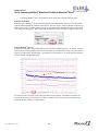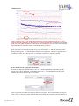
1
Patents Pending
User Manual
STARR Life Sciences Corp.
333 Allegheny Avenue, Suite 300
Oakmont, PA 15139
© STARR Life Sciences Corp., 2010
P/N 100017, Rev.5.3.0
USER MANUAL
Table of Contents
1. MOUSEOX® PACKAGED PARTS........................................................................................................ 1
2. COMPUTER REQUIREMENTS ........................................................................................................... 1
3. POWER REQUIREMENTS ................................................................................................................... 2
4. WARNINGS, CAUTIONS AND OPERATIONAL NOTES ................................................................ 2
5. USING THE MOUSEOX® CONTROL UNIT....................................................................................... 3
6. INSTALLING THE MOUSEOX® HARDWARE AND SOFTWARE................................................ 4
6.1. HARDWARE INSTALLATION .................................................................................................................. 4
6.2. UN-INSTALLING OLD SOFTWARE ......................................................................................................... 5
6.3. SOFTWARE INSTALLATION ................................................................................................................... 5
7. PHYSIOLOGIC PARAMETERS........................................................................................................... 7
8. USING THE SENSOR AND SENSOR CLIPS .................................................................................... 10
8.1. SEMI-DISPOSABLE SENSORS .............................................................................................................. 10
8.1.1. Mouse/Rat Pup CollarClip™ – Sizes XS and S ........................................................................ 11
8.1.2. Mouse/Rat Pup ThroatClip™ – Sizes XS and S ........................................................................ 13
8.1.3. Thigh Sensor ............................................................................................................................. 14
8.1.4. Rat CollarClip™ – Sizes M, L, XL and 2XL ............................................................................. 15
8.1.5. Rat ThroatClip™ – Sizes M, L, XL and 2XL............................................................................. 16
8.1.6. Rat Foot Sensor......................................................................................................................... 17
8.2. PULSAR™ SMALL-BORE MRI SENSOR .............................................................................................. 18
9. USING THE LIGHT-BLOCKING CLOTH........................................................................................ 19
10. USING THE MOUSEOX® SOFTWARE........................................................................................... 20
10.1. DESCRIPTION OF THE MOUSEOX® USER SCREENS ........................................................................... 20
10.1.1. MouseOx® Main – Startup Screen .......................................................................................... 20
10.1.2. Monitor Subject – Data Collection Screen.............................................................................. 21
10.1.3. Quick View Trends – Text File Review Screen........................................................................ 33
10.1.4. Compatibility of Old Text Data Files with Revision 5.0 Software .......................................... 35
10.2. PULSE PLETH SIGNAL QUALITY ....................................................................................................... 36
10.2.1. Good Signals ........................................................................................................................... 36
10.2.2. Undesirable Signals ................................................................................................................ 37
10.2.2.1. System Controller Changes ........................................................................................................... 38
10.2.2.2. Railed Signals.................................................................................................................................. 38
10.2.2.3. Motion Artifact ............................................................................................................................... 39
10.2.2.4. False Signals Due To Close Proximity of Artificial Light............................................................ 40
10.2.2.5. Poor Tissue Perfusion..................................................................................................................... 41
10.3. MEASUREMENT PARAMETER REJECTION CRITERIA AND ERROR CODES .......................................... 41
10.3.1. Parameter Rejection Criteria Specifications........................................................................... 41
10.3.2. Error Code Definitions............................................................................................................ 43
10.4. FILE SAVING OPTIONS AND UTILITIES .............................................................................................. 45
10.4.1. Opening Text Data Files Using Microsoft Excel .................................................................. 45
P/N 100017, Rev. 5.3.0
i
USER MANUAL
10.4.2. Filtering Files in Microsoft Excel to Blank Out Error Coded Data ..................................... 46
10.4.3. WinDaq® Waveform Browser.................................................................................................. 47
10.4.4. Converting WinDaq® Waveform File Data to Microsoft® Excel ............................................ 48
11. MOUSEOX® WITH ANALOG PULSE SIGNAL OUTPUT ........................................................... 51
12. EFFECT OF COMPUTER SLEEP MODES ON THE MOUSEOX® ............................................. 52
13. TROUBLESHOOTING ....................................................................................................................... 53
13.1. QUESTIONS AND ANSWERS .............................................................................................................. 53
13.2. ITEMS RELATED TO INSTACAL™...................................................................................................... 55
13.3. SYSTEM RESOURCE USAGE .............................................................................................................. 56
14. MOUSEOX® PARAMETER SPECIFICATIONS ............................................................................ 58
14.1. OXYGEN SATURATION (%) .............................................................................................................. 58
14.2. HEART RATE (BPM).......................................................................................................................... 58
14.3. PULSE DISTENTION ( M).................................................................................................................. 58
14.4. RESPIRATORY RATE (BRPM)............................................................................................................. 59
14.5. BREATH DISTENTION ( M)............................................................................................................... 59
15. CLEANING THE MOUSEOX® .......................................................................................................... 59
16. ACCESSORIES & REPLACEMENT PARTS.................................................................................. 60
17. SOFTWARE REVISIONS AND UPGRADES .................................................................................. 61
18. CONTACTING STARR LIFE SCIENCES™ FOR TECHNICAL SUPPORT ............................. 61
19. INTELLECTUAL PROPERTY PROTECTING THE MOUSEOX® ............................................. 62
20. WARRANTIES AND LIABILITIES.................................................................................................. 62
21. MOUSEOX® LABELS ......................................................................................................................... 63
P/N 100017, Rev. 5.3.0
ii
USER MANUAL
1. MouseOx® Packaged Parts
The following is a list of parts that should be included in your MouseOx® shipping box:
MouseOx® Oximeter Control Box
12 VAC Power Supply (you will have one or the other of the following)
o US and Canada……Standard External Plug-in Power Supply
o International……….Power Transformer and Power Cord to fit your wall receptacle
6-foot 2.0 USB Cable
CD with MouseOx® Drive Software and Electronic User Manual
Universal Cable
ManualSpin™ Low-Torque Commutator (optional)
MouseOx® User Manual (new customers only)
Under separate cover, you should also receive your choice of the following:
Selected size of CollarClip™, ThroatClip™, Rat Foot Clip and/or Mouse Thigh Clip chosen at
purchase
Please check to see that you have all of these parts. If you find any part missing, please contact STARR
Life Sciences™ at 1-866-9STARR9, or by e-mail at [email protected].
2. Computer Requirements
The MouseOx® has the following computer hardware requirements:
Processor
PC with Pentium -class processor (Pentium 1 GHz or higher recommended)
Computer Hardware
CD-ROM drive
VGA or higher resolution monitor (Super VGA recommended)
2.0 USB port
Operating System
Windows® 2000, XP, Vista or 7
MouseOx® is currently NOT compatible with Macintosh-based operating systems
Memory
1 GB RAM
5 MB Hard-Drive Space for program (does not include data files)
Minimum Screen Resolution
1024 by 768 pixels
If your hardware does not meet these requirements, please call or email STARR Life Sciences™ at: 1866-978-2779, or [email protected].
P/N 100017, Rev. 5.2.4
1
USER MANUAL
3. Power Requirements
Operating Wall Voltage:
100-240 VAC @ 50-60 Hz (You must use ONLY the STARR Life
Sciences power supply sent with your MouseOx®!)
Device Operating Voltage: 12 VAC
Max Operating Current:
500 mA
4. Warnings, Cautions and Operational Notes
WARNING
Replace control unit if housing is broken or cracked.
WARNING
DO NOT substitute cables or power supply. Use only parts that are packaged with the
MouseOx® device.
WARNING
Investigational device only.
WARNING
NOT for use on humans.
WARNING
DO NOT place control box in an environment with oxygen fraction greater than 30%.
WARNING
DO NOT immerse control box in liquids.
WARNING
DO NOT immerse lead ends of sensor cable in liquids.
WARNING
DO NOT directly expose control box to flammable or explosive gases.
WARNING
Replace power supply if cable is frayed.
WARNING
Inspect sensor cables after each use. Replace sensor cables if cable insulation is frayed
or wires are exposed.
WARNING
Replace control box if inside is suspected to be wet.
WARNING
DO NOT use STARR Life Sciences™ MouseOx® power supply or sensors with any
other device.
WARNING
Wipe external surfaces of box, sensors and leads, and sensor clip with light detergent
and water after each use on animals.
WARNING
Avoid spilling fluids on control box.
WARNING
Unplug the power cord to the controller when the device is unattended.
CAUTION
The MouseOx® uses an AC power supply. Do not substitute the power supply.
OPERATIONAL
NOTE
P/N 100017, Rev. 5.2.4
Do not place the sensor on the appendage of a cannulated leg/arm. Doing so could
result in a poor signal due to blood flow obstruction.
2
USER MANUAL
OPERATIONAL
NOTE
The MouseOx® will not work if the sensor pads are mounted on the incorrect cliphalves.
OPERATIONAL
NOTE
If the animal has dark fur, or signals are very small, the sensor location should be
shaved.
OPERATIONAL
NOTE
The user must have administrative rights to access computer USB ports and access to
the software that is loaded onto the computer from the MouseOx® CD.
OPERATIONAL
NOTE
OPERATIONAL
NOTE
Before installing an upgrade or re-installing a current software version, you must first
un-install any old versions of MouseOx® software.
It is recommended that the computer to be used with MouseOx® not be set to go into
System Standby or System Hibernate sleep modes.
OPERATIONAL
NOTE
Do not unplug any of the MouseOx® cables while running the MouseOx® software.
These cables include the power cable, the USB cable and the sensor cable. In the
event that a cable is unplugged, you must close completely out of the MouseOx®
software and rerun it.
OPERATIONAL
NOTE
The Sensor Power LED Indicator only lights when the MouseOx® controller board is
being energized. Therefore, this light will only illuminate after you completely
connect the system, start the MouseOx® program, and hit the "Monitor Subject" button
on the main screen.
OPERATIONAL
NOTE
The MouseOx® system DOES compensate for dysfunctional hemoglobin.
OPERATIONAL
NOTE
Always use the Light-Blocker cloth when in the presence of incandescent or
fluorescent lighting.
OPERATIONAL
NOTE
Always use only the specified sensor clip for your particular application, paying
particular attention to the weight range. See Section 8 for more detail.
OPERATIONAL
NOTE
You must never attempt to open a data file while it is open and being written to by the
MouseOx® software.
5. Using the MouseOx® Control Unit
Power Supply
The MouseOx® Control Unit does not have an on-board battery, so all power is provided by the
external plug-in power supply. The power supply should be connected to the control unit first, then
plugged into the wall. The MouseOx® power supply is non-standard, so DO NOT substitute power
supplies!!
For MouseOx® devices sold in the US and Canada, your MouseOx® was shipped with a standard
extermal plug-in power supply. For MouseOx® devices sold internationally (outside US and Canada), your
MouseOx® was shipped with a transformer and the appropriate wall receptacle power cord for your
country.
Sensor Power LED Indicator
The green Sensor Power LED Indicator will light when the monitoring mode is started using the
MouseOx® software by clicking the “Monitor Subject” button from the MouseOx® Main screen. The
Sensor Power LED Indicator illuminates whenever current is flowing through the main MouseOx®
P/N 100017, Rev. 5.2.4
3
USER MANUAL
controller electronics board. The MouseOx® control box does not allow power to be supplied to the sensor
until the software “Start/Reset” button on the “Monitor Subject” screen is clicked.
USB Cable
The USB cable must be connected between the MouseOx® and the computer in order for data to be
transferred. The order of connection is inconsequential.
Sensor Cable
The Sensor Cable Assembly should be connected as shown in the following figure.
MouseOx®
6” Sensor Extension
(optional)
Universal
Cable
ManualSpin™ Commutator
(optional)
Sensor Clip
Note that the Commutator is only necessary when making unanesthetized CollarClip™ measurements.
Note also that the 6” Sensor Extension is only necessary when using the Rat CollarClip™.
Calibration of the MouseOx®
Your MouseOx® was calibrated at the factory, and will be fully calibrated when you receive it.
Calibration constants are contained in the FLASH memory of the device. If a change in calibration is
suspected, please contact STARR Life Sciences™ Corp. technical service at 1-866-9STARR9, or by e-mail
at [email protected].
6. Installing the MouseOx® Hardware and Software
If you are installing an upgrade or are re-installing a current version of the
MouseOx® software, skip directly to Section 6.2.
If you are installing MouseOx® software for the first time, continue with Section 6.1.
6.1. Hardware Installation
Install Hardware BEFORE Installing Software!!
1] Power up the computer and allow Windows® to fully launch.
2] Connect the oximeter sensor to the oximeter control box. For US and Canada, connect the power supply
to the oximeter control box and insert the power supply into a wall receptacle to power the oximeter
control box. If you reside outside of the US and Canada, you will have received a separate power
transformer and power cord. You must plug the appropriate end of the power cord into the transformer,
plug the other end into to the wall receptacle, then plug the transformer low-voltage power cable into your
MouseOx®. NOTE: The green LED does NOT light at this point – it only lights when the software runs.
3] Connect one end of the USB cable to the USB port of the computer and connect the other end of the
USB cable to the oximeter control box.
P/N 100017, Rev. 5.2.4
4
USER MANUAL
a] After about 30 seconds, the message "your new hardware is installed and ready to use" should
appear, indicating that the correct driver was found and that the device is ready for use.
b] If the message "your new hardware is installed and ready to use" does not appear, disconnect
the USB Cable from the computer and repeat step 3] after unplugging your USB cable from the
computer.
c] If the message "your new hardware is installed and ready to use" still does not appear, the
correct hardware drivers may have already been installed. At this point, simply move to the next
step.
4] Unplug the USB Cable from the oximeter control box before proceeding to
installation of the software!!!
6.2. Un-Installing Old Software
Skip this section if you are installing MouseOx® software for the first time.
1] On your computer, click the “Start” button from the desktop, then click on either “Settings” then
“Control Panel,” or just “Control Panel.” The order depends on which version of Microsoft Windows®
you have.
2] On the Control Panel Menu, double-click on “Add or Remove Programs” or “Add/Remove Programs”
or “Uninstall a program (under “Programs)” or “Programs and Features” button.
3] After the list of software has been populated, find “MouseOx Revision X.X,” then click on the
“Add/Remove” or “Change/Remove” or “Uninstall/Change” button. For Vista, you must give permission
whenever asked.
4] Make sure all programs named “MouseOx” are removed from this list, in the event that a previous
version was installed more than once without first un-installing.
5] While remaining in the “Add/Remove Programs” section of the “Control Panel,” find “InstaCal and
Universal Library,” then click on the “Add/Remove” or “Change/Remove” or “Uninstall a program (under
“Programs)” or “Programs and Features” button.
6] Proceed to Section 6.3 and follow the instructions from there.
6.3. Software Installation
1] Be sure that the USB Cable is disconnected from the oximeter control box.
2] Insert the MouseOx® CD into the drive and the MouseOx® installation menu will appear. However, if
you have Vista, a dialog box will first appear – click “Install or run Program,” then the installation menu
will appear.
3] Click on Step 1 “Install InstaCal and Universal Library,” then do the following:
P/N 100017, Rev. 5.2.4
5
USER MANUAL
a] Select “OK” on the WinZip Self-Extractor dialog box. With Vista, you must first “Allow” the
download.
b] On the WinZip Self-Extractor – icalsetup.exe dialog box, select “Setup.”
c] On the Welcome Screen window, select “Next.”
d] On the Destination Folder window, accept the default and press “Next.” DO NOT change the
default file location.
e] On the Ready to Install Program window, select “Install.” At this point, the InstaCal software
will load. However, if you have Vista, a Windows Security dialog box may pop up to ask if you
want to install this software. This will occur at various stages during the operation. Click
“Install” each time, or click the “Always trust software from Measurement Computing
Corporation” check box, then click “Install.”
f] On the InstallShield Wizard Completed window, select “Finish.”
g] When asked to restart the computer, answer “No.”
4] Once you have finished, the MouseOx® installation menu will reappear. Click on Step 2 “Install
MouseOx,” then do the following:
a] On the MouseOx Revision X.X Setup dialog box (X.X represents the version of software that
you are currently loading), select “OK.” With Vista, you must first “Allow” the download.
b] On the next dialog box, click on the computer icon button to start the download. Although you
have the option to change the directory, DO NOT DO THIS.
c] On the MouseOx Revision X.X Setup – Choose Program Group dialog box, although you have
the option to choose a new program group, DO NOT DO THIS. Simply select “Continue” to
accept the default group “STARR Life Sciences.” After selecting “Continue,” the files will be
downloaded.
d] Select “OK” at the prompt after the download, and the installation menu will reappear.
5] (OPTIONAL) If you want to download the WinDaq® Waveform Browser, click on Step 3 “Install
WinDaq Waveform Browser,” then do the following (again, with Vista, you must allow the operation):
a] On the Welcome dialog box, select “OK.”
b] On the License Agreement dialog box, select “Accept and Continue.”
c] On the Select Destination Directory dialog box, although you have the option to choose a new
destination directory, DO NOT DO THIS. Simply select “OK” to accept the default directory
“C:\WINDAQ.” After selecting “OK,” the files will be downloaded.
d] On the Select Program Manager Group dialog box, although you have the option to choose a
new group name, DO NOT DO THIS. Simply select “OK” to accept the default group name
“WinDaq.”
6] (OPTIONAL) If you want to download the MouseOx® User Manual, click on Step 4 “Install MouseOx
User Manual”, then do the following (again, with Vista, you must allow the operation):
P/N 100017, Rev. 5.2.4
6
USER MANUAL
a] On the Welcome dialog box, select “Next.”
b] On the Choose Destination Location dialog box, select “Next.”
c] On the Setup Complete dialog box, select “Finish.” The MouseOx® User Manual will now be
accessible from your Desktop.
7] (OPTIONAL) If you want to download the WinDaq® Waveform Browser User Manual, click on Step 5
“Install WinDaq User Manual”, then do the following (again, with Vista, you must allow the operation):
a] On the Welcome dialog box, select “Next.”
b] On the Choose Destination Location dialog box, select “Next.”
c] On the Select Program Folder dialog box, although you have the option to choose a new
program folder, DO NOT DO THIS. Simply select “Next” to accept the default program folder
“WinDaq Documentation.” After selecting “Next,” the files will be downloaded.
d] On the Setup Complete dialog box, select “Finish.” The WinDaq® Waveform Browser User
Manual will now be accessible from your Desktop.
8] After completing the download of any of the optional software, the MouseOx® installation menu will
reappear. At this time, all of the software is now loaded and ready to use. You may now close the
installation menu.
9] At this point, you must restart your computer before you can run any software.
10] After restarting, you will need to complete a check to ensure that proper USB communication with the
computer has been established. Please do the following:
a] Reconnect the USB Cable to the oximeter control box.
b] Select “InstaCal” inside the “Measurement Computing” group under the Windows® Start
Menu.
c] After about 30 seconds, a message should appear indicating that InstaCal detected a new Plug
and Play Board.
d] If the new Plug and Play board message does not appear, then exit InstaCal and restart InstaCal
a second time (step 10b).
e] Select “OK” on the message that indicates that the device has been recognized.
f] Close InstaCal.
11] Start the MouseOx® program using the new “MouseOx” icon inside the “STARR Life Sciences” group
under the Windows® Start Menu. You are now ready to go.
7. Physiologic Parameters
The MouseOx® provides the following physiologic measurements:
P/N 100017, Rev. 5.2.4
7
USER MANUAL
Oxygen Saturation (SpO2)
The MouseOx® provides real-time percent oxygen saturation, in %, of functional arterial hemoglobin.
Heart Rate
The MouseOx® provides real-time cardiac pulse rate. Heart rate is given in bpm.
Breath Rate
The MouseOx® provides a real-time breath rate measurement that is updated every few seconds. Note
that this parameter is actually derived from respiratory effort, not airflow, and will be present even if the
animal is experiencing an obstructive apnea, as long as breathing effort is present. Breath rate is given in
breaths per minute or brpm.
Pulse Distention
Pulse distention is a measurement of the change in distention of the arterial blood vessels residing
between the sensors pads due to a cardiac output pulse. It is a direct measurement of changes in local
blood volume that accompany each cardiac pulse. For a given vascular compliance, pulse distention can
also provide a surrogate for pulse pressure. This will be explained in more detail below.
Pulse oximetry measures the oxygen content of arterial blood. Blood is identified as being arterial
because of its pulsatile nature. This pulsation is identifiable because it causes a cyclic change in the
absorption of light energy from the red and infrared LEDs (Light Emitting Diodes) as it passes through the
tissue due to the presence of changing quantities of blood that occur with every heart beat. All other nonblood material such as skin, fat and muscle do not change light absorption level with heart rate.
Additionally, non-arterial blood (venous or capillary) does not change light absorption level with heart rate
either, because pulsation is essentially completely diminished by the termination of the arterioles1. Thus,
our pulse oximetry measurement is being made across only pulsating blood, which can only be arterial.
Because this blood is arterial, it possesses systemic arterial oxygen content, which is what we measure.
Pulse distention is simply a measurement of the change in the effective path length of the light that
passes through only the arterial or pulsating blood, and it has true linear distance units of m. You could
picture this by thinking of placing all of the arterial blood residing in the light path between the sensor pads
into a cylinder that has a cross-sectional area equal to the cross-sectional area of the column of the light
beam passing from the LEDs to the photodiode. If the cylinder had one inlet and one outlet for the blood
to enter and exit, then the level of blood in the cylindrical chamber would rise with each cardiac ejection
stroke, and lower during each subsequent cardiac filling phase. The change in height of the blood in that
cylinder between ejection and filling, or systole and diastole, would then be measured directly as pulse
distention.
Pulse distention can be used to determine if you have sufficient arterial blood to be able to make good
oximetry measurements. As a practical example, if you were to place the sensor across your Achilles
tendon, which is very poorly perfused, you would not be able to make an oximetry measurement, and the
pulse distention would be nearly 0 because there is essentially no arterial blood in the tendon. On the other
hand, placing the sensor across a small artery would provide very large signals, indicated by large pulse
distention values.
Thus, in order to make an oximetry measurement, there must be sufficient arterial blood in the light
path, and because pulse distention quantifies the amount of arterial blood in the light path, it provides a
good indicator of the ability to make an oximetry measurement. The larger the pulse distention value,
the more arterial blood will be available to make oximetry, as well as heart rate and breath rate,
measurements.
Breath Distention
Breath distention is a measurement of the change in distention of the arterial blood vessels residing
between the sensor pads due to breathing effort. For a given vascular compliance, we have shown in our
lab that breath distention provides a surrogate for intrapleural pressure.
1
AC Guyton and JE Hall, Textbook of Medical Physiology, 9th ed., W.B. Saunders Co., 163, 1996.
P/N 100017, Rev. 5.2.4
8
USER MANUAL
The mechanisms that cause the breath distention signal at the sensor site are the same as those for
pulse distention, and are described in detail just above. The reason that we can detect a pulsation at
breathing rate however, is due to a cyclical pumping mechanism, just like that produced by the heart, that
provides a pulse of blood to the periphery, except at the rate of breathing instead of heart rate.
From our own investigation, we have observed that the breath distention signal is caused by a
phenomenon that we term the thoracic pump. In the thoracic pump model, pressure changes in the thoracic
cavity that accompany breathing or mechanical ventilation, act directly on the large arteries present there.
When intra-thoracic pressure is less than its mean, such as during inhalation, the great arteries become
slightly dilated in response, causing blood to fill the new dilated volume, which reduces blood flow to the
periphery. Conversely, when intra-thoracic pressure rises above its mean level, as occurs during
exhalation, this pressure exerts external force on the great arteries in the thorax, pushing more blood into
the periphery. This oscillatory increase and decrease in peripheral blood flow that accompanies breathing
effort, and which causes a subsequent oscillatory change in LED light absorption at the sensor site, can be
calculated as a distention, as is done with the cardiac blood pulse in Pulse Distention described above.
Note also, that this respiratory blood pumping occurs simultaneously with cardiac blood pumping, and
the two are actually super-imposed on one another on the actual light absorption signals that we measure.
In the human clinical realm, this respiratory effect, if it is large enough to become visible, is referred to as
pulsus paradoxus, although one should not also apply the connotation of abnormality commonly associated
with that term.
Given this model, the breath distention signal would have some useful applications. Because it is
caused by changes in intra-thoracic pressure, it may be considered to be a surrogate for intra-thoracic or
intra-pleural pressure. Measurement of intra-pleural or intra-thoracic pressure generally involves some sort
of invasive or semi-invasive physiologic intrusion, such as use of an esophageal pressure balloon sensor.
Correlation of breath distention with intra-thoracic pressure is not absolute without calibration however,
but it can be used for trending.
One specific application of breath distention is that it can provide monitoring of breathing effort put
forth by the animal. An example of its utility is when an animal becomes over-anesthetized. As breathing
effort is impeded by excessive anesthesia, the animal will start to gasp in order to move air as it becomes
hypoxic and hypercapnic. The increase in effort that accompanies gasping can greatly increase the breath
distention, and this phenomenon is particularly notable when it is compared with the pulse distention,
which does not change nearly as significantly as does breath distention. This utility of these dual
phenomenon is captured in our distention-based anesthesia alarms. Thus, breath and pulse distention may
be used to alert you to excessive levels of anesthesia.
Another application is an extension of intra-thoracic pressure measurement when it is used in
conjunction with tidal volume or air flow rate. In such a case, breath distention can provide a surrogate or
trending parameter for work of breathing, or power of breathing.
As with pulse distention, breath distention is given in true linear distance units of m.
P/N 100017, Rev. 5.2.4
9
USER MANUAL
8. Using the Sensor and Sensor Clips
In the following sections, instruction on the use of each type of STARR Life Sciences sensor is described.
8.1. Semi-Disposable Sensors
Clip Application and Sizing Chart
Following is the Semi-Disposable Sensor sizing chart. To get maximum performance from your clips,
it is imperative that you follow this chart when choosing a sensor for a given animal. Note that the Clip
Size is engraved on the back of each of the collar and throat clip handles.
Mouse
Size
Rat
Unshaven
Shaven
XS
1 - 15 gm
1 – 20 gm
N/A
S
> 15 gm
> 20 gm
5 – 30 gm
M
N/A
N/A
30 – 100 gm
L
N/A
N/A
101 – 300 gm
XL
N/A
N/A
301 – 500 gm
2XL
N/A
N/A
> 500 gm
< 60 gm
Thigh
Foot
N/A
= 100 gm
N/A
> 100 gm
System Configuration
To assemble and use your disposable sensors, the components should be connected as illustrated in the
following schematic.
MouseOx®
6” Sensor Extension
(optional)
Universal
Cable
ManualSpin™ Commutator
(optional)
Sensor Clip
CollarClip™/ThroatClip™ Differentiation
The CollarClip™, which is colored blue, is to be applied from the back of the neck of an animal, and
can be used in both awake and anesthetized applications. The ThroatClip™ is colored black, and it is
applied fromt the front of the neck. Thus, it is used only in anesthetized applications. It is important
that you use the correct clip for the correct application in order to get good measurements.
P/N 100017, Rev. 5.2.4
10
USER MANUAL
Collar Training
Each Clip Pack comes with a Blank Clip that matches the size of the sensor clip. It can be used for
training an animal to accept the CollarClip™ by simply placing the clip on the animal for a period of time
prior to your experiments. A minimum of 1 hour is recommended, but longer (3-6 hrs.) is preferred.
When to Replace Sensor Clips
The clips are designed to be semi-disposable. You should replace them in the following situations:
1] significant buildup of hair or material covering the clip faces,
2] good signals (data values on the software screen are in color) take an inordinately long time to appear,
3] presence of any mechanical defect in the sensor.
Chemical Hair Removal
The Blank Clips that come with each Clip Pack can also be used to chemically remove hair from the
CollarClip™ sensor site on the animal. This method is preferred over shaving, although not necessary
unless the animal has dark fur. If you do use a depilatory, we suggest that you do so a few hours to a day
before you will be conducting tests on your animals.
NOTE: You MUST remove hair from the sensor site of non-white animals.
1] It is recommended that you anesthetize the animal for hair removal.
2] Lay a bead of depilatory cream (e.g., Nair®) around the entire inside of the Blank Clip as shown.
3] Place the clip on the animal in the same location in which testing will be done, and let the clip and
depilatory cream remain in place for the duration specified by the manufacturer.
4] Remove the clip, then carefully wash the cream completely off of the animal using swabs and a small
water dish. The hair will be removed with the cream when you do this.
8.1.1. Mouse/Rat Pup CollarClip™ – Sizes XS and S
Mouse/Rat Pup CollarClip™ Sizing
Be sure to choose the correct clip for use with a given animal. Failure to do so can result in
inaccurate measurements. Use the following chart to pick the correct clip.
P/N 100017, Rev. 5.2.4
11
USER MANUAL
Mouse Weight Range
(gm)
Clip Size
Rat Pup
Weight Range
(gm)
1 - 20
XS
N/A
>20
S
5 - 30
Unshaven
Shaven
1 – 15
>15
Note that the Clip Size is engraved on the back of the clip handles.
Attaching the Collar and Making Measurements (Awake Animal)
1] Connect the MouseOx®, Universal Cable, Optional ManualSpin™ Commutator and Optional 6” Senor
Extension as shown in the figure at the top of the section.
2] If you are using an animal with non-white fur, you MUST either shave the entire sensor site WELL, or
use a depilatory product such as Nair® (See Chemical Hair Removal at the top of the section).
3] Place the animal on the lab bench surface, holding it by the tail. With the other hand, place the clip on
the animal from the back of its neck as shown below. Keep in mind the following:
- Place the clip BEHIND the ears and in FRONT of the paws. DO NOT capture the ears under
the clip!
- Center the clip handle on the vertebral column of the animal.
- Make sure that the clip is pushed down onto the back of the neck.
- For smaller animals, locate the clip further back away from the head to capture more tissue
between the clip tines.
- For neonates, place the clip on the skull across the ears.
- If the animal grabs the clip assembly out of your hand, DO NOT try to pull it back out. Set the
animal down on the lab bench and it will release the clip.
4] Other approaches for attaching the clip if the animal is resistant:
a] While trying to attach the clip, push down on the animal’s neck with the clip. It helps to lift the
animal so that its hind paws do not touch the ground. It is further helpful to allow the animal’s
front paws to grab a wire or box lid edge with its rear paws not touching anything.
b] Grab the tail using your ring finger, squeeze across its hind quarters with thumb and forefinger,
then push down. This immobilizes the animal quite well.
c] You can also anesthetize the animal to place the clip.
5] Pick the animal up by the tail while lifting the clip cable to keep it away from the animal. Gently set the
animal down inside its cage. DO NOT carry the weight of the animal by the clip wire as this can
dislodge the clip.
6] Once the animal is in the cage, connect the assembled wire to the CollarClip™ through an access port
P/N 100017, Rev. 5.2.4
12
USER MANUAL
on the cage lid. Make sure that you pull enough cable out of the cage so that it does not hang in the
animal’s face, as the animal may try to chew on the loose cable.
7] Check that the wiring assembly is connected to the MouseOx®, then start the software. You MUST
USE Revision 6.3 or higher with the CollarClip™. Follow the menu of buttons, choosing those
appropriate to your measurement conditions.
8] If you are making unanesthetized measurements and are using the Optional ManualSpin™ Commutator,
you can release rotation in the cable manually at any time while the system is running.
9] If signals do not come in quickly, or performance degrades over time, try the following:
- Re-position the sensor
- Replace the sensor
- Shave the animal
8.1.2. Mouse/Rat Pup ThroatClip™ – Sizes XS and S
Mouse/Rat Pup ThroatClip™ Sizing
Be sure to choose the correct clip for use with a given animal. Use the following chart to pick the
correct clip.
Mouse Weight Range
(gm)
Clip Size
Rat Pup
Weight Range
(gm)
1 - 20
XS
N/A
>20
S
5 - 30
Unshaven
Shaven
1 – 15
>15
Note that the Clip Size is engraved on the back of the clip handles.
Attaching the ThroatClip™ and Making Measurements
1] Anesthetize the animal, then connect the MouseOx®, Universal Cable and ThroatClip™ Sensor.
2] If you are using an animal with non-white fur, you MUST either shave the entire sensor site WELL, or
use a depilatory product such as Nair® (See Chemical Hair Removal at the top of the section).
3] With the animal laying in the supine position, push/slide the skin down on either side of the throat, then
place the clip over the throat while holding the skin down so that the tines end up BEHIND the ears and
in FRONT of the paws. The clip handles should be centered on the throat of the animal. Be careful
that the tips of the tines do not catch on the lab surface such that the clip does not close completely.
P/N 100017, Rev. 5.2.4
13
USER MANUAL
4] Check that the wiring assembly is connected to the MouseOx®, then start the software. You MUST
USE Revision 6.3 or higher with the ThroatClip™. Follow the menu of buttons, choosing those
appropriate to your measurement conditions.
5] If signals do not come in quickly, or performance degrades over time, try the following:
- Re-position the sensor
- Replace the sensor
- Shave the animal
8.1.3. Thigh Sensor
Thigh/Rat Foot Clip Sizing
Be sure to choose the correct clip for use with a given animal. Use the following chart to pick the
correct clip.
Sensor
Type
Mouse
Weight
Range (gm)
Rat Weight
Range (gm)
Thigh
< 60
= 100
Rat Foot
N/A
> 100
Attaching the Sensor and Making Measurements
1] Anesthetize the animal, then connect the MouseOx®, Universal Cable and Mouse Thigh Sensor.
2] If you are using an animal with non-white fur, you MUST either shave the entire sensor site, or use a
depilatory product such as Nair®.
3] Place the animal either prone or supine on the lab bench surface. Place the Thigh Sensor on the THIGH
(not the lower leg!) of the animal by using the half arc shape of the sensor to grasp tissue around the
thigh bone so that the sensor protrudes in a perpendicular orientation from the thigh. This will help
align the sensor with the large arteries that reside next to the bone. Gently press the clip together on the
thigh to improve the surface contact.
NOTE: Do not use this sensor on an area other than the upper thigh.
4] Check that the wiring assembly is connected to the MouseOx®, then start the software. You MUST
USE Revision 6.3 or higher with the ThroatClip™. Follow the menu of buttons, choosing those
appropriate to your measurement conditions.
5] If signals do not come in quickly, or performance degrades over time, try the following:
- Re-position the sensor
- Replace the sensor
- Shave the animal
NOTE: Shaving is NECESSARY for any animal with non-white fur.
P/N 100017, Rev. 5.2.4
14
USER MANUAL
8.1.4. Rat CollarClip™ – Sizes M, L, XL and 2XL
Rat CollarClip™ Sizing
Be sure to choose the correct clip for use with a given animal. Use the following chart to pick the
correct clip.
Clip Size
Animal Weight Range
(gm)
M
31 – 100
L
101 – 300
XL
301 - 500
2XL
500 +
Note that the Clip Size is engraved on the back of the clip handles.
Attaching the Collar and Making Measurements (Awake Animal)
1] Connect the MouseOx®, Universal Cable, Optional ManualSpin™ Commutator, Optional 6” Senor
Extension and CollarClip™ as shown in the figure at the top of the section.
2] Remove the lid from whatever cage is being used, and slide the CollarClip™ sensor through an access
on the top of the lid, then set it on the bench.
3] If you are using an animal with non-white fur, you MUST either shave the entire sensor site WELL, or
use a depilatory product such as Nair® (See Chemical Hair Removal at the top of the section).
4] Put the animal inside the cage. Pull enough cable through the lid so that you can move it around to
attach the clip to the animal. Place the sensor on the animal from the back of its neck as shown below.
Keep in mind the following:
- Place the clip BEHIND the ears and in FRONT of the paws. Do not capture the ears under the
clip!
- Center the clip handle on the vertebral column of the animal.
- Make sure that the clip is pushed straight down on the back of the neck.
- Once the clip is in place, squeeze the tines around the animal’s neck to assure good contact.
- If the animal grabs the clip assembly out of your hand, DO NOT try to pull it back out. Release the
tension on the cable and the animal will release the clip.
5] Once the clip is in place, re-assemble the lid back onto the cage, then pull the cable out of the box until
there is just enough length for the animal to move freely in its cage without pulling the clip off. Make
sure that you pull enough cable out of the cage so that it does not hang in the animal’s face, as the
animal may try to chew on the loose cable.
6] Check that the wiring assembly is connected to the MouseOx®, then start the software. You MUST
USE Revision 6.3 or higher with the CollarClip™. Follow the menu of buttons, choosing those
appropriate to your measurement conditions.
7] If you are making unanesthetized measurements and are using the Optional ManualSpin™ Commutator,
you can release rotation in the cable manually at any time while the system is running.
8] If signals do not come in quickly, or performance degrades over time, try the following:
P/N 100017, Rev. 5.2.4
15
USER MANUAL
- Re-position the sensor
- Replace the sensor
- Shave the animal
NOTE: When using the 2XL clip, it is recommended that the animal be shaved regardless.
Attaching the Collar and Making Measurements (Anesthetized Animal)
Anesthetize the animal, then simply follow steps 4, 6 and 8 above.
8.1.5. Rat ThroatClip™ – Sizes M, L, XL and 2XL
Rat ThroatClip™ Sizing
Be sure to choose the correct clip for use with a given animal. Use the following chart to pick the
correct clip.
Clip Size
Animal Weight Range
(gm)
M
31 – 100
L
101 – 300
XL
301 - 500
2XL
500 +
Note that the Clip Size is engraved on the back of the clip handles.
Attaching the ThroatClip™ and Making Measurements
1] Anesthetize the animal, then connect the MouseOx®, Universal Cable and ThroatClip™ Sensor.
2] If you are using an animal with non-white fur, you MUST either shave the entire sensor site WELL, or
use a depilatory product such as Nair® (See Chemical Hair Removal at the top of the section).
3] With the animal laying in the supine position, push/slide the skin down on either side of the throat, then
place the clip over the throat while holding the skin down so that the tines end up BEHIND the ears and
in FRONT of the paws. The clip handles should be centered on the throat of the animal. Be careful
that the tips of the tines do not catch on the lab surface such that the clip does not close completely.
When applying the clip, to facilitate good contact,
1] push the skin down on either side of the neck,
2] push down on the handles, then,
P/N 100017, Rev. 5.2.4
16
USER MANUAL
3] squeeze the tines together.
These steps are VERY important in order to obtain good signals.
4] Check that the wiring assembly is connected to the MouseOx®, then start the software. You MUST
USE Revision 6.3 or higher with the ThroatClip™. Follow the menu of buttons, choosing those
appropriate to your measurement conditions.
5] If signals do not come in quickly, or performance degrades over time, try the following:
- Re-position the sensor
- Replace the sensor
- Shave the animal
NOTE: When using the 2XL clip, it is recommended that the animal be shaved regardless.
8.1.6. Rat Foot Sensor
Thigh/Rat Foot Clip Sizing
Be sure to choose the correct clip for use with a given animal. Use the following chart to pick the
correct clip.
Sensor
Type
Mouse
Weight
Range (gm)
Rat Weight
Range (gm)
Thigh
< 60
= 100
Rat Foot
N/A
> 100
Attaching the Sensor and Making Measurements
1] Anesthetize the animal, then connect the MouseOx®, Universal Cable and Mouse Thigh Sensor.
2] Place the animal either prone or supine on the lab bench surface. Place the Rat Foot Sensor on the foot
of the animal so that the sensor protrudes directly off the end of the foot past the toes, as shown in the
figure below.
It is also important to observe the following:
- Make sure that the clip is CENTERED on the foot laterally
- Do not place the clip on the base of the foot near the ankle
- Do not place the clip too far toward the toes such that light can pass directly between the sensor pads
without passing through tissue.
3] Check that the wiring assembly is connected to the MouseOx®, then start the software. You MUST
USE Revision 6.3 or higher with the ThroatClip™. Follow the menu of buttons, choosing those
appropriate to your measurement conditions.
4] If signals do not come in quickly, or performance degrades over time, try the following:
P/N 100017, Rev. 5.2.4
17
USER MANUAL
- Re-position the sensor
- Replace the sensor
8.2. Pulsar™ Small-Bore MRI Sensor
Assemble your Pulsar™ Small-Bore MRI Sensor in the configuration shown in the following figure.
Single Black Cable – 15 ft
Disposable Sensor Clip
Converter Box
1] Connect the DB9 Connector (9-pin connector) on the end of the thin black cable to the MouseOx®
Control Box. Be sure that the MouseOx® software is not running when you do this.
2] Connect a disposable sensor clip to the fiber-optic cable. You MUST USE a Mouse Thigh Clip (any
mouse or a rat < 101 gm) or a Rat Foot Clip (for rats > 100 gm). Place the protrusion on the end
of the fiber-optic cable labeled “LED” into the hole on the clip half marked “LED,” then do the same
for the side marked “PD.” Make sure that the fiber-optic cable is oriented so that it aligns over the
handle as shown.
3] Connect the sensor clip to the animal:
A] Mouse Thigh – Place the clip on the thigh of a mouse as shown. For non-white fur, you MUST shave
both locations of the sensor site. On white fur, shaving is not necessary, but will improve signal
strength.
B] Rat Foot – Place the clip over the toes and locate it so that light shines through the CENTER of the
foot. Support the clip/cable so that the animal’s foot is NOT TWISTED relative to its position before
attaching the clip. The clip half marked “PD” should be on the bottom side of the foot.
Mouse Thigh
Rat Foot
4] After locating the clip on the animal, distribute the dual black fiber-optic cable such that it proceeds
straight from the animal and that it DOES NOT twist the animal’s foot. Try to lay the sensor clip so
that both the LED and PD cables are laying on the table.
P/N 100017, Rev. 5.2.4
18
USER MANUAL
5] Run the MouseOx® software (you should use Rev 6.3 or higher). To get to the Monitor Subject screen,
choose “Anesthetized Measurements” then “Adult Mouse” or the appropriate rat button, then pick the
MRI sensor, which is at the bottom of the list.
6] Other recommended guidelines:
- Keep the body (rectal) temperature of the animal above 36°C.
- Make sure that the Pulse Distention exceeds 20 m when operating the system. If the Pulse Distention is
less 20 m, it is recommended that you try to relocate the sensor clip to improve it or warm the
animal.
- If you are having trouble getting a good signal, try shaving the sensor location if applicable.
- The non-ferrous spring will weaken with multiple uses. An unreasonably low oxygen saturation
measurement (a healthy subject with a sat of 88% or less) is a clear sign of an over-used spring.
To prevent this, and to promote infection control, replace the clip before each MRI session.
- CAUTION: The converter box contains trace amounts of ferrous material. Keep it away from the magnet
bore.
9. Using the Light-Blocking Cloth
The light-blocking cloth (Light Blocker) included in your MouseOx® package is intended to be used to
reduce the brightness of ambient light that enters the photodiode on your sensor. Excessive ambient light,
such as that from surgical lighting or direct sunlight, can reduce the ability of the MouseOx® device to
provide consistent and accurate data. Thus, it is recommended that you always use the Light Blocker when
possible, or make sure that the amount of ambient light reaching the sensor be as low as possible.
We must also caution users that fluorescent lighting can produce interference that can simulate
breathing or cardiac input to the MouseOx®. Such input can disrupt proper operation of the MouseOx®.
Anesthetized Applications
To use the cloth, set up the animal with the clip as described in the previous section. After you have
determined that the sensor clip placement is sufficient, simply place the Light Blocker directly over the
sensor clip. If positional adjustments must be made to the sensor clip to improve signal levels, remove the
cloth, adjust the sensor position, and then replace the cloth over the sensor. When optimal signals have
been achieved, leave the cloth in place over the sensor clip. If signals become poor while you make
measurements, remove the cloth and check that the sensor has not moved.
Awake Applications
Obviously, the Light Blocker cannot be used on an awake, mobile animal. Regardless, we must stress
that you reduce the ambient light reaching the sensor. This can be done by reducing the room lighting, or
by covering the cage with something to help block out excessive light.
When making measurements using the STARR-Gate™ rat tubes, the Creature Comforter™ should be
placed on the tube to darken the environment for the subject, and the Light Blocker should be used to cover
the sensor, which should be located near the tip of the subject’s tail.
P/N 100017, Rev. 5.2.4
19
USER MANUAL
10. Using the MouseOx® Software
10.1. Description of the MouseOx® User Screens
Before describing the MouseOx® software, we note that you can double-click in the title bar at the top of
any of the screens to maximize that screen across your display. To return a screen to its original size, you
can simply double-click again in the title bar.
You also have the ability to resize all of your screens and windows using the standard Windows® resizing
feature. This is done by simply moving the mouse pointer to any edge of the screen or window until the
resizing arrow appears. Once the arrow appears, right-click and drag the edge to the desired shape and
size.
10.1.1. MouseOx® Main – Startup Screen
The following is a picture of the MouseOx® Main screen that appears at startup of the software.
BUTTONS
Monitor Subject
The Monitor Subject button takes you to the screen that controls the recording and display of MouseOx®
data received from the test subject. When this button is pressed, a screen appears inquiring whether your
subject is anesthetized or awake.
P/N 100017, Rev. 5.2.4
20
USER MANUAL
After pressing one or the other of these buttons, you will be taken to a series of screens with selfexplanatory buttons that eventually lead to the Monitor Subject screen.
Quick View Trends
The Quick View Trends button takes you to the screen that is used to replay data previously recorded from
the Monitor Subject screen in text file form. Its appearance is nearly identical to that of the Monitor
Subject screen, but it can be identified by its title at the top left of the screen. More detail about this screen
is provided in Section 10.1.3 below.
Close Program
This button simply closes the MouseOx® program.
Open Windaq
This button opens the WinDaq Waveform Browser software from Dataq Instruments. The WinDaq
Waveform Browser software allows you to perform a variety of post-processing functions on data recorded
from the MouseOx®.
Free MouseOx Training (click for info)
This button has a surrounding frame that flashes colors for about 2 seconds when you start the MouseOx®
software. When you click this button, a dialog box opens that describes the free, interactive, web-based
training and how to schedule a session. To schedule a session, simply call (866) 978-2779.
HARDWARE CONNECTION MESSAGE
In the space just below the user-controlled buttons, a message may appear that indicates that the
MouseOx® is not connected to the computer or is not recognized by the software. The self-explanatory
message is:
MouseOx® Not Detected
If you see this message, check that the USB cable is plugged into both the MouseOx® and the computer. If
the USB cable is not plugged in, close down this program completely, plug in the USB cable, then open up
the MouseOx® software again.
If the USB cable appears to be plugged in, unplug one end of the USB cable, close down this program
completely, re-plug in the USB cable, then open up the MouseOx® software again.
If neither case applies, or either is unsuccessful, please contact technical service by calling or emailing
STARR Life Sciences™ at: 1-866-978-2779, or [email protected].
10.1.2. Monitor Subject – Data Collection Screen
The following is a picture of the “Monitor Subject” screen. The Monitor Subject screen uses the same
form, but has 2 titles in the upper left corner of the title bar. These are “Monitor Anesthetized Subject” and
“Monitor Awake Subject,” corresponding to an answer of “Anesthetized” or “Awake” respectively, to the
question posed when entering the Monitor Subject Screen.
BUTTONS
Start/Reset
At the beginning of use, the Start/Reset button initiates data collection by the MouseOx®. You will notice
that the sensor light comes on when this button is pressed. No data are being saved or recorded if only this
button is pressed.
P/N 100017, Rev. 5.2.4
21
USER MANUAL
If you start the software by hitting the Start/Reset button before attaching the sensor clip to the animal, or if
you detach the sensor clip from the animal after hitting the Start/Reset button, and then apply the sensor to
the animal again, it is recommended that you press the Start/Reset button again. This will reset the
amplifier gains so that the MouseOx® can hone in on measured values more quickly.
Lastly, if you are making measurements on un-anesthetized animals using the STARR-Gate™, it is
recommended that you press Start/Reset if the Pulse Pleth signals appear to be noisy (i.e., they are not
smooth or they do not have coincidental phase). Also, it often takes some time for the animals to calm
down once they are in the tube and you begin making measurements. It is recommended that you press
Start/Reset if an animal has been in the tube for 10-15 or more minutes, and good Pulse Pleth signals are
beginning to appear.
Pause
This button stops data monitoring by the MouseOx®. If Start/Reset is pressed after Pause, the system will
continue monitoring from the point at which it was stopped, including resuming at all of the previous
control settings and chart data streams. If data files are being recorded, pressing the Pause button will stop
the program from writing to the file until Start/Reset is pressed again. Once Start/Reset is pressed again,
the program will resume writing to the file at the next available location. However, no marker will be
placed into the file to indicate the stoppage location.
Stop
This button stops data monitoring by the MouseOx®. If Start/Reset is pressed after Stop, the system will
reset. Control settings will be re-initialized and Data Charts will be cleared. If you are writing to a data
file and press the Stop button, you will be prompted to first cease file saving before you can use the Stop
button.
Clear Charts
This button clears all of the data charts while data are being collected. It has no effect if pressed while the
MouseOx® is either paused or stopped. It does not affect the writing of data to a file.
Cruise Control ON/OFF Toggle
Cruise Control ON should be the normal operating condition for the MouseOx®. When Cruise Control is
ON, the MouseOx® controller can optimize all signals at any time. When Cruise Control is OFF, the
internal data monitoring electronics lock in the current state to permit continued data monitoring when
normally the system would try to optimize gains because of low or high signal levels. This condition
P/N 100017, Rev. 5.2.4
22
USER MANUAL
permits you to effect some new condition on the animal without the data collection system trying to
optimize itself. The benefit is that if the condition is reversed, the system is in the same electronic state in
which it started prior to initiation of the new condition. This functionality is particularly useful when using
the STARR-Gate™ restraint tubes, because you can turn the Cruise Control OFF when you get generally
consistent signals that the controller would normally and unnecessarily keep trying to optimize in response
to random, occasional events such as tail motion.
If you press Start/Reset while the Cruise Control is OFF, the system will not be affected until the Cruise
Control is turned back ON, at which point the gains will be reset to their default start values.
The default value at startup is Cruise Control ON.
Main Menu
This button returns you to the MouseOx® Main screen. If monitoring has been started and not stopped, a
message box will prompt you to first stop monitoring. If data are being recorded, a message box will
appear to remind you to stop recording data and to stop monitoring.
Summary
This button condenses the Monitor Subject screen to a much smaller screen with a reduced data
presentation. This function allows you to have better access to your computer screen. You can also
double-click in the title bar of this screen to expand it to fill your viewing screen, which will provide you
with a very uncomplicated, easy-to-read interface for animal monitoring.
This screen only displays the Pulse Pleth window, each of the four Chart Data boxes and the Mark Trend
File count window. There are only two control buttons on this screen: Mark Trend File and Monitor
Subject. The Mark Trend File button is described just below. The Monitor Subject button returns you to
the normally-sized screen. User-set alarm conditions will be flagged both audibly and by a color change
(white to yellow) on the pertinent parameter Chart Data Box. An image of the Summary screen appears in
the following figure.
Mark Trend File
This button only functions when data are being recorded. It places a marker in the data stream at each file
location at the time the button is pressed. It also places a vertical line on the Oxygen Saturation chart at the
location at which time the button was pressed. This line follows across the scrolling screen. You are
permitted 50 file markers per data file. If the data file was saved in text format, the markers can be
accessed from a drop down menu in the Quick View Trends screen, which can be reached from the
MouseOx® Main screen. If the data file was saved in WinDaq® format, the file markers are located in
Channel 1. During data recording, the currently available marker number is displayed in the adjacent box.
Note that markers can also be placed in the file by pressing the keyboard space bar, after the Mark Trend
File button has been highlighted once. Also, this button performs the same function whether on the main
Monitor Subject screen, or on the Summary screen.
P/N 100017, Rev. 5.2.4
23
USER MANUAL
Because the file marker only indicates a location within a file, it is up to you to separately record the
significance of any marked event.
Set Alarms
Upon pressing this button, a separate screen titled Alarm Values appears over the top of the monitoring
charts. A pictorial example is given in the next figure below.
Parameter Alarms
You can separately adjust the upper and lower alarm thresholds of each of 4 chart signals (Breath
Distention does not have its own alarm). All upper and lower threshold alarms can be turned on
or off separately by clicking the box adjacent to the chosen Alarm On title. If a given signal
passes above its high alarm setting, or if a given signal passes below its low alarm setting, an
audible alarm sound will be initiated and the corresponding data chart will change background
color. The audible alarm can be shut off by pressing the Alarm Silent toggle button on the
Monitor Subject screen. In addition to the audible and visual alarms, a message will appear in red
in place of the pertinent chart title describing the possible cause of the alarm.
All alarms will be automatically de-activated if the pertinent parameter in that alarm is under an
active error code. Also, the default position for the alarms is that all Low Alarms will be active,
and all High Alarms will be inactive. The values are defaulted to the following (Breath Distention
does not have an alarm):
High Alarms (all high alarm defaults are also the maximum value for each parameter)
- Pulse Distention
800 m
- Pulse Rate
900 bpm
- Oxygen Saturation
100 %
- Breath Rate
450 brpm
Low Alarms
- Pulse Distention
P/N 100017, Rev. 5.2.4
10
m
24
USER MANUAL
- Pulse Rate
- Oxygen Saturation
- Breath Rate
120 bpm
80 %
40 brpm
The alarm levels can be adjusted by choosing one of the incremental values from the pull-down
menu associated with a given alarm, or by typing a new value directly into the pull-down menu
box for that alarm.
Anesthesia Alarm Settings
The MouseOx® software provides you with two types of alarms that can be used specifically to
monitor depth of anesthesia. These are the Distention Compare Alarm and the Rate Compare
Alarm. It should be noted that we do not recommend specific values for these alarms, and that
they are available for your use based on your own experience. The reason that we do not make
specific recommendations is because parameter levels may be dependent on items that we cannot
control, such as dosage, time after dosage, type of anesthesia, where it is delivered, etc.
NOTE: When monitoring awake subjects, these alarms are deactivated and cannot receive input.
In our alarm configuration, parameter alarms (described above) have precedence over anesthesia
alarms. Additionally, an anesthesia alarm will be disabled whenever an error code for a pertinent
parameter within the given alarm is active.
Distention Compare Alarm
The Distention Compare Alarm allows you to use the relative percentage of Pulse
Distention to Breath Distention as a means of monitoring depth of anesthesia. The value of
the alarm limit is literally the percent that Breath Distention is to Pulse Distention, just as it
reads on the screen. Because this alarm is a little confusing, an example of each type of alarm
is given below.
Example 1 – Anesthesia Too Deep Distention Compare Alarm
If the Anesthesia Too Deep alarm is set to 80%, the alarm threshold would be met
when the Breath Distention becomes greater than 80% of the Pulse Distention. The idea
behind this alarm is that excessive anesthesia levels generally tend to suppress breathing.
The response to decreased ventilation is gasping, the result of which, is an increase in
breathing effort, which increases Breath Distention relative to Pulse Distention.
Example 2 – Anesthesia Too Light Distention Compare Alarm
If the Anesthesia Too Light alarm is set to 20%, then the alarm threshold would be
met when the Breath Distention becomes less than 20% of the Pulse Distention.
The alarm settings cannot be set such that there is overlap between the Too Deep and
Too Light alarm settings. Additionally, you do not have the ability to type in individual
alarm values. You can only choose from the lists provided on the drop-down menus in the
combo-boxes. The limits and steps for each of the Distention Compare Alarms are given
below.
Anesthesia Too Deep
Range:
70 – 140%
Step:
10% increments
Default:
100%
Anesthesia Too Light
Range:
10 – 40%
Step:
10% increments
P/N 100017, Rev. 5.2.4
25
USER MANUAL
Default:
20%
Note that this alarm is automatically disabled if the Pulse Distention drops below 25 m.
Rate Compare Alarm
The Rate Compare Alarm allows you to adjust the ratio of Heart Rate to Breath Rate as
another means of monitoring depth of anesthesia. The value of the alarm limit is literally the
direct ratio of Heart Rate to Breath Rate, as it reads on the screen. Examples of each type of
alarm are given below.
Example 1 – Anesthesia Too Deep Rate Compare Alarm
If the Anesthesia Too Deep alarm is set to 7.5, the alarm threshold would be met
when the Heart Rate becomes greater than 7.5 times the Breath Rate. The idea behind
this alarm is that excessive anesthesia levels generally tend to suppress breathing, causing
the breath rate to drop, usually without a concomitant change in heart rate.
Example 2 – Anesthesia Too Light Rate Compare Alarm
If the Anesthesia Too Light alarm is set to 3.5, then the alarm threshold would be
met when the Heart Rate becomes less than 3.5 times the Breath Rate, irrespective of
which one actually changed.
With this alarm, you can insert your own limit values in the combo boxes provided.
However, the system will not permit you to insert values that are overlapping, i.e., the Too
Deep alarm limit less than the Too Light alarm limit. You can also choose from the list
provided on the drop-down menu in the combo-box. The limits and steps for each of the Rate
Compare Alarms are given below.
Anesthesia Too Deep
Range:
5.0 – 10.0 bpm/brpm
Step:
0.5 bpm/brpm increments
Default:
7.5 bpm/brpm
Anesthesia Too Light
Range:
2.5 - 4.5 bpm/brpm
Step:
0.5 bpm/brpm increments
Default:
3.5 bpm/brpm
Default Values
The Default Values button resets the alarm parameters to the default values listed in the Parameter
Alarms section above.
Set Alarms and Close Window
This button must be pressed in order to set any changed alarm values. This button also closes the
Alarm Values screen.
Alarm Silent (toggle)
This button shuts off all audible alarms until the button is pressed again, which restarts the audible alarms.
When first pressed, its name changes to “Turn Sound On.” Audible alarms occur whenever one of the 4
chart signals crosses either above or below the user-selected threshold. For all signals, alarms will initiate
as soon as a new calculated value falls above or below the set threshold.
Under an alarm condition, the monitoring screens will indicate the source by changing the associated
parameter chart background to a caution yellow. Additionally, the chart title is replaced by a red label
warning of the infraction.
P/N 100017, Rev. 5.2.4
26
USER MANUAL
View Breath Pleth Window
This button opens the Breath Pleth window, which appears in the figure below.
This screen has two plots on it. The filled, lighter blue waveshapes in the background represent the
breathing oscillation, and indicate the breathing signal frequency. The darker blue waveform
superimposed over the filled breath waveshapes is the combined heart rate and breathing signal. It is the
same signal that appears in the Pulse Pleth window of the Monitor Subject screen, albeit with a wider
swath of data. Clicking on the Close Window button in the bottom right corner will close this window.
Note also that the Error Code messages that appear in the Pulse Pleth window will also appear at the top
center of the View Breath Pleth Window screen when an error code is active.
View Diagnostic Window
This button opens the Quick Averaging Diagnostics window, which appears in the figures below. The
Quick Averaging Diagnostics window allows you to collect an average value for each of the 5 MouseOx®
parameters. The length of the average is selectable by grabbing the maroon arrow slider and moving it
vertically. You can choose averaging durations from 5 to 30 seconds, in 1 second increments.
Averaging can be done manually, or automatically at constant intervals of your choosing. Automated
averages always take precedence over manual averages.
Once a diagnostic average (manual or automatic) is initiated, each parameter window will display the
message “Processing…” until the first valid value is entered into the averaging calculation. At this point,
each window will display the running average, but in gray text, as shown in the leftward figure below.
Once each parameter calculation is completed, it will change from gray to color, as shown in the rightward
figure below. Because the system rejects values that are error-coded independently for each parameter,
some averaged values may appear in their respective slots before others.
The duration of a given averaging operation for a parameter may be longer than the period set using the
slider arrow. The duration corresponding to the position of the slider arrow simply indicates the sum total
duration of valid data collected and averaged, irrespective of how long that takes. This is due to the fact
that any data subject to an active error code are automatically rejected by the averaging algorithm and are
not included in the average. However, all diagnostic runs will time out at a value of 3 times the time
selected using the slider arrow, and a value of 0 will be written for any parameters that have not completed
an average in that period. In summary, once an average is initiated, the averaging algorithm will simply
continue until the full period of non-error code-rejected data are averaged, or one or more uncompleted
parameter averages times out.
Data used in the averaging algorithm of the Quick Diagnostics window are NOT affected by your choice of
data averaging on the Monitor Subject Screen (i.e., only 1X data are averaged).
P/N 100017, Rev. 5.2.4
27
USER MANUAL
Run New Diagnostics (Manual Only)
This button will initiate a manual average. If you choose to initiate another average, you can
simply press the Run New Diagnostics button without having to close the window. Also, if you
press the Run New Diagnostics button while a previous data averaging is still in progress, you
will override that data averaging and initiate a new one. If you press this button while an
automatic average is active (not executing), the manual average will proceed, but will be
overridden if an auto-run average executes while the manual average is running.
Write Data to File (Manual Only)
This button saves the current manual diagnostic data to a text file of your choice. This text file
can easily be read into Microsoft® Excel using the same instructions for reading a Text File into
Excel as described in Section 10.4.1 below titled “Opening Text Data Files Using Microsoft®
Excel.”
When you press the Write Data to File button, a standard Windows® file saving dialog box will
appear, allowing you to name the file and store it in a location of your choice. A default filename
will be provided with the default save location being “c:\”, and the default name being
“Diagnostic mmddyyhhmmss.txt” (month, day, year, hour, minute, second). The data set will be
time-stamped at the point at which you press the Write Data to File button. The currently active
filename will appear in light text above this button. A sample Excel file with 5 data sets saved is
provided below.
If you press the Write Data to File button before a new diagnostic data collection has been
completed, a data set will still be saved, but will contain all zeros. If you press this button while
all data are still being averaged, the currently completed data values will be saved, along with
zeros for those that are still processing. You cannot write data from the same diagnostic run to a
file more than once, so be sure that you have all of your desired parameter calculations completed
before saving.
P/N 100017, Rev. 5.2.4
28
USER MANUAL
Start Auto Run Diagnostics (Automatic Only)
This button will initiate automatic averaging that will execute at time intervals of your choosing
from the palette of duration toggles directly above this button. The available time intervals are 5,
10 and 15 minutes. If you want to change this time interval after having started, without creating
a new file, you can simply press the desired duration toggle, then press Start Auto Run
Diagnostics again, which will initiate a new automatic averaging at that point.
When you first press the Start Auto Run Diagnostics button, AND you have not opened a file
using the Write Data to File button, a standard Windows® file saving dialog box will appear,
allowing you to name the file and store it in a location of your choice. A default filename will be
provided with the default save location being “c:\”, and the default name being “Diagnostic
mmddyyhhmmss.txt” (month, day, year, hour, minute, second). The data set will be time-stamped
at the point at which each auto run is executed. The currently active filename will appear in light
text above the Write Data to File button.
In order to initiate saving data to a new file, you must first press the Stop button, then press the
New File toggle, then press Start Auto Run Diagnostics again.
Once you press the Start Auto Run Diagnostics button, a yellow banner will appear below the
Stop button that says “Auto Run Diagnostic is Executing” (see leftward figure above). Once the
data are calculated and stored to the file, the banner will change color and title to green and “Auto
Run Diagnostic is Active” (see rightward figure above). If the Auto Run feature has not been
initiated, this area will remain gray.
New File/Append to File Toggles
When you first open the Quick Averaging Diagnostics window, the New File toggle will be
active. It will remain so until you open a file by pressing either the Write Data to File or Start
Auto Run Diagnostics buttons. Once the data set associated with that action is saved, the New
File toggle will deactivate and the Append to File toggle will be active. From that point forward,
each manual or automatic data set will be appended to the current file. If you would like to open a
new file, simply activate the New File option box, and the same process will repeat.
Data Set Counter
The data set counter will increment each time a data set is saved to a given data file, regardless if
it was a manual or automatic execution. If you save to a new file, the counter will be reset to 0.
Close Window
This button closes the Quick Averaging Diagnostics Window. It does not affect execution of
automatic diagnostics.
Trend Charts
Four Trend Charts appear on the left side of the monitoring screens. These are:
- Pulse and Breath Distention ( m)
- Heart Rate (bpm)
- Oxygen Saturation (%)
- Breath Rate (brpm)
These charts scroll horizontally from right to left, but each data point represents data collected for 1
heartbeat, which means that the points are not necessarily spaced evenly in time. Each chart holds 1000
heartbeats of information across the screen, but new data simply continue to scroll. In general, heartrelated parameters are in red, while breathing-related parameters are in blue.
For all 4 charts, any signal that passes below the alarm threshold (either default or user-set) will cause that
P/N 100017, Rev. 5.2.4
29
USER MANUAL
chart background color to change from white to yellow, and its title to turn red and display a message. The
alarm condition will occur after the first calculated value that falls below the set threshold for that
parameter.
The Pulse and Breath Distention chart includes both pulse and breath distention signals. These signals are
included on the same graph because they have similar properties and have the same units ( m). The pulse
distention plot is in red, and the breath distention plot is in blue. Descriptors that detail the color coding
appear in opposite upper corners of the plot.
Note that when you use the cursors on a data location at which an error code is active, the error code title
will appear for a short period of time directly below the Breath Rate data chart. Also, when an error code
is active, we do not adjust the color or appearance of any of the data charts.
Start Recording Data
This button initiates data-writing to a text file or a WinDaq file. When pressed, a small dialog box with 3
buttons will appear (“Text File,” “Windaq File” and “Cancel”) that will allow you to choose either format,
or cancel a file-saving request. The dialog box will also have 3 option boxes that will allow you to choose
the data file saving sample rate (1, 5 or 15 Hz). Data points are saved in these files at a constant
frequency, not by heartbeat, as is the case with data posted to the Monitor Subject chart displays.
Be sure that you have pressed Start/Reset before recording data, as the software will not indicate that no
data are being collected or saved. The reason that no indicator is present is so that you can Pause and
Restart data collection without having to stop data recording, which would necessitate the recording of
multiple files. If you do Pause and Restart monitoring/collecting, the data will be saved sequentially in the
file as if no break had occurred.
Note also, that if you plan to use Microsoft® Excel to view text files, Excel limits data file size to
64,000 rows. Therefore, you must limit your data files sizes to 64,000 points, or about 70 minutes at
15 Hz, 210 minutes (3.5 hrs) at 5 Hz and 1050 minutes (17.5 hrs) at 1 Hz. However, if you will be
using the MouseOx® Quick View Trends, or the WinDaq® Waveform Browser, the file size limit will
be based on your available computer memory.
Text File
When this button is pressed, a Windows® file saving utility dialog box will appear. This box will
provide you with the option to direct the location of the saved file, and to rename it as you wish.
The suggested location and file name will appear as in previous software revisions with the
default save location being “c:\” and the default name being “XTrend mmddyyhhmmss.txt”
(month, day, year, hour, minute, second). The character “X” shown at the beginning of the file
name will not actually appear, but is a placeholder for the frequency at which the file is going to
be saved, and will actually be a value of 1, 5 or 15, depending on that frequency. Information
about the file format is given in Section 10.4.1 below titled “Opening Text Data Files Using
Microsoft® Excel.”
WinDaq File
When this button is pressed, a Windows® file saving utility dialog box will appear as shown
below. This box will provide you with the option to direct the location of the saved file, and to
rename it as you wish. The suggested location and file name will appear as in previous software
P/N 100017, Rev. 5.2.4
30
USER MANUAL
revisions with the default save location being “c:\Windaq\” and the default name being “Xwindaq
mmddyyhhmmss.wdq” (month, day, year, hour, minute, second). The character “X” shown at the
beginning of the file name will not actually appear, but is a placeholder for the frequency at which
the file is going to be saved, and will actually be a value of 1, 5 or 15, depending on that
frequency. Information about the file format is given in Section 10.4.3 below titled “WinDaq®
Waveform Browser.”
Pulse Pleth
The Pulse Pleth chart in the upper right-hand corner displays the real-time red and infrared (in yellow)
signals. The signals scroll right-to-left whenever monitoring is active. This information helps you to
determine the quality of the signals and improves your ability to diagnose problems. Generally, a good
signal should appear as overlapping sinusoids of both the red and yellow traces. In Section 10.2.1 below,
the characteristics of good traces are described in detail.
Error codes that become active during a data monitoring session will be displayed with their numerical
designation in the lower center of the Pulse Pleth window in yellow, during the period at which that given
code is active. Additionally, the Pulse Pleth chart background will change from black to a translucent
yellow-on-black whenever any error code is active, except for “3-Lost Breath Rate.” More detail on error
codes is given in Section 10.3.2 below.
If you continually receive error codes, particularly “6-Lost Signal,” “8-Breathing Artifact” and “9Signal Optimization,” it is recommended that you first try pressing the Start/Reset button. If the problem
persists, try a new sensor location, with the following cautions:
- The animal may not be warm enough.
- Do not place the sensor on a limb with an inserted cannula.
- If the animal has dark fur, the sensor location should be shaved.
- The animal may be over-anesthetized.
CHART DATA
Chart Data displays appear as the leftward column of boxes on the right side of the monitoring screen. In
these boxes, we display current numerical values for each parameter, and they are updated continuously
(with each heart beat).
When an error code becomes active, the Chart Data displays associated with that error code will change
from red and/or blue to gray. Their colors will revert back as soon as the error code disappear. Parametric
information for the Chart Data displays is given in the following table.
Parameter
Time (BIOS clock)
Pulse Distention
Breath Distention
Heart Rate
Oxygen Saturation
Breath Rate
P/N 100017, Rev. 5.2.4
Units
seconds
m
m
bpm
%
brpm
Averaging Information
N/A
10 heart beats
10 breath distention measurements
5 heart beats
10 heart beats
10 breath cycles
31
USER MANUAL
CURSOR DATA
The cursor data displays appear in the rightward boxes on the right side of the screen adjacent to the Chart
Data Displays. They display the same data as their Chart Data Display counterparts, and appear without
values until you click on one of the main charts on the monitoring screen at a given time, or heartbeat,
location. Once the chart is clicked, new data from the location of the click will be placed in the Cursor
Data boxes and will remain there until the next click at a different location on one of the charts. With each
click, all Cursor Data boxes will update to the values at that cursor location.
During monitoring, the cursor position is only visually identifiable for the current heartbeat. However, the
data placed in the Cursor Data boxes will be retained until a new cursor position is chosen.
If an error code was active at the location of the cursor, the cursor data associated with that code in these
boxes will be displayed in gray rather than in black. Additionally, the name of the error code will appear
in a box directly below the Data Charts for a short period of time after you locate the cursor.
AVERAGING
The averaging slider allows you to adjust the smoothness of your data by changing the amount of
averaging that is included in data calculations and display. It provides settings of 1X, 5X, 10X and 20X,
and is set to a default of 10X. These settings represent multipliers on the normal number of points
averaged for each parameter as described just above under CHART DATA, with 1X representing the
averaging exactly as given in that chart. However, it should be noted for breathing parameters, that the
additional averaged values are based on the multiple of heart beats, not the multiple of breaths.
Averaged data are not used in all MouseOx® data functions. The types of data function in which the userselected averaging is employed are delineated in the following table.
Data Type
Trend Charts
Chart Data
Data Files
STARR-Link™ Output
Alarms
Quick Averaging Diagnostics
Display Error Codes
Data File and STARR-Link™ Error Codes
1X, 5X, 10X or 20X
x
x
x
x
x
1X Only
x
x
x
During normal operation of the MouseOx®, error codes that are displayed on the Monitor Subject Screen
are based on 1X averaging, regardless of the averaging setting. This feature allows you to see when
signals are starting to become poor, so that you can react to improve them. However, when data are saved
to files, or output by the STARR-Link™, this type of error coding would not be so meaningful since each
saved parameter represents data averaged over a number of heart beats, or 1X data points. Thus, error
codes that only apply to SAVED data AVERAGED at 5X, 10X or 20X, follow slightly different rules to
accommodate this averaging. Whenever any parameter accumulates points associated with instantaneous
error codes that comprise 50% or more of the span of the average, an error code becomes active, and
applies exactly as defined in Section 10.3.2 below, Error Code Definitions.
Up - Down Scroll
These buttons, which appear to the left of each main chart, allow you to scroll the vertical axis range up or
down. If data are being collected when a given chart appears to be railed (i.e., the Up-Down Scroll has not
been adjusted and the trace falls along the top or bottom of the plot), the true data will still be saved. The
vertical spans of the charts are not adjustable.
P/N 100017, Rev. 5.2.4
32
USER MANUAL
2X and 1/2X Toggles
Activating the 2X toggle zooms the y-axis span IN by a factor of 2. When you de-activate this toggle, the
span is doubled back to its original range.
Activating the 1/2X toggle zooms the y-axis span OUT by a factor of 2. When you de-activate this toggle,
the span is zoomed back by half to its original range.
10.1.3. Quick View Trends – Text File Review Screen
The screen “Quick View Trends,” shown below, is used to view saved text data files created while in the
“Monitor Subject” screen. Recall that the data in any text file were saved at whatever averaging level you
used when the data file was created. You cannot use Quick View Trends to analyze WinDaq files. In the
following section, a detailed description of the buttons, the Zoom and Scroll section, the charts and the data
boxes will be given.
BUTTONS
Up - Down Scroll
These buttons, which appear to the left of each main chart, allow you to scroll the vertical axis range up or
down. If data are scrolling at the upper or lower range of the chart (i.e., the Up-Down Scroll had not been
adjusted and the trace falls along the top or bottom of the plot), they can be brought into view simply by
using these buttons.
2X Option Boxes
Activating the 2X option box zooms the y-axis default span in by a factor of 2. When you de-activate this
option box, the span is doubled back to its original range.
Open Trend File
This button opens a dialogue box that allows you to select or type in the name of the file to be opened.
After the file is opened, no data appear on the charts until any of the buttons under the Zoom and Scroll
section is pressed. If you open a second file, the data from the first file will be retained on the screen after
opening the new file, until you again press one of the buttons under the Zoom and Scroll section. For your
reference, the name and location of the opened file is written to the right of the Open Trend File button.
P/N 100017, Rev. 5.2.4
33
USER MANUAL
Main Menu
This button returns you to the MouseOx® Main screen.
ZOOM AND SCROLL
Zoom 1X
This button places 2000 (667, 134) samples of information across the data charts for a 15 Hz (5, 1 Hz) file.
This is the default size when you open your first file in Quick View Trends. The zoom will retain its value
if subsequent files are opened. Note that while collecting data in the Monitor Subject screen, the data are
placed on the charts by heartbeat, but data are saved to files at a rate of 1, 5 or 15 Hz, and will thus appear
in this screen at 1, 5 or 15 Hz.
Zoom 2X
This button places 1000 (333, 67) samples of information across the data charts for a 15 Hz (5, 1 Hz) file.
The zoom will retain its value if subsequent files are opened. Note that while collecting data in the
Monitor Subject screen, the data are placed on the charts by heartbeat, but data are saved to files at a rate of
1, 5 or 15 Hz, and will thus appear in this screen at 1, 5 or 15 Hz.
‘<’
This button scrolls the data charts to the left by half of a window length of the currently active zoom.
‘>’
This button scrolls the data charts to the right by half of a window length of the currently active zoom.
Select File Marker #
This tab, contained in the File Marker Finder box, opens a drop-down menu that shows all of the file
markers that could have been used (up to 50) when the file was created. All markers appear, but a message
will inform you that no such marker exists if you select a number that was not used when the file was
created. Upon selecting a file marker, the charts move to that point, with the marked data location
placed on the far left side of the chart. There is no identifier on the charts to indicate the actual location
of the marker. You will only know that the location is on the left side of the charts when you click the
button on the drop down menu. This is true in either zoom 1X or 2X.
Start Sample #
This box displays the current sample number on the left side of the charts.
Start Time Box
This box displays the current elapsed time value on the left side of the charts. Note that all files start at
time 0.
End Sample # Box
This box displays the current sample number on the right side of the charts.
End Time Box
This box displays the current elapsed time value on the right side of the charts.
Sample # Box
This box displays the sample number at the current cursor location.
Time Box
This box displays the elapsed time at the current cursor location.
CHARTS
The same 4 data charts that appear on the left side of the Monitor Subject screen also appear on the Quick
P/N 100017, Rev. 5.2.4
34
USER MANUAL
View Trends screen. These are, in order:
- Pulse and Breath Distention
- Heart Rate
- Oxygen Saturation
- Breath Rate
These charts can be scrolled horizontally from right to left, and each data point represents data that was
stored at 1, 5 or 15 Hz, which means that the points are spaced evenly in time (by 1 sec, 200 msec or 67
msec).
CURSOR DATA
The cursor data displays appear in the boxes on the right side of the screen. These are the parameter values
at the current cursor location. The values are retained until you choose a new cursor position on any one of
the chart screens. In the file marker box, the most recent file marker to the left of the current cursor
position will be displayed.
If an error code was active at the location of the cursor, the cursor data associated with that code in these
boxes will be displayed in gray rather than in black. If an error message appeared on the Pulse Pleth
screen during data recording, that message will appear underneath the Cursor Data boxes when the cursor
passes over those particular points in the file.
10.1.4. Compatibility of Old Text Data Files with Revision 5.0 Software
If you have text data files created before Version 3.0, you cannot use them with newer versions of
MouseOx® software. Thus, we will only address Version 3.0 and higher in this section.
Version 3.0 and Greater Software
Data files, either text or Windaq®, saved with Version 3.0 software include the Breath Distention
channel, but do not include the elapsed time column, or column headers. This differentiates these file types
from those created using Version 4.0 and higher. This is important to know if you are going to use the
error code filters described in Section 10.4.2 below, because the data in Version 3.0 text data files read into
Excel® will start on the first row rather than the fourth, as is the case with Version 4.0 and 5.0 text files.
This means that the filtering equations in that section would need to be altered slightly to deal with
Version 3.0 files. If you have questions, please contact technical service at 1-866-978-2779, or
[email protected]. Regardless, Version 3.0 files can still be read into the Version 5.0
Quick View Trends.
Version 4.0 and Greater Software
In Version 4.0 software, two additional error codes were added. The first was “8-Breathing
Artifact.” This code detects when the MouseOx® algorithms have detected breathing instead of heart rate.
The other code was “9-Signal Optimization.” This code indicates that values calculated or displayed
while system controls are changing are not misconstrued to be unconditionally valid. One additional
change was made to the error code list. The code “6-Lost Pulse & Lost SpO2” was changed to “6-Lost
Signal.” None of these alterations impact the error code filters described in Section 10.4.2 below, and
Version 4.0 files can be read into the Version 5.0 Quick View Trends.
Version 5.0 Software
In Version 5.0, we have added data file saving sample rates of 5 and 1 Hz to the previous single rate of
15 Hz. We have also added the anesthesia filters and increased the number of file markers available for
your use from 20 to 50. Lastly, the error codes “1-Unstable Pulse” and “5-Unstable Pulse & Lost SpO2”
have been eliminated. This change will not affect evaluation of data collected using previous versions of
software. All other functional attributes of Version 5.0 software are identical to those for Version 4.0
software.
P/N 100017, Rev. 5.2.4
35
USER MANUAL
10.2. Pulse Pleth Signal Quality
In order to get consistently successful measurements using your MouseOx®, it is important that you
have good, strong light signals. There are a couple of ways to determine when you have good signals, but
the primary method of determining if you have good signals is through understanding the proper
characteristics of the signals in the Pulse Pleth window on the MouseOx® Monitor Subject screen.
It is important that you understand that the Pulse Pleth screen shows the raw light received by the
photodiode sensor (sensor pad with black sheath on sensor wire) that was transmitted by the red (red trace)
and infrared (yellow trace) LEDs (Light-Emitting Diodes) through the tissue. In general, the oscillations
that appear on this screen are due to light absorption changes resulting from blood volume variations that
accompany the cardiac cycle. In other words, the red and yellow traces show the effect of the heart
beating. Below, we will provide specific examples of the content and type of signals that you will see, and
a description of what you are trying to obtain.
10.2.1. Good Signals
We first want to review the characteristics of good signals. The following are examples of good
signals from which measurements can easily be made by the MouseOx®. Note that we will simply call
what is actually the infrared trace, the “yellow trace,” since that is its color on the Pulse Pleth screen.
In the first pair of pictures, we see both small and large sinusoids that are coincidental in both phase
and amplitude. These are unquestionably good signals from which good measurements can consistently be
made.
In the second two pictures, we see clean sinusoids, with the simple exception that one signal is larger
than the other. This disparity in signal strength is generally inconsequential with regard to making
measurements because the MouseOx® algorithms actually optimize the amplification of these signals for
proper measurement independently, and the result is this disparity. Thus, even though the red signal in the
first picture and the yellow signal in the second picture are significantly smaller than their alternates,
consistent measurements will still be made.
In the last pair, we see some variability in the amplitudes of the signals in the first picture, and an
apparent drifting effect in the second picture. Such signals are typical of motion artifact. Depending on
P/N 100017, Rev. 5.2.4
36
USER MANUAL
the severity of these phenomena, the MouseOx® algorithms will generally be able to compensate for them.
If compensation cannot be made, error codes may result. This pair of signals begins to approach the edge
of what we consider to be good signals.
Based on the figures above, we can write a list of the characteristics that good signals possess:
1] Both the red and yellow signals should be fairly smooth and have a sinusoidal or oscillatory shape.
2] Both signals must be in phase (peaks and troughs line up).
3] Relative amplitudes of the red and yellow signals are mostly unimportant, as long as the
characteristic in-phase sinusoid of each is discernable. The MouseOx® actually controls the
amplitude, or height, of the sinusoid limits, so you should not be concerned if the signals do not
reasonably coincide in amplitude.
4] Because the sampling rate for the Pulse Pleth window is constant, a given heart rate will always
produce the same number of complete waveforms or sinusoids across the Pulse Pleth window.
The following table illustrates the expected number of sinusoids for given heart rates.
Heartbeat Range
(bpm)
90
300
600
900
# of Sinusoids on the Pulse Pleth
Window
1-2
5
10
15
5] Sometimes, the Pulse Pleth signals will have strong “blips” or “disturbances” on them. Generally,
as long as the other rules listed above apply to the signals, you can assume that these “blips” or
“disturbances” are due to gasping, or intermittent strong breath efforts. Regardless, the
MouseOx® will still be able to make measurements. An example of a strong breath effort that
appears on a good Pulse Pleth signal is given below.
“”disturbance”
10.2.2. Undesirable Signals
We define undesirable signals to be those that result in inconsistent measurements, meaning that there
are many error codes present. This results from either the lack of sufficient arterial blood from which to
make measurements, or any sort of motion of the sensor clip. In the case of tail measurements, there is an
inherent low level of arterial blood flow in the tails of small animals, which can make tail measurements
very difficult to obtain.
Generally, certain specific occurrences will result in repeatable characteristics of undesirable signals.
We have categorized those signals and have provided samples in the sections below. Additionally, there
are many techniques to help you get consistent measurements, and they are described as “User Actions,”
listed just below the pictures under each type of undesirable signal.
In general, undesirable signals are characterized by the opposite properties of those of good signals.
These properties include:
1] One or both signals are flat.
2] One or both signals are non-sinusoidal.
3] Signals are not in phase with each other.
P/N 100017, Rev. 5.2.4
37
USER MANUAL
10.2.2.1. System Controller Changes
When the MouseOx® controller resets its gains to the LEDs or photodiode in an effort to optimize the
signals that it receives for measurement, a brief discontinuity occurs on the Pulse Pleth window. The
discontinuity nearly always takes the form shown in the first picture below, in which there is a large
sweeping curve on one or both of the signals that spans the entire screen vertically and horizontally. After
the controller chooses its new position, the signal will remain flat for some period of time until new
measurements can be made (second picture below). Depending on the type of controller change, this
excursion usually spans about 1-5 seconds, so it is imperative at this point that you wait until you see
measurement signals reappear.
If after this period, the control algorithm is still not satisfied with a given gain change, the system will
continue to reset gains until the signals are optimized. During this period, you will see screen images like
those directly above. In any case, if the control algorithm has determined that the signals are optimized,
the sinusoidal signal will appear following the flat section as is depicted in the next picture.
System Controller Changes - User Action:
There is no specific action required by you when a controller change occurs, other than to wait
until it passes. The error code, “9-Signal Optimization,” will be active during occurrences of
signal optimization, which renders all measurements during the signal optimization “Unsure”,
such that they should be ignored.
10.2.2.2. Railed Signals
A railed signal occurs when there is too much light entering the photodiode (light receiver), which
saturates the output of the photodiode to the amplifier “rail.” This can occur for a couple of reasons:
1] Any time that the gains change, there is a chance that one of the amplification methods will drive
the photodiode to saturation. In this case, you must simply wait until the light resets.
2] Sometimes, if too much ambient light is entering the photodiode, it will help cause saturation of the
photodiode output. Make sure that the MouseOx® Light Blocker is placed over the sensor, or that
the Creature Comforter™ is placed over the back plate of the STARR-Gate™ in order to try to
eliminate input from ambient light.
In the next picture, we show a signal that is in the rail. The characteristic of a railed signal is that one
or both of the Pulse Pleth traces is completely flat, and resides in the vertical center of the screen, and not
at the top or bottom. In this picture, we present an example of a railed yellow signal.
P/N 100017, Rev. 5.2.4
38
USER MANUAL
Railed Signals - User Actions:
1] When you see this characteristic shape, you generally only need to wait for the controller to
optimize the signals to back them off of the rail.
2] If the controller takes an inordinately long time, or you are very impatient, you can press the
“Start/Reset” button. This resets the controller to the start condition, which can allow the signals
to be optimized more quickly.
3] Make sure that the Creature Comforter™ and/or Light-Blocker are in place.
10.2.2.3. Motion Artifact
One of the primary problems with measuring parameters in un-anesthetized animals is motion artifact,
although you can also get breathing-derived body motion to cause motion artifact in anesthetized animals.
Arterial saturation measurements made from pulse oximetry are extremely susceptible to motion of the
sensors. Even with the slightest, nearly imperceptible motion, oximetry measurements are rendered
undoable. Some samples of bad signals due to motion artifact are presented here.
Motion artifact is generally characterized by the following:
1] Large random oscillations that are mostly in phase.
2] Often, an underlying normal signal is present but is riding on top of large oscillations.
3] The frequency of these oscillations will be lower than the prevailing heart rate.
Motion Artifact - User Actions:
1] If the animal is anesthetized and is moving or twitching, you should try increasing the
anesthesia level, but keep an eye on the SpO2.
2] If the animal is anesthetized and is apparently not moving, you may be getting motion of the
clip associated with body motion resulting from breathing effort. If this is the case, you should try
P/N 100017, Rev. 5.2.4
39
USER MANUAL
to move the clip to a location that is less subject to such noise. You can also try taping the limb on
which the sensor is located to the lab bench surface in order to restrict motion.
3] If the animal is not anesthetized and is not in a STARR-Gate™ tube, you will need to improve
whatever method you are using for restraint such that this motion is reduced. Note that handholding techniques can transfer motion from your body to the animal. In such a case, you can try
blocking your hand against a steady surface while you hold the animal.
4] If the animal is in a STARR-Gate™ and is moving and twitching, you will have to wait until it
calms down. This may take 10-15 minutes. Please follow the directions in your STARR-Gate™
manual for handling and loading the animal.
5] If the animal is in a STARR-Gate™ and is not apparently moving or twitching, breathing effort
body motion is likely the culprit. In such a case, you will need to further apply tape to try to
restrain the motion, or attempt to loosen the animal in the tube by moving the Nose Cone
Assembly forward slightly, in the case of an animal that fits very snugly in the tube. A tight fit
facilitates transmission of breathing-derived motion, and can be mitigated by releasing that
pressure through providing the animal more room in the tube.
10.2.2.4. False Signals Due To Close Proximity of Artificial Light
Another issue that can arise while making measurements with the MouseOx® involves noise from
ambient, artificial light. All electrical lighting sources that plug directly into wall AC power, or at least
those that do not run on power converted to DC, flash their output at the frequency of the AC power.
Because of the highly sensitive response capability of the MouseOx® electronics, we can pick that signal
up through the photodiode. It becomes problematic when the light is very close to the photodiode,
resulting in the ambient light providing an extremely strong signal that cannot fully be quelled by our
filters. The result can be a signal that appears on the Pulse Pleth screen that looks like the one in the next
figure below. The characteristics of this signal include:
1] Both traces are a very high frequency oscillation.
2] The signal levels have very low amplitude.
3] The red and yellow signals are usually mostly in phase.
It should further be noted that sometimes, fluorescent light can cause the MouseOx® to generate
signals that look “good” as defined in Section 10.2.1 above. This is due to the fact that sometimes the
frequency of these lights can be reduced enough such that these signals are not filtered at all because they
are in our operational band. Thus, it is imperative that you use the Light Blocker at all times.
Artificial Light Noise - User Actions:
1] For normal MouseOx® use, you can eliminate this problem simply by placing the MouseOx®
Light Blocker over the sensor clip.
2] If you are using a STARR-Gate™ mouse tube (S, M), place the Creature Comforter™ over the
entire tube assembly and back plate so that the sensor pads are shielded from the offending light.
P/N 100017, Rev. 5.2.4
40
USER MANUAL
3] If you are using a STARR-Gate™ rat tube (L,XL,XXL,XXXL), the Creature Comforter™
might not be able to cover both the tube and the sensor. In such a case, use the Creature
Comforter™ to cover the tube, and use your Light Blocker to cover the sensor clip.
10.2.2.5. Poor Tissue Perfusion
A final complication that is very common, particularly in small, anesthetized mice, and when making
tail measurements, is poor perfusion of the tissue between the sensor pads. MouseOx® measurements are
very difficult to make in the presence of small quantities of arterial blood. Additionally, when doing tail
measurements, murine animals have the ability to shunt blood away from the tail in response to low
temperature and stress. The result of low or no blood flow is an inability to make measurements, and is
manifested by a very characteristic type of signal, which is shown below.
In the presence of little or no blood flow, the gain control algorithms will repeatedly keep adjusting
toward higher gains, and the signals will eventually begin to look like they do in this example. In this
picture, we see that both signals have the following characteristics:
1] If there is a sinusoidal oscillation, it is very distorted.
2] Signals have little to no phase relationship.
3] The signals both appear to be small in amplitude.
The shape and character of this signal are derived from the high amplification of what is essentially
noise.
Poor Tissue Perfusion - User Actions:
1] If you are not using a STARR-Gate™, simply re-position the clip, and press “Start/Reset.” If
you still have the same problem, try moving the clip to a thinner, perfused location that includes
muscle tissue under the hide. If you still have problems, try shaving the sensor location.
2] If you are making any type of measurement on the tail, including use of a STARR-Gate™, first
try removing and replacing the sensor while changing its relative orientation around the
circumference of the tail slightly. If you still see this type of signal, it probably means that the
animal is not getting sufficient arterial blood in its tail, and thus you must address issues of stress
and temperature, as detailed in your STARR-Gate™ User Manual.
10.3. Measurement Parameter Rejection Criteria and Error Codes
10.3.1. Parameter Rejection Criteria Specifications
Pulse Pleth Error Messages
When MouseOx® measured parameters are not passing all of our acceptance criteria, one of seven
(numbers up to nine) numerical error code messages will be displayed at the bottom of the Pulse Pleth
window. These error codes are used to show you which data points for which parameter are valid at any
given time during data collection. The numerical designation of any of these codes will be placed in both
P/N 100017, Rev. 5.2.4
41
USER MANUAL
the text and WinDaq data files at every temporal location for which the given error message applies.
Before describing each of the codes, we must first explain the basic acceptance criteria for each parameter
in the MouseOx®.
Heart Rate – Rev 5.0 and Later Versions
In order to calculate the displayed heart rate, an average of 5 consecutive heart rate measurements
is used. For the displayed value, we define an erroneous heart rate as occurring after we calculate
5 consecutive individual heart rates that do not meet our internal acceptance criteria. During the
period when heart rate is considered not valid by this algorithm, the last value displayed will be
retained on the screen (and written to the data file) and the error code “2-Lost Pulse” will appear.
This code will clear after the passage of 1 heart beat that meets our acceptance criteria.
Heart Rate – Software Versions Prior to Rev 5.0
In versions of software prior to Rev 5.0, there were two levels of error signaling that occurred at
different numbers of beats, and that were erroneous based on different criteria. For the first level,
we defined a questionable heart rate as occurring after we calculated 5 consecutive individual
heart rates that met only secondary internal acceptance criteria, but that were still considered to be
valid. During this period, the last value displayed was retained on the screen (and written to the
data file) and the error code “1-Unstable Pulse” appeared. This code cleared after the passage of
1 heart beat that met our primary acceptance criteria. If we found 20 consecutive heart rates that
did not meet our acceptance criteria, we changed the error code to “2-Lost Pulse.” This code also
cleared after the passage of 1 heart beat that met our acceptance criteria.
Breath Rate
Breath rate may appear to have a locked-in value due to its slow update rate, since it is naturally
slower than heart rate. It is also derived from a running average of 10 calculated breaths. For the
displayed value, we define an erroneous breath rate as occurring after we calculate 10 consecutive
breath rates that do not meet our internal acceptance criteria.
During the period when breath rate is considered not valid by this algorithm, the last value
displayed will be retained on the screen (and written to the data file), and the error code “3-Lost
Breath Rate” will appear. This code will clear after the presence of 1 breath that meets our
acceptance criteria.
We have an additional complex criterion that activates the breath error code “3-Lost Breath
Rate” instantaneously. When this criterion triggers the error code, the breath rate value will
continue to be updated even though the error code is active and the associated values in the Chart
Data displays are grayed out.
Additionally, because breath rate requires even better signal quality than does heart rate, when one
of the heart rate error codes occurs, we consider that breath rate is also erroneous, and its value is
turned gray while that heart rate error code is active. Thus, breath rate has its own error code, as
well as all of the heart rate error codes.
Oxygen Saturation
Although oxygen saturation or SpO2 is calculated at every heart beat, highly accurate values of
heart rate are not necessary for accurate calculation of SpO2. This means that we can still calculate
SpO2 in the absence of accurate heart rate values (i.e., when a heart rate error code is active). SpO2
is calculated based on an average of 10 SpO2 calculations that have satisfied the acceptance
criteria. If we calculate 10 consecutive values that do not meet the acceptance criteria, the error
code “Lost SpO2“ will appear. This code will clear after the passage of 1 SpO2 value that meets
our acceptance criteria.
P/N 100017, Rev. 5.2.4
42
USER MANUAL
Pulse Distention
At this time, we do not have an error code specifically for pulse distention. However, because
pulse distention requires signal qualities similar to those for measuring heart rate, we have tied
pulse distention to the heart rate error codes. Thus, when any of the heart rate error codes are
active, the last pulse distention value is turned gray until the error code clears. Additionally, if
pulse distention drops below 5.0 m, we consider the signal to be lost and we display the error
code “6-Lost Signal.”
Breath Distention
At this time, we do not have an error code specifically for breath distention either. However,
because breath distention requires signal qualities similar to those for measuring breath rate, we
have tied breath distention errors to the breath rate error codes, which include all of the heart rate
error codes as well. Thus, when any of the breath rate error codes are active, the last breath
distention value is turned gray until the error code clears.
We should finally mention that when an error code is active, the parameters associated with that error code
will be displayed in gray rather than in their normal colors in the Chart Data displays. Also, a parameter
that is subject to any error code will not be included in an average of that parameter when using the Quick
Averaging Diagnostics window. The parameters that are affected by each error code are detailed just
below.
10.3.2. Error Code Definitions
In the conversion to Rev 5.0 software, 2 errors codes were removed: “1 - Unstable Pulse” and “5 –
Unstable Pulse & Lost SpO2.” This was done because of the improvements that we have made in our
ability to make the associated measurements using only our primary algorithms. In the following
definitions of each error code, we have retained the descriptions for these eliminated error codes to allow
you to have them as a reference for data collected using versions of software prior to Rev 5.0.
0
An error code of 0 indicates the absence of any problematic error codes.
1 - Unstable Pulse (No longer active in Rev 5.0 and greater)
This error code is really a warning message that indicates that signal quality is starting to erode. It
occurs when 5 consecutive heart rate measurements do not pass the primary measurement
accuracy acceptance criterion. When this code occurs, we still consider that all measured
parameters are valid. This code will be removed when 1 good heart rate measurement is obtained.
2 - Lost Pulse
This error code occurs when 5 consecutive heart rate measurements do not pass the accuracy
acceptance criterion (prior to Rev 5.0 software, this error code became active when 20
consecutive heart rate measurements did not pass the accuracy acceptance criterion). This code
also applies to breath rate, pulse distention and breath distention, but not to oxygen saturation.
This code will be removed when 1 good heart rate measurement is obtained.
3 – Lost Breath Rate
This error code occurs when 10 consecutive breath rate measurements do not pass the accuracy
acceptance criterion. If the error code Lost Pulse becomes active while the Lost Breath Rate code
is active, the Lost Breath Rate code will be overridden. The Lost Breath Rate code also becomes
active if the calculated breath rate falls below 25 brpm. This code will be removed when 1 good
breath rate measurement is obtained.
4 - Lost SpO2
This error code occurs when approximately 10 consecutive SpO2 measurements do not pass the
P/N 100017, Rev. 5.2.4
43
USER MANUAL
accuracy acceptance criteria. This code will be removed when 1 valid SpO2 measurement is
obtained.
5 – Unstable Pulse & Lost SpO2 (No longer active in Rev 5.0 and greater)
This error code occurs whenever the conditions for both the Unstable Pulse and Lost SpO2 error
codes are valid simultaneously.
6 – Lost Signal
This error code occurs whenever the conditions for both the Lost Pulse and Lost SpO2 error codes
are valid simultaneously. This error code can also become active if the Pulse Distention drops
below 5.0 m.
7 - Lost Breath Rate & Lost SpO2
This error code occurs whenever the conditions for both the Lost Breath Rate and Lost SpO2 error
codes are valid simultaneously.
8 – Breathing Artifact
This error code occurs when the MouseOx® detects that breathing, or motion caused by breathing,
overrides the heart pulse signal, which makes all other parameters difficult to measure accurately.
This code will be removed when cardiac pulse signals become strong enough such that accurate
measurements can again be made.
9 – Signal Optimization
This error code becomes active whenever the MouseOx® controller is adjusting system gains.
This code will be removed after the refractory period of the given controller change has elapsed.
The table below provides guidance as to the validity of specific parameters for each error code. In this
table, each of the error codes is defined for the MouseOx® parameters listed on the right-hand side of the
table. For a given error code, the parameter at that location will be either Good or Unsure. Note that error
codes 1 and 5 are no longer active in MouseOx® software Version 5.0 or higher.
Good means that the given measurement satisfies all of our acceptance criteria.
Unsure means that the given measurement does not meet our acceptance criteria during the presence of
that particular code.
(If using pre-Version 5.0 software) Probable means that signal quality is beginning to erode but the
pertinent displayed values are still valid.
Error
Error Description
Codes
No Error
0
1*
Unstable Pulse
2
Lost Pulse
3
Lost Breath Rate
4
Lost SpO2
5*
Unstable Pulse & Lost SpO2
6
Lost Signal
7
Lost Breath Rate & Lost SpO2
8
Breathing Artifact
9
Signal Optimization
*
Error code no longer active
P/N 100017, Rev. 5.2.4
Oxygen
Saturation
Good
Good
Good
Good
Unsure
Unsure
Unsure
Unsure
Unsure
Unsure
44
Heart
Rate
Good
Probable
Unsure
Good
Good
Probable
Unsure
Good
Unsure
Unsure
Breath
Rate
Good
Probable
Unsure
Unsure
Good
Probable
Unsure
Unsure
Unsure
Unsure
Pulse
Distention
Good
Probable
Unsure
Good
Good
Probable
Unsure
Good
Unsure
Unsure
Breath
Distention
Good
Probable
Unsure
Unsure
Good
Probable
Unsure
Unsure
Unsure
Unsure
USER MANUAL
10.4. File Saving Options and Utilities
10.4.1. Opening Text Data Files Using Microsoft Excel
MouseOx® text (or trend) data files and Quick Averaging Diagnostics data files can be opened easily
in Microsoft Excel. To do so, simply open Microsoft Excel, and click “Open” under the “File” tab, just
as you would open any Excel file. A dialogue box titled “Open” will appear. Because MouseOx® text
files use a “.txt” extension, you must open the pull-down menu at the bottom left titled “Files of Type,” and
choose “All Files.” This will permit the text files to appear. Simply highlight the desired file and click the
“Open” button. Another dialogue box will appear with the title “Text Import Wizard – Step 1 of 3.” When
this appears, choose the following steps:
- Choose “Delimited,” then select “Next>.”
- Check the “Comma” box, then select “Next>.”
- Choose “General” as the column data format, then select “Finish.”
The data will appear in 8 columns as shown in the figure just below.
Note that Microsoft® Excel limits data file size to 64,000 rows. Therefore, you must limit your
data files sizes to 64,000 points, or about 70 minutes at 15 Hz, 210 minutes (3.5 hrs) at 5 Hz and 1050
minutes (17.5 hrs) at 1 Hz. This limitation does not exist for the MouseOx® Quick View Trends or
the WinDaq® Waveform Browser.
File Marker
The File Marker column will start at 0 and hold the value of the last file marker that was inserted.
Thus, the location of the next file marker is at the location of the change in numerical value in this column.
Error Code
The Error Code can have values 0 through 9, excluding 1 and 5 for files saved with software versions
5.0 or greater. If the value is 0, all data points are valid. A non-zero error code indicates that some or all
of the data points may be unreliable at that temporal location. The error code table just above provides
guidance as to the validity of specific parameters for each error code value. For more specific information,
see the description in Section 10.3.2 above.
P/N 100017, Rev. 5.2.4
45
USER MANUAL
We have also added some useful functionality to help you deal with error codes in your Excel files. In
the following section, we provide you with equations that you can type into your data file after it has been
loaded into Excel, which will allow you to clear cells that have parameters that are associated with an
active error code.
10.4.2. Filtering Files in Microsoft Excel to Blank Out Error Coded Data
So now you have moved your data into Microsoft® Excel, but you still have to figure out how you are
going to deal with data points that have associated active error codes. As you know, the error codes
provided with your data indicate which data points do not pass our internal acceptance criteria. Thus, we
will describe here how you can filter your data so that you can CLEAR cells that have an associated error
code.
If you load your data into Excel exactly as described in the previous section (Section 10.4.1), you will
have the parameters loaded into the Excel columns as shown in the figure above, and as detailed in the
following table.
Immediately below this table are 3 tables that provide expressions to be used in Excel to filter your
data. The first table is to be used for data collected using software versions Rev 5.0 and greater. The next
two tables are to be used for data collected using software versions prior to Rev 5.0. The second table
provides expressions that allow data subject to the Unstable Pulse Error Codes 1 and 5 to be accepted,
while the third table provides expressions that allow data subject to the Unstable Pulse Error Codes 1 and 5
to be rejected.
To filter the data for each parameter, you can simply type the text of the given formula from the
middle column of one of the following tables into the associated cell in the right column of that particular
table. Note that there are no filters for the Elapsed Time and File Marker parameters.
Parameter
Elapsed Time
File Marker
Oxygen Saturation
Heart Rate
Pulse Distention
Breath Distention
Breath Rate
Error Code
Excel Column
A
B
C
D
E
F
G
H
Rev 5.0 and Later
Parameter
Filter Function
Oxygen Saturation
Heart Rate
Pulse Distention
Breath Distention
Breath Rate
=IF(OR(H4=0,H4=2,H4=3),C4,””)
=IF(OR(H4=0,H4=3,H4=4,H4=7),D4,””)
=IF(OR(H4=0,H4=3,H4=4,H4=7),E4,””)
=IF(OR(H4=0,H4=4),F4,””)
=IF(OR(H4=0,H4=4),G4,””)
Formula Cell
Location
I4
J4
K4
L4
M4
Pre-Rev 5.0 – Accept Unstable Pulse Error Codes 1 and 5
P/N 100017, Rev. 5.2.4
Parameter
Filter Function
Oxygen Saturation
Heart Rate
Pulse Distention
Breath Distention
Breath Rate
=IF(OR(H4=0,H4=1,H4=2,H4=3),C4,””)
=IF(OR(H4=0,H4=1,H4=3,H4=4,H4=5,H4=7),D4,””)
=IF(OR(H4=0,H4=1,H4=3,H4=4,H4=5,H4=7),E4,””)
=IF(OR(H4=0,H4=1,H4=4,H4=5),F4,””)
=IF(OR(H4=0,H4=1,H4=4,H4=5),G4,””)
46
Formula Cell
Location
I4
J4
K4
L4
M4
USER MANUAL
Pre-Rev 5.0 – Reject Unstable Pulse Error Codes 1 and 5
Parameter
Filter Function
Oxygen Saturation
Heart Rate
Pulse Distention
Breath Distention
Breath Rate
=IF(OR(H4=0,H4=1,H4=2,H4=3),C4,””)
=IF(OR(H4=0,H4=3,H4=4,H4=7),D4,””)
=IF(OR(H4=0,H4=3,H4=4,H4=7),E4,””)
=IF(OR(H4=0,H4=4),F4,””)
=IF(OR(H4=0,H4=4),G4,””)
Formula Cell
Location
I4
J4
K4
L4
M4
Instructions for Implementing the Error Code Filtering Formulae:
1] Make sure that the numerical data in your Excel file start in Row 4 and that the last column of data
(titled Error Code) are in Column H.
2] Type the EXACT text of the Filter Function from the middle column of one of the three tables
immediately above, into the Formula Cell Location listed to the right of that formula.
3] After the formulas for each parameter are typed in, highlight all of the formulas along that row
(Row 4, Columns I through M), then drag them or copy them down to the last row of your data
set. The result of this will be that the data will be copied, but for every active error code, the cells
for the associated parameter values will be left blank.
4] In order for these filtering functions to work as written, your original data must be in the
rows and columns specified above (start at Row 4 and encompass Columns A-H), AND you
must place each function in the given Formula Cell Location specified in the tables just
above!
10.4.3. WinDaq® Waveform Browser
The WinDaq Waveform Browser is an advanced data analysis software package from Dataq
Instruments. A copy of this software is included with your MouseOx® software and can be downloaded
onto your computer from the MouseOx® CD. For information regarding the use of the WinDaq
Waveform Browser, Dataq Instruments provides a very detailed user manual for this software, and we
have included a copy of it (wwb.pdf) on your MouseOx® CD. This file can be accessed by opening
Windows® Explorer and highlighting the CD drive. The file should appear in the list on the right-hand side
of the screen. Do not double-click the CD icon, as this will simply open the auto-load menu. Below is a
sample WinDaq Waveform Browser screen.
P/N 100017, Rev. 5.2.4
47
USER MANUAL
10.4.4. Converting WinDaq® Waveform File Data to Microsoft® Excel
Converting WinDaq® files to be readable by Excel can be done using the following steps:
1] Compress the Data
When you open a WinDaq® file, all of the data channels will be displayed on one screen, but only a small
segment of the total duration of the file will be shown. The first step is to compress the data so that all of it
will be on the screen. To do this, simply press the F7 key and select the “Maximum” button on the bottom
of the dialog box that appears. When you do this, all channels of all of your data will appear on the screen.
2] Insert WinDaq® Cursors
You must now select the section of data that you are interested in loading into Excel. To do this, you must
move the cursor to the beginning of the section of data that you are interested in saving. Once the cursor is
in place, press the F4 key. By doing this, you will have set a time marker to “0” at the cursor location that
you have chosen. See the figure below.
Cursor Start Point
You must now move the cursor to the end of your data section of interest. The data between these two
cursor locations can now be saved for use in Excel. Note that a cursor start point mark is placed at the
starting location on channel 1, as shown below.
P/N 100017, Rev. 5.2.4
48
USER MANUAL
Cursor Start
Point
Cursor End Point
Please note that Excel can only handle about 70 minutes (210 min, 1050 min) worth of your data
collected at 15 Hz (5 Hz, 1 Hz), so do not choose a section longer than this. The data box at the bottom
of the screen will update the period of the data capture in seconds as you move the second cursor along the
data stream. The location of this counter is circled in the picture just above.
3] Uncompress the Data
After your cursors are set, simply press the F7 key again, and select the “1” button on the bottom of the
dialog box that appears. This will uncompress the data to its original span. This step is important since it
will allow the data to be saved in the 15, 5 or 1 Hz format in which it was originally captured.
4] Save the Data to the Appropriate File Format
Go to the File drop-down menu and choose “Save As.” On the right-hand side of the dialog box that
appears, please choose the format “5) Spreadsheet print (CSV)”. You should then choose the desired
filename and location, then press the “Save” button.
After you press the save button, the following dialog box will appear. We recommend that you just press
“OK.” However, there are a couple of options that you can choose that will improve the representation of
P/N 100017, Rev. 5.2.4
49
USER MANUAL
your data once it is in the Excel format. Firstly, if you want to transfer the time-base to your Excel file,
you should check the “Relative Time” box. Secondly, if you desire to transfer the channel names that
appear on the left side of the Windaq® charts (see Figure in Step 2 above) to your Excel file, you can
simply select “Engineering Units” and all data columns will have the channel names appear as column
headers. Once you have done this, you can then press “OK.”
5] Opening the File in Excel
The data are now in a file format similar to a MouseOx® text file. To read them into Excel, simply follow
the instructions given in Section 10.4.1 above, “Opening Text Data Files Using Microsoft® Excel.”
P/N 100017, Rev. 5.2.4
50
USER MANUAL
11. MouseOx® with Analog Pulse Signal Output
The MouseOx® with Analog Pulse Signal Output (APSO) is a special version of the regular
MouseOx® Small Animal Pulse Oximeter that provides you with the capability of looking at raw infrared
light signals before they have been processed with our software algorithms, in addition to all of the
functionality and capability of the regular MouseOx®. The device is identifiable by the BNC connector
protruding from the right side of the control unit. You can use a BNC cable to connect the MouseOx®
device directly to a measuring device such as an oscilloscope, or to an alternate measurement device for
triggering. Note that the signal operates on a floating ground, and that you must be sure that the
device to which you connect the signal has a floating, differential input.
The primary application for the MouseOx® APSO is event triggering. The signal provided is an
electronic version of the yellow Pulse Pleth trace that you see on the Monitor Subject screen, which can be
used for cardiac triggering. There are some things to be aware of before you use this signal however. The
output signal from the APSO spans ± 5 volts, but is usually much smaller. It is AC-coupled, indicating that
its mean value resides around 0 volts, so it can easily be amplified externally.
The signal does undergo the analog filtering and amplification that occurs before we digitize it and use
it to calculate the oximetry parameters. However, its amplitude is subject to the MouseOx® control system,
which can adjust the input light strength, as well as the strength of the amplification of the photodiode
output signal directly. Thus, it can suddenly increase or decrease in amplitude as the MouseOx® controller
makes its adjustments. You can eliminate this by turning off the Cruise Control.
The signal is continuous and has no delay, outside of that associated with both low and high-pass
analog filtering. We would consider this delay to be inconsequential for the purposes of cardiac triggering,
although we do not know its magnitude specifically.
Additionally, the signals are subject to various forms of noise in our system, which includes motion
artifact due to animal motion, variations in amplitude due to changes in signal amplification from the
MouseOx® controller as we just described, small variations in amplitude due to breathing artifact on the
signal and various characteristics of peripheral microvascular physiology that can make the signal shape
vary.
In the following pictures, we see sample signals from the APSO on an oscilloscope in three different
stages of time compression. Note that the signal is not completely repetitive in shape or amplitude as is
the case with a pure sine wave. The reason for this is that the signal is simply the received infrared light
from the photodiode that has not been digitized and undergone the filtering used to optimize the signals to
allow measurements to be made from them by the MouseOx®.
P/N 100017, Rev. 5.2.4
51
USER MANUAL
If you observe the figure directly above, you can see that there is a fast downstroke followed by a slow
upstroke. The fast downstroke represents increased peripheral blood flow associated with cardiac ejection,
or systole. The slow upstroke is associated with diastole. It is our observation that the most repeatable
(jitter-free) portion of the signal is the vertical middle of the downstroke, which represents some part in the
middle of the cardiac ejection phase. We cannot say exactly what portion this is, since we do not know
what delays are associated with blood flow to the periphery. Regardless, it might be best to set the trigger
at this point, and add any desired delay externally, if possible.
Recommendations for use as a trigger:
1] Once you get good signals as indicated by the MouseOx® software, turn off the Cruise Control.
2] Use the middle portion of the downstroke as a trigger point, then delay from that location if
necessary.
3] Make sure that the animal is anesthetized or restrained sufficiently to prevent motion from
deleteriously affecting the signal. However, you do not want to over-anesthetize the animal such
that it starts to gasp, as this will cause the signal to “jump” at each inhalation.
4] Make sure that the device into which you are plugging the APSO signal has a high input impedance
and that it is non-grounded (i.e., has a floating differential input).
12. Effect of Computer Sleep Modes on the MouseOx®
Screen Saver
If the MouseOx® software is running or collecting data or writing to a data file, and the computer
screen saver activates, there will be no effect on any of the operations. As is true any time the computer
goes into screen saver mode, you simply need to move the mouse or touch a key to return the active screen.
P/N 100017, Rev. 5.2.4
52
USER MANUAL
System Standby
If the MouseOx® software is open, but data are not being collected, and the computer goes into System
Standby, you must close and reopen the program to re-establish USB communications. We also
recommend that you Restart the computer before restarting the MouseOx® software. If the MouseOx®
software is open and data are being collected, the computer will not go into System Standby.
We recommend that the dedicated MouseOx® computer have the System
Standby sleep mode deactivated.
System Hibernate
If the MouseOx® software is open, but data are not being collected, and the computer goes into System
Hibernate, you must close and reopen the program to re-establish USB communications. We also
recommend that you Restart the computer before restarting the MouseOx® software. If the MouseOx®
software is open and data are being collected, the computer will not go into System Hibernate.
We recommend that the dedicated MouseOx® computer have the System
Hibernate sleep mode deactivated.
13. Troubleshooting
13.1. Questions and Answers
As a starting point, make sure that you have properly followed all of the directions in your User
Manual for installing your software and hardware. Please note that the MouseOx® device will not appear
to function until you have started data collection by hitting the "Start/Reset" button on the "Monitor
Subject" screen in the software.
Q. Why does the green LED on my MouseOx® unit not illuminate when I plug in the power supply?
A. The green LED on the power MouseOx® unit is not a traditional POWER ON indicator. It only lights
when the MouseOx® controller board is being energized. Therefore, this light will only illuminate after
you completely connect the system, start the MouseOx® program, and hit the "Monitor Subject" button on
the main screen.
Q. What if I experience either of the following issues during installation or operation?:
1] During step 10c of the installation, InstaCal® does not display a message that it has
detected a plug-and-play board, as it is supposed to.
2] When starting the MouseOx® program, a message appears saying "PMD-1208FS
with serial #nn is stored in CB.CFG but has not been detected."
A1. If you have properly followed all of the instructions to load the software but get one of these messages,
you should first try the following. Exit the MouseOx® program, remove the USB cable (from either the
computer or device) for a few seconds, then plug it back in. Try restarting the program.
A2. If this did not work, you may not have administrative rights to the computer. To address this problem,
you will need to contact an IT (information technology) representative to provide the following:
- Grant you read and write access to all USB ports on the computer AND
P/N 100017, Rev. 5.2.4
53
USER MANUAL
- Grant you access/rights to any of the following (the following list goes from general to specific,
but provides access to all of the necessary files):
- all files in the computer OR
- all files in the C:\MCC folder AND all MouseOx® files located in C:\program files\playback OR
- the CB.CFG file located in the C:\MCC folder AND all MouseOx® files located in C:\program
files\playback
If you or the IT representative have any questions, please contact STARR Life Sciences™ at 1-866-9782779.
Q. I had a MouseOx® setup in the lab and everything was working. After about 15 seconds of
recording however, the device just stopped. This was identified by the fact that the sensor LED and
green LED on the box both shut off. What happened?
A. In cases where the LED lights go off, the likely culprit is that USB communication with the computer
has ceased. In the case described here, it is likely that the user dislodged the USB cable from either the
computer or the MouseOx® device. You should first make sure that the USB cable is attached on both
ends, then press “Stop,” followed by “Start/Reset.” If the sensor light does not come on after this, it will be
necessary to exit the “Monitor Subject” screen by pressing the “Main Menu” button. You should be able
to go immediately back to the “Monitor Subject” screen and restart. If this still does not work, close all the
way out of MouseOx®, pull the USB cable from either the computer or MouseOx® device, then plug it
back in. After the USB cable is re-inserted, restart the MouseOx® software.
Q. Why do I lose my signal when I put ground shielding around the BNC cable connected to the
MouseOx® with APSO (analog pulse signal output)?
A. The ground plane on the APSO is floating. The exposed metal on a BNC connector is attached directly
to one of the conductors in the wire. When you apply noise shielding that touches this exposed metal, you
drive the signal to ground, causing its loss. The APSO connector must be attached to a floating,
differential input.
Q. What other things can I check if signals are poor?
A. You should check on the following if signals are poor:
- If you have detached the sensor while the sensors lights are illuminated, you should press the
“Start/Reset” button. This will reset the amplifier gains and allow the MouseOx® device to
converge on the correct values faster. Note that this will not interrupt data recording.
- Because pulse distention provides a direct measurement of signal strength, low values of pulse distention
(<5.0 m) indicate a need to reposition the sensor if the weak signal is due to poor access to
arterial blood. Note that it is also possible that the animal actually does have a low pulse
distention, which may be due to a number of factors such as low fluid volume, low cardiac
contractility, making tail measurements, etc.
- Be aware that LED light reflecting off of shiny surfaces can sometimes make signal processing difficult
for the MouseOx®. We have observed this from materials as seemingly innocuous as Formica.
We recommend that animals be placed on a non-reflective material that hides a reflective lab
bench surface.
P/N 100017, Rev. 5.2.4
54
USER MANUAL
- The light-blocking cloth should be used if the room is extremely bright from either artificial lights (such
as surgical lights) or direct sunlight. Fluorescent lighting in particular, is more problematic than
other types of artificial lighting or direct sunlight. The Light Blocker should always be used in
the presence of fluorescent lighting.
- Keep the subject warm. If the appendage on which the clip is placed feels cool to the touch, the subject
probably needs to be warmed. We suggest a drug store heating pad set on “Low.”
- Did you deliver too much anesthesia? Very high anesthesia levels can lower the breath rate (BR must be
= 25 brpm), and even sometimes the heart rate (HR must be > 90 bpm), outside of the valid ranges
of the MouseOx®. You can use the anesthesia alarms to help you to assess this, or you can simply
note if the animal is gasping, a sure sign of over-anesthesia.
- If signals are poor after trying all of the above, move the sensor clip to the other foot or thigh, keeping in
mind that it should not be placed on an appendage that is cannulated.
- Dark fur can impede transmission of light. Make sure that you shave the sensor sight if the animal has
dark fur. Note as well, that animals with dark fur may also have highly pigmented skin, which
can also impede light transmission. If this is the case, it may be necessary to find a thinner section
of perfused tissue, or a section of tissue that is less pigmented.
- [For mice only] The black side of the sensor should be on the side of the thigh that is not directly exposed
to room lighting. Make sure that the sensor clip is covering muscle tissue, and not just skin, since
skin is poorly perfused. Also, if you locate the sensor close to the femur, you may get some light
obstruction, but more likely, you will be able to shine through a large artery, which will greatly
improve your signal strength.
- [For mice only] Even if the subject has light-colored hair, signal quality can always be improved by
shaving the sensor site.
- [For mice only] Try moving the sensor inward on the thigh, toward the pelvis. Perfusion generally
improves proximally.
- [For rats only] The black side of the sensor should go on the bottom of the foot (the pad) and the slot on
the black side of the sensor should run lengthwise with the foot, not across it. The slot on the
black side of the sensor should always be COMPLETELY covered/filled by perfused tissue.
Thus, the clip should be placed on the center of the foot from side to side.
- [For rats only] If good signals cannot be obtained from the foot, try the sensor on the calf or thigh.
Remember, if the subject has dark fur, the sensor site will need to be shaved.
13.2. Items Related to InstaCal™
The MouseOx® device contains embedded calibration software and hardware from Measurement
Computing Corp. These embedded components are controlled by the InstaCal™ software, which you have
loaded into the computer as part of the MouseOx® software. There are some requirements given by
Measurement Computing related to use of this software. These are:
1] Windows® Updates
The InstaCal™ program that is included with your MouseOx® software requires that you have
conducted all of the most recent Windows® Updates. There are a few ways to get to the Windows® Update
P/N 100017, Rev. 5.2.4
55
USER MANUAL
to check that you have these. These include:
- Click the "Start" button. For the older Windows® Start button format, there will simply be a button titled
"Windows Update" at the top of the menu that appears when you have clicked the "Start" button.
Click on that “Windows Update” button and follow the instructions. Select the "Express" option
once you are at the Microsoft® website. OR
- Click the "Start" button. For the newer Windows® Start button format, click on "All Programs." At the
top of the menu that appears, click on "Windows Update," and follow the instructions. Select the
"Express" option once you are at the Microsoft® website.
[For Windows® XP only] Make sure that the Windows® XP Hotfix KB822603 has been installed on any
machine with Windows® XP. This patch fixes an error associated with operation of the USB ports.
2] USB Cable Usage
Do not disconnect the USB cable from either the computer or control unit end while communications
between the computer and control box are occurring (when the green LED on the control box is lit). Doing
so may cause loss of data and/or cease communicate between the MouseOx® and the computer.
If the LED on the sensor is illuminated then goes dark, the computer has lost communication with the
control box. In order to re-establish communications, you must first exit the MouseOx® software by
pressing “Main Menu” from the Monitor Subject screen, then press “Close Program” from the Main Menu.
After closing the program, disconnect the USB cable, then reconnect it. This should reset communications.
At this point, the software can be rerun, and the sensor LED should illuminate.
3] Major Problems with Your Connection
On very rare occasions, you may lose communications between the MouseOx® and the computer that
are not solved using the steps described just above. Such problems are usually related to a Windows®
application issue. Such an example is if you try to open a MouseOx® data file that is still being written to.
This can cause a serious communication problem that requires a little effort to correct. If you have
unsuccessfully tried other methods to re-establish communications, you should do the following (the basic
plan of this correction is to electrically disconnect the MouseOx® from the computer, then to have
InstaCal™ remove the software connection to the board, then to reconnect the software connection, and
finally, to reconnect the electrical connection):
1] Close the MouseOx® software.
2] Unplug the USB cable from either end.
3] Open InstaCal™. A dialog box titled “PnP Board Detection” will appear. Select “OK.”
4] Close InstaCal™.
5] Reconnect the USB cable.
6] Open InstaCal™ again. A dialog box titled “Plug and Play Board Detection” will appear.
Select “OK.”
7] Close InstaCal™.
8] Open the MouseOx® software and you should be good to go.
13.3. System Resource Usage
In some cases, your computer may be running other programs simultaneously with the MouseOx®
software, which may use up system resources significantly. System resources are simply defined as
computer RAM. This can cause the MouseOx® system to sporadically skip groups of data because
calculations for a current set of data are not completed before the next set of data are collected. This
problem is not obvious because it does not cause a loss of communication between the MouseOx® and the
P/N 100017, Rev. 5.2.4
56
USER MANUAL
computer. The solution to this problem is simply to minimize usage of the memory from other applications
by closing them whenever you use the MouseOx®.
Windows® does provide a means for assessing what resources are available, but we have found that
these are inconsistent and do not provide a clear means for identifying what you have the capacity to
actually do at any given time. Thus, we will simply recommend that you close any applications that are not
needed when using the MouseOx®.
P/N 100017, Rev. 5.2.4
57
USER MANUAL
14. MouseOx® Parameter Specifications
14.1. Oxygen Saturation (%)
Measurement Range (pulse rate 90 to 900 bpm)
0 to 100% Arterial Blood Oxygen Saturation
Measurement Resolution (pulse rate 210 to 900 bpm)
Resolution 1.5%, across entire range
Measurement Response Time
SpO2 is reported to the user after each heartbeat. Every 0.72 seconds the screen is refreshed to
display all of the SpO2 values measured since the previous screen refresh.
14.2. Heart Rate (bpm)
Measurement Range
90 to 900 bpm (beats per minute)
Measurement Resolution (heart rate 210 to 900 bpm)
Rate (bpm)
Resolution (bpm)
210
2.4
300
4.9
400
8.7
500
13.5
600
19.4
700
26.2
800
34.0
900
42.9
Measurement Response Time
Heart rate is reported to the user after each heartbeat. Every 0.72 seconds the screen is refreshed
to display all of the heart rate values measured since the previous screen refresh.
14.3. Pulse Distention ( m)
Measurement Range (pulse rate 90 to 900 bpm)
0 to 800 m
Measurement Resolution
2.4% of measurement
Measurement Response Time
Pulse distention is reported to the user after each heartbeat. Every 0.72 seconds the screen is
refreshed to display all of the pulse distention values measured since the previous screen refresh.
P/N 100017, Rev. 5.2.4
58
USER MANUAL
14.4. Respiratory Rate (brpm)
Measurement Range (pulse rate 90 to 900 bpm)
25 to 450 brpm (breaths per minute)
Measurement Resolution
Rate (brpm)
Resolution (brpm)
25
< 0.1
100
0.6
200
2.2
300
4.9
400
8.7
Measurement Response Time
Respiratory rate is reported to the user every 1.7 sec. The value reported is the result of a moving
average of the rate of breathing for the 10 previous breath rate measurements. Note that the
screen still updates at the rate used for the other parameters (0.72 seconds), but new breath rate
data are presented at the lower rate.
14.5. Breath Distention ( m)
Measurement Range (pulse rate 90 to 900 bpm)
0 to 800 m
Measurement Resolution
2.4% of measurement
Measurement Response Time
Breath distention is reported to the user as it can be validly calculated. It cannot update any faster
than breath rate.
15. Cleaning the MouseOx®
The MouseOx® device and all accompanying cables should be cleaned using only light detergent and
water.
P/N 100017, Rev. 5.2.4
59
USER MANUAL
16. Accessories & Replacement Parts
Oximeter Sensors
Mouse
Size
Rat
Unshaven
Shaven
XS
1 - 15 gm
1 – 20 gm
N/A
S
> 15 gm
> 20 gm
5 – 30 gm
M
N/A
N/A
30 – 100 gm
L
N/A
N/A
101 – 300 gm
XL
N/A
N/A
301 – 500 gm
2XL
N/A
N/A
> 500 gm
< 60 gm
Thigh
Foot
N/A
= 100 gm
N/A
Mouse CollarClip™
#000749 XS CollarClip™ w/ Blank Clip
#000750 S CollarClip™ w/ Blank Clip
Mouse ThroatClip™
#000752 XS ThroatClip™ w/ Blank Clip
#000753 S ThroatClip™ w/ Blank Clip
Rat CollarClip™
#000751 M CollarClip™ w/ Blank Clip
#000756 L CollarClip™ w/ Blank Clip
#000757 XL CollarClip™ w/ Blank Clip
#000758 2XL CollarClip™ w/ Blank Clip
Rat ThroatClip™
#000754 M ThroatClip™ w/ Blank Clip
#000759 L ThroatClip™ w/ Blank Clip
#000760 XL ThroatClip™ w/ Blank Clip
#000761 2XL ThroatClip™ w/ Blank Clip
Foot/Thigh Clips
#000755 Thigh Sensor
#000762 Rat Foot Sensor
MRI-Compatible Sensor
MRI Sensor
#001300 Pulsar™ Small-Bore MRI Compatible Sensor
MRI Sensor Replacement Clips
#001304 Replacement MRI Sensor Clip for Mouse Thigh
#001305 Replacement MRI Sensor Clip for Rat Foot
#001306 Replacement MRI Sensor Clip Multi – Pack
P/N 100017, Rev. 5.2.4
60
> 100 gm
USER MANUAL
MouseOx® Small-Animal Pulse Oximeter
Standard Oximeter
#000765 MouseOx® 120V
#000766 MouseOx® 230V
Oximeter w/ Analog Pulse Signal Output
#000767 MouseOx® Pulse Output 120V
#000768 MouseOx® Pulse Output 230V
Standard Oximeter for MRI
#000029 MouseOx® 120V w/ MRI Compatible Sensor
#000737 MouseOx® 230V w/ MRI Compatible Sensor
Oximeter w/ Analog Pulse Signal Output for MRI
#000034 MouseOx® Pulse Output 120V w/ MRI Compatible Sensor
#000738 MouseOx® Pulse Output 230V w/ MRI Compatible Sensor
Oximeter Replacement Parts
#000006 Replacement USB Cable, MouseOx® or STARR-Link
#000007 MouseOx® 120V Power Supply
#000018 MouseOx® 230V Power Supply
#000702 Universal Cable
#000704 ManualSpin™ Low-Torque Commutator
STARR-Link™
#000003 STARR-Link™ - Analog Data Output Module
STARR-Gate™
Tube
Size
Mouse
Tubes
Rat
Tubes
Small
Medium
Large
X-Large
XX-Large
XXX-Large
Animal
Weight (gm)
20-30
30-70
70-125
125-250
250-400
400-600
Tube Inner
Diameter (in) [cm]
1.00 [2.54]
1.25 [3.18]
1.50 [3.81]
2.00 [5.08]
2.50 [6.35]
3.00 [7.62]
Part Order
#
000101
000102
000200
000201
000202
000203
Accessories and replacement parts can be purchased from the STARR Life Sciences™ website, or by
contacting STARR Life Sciences™ at 1-866-9STARR9, or by e-mail at [email protected].
17. Software Revisions and Upgrades
REVISIONS: STARR Life Sciences™ Corp. provides free software revisions to users. Please refer to the
website for the most current version and contact customer service for updates.
18. Contacting STARR Life Sciences™ for Technical Support
STARR Life Sciences™ values its customers and is dedicated to providing quality products and
solutions to meet your needs. For technical support, please call or email STARR Life Sciences™ at: 1866-978-2779, or [email protected].
P/N 100017, Rev. 5.2.4
61
USER MANUAL
19. Intellectual Property Protecting the MouseOx®
© 2005-2009 STARR Life Sciences™ Corp, All rights reserved. STARR Life Sciences™, the
STARR Life Sciences™ logo and other STARR Life Sciences™ marks are owned by STARR Life
Sciences™ and may be registered.
STARR Life Sciences™, the STARR Life Sciences™ logo and other STARR Life Sciences™
marks are owned by STARR Life Sciences™ Corporation and may not be used without express
written permission of STARR Life Sciences™ Corporation.
Technology Warning: US and foreign patents are pending. STARR Life Sciences™ Software
is copyright protected. The oximeter sensor, measurement control unit and software are all
protected by U.S. and international intellectual property laws. Unauthorized reproduction or
distribution of this oximeter system, or certain portions of it, will result in civil and criminal
penalties, and will be prosecuted to the fullest extent possible under the law.
20. Warranties and Liabilities
Limited Warranty & Disclaimer
STARR Life Sciences™ (“SLS”) warrants its non-disposable hardware and sensors (the “Nondisposable Products”) against defects in material, workmanship, and performance for a period of one (1)
year from the date of shipment by SLS (the “Warranty Period”). SLS warrants that the Non-disposable
Products will meet the electronic and mechanical specifications stated in the SLS user manual, although the
specifications are subject to change without notice. SLS, at its option, will repair or replace a Nondisposable Product that is found to be defective during the Warranty Period. Defective Non-disposable
Products must be received at STARR Life Sciences Corp., 333 Allegheny Ave., Suite 300, Oakmont, PA
15139 no more than thirteen (13) months from the original date of shipment by SLS. All shipments must
include a Return Authorization Number (RMA #), obtainable from SLS, and must be sent freight prepaid
by the sender.
This warranty is nontransferable. This warranty does not apply to any defects or damages caused by
an animal or resulting from alteration, modification, neglect, misuse, usage of improper power sources,
damage in transportation, abuse, or any cause other than normal use of the Non-disposable Products. This
warranty does not apply to products resold by SLS that are manufactured by other companies. No
warranty or claim is made by SLS, regarding the efficacy of any product for any particular application.
Except for the Limited Warranty expressly set forth above, SLS MAKES NO WARRANTIES
WHATSOEVER. SLS HEREBY DISCLAIMS ALL WARRANTIES, EXPRESS OR IMPLIED,
ORAL OR WRITTEN, INCLUDING, WITHOUT LIMITATION, ANY IMPLIED WARRANTIES
OF MERCHANTABILITY OR FITNESS FOR A PARTICULAR PURPOSE, OR ANY IMPLIED
WARRANTIES ARISING FROM ANY COURSE OF DEALING, USAGE, OR TRADE
PRACTICE.
In no event, shall SLS be liable for any damages whatsoever arising out of the use of its product,
including without limitation any direct, incidental or consequential damages, any damages for loss of
profits, business interruption, loss of information or any pecuniary loss even if SLS has been advised of the
possibility of such damages.
Important Notice
SLS products are designed to be used while under the supervision of research scientists in an
experimental application. All items sold by SLS are for NON-HUMAN use. The user is solely responsible
for determining the suitability of any items from SLS for their particular application. ALL SOFTWARE,
MANUALS, COMMUNICATIONS, ETC. WILL BE IN THE ENGLISH LANGUAGE ONLY.
P/N 100017, Rev. 5.2.4
62
USER MANUAL
21. MouseOx® Labels
Controller Top Label
Controller Bottom Label
P/N 100017, Rev. 5.2.4
63
USER MANUAL
Power Cable Label
Sensor Cable Label
P/N 100017, Rev. 5.2.4
64