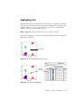
1
BD CellQuest Pro Software User’s Guide http://www.bdbiosciences.com/ Part No. 349226 Rev. A July 2002 BD Biosciences 2350 Qume Drive San Jose, CA 95131-1807 USA Tel (877) 232-8995 Fax (408) 954-2347 Asia Pacific Tel (65) 6-861-0633 Fax (65) 6-860-1590 Brazil Tel (55) 11-5185-9995 Fax (55) 11-5185-9895 Canada Tel (888) 259-0187 (905) 542-8028 Fax (905) 542-9391 [email protected] Europe Tel (32) 53-720211 Fax (32) 53-720450 Japan Nippon Becton Dickinson Company, Ltd. Tel 0120-8555-90 Mexico Tel (52) 5-284-8281 Fax (52) 5-284-8288 © 2002, Becton, Dickinson and Company. All rights reserved. No part of this publication may be reproduced, transmitted, transcribed, stored in retrieval systems, or translated into any language or computer language, in any form or by any means: electronic, mechanical, magnetic, optical, chemical, manual, or otherwise, without prior written permission from BD Biosciences. The information in this guide is subject to change without notice. BD Biosciences reserves the right to change its products and services at any time to incorporate the latest technological developments. Although this guide has been prepared with every precaution to ensure accuracy, BD Biosciences assumes no liability for any errors or omissions, nor for any damages resulting from the application or use of this information. BD Biosciences welcomes customer input on corrections and suggestions for improvement. BD CellQuest Pro software © 2002, Becton, Dickinson and Company. This software is the property of Becton, Dickinson and Company. Each sale of a stored unit of this software grants the purchaser a nontransferable, nonexclusive, personal license. This software may not be duplicated, reproduced, or copied in any form or by any means whatsoever, except as otherwise permitted by law. This program was written with MacApp®, © Apple® Computer, Inc. The MacApp software is proprietary to Apple Computer, Inc. and is licensed to BD Biosciences for distribution only for use in combination with this software. Apple Computer, Inc. makes no warranties whatsoever, either expressed or implied, regarding this product, including warranties with respect to its merchantability or its fitness for any particular purpose. BD, the BD logo, BD CaliBRITE, BD CellQuest, BD CloneCyt, BD FACS, BD FACSCalibur, BD FACScan, BD FACSComp, BD FACSConvert, BD FACSDiVa, BD FACSort, BD FACStation, BD FACSVantage, BD MultiSET, BD ProCOUNT, BD QuantiBRITE, and BD SimulSET are trademarks of Becton, Dickinson and Company. Apple, Mac, Macintosh, and Power Macintosh are trademarks of Apple Computer, Inc., registered in the US and other countries. Finder is a trademark of Apple Computer, Inc. Adobe and Acrobat are registered trademarks of Adobe Systems Incorporated. All other company and product names might be trademarks of the respective companies with which they are associated. Guide updated by Linda Jackson; edited by Kim Gautho; produced by Pushpa MacFarlane. History Revision Date Change Made A 11/00 New software features 349226 8/02 Revised for Mac OS X Contents About This Guide ix Conventions . . . . . . . . . . . . . . . . . . . . . . . . . . . . . . . . . . . . . . . . . . . . . . . . . x Technical Assistance . . . . . . . . . . . . . . . . . . . . . . . . . . . . . . . . . . . . . . . . . . . xi Limitations . . . . . . . . . . . . . . . . . . . . . . . . . . . . . . . . . . . . . . . . . . . . . . . . . . xi Chapter 1: Introduction 13 Requirements . . . . . . . . . . . . . . . . . . . . . . . . . . . . . . . . . . . . . . . . . . . . . . . . 14 Hardware . . . . . . . . . . . . . . . . . . . . . . . . . . . . . . . . . . . . . . . . . . . . . . . . 14 Software . . . . . . . . . . . . . . . . . . . . . . . . . . . . . . . . . . . . . . . . . . . . . . . . . 15 Compatibility . . . . . . . . . . . . . . . . . . . . . . . . . . . . . . . . . . . . . . . . . . . . . . . . 15 Data Files . . . . . . . . . . . . . . . . . . . . . . . . . . . . . . . . . . . . . . . . . . . . . . . . 15 Other Software . . . . . . . . . . . . . . . . . . . . . . . . . . . . . . . . . . . . . . . . . . . . 16 Installing BD CellQuest Pro . . . . . . . . . . . . . . . . . . . . . . . . . . . . . . . . . . . . . 16 Installing BD Inits . . . . . . . . . . . . . . . . . . . . . . . . . . . . . . . . . . . . . . . . . 18 Launching BD CellQuest Pro Software . . . . . . . . . . . . . . . . . . . . . . . . . . . . . 20 Registering the Software . . . . . . . . . . . . . . . . . . . . . . . . . . . . . . . . . . . . . 21 Quitting BD CellQuest Pro Software . . . . . . . . . . . . . . . . . . . . . . . . . . . . . . 21 Chapter 2: Becoming Familiar with BD CellQuest Pro Software 23 BD CellQuest Pro Files . . . . . . . . . . . . . . . . . . . . . . . . . . . . . . . . . . . . . . . . . 24 Default File Naming . . . . . . . . . . . . . . . . . . . . . . . . . . . . . . . . . . . . . . . . 25 Experiment Document Components . . . . . . . . . . . . . . . . . . . . . . . . . . . . . . . 26 Tool Palette . . . . . . . . . . . . . . . . . . . . . . . . . . . . . . . . . . . . . . . . . . . . . . 27 View Area Objects . . . . . . . . . . . . . . . . . . . . . . . . . . . . . . . . . . . . . . . . . 30 iii Inspector . . . . . . . . . . . . . . . . . . . . . . . . . . . . . . . . . . . . . . . . . . . . . . . . . . . . 31 Changing Text Style and Color . . . . . . . . . . . . . . . . . . . . . . . . . . . . . . . . 32 Changing Object Geometry . . . . . . . . . . . . . . . . . . . . . . . . . . . . . . . . . . 34 Customizing Experiment Documents . . . . . . . . . . . . . . . . . . . . . . . . . . . . . . 35 Document Size . . . . . . . . . . . . . . . . . . . . . . . . . . . . . . . . . . . . . . . . . . . . 35 Text . . . . . . . . . . . . . . . . . . . . . . . . . . . . . . . . . . . . . . . . . . . . . . . . . . . . 37 Text Settings . . . . . . . . . . . . . . . . . . . . . . . . . . . . . . . . . . . . . . . . . . . . . . 40 Color Palette . . . . . . . . . . . . . . . . . . . . . . . . . . . . . . . . . . . . . . . . . . . . . . 41 Banners . . . . . . . . . . . . . . . . . . . . . . . . . . . . . . . . . . . . . . . . . . . . . . . . . . 44 Grids . . . . . . . . . . . . . . . . . . . . . . . . . . . . . . . . . . . . . . . . . . . . . . . . . . . . 45 Chapter 3: BD CellQuest Pro Plots iv 49 Plot Types . . . . . . . . . . . . . . . . . . . . . . . . . . . . . . . . . . . . . . . . . . . . . . . . . . . 50 Creating Plots . . . . . . . . . . . . . . . . . . . . . . . . . . . . . . . . . . . . . . . . . . . . . . . . 52 Deleting Plots . . . . . . . . . . . . . . . . . . . . . . . . . . . . . . . . . . . . . . . . . . . . . 55 Formatting Plots . . . . . . . . . . . . . . . . . . . . . . . . . . . . . . . . . . . . . . . . . . . . . . 56 Dot Plots . . . . . . . . . . . . . . . . . . . . . . . . . . . . . . . . . . . . . . . . . . . . . . . . . 56 Contour Plots . . . . . . . . . . . . . . . . . . . . . . . . . . . . . . . . . . . . . . . . . . . . . 58 Density Plots . . . . . . . . . . . . . . . . . . . . . . . . . . . . . . . . . . . . . . . . . . . . . . 60 3D Plots . . . . . . . . . . . . . . . . . . . . . . . . . . . . . . . . . . . . . . . . . . . . . . . . . 63 Plot Parameters . . . . . . . . . . . . . . . . . . . . . . . . . . . . . . . . . . . . . . . . . . . . 64 Formatting Histograms . . . . . . . . . . . . . . . . . . . . . . . . . . . . . . . . . . . . . . . . . 65 Creating Overlaid Histograms . . . . . . . . . . . . . . . . . . . . . . . . . . . . . . . . 66 Using Histogram Tools . . . . . . . . . . . . . . . . . . . . . . . . . . . . . . . . . . . . . . 71 Saving Experiment Documents . . . . . . . . . . . . . . . . . . . . . . . . . . . . . . . . . . . 73 Saving Experiment Documents as Templates . . . . . . . . . . . . . . . . . . . . . 73 Saving Data . . . . . . . . . . . . . . . . . . . . . . . . . . . . . . . . . . . . . . . . . . . . . . 75 BD CellQuest Pro Software User’s Guide Chapter 4: Optimizing Instrument Electronics 77 Performing Instrument Quality Control . . . . . . . . . . . . . . . . . . . . . . . . . . . . 78 Instrument Controls in BD CellQuest Pro . . . . . . . . . . . . . . . . . . . . . . . . . . . 79 Adjusting Instrument Controls . . . . . . . . . . . . . . . . . . . . . . . . . . . . . . . . 79 Adjusting Detectors . . . . . . . . . . . . . . . . . . . . . . . . . . . . . . . . . . . . . . . . 80 Adjusting Amplifiers . . . . . . . . . . . . . . . . . . . . . . . . . . . . . . . . . . . . . . . 82 Adjusting Threshold . . . . . . . . . . . . . . . . . . . . . . . . . . . . . . . . . . . . . . . . 83 Adjusting Compensation . . . . . . . . . . . . . . . . . . . . . . . . . . . . . . . . . . . . 84 Optimizing the Instrument for Biological Samples . . . . . . . . . . . . . . . . . . . . 85 Starting Up the Cytometer . . . . . . . . . . . . . . . . . . . . . . . . . . . . . . . . . . . 86 Setting Up an Acquisition Experiment Document . . . . . . . . . . . . . . . . . . 86 Restoring Calib Settings to the Cytometer . . . . . . . . . . . . . . . . . . . . . . . 88 Adjusting FSC, SSC, and FSC Threshold . . . . . . . . . . . . . . . . . . . . . . . . 90 Gating on the Population of Interest . . . . . . . . . . . . . . . . . . . . . . . . . . . 91 Adjusting Fluorescence Detectors . . . . . . . . . . . . . . . . . . . . . . . . . . . . . . 92 Adjusting Fluorescence Compensation . . . . . . . . . . . . . . . . . . . . . . . . . . 96 Instrument Settings . . . . . . . . . . . . . . . . . . . . . . . . . . . . . . . . . . . . . . . . . . . . 99 Saving Instrument Settings . . . . . . . . . . . . . . . . . . . . . . . . . . . . . . . . . . . 99 Restoring Instrument Settings . . . . . . . . . . . . . . . . . . . . . . . . . . . . . . . . . 100 Printing Instrument Settings . . . . . . . . . . . . . . . . . . . . . . . . . . . . . . . . . . 101 Checking Cytometer Status . . . . . . . . . . . . . . . . . . . . . . . . . . . . . . . . . . . . . . 101 BDPAC Application . . . . . . . . . . . . . . . . . . . . . . . . . . . . . . . . . . . . . . . . 102 Status Window . . . . . . . . . . . . . . . . . . . . . . . . . . . . . . . . . . . . . . . . . . . . 103 Chapter 5: Acquiring Data 105 Using the Acquisition View of the Browser . . . . . . . . . . . . . . . . . . . . . . . . . . 106 Using Acquisition Controls . . . . . . . . . . . . . . . . . . . . . . . . . . . . . . . . . . 109 Getting Ready to Acquire . . . . . . . . . . . . . . . . . . . . . . . . . . . . . . . . . . . . . . . 110 Defining Global Acquisition and Storage Settings . . . . . . . . . . . . . . . . . 111 Selecting Parameters to Save . . . . . . . . . . . . . . . . . . . . . . . . . . . . . . . . . . 116 Defining File Name and Storage Location . . . . . . . . . . . . . . . . . . . . . . . 118 Defining Parameter Labels . . . . . . . . . . . . . . . . . . . . . . . . . . . . . . . . . . . 120 Contents v Setting Up an Acquisition Tube List . . . . . . . . . . . . . . . . . . . . . . . . . . . . . . . 120 Editing Tubes Using the Inspector . . . . . . . . . . . . . . . . . . . . . . . . . . . . . . 122 Creating Plots and Associating With Tubes . . . . . . . . . . . . . . . . . . . . . . 123 Defining Tube-Specific Settings . . . . . . . . . . . . . . . . . . . . . . . . . . . . . . . . 124 Using Counters . . . . . . . . . . . . . . . . . . . . . . . . . . . . . . . . . . . . . . . . . . . . . . . 127 Displaying Live Statistics . . . . . . . . . . . . . . . . . . . . . . . . . . . . . . . . . . . . . . . . 128 Acquiring Data . . . . . . . . . . . . . . . . . . . . . . . . . . . . . . . . . . . . . . . . . . . . . . . 129 Monitoring Acquisition in the Browser . . . . . . . . . . . . . . . . . . . . . . . . . . 131 Pausing Acquisition . . . . . . . . . . . . . . . . . . . . . . . . . . . . . . . . . . . . . . . . 132 Ending Acquisition . . . . . . . . . . . . . . . . . . . . . . . . . . . . . . . . . . . . . . . . . 133 Using Sorting Controls . . . . . . . . . . . . . . . . . . . . . . . . . . . . . . . . . . . . . . . . . 133 Optional Acquisition Features . . . . . . . . . . . . . . . . . . . . . . . . . . . . . . . . . . . . 134 Using the Reagent List . . . . . . . . . . . . . . . . . . . . . . . . . . . . . . . . . . . . . . 134 Creating Panels . . . . . . . . . . . . . . . . . . . . . . . . . . . . . . . . . . . . . . . . . . . . 135 Defining Custom Keywords . . . . . . . . . . . . . . . . . . . . . . . . . . . . . . . . . . 138 Chapter 6: Regions and Gates vi 141 Creating Regions . . . . . . . . . . . . . . . . . . . . . . . . . . . . . . . . . . . . . . . . . . . . . . 142 Histogram Regions . . . . . . . . . . . . . . . . . . . . . . . . . . . . . . . . . . . . . . . . . 142 Rectangular, Elliptical, and Polygonal Regions . . . . . . . . . . . . . . . . . . . . 143 Snap-To Regions . . . . . . . . . . . . . . . . . . . . . . . . . . . . . . . . . . . . . . . . . . . 146 Editing Regions . . . . . . . . . . . . . . . . . . . . . . . . . . . . . . . . . . . . . . . . . . . . . . . 152 Rotating Regions . . . . . . . . . . . . . . . . . . . . . . . . . . . . . . . . . . . . . . . . . . 153 Deleting or Hiding Regions . . . . . . . . . . . . . . . . . . . . . . . . . . . . . . . . . . . 154 Using the Region List . . . . . . . . . . . . . . . . . . . . . . . . . . . . . . . . . . . . . . . . . . 156 Copying Regions . . . . . . . . . . . . . . . . . . . . . . . . . . . . . . . . . . . . . . . . . . . 158 Defining Gates . . . . . . . . . . . . . . . . . . . . . . . . . . . . . . . . . . . . . . . . . . . . . . . 160 Using Combined Regions in Gate Definitions . . . . . . . . . . . . . . . . . . . . . 160 Using the Gate List . . . . . . . . . . . . . . . . . . . . . . . . . . . . . . . . . . . . . . . . . 161 Using Multicolor Gates for Dot Plots . . . . . . . . . . . . . . . . . . . . . . . . . . . . . . 163 Setting Up a Gate List for Multicolor Gating . . . . . . . . . . . . . . . . . . . . . 164 Highlighting Dots . . . . . . . . . . . . . . . . . . . . . . . . . . . . . . . . . . . . . . . . . . 165 BD CellQuest Pro Software User’s Guide Chapter 7: Statistics 167 Calculating Data . . . . . . . . . . . . . . . . . . . . . . . . . . . . . . . . . . . . . . . . . . . . . . 168 Automatic vs Manual Calculation . . . . . . . . . . . . . . . . . . . . . . . . . . . . . 168 Selecting Log Data Units . . . . . . . . . . . . . . . . . . . . . . . . . . . . . . . . . . . . 170 Calculating Percent Gated and Percent Total Events . . . . . . . . . . . . . . . 171 Calculating Means . . . . . . . . . . . . . . . . . . . . . . . . . . . . . . . . . . . . . . . . . 172 Using Histogram Markers . . . . . . . . . . . . . . . . . . . . . . . . . . . . . . . . . . . . . . . 173 Creating Histogram Markers . . . . . . . . . . . . . . . . . . . . . . . . . . . . . . . . . 173 Editing Histogram Markers . . . . . . . . . . . . . . . . . . . . . . . . . . . . . . . . . . 173 Displaying a Marker List . . . . . . . . . . . . . . . . . . . . . . . . . . . . . . . . . . . . 174 Copying Markers . . . . . . . . . . . . . . . . . . . . . . . . . . . . . . . . . . . . . . . . . . 175 Converting Markers to Regions . . . . . . . . . . . . . . . . . . . . . . . . . . . . . . . 176 Displaying Histogram Statistics . . . . . . . . . . . . . . . . . . . . . . . . . . . . . . . . . . 177 Generating Overlaid Histogram Statistics . . . . . . . . . . . . . . . . . . . . . . . . . . . 178 Calculating Percentages From Overlaid Histograms . . . . . . . . . . . . . . . . 178 Calculating Kolmogorov-Smirnov (K-S) Statistics . . . . . . . . . . . . . . . . . 180 Creating Quadrant Markers . . . . . . . . . . . . . . . . . . . . . . . . . . . . . . . . . . . . . 182 Editing Quadrant Markers . . . . . . . . . . . . . . . . . . . . . . . . . . . . . . . . . . . 183 Displaying Statistics . . . . . . . . . . . . . . . . . . . . . . . . . . . . . . . . . . . . . . . . . . . 184 Editing Statistics . . . . . . . . . . . . . . . . . . . . . . . . . . . . . . . . . . . . . . . . . . . . . . 186 Formatting Statistics Views . . . . . . . . . . . . . . . . . . . . . . . . . . . . . . . . . . 192 Using the Expression Editor . . . . . . . . . . . . . . . . . . . . . . . . . . . . . . . . . . . . . 192 Editing Expressions . . . . . . . . . . . . . . . . . . . . . . . . . . . . . . . . . . . . . . . . 195 Chapter 8: Analyzing Data 197 Using the Analysis Browser . . . . . . . . . . . . . . . . . . . . . . . . . . . . . . . . . . . . . . 198 Analyzing Data . . . . . . . . . . . . . . . . . . . . . . . . . . . . . . . . . . . . . . . . . . . . . . . 200 Setting Up an Analysis Experiment Document . . . . . . . . . . . . . . . . . . . . 201 Displaying Statistics . . . . . . . . . . . . . . . . . . . . . . . . . . . . . . . . . . . . . . . . 204 Using the Next Data File Command . . . . . . . . . . . . . . . . . . . . . . . . . . . . 206 Contents vii Analyzing Groups of Data Files . . . . . . . . . . . . . . . . . . . . . . . . . . . . . . . . . . . 211 Using the Load Sample Feature . . . . . . . . . . . . . . . . . . . . . . . . . . . . . . . . 211 Setting Up Batch Analysis . . . . . . . . . . . . . . . . . . . . . . . . . . . . . . . . . . . . 216 Saving FCS Files . . . . . . . . . . . . . . . . . . . . . . . . . . . . . . . . . . . . . . . . . . . . . . 219 Exporting Statistics . . . . . . . . . . . . . . . . . . . . . . . . . . . . . . . . . . . . . . . . . . . . 220 Using Quantitative Calibration . . . . . . . . . . . . . . . . . . . . . . . . . . . . . . . . . . . 221 Setting Up for Quantitative Calibration . . . . . . . . . . . . . . . . . . . . . . . . . 222 Chapter 9: Publishing and Presenting Data 229 Exporting Plots . . . . . . . . . . . . . . . . . . . . . . . . . . . . . . . . . . . . . . . . . . . . . . . 230 Helpful Hints for Publication-Quality Graphics . . . . . . . . . . . . . . . . . . . 231 Saving Experiment Documents as PDFs . . . . . . . . . . . . . . . . . . . . . . . . . . . . 232 Appendix A: Menus and Keyboard Shortcuts 233 Menus . . . . . . . . . . . . . . . . . . . . . . . . . . . . . . . . . . . . . . . . . . . . . . . . . . . . . . 234 Keyboard Shortcuts . . . . . . . . . . . . . . . . . . . . . . . . . . . . . . . . . . . . . . . . . . . . 236 Appendix B: Using Snap-To-Regions 239 Becoming Familiar With Snap-To Regions . . . . . . . . . . . . . . . . . . . . . . . . . . 240 Tethering Snap-To Regions . . . . . . . . . . . . . . . . . . . . . . . . . . . . . . . . . . . . . . 244 Glossary 247 Index 251 viii BD CellQuest Pro Software User’s Guide About This Guide This user’s guide describes the setup and operation of BD CellQuest™ Pro software. This guide is intended for laboratory personnel and flow cytometry operators experienced in flow cytometry instrument operation. For information on using the BD FACS™ Loader or other instrument components, refer to the appropriate user’s guide. The BD CellQuest Pro Software User’s Guide assumes you have a working knowledge of the Mac® OS X operating system. If you are not familiar with this system, refer to the documentation provided by Apple Computer, Inc. Before using BD CellQuest Pro software, review the ReadMe file that was copied to your hard disk during software installation. The file contains late-breaking information that is not printed in this user’s guide. First-time users of BD CellQuest Pro software should read • Chapter 1 to learn about compatibility and installation • Chapters 2 and 3 to learn about BD CellQuest Pro components • Chapters 6 and 7 to learn about analysis tools like regions, gates, and statistics You will find instructions for routine acquisition and analysis in Chapter 4, Chapter 5, and Chapter 8. For tips on publishing and presenting data, see Chapter 9. Appendix A has a useful list of keyboard shortcuts. ix Conventions The following tables list conventions used throughout this guide. Table 1 Notice icons Icon Notice Type Use NOTE Describes important features or instructions CAUTION Alerts you to potential loss of data or potential damage to an application, system, or device Ꮬ WARNING Alerts you to potential personal injury ! Tip Highlights features or hints that can save time and prevent difficulties Table 2 Text conventions x Convention Use Italics Italics are used to highlight book titles and new or unfamiliar terms on their first appearance in the text. A glossary of terms is listed at the end of this manual. > The arrow indicates a menu choice. For example, “choose File > Print” means to choose Print from the File menu. Command Keyboard shortcuts use the Command key (") in combination with another indicated keystroke. For example, Command-K means to hold down the Command key while pressing the letter k. BD CellQuest Pro Software User’s Guide Technical Assistance For technical questions or assistance in solving a problem, read the section of the user’s guide specific to the operation you are performing. If additional assistance is required, contact your local BD Biosciences technical support representative or supplier. When contacting BD Biosciences, have the following information available: • product name, part number, and serial number • any error messages • details of recent system performance For instrument support from within the US, call (877) 232-8995, prompt #2-2. For support from within Canada, call (888) 259-0187. Customers outside the US and Canada, contact your local BD representative or distributor. Limitations For in vitro diagnostic (IVD) use when used with IVD reagents. Refer to the appropriate reagent package insert for application-specific limitations. About This Guide xi xii BD CellQuest Pro Software User’s Guide 1 Introduction BD CellQuest Pro software allows you to acquire and analyze data from your flow cytometer on a Macintosh® computer. Working in the BD CellQuest Pro Experiment document window, you can create several types of plots, including multicolor contour plots and overlaid histograms, and you can generate statistics for dot plots, histograms, density plots, 3D plots, and contour plots. The versatile graphics tool palette helps you produce presentation-quality documents. You can also save everything in an Experiment document, including plots, regions, gates, markers, statistics, calculated expressions, Browser contents, annotations, and color preferences, and then restore it all later. The following topics are covered in this chapter: • Requirements on page 14 • Compatibility on page 15 • Installing BD CellQuest Pro on page 16 • Launching BD CellQuest Pro Software on page 20 • Quitting BD CellQuest Pro Software on page 21 13 Requirements Hardware • BD FACSCalibur™, BD FACScan™, BD FACSort™, or BD™ LSR flow cytometer; BD FACSVantage™ family of cytometers (BD FACSVantage, BD FACSVantage SE, or BD FACSVantage SE with the BD FACSDiVa option) • BD FACStation™ Power Macintosh® G4 computer purchased through BD Biosciences Other platforms might also be supported; contact your BD representative for more information. CAUTION If you have a modem on your system, BD recommends turning it off while running our software. • Universal Serial Bus (USB) security module The security module is provided with the software. 14 • (optional) BD FACS Loader for automated acquisition from tubes; BD™ WorklistManager software version 4.0 or later is required. • (optional) BD™ Multiwell AutoSampler for automated acquisition from plates; BD™ Multiwell Plate Manager software version 4.0 or later is required. BD CellQuest Pro Software User’s Guide Software • Mac OS X.1 or later BD recommends turning off file sharing and program linking. • BD Inits version 4.0 or later (see Installing BD Inits on page 18) NOTE BD Biosciences has tested many common INITs (eg, extensions, control panels) that do not adversely affect software performance; however, some INITs might interfere with the software. Therefore, BD Biosciences does not recommend installing additional INITs. • For cytometers equipped with the BD FACS Loader, BD WorklistManager software version 4.0 or later • For cytometers equipped with the BD Multiwell AutoSampler, BD Multiwell Plate Manager software version 4.0 or later Compatibility Data Files BD CellQuest Pro software reads and writes FCS 2.0 data files. See Other Software on page 16. BD CellQuest Pro software can analyze data files produced by BD CellQuest, BD FACSDiVa™, BD SimulSET™, BD MultiSET™, BD ProCOUNT™, or any other BD software. FCS 2.0 data files from the PC (ie, from the BD FACSDiVa option) must be formatted using BD FACSConvert™ software. Refer to the BD FACSConvert User’s Guide for instructions. Chapter 1: Introduction 15 Other Software BD CellQuest • BD CellQuest Pro software can open and read data files and Experiment documents created with BD CellQuest software version 3.3 or later. BD CellQuest Pro Experiment documents cannot be read by BD CellQuest software. • Preferences from BD CellQuest software version 3.3 or later are not deleted or overwritten by BD CellQuest Pro software. BD CellQuest Pro preferences will retain older version BD CellQuest preferences. BD CloneCyt™ Plus For single-cell sorting, BD CellQuest Pro, in conjunction with BD CloneCyt Plus software and a BD FACSVantage SE flow cytometer, helps identify a single cell’s position on a plot when the corresponding clone number is known. BD CellQuest Pro software is compatible with BD CloneCyt Plus software version 4.0 or later. BD WorklistManager BD CellQuest Pro Experiment documents can serve as assay templates in BD WorklistManager software. BD CellQuest Pro software is compatible with BD WorklistManager software version 4.0 or later. Adobe® Acrobat® software BD CellQuest Pro Experiment documents can be saved as Adobe PDF (portable document format) files that can be viewed on any computer using Adobe Acrobat Reader software. Installing BD CellQuest Pro Follow these steps to install BD CellQuest Pro software on your computer’s hard disk. 1 Shut down the computer. 2 Plug the security module into the USB hub. The security module is packaged with this user’s guide. BD CellQuest Pro software will not launch if the security module is not attached. 16 BD CellQuest Pro Software User’s Guide 3 Restart the computer. 4 Insert the BD FACStation Basic Software CD into the CD-ROM drive. 5 Double-click the CD icon. A window appears showing the contents of the CD. The BD FACStation Basic Software disc includes installers for several BD applications and corresponding electronic documentation. Review the BD FACStation ReadMe file for more information. 6 Double-click the BD CellQuest Pro installer icon. 7 Follow the instructions in the dialog to install the software. The installer loads BD CellQuest Pro software and its support files on the hard disk. A message appears when installation is complete. 8 Click Quit when you see a message reporting a successful installation. 9 (Optional) Restart the computer. BD recommends that you restart your computer in order to detect the Eve Init the installer places in the System/Library/Frameworks/ Eve3.Framework folder. This Init is required to run BD CellQuest Pro software on the G4 Power Macintosh computer. If you will be performing additional installations, restart the computer after all installations are complete. The software installer creates two folders on the hard disk: the BD Applications folder and the BD Files folder. If another BD application was installed previously, these folders already exist and the installer adds BD CellQuest Pro files to these folders. The CellQuest Pro folder, located in the BD Applications folder, contains the BD CellQuest Pro application, a copy of the ReadMe file, and a Sample Files folder. The CellQuest Files folder, located in the BD Files folder, is the default storage location for Experiment documents. See Figure 1-1 on page 18. Chapter 1: Introduction 17 Figure 1-1 BD Applications and BD Files folder contents Installing BD Inits All BD applications that acquire data from a flow cytometer require BD startup files, or BD Inits, that extend the capabilities of the computer. These files are already installed in all BD FACStation computer systems. However, if it is necessary to reinstall or update BD Inits, follow these instructions. NOTE You will need to update the BDPAC configuration information if you connect your computer to another cytometer or if you upgrade your cytometer with new options. Verify the configuration information after you reinstall the software. You must quit BD CellQuest Pro software to reconfigure BDPAC. 1 Insert the BD FACStation Basic Software CD into the CD-ROM drive. 2 Double-click the CD icon. 18 BD CellQuest Pro Software User’s Guide 3 Double-click the Install BD Inits icon. 4 Follow the instructions that appear on the screen. 5 Click Restart when installation is complete. The installer automatically places the following BD Init files into the indicated folders: • BDPAC and BDPAC Startup Check into the BD Applications folder • BDAcquisitionServices into the Library > CFMSupport folder • Com_bd_bdpac.kext into the System > Library > Extensions folder 6 Launch the BDPAC application. Locate the BDPAC application in the BD Applications folder, and double-click the icon. icon 7 Enter the cytometer serial number, if needed, and choose the cytometer type from the drop-down menu. 8 Select the appropriate checkboxes for your cytometer setup; click OK. Chapter 1: Introduction 19 Launching BD CellQuest Pro Software If you are using BD CellQuest Pro software to acquire samples from the cytometer, make sure the cytometer is turned on and connected to the computer through the GPIO interface cable. CAUTION Turn on the cytometer before you turn on the computer to correctly establish the communication link. NOTE Before launching the software, verify that the security module is attached. BD CellQuest Pro software will not start without the security module attached. Launch BD CellQuest Pro software in one of the following ways: • Navigate to the BD Applications > CellQuest Pro Folder > CellQuest Pro icon in the Finder window (Figure 1-1 on page 18). Double-click the icon. The software opens with a new, untitled Experiment document. ! Tip After launching the application, press the BD CellQuest Pro icon in the Dock and choose Keep In Dock. (You can also drag the icon from the Finder window into the Dock.) You can then open the application by clicking its icon in the Dock. BD CellQuest Pro icon • Double-click the icon for a saved BD CellQuest Pro Experiment document. The software opens to the saved Experiment document. Experiment document icon • 20 Drag one or more Experiment document icons onto the BD CellQuest Pro application icon. This launches BD CellQuest Pro software and allows you to open several Experiment documents at one time. BD CellQuest Pro Software User’s Guide Registering the Software The first time you launch BD CellQuest Pro software, a registration dialog appears. 1 Enter your name and the name of your institution. 2 Click Save or press the Return key. Quitting BD CellQuest Pro Software Do either of the following to quit the software: • Choose Quit from the File menu. • Hold down the Command key while pressing the Q key (Command-Q). Chapter 1: Introduction 21 22 BD CellQuest Pro Software User’s Guide 2 Becoming Familiar with BD CellQuest Pro Software The following topics are covered in this chapter: • BD CellQuest Pro Files on page 24 • Experiment Document Components on page 26 • Inspector on page 31 • Customizing Experiment Documents on page 35 23 BD CellQuest Pro Files This section describes the several types of files BD CellQuest Pro software can create: Experiment document files, FCS data files, statistical files for export, and instrument settings files. See Default File Naming on page 25 for a summary of default file-naming conventions and storage locations. Experiment Document Files A BD CellQuest Pro Experiment document is a document that can contain user-defined plots, regions, gates, markers, statistics, tube lists in the Browser, calculated expressions, text annotations, and color preferences. When saving an Experiment document, BD CellQuest Pro software saves a reference to the data files used in the Experiment document, not the data files themselves. See Experiment Document Components on page 26 for general information about Experiment documents; see Saving Experiment Documents on page 73 for information about saving Experiment documents. Data Files Flow cytometric data is stored according to a standard format, the Flow Cytometry Standard (FCS) format, developed by the International Society for Analytical Cytology.a After acquisition, the parameter measurements for each event and the instrument settings of the cytometer are automatically saved in an FCS data file. See Saving Data on page 75 for more information about data files. a. For a detailed description of FCS 2.0, refer to Data file standard for flow cytometry. Data File Standards Committee of the International Society for Analytical Cytology. Cytometry. 1990;11(3):323-332. 24 BD CellQuest Pro Software User’s Guide Statistics Files Any combination of histogram, region, gate, and quadrant statistics can be exported as tab-delimited text to a file, with the exception of statistics from overlaid histograms and the Kolmogorov-Smirnov (K-S) two-sample test. These export files can be used to transfer data to other applications, such as spreadsheet programs or databases. See Exporting Statistics on page 220 for more information. Instrument Settings Files An instrument settings file contains detectors/amps settings, the threshold parameter and value, compensation settings, pulse processing settings (if applicable to your cytometer), and special setup values (if applicable to your cytometer). These settings are saved automatically in the text section of FCS files; they can also be saved in a separate instrument settings file. See Saving Instrument Settings on page 99 for information about saving and restoring instrument settings. Default File Naming Table 2-1 summarizes the file naming conventions for the different types of BD CellQuest Pro files. Table 2-1 BD CellQuest Pro file names File Type File Name Experiment document Untitledn (The software adds a number to each subsequent file) FCS data file Data.nnn Export statistics untitled stats Instrument settings INSTRSettings Chapter 2: Becoming Familiar with BD CellQuest Pro Software 25 Experiment Document Components When you start BD CellQuest Pro software, an Experiment document window appears. The BD CellQuest Pro Experiment document is where you create plots for acquisition and analysis, show statistics, create calculated expressions, enter text, and customize preferences. Software functions are controlled using the tool palette and the menu bar above the window. Other Experiment document window elements include the ribbon, view area, resize control, and window controls. menu bar window controls ribbon tool palette view area resize control Figure 2-1 BD CellQuest Pro Experiment document window • menu bar—displays BD CellQuest Pro pull-down menus with the commands you will need to operate the software For a complete description of the menus, see Menus and Keyboard Shortcuts on page 233. • ribbon—shows the cursor coordinates in a plot It is often helpful to know the cursor coordinates when you create markers or regions. 26 BD CellQuest Pro Software User’s Guide When the cursor is on a dot, density, or contour plot, the ribbon displays the channel number or linear value of the x-axis (X) and the y-axis (Y). When the cursor is on a histogram, the ribbon displays the channel number or linear value of the x-axis (X) and the event count (Y). For overlaid histograms, the Y value is blank. • tool palette—appears when you open an Experiment document and contains tools for creating plots, regions, markers, and text For a complete description, see the following section. • view area—displays all Experiment document elements you create; for a complete description, see View Area Objects on page 30. • window controls—close (red), minimize (yellow), enlarge or reduce (green) the window when you click the corresponding button • resize control—resizes the window when you drag the control Tool Palette The tool palette appears when you launch BD CellQuest Pro software. Use the tools in the palette to create plots, regions, markers, text, and calculated expressions. • To select a tool, click once to highlight it. • To move the tool palette to a different location, click the shaded bar at the top of the palette and drag to the new location. • To remove the tool palette from the desktop, click the close button in the upper-left corner of the palette, or choose Hide Palette from the Windows menu. To make the palette reappear, choose Show Palette from the Windows menu. Chapter 2: Becoming Familiar with BD CellQuest Pro Software 27 Here is a summary of the tool functions. Selection tool—selects (by clicking), moves (by dragging), or resizes (by dragging) items in the Experiment document. C 3D H D d H Q Plot tools—create contour (C), 3-dimensional (3D), density (D), histogram (H), and dot (d) plots. Select the tool, click in an unused section of the Experiment document, and drag diagonally until the plot outline is the proper size. For more information, see Creating Plots on page 52. Marker tools—The Histogram-Marker tool (H) creates a marker on a histogram plot allowing you to obtain statistics on the data. The Quadrant-Marker tool (Q) draws a quadrant on a dot, density, or contour plot. Select the tool, click in the plot, and drag to set the marker. See Using Histogram Markers on page 173 and Creating Quadrant Markers on page 182 for more information on setting markers. Zoom tools—The Zoom-In tool (+) magnifies an area of a plot. Select the tool, click in the plot, and drag to define the area of the plot you want to enlarge. The Zoom-Out tool (–) returns the plot to its original size. Select the tool and click in the plot. R P H E S Region tools—create regions on specific plots. • The Rectangular-Region tool (R) creates a rectangular region on a dot, density, or contour plot. Select the tool, click in the plot, and drag diagonally until the region outline is the proper size. • The Polygonal-Region tool (P) creates a polygonal region on a dot, density, or contour plot. Select the tool and click in the plot to establish the starting point (first vertex). Move the cursor to create the next vertex and click. Continue moving the cursor and setting vertices; click the first vertex to complete the region. 28 BD CellQuest Pro Software User’s Guide • The Histogram-Region tool (H) creates a histogram region on a histogram plot. These regions are created just like histogram markers. However, histogram regions are used for gates; markers are used for statistics. • The Elliptical-Region tool (E) creates a circular or elliptical region on a dot, density, or contour plot. Select the tool, click in the plot, and drag diagonally until the region outline is the proper size. • The Snap-To–Region tool (S) draws a region around a distinct cluster on a dot, density, or contour plot. Select the tool and click the cluster on the plot. The region is drawn automatically. For more information, see Creating Regions on page 142. Text tool—creates or edits free text anywhere in the Experiment document. Select the tool and click to position the insertion point; type the text and click outside the text box to complete the entry. For information on adding free text, see page 39. For information on customizing the style of free text, see Changing Text Style and Color on page 32 or Text Settings on page 40. Arrow tool—draws arrows in the Experiment document. Select the tool, click in the view area and drag the line out. Arrows must be started in an empty area of the Experiment document but they can extend into objects. Expression Editor tool—opens a dialog where you can enter custom mathematical expressions (formulas). The results of your calculated expression appear on the Experiment document with a label you have assigned. For more information on calculated expressions, see Using the Expression Editor on page 192. Calculator tool—recalculates data. This tool is enabled only if Auto Recalculate (Gates menu) is turned off. Click the tool to recalculate data. For more information, see Automatic vs Manual Calculation on page 168. Chapter 2: Becoming Familiar with BD CellQuest Pro Software 29 View Area Objects Plots, statistics, expressions, and free text that appear in an Experiment document view area are collectively referred to as objects. Objects appear in three different states: active, selected, or deselected. • An active object has a light-blue border (Figure 2-2); it is the target of any action you take. Any commands you choose affect only the active object. For example, if you make a plot active and then choose Select All from the Edit menu, everything within that plot, such as markers and regions, becomes selected. On the other hand, if no objects are active and you choose Select All, all objects in the Experiment document become selected. To make an object active, click anywhere inside the object except on the frame. Only one object can be active at a time. frame Figure 2-2 Active object (plot) • A selected object has a black handle on each corner (Figure 2-3 on page 31); it has many properties of an active object. However, only a selected object can be inspected, deleted, resized, or moved. Only one object can be active at a time, but multiple objects can be selected. To select an object, click its frame. To select more than one object, click each frame while holding down the Shift key or click in the view area and drag the cursor diagonally to surround the appropriate objects. To select all objects, choose Select All from the Edit menu; make sure no objects are active before choosing Select All. 30 BD CellQuest Pro Software User’s Guide selection handles Figure 2-3 Selected object (plot) • A deselected object has a clear border (Figure 2-4). To deselect an active or selected object, click anywhere outside the object. Figure 2-4 Deselected object (plot) Inspector The BD CellQuest Pro Inspector provides an easy-to-use interface for viewing or modifying the attributes of a single object or set of objects in the Experiment document. For example, you can use the Inspector to change text and color settings, resize multiple objects to the same size, align multiple objects in an Experiment document, select or change the file displayed in an analysis plot, or enter or change parameter labels. Chapter 2: Becoming Familiar with BD CellQuest Pro Software 31 The type of Inspector varies depending on the objects selected in the view area. Common Inspector elements are discussed in the following section; for Annotation Inspectors, see Adding Free Text on page 39. For plot Inspectors, see Formatting Plots on page 56. For statistics Inspectors, see Formatting Statistics Views on page 192. Changing Text Style and Color You can change the text style of any selected object or set of objects in an Experiment document using the Text Style options in the Inspector. When a plot is selected, for example, text attributes apply to all text within the plot, including the plot title and axes labels. 1 Select one or more objects in the Experiment view area. Select more than one object by holding down the Shift key as you click individual objects or by clicking in the view area and holding down the mouse button as you drag across the view. ! Tip To select all objects, choose Edit > Select All (Command-A). 2 Choose Windows > Show Inspector (Command-I). The Inspector appears, indicating in the title bar the type of object or number of objects selected. The contents of the Inspector vary depending on the objects selected; however, any objects including text will show Text Style options within the Inspector window. Inspector contents—dot plot selected Inspector contents—multiple objects selected Inspector contents—nothing selected 32 BD CellQuest Pro Software User’s Guide NOTE The disclosure triangles are closed in this illustration. When the Inspector window appears on the screen, the triangles are open, revealing the options for each category. 3 Scroll down to the Text Style header line; if needed, click once on the disclosure triangle to display Text Style options. disclosure triangle to hide/show style options pop-up controls 4 Specify the text style. • Click once on the Font pop-up control to change the font; select a font from the menu that appears. All fonts installed on your computer are available. • Enter the appropriate font size in the Size entry field or click on the pop-up control and select a size from the menu that appears. • Specify the text style. You can select bold, italic, underlined, outlined, or shadowed. Select condense (C) to compress spacing between characters; select extend (E) to stretch out spacing. Remember that the selected style and justification applies to the text in all selected objects. • Specify the justification (alignment) for any free text or text annotations. Justification cannot be applied to the text in plots or statistics views. • Specify the text color. Click once on the white square and choose from the 24 available colors. You can change the available color selections using the Color Palette, as described in Color Palette on page 41. • Click on the Background pop-up control to choose the background for any free text or text annotation that overlaps an object. As shown in Figure 2-5, a Transparent background reveals overlapping items in the Chapter 2: Becoming Familiar with BD CellQuest Pro Software 33 text field; an Opaque background hides overlapping items in the text field. transparent background Figure 2-5 opaque background Text background options 5 Close the Inspector by clicking the close button or by choosing Hide Inspector from the Windows menu. Changing Object Geometry You can resize multiple objects to the same size or align multiple objects in an Experiment document using the Geometry feature. Location and size settings are in inches or centimeters. These settings are specified in the Grid Settings dialog, described in Grids on page 45. 1 Select one or more objects in the Experiment view area. For example, to make a series of plots all the same size, select all the plots in the Experiment view area. 2 Choose Windows > Show Inspector (Command-I). 3 Scroll down to the Geometry header line; if needed, click once on the disclosure triangle to display Geometry options. disclosure triangle to hide/show geometry options 34 BD CellQuest Pro Software User’s Guide 4 Enter a location and size of the selected objects. You can change unit settings from inches to centimeters in the Grid dialog (see Grids on page 45). If two or more items are selected, only attributes that are common to all the selected objects are displayed. • To make all selected objects the same size, enter a width and height in the Size boxes. • To align multiple objects horizontally, enter a distance from the top of the page; leave the Left distance blank. • To align multiple objects vertically, enter a distance from the left side of the page; leave the Top distance blank. Customizing Experiment Documents You can customize certain features of an Experiment document and save them with the document. These include the document size, the presence and location of text or annotations, the text and color settings for all objects in the view area, the appearance of a custom banner, and the presence or absence of grids. These features are described in this section. Document Size BD CellQuest Pro software sets the size of each new Experiment document to a 10.19 x 8-inch page. The view area is set to this page size. To increase the size of the view area, you can increase the Experiment document size up to a maximum of 20 pages. The actual number of pages needed to print an Experiment document is determined by the paper size selection and the reduction or enlargement percentage set in the Page Setup dialog. BD CellQuest Pro software recalculates the minimum number of pages needed to show all the information in a document when changes are made to Page Setup. Refer to your Macintosh documentation for information about setting up pages for printing. Chapter 2: Becoming Familiar with BD CellQuest Pro Software 35 1 Choose File > Document Size. 2 Click the rectangles to the right or below the current page selection to add additional pages (Figure 2-6). Figure 2-6 Document Size dialog Each rectangle represents one page. As you add pages, the corresponding height and width measurements are updated to the left of the grid. NOTE The number of pages selected appears in an uneditable field. BD CellQuest Pro software automatically adds pages to maintain a rectangular shape in the grid. The number of pages cannot be reduced if there is any overlapping information on a page you want to eliminate. 3 Specify the scale (inches/cm) and page sequence. The page sequence refers to the printing order. 4 Click OK when you are finished. The view area will increase to encompass the number of pages you have selected; page breaks are indicated by a dotted line. Use the scroll bar to navigate within the enlarged view area. NOTE Do not place Experiment document objects on the dotted line representing the page break. 36 BD CellQuest Pro Software User’s Guide Text There are two ways to add text to an Experiment document. The first method, annotating, allows you to extract and display text that was saved with the FCS data file. Annotations are linked to the data file and are updated when the data file displayed in a plot changes. The second method, adding free text, allows you to add any text you type or paste. You can place text and annotations anywhere in the Experiment document. Adding an Annotation Use the Annotation feature to display text from an FCS data file. If a linked data file cannot be found, the annotation changes to “***”; if a plot is deleted, its annotation is also deleted. 1 Select a plot. 2 Click on the Plots menu and hold the mouse button over Annotation to view Annotation options. A submenu with a list of annotation options appears (Figure 2-7). The available choices vary depending on which items were saved in the data file. Figure 2-7 Annotation submenu from Plots menu Chapter 2: Becoming Familiar with BD CellQuest Pro Software 37 3 Choose the required type of annotation. The annotation, drawn from the current data file, appears. When the annotation is selected, handles appear around it. selected annotation 4 Select the annotation if you want to edit it. You can change the text style or background of a selected annotation using the Inspector or the Text Settings dialog, but you cannot edit the annotation text. See Changing Text Style and Color on page 32 or Text Settings on page 40. 38 • To move a selected annotation to a different location, click and drag it. Annotations can be placed anywhere in the Experiment document. • To change the width of a selected annotation, drag a handle in or out. • To delete a selected annotation, press Delete or choose Cut or Clear from the Edit menu. • To change the type of annotation, choose Windows > Show Inspector (Command-I) and choose a different annotation from the Attribute pop-up menu. BD CellQuest Pro Software User’s Guide Adding Free Text 1 Select the Text tool from the tool palette ( ). 2 Click on the Experiment document to position the insertion point. You can place free text anywhere in the Experiment document. A blinking vertical bar appears at the insertion point. 3 Enter the text you want. The text wraps to the next line after reaching a default width. Click anywhere outside the text box to complete the entry. 4 Select the free text to move or delete it. When the free text is selected, handles appear around it. selected free text • To move selected free text to a different location, click and drag it. • To change the width of selected free text, drag a handle in or out. • To delete selected free text, press Delete or choose Cut or Clear from the Edit menu. 5 To edit free text, select the Text tool and position the cursor where you want to edit the text. You can also change the text style or background of selected free text using the Inspector or the Text Settings dialog. See Changing Text Style and Color on page 32 or Text Settings on page 40. Chapter 2: Becoming Familiar with BD CellQuest Pro Software 39 Text Settings You can specify the font, size, color, and style for free text, annotations, plots, and statistics views. In contrast to the Inspector, text settings apply to all objects in the Experiment document while Inspector text settings apply only to selected objects. You cannot change text settings of marker, region, or gate labels and definitions. 1 Select the text or views you want to change. 2 Choose Edit > Text Settings (Command-T) to access the Text Settings dialog (Figure 2-8). 3 Select a font and point size. Figure 2-8 Free Text Inspector 4 Select any combination of Style checkboxes. Select Condense to compress spacing between characters; select Extend to stretch out spacing. If both are selected, they cancel each other. 5 Select a font color. 40 BD CellQuest Pro Software User’s Guide Choose from the 24 available colors. You can change the available color selections using the Color Palette, as described in Color Palette on page 41. 6 Select the Background and Justification. • A Transparent background reveals overlapping items in the text field; an Opaque background hides overlapping items in the text field. See Figure 2-5 on page 34. • Justification applies to the alignment of free text or annotations within a text box; it cannot be applied to the text in plots or statistics views. Select Left, Center, or Right alignment. Color Palette You can select and apply 24 colors to text, histogram fill, contours, overlaid histograms, and 3D plots in an Experiment document. Use the Color Palette to change the available colors. NOTE Color is not an option if you are using a monochrome or gray-scale monitor; the palette contains varying shades of gray. 1 Choose Edit > Color Palette. Chapter 2: Becoming Familiar with BD CellQuest Pro Software 41 2 Click on the color you want to change to access the Color Picker dialog. NOTE Color options vary depending on what is installed on your computer. See the following table for a summary of color picker options. Table 2-2 Color Picker Options Color Picker Option 42 Color Picker Dialog Function CMYK Select colors based on a cyan, magenta, yellow, and black (CMYK) color model. Move the sliders to create a color. Each color value ranges from 0–100%. Crayon Select colors from a palette of crayons. BD CellQuest Pro Software User’s Guide Table 2-2 Color Picker Options (continued) Color Picker Option Color Picker Dialog Function HSV Select colors based on a hue, saturation, and value (HSV) color model. Select a color on the color wheel. Hue is determined by an angle value instead of a percent value. Name Select colors from a list of number codes. Use the Name Picker to select colors that are compatible with the Web. RGB Select colors based on a red, green, and blue (RGB) color model. Move the sliders to create a color. Each color value ranges from 0–100%. 3 Specify the new color and click OK. The color palette reflects your selection. 4 Click Apply when you are satisfied with the 24 palette colors. This saves the new colors in the palette, but does not close the window. 5 Click the close button to close the window. NOTE You must click Apply before closing the window or color changes are lost. These color changes apply to the current Experiment document; default settings return when you open another document. Chapter 2: Becoming Familiar with BD CellQuest Pro Software 43 Banners You can add a standard BD banner or a custom banner of your own design to an Experiment document. Design a banner in any graphics program and copy and paste it into BD CellQuest Pro software. Position the banner anywhere on the page and specify which pages of a multipage document receive the banner. The banner will never cover an object; objects and windows can overlap the banner, but the banner will not overlap an object. 1 Create a custom banner in a third-party application and copy it to the Clipboard. Use any software application that can copy graphical objects to the Clipboard. 2 Choose Edit > Banner. 3 Select Custom to add your own banner, or select Becton Dickinson to insert a BD banner. 4 If you are adding a custom banner, paste it into the Picture Preview box where indicated (Figure 2-9). Figure 2-9 Banner options 44 BD CellQuest Pro Software User’s Guide 5 Click Positioning to specify the banner position on the page. • A dialog appears with the banner on a scaled page. Drag the banner to position it. • The time and date stamp appear if you selected the respective checkboxes; drag these if you want to change their position. • Click OK to return to the Banner dialog. 6 Specify options for multiple pages. You can limit where the banner appears or have it appear on all pages. 7 Click Apply to see the banner on your document. 8 Close the dialog when you are satisfied with the appearance of your document. NOTE The banner and the time and date stamps can be positioned or removed only from within the Banner window; they cannot be changed from within the Experiment document itself. The time and date stamps update each time the document is opened, saved, or printed and when Apply is clicked. Grids If you want to display a grid in the Experiment document, use the Grid command to set the grid size and determine the units of measure. A grid is saved in the Experiment document file and restored when the document is opened; it will not appear on printed Experiment documents. NOTE The unit settings specified in the Grid dialog are used by the Inspector (see Changing Object Geometry on page 34). 1 Choose Windows > Grid. 2 Specify grid options in the Grid dialog (Figure 2-10 on page 46). Chapter 2: Becoming Familiar with BD CellQuest Pro Software 45 Figure 2-10 Grid options • Choose inches or centimeters from the Grid Size pop-up menu; enter the grid size within 0.10–2.00 inches or 0.25–5.08 cm. • Select Show Grid to display the grid within the Experiment document view area. Choosing Snap to Grid from the Windows menu does not display the grid; you must select Show Grid in the Grid dialog. • Select Snap to Grid to have view area objects snap to the nearest grid point. See Snap to Grid on page 47 for more information. 3 Click OK. The Experiment document appears with the grid displayed as a pattern of dots (Figure 2-11). Figure 2-11 Experiment document view area with grid 46 BD CellQuest Pro Software User’s Guide Snap to Grid The Snap to Grid command affects how you create and move objects by restricting cursor movement to a selected increment (inches or centimeters) within a grid. This option is helpful if you are trying to align objects precisely. Snap to Grid does not move objects automatically to align them; you must select each plot, statistics view, or text object you want to align, and then move or resize it. You can also use the Inspector to align objects or to make a group of objects the same size. See Changing Object Geometry on page 34. 1 Choose Windows > Snap to Grid. A checkmark next to the menu option indicates Snap to Grid is turned on. Alternatively, choose Windows > Grid, and select the Snap to Grid checkbox (Figure 2-12). checkbox menu Figure 2-12 Turning on Snap to Grid 2 Drag or resize any object. The upper left corner snaps to the grid. NOTE If you add new plots, statistics views, or text annotations, the new objects will not be snapped to the grid when they first appear; you must select each object and move or resize it to align it to the grid. Chapter 2: Becoming Familiar with BD CellQuest Pro Software 47 3 Choose Windows > Snap to Grid to turn it off. Alternately, choose Windows > Grid, and deselect the Snap to Grid checkbox. 48 BD CellQuest Pro Software User’s Guide 3 BD CellQuest Pro Plots BD CellQuest Pro software allows you to acquire or collect data from a flow cytometer, then analyze the data. During acquisition and analysis, data is displayed in plots. Data plots can be set up to display all or a selected subset of the data. This section describes BD CellQuest Pro plots. The following topics are covered in this chapter: • Plot Types on page 50 • Creating Plots on page 52 • Formatting Plots on page 56 • Formatting Histograms on page 65 • Saving Experiment Documents on page 73 49 Plot Types There are three types of plots in BD CellQuest Pro software. • Acquisition plots can consist of histograms, dot plots, or density plots. These plots are used to display data while optimizing instrument electronics and to define the conditions for data collection and storage. To perform acquisition, you must first create a plot. • Analysis plots can consist of histograms, dot, density, contour, or 3D plots. During analysis, a saved data file is displayed in one or more plots. Data can be analyzed by creating regions, setting markers, and generating statistics. • Acquisition-to-analysis plots can consist of histograms, dot plots, or density plots. These plots change from an acquisition plot to an analysis plot after data acquisition is complete. The data from the file just acquired appear in the plot with any formatting defined during acquisition, for example, regions, markers, or colors. Dot Plot A dot plot provides a two-parameter data display. Each dot in a dot plot represents one or more events (cells or particles). The dot location is defined by two values, one for each parameter. For dot plot formatting options, see Dot Plots on page 56. Figure 3-1 Dot plot 50 BD CellQuest Pro Software User’s Guide Contour Plot Like a dot plot, a contour plot provides a twoparameter data display. Each event has a position in the plot according to its values for both parameters. Contour lines in the plot provide a third dimension by joining x- and y-coordinates with similar event counts. These plots are similar to topographic maps which use contour lines to show points at the same elevation. For contour plot formatting options, see Contour Plots on page 58. Figure 3-2 Contour plot Density Plot A density plot simulates a three-dimensional display of events. It is similar to a dot plot except it uses colors to show the number of events. As BD CellQuest Pro software acquires more events, the number of events change and the colors in the density plot change. The position of each event on the x- and y-axis reflects its parameter values. The color shows the number of events at each x and y position on the plot. For density plot formatting options, see Density Plots on page 60. Figure 3-3 Density plot Chapter 3: BD CellQuest Pro Plots 51 Histogram A histogram plot is a graphical means of presenting a single parameter of data. The horizontal axis of the graph represents the signal intensity of the parameter, and the vertical axis represents the number of events (counts). For histogram formatting options, see Formatting Histograms on page 65. Figure 3-4 Histogram plot 3D Plot Like a contour plot, a 3D plot provides a twoparameter display of data. The same data displayed in a contour plot can be shown from a different perspective in a 3D plot. Figure 3-5 shows the same data file as that displayed in Figure 3-2 as a contour plot. For 3D plot formatting options, see 3D Plots on page 63. Figure 3-5 3D plot Creating Plots You can create plots by choosing the appropriate plot type from the Plots menu or by drawing a plot using the appropriate tool. You can also duplicate any plot by choosing the Duplicate option under the Edit menu. Plot options are specified in the plot Inspector that appears when you create the plot. The contents of the plot Inspector vary depending on which type of plot is created, but all plot Inspectors contain Basic Plot, Geometry, Text Style, and FCS Keyword options. Inspector options for individual plot types are described in Formatting Plots on page 56. 52 BD CellQuest Pro Software User’s Guide 1 Choose the appropriate plot type from the Plots menu. The plot and its corresponding Inspector appear in the Experiment document. Figure 3-6 shows an example of a plot Inspector. 2 Alternatively, draw a plot using one of the plot tools. Contour 3D Using a plot tool allows you to specify the size and location of the plot. Density plot • Select the appropriate plot tool from the tool palette. • Click in an unused section of the Experiment document. • Drag diagonally until the plot outline is the appropriate size. Histogram Dot plot The corresponding plot Inspector appears where you can select plot options. See Figure 3-6 for an example. pop-up control Figure 3-6 Example of a plot Inspector Chapter 3: BD CellQuest Pro Plots 53 3 Select the Plot Type in the plot Inspector. Click once on the Plot Type pop-up control under Basic Plot and choose Acquisition, Analysis, or Acq –> Analysis from the menu that appears. If you create an Analysis plot, you can choose Select File from the File pop-up menu and select a data file to display in the plot. 4 Choose the X and Y parameters. Click the Parameter pop-up menus in the Inspector to display the available choices. You can customize parameter labels in the Acquisition Browser (see Defining Parameter Labels on page 120) or in the Reagent List (see Using the Reagent List on page 134). NOTE Histogram plots are single-parameter plots; the Y parameter is not available. 5 To select a gate, use the Gate pop-up menu. For information about gates, see Defining Gates on page 160. 6 Display the plot title by clicking the Show Title checkbox. 7 To specify the plot size and location, use the Geometry options. See Changing Object Geometry on page 34. 8 Specify the Text Style, if necessary. See Changing Text Style and Color on page 32. The text style applies to the plot title, axes labels, and axes units. The new plot appears in the Experiment document according to your specifications. 54 BD CellQuest Pro Software User’s Guide Creating Additional Plots Do the following to create additional plots on the Experiment document. • Choose a plot type from the Plots menu. • Draw a new plot using one of the plot tools. • Select an existing plot and choose Duplicate from the Edit menu. Inspector attributes can be applied to more than one plot at a time. To resize multiple plots, for example, use the Geometry > Size options in the Inspector. • Select the plots to be resized. • Enter the new plot dimensions in the Size text boxes. To align multiple plots, use the Geometry > Location options in the Inspector. • Select the plots to be aligned. • Enter a distance from the top or left margin. ! Tip To avoid overlapping plots, enter either a Top value or a Left value. Deleting Plots 1 Click on a plot frame to select it. Selection handles appear on the four corners of the plot. Only a selected plot can be deleted. Chapter 3: BD CellQuest Pro Plots 55 2 Choose Cut or Clear from the Edit menu or press Delete. The plot and any associated views (statistics, annotations) are removed. You can undo the deletion of a plot by choosing Undo from the Edit menu. Formatting Plots Plot-specific Inspector options for all plots except histograms are described in this section. Because histograms have multiple formatting options, they are discussed in a separate section, Formatting Histograms on page 65. There are slight differences in the format options available for acquisition and analysis plots. These differences are described under each plot type where applicable. For all acquisition plots, the File selection button at the top of the Inspector is disabled. The appropriate plot Inspector appears when you create a new plot. To format an existing plot, select the plot, and choose Windows > Show Inspector (Command-I). Dot Plots Use the Dot Plot Inspector to format a dot plot. pop-up control Figure 3-7 Dot Plot Inspector options 56 BD CellQuest Pro Software User’s Guide 1 Click once on the Show Dots pop-up control and choose an option to display fewer than all dots. • In acquisition, select the number of dots to display (from 100 to All). For example, if you choose 100, you will see the most recently acquired 100 events. • In analysis, select the percentage of dots to display (from 1% to 100%). When analyzing large data files, you might want to display only a portion of the events. Displaying 50% of dots for a 2,000-event file will display every other event and not the first 1,000 events. Statistics are always calculated on all events in the file regardless of the display. 2 Select the Identify Event checkbox to locate a specific, numbered event on a plot. This option is useful when performing single-cell sorting into a 96-well plate using BD CloneCyt Plus software and a BD FACSVantage SE flow cytometer. BD CellQuest Pro software helps you identify a single cell’s position on a plot when the corresponding clone number is known. For more information, refer to the BD CloneCyt Plus User’s Guide. 3 Click the Event Color swatch to specify the dot color. Select any color in the color selection box that appears. To create custom colors, see Color Palette on page 41. 4 Select the MultiColor Gating checkbox to display gated data in different colors. For information about multicolor gating, see page 164. Chapter 3: BD CellQuest Pro Plots 57 Contour Plots Use the Contour Plot Inspector to format a contour plot. Figure 3-8 Contour Plot Inspector options 1 Click once on the Scale pop-up control and choose an option to change the scale. BD CellQuest Pro software has three methods of calculating contour lines: linear density, logarithmic density, and probability. The data used by contour plots have a resolution of 64 x 64 channels. The data from adjacent channels are added to condense higher resolution data into 64 channels. Statistics calculated on a contour plot use data in the original resolution, either 256 or 1024. Choose: • Linear Density, and enter a % value, to calculate contour lines as a percentage of the maximum event number (peak height) and to space contour lines equally (linearly). The starting value (the outermost contour) will be half the value you requested. Example: Enter 20% linear density. The outermost contour represents 10% of the peak height, the next contour represents 30%, then 50%, 70%, and 90%. Equal spacing tends to put most of the contour lines on the higher peaks (representing larger numbers of events) and might not show lower features. • 58 Log Density, and enter a % value, to calculate contour lines as a percentage of the maximum event number and to space contour lines logarithmically. Log-density contours begin at the percentage of peak BD CellQuest Pro Software User’s Guide height you entered and continue until they have reached a threshold value or 1. Example: Enter 50% log density. The innermost contour represents 50% of the peak height, the next contour represents 50% of the first contour (or 25% of peak height), and so on. The corresponding percentages based on peak height are 50%, 25%, 12%, 6%, 3%, and 1%. This method shows more detail in the lower regions, while still showing high peaks. • Probability, and enter a % value, to draw contour lines as a percentage of the total event number. The contour lines are not based on maximum number of events. Instead, the area between each pair of contour lines contains an equal percentage of the total events. The starting value (the outermost contour) is half the value you entered. Example: Enter 20% probability. The outermost contour represents 10% of the total number of events, the next contour represents 30%, then 50%, 70%, and 90%. 2 Select the Smoothing checkbox and enter a value to decrease irregularities in the contour plot profile. Enter a value from 1 (minimum smoothing) to 5 (maximum smoothing). Smoothing does not affect calculated statistics on contour plots. 3 Select the Threshold checkbox and enter a value to set a threshold level for the outermost (lowest) contour line. Enter a value from 0% (no threshold) to 100% (maximum threshold). A threshold provides a level of discrimination to eliminate unwanted data and is a percentage of the peak. Contour lines below this threshold are not shown. Thresholding does not affect statistics. 4 Click on the Color swatch to specify the line color. Select any color in the color selection box that appears. To create custom colors, see Color Palette on page 41. Chapter 3: BD CellQuest Pro Plots 59 5 Select the MultiColor checkbox to display each contour line in a different color. The first, outermost contour line is drawn with the second color in the color palette, the second line with the third color, and so on. Density Plots Use the Density Plot Inspector to format a density plot. Figure 3-9 Density Plot Inspector options 1 Click once on the Scale pop-up control and choose an option to change the scale. BD CellQuest Pro software has three methods of calculating density levels: linear density, logarithmic density, and probability. The data used by density plots have a resolution of 128 x 128 channels. The data from adjacent channels are added to condense higher resolution data into 128 channels. NOTE Probability scaling is not available during acquisition. Choose: • 60 Linear Density, and enter a % value, to calculate density levels as a percentage of the maximum event number (the peak height) and to space density levels equally (linearly). The starting value (the lowest level) will be half the value you requested. BD CellQuest Pro Software User’s Guide Example: Enter 20% linear density. The lowest level represents 10% of the peak height, the next level represents 30%, then 50%, 70%, and 90%. Equal spacing tends to put most of the density levels on the higher peaks (representing larger numbers of events) and might not show lower features. • Log Density, and enter a % value, to calculate density levels as a percentage of the maximum event number and to space density levels logarithmically. Log-density levels begin at the percentage of peak height you entered and continue until they have reached a threshold value or 1. Example: Enter 50% log density. The highest level represents 50% of the peak height, the next level represents 50% of the first level (or 25% of peak height), and so on. The corresponding percentages based on peak height are 50%, 25%, 12%, 6%, 3%, and 1%. This method shows more detail in the lower regions, while still showing high peaks. • Probability, and enter a % value, to calculate density levels as a percentage of the total event number (available only during analysis). The density levels are not based on the maximum number of events. Instead, the number of events between each level is the same. The starting value (the outermost color) is half the value you entered. Example: Enter 20% probability. The lowest level represents 10% of the total number of events, the next level represents 30%, then 50%, 70%, and 90%. 2 In acquisition, enter the maximum event level, or peak, of dots to display (from 1 to 999,999). NOTE Peak is not available during analysis. Chapter 3: BD CellQuest Pro Plots 61 3 Select the Smoothing checkbox and enter a value to decrease irregularities in the density plot profile. Enter a value from 1 (minimum smoothing) to 5 (maximum smoothing). Smoothing does not affect calculated statistics on density plots. NOTE Smoothing is not available during acquisition. 4 Select the Threshold checkbox and enter a value to set a threshold level for the outermost (lowest) density level. Enter a value from 0% (no threshold) to 100% (maximum threshold). A threshold provides a level of discrimination to eliminate unwanted data and is a percentage of the peak. Data below this threshold are not shown. Thresholding does not affect statistics. NOTE Thresholding is not available during acquisition. 5 Click on the Color swatch to specify the density level color. Select any color in the color selection box that appears. To create custom colors, see Color Palette on page 41. If a single color is selected (MultiColor left unchecked), all levels are displayed in different shades of the same color. The color gets lighter at higher levels. 6 Select the MultiColor checkbox to display the density levels in different colors. The first density level is drawn with the second color in the color palette, the second level with the third color, and so on. 62 BD CellQuest Pro Software User’s Guide 3D Plots Use the 3D Plot Inspector to format a 3D plot. 1 Enter new values to change the 3D View Angles. • Enter a number between 0 and 90 degrees to change the tilt angle of the plot. • Enter a number between –360 and +360 degrees to change the rotation angle. The plot rotates around an imaginary axis at right angles to, and through the center of, the 3D plot grid. The default rotation is 315; to rotate to the left, enter a number greater than 315; to rotate to the right, enter a number less than 315. 2 Select the Smoothing checkbox and enter a value to decrease irregularities in the 3D plot profile. Enter a value from 1 (minimum smoothing) to 5 (maximum smoothing). Smoothing does not affect calculated statistics on 3D plots. 3 Select the Threshold checkbox and enter a value to set a threshold level. Enter a value from 0% (no threshold) to 100% (maximum threshold). A threshold provides a level of discrimination to eliminate unwanted data and is a percentage of the peak. Data below this threshold are not shown. Thresholding does not affect statistics. 4 Click on the Line Color swatch to specify the line color. Select any color in the color selection box that appears. To create custom colors, see Color Palette on page 41. Chapter 3: BD CellQuest Pro Plots 63 Plot Parameters You can change X and Y parameters using the Parameter pop-up menus in the Inspector. Alternatively, you can change parameters directly on any plot except overlaid histograms and 3D plots. 1 Place the cursor over the plot axis to be changed. The cursor changes to a pop-up cursor whenever it is in the x- or y-axis area (Figure 3-10). pop-up cursor pop-up menu Figure 3-10 Changing plot parameters 2 Click the mouse button to display a pop-up menu listing the parameters. 3 Choose the required parameter from the menu. Using the Time Parameter All acquisition plots are time dependent since events are displayed as they come from the cytometer. For kinetic experiments, you can introduce time as a parameter in any acquisition plot (dot, density, or histogram). 64 BD CellQuest Pro Software User’s Guide To enable the time parameter, a time limit must be specified under Collection Criteria for acquisition and storage settings. See Defining Global Acquisition and Storage Settings on page 111. • A dot plot with a time parameter shows events acquired over time. The time interval you set for acquisition determines the plot axis. The dot plot shows dots moving left to right, assuming time is the X parameter. • A density plot with a time parameter is similar to a dot plot with a time parameter, with the addition of two features: • - The density plot keeps a history of the acquisition. It adds events to the latest time interval while keeping the information from previous time intervals. - The plot colors the dots according to the number of events at a particular channel location. A histogram with a time parameter becomes a bar plot showing data acquired during each time interval. It maintains a history of all events acquired throughout the acquisition, assuming you have chosen to show all events. See the following section on formatting histograms. Formatting Histograms Use the Histogram Inspector to format a histogram. Figure 3-11 Histogram Inspector options Chapter 3: BD CellQuest Pro Plots 65 1 Select the Log Y Axis option to change from linear to logarithmic scaling. When the checkbox is deselected, the y-axis has a linear scale; when the checkbox is selected, the y-axis has a logarithmic scale. 2 Select the Manual Scale checkbox and enter a value to specify the maximum y-axis value. Enter a value from 1 to 32,767. When the checkbox is deselected, the y-axis automatically scales to the highest peak in the histogram. 3 Click once on the Show Events pop-up control and choose the number of events to display. • In acquisition, select from 100 to All. For example, if you choose 100, you will see only the last 100 acquired events. • In analysis, all events are automatically shown. 4 Select the Smoothing checkbox and enter a value to decrease irregularities in the histogram profile. Enter a value from 1 (minimum smoothing) to 5 (maximum smoothing). NOTE Smoothing is not available during acquisition. 5 Click on the Color swatches to specify outline and fill colors. Select any color in the color selection box that appears. To create custom colors, see Color Palette on page 41. Creating Overlaid Histograms An overlaid histogram plot is a plot containing more than one histogram (Figure 3-12 on page 67). You can overlay histograms showing any parameter from any file. The initial histogram appears in the back, with the overlays in front in the order they were created. An overlaid histogram cannot be saved as an FCS file. 66 BD CellQuest Pro Software User’s Guide Figure 3-12 Two-dimensional overlaid histogram 1 Select a histogram or overlaid histogram plot. 2 Choose Plots > Overlay to access the Histogram Overlay dialog. 3 Click Select File to access a file location dialog. 4 Locate and select the file to overlay, and then click Open. The Histogram Overlay dialog lists the selected file. 5 Specify formatting options for the overlaid histogram; then click OK. Chapter 3: BD CellQuest Pro Plots 67 The first four fields in the dialog—Plot Source, File, Parameter, and Gate— describe the histogram to be added. The fields in the lower half of the dialog apply to all histograms in the overlaid histogram plot with the exception of overlay color and line style. After clicking OK, the overlaid histogram appears. Formatting Overlaid Histograms After creating an overlaid histogram, you might want to change the formatting. 1 Select the overlaid histogram to reformat. 2 Choose Plots > Format Histogram Overlay (Command-F). selection arrow 3 Change the order of the overlays, if necessary. • Click on the selection arrows to the left of the file name. • While holding down the mouse button, drag the file name to the appropriate position in the list. The first file appears in the back, with subsequent files overlaid on top in the order they are listed. 68 BD CellQuest Pro Software User’s Guide 4 Change the histogram color and line style of any overlay, if necessary. If you are using a black-and-white printer, click the Line Style box so the overlaid histograms will be printed in different line styles to differentiate them. A dialog appears with 10 line style options. Click the appropriate style. 5 Select an Overlay View. • Select Normal to display plots in a two-dimensional view. • Select 3D View to display plots in a three-dimensional view. • For 3D View, you can enter values for 3D Tilt and Rotation. Enter a number between 0 and 90 degrees to change the tilt angle of the plot. Enter a number between –360 and +360 degrees to change the rotation angle. The plot rotates around an imaginary axis at right angles to, and through the center of, the 3D plot grid. The default rotation is 315; to rotate to the left, enter a number greater than 315; to rotate to the right, enter a number less than 315. Figure 3-13 shows the same histogram in 2D and 3D view. Figure 3-13 Overlaid histogram, 2D (left) vs 3D (right) view NOTE When the histogram is in 3D view, markers and regions are not shown and cannot be added. Chapter 3: BD CellQuest Pro Plots 69 6 Select the Log Y Axis option to change the y-axis from a linear to logarithmic scale. 7 Select a Normalization option. Normalization applies only to the display of data. It does not affect the actual number of events in a histogram. • Select Global Maximum to normalize the vertical scale to the highest value found in any histogram in the plot. All histograms appear in their true relation to each other. • Select Each Histogram to normalize each histogram to itself. Peak heights in each histogram do not necessarily appear in their true relation to other histograms. • Select Manual Scale to assign an absolute value to the vertical scale. Enter a number from 1 to 32,767 in the designated field. All histograms appear in their true relation to each other. 8 Click OK to apply your formatting options to the displayed plot. Displaying Histogram Legends A legend applies only to overlaid histogram plots; it lists all histogram names, parameters, gates, and associated colors in the selected plot. Because of space constraints, individual histograms are not labeled in the plot; the legend allows you to identify the histograms. 1 Select an overlaid histogram plot. 2 Choose Plots > Legend to display the histogram legend. legend for histogram in Figure 3-13 You cannot edit this view. To delete the legend, select it and choose Cut or Clear from the Edit menu, or press Delete. 70 BD CellQuest Pro Software User’s Guide Using Histogram Tools Histogram tools can be used to smooth or scale histograms or to add or subtract channel counts. 1 Select a histogram. 2 Choose Plots > Histogram Tools to access the Histogram Tools dialog. selection arrow 3 Select the histograms to change by clicking the selection arrows. To select more than one histogram, click each histogram selection arrow while holding down the Command key (discontinuous selection) or click the first histogram and last histogram in the list while holding down the Shift key (continuous selection). 4 Enter a value for Smoothing Iterations to decrease irregularities in the histogram profile. • Enter a value of 1 (minimum smoothing) to 5 (maximum smoothing). • Click Smooth. 5 Enter a value for Scale to multiply the channel counts. • Enter a value from 0.1 to 99.9. • Click Scale. Chapter 3: BD CellQuest Pro Plots 71 New histograms replace the old ones. Scaling affects histogram and K-S statistics. If you have histograms with different event counts and want to normalize them, you must reset the scale. Consider the following example: Histogram A has 10,000 events and histogram B has 8,000 events. Divide 8,000 by 10,000 to determine a scaling factor of 0.8. Select histogram A, enter 0.8 in the “Scale by” field, and click Scale. Now both histograms have 8,000 events and can be added or subtracted. CAUTION Do not use Scale to adjust the y-axis height. Use the histogram Inspector for a single histogram or choose Format Histogram Overlay from the Plots menu for an overlaid histogram. 6 Add or subtract channel counts, if appropriate. • Click Add to sum the channel counts. A third histogram is created that is the sum of the two added histograms. You must name and select a storage location for the added histogram. The original histograms are unchanged and the new added histogram appears in the same plot. If two histograms with different parameters are added or subtracted, the parameter in the histogram list will be undefined. • Click Subtract to subtract the channel counts. If the resulting channel count is a negative number, it is assigned a channel count of zero. You must name and select a storage location for the subtracted histogram. The original histograms are unchanged, and the new subtracted histogram appears in the same plot. The order in which the two histograms are selected is not important. The highlighted histogram nearer the bottom of the list will be subtracted from the one above it. Reorder the histogram list, if necessary, by clicking the selection arrows and dragging. If you do not know the number of events in each histogram, you can display histogram statistics to obtain the counts. You can calculate statistics on added and subtracted histograms. See Generating Overlaid Histogram Statistics on page 178. 72 BD CellQuest Pro Software User’s Guide 7 Click Delete to remove an overlaid histogram. The files created by adding or subtracting histograms are not deleted; only the histogram images are removed from the plot. Saving Experiment Documents Choose Save from the File menu to save your Experiment document to a file on your hard disk. An Experiment document file can contain any combination of plots, regions, gates, markers, statistics, acquisition tube lists in the Browser, and calculated expressions. In addition, the Experiment document file retains all customization options: document size, text and color settings, free text and annotations, banners, and grids. When saving an Experiment document, BD CellQuest Pro software saves a reference to the data files used in the Experiment document, not the data files themselves. See Saving Data on page 75 for information about preserving data file links. Note that instrument settings are not saved with the Experiment document; settings are automatically saved in the linked FCS data file or can be saved separately (see Saving Instrument Settings on page 99). The appearance of data in plots or statistics views is not affected when you save an Experiment document. However, when you open a saved Experiment document containing acquisition plots, the plots and their associated statistics views are cleared. Experiment documents containing analysis plots will display data in the plots unless you specifically instruct BD CellQuest Pro software to ignore the data (for example, when creating an analysis template). Saving Experiment Documents as Templates An Experiment document can be saved as a generic template so it can be reused for routine acquisition or analysis work. 1 Set up a routine acquisition or analysis Experiment document. The document can have a combination of acquisition and analysis plots with regions, gates, markers, statistics, colors, and banners defined. You Chapter 3: BD CellQuest Pro Plots 73 can create a generic analysis plot by choosing the parameters and gates in the Plot Inspector without selecting a data file. 2 If any analysis plots have data files associated with them, make the plots generic by choosing No File from the File pop-up menu in the Inspector. 3 Save the document with a name like Analysis Template, then click the close button. 4 Open the folder where the document was saved and click once on the document icon ( ). 5 Choose File > Show Info (Command-I). 6 Select the Stationery Pad checkbox, and close the dialog. checkbox Once a document is saved as a Stationery Pad, you can open the document to acquire or analyze data while preserving the original template. Each time you open the saved Experiment document, the statistics views and plots will appear exactly as you left them. NOTE In the Mac OS X operating system, the Experiment document icon does not change for Stationery Pad documents. 74 BD CellQuest Pro Software User’s Guide Using an Experiment Document Template 1 Open the document template. 2 For analysis plots, select the plots, and choose Select File from the File pop-up menu in the Inspector. Select the file you want to analyze; adjust markers and regions as necessary. 3 For acquisition plots, choose Connect to Cytometer; then click Acquire. Saving Data Data files are automatically saved as separate FCS data files following acquisition (see Data Files on page 15). You can also save a subset of the data in a separate FCS data file (see Saving FCS Files on page 219). Experiment documents displaying data from a saved FCS file preserve a link to the file when they are saved. If you move an Experiment document to a different disk, locate the data files and the Experiment document in the same folder on the new disk to help preserve data file links. If a data file used in an Experiment document is moved or deleted and cannot be found when the document is opened, the following message appears. Any plots and statistic views using that data file appear without data. To restore the data, do the following: • Click OK to dismiss the message. Chapter 3: BD CellQuest Pro Plots 75 • Move the missing data files into the same folder as the Experiment document, and then try opening the Experiment document again. Alternatively, you can reassociate an FCS file with a plot using the File pop-up control in the Inspector window. See Creating Plots on page 52. 76 BD CellQuest Pro Software User’s Guide 4 Optimizing Instrument Electronics Before collecting data, you must optimize instrument electronics for your sample type. This chapter describes how to use BD CellQuest Pro instrument controls to optimize instrument electronics before acquisition. The following topics are covered in this chapter: • Performing Instrument Quality Control on page 78 • Instrument Controls in BD CellQuest Pro on page 79 • Optimizing the Instrument for Biological Samples on page 85 • Instrument Settings on page 99 • Checking Cytometer Status on page 101 77 Performing Instrument Quality Control An instrument quality control (QC) procedure, performed on a regular basis, provides a standard for monitoring instrument performance over time. A general instrument QC procedure consists of the following: • running QC particles such as BD CaliBRITE™ beads to generate instrument settings ! Tip If you are using a BD FACSCalibur, BD FACScan, or BD FACSort instrument, BD Biosciences recommends running BD FACSComp™ software on a daily basis to perform photomultiplier tube (PMT) setup, compensation adjustment, and sensitivity tests. Refer to the BD FACSComp Software User’s Guide for instructions. You can use BD FACSComp calibration files as a starting point to optimize samples in BD CellQuest Pro software. • recording settings on an instrument QC log Sample logs are provided in your instrument user’s guide or training manual, or you can generate one for your use. Refer to your cytometer manual or the BD FACSComp Software User’s Guide for more information about instrument QC. Before beginning this chapter, you should be familiar with the following: 78 • general flow cytometer operation (Refer to your instrument manual for instructions.) • Experiment document components and views (Review Chapter 2, if needed.) • how to create and format plots and how to save Experiment documents (Review Chapter 3, if needed.) BD CellQuest Pro Software User’s Guide Instrument Controls in BD CellQuest Pro Before collecting sample data, you must optimize instrument electronics for the samples being run. During sample optimization, the following instrument controls are adjusted to optimally display cell populations of interest on the data plots: • detectors • amplifiers • threshold • compensation Each instrument control is described in the following sections. To skip to the optimization exercise, turn to Optimizing the Instrument for Biological Samples on page 85. Adjusting Instrument Controls To access instrument controls, you must be connected to the cytometer. 1 Choose Acquire > Connect to Cytometer (Command-B). 2 Choose any of the instrument controls to display the associated control window. Each window is described in detail in the sections that follow. • Choose Detectors/Amps (Command-1) to adjust detector and amplifier settings. (See Adjusting Detectors on page 80 or Adjusting Amplifiers on page 82.) • Choose Threshold (Command-2) to select threshold parameters and values. (See Adjusting Threshold on page 83.) Chapter 4: Optimizing Instrument Electronics 79 • Choose Compensation (Command-3) to adjust one of the compensation networks. (See Adjusting Compensation on page 84.) 3 Use the pop-up slider controls to adjust the settings within each window. There are three ways to change settings using these controls. • Click the up and down arrows. The number is increased or decreased by one. Click and hold the arrows for faster adjustments. • Click the up and down arrows while pressing the Option key. For whole numbers, the number is increased or decreased by ten. For decimals, the number is increased or decreased by one. Click and hold the arrows for faster adjustments. • Click the icon between the up and the down arrows to display a slider. Drag the slider up or down. The new value appears in the settings box. After releasing the mouse button, the slider disappears. NOTE Detectors/Amps, Threshold, and Compensation windows can vary by instrument. The examples shown in this chapter are from a BD FACSCalibur flow cytometer. Refer to the appropriate instrument manual for other options. Adjusting Detectors Light signals are generated as particles in a fluid stream pass through the laser beam. These light signals are converted to electronic signals (voltages) and then assigned a channel number on a data plot. Channel numbers range from 0 through 255 or from 0 through 1023, depending on the chosen resolution. Detectors and amplifiers adjust these signals so they appear in an appropriate place on data plots. The two types of signal detectors in the flow cytometer—a photodiode and photomultiplier tubes—are described in the following sections. For amplifiers, see Adjusting Amplifiers on page 82. 80 BD CellQuest Pro Software User’s Guide Photodiodes The photodiode is less sensitive to light signals than a PMT, and is therefore more suitable for detecting the stronger FSC signal. FSC photodiode signals are amplified using the Voltage pop-up menu in the Detectors/Amps window. 1 Choose Cytometer > Detectors/Amps (Command-1) to access the Detectors/Amps window (Figure 4-1). This window has three different sizes. To resize the window, click the zoom button. zoom button Figure 4-1 Detectors/Amps windows for the BD FACSCalibur flow cytometer 2 Click the Voltage pop-up menu and choose a setting. E00 Multiplies the signal by 100 or 1 E01 Multiplies the signal by 101 or 10 E02 Multiplies the signal by 102 or 100 E03 Multiplies the signal by 103 or 1,000 E-1 Multiplies the signal by 10-1 or 0.1 Chapter 4: Optimizing Instrument Electronics 81 E01, E02, and E03 increase the signal of small events; E-1 reduces the signal of large events. Photomultiplier Tubes Photomultiplier tubes (PMTs) are used to detect the weaker signals generated by SSC and all standard or optional fluorescence channels. These signals are amplified by applying a voltage to the PMTs. As the voltage is increased, the detector sensitivity increases, resulting in increased signal. As the voltage is decreased, the detector sensitivity decreases, resulting in decreased signal. PMT voltages are adjusted using the Voltage sliders in the Detectors/Amps window. 1 Choose Cytometer > Detectors/Amps (Command-1) to adjust signal detector voltages. 2 Adjust detector voltages from 150 to 999 V using the Voltage sliders. See Adjusting Instrument Controls on page 79 for instructions. Adjusting Amplifiers Adjust amplifier settings to make fine adjustments to signals. Choose from linear or log amplification. • Log amplification is often used to analyze samples with a large dynamic range of fluorescence signals. The log scale has four decades of range. • Linear amplification is usually used for all light scatter parameters. It is also useful for fluorescent parameters used in DNA quantitation experiments. Linear amplifier gain can be adjusted from 1.00–9.99. The chosen value multiplies the signal by a factor of 1.00–9.99. 1 Choose Cytometer > Detectors/Amps (Command-1). 2 Click the Mode pop-up menu and choose Lin or Log. 3 For linear mode, use the Amp Gain sliders to adjust the amplifier gain. See Adjusting Instrument Controls on page 79 for instructions. 82 BD CellQuest Pro Software User’s Guide Adjusting Threshold Set a threshold to select a channel number below which data will not be processed. Depending on your instrument and configuration, threshold can be set for one or two parameters at a time. Only signals with an intensity greater than or equal to the threshold channel value will be processed and sent to the computer. When running immunophenotyping samples, for example, the threshold can be set on FSC to eliminate events such as debris that are smaller than the threshold channel number. When using propidium iodide (PI) in DNA analysis, threshold can be set on FL2. 1 Choose Cytometer > Threshold (Command-2) to access the Threshold window (Figure 4-2). To resize the window, click the zoom button. zoom button Figure 4-2 Threshold windows for the BD FACSCalibur flow cytometer 2 Click a radio button to select a threshold parameter. 3 Adjust the threshold value using the corresponding slider. See Adjusting Instrument Controls on page 79 for instructions. Chapter 4: Optimizing Instrument Electronics 83 Adjusting Compensation Compensation allows you to adjust for spectral overlap when samples are stained with two or more fluorochromes. Fluorochromes emit light over a range of wavelengths. As a result, a signal from one fluorochrome might appear in a detector used for another fluorochrome. For example, FITC appears primarily in the FL1 detector, but some of its fluorescence overlaps into the FL2 detector. PE appears primarily in the FL2 detector, but some of its fluorescence overlaps into the FL1 and the FL3 detectors. Compensation allows you to eliminate this spectral overlap electronically. 1 Choose Cytometer > Compensation to access the Compensation window (Figure 4-3). This window has three different sizes. Resize the window by clicking the zoom button. zoom button Figure 4-3 Compensation windows for the BD FACSCalibur flow cytometer 2 Adjust the compensation network using the appropriate slider. See Adjusting Instrument Controls on page 79 for instructions. 84 BD CellQuest Pro Software User’s Guide Optimizing the Instrument for Biological Samples Sample optimization consists of five steps. It is important to perform these steps in the order in which they are listed. Procedures for each of these steps are described in the following sections and lysed whole blood is used as an optimization example. You might have to adapt some of these procedures for other sample types. 1 Set up a BD CellQuest Pro Acquisition Experiment document. (See Setting Up an Acquisition Experiment Document on page 86.) 2 Adjust the FSC and SSC detectors and FSC threshold. (See Adjusting FSC, SSC, and FSC Threshold on page 90.) 3 Gate on the population of interest; in this case, the lymphocytes. (See Gating on the Population of Interest on page 91.) 4 Adjust the fluorescence detectors. (See Adjusting Fluorescence Detectors on page 92.) 5 Adjust fluorescence compensation. (See Adjusting Fluorescence Compensation on page 96.) When performing sample optimization, prepare proper controls. For example, include a subclass control to adjust for non-specific antibody binding and compensation controls to adjust for spectral overlap. In addition, include a normal sample stained with antibodies to check for normal antibody binding. Use the subclass control to adjust forward scatter (FSC), side scatter (SSC), and the FSC threshold; to set a region around the cells of interest; to check FL1, FL2, FL3, and FL4 settings; and to set quadrants. In our three-color example, the following subclass control will be used: Mouse IgG1 FITC/Mouse IgG1 PE/Mouse IgG1 PerCP Use compensation control samples to adjust the compensation. Each control should contain a single positive fluorochrome reagent combined with subclass control reagents. Multiple positive fluorochrome reagents can be used if they are Chapter 4: Optimizing Instrument Electronics 85 bound to mutually exclusive antibodies. That is, the cells will bind to only one antibody or the other and none of the cells will bind to both. In our three-color example, the following compensation controls will be used: • CD3 FITC/Mouse IgG1 PE/Mouse IgG1 PerCP • Mouse IgG1 FITC/CD8 PE/Mouse IgG1 PerCP • Mouse IgG1 FITC/Mouse IgG1 PE/CD45 PerCP Starting Up the Cytometer 1 Turn on your flow cytometer, and then turn on the BD FACStation computer. CAUTION To ensure proper communication between the cytometer and the computer, always turn on the cytometer before turning on the computer. 2 Perform daily instrument quality control. See Performing Instrument Quality Control on page 78. 3 Launch BD CellQuest Pro software. See Launching BD CellQuest Pro Software on page 20. Setting Up an Acquisition Experiment Document Follow these steps to prepare an acquisition Experiment document. 1 Expand the size of the Experiment view. • Choose File > Document Size. • Click the square to the right of the highlighted square, and click OK. See Document Size on page 35 for more details. 86 BD CellQuest Pro Software User’s Guide 2 Create an FSC vs SSC Acquisition dot plot. See Creating Plots on page 52. 3 Select MultiColor Gating in the Dot Plot Inspector. MultiColor gating allows you to view gated events in color. 4 Choose Acquire > Connect to Cytometer (Command-B). The Acquisition Browser and the Acquisition Control windows appear. Click on the title bars and drag the windows to a clear area of the screen. • The Acquisition Browser contains all the tools needed for acquisition. For a full description, see Using the Acquisition View of the Browser on page 106. • The Acquisition Control window contains buttons used to start, stop, and pause acquisition. For more information, see Using Acquisition Controls on page 109. Chapter 4: Optimizing Instrument Electronics 87 5 Choose Cytometer > Detectors/Amps (Command-1). Drag the Detectors/Amps window to a clear area of the screen. 6 Choose Cytometer > Threshold (Command-2). Drag the Threshold window to a clear area of the screen. Restoring Calib Settings to the Cytometer To practice sending saved instrument settings to the cytometer, you will retrieve the Calib file containing instrument settings created by BD FACSComp software and send the settings to the cytometer. For general information about instrument settings, see Instrument Settings on page 99. 88 BD CellQuest Pro Software User’s Guide 1 Choose Cytometer > Instrument Settings to access the Instrument Settings window. 2 Click Open. 3 Navigate to the Instrument Settings Files folder. This folder is in the BD Files folder on the FACStation hard drive. 4 Double-click the Calib file. You can also click once on the file name and then click Open. The settings change in the Instrument Settings window. 5 Click Set in the Instrument Settings window. The Calib settings are sent to the cytometer. NOTE You must click Set to download instrument settings; otherwise, previous settings will still apply. 6 Click Done. Chapter 4: Optimizing Instrument Electronics 89 Adjusting FSC, SSC, and FSC Threshold Adjust the FSC Amp Gain, SSC Voltage, and FSC Threshold to appropriately display the scatter properties of the sample. For example, Figure 4-4 shows a lysed whole blood sample. unadjusted adjusted Figure 4-4 Optimizing FSC and SSC The FSC Amp Gain and SSC Voltage are used to place the population of interest on scale. FSC Threshold is adjusted to exclude any unwanted debris and noise. These settings are adjusted in the Detectors/Amps window while running the subclass control. ! Tip You can make these adjustments on any sample tube because the scatter properties of cells are the same no matter which antibodies are added to the tube. 1 Place the flow cytometer in RUN mode. 2 Place the subclass control tube on the sample injection port (SIP). For our example, the subclass control is stained with Mouse IgG1 FITC/ Mouse IgG1 PE/Mouse IgG1 PerCP. 3 Click Acquire in the Acquisition Control window or the Browser. 90 BD CellQuest Pro Software User’s Guide Events appear in the FSC vs SSC dot plot. Because the Setup checkbox is selected, you can click Acquire and view a real-time acquisition display without saving the data to a file. 4 Adjust the FSC Amp Gain in the Detectors/Amps window. Select a linear gain setting that positions all clusters on scale relative to the x-axis (see Figure 4-4 on page 90). 5 Adjust the SSC Voltage and/or Amp Gain in the Detectors/Amps window. For instructions on using the slider controls, see Adjusting Instrument Controls on page 79. Select a linear gain setting that positions all clusters on scale relative to the y-axis (see Figure 4-4 on page 90). 6 Adjust the FSC threshold in the Threshold window. Set the threshold to remove most of the debris without cutting off the lymphocytes (see Figure 4-4 on page 90). 7 Click Pause, then Abort. 8 Remove the subclass control tube from the SIP. Gating on the Population of Interest Next, you draw a region around the lymphocytes, the cell population of interest. Each cell population differs in autofluorescence and nonspecific antibody binding. By gating this population, you can adjust the fluorescence detectors, taking into account the autofluorescence and nonspecific binding of these cells. For information about creating and editing regions, see Chapter 6, Regions and Gates on page 141. 1 Select the Polygon-Region tool in the tool palette ( ). The tool becomes highlighted. Chapter 4: Optimizing Instrument Electronics 91 2 Draw a polygon region around the lymphocyte population. • Move the cursor over the plot; the cursor changes to a crosshair. • Position the cursor on the FSC vs SSC dot plot and click to set the first vertex. • Release the mouse button, move the cursor to where you want the second vertex, and click again. • Continue clicking around the population until it is encompassed by the region. • Close the region either by double-clicking or by clicking on the first vertex. To edit the region, double-click the polygon outline. The vertices become handles that can be clicked and dragged to a new location. Figure 4-5 Region surrounding lymphocytes Adjusting Fluorescence Detectors Mouse anti–keyhole limpet hemocyanin (KLH) antibodies are used in the subclass control. When these antibodies are added to human cells, any binding that occurs is nonspecific. Nonspecific antibody binding occurs when the Fc portion of the antibodies binds to Fc receptors on the cell membrane. This, together with autofluorescence, is considered background fluorescence. If necessary, you can ignore the signal from background fluorescence by adjusting fluorescence voltages on the gated lymphocytes from the previous section. These 92 BD CellQuest Pro Software User’s Guide adjustments are made in the BD CellQuest Pro Detectors/Amps window while running the subclass control. Creating Plots 1 Create an FL1 vs FL2 Acquisition dot plot. See Creating Plots on page 52. 2 Choose Gate G1 = R1 from the Gate pop-up menu in the Inspector. R1 contains the gated lymphocytes from the previous section. 3 Click on the frame of the FL1 vs FL2 plot and drag it to the right of the FSC vs SSC plot. 4 Create an FL3 vs FL2 Acquisition dot plot and choose G1 = R1 for the gate. Move the plot to the right of the FL1 vs FL2 dot plot. 5 Select the Quadrant-Marker tool in the tool palette ( ) to highlight it. Quadrant markers are set around the gated subclass control data on the FL1 vs FL2 dot plot. These markers designate areas of negativity and positivity. 6 Click in the FL1 vs FL2 plot and drag the handle of the marker so the marker is set at 101 in FL1 and 101 in FL2. Chapter 4: Optimizing Instrument Electronics 93 Look at the ribbon to see the x- and y-coordinates of the marker. Release the mouse to set the quadrants. To change the position of the marker, click on the intersection and drag the marker to a new location. Your plot should look like the one that follows. 7 Choose Quadrant Stats from the Stats window. The statistics view appears. 8 To make changes, edit the statistics by choosing Stats > Edit Quadrant Stats. Selected statistics will appear in the statistics view. Only the statistics you select in the Edit Quadrant Statistics dialog are exported. See Editing Statistics on page 186 for more information. 94 BD CellQuest Pro Software User’s Guide Since the statistics view usually contains more information than you need for spreadsheet analysis, deselect some of the extraneous information. Click in the checkbox to the left of the statistic to deselect that statistic. 9 Repeat steps 6 through 8 for the FL3 vs FL2 plot. Adjusting Voltages 1 Install the subclass control tube on the SIP. 2 Click Acquire in the Acquisition Control window or the Browser. 3 Set the Amp Gain to Log for all fluorescence channels in the Detectors/ Amps window. 4 If necessary, adjust FL1 and FL2 Voltages in the Detectors/Amps window. Adjust the Voltage until the events are clustered within the lower-left quadrant of the quadrant markers (Figure 4-6). unadjusted adjusted Figure 4-6 Adjusting the fluorescence detectors 5 If necessary, adjust the FL3 voltage to place the negative population in the lower-left quadrant of the FL3 vs FL2 plot. Once you have adjusted all fluorescence detectors, events should be aligned within the lower-left quadrant in the FL1 vs FL3 plot and no further adjustment should be necessary. 6 Click Pause in the Acquisition Control window or the Browser. Chapter 4: Optimizing Instrument Electronics 95 7 Remove the subclass control tube from the SIP. 8 Click the close button in the Detectors/Amps and Threshold windows. Adjusting Fluorescence Compensation The last step in the optimization process is adjusting the compensation. Check and adjust the following compensation networks for three-color samples, if necessary. • FL2–%FL1 • FL1–%FL2 • FL3–%FL2 • FL2–%FL3 Compensation controls are used to adjust for spectral overlap and account for specific binding of antibodies or positive fluorescence. Specific binding of antibodies occurs when the Fab portions of the antibodies bind to antigens on the cell surface to which particular antibodies are directed. It is important to choose appropriate reagents for compensation. Base the selection on the panel of reagents being used. Set compensation using the brightest stained population for each color. For example, in a panel containing CD3 FITC, CD4 FITC, and CD8 FITC, set FITC compensation using CD8 FITC, the brightest FITC-stained population in the panel. Compensation adjustments depend on the detector voltages established for the subclass control. Once these voltages are set, compensation is adjusted on the fluorescence-positive cells. 96 BD CellQuest Pro Software User’s Guide 1 Choose Cytometer > Compensation. Drag the Compensation window to a clear area of the screen. 2 Place the CD3 FITC/Mouse IgG1 PE/Mouse IgG1 PerCP tube on the SIP. 3 Click Restart in the Acquisition Control window or the Browser. 4 If necessary, adjust FL2–%FL1 to place the FITC-positive population in the lower-right quadrant. adjusted unadjusted 5 Remove the tube and place the Mouse IgG1 FITC/CD8 PE/Mouse IgG1 PerCP tube on the SIP. 6 Click Pause and then click Restart in the Acquisition Control window or the Browser. Chapter 4: Optimizing Instrument Electronics 97 7 If necessary, adjust FL1–%FL2 to place the PE population in the upper-left quadrant. unadjusted adjusted 8 If necessary, adjust FL3–%FL2 to place the PE population in the upper-left quadrant. unadjusted adjusted 9 Remove the tube and place the Mouse IgG1 FITC/Mouse IgG1 PE/ CD45 PerCP tube on the SIP. 10 Check the FL2–%FL3 compensation. FL2–%FL3 compensation should not need adjusting because PerCP does not have much fluorescence in the FL2 channel. Adjust the compensation, if necessary. 11 Click the close button to close the Compensation window. 12 Click Pause and then Abort in the Acquisition Control window or the Browser. 98 BD CellQuest Pro Software User’s Guide 13 Remove the sample tube; then install a tube of DI water on the SIP. 14 Save your instrument settings as described in the following section. Instrument Settings Instrument settings are saved automatically in the text section of FCS data files. However, you can also save settings established for different applications in a separate instrument settings file. For example, you can save the instrument settings established for instrument quality control to a file and they will be restored to the flow cytometer the next time instrument QC is performed. An instrument settings file contains detectors/amps settings, the threshold parameter and value, compensation settings, pulse processing settings (if applicable to your cytometer), and special setup values (if applicable to your cytometer). The Instrument Settings dialog displays a summary of the current settings. From this window, you can print, save, or restore the settings. Saving Instrument Settings 1 Choose Cytometer > Instrument Settings to access the following dialog. Chapter 4: Optimizing Instrument Electronics 99 2 Click Save. 3 Enter a name (up to 31 characters) in the location dialog, choose a location for the settings file, and click Save. 4 Click Done in the Instrument Settings dialog. Restoring Instrument Settings 1 Choose Cytometer > Instrument Settings. 2 Click Open. 3 Locate the instrument settings file or FCS file in the location dialog that appears. 4 Select the file, and click Open. The settings change in the Instrument Settings window. 5 Click Set in the Instrument Settings window. The new settings are sent to the cytometer. NOTE You must click Set to download instrument settings; otherwise, previous settings will still apply. If you want to return to the previous settings, click Revert. 6 Click Done. 100 BD CellQuest Pro Software User’s Guide Printing Instrument Settings 1 Choose Cytometer > Instrument Settings. 2 Click Print. The Print dialog appears. 3 Click Print. ! Tip To preview the printout before printing, click the Preview button. Checking Cytometer Status The operating status of the cytometer can be checked anytime during acquisition. Instrument status can be used as a troubleshooting resource. There are two ways to check cytometer status: in the BDPAC application window or in the BD CellQuest Pro Status window. Chapter 4: Optimizing Instrument Electronics 101 BDPAC Application The BDPAC is a computer board that allows the computer to communicate with the cytometer. The BDPAC application is used to configure the computer board for your cytometer type. You can also use the BDPAC application to check instrument status. 1 Launch the BDPAC application. Double-click the BDPAC icon in the BD Applications folder: 2 Check the instrument status. The BD CellQuest Pro Cytometer menu and most Acquire menu items are disabled if the: • computer is turned on before the cytometer • GPIO cable is not connected • acquisition card is not present • BDPAC is set to None for cytometer type Some examples of these cytometer problems are shown in Figure 4-7 on page 103. 102 BD CellQuest Pro Software User’s Guide computer turned on before cytometer GPIO cable not connected Figure 4-7 Examples of cytometer status messages If the Status indicates a condition you cannot remedy, contact BD Biosciences technical support. NOTE Configuration options vary depending on the cytometer chosen. Figure 4-7 shows BD FACSCalibur flow cytometer options; refer to your instrument user’s guide for other instrument options. 3 Click OK to close the application window. Status Window You can use the instrument Status window to check the current state of the cytometer anytime during a run. You must be connected to the cytometer to display the Status window. For the BD FACScan flow cytometer, this is a readonly window. For BD FACSCalibur or BD FACSort flow cytometers, this is also the test pulse controller. Chapter 4: Optimizing Instrument Electronics 103 NOTE Instrument status is not available via BD CellQuest Pro software when using a BD LSR or BD FACSVantage family flow cytometer. Status information appears on the video monitor of the BD FACSVantage or BD FACSVantage SE flow cytometer; use the BDPAC window to check the status of the BD LSR cytometer. 1 Choose Cytometer menu > Status (Command-4). 2 Verify the instrument status in the window that appears. • NOT READY could mean that the laser is warming up, the sheath fluid reservoir is empty, or the waste reservoir is full. Sheath and waste status are indicated next to Sheath Fluid and Waste Tank in the status window. • STANDBY could mean that the fluidics is set to Standby mode. This is also referred to as hard standby. If the fluidics is set to RUN mode, STANDBY could mean that there is no sample on the injection port, the arm is not under the tube, or the tube is not fully pressurized. 3 Choose FSC, All, or Off from the Test Pulses pop-up menu. Test pulses are electronically generated signals. On the BD FACScan and FACS Vantage family of cytometers, turn test pulses on and off from the cytometer’s control panel. On the BD FACSCalibur and BD FACSort cytometer, turn test pulses on and off in the Status window. 104 • Choose FSC to generate test pulses in the forward scatter channel only. • Choose All to generate test pulses in all channels. BD CellQuest Pro Software User’s Guide 5 Acquiring Data During acquisition, data is collected from the flow cytometer and displayed in data plots. Before acquiring data, you must define the events you want to acquire, how many events to acquire, and where to store the data files. These settings can apply to the entire Experiment document (global settings) or specified on a tube-by-tube basis (tube-specific settings). This chapter describes how to use the Acquisition Browser to define acquisition and storage settings, how to associate these settings with tubes or experiments, how to specify acquisition instrument settings, and how to acquire data. The following topics are covered in this chapter: • Using the Acquisition View of the Browser on page 106 • Getting Ready to Acquire on page 110 • Setting Up an Acquisition Tube List on page 120 • Using Counters on page 127 • Displaying Live Statistics on page 128 • Acquiring Data on page 129 105 • Using Sorting Controls on page 133 • Optional Acquisition Features on page 134 Before beginning this chapter, you should be familiar with the following: • general flow cytometer operation (Refer to your instrument user’s guide for instructions.) • Experiment document components and views (Review Chapter 2, if needed.) • how to create and format plots and how to save Experiment documents (Review Chapter 3, if needed.) • how to optimize instrument electronics for the sample type to be run (Review Chapter 4, if needed.) Using the Acquisition View of the Browser The Browser is the command center for monitoring data acquisition and analysis. There are two views in the Browser: the acquisition view and the analysis view. You can switch between the two views by clicking on the tabs at the top of the window. For information about the Analysis view, see Using the Analysis Browser on page 198. • To view the Browser, choose Windows > Show Browser. Alternatively, the acquisition view of the Browser appears when you connect to the cytometer (Command-B) (Figure 5-1 on page 107). • 106 Click on the Acquisition tab, if needed, to show Acquisition options. BD CellQuest Pro Software User’s Guide pop-up control to add tubes or panels Figure 5-1 Acquisition Browser Use the Acquisition view of the Browser to set up and monitor data acquisition. The Browser lists current parameter settings, all acquisition and acquisition-toanalysis plots in the Experiment document, global and tube-specific acquisition and storage settings, and a list of tubes to be acquired. You can use the Browser to set up a list of tubes to be acquired and to monitor the acquisition status. You can also delete, skip, reorder, or add new tubes to the acquisition tube list. The following items can be found in the Acquisition Browser: • Acquisition Controls—a Setup checkbox that, when selected, allows you to acquire events without saving to a data file; control buttons to start, stop, pause, or resume acquisition; buttons to specify file name and storage location; fields where you can enter sample ID, patient ID, and comments; and fields where you can select or enter Parameter labels. These features are described in more detail in the sections that follow. • Global Settings—acquisition and storage settings that apply to all tubes in an Experiment document except those that have tube-specific settings. Click once on the disclosure triangle next to the folder to view the icon for global settings. Chapter 5: Acquiring Data 107 • Acquisition Plots—icons representing all acquisition and acquisition-toanalysis plots that are not associated with specific tubes. Click once on the disclosure triangle next to the folder to view the list of plots. • Acquisition Tube List—list of tubes to be acquired (in order of acquisition), the file names to be assigned to the tubes, and any plots or settings associated with the tubes. Click once on the disclosure triangle next to the folder to view the list of tubes; click once on the triangle next to a tube to view the settings and plots associated with that tube. Use the pop-up control next to the Acquisition Tube List folder to add tubes to the list or replace the panel (Figure 5-1 on page 107). The acquisition tube list is described in Setting Up an Acquisition Tube List on page 120. Within the acquisition tube list, the Browser contains: • Tube-specific settings—includes a tube pop-up control to add acquisition and storage settings or instrument settings specific to that tube. If no settings are specified, the tube uses global acquisition and storage settings. For more information, see Defining Tube-Specific Settings on page 124. acquisition pointer • 108 Acquisition pointer—indicates the tube currently being acquired. You can drag this pointer to any tube in the list to specify the next tube to be acquired. BD CellQuest Pro Software User’s Guide • Acquisition status icon—indicates the status of each tube in the acquisition list. The following icons indicate status: u Ready for acquisition 4 Acquisition complete and successful j Acquisition in progress Using Acquisition Controls When the workstation is connected to the cytometer, an Acquisition Control window appears. This window cannot be closed or minimized; it closes automatically when you disconnect from the cytometer. Use the buttons in the window for the following: • Setup checkbox—Select the checkbox to acquire data in Setup mode. During Setup mode, no data is saved to the data file. Deselect the checkbox to save acquired data to a file. • Acquire—begin acquisition for the tube indicated by the acquisition pointer. Events are displayed in any plots created for the tube; statistics are displayed in the statistics views and the values are updated with the event display. During acquisition, the Acquire button changes to Pause. The remaining buttons are enabled only when you click Pause. • Resume—continue collecting events, adding events to those already acquired • Restart—begin collecting events, discarding the events already acquired • Save—stop collecting events, saving the events already acquired to the data file Chapter 5: Acquiring Data 109 • Abort—stop collecting events, discarding the events already acquired Getting Ready to Acquire Use Global Acquisition & Storage settings to define the number and type of events to acquire and store. Global acquisition settings apply to all tubes in an Experiment document that do not have specific settings associated with them. For tube-specific settings, see Defining Tube-Specific Settings on page 124. Figure 5-2 illustrates the application of different gates in BD CellQuest Pro software. acquisition gate plot gate data plot cytometer FCS file events storage gate Figure 5-2 Event flow through gates during acquisition 110 • Acquisition gates are specified as part of acquisition and storage settings. Only events that pass through the acquisition gate are available for data storage, display in plots, and statistical calculations. • Plot gates are defined by one or more regions drawn on a plot. A plot gate defines only the data displayed in the plot and not the data saved to a file. Regions and gates are explained in Chapter 6, Regions and Gates on page 141. • Storage gates are also specified in acquisition and storage settings. Events that pass the acquisition gate and the storage gate are saved in the FCS data file. The storage gate does not affect the data shown in acquisition plots. BD CellQuest Pro Software User’s Guide Defining Global Acquisition and Storage Settings 1 Access global acquisition and storage settings by opening the Acquisition & Storage settings dialog or the Inspector. • Choose Acquire > Global Acquisition & Storage to access the Acquisition & Storage dialog (Figure 5-3). Figure 5-3 Acquisition & Storage settings dialog • Alternatively, select the Global Acquisition & Storage Settings icon in the Acquisition Browser; then choose Windows > Show Inspector. You can also press Command-I to access the Inspector (Figure 5-4 on page 112) or double-click the Global Acquisition & Storage Settings icon. Chapter 5: Acquiring Data 111 Acquisition Browser icon Acquisition & Storage Inspector Figure 5-4 Acquisition & Storage Inspector 2 Specify the Acquisition Gate. Inspector Acquisition & Storage dialog • Choose whether to accept or reject events that fall within a particular gate. • Specify the events in the gate by choosing from All events or from gated events (G1 = R1, for example). 3 Specify the Collection Criteria. Use Collection Criteria to stop collecting events after a certain number of events have been acquired (event count) or a certain amount of time has elapsed (time limit). 112 BD CellQuest Pro Software User’s Guide • To specify an event count, enter the number of events to collect in the Events field (Inspector) or Acquisition will stop when field (dialog). Make sure the Collection Criteria drop-down menu in the dialog is set to Event Count. Select a gate if you want acquisition to stop when the specified number of events are in the gate. Inspector Acquisition & Storage dialog Note that the collection criteria will not change the number of events stored in the data file. For example, if you want to acquire until you get 5,000 events in G1=R1 and no storage gate is applied, the data file will contain the 5,000 events as well as the events that were not in the gate. • To specify a time limit using the Inspector, select the Time Limit checkbox and enter the appropriate time. To specify a time limit using the dialog, choose Event Count or Time from the Collection Criteria drop-down menu and enter the appropriate time in the OR after field. BD CellQuest Pro software will stop collecting data after acquiring the specified number of events or after the specified amount of time has elapsed, whichever comes first. Inspector Acquisition & Storage dialog NOTE Before every acquisition begins, BD CellQuest Pro software compares the number of events to acquire with the available storage space to verify there is enough space for all data. Even when you want to acquire events based on time, you must enter a number in the event count field so BD CellQuest Pro software can check for storage space. To make sure the Chapter 5: Acquiring Data 113 collection criteria is based on time, enter a number of events greater than you would expect to collect in this amount of time so the time will expire before the event count is reached. • Choose a Time Resolution value from 10 ms to 10 sec to stop collecting data after a specified time. Time resolution is the time interval used for each channel. For example, if the time resolution is 100 ms, you see all the events acquired during a 100-ms time interval in one channel. The total acquisition time and the time resolution are related; you can either enter the total acquisition time and have BD CellQuest Pro software calculate the time resolution, or you can choose a time resolution and have BD CellQuest Pro software calculate the total time. If the calculated time resolution falls between two values shown in the pop-up menu, the greater of the two values is used. The following formula shows this relationship: (time resolution) x (1024 resolution) = total time NOTE If you pause a timed acquisition, saving of events to the data file is suspended, but the elapsed time continues. 4 Choose a storage gate, if necessary. Inspector Acquisition & Storage dialog Specify the events to be stored in the data file—all or gated events. The storage gate does not limit the data shown in acquisition plots. See Figure 5-2 on page 110. 5 Select the parameters to save and their resolution. 114 BD CellQuest Pro Software User’s Guide Inspector Acquisition & Storage dialog • Choose a resolution of either 256 or 1024; you cannot have mixedresolution parameters. NOTE Always choose 1024 resolution if the time parameter will be saved in data files. Choosing 256 results in a mixed-resolution file that might not be compatible with other software programs. • Specify the parameter information to save in the data file by selecting the parameters to be saved. You can save disk space by deselecting unnecessary information. The available parameters depend on the cytometer type and configuration. See the following section for information on selecting the parameters to save. In the Inspector, click the checkboxes to select or deselect parameters. In the Acquisition & Storage dialog, click the Parameters Saved button; then specify parameters in the dialog that appears. Click OK to return to the Acquisition & Storage dialog (Figure 5-5). Figure 5-5 Selecting parameters to save in Acquisition & Storage dialog Chapter 5: Acquiring Data 115 NOTE To enable the time parameter, a time limit must be specified under Collection Criteria. 6 Click OK in the Acquisition & Storage dialog to apply your settings. Inspector settings are automatically updated. Selecting Parameters to Save The parameters used in a plot are defined by number rather than by name. To work successfully with acquisition-to-analysis plots, it is important to understand the BD CellQuest Pro software parameter numbering system. Acquisition-to-analysis plots can display any parameter analyzed by a cytometer as well as the time parameter. When saving a subset of a cytometer's parameters to a data file, the parameter numbering changes. This can affect the parameters displayed by the plots. Consider the following example for a BD FACSCalibur flow cytometer. 1 Select P1, P3, and P5 in the Select Parameter dialog (Figure 5-6). Figure 5-6 Select Parameters dialog 116 BD CellQuest Pro Software User’s Guide On the cytometer, these parameters are listed as P1, P3, and P5. When they are saved to the data file, however, they will be listed as P1, P2, and P3 so no gaps will occur in the numbering. See the following table. Cytometer (Live Data) Data File (Saved Data) Parameter Number Parameter Name Parameter Number Parameter Name 1 FSC-H 1 FSC-H 2 SSC-H 2 FL1-H 3 FL1-H 3 FL3-H 4 FL2-H 5 FL3-H 6 FL2-A 7 FL2-W 2 Create an Acquisition-to-Analysis dot plot of P1: FSC-H vs P2: SSC-H. 3 Create an Acquisition-to-Analysis dot plot of P3: FL1-H vs P5: FL3-H. When the P1 vs P2 dot plot changes to an analysis plot upon completion of acquisition, it updates using parameters 1 and 2 from the data file. Parameter 2 for the data file (FL1-H) is not the same as parameter 2 for the cytometer (SSC-H). Since there is no fifth parameter in the data file for this example, the P3 vs P5 dot plot must use parameter numbers that are in range. If one or both parameters of a dot plot (or density plot) are out of range, the plot changes its parameter numbers to P1 and P2. The parameter for a histogram plot changes to P1. In this example, therefore, the P3 vs P5 dot plot becomes a P1 vs P2 plot and shows FSC-H vs FL1-H for the data file. Chapter 5: Acquiring Data 117 Defining File Name and Storage Location Use the acquisition view of the Browser to define the file name, storage location, and parameter labels. 1 Choose Windows > Show Browser. 2 Click on the Acquisition tab, if needed, to display the acquisition view. 3 Click once on the disclosure triangle next to Acquisition Controls, if needed, to view the acquisition controls (Figure 5-7). Figure 5-7 Acquisition Controls in Browser 4 Click the Change button next to Directory to change the file storage location. A location dialog appears. Navigate to the folder where you want to store the files; then click the Choose button at the bottom of the dialog. ! Tip BD recommends that you do not save files directly to an external storage device, such as a zip or jaz drive, a file server, or a remote computer. Save files to the hard drive and move them at a later time. 118 BD CellQuest Pro Software User’s Guide 5 Click the Change button next to File to specify the file name in the File Name Editor dialog. 6 Choose a File Name Prefix and File Name Suffix from the pop-up menus. • Choose Custom Prefix, Sample ID, or Patient ID for the prefix. Enter a custom prefix of up to 27 characters in the File Name Editor box; enter a sample ID or patient ID in the Acquisition Browser window. To use the Load Sample feature, you must enter a unique Sample ID or Patient ID before acquiring samples • Choose File Count, Tube Number, or None for the suffix. If you choose File Count, BD CellQuest Pro software appends an .nnn to the file name, where nnn is the number of files acquired since starting acquisition. You can change the starting number of the File Count by entering a number in the File Count field. Choose Tube Number if you are going to use a panel for acquisition (see Creating Panels on page 135). The tube number field is appended to the file name. 7 Click OK; your choices appear in the Acquisition Browser. 8 Enter comments in the Browser. Enter up to 32,000 characters. The text automatically scrolls when it reaches the end of a line. These comments are saved in the data file. NOTE If you export this field, note that some applications have difficulty processing text lines longer than 63 characters. Chapter 5: Acquiring Data 119 Defining Parameter Labels Parameter labels can be selected or defined in the Browser. • Select an existing parameter label by clicking on the double-headed arrow icon next to each parameter label and choosing a label from the pop-up menu that appears. • Define a new parameter label by entering the label in the appropriate field. The new labels are saved with the Experiment document. You can add or delete a reagent label from a parameter label list by editing the reagent list. See Using the Reagent List on page 134. Setting Up an Acquisition Tube List Use the acquisition view of the Browser to define a list of tubes to be acquired, specify the acquisition order, and associate plots with tubes. You can also use the Browser to define tube-specific settings. All Acquisition Browser options— acquisition tube list, acquisition order, and tube-specific plots and settings—are saved when you save the Experiment document. 120 BD CellQuest Pro Software User’s Guide 1 Click the Acquisition Tube List pop-up control and choose an option from the menu that appears. click here to add tubes or panels Select any combination of the following options: • Choose Load Tubes from Panel to add a predefined panel of tubes and plots (see Creating Panels on page 135). The Acquisition List folder is renamed with the name of the panel and the tubes in the panel are listed under the folder. • Choose New Tube From Panel to add a single tube from a predefined panel. • Choose New Tube to add a single tube; use the Inspector to define the tube name and parameter settings (see Editing Tubes Using the Inspector on page 122). The selected tubes or panels of tubes are listed under the Acquisition List folder. Click the disclosure triangle next to the Acquisition List folder to hide or show the list of tubes. Any plots not associated with a tube are listed in the Acquisition Plots folder. Chapter 5: Acquiring Data 121 2 Change the acquisition order by selecting a tube and dragging it up or down in the list. The tubes are listed in the order of acquisition. Change the tube order by clicking on a tube icon and moving it while holding down on the mouse button. 3 Delete a tube by clicking on the tube icon, then pressing Delete. The selected tube is removed from the acquisition tube list. Editing Tubes Using the Inspector Use the Inspector to change tube names and parameter labels. 1 In the acquisition tube list in the Browser, select the tube to be edited. Click on the tube icon to select it. 2 Press Command-I to display the Tube Inspector if it isn’t showing. 3 Enter or change the tube name. 4 Choose parameters for the tube. Use the parameter pop-up menus or enter a name in each field. 122 BD CellQuest Pro Software User’s Guide Creating Plots and Associating With Tubes After defining an acquisition tube list, you will need to create appropriate plots on your Experiment document. You can then associate the plots with specific tubes. During acquisition, data will be displayed in the associated plots and remain on display until all tubes in the panel are acquired. 1 Create appropriate acquisition plots in the Experiment document. See Creating Plots on page 52. Each new plot that is created is automatically listed under the Acquisition Plots folder in the Browser. 2 Select one or more plots to be associated with a particular tube. You can select the plots in the Experiment document or select one or more plot icons in the Browser. Hold down the Command key to select multiple plots that are not listed next to each other in the plot list. 3 Click the Tube pop-up menu in the Inspector and choose a tube. The Plot Inspector automatically appears after a new plot is created. If you need to display the Inspector, press Command-I. All tubes in the acquisition tube list are shown. After choosing a tube, the selected plots are specific for that tube. When the tube is acquired, live data will appear in the designated plots. 4 Repeat steps 2 and 3 for the remaining tubes in the panel. Chapter 5: Acquiring Data 123 Defining Tube-Specific Settings You can define tube-specific acquisition and storage settings and select a different set of instrument settings for specific tubes in the acquisition tube list. If tubespecific settings are not defined, the tube will be acquired using global acquisition and storage settings and the current instrument settings for the Experiment document. All tube-specific settings defined in the Acquisition Browser can be saved with the Experiment document. Tube-specific settings are loaded only when acquisition is started and tubespecific settings for the current tube have not yet been downloaded. Global settings (or the settings that were in effect before tube-specific settings were activated) are reloaded when acquisition is complete, or when you click Save or Abort in the Acquisition Control window. Global settings are not reloaded when acquisition is paused or when you click Restart. Defining Tube-Specific Acquisition and Storage Settings 1 Click once on the pop-up control next to a tube in the tube list. 2 Choose Add Acquisition & Storage Settings from the menu that appears. pop-up control An Acquisition & Storage Settings icon is added below the tube name. Click once on the disclosure triangle next to the tube name to display the icon, if needed. icon 3 Select the Acquisition & Storage Settings icon and press Command-I to display the Acquisition & Storage Inspector (or double-click the icon) (Figure 5-8 on page 125). 124 BD CellQuest Pro Software User’s Guide Figure 5-8 Acquisition & Storage Inspector 4 Make any necessary changes to the acquisition and storage settings. All changes apply as they are entered. 5 Repeat steps 1 through 4 for any other tube-specific settings. 6 Press Command-I to hide the Inspector. Defining Tube-Specific Instrument Settings Once you have defined instrument settings for your experiment (see Restoring Instrument Settings on page 100), all tubes in the experiment are acquired using these settings. However, you also have the option of defining tube-specific instrument settings. BD CellQuest Pro software creates a reference to one or more alternative instrument settings files according to your definition. These references are saved with the Experiment document. 1 Click once on the pop-up control next to a tube in the tube list. 2 Choose Add Instrument Settings from the menu that appears (Figure 5-9 on page 126). Chapter 5: Acquiring Data 125 pop-up control Figure 5-9 Adding tube-specific instrument settings An Instrument Settings icon is added below the tube name. The icon is crossed out until instrument settings are defined. Click once on the disclosure triangle next to the tube name to display the icon, if needed. icon 3 Select the Instrument Settings icon and press Command-I to display the Instrument Settings Inspector (or double-click the icon). . 4 Click the File button and select the appropriate instrument settings file in the location dialog that appears. The file name appears in the Inspector. 5 Repeat steps 1 through 4 for any other tube-specific settings. 6 Press Command-I to hide the Inspector. 126 BD CellQuest Pro Software User’s Guide Using Counters BD CellQuest Pro software provides several different types of counters so you can monitor the progress of acquisition. You can leave the Counters window open during acquisition to view a running total of the number of acquired events. 1 Choose Acquire > Counters. The Counters window appears (Figure 5-10). This window has two sizes. To resize the window, click the zoom button. The additional counters in the enlarged window apply only if you are acquiring gated data. zoom button Figure 5-10 Counters window, two sizes 2 Choose the Source for each counter. You can choose between three different sources for each counter using the pop-up menus under Source. Each Source corresponds to a gate chosen in Acquisition & Storage settings; the gate label appears between the Source and Mode pop-up menus. If you change the gates in the Acquisition & Storage settings, the gates in the Counters window are updated. • Choose Accept to count the events that pass through the gate specified in the Acquisition Gate section of Acquisition & Storage settings. The Source reads Reject if a reject gate is set. If no gate is defined, this count is the same as the total event count. Chapter 5: Acquiring Data 127 • Choose Collect to count the events that pass through the gate specified in the Collection Criteria section of Acquisition & Storage settings. • Choose Store to count the events that pass through the gate specified in the Storage Gate section of Acquisition & Storage settings. 3 Choose the Mode for each counter. • Choose Accumulate to display a running total of the events counted. • Choose Rate to display the number of events counted over one second. • Choose % of Total to display the number of events counted as a percentage of the total number of events coming from the cytometer. • Choose % of Accept to display the number of events counted as a percentage of the number of events that pass through the Acquisition Gate set in the Acquisition & Storage dialog. Displaying Live Statistics BD CellQuest Pro software can display region, histogram, and quadrant statistics while acquiring data. First you must create regions or markers; then you can display the statistics. Live statistics can be displayed as last second or cumulative. See Creating Regions on page 142 for information on creating regions; see Chapter 7, Statistics on page 167 for information on creating markers and displaying statistics. 1 Select an acquisition or acquisition-to-analysis plot with defined markers or regions. 2 Choose the type of statistics you want to display from the Stats menu (Figure 5-11 on page 129). 128 BD CellQuest Pro Software User’s Guide Figure 5-11 Stats menu The corresponding statistics view appears. Live statistics are displayed as data is acquired. NOTE Undefined values are represented by *** in a statistics view. Acquiring Data Once you have defined acquisition and storage settings and an acquisition tube list, you are ready to acquire data. ! Tip BD recommends you turn off file sharing during acquisition. NOTE These instructions are for a BD FACSCalibur flow cytometer. For other flow cytometers, refer to the appropriate instrument user’s guide. 1 Choose Acquire > Counters; move the window to a clear area of the screen. Click the zoom button to display the enlarged window if you are acquiring gated data. zoom button 2 Choose Acquire > Connect to Cytometer (Command-B), if you are not already connected. Chapter 5: Acquiring Data 129 The Acquisition Control window and Acquisition Browser appear. 3 Deselect the Setup checkbox under Acquisition Controls. In the Acquisition Browser, the Directory and File names change from italics to a plain font, indicating the next acquisition will save events to a file. The directory path and file name also appear in the Acquisition Control window (only when the checkbox is deselected in the Browser). 4 Place the flow cytometer in RUN mode. 5 Install the first tube on the sample injection port (SIP); immediately place the support arm under the tube. 6 Wait approximately 5 seconds; then click Acquire. NOTE There is a brief period during which the sample tube is pressurized. It is important to wait for the pressure in the sample tube to stabilize before you click Acquire. Acquisition to the file begins. The acquisition pointer in the Browser indicates the tube currently being acquired. If statistics are displayed (see Displaying Live Statistics on page 128), live statistics update as data is acquired. Acquired events are counted in the Counters window. The system beeps at the completion of acquisition. 7 Remove the tube after the beep. 8 Place the next tube on the SIP. 9 Wait approximately 5 seconds and click Acquire; remove the tube after the beep. 130 BD CellQuest Pro Software User’s Guide If acquiring tube lists, BD CellQuest Pro software automatically updates the parameter labels for the next tube and increments the file name suffix. If panels are not being used, only the file count increments. 10 Repeat steps 8 and 9 for the remaining tubes in the panel. 11 Place a tube of DI water on the SIP. 12 Select the Setup checkbox; place the flow cytometer in STANDBY mode. 13 Close the Acquisition Browser and the Counters window when all tubes have been acquired. NOTE This does not disconnect you from the cytometer. To do so, choose Acquire > Disconnect from Cytometer. Monitoring Acquisition in the Browser The acquisition pointer in the Browser indicates the tube currently being acquired. The tube and its parameter labels appear in the Acquisition Controls section of the Browser (Figure 5-12). You cannot edit the parameter labels at this point because the panel is already defined. parameter labels for tube being acquired acquisition pointer Figure 5-12 Tube being acquired in Acquisition Browser Chapter 5: Acquiring Data 131 When acquisition of the first tube is complete, the file name, acquisition pointer, and the parameter labels automatically increment to the next tube. If acquisition was successful, a green checkmark appears next to the tube. If you chose Tube Number for the File Name Suffix (see Defining File Name and Storage Location on page 118), the tube number is appended to the file name. After acquiring all the tubes in the panel for the first sample, the Tube Number returns to 001 for the next sample. Pausing Acquisition Acquisition can occur only in one Experiment document at a time. If you pause acquisition, you cannot open or create another Experiment document until you save or abort acquisition. Any additional open documents are hidden; only the active acquisition document is shown. Folders in the Acquisition Browser are locked and all Experiment Components are inactive. 1 Click Pause in the Acquisition Control window. The Pause button changes to Resume. 2 Select from the following to continue. 132 • Resume—continue collecting events, adding events to those already acquired • Restart—begin collecting events, discarding the events already acquired • Save—stop collecting events, saving the events already acquired to the data file • Abort—stop collecting events, discarding the events already acquired BD CellQuest Pro Software User’s Guide Ending Acquisition There are three ways to end data acquisition: • Manually—Click Pause; then click Abort or Save. • By count—Set an event count in the Acquisition & Storage dialog. • By time—Set a time limit in the Acquisition & Storage dialog. BD CellQuest Pro software stops acquiring data as soon as one of these conditions is met. You must either quit the software or choose Acquire > Disconnect from Cytometer to use a different acquisition software. When you disconnect from the cytometer, all instrument control windows automatically close and the Cytometer menu is disabled. NOTE When you open a saved Experiment document, acquisition plots and their associated statistics views are cleared. Using Sorting Controls In BD CellQuest Pro software, sorting controls are available in the Cytometer menu and the Acquire menu if one of the following cytometers have been chosen in the BDPAC application: • BD FACSCalibur with Sort Module Present checkbox selected • BD FACSVantage or BD FACSVantage SE • BD FACSort Menu options vary depending on which cytometer is specified. For a full description of sorting options and instructions on how to sort, refer to the appropriate instrument manual. Chapter 5: Acquiring Data 133 Optional Acquisition Features This section describes the following optional BD CellQuest Pro software features that can be used to customize or simplify acquisition. • Using the Reagent List on this page • Creating Panels on page 135 • Defining Custom Keywords on page 138 Using the Reagent List The Reagent List is a list of reagent names that can be used as parameter labels or elements of a panel. Use the Edit Reagent List command to add new reagents or remove existing reagents from the Reagent List. 1 Choose Acquire > Edit Reagent List. 2 Choose the parameter label list to edit from the Label pop-up menu. The current choices for that parameter appear in the dialog. Many BD reagents are defined in the default reagent list. 134 BD CellQuest Pro Software User’s Guide 3 Click New to add a reagent name or parameter label to the list. 4 Enter the reagent name in the dialog that appears; then click OK. 5 To delete a reagent name or parameter label from the list, select the reagent name; then click Delete. 6 Click OK when the reagent lists are complete. Creating Panels A panel is a group of reagents commonly used together in the same experiment. Acquiring and analyzing panels, rather than individual tubes, can save time because acquisition and analysis can proceed automatically, updating parameter labels for each subsequent tube. Choose Edit Panels from the Acquire menu to access the Edit Panels dialog. The Edit Panels dialog contains a list of current panels, a list of the tubes in a selected panel, and a list of the parameter labels for a selected tube. To view the tubes for a panel, click the icon to the left of the panel name. Click the icon to the left of a tube to see the parameter labels. The Tube and Labels lists are empty if no panel is selected or if more than one panel is selected in the Panels list. Similarly, the Labels list is empty if no tube is selected or more than one tube is selected in the Tubes list. Adding a New Panel 1 Choose Acquire > Edit Panels. The Edit Panels dialog appears with an IMK-Lymph panel defined in the default panel list (Figure 5-13 on page 136). Chapter 5: Acquiring Data 135 Figure 5-13 Edit Panels dialog 2 Click the Add button. A new panel is added to the bottom of the list with the default name new panel. 3 Enter a new panel name (up to 32 characters). 4 Select the new panel, and then click the Add button above the tube list. A new tube is added to the tube list. 136 BD CellQuest Pro Software User’s Guide 5 Enter a name for the tube (up to 32 characters). 6 Select the tube and choose labels for the parameters. Use the pop-up menus in the Labels list or enter a name in each field. 7 Click Add above the Tubes list to add another tube to the panel. 8 Enter a name for the tube and define parameter labels. 9 Repeat steps 7 and 8 for the remaining tubes in the panel. Editing Panels You can change a panel or tube name or a parameter label; reorder panels; duplicate a panel or tube; or delete panels or tubes. To edit a panel, tube name, or parameter label: 1 Select the panel, tube name, or parameter label to be changed. 2 Enter the new name (up to 32 characters). Alternately, you can change a parameter label by choosing a new label from the pop-up menu to the right of the label. Chapter 5: Acquiring Data 137 To reorder panels or tubes: 1 Select one or more panels or tubes in the list. 2 Drag them to a new location. A small arrow in the space to the left of the reordering icons guides you. Sort panels into alphabetical order by clicking Sort above the Panels list. To duplicate panels or tubes: 1 Select a panel or tube. 2 Click Duplicate above the Panels list or Tubes list. ! Tip To copy all panels to another computer, copy the BD CellQuest Pro Prefs file in the Library > Preferences > BD Preferences folder into the same folder on the other computer. Doing this overwrites previous preference and panel information. To delete panels or tubes: 1 Select the panels or tubes to delete. 2 Click the Delete button above the panel or tube list. Defining Custom Keywords You can define custom keywords to be saved in the FCS data file with your acquired data. Keywords are written to the text section of an FCS file during acquisition. When the file is opened during analysis, keywords can be displayed in the Inspector or the statistics view. Custom keywords each start with an ampersand (&) and a number to identify them. If a keyword value is not defined, the keyword is not written to the data file. 138 BD CellQuest Pro Software User’s Guide 1 Choose Acquire > Custom Keywords. 2 Click the New button in the Custom Keywords dialog. A new line for entering keywords is added. New keywords are automatically prefixed with an ampersand (&) and a number. There is no limit to the number of custom keywords, but only the first three can be shown in any statistics view. If you define two duplicate keywords, the value from the newer keyword is written to the file and older values from duplicate keywords are deleted. 3 Enter a keyword of up to 62 characters; press the Tab key to move to the next field and enter a value of up to 62 characters. To delete a keyword, click to the left of the & symbol in the Custom Keyword dialog. The entire line is highlighted. Choose Edit > Clear or press Delete. If you delete keywords from the list, the remaining keywords are contiguously renumbered. NOTE If you are acquiring a panel and have created tube-specific keywords, you will need to redefine the custom keywords before each tube is acquired. Otherwise, each tube in the panel will contain the same keywords. Chapter 5: Acquiring Data 139 Displaying and Exporting Custom Keywords During analysis, you can display up to three custom keywords in the statistics view or up to five keywords in any acquisition plot Inspector. • Select the custom keywords to display in the statistics view using the Edit Statistics dialog. See Editing Statistics on page 186. Any keywords displayed in the statistics view will be exported with the statistics. See Exporting Statistics on page 220. • Select up to five keywords (including any custom keywords) to display in the Inspector by choosing them from the pop-up list in the window. You can view the value of any FCS keyword in the referenced data file. Click on each pop-up control to display a list of keywords. pop-up control 140 BD CellQuest Pro Software User’s Guide 6 Regions and Gates A region is a boundary you draw around a subpopulation of events on a plot. You can define up to 16 regions in each Experiment document and use them to define gates. A gate, which is a combination of regions, is used to isolate subsets of data. NOTE Regions cannot be drawn on a 3D plot or a 3D view of an overlaid histogram plot. This chapter provides an overview of the different kinds of regions available in BD CellQuest Pro software: how to create and edit regions, and how to combine regions into gates. You should be familiar with regions and gates before analyzing your data. The following topics are covered in this chapter: • Creating Regions on page 142 • Editing Regions on page 152 • Using the Region List on page 156 • Defining Gates on page 160 • Using Multicolor Gates for Dot Plots on page 163 141 Before beginning this chapter, you should be familiar with the following: • Experiment document components and views (Review Chapter 2, if needed.) • how to create and format plots and how to save Experiment documents (Review Chapter 3, if needed.) Creating Regions Use the Region tools on the tool palette to create regions on plots. Histogram Regions Histogram regions resemble histogram markers on a histogram plot, except that regions are labeled with an R and markers are labeled with an M (Figure 6-1). Histogram regions are used to define subpopulations of data for analysis and to define gates, while histogram markers are used to generate statistics. For information about histogram markers, see Using Histogram Markers on page 173. Figure 6-1 Histogram region vs marker 142 BD CellQuest Pro Software User’s Guide To create a histogram region: 1 Select the Histogram-Region tool in the tool palette ( ). 2 Click in the histogram plot to position the left edge of the region; then drag to position the right edge of the region. As the cursor passes over the histogram, its shape changes to a crosshair to indicate that you can draw a region. The ribbon under the title bar displays the channel number or linear value of the x-axis (X) and the event count (Y) as you draw the region. 3 Release the mouse button to complete the region. The region appears with a handle on each endpoint. Click and drag on the horizontal line of the region to move it; click on either handle and drag the edge to resize it. Rectangular, Elliptical, and Polygonal Regions Draw a region on a dot, density, or contour plot using one of three tools from the tool palette. • Use the Rectangular-Region tool to create a square or rectangular region. • Use the Elliptical-Region tool to create a circular or elliptical region. • Use the Polygonal-Region tool to create a free-form region. The following figure shows these three types of regions. R2 = rectangular region R5 = elliptical region R6 = polygonal region Chapter 6: Regions and Gates 143 To create a rectangular or elliptical region: 1 Select the Rectangular- ( ) or Elliptical-Region ( ) tool in the tool palette. 2 Click in a dot, density, or contour plot to position the region and drag diagonally until the region is the appropriate shape. As the cursor passes over the plot, its shape changes to a crosshair to indicate that you can draw a region. The ribbon under the title bar displays the x and y location of the cursor as you draw the region. 3 Release the mouse button to complete the region. When you have completed the region, it appears as a solid outline with a handle on each corner. These handles indicate the region is selected. Click and drag any of the handles to resize the region. Click on the border of the region to drag it to a new position. To create a polygonal region: 1 Select the Polygonal-Region tool in the tool palette ( ). 2 Click in a dot, density, or contour plot to position the first vertex of the polygon region. 3 Move the cursor to the next position and click to add another vertex. A solid line appears connecting the two vertices. To restart or cancel the region after setting the first vertex, click outside the plot view. 4 Continue moving and clicking around the population until you reach the first vertex. 5 Click on the first vertex to complete the polygon. You can also double-click the mouse button to set the last vertex and complete the polygon. 144 BD CellQuest Pro Software User’s Guide When you have completed the region, it appears as a solid outline with handles at each corner of the polygon. These handles indicate the region is selected (Figure 6-2). Click and drag any of the handles to resize the region. Click on the border of the region to drag it to a new position. Figure 6-2 Polygon region (selected) NOTE A polygon region can only be concave in one direction (X or Y) (Figure 6-3). BD CellQuest Pro software prevents you from adding a vertex that would create an illegal shape or rotating the polygon into an illegal shape. polygon concave in y direction polygon concave in x direction Figure 6-3 Concave polygon in x and y directions Chapter 6: Regions and Gates 145 Snap-To Regions Use the Snap-To–Region tool to automate cluster analysis in Analysis dot, density, or contour plots and Acquisition-to-analysis dot or density plots. When new data is read into the plot, a Snap-To region automatically adjusts to encompass the new population. NOTE Snap-To regions cannot be created in Acquisition only plots. You cannot use a Snap-To region as an acquisition gate. 1 Select the Snap-To–Region tool in the tool palette ( ). 2 Click on a cluster inside an appropriate plot. A Snap-To region is automatically drawn around the population in the plot (Figure 6-4). The region appears with selection handles at each corner; click anywhere outside the region to deselect it. Note that the region outline appears thicker than that of other regions to differentiate the Snap-To feature. Snap-To region Figure 6-4 Snap-To region To edit the Snap-To region, click and drag any of the handles to resize the region. Click on the border of the region to drag it to a new position. See Editing Regions on page 152. 146 BD CellQuest Pro Software User’s Guide Adjusting the Movement of Snap-To Regions A Snap-To region is automatically redrawn when the data in the plot changes, such as when the plot’s gate changes or new data is read into the plot. When updating, the Snap-To region searches for a cluster closest to the area it was originally placed. If no cluster can be found, the system beeps and the Snap-To region maintains its original position. You can change how far the region moves to find a new cluster by adjusting the Auto Movement value. For an example showing how this feature works, see Becoming Familiar With Snap-To Regions on page 240. 1 Select one or more Snap-To regions in a plot. 2 In the Inspector, deselect the Auto Movement checkbox. When the checkbox is selected, movement of the Snap-To region is limited to the software default value of 18 (not much movement). 3 Adjust the slider control toward the right until the region encompasses the cluster of interest. Alternatively, enter a value in the Auto Movement field and press Return. slider control The Auto Movement range is a percentage of the plot width, or resolution, which can be 256 or 1024 channels. Changing the number of channels will not affect the relative movement of the Snap-To regions. A higher Auto Movement value allows the Snap-To region to travel greater distances to locate a cluster. The Snap-To region retains this setting if another data file is read into the plot. Chapter 6: Regions and Gates 147 Adjusting the Size of Snap-To Regions Cluster variability can cause BD CellQuest Pro software to draw a Snap-To region around only a portion of a cluster. Use the Auto Size feature to adjust automatic sizing of the Snap-To region. For an example showing how this feature works, see Becoming Familiar With Snap-To Regions on page 240. 1 Select one or more Snap-To regions in a plot. 2 In the Inspector, deselect the Auto Size checkbox. When the checkbox is selected, the software automatically determines cluster size. 3 Adjust the slider control toward the right until the region encircles the entire cluster. Alternatively, enter a value in the Auto Size field and press Return. slider control A higher Auto Size value allows the Snap-To region to encompass a greater number of outlying events; a lower value restricts the region to fewer outlying events. The Snap-To region retains this setting if another data file is read into the plot. ! Tip Display region statistics to view the effects of the region changes. Figure 6-5 on page 149 shows how adjusting Auto Size affects the Snap-To region in the plot. 148 BD CellQuest Pro Software User’s Guide Auto Size on Auto Size off Figure 6-5 Snap-To region with automatic sizing (left) and user-adjusted sizing (right) Tethering Snap-To Regions A Snap-To region requires a minimum number of events in order to find a cluster. When you want to automatically gate a small number of events or analyze an area of a plot that might or might not contain events, you can tether one or more manual regions to a Snap-To region. Tethered region(s) move relative to the Snap-To region. This feature is useful when you expect changes in the population of interest in relation to another population. You can use tethered regions to help automate rare event analysis. Tethered regions have the same properties as regular regions. For example, a plot can be gated and statistics can be generated from a tethered region. NOTE The following restrictions apply to tethered regions. • Only regions with the same parameters as the Snap-to region can be tethered. • Only manually drawn regions can be tethered to Snap-To regions. • Only one manual region can be tethered to one Snap-To region; however, one Snap-To region can be tethered to many manually drawn regions. Chapter 6: Regions and Gates 149 • If you move, resize, or reshape the Snap-To region, the tethered regions remain the same. When you read in the next or previous file, the Snap-To region reverts to its previous position, size, or shape. • If you move the tethered region, the relative position is remembered and used when reading in the next or previous file. Follow these steps to create a tethered region. For an example showing how tethered regions adjust for subsequent data files, see Tethering Snap-To Regions on page 244. 1 Create a Snap-To region (R1) and a polygon region (R2) in an appropriate plot. 2 Select R2 in the plot. 3 In the Region Inspector, click the Tethering pop-up control. A pop-up menu appears listing all regions in the plot; only Snap-To regions are enabled. Snap-To regions on other plots with the same parameters also appear in the list. NOTE If you select a Snap-To region in the plot, the Snap-To region is disabled in the Tethering menu. 4 Choose R1 from the Tethering menu. A checkmark appears next to the region name in the menu indicating that R1 is tethered to R2. The border of the region changes and a chain-link icon appears next to the region label to show that the region is tethered. (A tethered region has a bold outline similar to a Snap-To region.) See Figure 6-6 on page 151. 150 BD CellQuest Pro Software User’s Guide Figure 6-6 Snap-To region (R1) with tethered polygon region (R2) Copying Snap-To Regions You can copy a Snap-To region just like any other region in BD CellQuest Pro software (see Copying Regions on page 158). However, any copies of the region appear and behave like a standard polygon region; that is, they do not change when a new data file is read into the plot containing the copied region. If you edit or move the original Snap-To region, you will edit or move all copies of the region. Similarly, if you edit or move any of the copies, you will edit or move the original region. Thus, reading a new data file into the plot displaying the original Snap-To region will affect all copies of the region in the Experiment document. Chapter 6: Regions and Gates 151 Editing Regions You can move or edit the shape of any region. You can edit a polygon region, but you can cannot add a vertex to an existing region. Region statistics are automatically updated after a region is edited (see Displaying Statistics on page 184 for instructions on displaying region statistics). Changes to a region will apply to all copies of the region in the Experiment document. 1 Click once on any region to select it. Handles appear at each corner of the region or each side of a histogram region to indicate it is selected (Figure 6-7). R3 = Rectangular region R7 = Polygon region R6 = Elliptical region R2 = Snap-To region R1 = Histogram region Figure 6-7 Examples of selected regions 2 To move a region, click on the border of a selected region and drag the border. Click on the horizontal line of a histogram region to move it. 3 To resize a region, click on one of the handles of a selected region and drag the handle. 4 To reposition the vertex of a polygon or Snap-To region, double-click on the region’s border. Alternatively, click once on the border; then choose Edit Polygon from the Gates menu. 152 BD CellQuest Pro Software User’s Guide The selection handles are now on the vertices of the polygon, not the corners of the rectangle circumscribing the polygon (Figure 6-8). deselected polygons selected polygons edit-mode polygons Figure 6-8 Deselected, selected, and edit-mode polygons 5 Click and drag a vertex to the appropriate position. To exit the edit mode, click outside the region to deselect it or choose Stop Editing Polygon from the Gates menu. 6 To move a region label, deselect the region; then click and drag the label to its new location. Rotating Regions All regions except histogram regions can be rotated. Region statistics are automatically updated after rotating a region. 1 Select the region. 2 Choose Gates > Rotate Region. 3 Click and drag one of the handles. The region rotates around the center of the region, not the handles. To exit the rotate mode, click outside the region to deselect it or choose Stop Rotating Region from the Gates menu. Chapter 6: Regions and Gates 153 Deleting or Hiding Regions You can delete a region from an Experiment document or hide a region from view. • Hidden regions no longer appear in the view but still appear in the Region List (see Using the Region List on page 156) and are still valid in gate definitions (see Defining Gates on page 160). Hidden regions are still defined and can be displayed again. • Deleted regions are removed from the Region List and from any plots where they appear. Any gate using the region becomes invalid and any plot using the invalid gate goes blank. In statistics views, data from an invalid gate is either undefined (denoted as ***) or 0, depending on the statistic. For example, the total events can be 0, but the CV cannot be 0 and is therefore undefined. Deleted regions cannot be displayed again. To delete a region: 1 Click on one or more regions to select them. To select multiple regions in a plot, click each region while holding down the Shift key. 2 Press the Delete key or choose Clear from the Edit menu. The region is removed from the Region List and from all plots in which it appears. NOTE You can also delete regions by selecting them in the Region List and pressing Delete. To hide a region: 1 Press the Option key while clicking inside a plot. A pop-up menu appears listing all the regions in the Experiment document. Regions outside the selected plot are grayed out (R4, in Figure 6-9 on page 155); regions currently displayed in the plot are enabled and checked. 154 BD CellQuest Pro Software User’s Guide Figure 6-9 Choosing a region to hide 2 Choose regions to hide from the pop-up menu. • To hide a single region, choose the region name to uncheck it. • To hide all regions, choose Hide All. 3 Alternatively, click on one or more regions in the plot to select them. 4 Press Option-Delete simultaneously. The regions are hidden on the plot but still exist in the Region List. To display hidden regions: 1 Press the Option key while clicking inside a plot. A pop-up menu appears; hidden regions are enabled and unchecked (Figure 6-10 on page 156). Chapter 6: Regions and Gates 155 Figure 6-10 Choosing a region to show 2 Choose a region name from the pop-up menu to recheck it. The region reappears on the plot. 3 To show all regions, choose Show All from the pop-up menu. Using the Region List Use the Region List to display a list of all regions in an Experiment document and to define labels for the regions. Regions are named with a region number in the order they are created (R1 through R16). When you delete a region, the next region you define is given the lowest R number available. For example, if you have R1, R2, and R3 defined and you delete R2, the next region you define will be named R2. 1 Choose Gates > Region List. The Region List window appears showing the name of the associated Experiment document (Figure 6-11 on page 157). 156 BD CellQuest Pro Software User’s Guide selection arrow Figure 6-11 Region List window 2 To name a region, double-click in the Label field. 3 Enter the new label, using up to 40 characters. 4 Click anywhere outside the Label field to complete the entry, or press Tab or Return. The new name appears in the Region List and on all plots using that region. NOTE If the region label consists of two or more words with spaces between the words, quotation marks are automatically put around the words in the Gate List. This allows the software to determine where the region label starts and ends. If you want to define a gate in the Gate List using a region labeled with more than one word, you must put quotation marks around the label. See Defining Gates on page 160. Chapter 6: Regions and Gates 157 5 To reorder the Region List, click the selection arrow (Figure 6-11 on page 157) of the region you want to move and drag it up or down to a new position on the list. Copying Regions You can select one or more regions to copy and paste back into a plot or into another plot with the same parameters. There are two ways to copy a region: copy the definition of the region from the Region List, or copy the image of the region from a plot. There are also two ways to paste a region: paste the region into a plot, or paste the region into the Region List. Keeping in mind that an Experiment document has only one Region List, you can copy a region definition and paste it into • another plot with the same parameters in the same Experiment document. The region is drawn in the plot. The region is already defined in the Region List, so there are no changes to the Region List. • a plot in a different Experiment document. The region is drawn in the plot and the definition is added to the Region List if there is room. • a Region List in a different Experiment document. It is added to the list if there is room, but no images are drawn. In addition, you can copy a region image and paste it into 158 • another plot with the same parameters in the same Experiment document. The region is drawn in the plot. The region is already defined in the Region List, so there are no changes to the Region List. • a plot in a different Experiment document. The region is drawn in the plot and the definition is added to the Region List if a region does not already exist with the same name and if the limit of 16 regions has not been exceeded. None of the regions in the Clipboard will be pasted if there is not enough room in the Region List for all of them. • a Region List in a different Experiment document. It is added to the list if there is room, but no images are drawn. BD CellQuest Pro Software User’s Guide In summary, if you are copying a region from one Experiment document to another and are pasting into a plot, the image is drawn and the definition added. If you are pasting into the Region List, the definition is added but the image is not drawn. See Copying Snap-To Regions on page 151 for information on copying Snap-To regions. To copy and paste a region: 1 Select either the region in a plot or the region in a Region List. To select more than one region in a plot, click each region while holding down the Shift key. To select more than one region in the Region List, click each region selection arrow while holding down the Command key (discontinuous selection) or click the first region and last region in the list while holding down the Shift key (continuous selection). 2 Choose Edit > Copy (Command-C). 3 Activate the destination plot or Region List, and paste the region in the appropriate location (Command-V). Keep in mind the following: • A histogram region cannot be pasted into a two-parameter plot and a region from a two-parameter plot cannot be pasted into a histogram. • A region cannot be pasted into a plot in a different Experiment document if the region has the same name as a region in the other document. • You cannot have more than 16 regions in each Experiment document. Chapter 6: Regions and Gates 159 Defining Gates A gate consists of one or more combined regions. Use gates to isolate subsets of data for analysis or sorting. By default, BD CellQuest Pro software assigns gate 1 to region 1 (G1 = R1), gate 2 to region 2 (G2 = R2), etc. You can modify these assignments according to your experiment. Using Combined Regions in Gate Definitions To define a gate using combined regions, use the logical operators AND, OR, and NOT. You can use the words or their equivalent symbols in your gate definitions. R1 AND R2 When the AND (*) operator is used, only events that are in both (R1 * R2) regions satisfy the gate. NOTE Be careful when using AND. If you wanted a gate that would show you the fluorescence of lymphocytes and granulocytes on a scatter plot, using AND to combine lymphocyte and granulocyte regions would not work since no cells fall in both regions. You would use the OR operator for this gate. R1 OR R2 (R1 + R2) When the OR (+) operator is used, an event that falls in either region satisfies the gate. NOT R1 (–R1) When the NOT (–) operator is used, any event that falls outside the region satisfies the gate. You can begin an expression with NOT, for example, NOT R1 AND R2. However, if NOT appears in the middle of the expression, it must be preceded by an operator. R1 AND NOT R2 is a valid expression; R1 NOT R2 is not. You must insert spaces between the logical operators and the definitions. For example, BD CellQuest Pro software cannot recognize R1ANDR2, but can recognize R1 AND R2. If the region label consists of more than one word, put quotation marks around the label in the Gate List. 160 BD CellQuest Pro Software User’s Guide The logical operators are applied in this order: NOT, then AND, then OR. You can use parentheses to change this order. The operations and regions in parentheses are read first, before the other operators. Consider the following examples: • NOT R1 AND R2—BD CellQuest Pro software first reads NOT R1, and then AND R2: all events outside R1 and also in R2. NOT (R1 AND R2)—BD CellQuest Pro software first reads R1 AND R2, and then applies NOT: all events outside the intersection of R1 and R2. • R1 OR R2 AND R3—BD CellQuest Pro software first reads R2 AND R3, and then R1 OR: all events in both R2 and R3 or events in R1. (R1 OR R2) AND R3—BD CellQuest Pro software first reads R1 OR R2, and then AND R3: all events in either R1 or R2 and also R3. • R1 OR G2 OR NOT R3 AND R4—BD CellQuest Pro software first reads NOT R3, then reads AND R4, and then R1 OR G2. (R1 OR G2 OR NOT R3) AND R4—BD CellQuest Pro software first determines the events within the parentheses, using the normal order of operations. Then it reads these events and R4. You can use a logical gate in the definition of another logical gate, for example, R1 and G2. You can use the same region or logical gate any number of times in a logical gate definition. Using the Gate List Use the Gate List to label, define, and display up to 16 gates for an associated Experiment document. By default, BD CellQuest Pro software assigns gate 1 to region 1 (G1 = R1), gate 2 to region 2 (G2 = R2), etc. If you rename a region, the Gate List is updated. 1 Choose Gates > Gate List (Command-G). The Gate List window appears showing the name of the associated Experiment document (Figure 6-12 on page 162). Chapter 6: Regions and Gates 161 selection arrow valid gates invalid gates (note italics) Figure 6-12 Gate List window 2 Enter the gate Definition. Use region labels, not region numbers, in gate definitions. If the region label contains more than one word, put quotation marks around the label. See Using the Region List on page 156. For a gate to be valid, the regions that comprise it must be valid, or defined. All labels and definitions of invalid gates are italicized. 3 To name a gate, double-click in the Label field. 4 Enter the new label, using up to 40 characters. 5 Click anywhere outside the Label field to complete the entry, or press Tab or Return. The new name appears in the Gate List. 6 To reorder the Gate List, click the selection arrow of the gate you want to move and drag it up or down to a new position on the list. 162 BD CellQuest Pro Software User’s Guide Copying a Gate Gates cannot be copied. However, the text of a gate label or definition can be cut or copied and pasted into another Gate List. The regions that comprise the gate are not copied when you copy the text of a gate label or definition. Using Multicolor Gates for Dot Plots For dot plots, you can use the multicolor option to color events that satisfy gates in the Gate List without actually gating the data. This allows you to visually compare subpopulations that appear in different dot plots. When you select multicolor gating for a dot plot, the events take on the colors of the gates listed in the Gate List according to the following conditions: • For each event, BD CellQuest Pro software scans down the Gate List, starting with the first gate listed at the top. An event is drawn in the color of the first valid gate that is satisfied and that has its Multicolor checkbox checked. • If a given event does not satisfy any of the gates in the Gate List, it will take on the color of the gate for the dot plot. • An event is drawn in the color selected for Single Color if it does not satisfy any other gate in the list, and there is no gate for the dot plot. Figure 6-13 on page 164 illustrates multicolor gating using two dot plot views of the same data and the associated Gate List, using data file NORM003 from the BD CellQuest Pro Sample Files folder. On the two plots, lymphocytes, monocytes, and granulocytes are colored according to which gates they satisfy. Notice that the population in G4 is not colored because the multicolor checkbox is unchecked in the Gate List. Any populations in undefined or invalid gates (listed in italics) are not colored even if their multicolor checkboxes are checked. NOTE You can reorder the gates in the Gate List to change the gate hierarchy, or priority (and therefore the color) of the events in multicolor dot plots. Chapter 6: Regions and Gates 163 Figure 6-13 Multicolor gating example Setting Up a Gate List for Multicolor Gating 1 Choose Gates > Gate List. 2 Click the checkboxes, if unchecked, to select gates for multicolor. 3 Click the color box to change the color of the gate. The Color Selection dialog appears with the 24 color choices. Click the color to make your selection. The new color appears in the color box. 164 BD CellQuest Pro Software User’s Guide Highlighting Dots When the Hilite box is checked in the Gate List window, any multicolored events in the selected gate are drawn larger, throughout the Experiment document. Use the Hilite feature to locate a rare population on a dot plot or to differentiate between a mixed population within a region. NOTE Highlighting does not add events nor does it affect statistics. Figure 6-14 and Figure 6-15 show how checking the Hilite checkbox changes the selected dot population. R3 + R4 events Figure 6-14 Area with mixed population (R3 + R4 events) Hilite box checked Figure 6-15 Area with R3 events highlighted Chapter 6: Regions and Gates 165 166 BD CellQuest Pro Software User’s Guide 7 Statistics BD CellQuest Pro software generates statistics from acquired events. The following types of statistical analysis can be displayed: • Histogram statistics for an entire histogram or sections of a histogram. • Kolmogorov-Smirnov (K-S) statistics—a two-sample test to determine if two histograms come from different populations. • Region statistics for two-parameter plots; statistics are displayed for all regions in the Region List. • Gate statistics for two-parameter plots; statistics are calculated for all valid gates in the Gate List. • Quadrant statistics for two parameter plots; statistics are calculated for each quadrant on the plot. This chapter contains an overview of the following statistical analysis features in BD CellQuest Pro software; you should be familiar with statistics before analyzing your data. • Calculating Data on page 168 • Using Histogram Markers on page 173 167 • Displaying Histogram Statistics on page 177 • Generating Overlaid Histogram Statistics on page 178 • Creating Quadrant Markers on page 182 • Displaying Statistics on page 184 • Editing Statistics on page 186 • Using the Expression Editor on page 192 Before beginning this chapter, you should be familiar with the following: • how to create and format plots and how to save Experiment documents (Review Chapter 3, if needed.) • how to create regions and define gates (Review Chapter 6, if needed.) Calculating Data For all types of statistical analysis, you can set up calculation to occur automatically or on demand. You can specify log data units for calculating and reporting statistics on logarithmically amplified data. BD CellQuest Pro software can calculate the percent gated vs percent total events in a plot and generate means and geometric means. These general statistical features are described in this section. Automatic vs Manual Calculation When you start the software for the first time, all statistical calculations are automatic. This means that immediately after you make changes in an Experiment document, BD CellQuest Pro software calculates any items affected. You can set automatic calculation to occur only when you want by turning off the Auto Recalculate feature. This allows you to delay certain time-consuming updates until you are ready. 168 BD CellQuest Pro Software User’s Guide Turning Auto Recalculate off suspends some operations that trigger data processing. These operations include: • changing the data displayed in a plot • changing plot parameters • changing the color of a gate • adding, deleting, or altering regions and gates Calculations are not affected by moving, resizing, or zooming plots. NOTE Some operations cause a partial update of information. For example, if you change the data file of a dot plot while Auto Recalculate is turned off, the plot axes and title update, but the data in the plot does not. 1 To turn off automatic recalculation, choose Auto Recalculate from the Gates menu. The checkmark in front of the menu item disappears. automatic recalculation on vs off 2 To make a calculation while in the manual mode (Auto Recalculate turned off), choose Gates > Recalculate Now. Alternatively, press Command-= or select the Calculator tool in the tool palette ( ). In manual mode, all calculations are kept in a processing queue until Recalculate Now is chosen or Auto Recalculate is turned back on. Chapter 7: Statistics 169 NOTE Because some information in the Experiment document might not be valid when items are awaiting calculation, printing, Export Stats, and Append Stats are not enabled. After recalculating the data, these items are once again enabled. Selecting Log Data Units BD CellQuest Pro software uses two methods to calculate and report statistics on logarithmically amplified data. The first method uses channel values; the second method uses linear values. • Channel values correspond to the data resolution value (0–255 or 0–1023). • Linear values refer to a dynamic range of signal intensities (100–104 or 1–10,000). The method you select applies to all types of statistics in the entire Experiment document, not just a particular plot. All calculations begin with a channel value (or number) because this is how the cytometer sends digital signals to the computer. If you select Channel Values as the method of calculation, calculations are performed on the channel numbers and the results are expressed as channel values. You might prefer to compare the relative intensities of logarithmically amplified signals by converting the channel value to a number that corresponds to the fluorescence intensity. This converted number is referred to as a linear value. If you select Linear Values as the method of calculation, the channel numbers are converted to linear values using the following equation: Linear value = 10(channel number/scaling factor*) After the conversion, statistics are calculated and the results are expressed as linear values. Remember that only parameters acquired with log amplification are affected. BD CellQuest Pro software uses information in the FCS file to * 170 For 256-channel resolution data, the scaling factor is 256/4 or 64. For 1024-channel resolution data, the scaling factor is 1024/4 or 256, where 4 is the number of log decades. BD CellQuest Pro Software User’s Guide identify these parameters. Parameters acquired with linear amplification are not affected. To change Log Data Units: 1 Choose Plots > Log Data Units from the menu. The Log Data Units dialog appears. 2 Click the radio button to change the current selection; then click OK. All plots and statistics views are updated with the new values. Calculating Percent Gated and Percent Total Events Percent of gated and percent of total events can be calculated for all types of BD CellQuest Pro statistics. These values can be displayed in all statistics views. • The percent gated is calculated by taking the number of events within a quadrant, region, gate, or marker and dividing by the number of events in the gate that was applied to the data. • The percent total is calculated by taking the number of events within a quadrant, region, gate, or marker and dividing by the total number of ungated events in the data file. Chapter 7: Statistics 171 For example, assume you have an FCS data file that contains 10,000 events. You can create a histogram that contains 8,000 events by gating the data. If you create a marker that encloses 2,000 events, you get the following percentage statistics for the marker: percent gated = ( 2000 ⁄ 8000 ) × 100 = 25% percent total = ( 2000 ⁄ 10,000 ) × 100 = 20% Calculating Means BD CellQuest Pro software calculates means and geometric means. Geometric means are preferable when dealing with skewed populations. The formulas for mean and geometric mean are as follows: mean = Xi ∑ ----------n log ( X ) ∑ -------------------------i geometric mean = 10 n where Xi is the channel value or the linear value for the ith event and n is the number of events used in the calculation. Remember that for data acquired with log amplification, you can convert the channel values to linear fluorescence values. Statistics can be calculated using linear or channel values. The formulas shown above are used in either case. Note that the mean channel value, when converted to a linear fluorescence value using the formula in Selecting Log Data Units on page 170, is not the mean intensity value. In fact, averaging the channels and converting them to linear is the equivalent of calculating the geometric mean. Be sure to convert to linear fluorescence before calculation if you want the mean or geometric mean of the linear fluorescence. The geometric mean can be calculated for any parameter regardless of the data mode, log or linear, and the log data units settings. 172 BD CellQuest Pro Software User’s Guide Using Histogram Markers BD CellQuest Pro software calculates statistics for an entire histogram or for sections of the histogram. Each section includes a range of channels on the histogram x-axis. The sections are defined by setting histogram markers. Histogram markers consist of an upper and lower boundary connected by a horizontal line. A marker generates statistics for the events in a designated section of the histogram. You can draw markers only on a single histogram plot or a 2D view of an overlaid histogram plot. You can define up to eight markers for each plot. Histogram regions resemble histogram markers on a histogram plot except that regions are labeled with an R and markers are labeled with an M (see Figure 6-1 on page 142). Creating Histogram Markers 1 Select the Histogram Marker tool in the tool palette ( ). 2 Click in the histogram plot to position the left edge of the marker; then drag to position the right edge of the marker. As the cursor passes over the histogram, its shape changes to a crosshair to indicate that you can define a histogram marker. The ribbon under the title bar displays the channel number or linear value of the x-axis (X) and the count (Y). For overlaid histograms, the Y value is blank. 3 Release the mouse button to complete the marker. The marker appears with a handle at each endpoint. Click and drag on the horizontal line of the marker to move it; click on either handle and drag the edge to resize it. The marker is added to the Marker List. Editing Histogram Markers Histogram statistics are automatically updated after editing a histogram marker. The Marker List is updated with the new location. Chapter 7: Statistics 173 1 Click on the horizontal line of the marker to select it. Handles appear at each corner of the region or each side of a histogram region to indicate it is selected. 2 To move the marker, drag the horizontal line to a new location. 3 To resize the marker, click selected (M1) vs deselected (M2) histogram markers on one of the handles of a selected region and drag the handle to the appropriate position. 4 To move a marker label, deselect the marker; then click and drag the label to its new location. 5 To delete a marker, select it; then choose Cut or Clear from the Edit menu or press Delete. If you delete a marker from the Marker List, it is automatically deleted from the plot. If you delete a marker from the plot, it is automatically deleted from the Marker List. Displaying a Marker List Use the Marker List to display a list of markers in a histogram. You can copy, paste, delete, reorder, or give a new label to a marker on the Marker List. Since each histogram plot can have its own markers, you can open a Marker List for each histogram plot in an Experiment document. 1 Select a histogram plot. The Marker List applies to that plot only. 2 Choose Gates > Marker List. 174 BD CellQuest Pro Software User’s Guide The Marker List window appears showing the name of the associated histogram, a list of all markers in the histogram, and the left and right boundries of each histogram marker. selection arrow 3 To name a marker, double-click in the Label field. 4 Enter the new label, using up to 40 characters. 5 Click anywhere outside the Label field to complete the entry, or press Tab or Return. The new name appears in the marker list and on the histogram plot using that marker. 6 To reorder the marker list, click the selection arrow of the marker you want to move and drag it up or down to a new position on the list. Copying Markers You can select one or more markers to copy and paste to another plot. There are two ways to copy a marker: copy the image of the marker from a plot, or copy the definition of the marker from the Marker List. There are also two ways to paste a marker: paste into a plot, or paste into the Marker List. If you paste an image, the definition is automatically added; if you paste a definition, the image is automatically drawn. Chapter 7: Statistics 175 1 Select the marker to be copied from the histogram plot or in the Marker List. 2 Choose Edit > Copy or press Command-C. The marker is copied to the Clipboard. 3 Click the plot or the Marker List where you want to paste the marker. 4 Choose Edit > Paste or press Command-V. The marker appears in the plot and is added to the Marker List if a marker with the same name does not already exist and there are less than eight markers already defined. If you copied more than one marker to the Clipboard, none of the markers in the Clipboard will be pasted if there is not enough room in the Marker List for all of them. The two plots do not need to have the same axis parameters. Converting Markers to Regions Markers can be converted to regions that can be used to define gates. Remember, markers provide statistics for histograms; regions are components of gates used to define subpopulations of events. 1 Select the marker to be converted in the histogram plot or in the Marker List. 2 Choose Edit > Copy (Command-C). 3 Choose Gates > Region List. 4 Choose Edit > Paste (Command-V). The information is added to the Region List if a region with the same name does not already exist and there are fewer than 16 regions already defined. Its image, however, is not drawn on any plots. If you copied more than one marker to the Clipboard, none of the markers in the Clipboard will be pasted if there is not enough room in the Region List for all of them. If you copy marker M1 to the Region List, its label is still M1, not R1. The default in the Gate List is G1=R1. Therefore, to use M1 in gates, either 176 BD CellQuest Pro Software User’s Guide change the label in the Region List to R1, or change the definition in the Gate List to M1. Displaying Histogram Statistics Statistical information from a histogram plot is displayed in the Histogram Statistics window. To display this window: 1 Select a histogram plot. 2 Choose Stats > Histogram Stats. The Histogram Statistics view appears (Figure 7-1). Figure 7-1 Histogram Statistics view Undefined values are represented by ***. To display more or less information in the window, edit the histogram statistics (see Editing Statistics on page 186). For a brief description of the items displayed in the window, see Table 7-1 on page 187 and Table 7-5 on page 191. Chapter 7: Statistics 177 Generating Overlaid Histogram Statistics There are two statistical options that apply only to overlaid histograms. One option allows you to select different histograms for the numerator and denominator and calculate percent gated and percent total events. The other, the Kolmogorov-Smirnov statistics option, determines if two histograms come from different populations. Calculating Percentages From Overlaid Histograms You can express the number of events within one histogram’s marker as a percentage of events in a second histogram. In other words, you can calculate percent gated and percent total events using the number of gated and total events from a different histogram as the denominators in the equations. This feature is commonly used with subtracted histograms. The overlaid histogram plot in Figure 7-2 shows a control histogram (NORM001) that is subtracted from another histogram (NORM002) to create a third histogram (NORM002–NORM001). (Instructions for creating subtracted histograms can be found in Using Histogram Tools on page 71.) NORM001 NORM002 NORM002–NORM001 Figure 7-2 Subtracted histogram example Histograms must be overlaid in the same plot to be subtracted. To better illustrate the subtraction, Figure 7-3 shows the two component histograms and the resulting subtracted histogram in separate plots. 178 BD CellQuest Pro Software User’s Guide (minus) (equals) Figure 7-3 Test histogram (NORM002) – control histogram (NORM001) = subtracted histogram To express the NORM002–NORM001 histogram as a percentage of the NORM002 histogram, do the following: 1 Select the subtracted plot and choose Stats > Histogram Stats to access the following dialog. 2 Select the numerator (NORM002–NORM001) and denominator (NORM002) histograms; then click OK. The Histogram Statistics view appears. In this example, there are 27.75% total events and 67.64% gated events in the subtracted histogram in comparison to the test histogram (NORM002). Chapter 7: Statistics 179 Remember, the selection of numerators and denominators affects only the percent gated and percent total in overlaid histograms; other statistics are not affected. Calculating Kolmogorov-Smirnov (K-S) Statistics The Kolmogorov-Smirnov (K-S) two-sample test determines if two overlaid histograms come from different populations. The calculation computes the summation of the curves and finds the greatest difference between the summation curves. You can calculate K-S statistics for the whole overlaid histogram or for a section of the histogram by setting a marker. Both histograms must have the same data mode, logarithmic or linear. Smoothing and scaling affect K-S statistics. 1 Select an overlaid histogram plot. 2 Choose Stats > K-S Stats. The Kolmogorov-Smirnov Statistics dialog appears. 3 Select two histograms to compare (Figure 7-4 on page 181). ! Tip To select multiple histograms, click the names while holding down the Command key. 180 BD CellQuest Pro Software User’s Guide Figure 7-4 Selecting histograms to compare You can also select a marker to restrict the range over which the statistics are calculated. 4 Click OK. The Kolmogorov-Smirnov Statistics view appears. Chapter 7: Statistics 181 The following statistics are reported: Channels The portion of the histogram used in the analysis D/s(n) An index of similarity for the two curves. If D/s(n) = 0, the curves are identical. In this equation, s(n) equals the square root of (n1 + n2)/(n1 * n2), where n1 is the number of events in the first histogram and n2 is the number of events in the second histogram. D The Kolmogorov-Smirnov statistic; simply, the greatest difference between the two curves Channel Channel number of D (where the greatest difference between the two curves occurs) P Value The probability of D being as large as it is given that the two selected histograms are from the same population For a complete discussion of these statistics, refer to the article written by IT Young in the Journal of Histochemistry and Cytochemistry. 1977;25:935-941. Creating Quadrant Markers A quadrant marker divides a 2D plot into four sections (quadrants) for calculating statistics. A plot can have only one quadrant marker. If you change the parameters of the plot, the quadrant marker and statistics are removed. 1 Select the Quadrant Marker tool in the tool palette ( ). 2 Click in the plot and drag to position the intersection of the quadrant markers (Figure 7-5 on page 183). As the cursor passes over the plot, its shape changes to a crosshair to indicate that you can define quadrant markers. The ribbon under the title bar displays the x and y location of the cursor as you draw the markers. 182 BD CellQuest Pro Software User’s Guide ribbon handle Figure 7-5 Creating quadrant markers 3 Release the mouse button to complete the marker. Quadrant markers appear with a handle at the intersection. To move the intersection, click on the handle and drag it to a new location. Editing Quadrant Markers Quadrant statistics are automatically updated after editing quadrant markers. 1 Click on the intersection of the quadrant markers to select them. A handle appears to indicate the markers are selected. • To delete the markers, choose Cut or Clear from the Edit menu, or press Delete. • If quadrant statistics are displayed, they are also deleted. • To move the markers, drag the handle to a new location. • To copy the markers, choose Edit > Copy or press Command-C. Click the plot where you want to paste the marker; choose Edit > Paste or press Command-V. Chapter 7: Statistics 183 The new marker appears. The two plots need not have the same axis parameters. If a quadrant marker already exists in the plot, pasting another quadrant marker updates the existing marker to the new location and updates the associated quadrant statistics. 2 Click anywhere outside the marker intersection to deselect the quadrant markers. Displaying Statistics To generate statistics for a dot plot, contour plot, or density plot, you must first define quadrants, regions, or gates. Display statistics by selecting a plot, then choosing the appropriate option from the Stats menu. • Quadrant Stats displays calculated statistics for each quadrant on the selected plot. • Region Stats displays calculated statistics for all regions in the Region List. You can generate region statistics for a plot even if the regions were drawn on another plot with different parameters. • Gate Stats displays calculated statistics for all valid gates in the Gate List. Figure 7-6 on page 185 shows an example of each statistics view. Undefined values are represented by ***. To display more or less information in the window, edit the histogram statistics (see Editing Statistics on page 186). For a brief description of items displayed in the windows, see Table 7-1 on page 187 through Table 7-4 on page 190. 184 BD CellQuest Pro Software User’s Guide Figure 7-6 Examples of statistics views To maintain statistical integrity, the following rules apply: • Statistical results cannot be modified. • You can cut or copy an entire statistics view, but not the individual items in the view. Individual items can only be extracted for use in calculated expressions. See Using the Expression Editor on page 192. Chapter 7: Statistics 185 Editing Statistics You can specify what information appears in each statistics view by editing the statistics. You can specify the formatting of the text and location of the view using the Inspector. To specify the information appearing in a statistics view: 1 Select the statistics view. 2 Choose Stats > Edit ... Stats. The menu item changes depending on what type of statistics view is selected. If you selected the histogram statistics view, for example, the Stats menu item reads Edit Histogram Stats. After making your selection, the appropriate Edit Statistics dialog appears. Figure 7-7 Edit Histogram Statistics dialog 186 BD CellQuest Pro Software User’s Guide 3 Click to place a checkmark in the checkboxes of items you want to appear; click to remove the checkmark in the checkboxes of items you don’t want to appear. A brief description of the items common to all statistics can be found in Table 7-1. Statistics-specific information is described in Table 7-2 on page 189 through Table 7-4 on page 190. 4 Click OK in the dialog to update any changes. NOTE The statistics view does not automatically resize itself when statistics are deselected. You can resize it by dragging the selection handles. The new size is remembered when you get another data file or save the Experiment document. Table 7-1 Items available in all statistics views Header Info Title Line Labels the view with the window title (Histogram Statistics, for example) File Name Name of data file Log Data Units Units used for calculating and displaying statistics (linear or channel values) Sample ID Sample identification Patient ID Patient identification Patient Name Patient name Case Number Case number Custom Keywords Up to three custom keywords can be displayed in each statistics view. The default is to display none. A custom keyword with its value is displayed if you have defined it. For example, if you select Custom Keyword #1 in the Edit Histogram Statistics dialog, but you have not defined that keyword, then it will be displayed as Custom Keyword #1: with no value following Tube Tube name Chapter 7: Statistics 187 Table 7-1 Items available in all statistics views (continued) Header Info (continued) Panel Panel name Acquisition Date Date file was acquired or created Gate Gate label or No Gate Total Events Total number of events in the data file Gated Events Number of events in the gate applied to the data file Parameter Histogram x-axis parameter and its data mode (linear or log) Smoothing Iterations Always reads zero because statistics are calculated on unsmoothed data Quad Location Labels the view with the x and y location of the quadrant (Quadrant Stats, only) markers in linear or channel values Live Stats Events (not available for gate statistics) Last second or Cumulative Calculate live stats for the last second or for all data accumulated since live acquisition began. This option disabled when selected view is in Analysis mode. Default setting is Last second. Header Columns 1 or 2 Columns 188 Select 1 or 2 header columns for an individual view; default setting is 2 Columns BD CellQuest Pro Software User’s Guide Table 7-2 Statistics specific to Quadrant Statistics Column Title (view) Item (Edit Stats dialog) Description Quad Quadrant Label Inserts a quadrant label before each quadrant statistic: UL = upper-left; UR = upper-right; LL = lower-left; LR = lower-right Events Event Count Number of events in each quadrant % Gated Percent of Gated Percentage of events in a quadrant compared to total events in the gate % Total Percent of Total Percentage of events in a quadrant compared to all events in the original file X Mean X Mean Average x-axis channel number or linear value for events in the quadrant X Geo Mean X Geometric Mean Average of the logarithm of the x-axis channel number or linear value for events in the quadrant, expressed as the anti-log Y Mean Y Mean Average y-axis channel number or linear value for events in the quadrant Y Geo Mean Y Geometric Mean Average of the logarithm of the y-axis channel number or linear value for events in the quadrant, expressed as the anti-log Table 7-3 Statistics specific to Region Statistics Column Title (view) Item (Edit Stats dialog) Description Region Region Label Region label from region list Events Event Count Number of events in each region % Gated Percent of Gated Percentage of events in the region compared to number of events in the gate % Total Percent of Total Percentage of events in the region compared to total events in the data file Chapter 7: Statistics 189 Table 7-3 Statistics specific to Region Statistics (continued) Column Title (view) Item (Edit Stats dialog) Description X Mean X Mean Average x-axis channel number or linear value for events in the region X Geo Mean X Geometric Mean Average of the logarithm of the x-axis channel number or linear value for events in the region, expressed as the anti-log Y Mean Y Mean Average y-axis channel number or linear value for events in the region Y Geo Mean Y Geometric Mean Average of the logarithm of the y-axis channel number or linear value for events in the region, expressed as the anti-log Px, Py Region Parameters Parameters used in the region (parameter number of the X and Y axes for each region) Table 7-4 Statistics specific to Gate Statistics 190 Column Title (view) Item (Edit Stats dialog) Description Gate Gate Label Gate label from Gate List Events Event Count Number of events in each gate % Gated Percent of Gated Percentage of events in the gate compared to total events in the gate applied to the plot % Total Percent of Total Percentage of events in the gate compared to all events in the data file BD CellQuest Pro Software User’s Guide Table 7-5 Statistics specific to Histogram Statistics Column Title (view) Item (Edit Stats dialog) Description Marker Marker Label Marker label from marker list Left, Right Marker Bounds Locations of the left and right boundaries Events Event Count Number of events in each marker of the selected histogram % Gated Percent of Gated Percentage of events in a marker compared to total number of events within the gate % Total Percent of Total Percentage of events in a marker compared to total events in the data file Mean Mean Average channel number or linear value of events within a marker Geo Mean Geometric Mean Average of the logarithm of the channel numbers or linear values of the events within a marker, expressed as the anti-log SD SD Standard deviation: a measure of the spread about the mean for events within a marker CV CV Coefficient of variation: the SD divided by the mean within a marker, expressed as a percentage Median Median Channel number or linear value that divides a histogram in the marker into two parts with equal numbers of events Peak Peak Number of events at the highest point within the marker PeakCh Peak Channel Channel number or linear value of the peak within a marker Chapter 7: Statistics 191 Formatting Statistics Views Use the Inspector to change the font style or the location and size of a statistics view. 1 Select the statistics view to reformat. 2 Choose Windows > Show Inspector or press Command-I. The Inspector window appears. 3 Make the appropriate selections in the Inspector window. For Text Style options, see Changing Text Style and Color on page 32. For Geometry options, see Changing Object Geometry on page 34. Using the Expression Editor Use the Expression Editor to combine statistics in user-defined mathematical expressions for data analysis. For example, you might want to use quadrant statistics from a lymphocyte fluorescence plot to calculate T-helper/suppressor ratios. Expressions update automatically as data in the document changes. 1 Display statistics for the appropriate plot. 2 Select the Expression Editor tool in the tool palette ( 192 BD CellQuest Pro Software User’s Guide ). Alternatively, choose Stats > New Expression; the Expression Editor appears. expression box 3 Enter a Label for your custom expression, if appropriate. The label will precede your expression in the Experiment document. 4 Click to place the cursor inside the expression box. 5 Enter operands for your custom expression. Operands can consist of the following: • Individual statistics from a statistics view—click on the statistic to add it to the expression box. Expression results will be updated each time the individual statistic changes. • A user-defined constant—use the keyboard to enter. • Results of another calculated expression—click on the expression to add it to the expression box. Expression results will be updated if the other calculated expression changes. 6 Use the function keys to build your custom expression. Chapter 7: Statistics 193 For functions containing parentheses, click inside the parentheses to enter an operand. Use function keys for the following: Multiplies the displayed number by the next entered quantity Divides the displayed number by the next entered quantity Adds the next entered quantity to the displayed number Subtracts the next entered quantity from the displayed number Calculates the natural logarithm (base e) of the number in the display Calculates the natural antilogarithm of the number in the display Calculates the common logarithm (base 10) of the number in the display Calculates the common antilogarithm of the number in the display Isolates numerical expressions for separate calculation; defines sequence of operation The result of your custom expression is automatically displayed in the Result window; invalid or incomplete entries are displayed as ######. 7 Enter a suffix and any comments. The suffix will appear after your expression in the Experiment document; comments will appear only in the Expression Editor window. Use the comments field to keep notes on how the expression was created. 8 Click OK to transfer your custom expression to the Experiment document. The result of your expression appears in the view area with its label and any specified suffix. Handles appear at each corner of the expression box to indicate it is selected (Figure 7-8 on page 195). 194 BD CellQuest Pro Software User’s Guide expression in view area Figure 7-8 Sample expression and appearance in view area Editing Expressions 1 Select an expression in the Experiment document view area. Handles appear at each corner of the expression to indicate it is selected. • Move the expression by clicking on it and dragging it to a new location. • Delete the expression by choosing Cut or Clear from the Edit menu or by pressing Delete. 2 Choose Edit Expression from the Stats menu to make changes to the expression. The Expression Editor appears displaying the current expression. Click in any field to enter new information. Chapter 7: Statistics 195 196 • Click OK to apply your changes and transfer the edited expression to the view area. • Click Clear to erase the current expression. • Click Cancel to leave the expression as is. BD CellQuest Pro Software User’s Guide 8 Analyzing Data During analysis, acquired data from FCS data files is displayed in appropriate plots. You can use regions and gates to restrict your analysis to the populations of interest. BD CellQuest Pro software analyzes the data displays and calculates statistics that you can print or export. This chapter describes how to perform data analysis, analyze groups of data files, and export statistics. At the end of the chapter, you will find a brief description of quantitative calibration. The following topics are covered in this chapter: • Using the Analysis Browser on page 198 • Analyzing Data on page 200 • Analyzing Groups of Data Files on page 211 • Saving FCS Files on page 219 • Exporting Statistics on page 220 • Using Quantitative Calibration on page 221 197 Before beginning this chapter, you should be familiar with the following: • Experiment document components and views (Review Chapter 2, if needed.) • how to create and format plots and how to save Experiment documents (Review Chapter 3, if needed.) • data acquisition (Review Chapter 4 and Chapter 5, if needed.) • how to create regions, define gates, and display statistics (Review Chapter 6 andChapter 7, if needed.) Using the Analysis Browser The Browser is the command center for monitoring data acquisition and analysis. You can switch between the two views in the Browser—the Acquisition view and the Analysis view—by clicking on the tabs at the top of the window. For information about the Acquisition view, see Using the Acquisition View of the Browser on page 106. 198 • To view the Browser, choose Windows > Show Browser. • Click on the Analysis tab, if needed, to access the Analysis view (Figure 8-1 on page 199). BD CellQuest Pro Software User’s Guide Load Sample pop-up shortcut Figure 8-1 Sample Analysis Browser The Analysis Browser provides a hierarchical listing of all panels, tubes, and plots in the Experiment document, grouped by data file. All plots using the same data file (or tube) are listed under the name of that tube; all tubes acquired are listed under the name of that panel. You can use the Browser to navigate to specific plots in the document, a handy feature when you are using a large view area with many plots. The following objects can appear in the Analysis Browser: • Panels—all tubes referenced in the Experiment document that were acquired as part of a panel are listed under the name of that panel. Click once on the disclosure triangle next to the panel name to see a list of tubes in the panel. For more information, see Creating Panels on page 135. The Load Sample pop-up control next to the panel name allows you to load new data files into plots currently displayed under the panel. For a full description, see Using the Load Sample Feature on page 211. Chapter 8: Analyzing Data 199 • Tubes—tubes that were acquired as part of a panel are listed under the sample. When not associated with a panel, tubes are listed individually with the name of the tube data file shown in the Filename column. Note the difference in icons between tubes ( )and panels ( ). Click once on the disclosure triangle next to the tube name to see a list of plots associated with that tube. • Plots—all Analysis plots and Acq–>Analysis plots associated with a tube or panel are listed under the name of their associated tube. Any plots not associated with a tube or panel are listed in the lowermost level of the Browser. ! Tip Use the Analysis Browser to find a plot in the Experiment document view area. To do so, hold down the Option key while clicking on the plot title in the Browser. The selected plot appears in the view area. Analyzing Data BD CellQuest Pro software comes with several sample data files that you can use to practice data analysis. Sample files are located in the BD CellQuest Pro folder inside the BD Applications folder (Figure 1-1 on page 18). In this section, we will show you an example of data analysis using the sample files. Alternatively, use these steps as a guideline to analyze your own data files. Data analysis consists of the following: • creating an Analysis Experiment document • displaying statistics • using the Next Data File command to analyze other data files ! Tip You can practice many of the steps in this section by performing the BD CellQuest Pro Analysis Tutorial located on your BD FACStation Basic Software CD. 200 BD CellQuest Pro Software User’s Guide Using Sample Files for Practice The Sample Files folder contains 12 data files from three different samples. Each sample was stained with the four-tube reagent panel listed in Table 8-1. Table 8-1 Data files in Sample Files folder sample 1 sample 2 sample 3 Tube 1: γ1 FITC/γ2a PE NORM001 NORM005 NORM009 Tube 2: CD3 FITC/CD19 PE NORM002 NORM006 NORM010 Tube 3: CD3 FITC/CD4 PE NORM003 NORM007 NORM011 Tube 4: CD3 FITC/CD8 PE NORM004 NORM008 NORM012 This section describes data analysis of a single panel. Once you are familiar with data analysis, you can set up BD CellQuest Pro software to automatically analyze multiple tubes in a panel and multiple panels. See Analyzing Groups of Data Files on page 211. Setting Up an Analysis Experiment Document 1 Launch BD CellQuest Pro, if you have not already done so. The software starts up with a new, untitled Experiment document. 2 Expand the Experiment document window to full size by clicking the zoom button in the title bar. 3 Choose Windows > Show Browser. 4 Click on the Analysis tab to view the Analysis Browser. The Experiment Components section is empty; as you create plots in the Experiment document, they will be added to the Browser. Chapter 8: Analyzing Data 201 Creating Analysis Plots 1 Create an Analysis dot plot. See Creating Plots on page 52 for assistance. 2 In the Dot Plot Inspector, select NORM001 for the File; change the Event Color to a color other than white. NORM001 is an isotype control data file in the Sample Files folder. 3 Draw a region around the lymphocytes on the NORM001 dot plot. ! Tip Use the Snap-To tool to draw a quick and simple region. See Snap-To Regions on page 146 for assistance. Your dot plot and Inspector should look like those shown in Figure 8-2. Note that the plot and tube have been added to the Acquisition Browser. Figure 8-2 Analysis dot plot with Snap-To region around the lymphocytes 4 Using NORM001, create an Analysis contour plot of FL1 vs FL2 and choose G1 = R1 for the gate. 5 Make sure the scale is set to Log Density; change the Contour Color to a color other than white. 202 BD CellQuest Pro Software User’s Guide 6 Draw quadrant markers around the negative population in the contour plot. Set the marker so that the subclass population is in the lower-left quadrant. See Creating Quadrant Markers on page 182 for assistance. Your contour plot and Inspector should look like those shown in Figure 8-3. Note that the plot has been added to the Analysis Browser. Figure 8-3 Analysis contour plot with quadrant markers around negative population 7 Create an Analysis FL1 histogram displaying NORM001 gated data. 8 Change the Line and Fill colors in the Inspector. 9 Draw a histogram marker around the negative population. See Creating Quadrant Markers on page 182 for assistance. 10 Draw a second histogram marker from the upper boundary of marker 1 to the end of the histogram plot. Your histogram, Inspector, and Browser should look like those shown in Figure 8-4 on page 204. Chapter 8: Analyzing Data 203 Figure 8-4 Analysis histogram with markers around negative population, Histogram Inspector, and Browser Displaying Statistics 1 Select the contour plot; choose Stats > Quadrant Stats. The Quadrant Statistics view appears displaying data from data file NORM001. 2 Select the Quadrant Statistics view and choose Stats > Edit Quadrant Stats. 3 Deselect all choices except the File Name, Quadrant Label, Percent of Gated, and Percent of Total; select 1 Column in the Header field (Figure 8-5 on page 205). 204 BD CellQuest Pro Software User’s Guide Figure 8-5 Editing Quadrant Statistics 4 Click OK; then resize and reposition the Quadrant Statistics view on the Experiment document. Drag on one of the handles to resize the view; drag the border to move the reduced view to the right of the FL1 vs FL2 contour plot. The calculated statistics for NORM001 show that most of the gated events fall in the lower-left (LL) quadrant. In this example, 98.94% of the gated events are negative for FL1 and FL2 which would be expected because the subclass control accounts for nonspecific antibody binding. 5 Select the histogram; choose Histogram Stats from the Stats menu. The changes you made in the Edit Quadrant Statistics dialog also apply to the histogram statistics. Position the statistics view to the right of the histogram. Chapter 8: Analyzing Data 205 6 Print the results for NORM001. ! Tip Choose File > Print One to print a single copy of the document. Using the Next Data File Command The Next Data File and Previous Data File commands in the Plots menu allow you to select one or more analysis plots and increment or decrement their data files by any number. The next data file is defined as the next file of the appropriate type in alphanumeric order within the same folder as the original file. The new data replaces the original data. Any associated views, such as statistics and annotations, are also updated with the new information. For example, if the original annotation was the patient ID, the new annotation is the patient ID from the new file. The Next Data File command is equivalent to choosing Select File from the File pop-up menu in the plot Inspector and choosing the file n positions down from the original file in the list, where n is the file increment. NOTE The Next Data File, Previous Data File, and Change Data File commands are not available for overlaid histograms. 1 (Optional) To analyze the tubes in order (ie, Tube 1 from Sample 1 followed by Tube 1 from Sample 2, etc.), change the File Increment; otherwise, skip to step 2. 206 • Choose Plots > File Increment. • Enter the number of the next file to analyze according its position in the list of files in the folder. For example, the sample files contain four tubes per sample, so to analyze the tubes in order, you would enter 4. BD CellQuest Pro Software User’s Guide The Next Data File command analyzes the data files in the following order: NORM001, NORM005, NORM009. 2 Select the three Analysis plots in your analysis document; then choose Plots > Next Data File (Command-]). The plots and statistics views display the data from NORM002 if you are analyzing with a file increment of 1 or from NORM005 if you specified a file increment of 4. 3 Print or save the results for the data file; then press Command-] to analyze the next data file. You can save an electronic copy of each analyzed file as an Experiment document or as a PDF file that can be opened and viewed on any computer using Adobe Acrobat Reader. See Saving Experiment Documents as PDFs on page 232 for more information. 4 Repeat step 3 for the remaining data files you want to analyze. BD CellQuest Pro will consecutively analyze each data file in the folder. • To return to a previous data file, choose Plots > Previous Data File (Command-[). • To switch to another folder, choose Plots > Change Data File (Command-D) and navigate to the new folder in the Location dialog that appears. 5 Save the Experiment document. To save your document as an acquisition template, see Saving Experiment Documents as Templates on page 73. Chapter 8: Analyzing Data 207 Special Considerations Keep the following in mind when using the Next Data File and Previous Data File commands. • Within a plot, Next and Previous Data File will call up the next file in the folder. However, when more than one plot is selected, each plot can display data from a different folder. In this case, you can increment data file A from Folder A and data file B from Folder B at the same time using the Next Data File command. • Only files of the appropriate type are considered in the increment. NonFCS files are not considered. If the current plot is a dot plot, a singleparameter histogram file is not counted in the increment. Folders within folders are not considered. This is consistent with manually changing the data file; only those files appropriate to the specific plot type appear in the Open a Data File dialog after clicking Select File. • Next Data File and Previous Data File are all-or-nothing functions. If, for some reason, the data file of one plot cannot be incremented, none of the plots will be incremented. If you reach the end of the list of files in a folder, a message appears, and the first plot that could not be incremented is selected. • If the next data file does not contain enough parameters, a message appears. BD CellQuest Pro software matches on parameter numbers, not parameter names, in FCS files. A given plot, for example, will remember that its x-axis is showing parameter 2 and its y-axis is showing parameter 5. No error occurs if subsequent data files have at least as many parameters as the highest numbered parameter being displayed. An example illustrates some of these points. There are two analysis plots in an Experiment document: a histogram from DATA001, which is located in the Data Files folder, and a dot plot from NORM001, which is located in the Sample Files folder. Applying the Next File command to the histogram is straightforward. All files in the Data Files folder (Figure 8-6 on page 209) are appropriate for histograms, so the next file is easy to identify. 208 BD CellQuest Pro Software User’s Guide • Set the file increment to 1, and the next file displayed is DATA002. • Set the increment to 2, and the next file displayed is DATA003, and so on. Figure 8-6 Data Files folder contents Applying the Next File command to the dot plot is more complex. Figure 8-7 shows the contents of the Sample Files folder from the Finder window. Figure 8-7 Sample Files folder contents shown from the Finder Chapter 8: Analyzing Data 209 Figure 8-8 shows the contents of the Sample Files folder when you choose Select File from the File pop-up menu in the Dot Plot Inspector. Note that not all files are enabled. Figure 8-8 Sample Files folder contents for a dot plot • A dot plot cannot be generated from a histogram file, so the Histogram File is not enabled when the dot plot is chosen. • A ReadMe file is not an FCS data file; thus it is not enabled when the dot plot is chosen. With this information in mind, you see that selecting the dot plot displaying Norm001 and choosing Next File with the file increment set to 1 displays Norm002. If you again choose Next File, Old Folder is ignored. Therefore, the next file displayed is Test1. The Previous File command works in the same way, only moving up, rather than down, the list of files. 210 BD CellQuest Pro Software User’s Guide Analyzing Groups of Data Files This section describes how to use BD CellQuest Pro software analysis features to analyze multiple tubes in a panel or multiple panels. There are several ways to analyze a series of data files. • You can use the Next File command to manually advance a series of data files through the designated analysis template until all files in a folder have been analyzed, as described in the previous section. • You can use the Load Sample command to associate a set of plots with a panel and to display similar data files in each plot, then use the Next Sample File command to analyze the next data file in the series. • You can set up batch analysis to automatically advance a series of data files through an analysis template, to pause between each data file, and to print a copy of each analysis document before proceeding with the next file. Using the Load Sample Feature NOTE To use the Load Sample feature, data files must have been acquired with a unique Sample ID or Patient ID. The Load Sample feature is useful for managing the analysis of large, multi-tube data files, especially when the files were acquired as part of a panel. The Load Sample command displays a set of FCS data files in a group of plots in an Experiment document. In many ways, the Load, Next, and Previous Sample commands are similar to the Next and Previous Data File commands, with additional capabilities. • When loading data files into multiple plots, if the data file for one plot is missing, the plot will be left blank but all other plots will still be loaded. • When using Next Sample, you don’t have to know the panel length or subsequent file increment. Only appropriate data files are loaded into each plot. For example, a 3/4/45 plot would only load 3/4/45 tube data. Chapter 8: Analyzing Data 211 To understand how to use the Load Sample feature, review the following example. The following IMK Lymph panels have been acquired and will be analyzed using Next Sample: • Panel A = Sample 1, tubes 1–6 • Panel B = Sample 2, tubes 1–3, 5, 6 (tube 4 is missing) • Panel C = Sample 3, tubes 1–8 The Experiment document displays Panel A plots for tubes 1, 2, 4, and 6. These plots are listed under the appropriate tube names in the Analysis Browser. You can use the Analysis Browser or the Plots menu to set up the Load Sample feature to display the next series of data files in the plots. Use Previous Sample to display the series of data files before the currently selected sample. Loading Samples Using the Browser The Load, Next, and Previous Sample commands are available from the pop-up menu located next to each panel row in the Analysis Browser (Figure 8-9 on page 213). When you use the Browser commands, the next set of data files will be displayed only in the plots shown in the Browser panel. 212 BD CellQuest Pro Software User’s Guide Figure 8-9 Choosing Load Sample from the Analysis Browser pop-up 1 In the Browser, click on the pop-up control next to the panel folder and choose Next Sample from the pop-up menu. The plots in the Experiment document automatically display the data files from Panel B. Note that the missing data file from Tube 2 (CD3/CD19 tube) has been skipped but all other plots have still been loaded. 2 Choose Next Sample again from the Browser pop-up menu. The plots in the Experiment document automatically display the data files from Panel C. Chapter 8: Analyzing Data 213 Now that data is available for the CD3/CD19 tube, the plot is able to display data and thus reappears in the analysis tube list. 3 Choose Previous Sample from the Browser pop-up menu to display the data files from Panel B. Loading Samples Using the Plots Menu Use the menu commands in the Plots menu if you want to display data in a selected set of plots rather than a predefined panel. 1 In the Experiment document, select the plots you want to display data. Select multiple plots by holding down the Shift key while clicking on the plot borders. NOTE 214 Selected plots must exist as part of the same panel and sample. BD CellQuest Pro Software User’s Guide 2 Choose Next Sample File from the Plots menu. The selected plots in the Experiment document automatically display the next series of data files. 3 Repeat step 2 for the remaining data files you want to analyze. In summary, choosing Next Sample File displays the following data files, using the panels described in the example in Loading Samples Using the Browser on page 212. Plots First Data Set Next Sample Next Sample Plot A Sample 1, Tube 1 Sample 2, Tube 1 Sample 3, Tube 1 Plot B Sample 1, Tube 2 Sample 2, Tube 2 Sample 3, Tube 2 Plot C Sample 1, Tube 4 (missing) Sample 3, Tube 4 Plot D Sample 1, Tube 6 Sample 2, Tube 6 Sample 3, Tube 6 Changing Directories Next Sample will continue displaying data files until you reach the end of the file list in the selected folder. Use Load Sample to switch to another file directory. NOTE The Analysis Browser and Experiment document should contain a tube list and plots appropriate for the data files to be displayed. 1 In the Browser, click on the pop-up control next to the panel or tube folder and choose Load Sample from the pop-up menu that appears. Alternatively, choose Plots > Load Sample Files. The Load Sample dialog appears (Figure 8-10 on page 216). 2 Navigate to the data file you want to display, select the data file, and then click Load. Chapter 8: Analyzing Data 215 Figure 8-10 Load Sample dialog The dialog lists data files for all samples in the current folder; only the file for the first tube in each samples is enabled. The panel and tube names for a selected file are shown below the list of files. 3 Choose Next Sample File from the pop-up menu or the Plots menu to continue displaying data files from the same folder. Setting Up Batch Analysis Batch analysis can be used to automatically advance a series of data files through an analysis template, to pause between each data file, and to print a copy of each analysis document before proceeding with the next file. This feature is based on the Next Data File command (see Using the Next Data File Command on page 206); the special considerations described in Special Considerations on page 208 apply. 216 BD CellQuest Pro Software User’s Guide 1 Choose Batch > Setup. 2 Select the Plots and Stats to Process in the Batch Setup dialog. • Select All to process all plots and statistics views in the BD CellQuest Pro Experiment document. • Select Currently selected to process a subset of plots and statistics views. 3 Select the amount of time to pause after each file increment. • Select None to process each file in turn without pausing. • Select until manual resume to stop after each file increment until you tell the computer to proceed. • Select for ... seconds to specify a pause time. 4 Select the Print after each file increment checkbox. A printout of the analysis is generated for each data file. 5 Select the Export statistics checkbox. Specify whether to create a new statistics file or append to an existing file. See Exporting Statistics on page 220. Chapter 8: Analyzing Data 217 6 Specify the File Increment. Enter the number of the next file to analyze according to its position in the list of files in the folder. 7 Click OK. The settings entered in the Batch Setup dialog are saved with the BD CellQuest Pro Experiment document. Running Batch Analysis 1 Choose Batch > Run (Command-M) to begin batch analysis. A Batch Control floater appears. stop pause resume 2 Use the batch controls if you want to stop or pause batch analysis. If you selected Pause until manual resume in the Batch Setup dialog, you will need to click the resume button after each data file is processed. Otherwise, batch analysis proceeds automatically unless you click the pause button. A dialog appears at the end of the batch run to notify you that the run is finished. The Batch Control floater closes automatically when a batch finishes or when you press the stop button in the floater. 218 BD CellQuest Pro Software User’s Guide Saving FCS Files BD CellQuest Pro software automatically saves FCS data files following acquisition. However, you can use the Save FCS File command to save a subset of the data in a separate data file without affecting the original data file. There is no reference in the new FCS file to the original file and the acquisition date of the new file is updated to reflect the date it was saved. Instrument settings are not saved in these new FCS files. To save a data subset: 1 Select a plot of interest. 2 Choose File > Save FCS File. The type of selected plot determines the information saved. • For a dot plot, gated list-mode data containing all the parameters of the original file is saved. • For example, you can analyze a four-parameter, 10,000-event file and apply a gate to include the population of interest. Assuming the gate contains 3,000 events, you can save all four parameters for just those 3,000 events of interest in a new FCS file. • For an unsmoothed histogram plot (one histogram), single-parameter (SP) data containing only the parameter currently displayed in the histogram are saved. You cannot save an SP file from a smoothed histogram. If items are waiting to be recalculated, Save FCS File is not enabled. See Automatic vs Manual Calculation on page 168. Chapter 8: Analyzing Data 219 Exporting Statistics BD CellQuest Pro software allows you to export the statistics generated during data analysis for use in a spreadsheet, word processing, or other program. Statistics in each statistics view in the document are exported to a file you specify. There are three ways to export statistics: • Choose File > Export Statistics—use this option to export a selected set of statistics during manual data analysis. • Choose File > Append Statistics—use this option to add a selected set of statistics to an existing statistics file during manual data analysis. • Select the Export Statistics checkbox in the Batch Setup dialog—use this option to export statistics during batch analysis. You can specify whether to create a new file or append to an existing file. Statistics are automatically exported after each Next Data File command. To export a selected set of statistics: 1 Select the statistics view you want to export. 2 Choose File > Export Statistics. 3 Enter a name for the statistics file and specify a storage location in the dialog that appears; click Save. To append a selected set of statistics to an existing statistics file: 1 Select the statistics view you want to export. 2 Choose File > Append Statistics (Command-’). 3 Locate the statistics file in the dialog that appears; click Append. 220 BD CellQuest Pro Software User’s Guide To export a batch of statistics: 1 Choose Batch > Setup. 2 Select the Export Statistics checkbox in the Batch Setup dialog. 3 Select the New File or Existing File button. • Click New File to create a new statistics export file. Enter a file name and specify a location in the dialog that appears; click Save. • Click Existing File to append statistics to an existing file. Navigate to the statistics file in the location dialog that appears; click Append. The Batch Setup dialog displays the current statistics export file and its path in the Export File field. 4 Click OK. Using Quantitative Calibration Traditional analysis paradigms in flow cytometry include: a) percent positives and negatives based on an isotype control, and b) absolute counts (cells/µL) using a known number of reference beads. The continuum of fluorescence staining intensities from the negative to the brightly positive is often expressed in relative terms such as dim and bright. Chapter 8: Analyzing Data 221 Quantitation, the expression of the staining intensity levels in terms of fluorophores per cell, can provide a more meaningful classification of data. Quantitation of fluorophores is a necessary first step in the assessment of antigen density on cells. Other antigen-specific parameters, for example, epitopes per antigen and epitopes bound per antibody, and variables associated with sample preparation, such as avidity and concentration, must be controlled as well. Quantitation can be done using BD QuantiBRITE™ beads with a known number of PE molecules for each of four bead populations. By plotting the logarithm of the geometric means of FL2-H of the four bead populations against the logarithm of the number of PE molecules per bead, a linear regression can be done on the data to get the equation: y = mx + c where m is the slope of the regression line and c is the y-intercept. y = 0.99707 x –1.7247 Log FL2 R2 = 1.000 Log PE/bead This equation is then used to transform FL2-H into the number of PE molecules for a series of assays. Refer to the BD QuantiBRITE PE product data sheet for more information. Setting Up for Quantitative Calibration While BD CellQuest Pro software cannot use the regression equation to transform FL2-H, you can use the Quantitative Calibration command in the Acquire menu to calculate the regression line and save the slope and intercept information in data files for use in other applications. 222 BD CellQuest Pro Software User’s Guide During installation, the Installer places a Quantitation Acquisition document in the Sample Files folder in the BD CellQuest Pro application folder. Use this document to set up quantitative calibration. 1 Launch BD CellQuest Pro software, if it isn’t already open. 2 Open the Quantitation Acquisition document in the Sample Files folder. The folder can be found in the CellQuest Pro folder in the BD Applications folder. The Quantitation Acquisition document appears. Notice that the document contains: • an Acquisition-to-Analysis dot plot of FSC-H vs SSC-H with a region for the bead singlets • an Acquisition-to-Analysis histogram plot of FL2-H gated on bead singlets Chapter 8: Analyzing Data 223 • a Histogram Statistics view 3 Customize your template. Add plots to this document that you will use to optimize the cytometer and to acquire data for your cellular sample. 4 Save your modified Experiment document. Change the folder location if necessary. 5 Place your cellular sample on the sample injection port (SIP) and optimize the instrument settings; save an instrument settings file. Using Setup mode, adjust all the parameters for your cellular assay. Make sure the instrument is properly compensated. See Optimizing the Instrument for Biological Samples on page 85. 6 Prepare the BD QuantiBRITE bead sample according to the package insert and place it on the SIP. You can change the FSC and SSC parameter settings to gate on bead singlets without altering quantitation. All instrument settings for fluorescence and compensation must be the same as the cellular assay settings. 7 Adjust the region in the scatter plot over the singlet bead populations (Figure 8-11). Figure 8-11 Gating on bead singlets 224 BD CellQuest Pro Software User’s Guide 8 Deselect the Setup checkbox in the Acquisition Control window, and acquire a data file of 10,000 events. 9 Adjust the markers in the histogram over each bead peak (Figure 8-12). Figure 8-12 FL2-H histogram plot with markers The statistics will be updated after adjustments have been completed. 10 Choose Acquire > QuantiQuest. The QuantiQuest dialog appears. 11 Enter the lot-specific values provided on the flyer packaged in the BD QuantiBRITE PE kit. Chapter 8: Analyzing Data 225 12 Select the Histogram Statistics view (FL-2 beads). 13 Click Copy Means in the QuantiQuest dialog. The geometric means in the selected Histogram Statistics view are copied to the QuantiQuest fields. The Copy Means button is enabled only when you have selected one FL2-H Histogram Statistics view and that statistics view displays all four of the following: • analysis statistics, not live statistics • the geometric mean column • exactly four markers, not including the All marker • Log Data Units set to Linear Values When you click Copy Means, the geometric means for the four markers in the view are sorted and copied in to the Geometric Mean column of the QuantiQuest window. The statistics data file name is also displayed. 14 Click Calibrate. BD CellQuest Pro software performs the regression analysis and displays the results in the Slope, Intercept, and R-Squared fields as illustrated in Figure 8-13 on page 227. 226 BD CellQuest Pro Software User’s Guide ] Figure 8-13 Results displayed in QuantiQuest window NOTE Once you click Calibrate, do not click the Copy Means button again or change a PE molecule value. If you do either, the results are presented as ***, or undefined values, and you will have to redo the calibration. To print the active QuantiQuest window, choose File > Print. 15 Save your Experiment document. When you save an Experiment document used to perform quantitative calibration, the PE molecules per bead values you entered in the Quantitative Calibration window are saved; the mean and regression results are not saved, however. You must recalculate to get the calibration information when you reopen the document for further data file acquisition. If the Quantitative Calibration window contains defined values when the calibration process is complete, the quantitation information is saved in all subsequently acquired data files. 16 Acquire your cellular samples. You must use the same instrument settings and the same Experiment document. Chapter 8: Analyzing Data 227 Data files saved using this document will contain new keywords in the text sections which define the results of the calibration. If you use a different document to collect your data files, the keywords will not be present in the file text section. 228 BD CellQuest Pro Software User’s Guide 9 Publishing and Presenting Data After acquiring and analyzing data in BD CellQuest Pro software, you can save plots as graphical objects to export into a word-processing or graphics program, send by electronic mail, or publish on a website. You can save entire Experiment documents as Adobe PDF (portable document format) files that can be opened and viewed on any computer equipped with Adobe Acrobat Reader software or Macintosh Preview software. The following topics are covered in this chapter: • Exporting Plots on page 230 • Saving Experiment Documents as PDFs on page 232 Before beginning this chapter, you should be familiar with the following: • Experiment document components and views (Review Chapter 2, if needed.) • data analysis (Review Chapter 8, if needed). 229 Exporting Plots BD CellQuest Pro software can export plots in the Experiment document view area in a variety of graphical formats. You can also copy plots from an Experiment document and paste them into any graphics or word processing application. When pasting plots, the background automatically defaults to white. 1 Select the plot you want to export. 2 Choose File > Export Graphic. The following dialog appears. more folder options 3 Enter a file name for the exported graphic in the Save As field. 4 Specify the storage location. ! Tip To change folders, click the arrow control button to display navigation panes where you can locate another folder by clicking. 5 Select the format of the exported graphic file from the Type menu. 230 BD CellQuest Pro Software User’s Guide Choose from the following: • JPEG—retains all color information in an RGB image • BMP—serves as standard bitmap image • PNG—developed as alternative to GIF format • PICT—compresses images having large areas of solid color 6 (Optional) Click the Options button to specify options for the chosen format. 7 Choose the resolution. Choose from 72 (default), 150, or 300 dpi. A higher resolution results in a higher quality graphical output, but a larger file size. 8 Choose the background color of the exported plot object. Specify a white or black background. If your plot uses light-colored dots, you might want to pick a black background or change the color of the dots using the Inspector. 9 Click Save. Helpful Hints for Publication-Quality Graphics • For presentations, use the Hilite Dots feature to enlarge the events of interest if you will be showing them on a slide. See Highlighting Dots on page 165. • Use the Inspector to format the text style of the plot to your satisfaction before exporting. • Use the Inspector to hide or show the plot title. • If you want to change the plot, export it to a graphics program and make any changes there. Chapter 9: Publishing and Presenting Data 231 Saving Experiment Documents as PDFs BD CellQuest Pro Experiment documents can be saved as PDF files that can be viewed on any computer equipped with Adobe Acrobat Reader software or Macintosh Preview software. The PDF version of a BD CellQuest Pro document contains all of the items in the view area of the Experiment document (plots, statistics, expressions, annotations, etc). It does not include the contents of any BD CellQuest Pro window such as the Inspector or the Browser. Use the following method to save an Experiment document as a PDF file. 1 Choose File > Print (Command-P). The Print dialog appears. 2 Choose Output Options from the printing options menu. printing options menu 3 Select the Save as File checkbox and choose PDF from the Format menu; click Save. Alternatively, click Preview to view a copy of the PDF file after it is created. 4 Enter the file name and navigate to a file location in the location dialog that appears; click Save. ! Tip Retain the .pdf suffix in the filename to open the file on multiple computer platforms. 232 BD CellQuest Pro Software User’s Guide Appendix A Menus and Keyboard Shortcuts This appendix provides a guide to all BD CellQuest Pro application menus and a list of the available keyboard shortcuts. For more information, see the following: • Menus on page 234 • Keyboard Shortcuts on page 236 233 Menus 234 BD CellQuest Pro Software User’s Guide Appendix A: Menus and Keyboard Shortcuts 235 Keyboard Shortcuts Command-key equivalents, also known as keyboard shortcuts, are combinations of key presses that you can use instead of mouse actions to perform a task. They are convenient alternatives to using the mouse to choose menu commands. They appear in the pull-down menus next to their menu items. From the BD CellQuest Pro Software menu To quit BD CellQuest Pro software Command-Q From the File menu To open a new Experiment document Command-N To open a saved Experiment document Command-O To close the active Experiment document Command-W To save the active Experiment document Command-S To append statistics to an exported file Command-' To print the active Experiment document Command-P From the Edit menu 236 To undo the most recent change Command-Z To redo the most recent change Command-R To cut an item to the Clipboard Command-X To copy an item to the Clipboard Command-C To paste an item from the Clipboard Command-V To select all items in the active document or view Command-A To access the Text Settings dialog Command-T BD CellQuest Pro Software User’s Guide From the Cytometer menu To open the Detectors/Amps window Command-1 To open the Threshold window Command-2 To open the Compensation window Command-3 To open the Status window Command-4 From the Plots menu To reformat the selected plot Command-F To change the data file for the selected plots Command-D To get the next data file for the selected plots Command-] To get the previous data file for the selected plots Command-[ To get the next sample file Command-. To get the previous sample file Command-, From the Gates menu To open the Gate List window Command-G To recalculate any data in the Experiment document Command-= From the Batch menu Run Command-M From the Acquire menu To connect/disconnect the cytometer Command-B From the Windows menu To show/hide the inspector Command-I Appendix A: Menus and Keyboard Shortcuts 237 238 BD CellQuest Pro Software User’s Guide Appendix B Using Snap-To-Regions The following topics are covered in this chapter: • Becoming Familiar With Snap-To Regions on page 240 • Tethering Snap-To Regions on page 244 239 Becoming Familiar With Snap-To Regions This tutorial describes how to create Snap-To regions and demonstrates how the regions change in response to different data displays. You will use the Auto Size and Auto Movement features to adjust the behavior of the Snap-To regions. 1 Create an Analysis dot plot. 2 Display data for the Tutorial.001 file in the plot. • Click the File pop-up control in the Dot Plot Inspector and choose Select File. • Navigate to the BD Applications > CellQuest Pro Folder > Sample Files folder. • Select Tutorial.001; click Open. Data appears in the plot. 3 Change the X parameter to CD3 APC. 4 Create Snap-To regions around the APC-negative and APC-positive populations. The regions are not completely visible because of the number of events displayed in the plot. 5 Change the color of the events to view the region boundaries (Figure B-1 on page 241). Select the plot, and click on the Event Color box in the Dot Plot Inspector. Choose another color from the menu that appears. 240 BD CellQuest Pro Software User’s Guide Figure B-1 Snap-To regions around CD3-negative and -positive populations Now, we will read new data into the plot to see how the regions change and shift. 6 Display data for the Tutorial.002 file in the plot. Select the plot, click the File pop-up control in the Dot Plot Inspector, and navigate to the Tutorial.002 file. Notice how the Snap-To regions change in response to the new data file (Figure B-2). Figure B-2 snap-To regions in response to a new data file Appendix B: Using Snap-To-Regions 241 Now, we will adjust the sizing of region R1 to enclose more events. 7 Select region R1. 8 In the Inspector, deselect the Auto Size checkbox and move the slider control to the right (Figure B-3). As you move the slider, the region changes to enclose progressively more events. Practice moving and releasing the slider control to view the effects of your changes. Figure B-3 Adjusting the sizing of region R1 NOTE You can also enter a value in the Auto Size field. The region changes when you press Return. 9 Display data for the Tutorial.003 file in the plot (Figure B-4 on page 243). Select the plot, click the File pop-up control in the Dot Plot Inspector, and navigate to the Tutorial.003 file. Notice that region R1 retains the Auto Size setting from step 8. The region has not shifted enough to enclose the negative population. We can use the Auto Movement feature to adjust the region. 242 BD CellQuest Pro Software User’s Guide Figure B-4 Snap-To regions in response to a new data file Now, we will adjust the movement of region R1 to enclose the negative population. 10 In the Inspector, deselect the Auto Movement checkbox for region R1 and move the slider control to the right (Figure B-5). Release the mouse button to view the effect of your change. If necessary, move the slider further to the right until the region shifts to enclose the negative population. Figure B-5 Adjusting the movement of region R1 11 (Optional) Display data for the Tutorial.001 file in the plot; notice that the Auto Movement and Auto Size settings are retained. Appendix B: Using Snap-To-Regions 243 Tethering Snap-To Regions This tutorial describes how to tether a manual region to a Snap-To region. 1 Create an Analysis dot plot. 2 Display data for the Tutorial.001 file in the plot. • Click the File pop-up control in the Dot Plot Inspector and choose Select File. • Navigate to the BD Applications > CellQuest Pro Folder > Sample Files folder. • Select Tutorial.001; click Open. Data appears in the plot. 3 Change the X parameter to CD3 APC. 4 Create a Snap-To region around the APC-negative population; draw a manual region (R2) around the APC-positive population. ! Tip Make the manual region large enough to capture outlying events. The Snap-To region boundary is not completely visible because of the number of events displayed in the plot. 5 Change the color of the events to better view the region boundaries (Figure B-1 on page 241). Select the plot, and click on the Event Color box in the Dot Plot Inspector. Choose another color from the menu that appears. 244 BD CellQuest Pro Software User’s Guide Figure B-6 Snap-To (R1) and polygon (R2) regions 6 Select region R2 in the plot. 7 In the Inspector, click the pop-up control and choose Region R1 from the Tethering menu (Figure B-7). NOTE Only Snap-To regions are enabled in the menu. If there were other Snap-To regions on other plots with the same parameters, they would also appear in the list. Figure B-7 Region R2 before tethering Notice how the border of region R2 changes after it is tethered (Figure B-8 on page 246). Appendix B: Using Snap-To-Regions 245 Figure B-8 Region R2 after tethering Now, we’ll read in another data file and see how the tethered region moves proportionately with the Snap-To region. 8 Display data for the Tutorial.003 file in the plot (Figure B-4). Select the plot, click the File pop-up control in the Dot Plot Inspector, and navigate to the Tutorial.003 file. Figure B-9 Tethered region in response to a new data file Notice that the Snap-To region population (R1) has shifted between the first and second decades. The tethered region (R2) has moved along with it in relative proportion. 246 BD CellQuest Pro Software User’s Guide Glossary analysis The software function of numerically and graphically manipulating data to generate statistics annotation A label or comment read from a data file and displayed in the Experiment document coefficient of variation (CV) The standard deviation of the data divided by the mean of the data; typically expressed as a percentage When applied to channel data measured on a population of cells, the CV is a measure of variation independent of the population mean. contour plot A graphical presentation of two-parameter data in which contour lines show the distribution of events Similar to a topographical map, contour lines show event frequencies as peaks and valleys. data file A collection of measured values from a single sample combined with text describing the sample that has been stored to disk See also list-mode file. dot plot A graphical means of representing two-parameter data Each axis of the plot displays values of one parameter; a dot represents an event (particle). Experiment document A collection of gates, plots, regions, statistics, and text, defined by the user Experiment document window The window for viewing parts of an Experiment document 247 See Flow cytometry standard. FCS Flow cytometry standard Standard format for flow cytometer data files A combination of one or more previously defined regions that define a subset of the total sample population gate The logical operators AND, OR, and NOT, along with parentheses, if necessary, are used to define logical gates. histogram A graphical means of presenting single-parameter data The horizontal axis of the graph represents the increasing signal intensity of the parameter, and the vertical axis represents the number of events (count). list-mode file An unprocessed data file containing all the measured parameters for each particle in the sample, as well as information describing the sample marker A lower and upper boundary on a histogram that allows you to obtain statistics for a particular section of a histogram mean channel The average channel number of a sample from a population of cells parameter A measurement of a cell property that is ascertained as the cell passes through the laser beam Each parameter is the output of a single photomultiplier tube or photodiode, measuring fluorescent or scattered light. peak The number of events in the peak channel peak channel The channel number containing the maximum number of events in a marker region A boundary drawn around a subpopulation to isolate events for analysis. Regions can be combined to create gates. single parameter (SP) file A file that contains event counts for one parameter 248 BD CellQuest Pro Software User’s Guide threshold A trigger signal and level of discrimination to eliminate unwanted events Only events with parameter values above the threshold will be analyzed. tool palette A collection of tools used for creating plots, markers, regions, free text, and recalculation Glossary 249 250 BD CellQuest Pro Software User’s Guide Index Numerics 3D plot Color Palette, using creating 28 defined 52 formatting 63 3D Plot Inspector 63 41–43 A Acquire menu commands 237 acquisition controls 90, 107, 109 displaying live statistics 128 ending 132 Experiment document setup 86–89 gate 110, 112 monitoring 131 of data 129–131 pausing 132 plots 50 pointer 108, 132 status icon 108 time limit 113 Tube List 108 tube list setup 120–122 tube order 122 using counters 127–128 Acquisition & Storage Inspector 125 Acquisition Browser 106–108 acquisition-to-analysis plots 50 active objects 30 Adobe Acrobat 16 PDF 16 Amp Gain, adjusting 82, 90–91 analysis batch 211–218 Experiment document setup 201– 210 Next Data File 206–210 plots 50, 202–204 Previous Data File 206–210 Sample Files, using 200–201 statistics, displaying 204–206 Analysis Browser 198–200 Annotation Inspector 38 annotations 37–38 Arrow tool 29 assistance, technical xi Auto Recalculate 168–170 B background, plot image 231 banners, adding 44–45 Basic Plot Inspector 52, 53 batch analysis 211–218 Batch menu commands 237 BD Applications folder 17 BD Files folder 17 BD Inits 15 251 BD Inits, installing 18 BDPAC 102–103 configuring 19 beads, CaliBRITE 78 BMP file format 231 Browser acquisition setup controls 107 Acquisition view 106–108 Analysis view 198–200 entering comments 119 setting up tube list 120–122 C Calculator tool 29, 170 Calib file, restoring settings 88–89 calibration, quantitative 221–228 setting up for 222 See also quantitative calibration. CaliBRITE beads 78 CellQuest Pro software batch analysis 211–218 file compatibility 15–16 file types 24 graphics, exporting 230–231 INITs, interference of 15 installing 16–17 launching 20 limitations xi modem, using with 14 quitting 21 registering 21 requirements 14 requirements, software 15 channel values 80, 170–171 threshold 83 CHRONYS software 15 CloneCyt Plus software 16, 57 collection criteria 112 Color Palette 41–43 252 Command-key equivalents x, 236–237 comments, entering 107, 119 compatibility, other software 16 compensation, adjusting 84, 96–98 CONSORT 30 software 15 contour plot defined 51 formatting 58–60 Contour Plot Inspector 58 Contour-Plot tool 28 controls acquisition 90, 107, 109 compensation 85 instrument 79–84 subclass 85 conventions file naming 25 text x coordinates, cursor 26 counters 127–128 cursor coordinates 26 custom keywords, defining 138–140 cytometer connecting to 79 controls 79–84 printing settings 100 quality control 78 restoring Calib settings 88–89 restoring settings 99–100 saving settings 99 starting 86 status, checking 101–104 Cytometer menu commands 237 BD CellQuest Pro Software User’s Guide D data acquisition 129–131 analysis See analysis. batch analysis 211–218 exporting 220–221 Sample Files, using 200–201 saving 75, 219 data files compatibility 15–16 FCS format 15 saving 75 density plot defined 51 formatting 60–62 Density Plot Inspector 60 Density-Plot tool 28 deselected objects 31 detectors adjusting fluorescence 92–95 signal 80 Detectors/Amps, adjusting 81–82 document size 35–36 dot plot creating gated 93–95 defined 50 formatting 56–57 multicolor gates 163–164 Dot Plot Inspector 56 Dot-Plot tool 28 Duplicate option 52, 55 Experiment document acquisition setup 86–89 analysis setup 201–210 banners, adding 44–45 components 26–27 customizing 35–48 files 24 grids, displaying 45–46 PDF files, saving as 232 resizing 35–36 saving 73–74 Snap to Grid 47–48 templates 73–75 exporting plots 230–231 Expression Editor 192–194 tool 29 expressions creating custom 192–194 editing 195 F FACS Loader 14 FACScan Research software 15 FACSComp 78 FACSConvert software 15 FACStar Plus Research software 15 FACStation 14 FACSVantage SE 16 FCS files 24, 75, 219 FCS Keyword Inspector 52 E Edit menu commands 236 Elliptical-Region tool 28, 143 Eve Init 17 Index 253 file CellQuest Pro 24 count 119 data files 24 Experiment document 24 instrument settings 25 name, defining 118–119 naming conventions 25 prefix name 119 sharing 129 statistics 25 storage location, defining 118–119 suffix name 119 File menu commands 236 flow cytometer compatibility 14 Flow Cytometry Standard format See FCS files. fluorescence, background 92 G Gate List 161–162 gated plots, creating 93–95 gates combined regions 160–161 copying 163 defined 141 defining 160–162 Gate List setup 164 multicolor 87, 163–164 setting 91–92 statistics 184, 190 storage 114 Gates menu commands 237 geometric mean 172 Geometry Inspector 34, 52 Global Acquisition & Storage settings 107, 110–116 graphics, exporting 230–231 254 grids displaying 45–46 units, specifying for 45 H hardware requirements 14 highlighting dots 165, 231 Hilite feature 165, 231 histogram plot Color Palette, using 41–43 creating regions 142–143 defined 52 displaying statistics 177 formatting 65–66 legends 70 markers 173 overlays 66–70 statistics 191 using tools for 71–73 Histogram-Marker tool 28 Histogram-Plot tool 28 Histogram-Region tool 28 I Inspectors 3D Plot 63 Acquisition & Storage 125 Annotation 38 Basic Plot 52 Contour Plot 58 Density Plot 60 Dot Plot 56 editing tubes 122 FCS Keyword 52 Geometry 34, 52 Instrument Settings 126 Stats 192 Text Style options 32–34 window contents 32 BD CellQuest Pro Software User’s Guide installing BD CellQuest Pro 16–17 BD Inits 18 security module 16 instrument controls 79–84 printing settings 100 quality control 78 restoring Calib settings 88–89 starting 86 status 101–104 instrument settings printing 100 restoring 99–100 saving 99 Instrument Settings Inspector 126 J JPEG file format 231 LYSYS II software M Mac OS 15 Marker List 174–175 Marker tools 28 markers converting to regions 176 copying 175 histogram 173 list of 174–175 quadrant 93, 182 means, calculating 172 menu bar 26 monitoring acquisition 131 multicolor gating 87, 163–164 MultiSET software 15 N K Next Data File keyboard shortcuts See Command-key equivalents. keywords, defining 138–140 Kolmogorov-Smirnov See K-S statistics. K-S statistics 180–182 O L labels, parameter 120 launching software See CellQuest Pro software. legends, histogram 70 linear amplification 82 values 170–171 log amplification 82 Log Data Units 170–171 lymphocytes, gating 91–92 15 206–210 objects in view area active 30 aligning 34 deselected 31 resizing 34 selected 30 optimization controls 85 overlaid histogram defined 66 statistics 178–180 P palette tools 27–29 Index 255 panels creating 135–137 editing 137–138 parameters labels 120 resolution 114 saving 114, 116–117 using time 64–65 patient ID 107, 119 pausing acquisition 132 PDF files 16, 232 percent, gated vs. total 171 photodiode 81–82 photomultiplier tubes See PMTs. PICT file format 231 plots acquisition 50 acquisition-to-analysis 50 aligning 55 analysis 50, 202–203 associating with tubes 123 creating 52–55 deleting 55 Duplicate option 52 exporting 230–231 formatting 56–73 gates 110 magnifying 28 tools, creating with 28–29, 53 Plots menu commands 237 PMTs defined 82 FACSComp setup 78 PNG file format 231 Polygonal-Region tool 28, 143 portable document format See PDF. Previous Data File, using 206–210 ProCOUNT software 15 pulses, test 104 256 Q quadrant markers 93, 182 statistics 94, 184, 189 Quadrant-Marker tool 28 quality control, instrument 78 QuantiBRITE beads 222 quantitative calibration 221–228 Quantitation Acquisition document 223 quantitation, defined 222 Quantitative Calibration window 223, 227 setting up for 222 R ReadMe file ix, 17 Reagent List 134 recalculation, automatic vs. manual 168–170 Rectangular-Region tool 28, 143 Region List 156–158 regions combined in gate definitions 160– 161 copying 158–159 creating 142–146 defined 141, 142 deleting 154 displaying hidden 155 editing 152–153 hiding 154 histogram 142–143 naming 156–158 polygonal 144–145 Region List, using 156–158 rotating 153 Snap-to 146 statistics 184, 189 BD CellQuest Pro Software User’s Guide registering software See CellQuest Pro software. requirements hardware 14 software 15 resolution parameter 114 plot image 231 ribbon 26 S Sample Files folder 17 sample ID 107, 119 Save button 109 saving data 75, 109, 219 Experiment documents 73–74, 232 security module 14 installing 16 selecting objects 30 Selection tool 28 settings Global Acquisition & Storage 107, 110–116 tube-specific 124–126 setup mode, acquisition 90, 107 signal detectors 80 SimulSET software 15 size box 26 Snap to Grid 47–48 Snap-To regions copying 151 creating 146 reading new data into 151 Snap-to regions tool 28 software compatibility 16 sorting controls 133 statistics Auto Recalculate 169–170 displaying analysis 204–206 displaying live 128 editing 186–187 exporting 220–221 files 25 formatting 192 gate 167, 184, 190 histogram 167, 177, 191 K-S 167, 180–182 means 172 overlaid histogram 178–180 percent gated vs. total 171 quadrant 94, 167, 184, 189 region 167, 184, 189 Stats Inspector 192 Status window 104 storage gate 110, 114 T technical assistance xi templates, Experiment document 73–75 test pulses 104 text 37–41 aligning 34, 41 background 34, 41 Color Palette, using 41–43 formatting 32–34 free, adding 39 Inspector options, using 32–34 settings 40–41 tool 29, 39 Text tool 29, 39 threshold, adjusting 83, 90–91 time limit 113 parameter 64–65, 114 resolution 113 Index 257 tool palette 26, 27–29 tools Arrow 29 Calculator 29, 170 Expression Editor 29 Marker 28 Plots 28 Regions 28 Selection 28 Text 29 using histogram 71–73 Zoom 28 Tube Inspector 122 tubes, PMT defined 82 tube-specific settings 105, 108, 124–126 U unit settings 45 Universal Serial Bus See security module. V values, channel 80, 83 voltage, adjusting 81–82, 90–91, 95 W Windows menu 198 WorklistManager software 14, 16 X X and Y parameters, changing in plots 64 Z zoom box 26 Zoom tools 28 258 BD CellQuest Pro Software User’s Guide