1
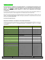
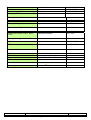
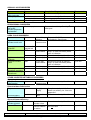
Great Ormond Street Hospital for Children
NHS Foundation Trust
Chemical Pathology Services
External User Guide
Updated August 2012
New website: www.labs.gosh.nhs.uk/
Accredited Medical Laboratory
External
Handbook
Page 1 of 26
This is a copy unless printed on controlled yellow paper.
No unauthorized amendments or photocopies to be made.
Doc. number: CCL 003
Version number: 1.5
Department of Chemical Pathology, Great Ormond Street Hospital for Children
Reference No: 0250
Index
Page
INTRODUCTION
Senior staff
Department sections and phone numbers
3
3
4
REQUESTING
4
SAMPLE COLLECTION
Storage/Packing/Transport
5
5
TURNAROUND TIMES
6
NEWBORN SCREENING
6
FACTORS affecting performance of biochemical investigations
7
SUDDEN INFANT DEATH/MORIBUND CHILD
Biochemical investigations
8
ASSAY DIRECTORY
Amino acid disorders
Carbohydrate Metabolism Disorders
Fatty acid oxidation defect / hypoglycaemia
Lactate / pyruvate disorders
Lysosomal storage disorders
Prenatal Diagnosis
Organic acid disorders
Peroxisomal disorders
Urea cycle disorders
Other inherited metabolic disorders:
Hypophosphatasia
Disacharidase deficiencies
Glycerol kinase deficiency
Neuroblastoma screen
Other tests:
Antimullerin hormone (AMH)
Busulphan
Inhibin B
Trace elements: copper, zinc, selenium, manganese
Vitamin A / E
Isoenzymes/macroenzymes – alkaline phosphatase, amylase,
creatine kinase
Renal tubular markers
9
10
12
12
12
17
19
19
19
19
20
20
20
21
APPENDICES: 1. Special enzyme assays;
2. Perchlorate precipitation
22
24
INDEX
25
External Handbook
Page 2 of 26
This is a copy unless printed on controlled yellow paper.
No unauthorized amendments or photocopies to be made.
Doc. number: CCL 003
Version number: 1.5
Department of Chemical Pathology, Great Ormond Street Hospital for Children
INTRODUCTION
Chemical Pathology Laboratory, Great Ormond Street Hospital for Children NHS Foundation Trust
(GOSH), is a CPA accredited laboratory providing a wide range of Chemical Pathology analyses with
a special interest in the diagnosis and monitoring of inborn errors of metabolism.
The laboratory is fully staffed between 9 am and 5.30 pm Monday to Friday and staff will be available
for any enquiries you may have. For sample requirements and general enquiries not dealt with by this
guide or for results, please contact the helpline in the first instance.
For other enquiries, advice on investigations, clinical advice and interpretation or to request an urgent
analysis, the duty biochemist is available on bleep 020 7405 9200 (hospital switchboard) bleep 0589.
The on duty clinical staff member can also be contacted by a long range message pager, via the
hospital switchboard, out of hours.
For additional information see website http://www.labs.gosh.nhs.uk/
This handbook contains of all the tests currently performed in house by the Department. There are a
number of investigations that are available to GOSH Clinicians provided by External Referral
Laboratories; details of which can be found in the Internal User’s Guide (CCL002).
Sample reception
Chemical Pathology Reception Level 1
Paediatric Laboratory Medicine
Camelia Botnar Building
Great Ormond Street Hospital for Children
Great Ormond Street
London
WC1N 3JH
Senior Staff
Prof Simon Heales
Professor of Clinical Chemistry
[email protected]
Laboratory Director, Clinical Lead
Enzyme Metabolic Unit
Director of Newborn Screening
Ms Helen Aitkenhead
Principal Clinical Scientist
[email protected]
Deputy Laboratory Director, Deputy Clinical Lead
Blood Sciences, Special Routine, QA, POCT
Mr David Wells
020 7813 8321 (DD)
020 7813 8318 (DD)
Bleep: 020 7405 9200
bleep 0200
Pathology Lead Laboratory Manager
020 7813 8320 (DD)
Principal Clinical Scientist
020 7405 9200 x 7843
Head of Newborn Screening
Enzyme Laboratory
Bleep: 020 7405 9200
[email protected]
Mrs Katie Harvey
(née Bainbridge)
[email protected]
bleep 0397
Ms Helen Prunty
Principal Clinical Scientist
[email protected]
Metabolic Laboratory
020 7813 8319 (DD)
Bleep: 020 7405 9200
bleep 2257
Dr Steve Krywawych
Principal Clinical Scientist
020 7405 9200 x 6758
Mr Daley Aolofaju
Chief Biomedical Scientist
020 7405 9200 x 0214
[email protected]
Blood Sciences / Special Routine
[email protected]
External Handbook
Page 3 of 26
This is a copy unless printed on controlled yellow paper.
No unauthorized amendments or photocopies to be made.
Doc. number: CCL 003
Version number: 1.5
Department of Chemical Pathology, Great Ormond Street Hospital for Children
Mr Derek Burke
Chief Biomedical Scientist
[email protected]
Enzyme Laboratory
Mr Ade Ifederu
Chief Biomedical Scientist
[email protected]
Newborn Screening
Ms Julie Leakey
Chief Biomedical Scientist
[email protected]
Metabolic Laboratory
020 7405 9200 x5290
020 7405 9200 x5290
020 7405 9200 x5290
DEPARTMENT SECTIONS AND PHONE NUMBERS
Departmental Office (results enquires)
020 7405 9200 ext 5076
020 7829 8624 (fax)
Email: [email protected]
Helpdesk (general enquires)
020 7405 9200 ext 5009
Metabolic Laboratory
Enzyme Laboratory
020 7405 9200 ext 5225
020 7405 9200 ext 2509/2440
Email: [email protected]
020 7405 9200 ext 5009
020 7829 8383 (DD)
Email: [email protected]
Specimen Reception/Routine Laboratory
Newborn Screening Laboratory
REQUESTING
A request giving the following information must accompany the specimen (apart from newborn
screening tests), a minimum of three identifiers are required:Patient ID:
surname or family name
forename or personal name
date of birth (many reference ranges are age dependent)
sex (some reference ranges are sex related)
patients reference i.e. Hospital number, laboratory, NHS number
Specimen:
type
date and time of collection
Assay(s) required:
Clinical details:
include medication, diet, fasting or fed sample
Sender:
name of sender
address for report and invoice
urgent contact, name, phone number (if different from sender):
Labeling of Specimens
Specimens should be legibly labeled with a minimum of three patient identifiers (see above) along
with the date and time of collection, type of specimen and specimen reference. To avoid results being
wrongly attributed to patients, unlabelled samples or samples that do not match the name on the
request form cannot be processed by the laboratory.
SAMPLE COLLECTION/HANDLING
External Handbook
Page 4 of 26
This is a copy unless printed on controlled yellow paper.
No unauthorized amendments or photocopies to be made.
Doc. number: CCL 003
Version number: 1.5
Department of Chemical Pathology, Great Ormond Street Hospital for Children
Requirements for sample collection and processing are listed under individual analyses further on in
this booklet.
Abbreviations used:
Li hep
Plain
RBC
WBC
S
P
B
BS
Lithium heparin
Plain container
Erythrocytes
Leucocytes
Serum
Li hep plasma
Whole blood
Blood spot
L
M
F
FB
VL
CV
CCV
AF
Liver
Muscle
Fibroblasts
Fetal blood
Vacuolated lymphocytes
Chorionic villus
Cultured chorionic villus
Amniotic fluid
STORAGE
Samples should be sent to us as soon as possible after collection. However, if storage is
unavoidable, guidance for sample storage is given under individual test.
Requesting additional tests and sample retention
If the sample is still available and sufficient in volume and is viable, additional tests may be added by
phoning the Helpdesk. On occasion, the requestor may be asked to send a further request form with
details of the test required.
Samples are retained in accordance to the Guidelines published by the Royal College of Pathologists
and the Institute of Biomedical Science. The retention and storage of pathological records and
specimens (4th edition, 2009). All samples are stored for a minimum of 48 hours after the report has
been issued; most samples are stored for at least two weeks and many are stored for longer periods.
Please contact the Helpdesk for further advice.
PACKING
The packing requirements for samples are specified under each analyte further on in the booklet.
General and room temperature
All specimens must be in leakproof containers. Seal cap of container with ‘parafilm’ or similar
waterproof tape. Wrap each container with sufficient absorbent material to completely absorb the
contents in case of breakage. There should be no contact between containers. Place the container(s)
and packing in plastic bag and seal the bag. Place the sealed bag, together with the request form, in
a rigid fibre or plastic outer case. The outer case should be sealed with tape.
NOTE – the request form must not be inside the plastic bag with the specimen.
Ice
Pack specimens as above. Place the ice in a leak-proof container (use a plastic bottle or bag). Ice
should not come into direct contact with the specimen container to avoid risk of contamination or
labels becoming illegible. Place the ice and specimen(s) in a plastic outer container and seal with
waterproof tape. Include sufficient ice to cover any possible delays in delivery.
Ice packs are suitable for a journey time of less than 6 hours. However, DO NOT place ice packs
from –20 °C freezer immediately next to whole blood or cells.
Dry Ice [Solid CO2]
Pack the specimens as above. The outer pack must be an insulator, e.g. expanded polystyrene.
State “CONTAINS SOLID CO2” on the outside. Seal outer case with tape. Include sufficient solid CO2
to cover any possible delays in delivery.
External Handbook
Page 5 of 26
This is a copy unless printed on controlled yellow paper.
No unauthorized amendments or photocopies to be made.
Doc. number: CCL 003
Version number: 1.5
Department of Chemical Pathology, Great Ormond Street Hospital for Children
TRANSPORT
First class post
When sending specimens by first class post, the packaging MUST comply with UN3733 packaging
regulations and postal regulations.
The package must be labeled ‘PATHOLOGICAL SPECIMEN’ and may only be sent 1st class letter
post. Where first class post is indicated this assumes that delivery will be made by the next day.
Please DO NOT POST on Friday or before a UK Bank Holiday.
Courier or express delivery.
A reliable service should be used and instructed to take the specimens to the Reception in Chemical
Pathology in the Camelia Botnar Building.
TURNAROUND TIME
Turnaround time given is the anticipated time taken between sample receipt and report under normal
operating conditions. Where the assays are batched and performed infrequently, the time is given as
a range up to the maximum anticipated time. Time taken for sample transport and posting the report
should be added to this. Where appropriate, abnormal results will be phoned or faxed to the
sending laboratory. In cases where results are required more urgently, please contact the
department to discuss your requirements prior to sending specimens so that samples can be fast
tracked.
NEWBORN SCREENING
Blood spot assays to screen for phenylketonuria (PKU), congenital hypothyroidism, medium chain
acyl coA dehydrogenase deficiency, sickle cell disorders and cystic fibrosis in the neonatal period are
available.
Sample requirement: 4 good blood spots collected between day 5 and 8 (day of birth = day 0) on a
standard screening card, dried at room temperature, and enclosed in a glassine cover. Please
provide the dates of birth and sampling. Send at room temperature by post immediately. Results will
be available within 4 working days
External Handbook
Page 6 of 26
This is a copy unless printed on controlled yellow paper.
No unauthorized amendments or photocopies to be made.
Doc. number: CCL 003
Version number: 1.5
Department of Chemical Pathology, Great Ormond Street Hospital for Children
FACTORS AFFECTING THE PERFORMANCE OF TESTS AND THEIR INTERPRETATION
METABOLIC INVESTIGATIONS
What samples?
It is important to check the fluid in which the metabolites of interest most obviously accumulate, e.g.
urine for organic acids. The next part of this booklet indicates the sample type required for the
investigations offered. When indicated (e.g. because of metabolite instability), it is necessary to make
arrangements with the laboratory prior to collecting the sample.
When?
The time of the sample collection is crucial where characteristic metabolites accumulate only
intermittently in the samples. Whenever possible, patients should be investigated during periods
when they are unwell. Samples should be taken as soon as possible after admission, before changes
in treatment and diet lead to the disappearance of relevant metabolites.
Sample integrity
Bacterial activity in poorly preserved samples produces a rise in pH and can lead to both the
appearance of bacterial metabolites and the breakdown of important components, especially sugars
and some amino acids. Samples with a high pH may not be analyzed for this reason. Faecal
contamination of urine produces a similar effect. Dilute urine makes the detection of urinary
constituents unreliable and samples with creatinine concentration >1 mmol/L are preferred.
Diet
Some metabolic disorders are related to a particular dietary intake or are produced only in the fasting
state. Investigations should be carried out, as far as possible, on samples taken at the time the
patient was symptomatic. Dietary restrictions or feeding may cause characteristic metabolites to
disappear and result in false negative results. Dietary metabolites may interfere with organic acid,
amino acid or carbohydrate chromatograms. Patients receiving intravenous amino acid mixture may
have amino aciduria, amino acidaemia or organic aciduria. Information on the type of diet and the
timing of the sample in relation to meals will aid in the interpretation of these complex analyses.
Drugs
Drugs influence metabolic investigations by analytical interference or by modifying metabolic
processes. Details of all medication should be provided with metabolic investigations.
Exchange transfusions / blood transfusions
These may affect the analytes measured in blood and especially erythrocytes. When requesting tests
in such patients, check whether adequate time has lapsed since the last transfusion. For assays of
enzymes and metabolites in erythrocytes, the time interval should be 6 weeks.
Other factors include
Age of specimen
Time of specimen separation
Specimen storage
Specimen haemolysis, icterus and lipaemia
Fasting state
External Handbook
Page 7 of 26
This is a copy unless printed on controlled yellow paper.
No unauthorized amendments or photocopies to be made.
Doc. number: CCL 003
Version number: 1.5
Department of Chemical Pathology, Great Ormond Street Hospital for Children
BIOCHEMICAL INVESTIGATION OF A SUDDEN INFANT DEATH
If an inborn error of metabolism is suspected in an infant who died suddenly, collect the following
samples as soon as practicable to minimize post mortem changes; blood spots, bile spots, plasma,
urine, CSF, aqueous humor. Blood stained CSF and urine should be spun and separated
immediately and this should be recorded. Freeze at –20 °C. Skin biopsy can also be taken (see
fibroblasts). Please discuss the request with the duty biochemist – 020 7405 9200 bleep 0589 before
sending the sample.
METABOLIC INVESTIGATION IN A MORIBUND CHILD
The diagnosis of metabolic disease cannot be made after death unless the correct specimens have
been appropriately collected. If metabolic disease is suspected and the child seems likely to die
before a diagnosis can be made, it would be advisable to collect the following specimens:
Blood - 10 ml in a heparinised tube. Separate plasma promptly. Freeze the bulk of the plasma, the
remaining plasma and red cells should be kept at 4 °C.
Urine - 20 ml in a plain container and deep freeze.
Blood spots for acylcarnitine
Bile spots for acylcarnitine
DNA - If the condition is one in which DNA studies are likely to be helpful, take 10ml blood into an
EDTA tube and deep freeze the whole blood.
Tissue biopsies (liver, muscle, heart – Label the plain container with the type of tissues prior to
taking the biopsies. Pre-cool a plain container in the deep freeze. Obtain dry ice, liquid nitrogen or a
freezing pack. Make a small boat with a piece of aluminium foil and place it on the dry ice / freezing
mixture. Take the biopsy (as many cores as possible, minimum two) and put it immediately in the
boat, it should freeze immediately and thereafter should be not allowed to thaw at any time. Wrap up
the core in the foil and put it in the pre-chilled container, making sure that the cap is tight and
immediately replace in the deep freeze (-40 °C or lower). A small part should be put into
glutaraldehyde and if necessary some into formalin, but the majority should be frozen for chemistry
and enzymology.
Skin Biopsy - See fibroblasts
External Handbook
Page 8 of 26
This is a copy unless printed on controlled yellow paper.
No unauthorized amendments or photocopies to be made.
Doc. number: CCL 003
Version number: 1.5
Department of Chemical Pathology, Great Ormond Street Hospital for Children
ASSAY DIRECTORY
Codes
Tests highlighted in turquoise are analysed in the Metabolic Laboratory
Test highlighted in green are analysed in the Enzyme Laboratory
Tests highlighted in yellow are analysed in the Routine Laboratory
AMINO ACID DISORDERS
Test
Amino acids
Plasma
Sample requirement
Sample handling
Turnaround
0.5 ml li hep plasma
Separate ASAP. Freeze immediately. Send
frozen
5 ml fresh random urine Freeze immediately. Send frozen
0.2 ml clear CSF
Freeze immediately. Send frozen
4 blood spots on std card Send by first class post.
1–2w
Urine
CSF
Blood spot
- branched chain
Homocysteine
Plasma
0.2 ml li hep plasma
3–6w
1–2w
4d
Urine
Succinylacetone
Sulphite
Sulphocystine
5 ml fresh random urine
5 ml fresh random urine
5 ml fresh random urine
5 ml fresh random urine
Separate ASAP. Freeze immediately. Send
frozen
Freeze immediately
Freeze immediately. Send frozen
Freeze immediately. Send frozen
Freeze immediately. Send frozen
1–2w
3–6w
2-4w
72 h
3–6w
Methionine load for the diagnosis of homocystinuria
Preparation:
Pre-load:
Methionine load:
Fast overnight (6 hours for infants).
At the beginning of the test, empty bladder and discard sample. Collect 1ml
blood in a lithium heparin tube, separate and precipitate immediately.
Empty bladder. Give L-methionine (100 mg/kg body weight) orally over 5
minutes in a flavoured drink or as tablets. Collect 1 ml blood in lithium heparin
tubes at 2, 4 and 6 hours. On each occasion, separate and precipitate
immediately. Collect all urine passed over the 6 hours after giving the
methionine load. Send plasma supernatant and urine on solid CO2 for amino
acid analysis.
Maple syrup urine disease enzyme diagnosis (Leucine decarboxylating system) (Skin
fibroblasts)
Send a skin biopsy sample in the culture medium (See Appendix 1).
External Handbook
Page 9 of 26
This is a copy unless printed on controlled yellow paper.
No unauthorized amendments or photocopies to be made.
Doc. number: CCL 003
Version number: 1.5
Department of Chemical Pathology, Great Ormond Street Hospital for Children
CARBOHYDRATE METABOLISM DISORDERS
Congenital Disorders of Glycosylation (CDG)
Initial investigation – serum transferrins isoelectric focusing at Institute of Neurology. If type I pattern
CDG 1a & 1b should be excluded first. If patterns are abnormal but clinically indicated, then measure
enzymes.
Test
Phosphomannomutase
Phosphomannose
isomerase
Type
WBC
Sample handling
5-10 ml well mixed li hep
whole blood with no clots
F
skin biopsy into culture
medium or saline
5-10 ml well mixed li hep
Whole blood with no
clots
WBC
F
skin biopsy into culture
Medium or saline
Send whole blood at
ambient temp. to reach
lab ideally within 24 h of
collection
Send at ambient temp.
by courier. Do not freeze
Send whole blood at
ambient temp. to reach
lab ideally within 24 h of
collection
Send at ambient temp.
by courier. Do not freeze
Turnaround
up to 6w
up to 10w
up to 6w
up to 10w
Galactose/ Fructose metabolism Disorders
Test
Reducing substances
Sugar
chromatography
Galactose -1phosphate uridyltransferase
[Gal-1-PUT]
Galactokinase
Type
U
Sample handling
5 ml fresh random
urine
RBC
UDP-galactose
RBC
Galactose-1
Phosphate
RBC
2 ml li hep whole
blood.
No transfusion prior 6
w
2 ml li hep whole blood
No transfusion prior
6w. Contact lab prior
sampling
2 ml li hep whole blood
No transfusion prior 6
w Contact lab prior
sampling
2 ml li hep whole blood
Fructose-1-P aldolase
L
liver biopsy
RBC
Turnaround
3–6w
Freeze immediately.
Send frozen
Send whole blood at
ambient temp. to reach
lab ideally within 48 h of
collection
Send whole blood at
ambient temp. to reach
lab within 24 h of
collection
Send whole blood at
ambient temp. to reach
lab ideally within 24 h of
collection
Send whole blood at
ambient temp. to reach
lab ideally within 24 h of
collection
Freeze immediately. Send
frozen without thawing
2-4w
4–6 w
4–6 w
4–6 w
4-6 w
Glycogen Storage Disorders (GSD)
Test
Ia Glucose-6phosphate hydrolase
Ib Glucose-6phosphate translocase
II α-1,4-glucosidase
External Handbook
Page 10 of 26
Type
L
Sample handling
Fresh liver biopsy
L
Fresh liver biopsy
BS,
Blood spots, 5 ml li
Turnaround
Contact enzyme lab prior
4-6 w
to sampling. Do not freeze.
Contact enzyme lab prior
4-6 w
to sampling. Do not freeze.
Send whole blood at
4-6 w
This is a copy unless printed on controlled yellow paper.
No unauthorized amendments or photocopies to be made.
Doc. number: CCL 003
Version number: 1.5
Department of Chemical Pathology, Great Ormond Street Hospital for Children
(acid maltase)
WBC
III glycogen
debrancher
WBC
IV glycogen brancher
hep blood+ EDTA
blood for vacuolated
lymphocytes
5-10 ml li hep whole
blood
M, L
muscle / liver biopsy
F
skin biopsy into culture
Medium or saline
5-10 ml li hep whole
blood
WBC
M, L
muscle / liver biopsy
F
skin biopsy into culture
Medium or saline
muscle biopsy
V phosphorylase
M
VI phosphorylase
WBC
5-10 ml lip hep whole
blood
L
liver biopsy
M
muscle biopsy
RBC
5 ml li hep whole blood
L
liver biopsy
WBC
5-10 ml lip hep whole
blood
L
liver biopsy
VII phospho
fructokinase
IX phosphorylase b
Kinase
IX fructose-1,6
bisphosphatase
ambient temp. to reach lab
ideally within 24 h of
collection
Send whole blood at
ambient temp. to reach lab
ideally within 24 h of
collection
Freeze immediately
Send frozen
Send at ambient temp.
Do not freeze
Send whole blood at 4-6
w ambient temp. to reach
lab within 18 h of
collection
Freeze immediately
Send frozen
Send at ambient temp.
Do not freeze
Freeze immediately
Send frozen
Send whole blood at
ambient temp. to reach lab
ideally within 24 h of
collection
Freeze immediately
Send frozen
Freeze immediately
Send frozen
Send whole blood at
ambient temp. to reach lab
ideally within 24 h of
collection
Freeze immediately
Send frozen
Send whole blood at
ambient temp. to reach lab
ideally within 24 h of
collection
Freeze immediately
Send frozen
4-6 w
4-6 w
4-6 w
4-6 w
4-6 w
4-6 w
4-6 w
4-6 w
4-6 w
4-6 w
4-6 w
4-6 w
4-6 w
4-6 w
Pompe CRIM testing – contact the Enzyme Laboratory Tel: 020 7405 9200 ext 2509/2440
Glycolytic enzymes
Test
Phosphoglucomutase
Type
WBC
Sample handling
5 ml li hep whole
blood
M, L
muscle / liver biopsy
Send whole blood at ambient
temp. to reach lab ideally within
24 h of collection
Freeze immediately
Send frozen
Turnaround
4-6 w
4-6 w
FATTY ACID OXIDATION DEFECT / HYPOGLYCAEMIA
Test
External Handbook
Page 11 of 26
Type
Sample handling
This is a copy unless printed on controlled yellow paper.
No unauthorized amendments or photocopies to be made.
Turnaround
Doc. number: CCL 003
Version number: 1.5
Department of Chemical Pathology, Great Ormond Street Hospital for Children
3-(Β)Hydroxybutyrate (BOHB)
Free fatty acids
(non-esterified
fatty acids, NEFA)
Acetoacetate
P
0.3 ml li hep plasma
P
0.3 ml li hep plasma
B
perchloric acid
supernatant
Organic acids
U
Acylcarnitines
BS
5 ml fresh random
urine
4 blood spot on
standard card
Freeze immediately. Send
frozen. Provide glucose result.
Freeze immediately. Send
frozen. Provide glucose result.
1-2w
Freeze immediately (see
appendix for protocol). Send
frozen
Freeze immediately. Send
frozen
Send by first class post
1–3w
1-2w
2–4w
1-2w
Diagnostic fast
All the above investigations to be carried out at the beginning and end of the fast under close medical
supervision in a Hospital unit experienced in carrying out these tests (not advisable in patients under
18 months or under 5 kg in weight)
LACTATE / PYRUVATE DISORDERS
Test
Lactate
Type
P
Sample handling
2 ml fluoride oxalate plasma
B
perchloric acid precipitation
(see appendix for protocol)
0.2 ml clear CSF
CSF
Pyruvate
B
perchloric acid precipitation
(see appendix for protocol)
perchloric acid precipitation
(see appendix for protocol)
CSF
Separate plasma assay
ASAP
Freeze immediately
Send frozen
Freeze immediately
Send frozen
Freeze immediately
Send frozen
Freeze immediately
Send frozen
Turnaround
6h
1–2w
1–2w
1–2w
1–2w
LYSOSOMAL STORAGE DISORDERS (LSD)
Initial investigations/monitoring
Test
Type Sample handling
Glycosaminoglycans
U
5 ml fresh random urine
Send at ambient temp. by
special post
Send at ambient temp. by
special post
Send at ambient temp. by
special post
Turnaround
up to 4 w
Sialic acid
U
5 ml fresh random urine
up to 4 w
Ceramide trihexoside
(CTH)
Globotriaosylceramide
(GL3/GB3)
Tetrasaccharides
(hex4)
Tetraglucose (glc4)
Vacuolated
lymphocytes
U
5 ml fresh random urine
U
5 ml fresh random urine
Send at ambient temp. by
special post
up to 4 – 6 w
B
2 ml EDTA whole blood
(see page 22)
Send at ambient temp. by
special post (done in
Histopathology)
Contact
histopathology
up to 4 – 6 w
Individual enzyme
Assays available individually for the diagnosis of lysosomal storage disorders are listed below with
samples suitable for the assay. Turnaround is 4-6 weeks
External Handbook
Page 12 of 26
This is a copy unless printed on controlled yellow paper.
No unauthorized amendments or photocopies to be made.
Doc. number: CCL 003
Version number: 1.5
Department of Chemical Pathology, Great Ormond Street Hospital for Children
Disease
Mucopolysaccharidoses
I-Hurler
II-Hunter
IIIA-Sanfilippo A
IIIB-Sanfilippo B
IIIC-Sanfilippo C
IIID-Sanfilippo D
IVA-Morquio A
IVB-Morquio B
VI-Maroteaux-Lamy
VII-Sly
Multiple enzyme defects
Mucolipidosis II (I-cell)
Mucolipidosis III (pseudo Hurler)
Multiple sulphatidosis
Gangliosidoses
GM1 gangliosidosis
GM2 gangliosidoses:
Tay Sachs / B1 variant
Sandhoff
Leucodystrophies
Krabbe
Metachromatic
Glycoproteinoses
Fucosidosis
α-Mannosidosis
β-Mannosidosis
Schindler
Sialidosis
Aspartylglucosaminuria
Galactosialidosis
Other lipid storage disorders
Fabry
Gaucher
Assay
Tissue
α-iduronidase
iduronate-sulphatase
heparan sulphamidase
α−N-acetyl-glucosaminidase
N-acetyltransferas
N-acetyl-glucosamine-6-sulphatase
N-acetyl galactosamine-6-sulphatase
β-galactosidase
arylsulphatase B
β-glucuronidase
WBC, F
WBC, P, F
WBC, F
WBC, P, F
WBC, F
WBC, F
WBC, F
WBC, F
WBC, F
WBC, P, F
multiple hydrolases
multiple hydrolases
multiple sulphatases
P, VL, F
P, VL, F
WBC, P, F
β-galactosidase
WBC, VL, F
hexosaminidase A
total β-hexosaminidase
WBC, P, F
WBC, P, F
galactocerebrosidase
arylsulphatase A
WBC, F
WBC, F
α-fucosidase
α-mannosidase
β-mannosidase
α-N-acetyl galactosaminidase
α-neuraminidase
aspartylglucosaminidase
α-neuraminidase/ β-galactosidase
WBC, P, VL, F
WBC, P, VL, F
P, WBC, F, VL
P, WBC, F
WBC, VL, F
P, F
WBC, VL, F
α-galactosidase
β-glucosidase
chitotriosidase
sphingomyelinase
acid esterase
WBC, P, F
WBC, F
P
WBC, VL, F,
WBC, VL, F,
Niemann-Pick A & B
Wolman & cholesteryl ester storage
disease (CESD)
Neuronal ceroid lipofuscinoses
(Batten disease)
Infantile (INCL, NCL1, CLN1)
palmitoyl protein thioesterase
Classic late infantile (LINCL, NCL2,
tripeptidyl peptidase I
CLN2)
Transport defects
Cystinosis
cystine
Sialic acid storage
sialic acid
NB: Prenatal diagnosis is available for these disorders.
External Handbook
Page 13 of 26
This is a copy unless printed on controlled yellow paper.
No unauthorized amendments or photocopies to be made.
WBC, F
WBC, F
WBC, F
U, VL, F
Doc. number: CCL 003
Version number: 1.5
Department of Chemical Pathology, Great Ormond Street Hospital for Children
Grouped Enzyme Screens for Lysosomal Disorders
The lysosomal storage disorders can be grouped according to clinical features and a group of
enzyme assays can be carried out on a single blood sample which provides both white blood cells
and plasma for analysis. The clinical signs of a lysosomal storage disease may eventually develop to
give a classic picture but diagnosis at an earlier stage can be more difficult, e.g. while Type II
Gaucher disease leads to hepato/splenomegaly, neurological signs may be more obvious initially. To
meet this and other concerns all patients have plasma chitotriosidase measured to exclude Gaucher
disease and other LSDs. Palmitoyl protein thioesterase and tripeptidyl peptidase I which are deficient
in infantile (INCL, NCL1, CLN1) and classic late infantile (LINCL, NCL2, CLN2) neuronal ceroid
lipofuscinosis are assayed in all patients under 16 years with neurological problems, and also in
adult patients if these disorders are suspected.
It is important that the laboratory is given full clinical details in order to carry out the
appropriate combination of tests. Turnaround time is 6 - 8 weeks
Note: Some diseases may present under more than one heading.
Neurodegenerative screen
Evidence of neurological regression, hypotonia, fits, etc.
Disease
GM1 gangliosidosis
GM2 gangliosidoses:
Tay Sachs / B1 variant
Sandhoff
Krabbe leucodystrophy
Metachromatic leucodystrophy
Fucosidosis
α-Mannosidosis
β-Mannosidosis
Schindler
MPS VII-Sly
I cell disease
Enzyme
β-galactosidase
hexosaminidase A
total β-hexosaminidase
galactocerebrosidase
arylsulphatase A
α-fucosidase
α-mannosidase
β-mannosidase
α-N-acetyl galactosaminidase
β-glucuronidase
I cell screen
Plasma chitotriosidase is assayed in all patients to exclude Gaucher disease
All patients under 16 years of age are tested for:
Infantile neuronal ceroid
lipofuscinosis(INCL, NCL1, CLN1)
Classic late infantile neuronal ceroid
lipofuscinosis (LINCL, NCL2, CLN2)
palmitoyl protein thioesterase
tripeptidyl peptidase I
Dysmorphic screen
The first line test for a dysmorphic child is screening for a mucopolysaccharidosis by urine GAGs.
The following enzymes are indicated if a mucopolysaccharidosis is excluded.
Disease
GM1 gangliosidosis
Sialidosis
Galactosialidosis
Fucosidosis
α-Mannosidosis
I cell disease
External Handbook
Page 14 of 26
Enzyme
β-galactosidase
α-neuraminidase
α-neuraminidase/ β-galactosidase
α-fucosidase
α-mannosidase
I cell screen
This is a copy unless printed on controlled yellow paper.
No unauthorized amendments or photocopies to be made.
Doc. number: CCL 003
Version number: 1.5
Department of Chemical Pathology, Great Ormond Street Hospital for Children
β-Mannosidosis
β-mannosidase
MPS VII-Sly
β-glucuronidase
Multiple sulphatidosis
arylsulphatase A
Aspartylglucosaminuria
aspartylglucosaminidase
Schindler
α-N-acetyl galactosaminidase
Plasma chitotriosidase is assayed in all patients to exclude Gaucher disease
Hepato/splenomegaly screen
For those patients with hepatomegaly and or splenomegaly suspected of having a lysosomal storage
disorder.
Disease
Enzyme
GM1 gangliosidosis
β-galactosidase
Sialidosis
α-neuraminidase
Galactosialidosis
α-neuraminidase/ β-galactosidase
Gaucher
β-glucosidase
Niemann-Pick A & B
sphingomyelinase
Wolman & CESD
acid esterase
Fucosidosis
α-fucosidase
α-Mannosidosis
α-mannosidase
I cell disease
I cell screen
β-Mannosidosis
β-mannosidase
MPS VII-Sly
β-glucuronidase
In all patients with hepato/splenomegaly plasma chitotriosidase is assayed
Cherry red spot screen
For patients with a cherry red spot on the macula.
Disease
GM1 gangliosidosis
GM2 gangliosidoses:
Tay Sachs / B1 variant
Sandhoff
Niemann-Pick A
Sialidosis
Galactosialidosis
Krabbe leucodystrophy
Enzyme
β-galactosidase
hexosaminidase A
total β-hexosaminidase
sphingomyelinase
α-neuraminidase
α-neuraminidase/ β-galactosidase
galactocerebrosidase
Angiokeratoma screen
For patients with an angiokeratoma.
Disease
Fabry
Fucosidosis
Sialidosis
Galactosialidosis
Adult GM1 gangliosidosis
α-Mannosidosis
β-Mannosidosis
Schindler
Aspartylglucosaminuria
Enzyme
α-galactosidase
α-fucosidase
α-neuraminidase
α-neuraminidase/ β-galactosidase
β-galactosidase
α-mannosidase
β-mannosidase
α-N-acetyl galactosaminidase
aspartylglucosaminidase
DNA Analysis
External Handbook
Page 15 of 26
This is a copy unless printed on controlled yellow paper.
No unauthorized amendments or photocopies to be made.
Doc. number: CCL 003
Version number: 1.5
Department of Chemical Pathology, Great Ormond Street Hospital for Children
The Enzyme Laboratory works closely with the Clinical Molecular Genetics Laboratory at Great
Ormond Street Hospital to offer mutational analysis for many of the lysosomal storage disorders. It is
essential to test for the presence of the polyA mutation encoding a pseudodeficiency of
arylsulphatase A in all patients with low arylsulphatase A activity. For other disorders the Enzyme
Laboratory will advise if mutational analysis is available and/or appropriate when a diagnosis is made.
External Handbook
Page 16 of 26
This is a copy unless printed on controlled yellow paper.
No unauthorized amendments or photocopies to be made.
Doc. number: CCL 003
Version number: 1.5
Department of Chemical Pathology, Great Ormond Street Hospital for Children
PRENATAL DIAGNOSIS
Prenatal diagnosis is available for the following disorders. It is important that the diagnosis in the
index case has been confirmed in an appropriate tissue. The tissues suitable for assay are stated in
the table. It is essential to contact the Enzyme Laboratory (020 7405 9200 ext 2509) before
taking any samples for prenatal diagnosis to discuss your requirements and transport
arrangements.
For chorionic villus specimens, it is our policy to assay the villi directly, where appropriate, and then to
check equivocal results or confirm diagnosis of an unaffected fetus on cultured cells.
Direct and cultured cell assays are charged separately and an additional charge is made for the cell
culture. For amniotic fluid samples where the assay is performed on cultured cells, the cost of the
cell culture is charged additionally.
Lysosomal storage disorders
Mucopolysaccharidoses, mucolipidoses and multiple sulphatidosis
Following amniocentesis, electrophoresis of amniotic fluid glycosaminoglycans (GAGs) is carried out
on all pregnancies at risk for a mucopolysaccharidosis, mucolipidoses II and III or a multiple
sulphatidosis.
Disorder
Mucopolysaccharidoses
Enzyme
Samples
I Hurler / Scheie
II Hunter
IIIA-Sanfilippo A
IIIB-Sanfilippo B
IIIC-Sanfilippo C
IVA-Morquio A
IVB-Morquio B
VI-Maroteaux-Lamy
VII-Sly
α-iduronidase
iduronate sulphatase
heparan sulphamidase
α-glucosaminidase
N-acetyltransferase
N-ac galactosamine-6-sulphatase
β-galactosidase
arylsulphatase B
β-glucuronidase
CV, CCV, CAC
CV, CCV, AF, CAC
CV, CCV, CAC
CV, CCV, CAC
CV, CCV, CAC
CV, CCV, CAC
CV, CCV, CAC
CV, CCV, CAC
CV, CCV, AF, CAC
Mucolipidosis II (I-cell)
multiple lysosomal hydrolases
CCV, AF, CAC
Mucolipidosis III (pseudo-Hurler)
multiple lysosomal hydrolases
CCV, AF, CAC
Multiple sulphatidosis
multiple sulphatases
CV, CCV, AF, CAC,
Lipidoses
GM1 gangliosidosis
GM2 gangliosidoses:
Tay Sachs
Sandhoff
Krabbe leucodystrophy
Metachromatic leucodystrophy
Fucosidosis
β-Mannosidosis
α-Mannosidosis
Schindler
Sialidosis
Galactosialidosis
External Handbook
Page 17 of 26
β-galactosidase
CV, CCV, CAC
hexosaminidase A
total β-hexosaminidase
galactocerebrosidase
arylsulphatase A
α-fucosidase
β-mannosidase
α-mannosidase
α-N-acetyl galactosaminidase
neuraminidase
α-neuraminidase/ β-galactosidase
CV, CCV, CAC
CV, CCV, AF, CAC
CV, CCV, CAC
CV, CCV, CAC
CV, CCV, CAC
CV, CCV, CAC
CV, CCV, CAC
CV, CCV, CAC
CV, CCV, CAC
CV, CCV, CAC
This is a copy unless printed on controlled yellow paper.
No unauthorized amendments or photocopies to be made.
Doc. number: CCL 003
Version number: 1.5
Department of Chemical Pathology, Great Ormond Street Hospital for Children
α-galactosidase
β-glucosidase
sphingomyelinase
acid esterase
Fabry
Gaucher
Niemann-Pick A & B
Wolman & CESD
CV, CCV, AF, CAC
CV, CCV, CAC
CV, CCV, CAC
CV, CCV, CAC
Other lysosomal disorders
Sialic acid storage
Cystinosis
Pompe (GSD type II)
sialic acid
cystine
α-glucosidase
CV, CCV, AF, CAC
CV, CCV, CAC
CV, CCV, CAC
palmitoyl protein thioesterase
tripeptidyl peptidase I
CV, CCV, CAC
CCV, CAC
GSD II (Pompe)
α-glucosidase
CV, CCV, CAC
GSD IV
brancher
CV, CCV, CAC
ornithine carbamoyl transferase
carbamoyl phosphate synthase
arginase
argininosuccinate lyase
fetal liver
fetal liver
FB
FB
release of 14CO2 from leucine
CV
Neuronal ceroid lipofuscinoses
Infantile (INCL, NCL1, CLN1)
Classic late infantile (LINCL, NCL2,
CLN2)
Glycogen storage disorders
Urea cycle disorders
OCT deficiency
CPS deficiency
Arginase deficiency
Argininosuccinate lyase deficiency
Other disorders
Maple syrup urine disease
External Handbook
Page 18 of 26
This is a copy unless printed on controlled yellow paper.
No unauthorized amendments or photocopies to be made.
Doc. number: CCL 003
Version number: 1.5
Department of Chemical Pathology, Great Ormond Street Hospital for Children
ORGANIC ACID DISORDERS
Test
Organic acids incl.
methylmalonate
N-acetylaspartate
Biotinidase
Type
5 ml fresh random urine
Sample handling
Freeze immediately. Send frozen
Turnaround
2–4w
5 ml fresh random urine
2 ml amniotic fluid
0.2 ml li hep plasma
Freeze immediately. Send frozen
By arrangement. Send by courier
Freeze ASAP. Send frozen
2–4w
as arranged
1–3w
Separate immediately. Send by 1st
class post.
2–4w
PEROXISOMAL DISORDERS
Very long chain
0.5 ml li hep plasma
fatty acids,
includes phytanate
& pristanate
UREA CYCLE DISORDERS
Amino acids
P
0.5 ml li hep
plasma
5 ml fresh
random urine
Organic acids,
includes orotic acid
U
N-acetylglutamate
Synthase
Arginase
Discuss with
enzyme lab
RBC
Argininosuccinate
Synthase
Argininosuccinate
Lyase
Carbamoyl
Phosphate synthase
Ornithine carbamoyl
Transferase
5 ml li hep
whole blood
liver biopsy
liver biopsy
RBC
5 ml li hep
whole blood
L
L
L
stool
Glycerol kinase deficiency
Organic acids,
U
includes glycerol
Glycerol kinase
F
External Handbook
Page 19 of 26
2–4w
Send whole blood at ambient
temp. to reach lab ideally within
24 h of collection
Freeze immediately. Send frozen
Freeze immediately. Send frozen
up to 6 w
up to 6 w
up to 6 w
up to 6 w
liver biopsy
liver biopsy
Send whole blood at ambient
temp. to reach lab ideally within
24 h of collection
Freeze immediately. Send frozen
Freeze immediately. Send frozen
up to 6 w
up to 6 w
liver biopsy
Freeze immediately. Send frozen
up to 6 w
Freeze immediately. Send frozen
3–6w
Snap freeze in liquid N2. Send
frozen on solid dry ice. Also see
appendix 1.
Freeze immediately. Send frozen
up to 8 w
Freeze immediately. Send frozen
2–4w
Send at room temperature.
Do not freeze
up to 10w
OTHER INHERITED METABOLIC DISORDERS
Hypophosphatasia
PhosphoU
5 ml fresh
ethanolamine
random urine
Sugar
chromatography
1–2w
up to 6 w
L
L
Disaccharidase Deficiencies
Enzymes
jejunum
Separate ASAP. Freeze
immediately. Send frozen
Freeze immediately. Send frozen
2 mg jejunum
biopsy
walnut size
stool
5 ml fresh
random urine
skin biopsy into
culture medium
or saline
This is a copy unless printed on controlled yellow paper.
No unauthorized amendments or photocopies to be made.
3–6w
Doc. number: CCL 003
Version number: 1.5
Department of Chemical Pathology, Great Ormond Street Hospital for Children
NEUROBLASTOMA SCREEN
Test
Type
HVA
U
VMA
U
Sample
handling
5 ml fresh
random urine
5 ml fresh
random urine
Turnaround
Freeze ASAP. Send frozen
3d
Freeze ASAP. Send frozen
3d
Separate and freeze plasma /
serum immediately after
collection. Send frozen
Arrange with lab prior to
sampling.
Send sample on ice
immediately to local Lab.
Separate and freeze plasma
ASAP
Label samples clearly with time
of collection.
Separate plasma ASAP
up to 2 m
Collect 24h urine into acid
washed container.
Note 24 h volume.
Separate and freeze plasma /
serum immediately after
collection. Send frozen
Separate plasma. Send on ice.
2–4w
OTHER TESTS
Antimullerian
hormone (AMH)
P, S
Busulphan
P
Copper
0.5 ml EDTA
plasma or
serum
1 ml EDTA
blood at 0, 0.5,
1, 1.5, 2, 4, 7 h
P
0.4 ml li hep
plasma
10 ml aliquot of
24 h
U
Inhibin B
P, S
Insulin
S, P
Manganese
P
Selenium
P
Sugar
chromatography
U, F
Vitamin A
S
0.5 ml li hep
plasma or
serum
0.3 ml
serum/plasma
0.5 ml whole
blood in Trace
metal container
0.4 ml li hep
plasma
5 ml random
urine
walnut size stool
0.5 ml serum
Vitamin E
S
0.5 ml serum
Zinc
P
0.4 ml li hep
plasma
Same day.
Must be prebooked.
1–2w
up to 3 m
1d
Send whole blood by first class
post
2–4w
Separate plasma ASAP
Send by first class post
Freeze immediately. Send
frozen
2-3w
3–6w
2-4w
Protect from light
Separate serum ASAP
Send by first post
Separate serum ASAP
Send by first post
Separate plasma ASAP
Send by first class post
2-4w
1–2w
ISOENZYMES
Alkaline phosphatase
isoenzymes
Amylase isoenzymes
Creatine kinase
isoenzymes
External Handbook
Page 20 of 26
P, S
0.5 ml li hep plasma / serum
Send by 1st class post
up to 4 w
P, S
P, S
0.5 ml li hep plasma / serum
0.5 ml li hep plasma / serum
Send by 1st class post
Separate and freeze
plasma / serum
immediately after
up to 5 w
up to 4 w
This is a copy unless printed on controlled yellow paper.
No unauthorized amendments or photocopies to be made.
Doc. number: CCL 003
Version number: 1.5
Department of Chemical Pathology, Great Ormond Street Hospital for Children
collection. Send frozen
RENAL TUBULAR MARKERS
Retinol binding Protein
[RBP]
N-acetylglucosaminidase
[NAG]
External Handbook
Page 21 of 26
U
1 ml fresh random urine
U
1 ml fresh random urine
Freeze soon after
collection
Send by 1st class post
This is a copy unless printed on controlled yellow paper.
No unauthorized amendments or photocopies to be made.
1-3w
1-3w
Doc. number: CCL 003
Version number: 1.5
Department of Chemical Pathology, Great Ormond Street Hospital for Children
APPENDIX 1: Special Enzyme Assays
It is important that full clinical details (especially presence or absence of neurological features,
hepatosplenomegaly, dysmorphic features) are given on the request form so that appropriate assays
can be carried out. Please let us know if the mother is pregnant as we can advise on prenatal
diagnosis.
Sample requirements
Enzyme assays are classified under separate diagnostic groups with abbreviations for samples
where appropriate. These abbreviations are explained below.
WBC (leucocytes) for white cell enzymes
Unless specified, enzymes are assayed according to the clinical details given. Blood transfusion
within 4 weeks may interfere with the result and sampling at this time is best avoided if possible.
Send 5 – 10 ml well mixed blood in lithium heparin (minimum of 5mls). The sample must not contain
any clots, heparinise the syringe if the patient is difficult to bleed. Send the whole blood sample to
reach the laboratory ideally within 24 hours of sample collection (the shorter the interval, the better
the quality of the sample). For most enzymes up to 48 hours is acceptable. However WBC cystine it
is essential that the sample is received within 24 hours. Send by courier or Royal Mail Special
Next Day delivery to arrive before 14:30 on a normal working day. Please avoid sending samples on
a Friday in case of delays in transport.
The turnaround time for these assays is approximately 6 weeks.
RBC (erythrocytes)
Blood transfusion in the previous 6 weeks invalidates results.
Send 2 ml heparinised blood to arrive in the laboratory within 24 hours of sample collection, except
for galactokinase + epimerase which has to be assayed on the day of sample collection and
should be arranged with the enzyme laboratory at least a day in advance. Send by courier or
Royal Mail Special Next Day delivery.
P (plasma) I cell screen etc
Send 1 ml plasma from a lithium heparin blood sample, to reach the laboratory within 24 hours of
collection. Send by courier or Royal Mail Special Next Day delivery.
F (fibroblasts) from skin biopsies
Taking a skin biopsy:
Proceed under aseptic conditions. Have sterile culture medium ready. The forearm and axilla are
suitable sites. Swab the skin with alcohol or chlorhexidine (not iodine or betadine). Approximately 0.2
ml to 0.4 ml of 0.5% lignocaine or similar local anaesthetic is injected intradermally and just
subcutaneously. Take a 3 mm punch biopsy (full thickness skin) or ellipse 4 mm x 2 mm, immediately
transfer the skin to the culture medium. IN EXCEPTIONAL circumstances, sterile dextrose / saline
may be used. Keep at 4 °C or room temperature (DO NOT FREEZE) and send by courier or
datapost. Fill the container to the top to avoid any airlock.
Storage: 4-8 oC for 24 hours in sterile saline, 3 to 5 days in sterile culture medium. It will take up to 6
weeks to grow fibroblasts.
VL (vacuolated lymphocytes)
Send 3 unstained and 1 stained blood film made and stained by your Haematology department or
200 μl blood in EDTA. This test is performed in the Dept. of Histopathology at GOSH. Tel: 020 7829
8663. Fax: 020 7813 1170.
External Handbook
Page 22 of 26
This is a copy unless printed on controlled yellow paper.
No unauthorized amendments or photocopies to be made.
Doc. number: CCL 003
Version number: 1.5
Department of Chemical Pathology, Great Ormond Street Hospital for Children
U (urine)
Send 5 ml urine. Keep frozen until dispatched and send by 1st class post. This is used for our
metabolic assays, not enzymes. Dilute urines (creatinine <1.0mmol/L) and infected urines (pH >8.0)
are unsuitable.
L (liver) M (muscle) J (jejunal)
Contact the enzyme laboratory for instructions before taking liver and muscle biopsies as some
assays require the biopsy in an unfrozen state. These assays are only available with prior
arrangement and when the tissue sample can be delivered to this laboratory within 1 hour after being
taken. Unfrozen samples must be transported in a sealed container on wet ice.
For most enzyme assays, including disaccharidases, a frozen biopsy is required. After wrapping in
aluminium foil, the sample must be frozen immediately, using solid CO2 or liquid nitrogen, then
placed in a labelled plastic bag. The sample must be stored and transported frozen. It is essential
that the sample remains frozen at all times until it is assayed.
External Handbook
Page 23 of 26
This is a copy unless printed on controlled yellow paper.
No unauthorized amendments or photocopies to be made.
Doc. number: CCL 003
Version number: 1.5
Department of Chemical Pathology, Great Ormond Street Hospital for Children
APPENDIX 2: Perchlorate Precipitation of Samples for Lactate/Pyruvate Ratios and
Acetoacetate
For each sample, prepare 2 tubes each with 500 µl of ice cold 0.46 mol/L perchloric acid, keep cold
at the bedside on an ice pack. Collect blood into a lithium heparin tube or CSF into a plain tube and
IMMEDIATELY pipette 100 µl of the sample into each of the perchloric acid tubes. Mix vigorously,
transport to the laboratory on the ice pack. Centrifuge within 10 minutes at 4°C, 3000 rpm for 5
minutes. Freeze supernatant in separate tubes and transport frozen. Any delay in sample
precipitation will result in rapid deterioration of the analyte level. Our method requires that the
proportion and concentration of perchloric acid is strictly adhered to in order to produce reliable
results. Manufacturers supply perchloric acid at a variety of strengths. Please prepare the working
perchloric acid as specified below:
NB: For β-hydroxybutyrate / acetoacetate ratio, a separate unprecipitated plasma sample should be
sent.
Stock perchloric acid
Supplied by manufacturer
---------------------------60 % w/w (SG 1.54)
70 % w/w (SG 1.70)
Preparation of 0.46 mol/L perchloric acid
-------------------------------------------------2.50 ml stock made up to 50 ml with distilled water
1.94 ml stock made up to 50 ml with distilled water
Keep the working reagent in a plastic bottle at 4 °C.
External Handbook
Page 24 of 26
This is a copy unless printed on controlled yellow paper.
No unauthorized amendments or photocopies to be made.
Doc. number: CCL 003
Version number: 1.5
Department of Chemical Pathology, Great Ormond Street Hospital for Children
Index of Contents
A
Acetoacetate ........................................................................... 12
Acetyl galactosamine-6-sulphatase ........................................ 13
Acetyl galactosaminidase ....................................................... 13
Acetyl-glucosamine-6-sulphatase .......................................... 13
Acetyl-glucosaminidase ........................................................ 13
Acetylglutamate Synthase ...................................................... 19
Acetyltransferase .................................................................... 13
Acid esterase .......................................................................... 13
Acid maltase........................................................................... 11
Acylcarnitines ........................................................................ 12
Alkaline phosphatase isoenzymes .......................................... 20
Amino acids ....................................................................... 9, 19
Amylase isoenzymes .............................................................. 20
Antimullerian hormone .......................................................... 20
Arginase ................................................................................. 19
Argininosuccinate lyase ......................................................... 19
Argininosuccinate synthase .................................................... 19
Arylsulphatase........................................................................ 13
Aspartylglucosaminidase ....................................................... 13
Aspartylglucosaminuria ......................................................... 13
B
Biotinidase ............................................................................. 19
Busulphan .............................................................................. 20
C
Carbamoyl phosphate synthase .............................................. 19
Ceramide trihexoside (CTH) .................................................. 12
Cherry red spot screen ........................................................... 15
Chitotriosidase ....................................................................... 13
Copper .................................................................................... 20
Creatine kinase isoenzymes ................................................... 21
CRIM testing .......................................................................... 11
Cystine ................................................................................... 13
Cystinosis ............................................................................... 13
Galactokinase......................................................................... 10
Galactose -1-phosphate uridyl-transferase ............................. 10
Galactose-1 Phosphate ........................................................... 10
Galactosialidosis ................................................................... 13
Galactosidase ......................................................................... 13
Gangliosidoses ...................................................................... 13
Gaucher ................................................................................. 13
Globotriaosylceramide ........................................................... 12
Glucose-6- phosphate hydrolase ............................................ 10
Glucose-6- phosphate translocase .......................................... 10
Glucosidase ............................................................................ 13
Glucuronidase ........................................................................ 13
Glycerol kinase ...................................................................... 19
Glycogen brancher ................................................................. 11
Glycogen debrancher ............................................................. 11
Glycosaminoglycans .............................................................. 12
H
Heparan sulphamidase ........................................................... 13
Hepato/splenomegaly screen ................................................. 15
Hexosaminidase ..................................................................... 13
Hunter .................................................................................... 13
Hurler .................................................................................... 13
HVA....................................................................................... 20
Hydroxy-butyrate (BOHB) .................................................... 12
Hypophosphatasia ................................................................. 19
I
I-cell ....................................................................................... 13
Iduronate-sulphatase .............................................................. 13
Iduronidase ............................................................................ 13
Infantile Battens ..................................................................... 13
Inhibin B ................................................................................ 20
Insulin .................................................................................... 20
K
Krabbe ................................................................................... 13
D
L
Disaccharidase Deficiencies ................................................... 19
DNA Analysis ........................................................................ 16
Dysmorphic screen ................................................................. 14
Lactate ......................................................................... 2, 12, 24
M
F
Fabry ...................................................................................... 13
Free fatty acids ....................................................................... 12
Fructose-1,6 bisphosphatase................................................... 11
Fructose-1-P aldolase ............................................................. 10
Fucosidase .............................................................................. 13
Fucosidosis............................................................................. 13
G
Galactocerebrosidase.............................................................. 13
External Handbook
Page 25 of 26
Manganese ............................................................................. 20
Mannosidase .......................................................................... 13
Mannosidosis ......................................................................... 13
Maroteaux-Lamy.................................................................... 13
Metachromatic ....................................................................... 13
Methionine loading test ........................................................... 9
Methylmalonate ..................................................................... 19
Morquio ................................................................................. 13
Mucolipidosis......................................................................... 13
Mucopolysaccharidoses ......................................................... 13
Multiple hydrolases ............................................................... 13
Multiple sulphatases .............................................................. 13
Multiple sulphatidosis ............................................................ 13
This is a copy unless printed on controlled yellow paper.
No unauthorized amendments or photocopies to be made.
Doc. number: CCL 003
Version number: 1.5
Department of Chemical Pathology, Great Ormond Street Hospital for Children
N
N-Acetylaspartate................................................................... 19
N-acetylglucosaminidase ....................................................... 21
Neuraminidase ....................................................................... 13
Neurodegenerative screen ...................................................... 14
Neuronal ceroid lipofuscinoses .............................................. 13
Niemann-Pick A & B .............................................................. 13
Non-esterified fatty acids ....................................................... 12
Sanfilippo ............................................................................... 13
Schindler ................................................................................ 13
Selenium ................................................................................ 20
Sialic acid .................................................................. 12, 13, 18
Sialidosis ................................................................................ 13
Sly .......................................................................................... 13
Sphingomyelinase .................................................................. 13
Succinylacetone .................................................................... 9
Sugar chromatography ............................................... 10, 19, 20
Sulphite ................................................................................... 9
Sulphocystine......................................................................... 9
O
T
Organic acids.................................................................... 12, 19
Ornithine carbamoyl Transferase ........................................... 19
Orotic acid .............................................................................. 19
Tay Sachs ............................................................................... 13
Tripeptidyl peptidase I ........................................................... 13
P
U
Palmitoyl protein thioesterase ................................................ 13
Phosphoethanolamine ............................................................ 19
Phosphofructokinase .............................................................. 11
Phosphoglucomutase .............................................................. 11
Phosphomannomutase ............................................................ 10
Phosphomannose isomerase ................................................... 10
Phosphorylase ........................................................................ 11
Phytanate ................................................................................ 19
Pristanate ................................................................................ 19
Pyruvate ................................................................................. 12
UDP-galactose ....................................................................... 10
R
V
Vacuolated lymphocytes ........................................................ 12
Very long chain fatty acids .................................................... 19
Vitamin A .............................................................................. 20
Vitamin E ............................................................................... 20
VMA ...................................................................................... 20
W
Reducing substances .............................................................. 10
Retinol binding Protein .......................................................... 21
Wolman & cholesteryl ester storage disease (CESD) ............ 13
S
Z
Sandhoff ................................................................................. 13
Zinc ........................................................................................ 20
External Handbook
Page 26 of 26
This is a copy unless printed on controlled yellow paper.
No unauthorized amendments or photocopies to be made.
Doc. number: CCL 003
Version number: 1.5
Department of Chemical Pathology, Great Ormond Street Hospital for Children