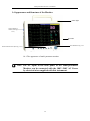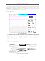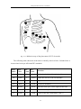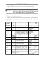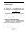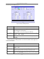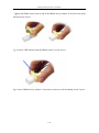
1
L505 Patient monitor user manual Technology that doesn’t miss a beat ® www.daray.com Tel: 0844 375 9000 Email: [email protected] Preface Thank you for using our Multi-parameter Monitor. In order to enable you to skillfully operate Multi-parameter Monitor as soon as possible, we provide this user’s manual with delivery. When you install and use this instrument for the first time, it is imperative that you read carefully all the information that accompanies this instrument. Based on the need to improve the performance and reliability of the parts and the whole instrument, we sometimes will make some amendments to the instrument (including the hardware and software). As a result, there might be cases of discrepancies between the manual and the actual situation of products. When such discrepancies occur, we will try our best to amend or add materials. Your comments and suggestions are welcome. Statement This manual contains exclusive information protected by copyright laws and we reserve its copyright. Without written approval, no parts of this manual shall be photocopied, Xeroxed or translated into other languages. The contents contained in this manual are subject to amendments without notification. Liabilities of the Manufacturer Only under the following circumstances will our company be responsible for the safety, reliability and performance of the instrument. All the installation, expansion, readjustment, renovation or repairs are conducted by the personnel certified by our company. The electrical safety status at the installation site of the instrument conforms to the national standards; The instrument is used in accordance with the operation procedures. CONTENTS CHAPTER 1 GENERAL INTRODUCTION ............................................................... 1-1 1.1 INTENDED USE .......................................................................................................... 1-1 1.2 ABOUT THIS MANUAL .............................................................................................. 1-1 1.3 BRIEF INTRODUCTION TO THE MONITOR ............................................................... 1-3 1.4 APPEARANCE AND STRUCTURE OF THE MONITOR ................................................. 1-4 1.5 SOCKETS .................................................................................................................. 1-5 1.6 FUNCTION BUTTONS AND TRIM KNOB ON THE FRONT PANEL ............................... 1-7 1.7 SPECIFICATIONS AND PERFORMANCE CRITERIA OF THE MONITOR ...................... 1-9 1.7.1 Classification ..................................................................................................... 1-9 1.7.2 Specifications .................................................................................................. 1-10 CHAPTER 2 IMPORTANT SAFETY NOTES ............................................................ 2-1 2.1 INTENDED USE ......................................................................................................... 2-1 2.2 GENERAL SAFETY .................................................................................................... 2-1 2.3 SOME IMPORTANT NOTES FOR SAFETY.................................................................... 2-3 2.4 CLASSIFICATIONS .................................................................................................... 2-5 2.5 SAFE OPERATING AND HANDLING CONDITIONS ..................................................... 2-7 CHAPTER 3 PREPARATIONS BEFORE THE USE OF THE MONITOR ............ 3-1 3.1 UNPACKING THE CASE ............................................................................................. 3-1 3.2 CONNECTING TO POWER ......................................................................................... 3-1 3.2.1 AC Power .......................................................................................................... 3-1 3.2.2 Battery Power .................................................................................................... 3-2 3.3 CONNECTING TO THE CENTRAL MONITOR SYSTEM .............................................. 3-4 3.4 START UP .................................................................................................................. 3-4 3.5 CONNECTING TO VARIOUS KINDS OF SENSORS....................................................... 3-4 3.6 PREPARATION OF RECORDER .................................................................................. 3-4 CHAPTER 4 OPERATION INSTRUCTIONS FOR THE MONITOR .................... 4-1 4.1 SYSTEM MENU ......................................................................................................... 4-1 4.2 MAIN MENU ........................................................................................................ 4-1 4.2.1 Setup patient information .................................................................................. 4-2 4.2.2 Setup system information .................................................................................. 4-2 4.2.3 Setup system time .............................................................................................. 4-3 4.2.4 Setup system sound volume .............................................................................. 4-4 4.2.5 Setup recorder .................................................................................................... 4-4 4.2.6 Restore default system setup ............................................................................. 4-5 4.2.7 Setup work-screen ............................................................................................. 4-5 4.2.8 Setup maintenance (do not belong to user operation item) ............................... 4-6 4.2.9 Setup Manufacturer maintenance ...................................................................... 4-7 4.2.10 Setup Demo function ....................................................................................... 4-7 4.2.11 Display of trend map ....................................................................................... 4-7 4.2.12 Display of trend data ....................................................................................... 4-8 4.2.13 Display of alarm recall .................................................................................... 4-9 4.2.14 Calculate drug concentration ......................................................................... 4-10 4.2.15 Titration table ................................................................................................ 4-12 4.3 SCREEN DISPLAY ................................................................................................... 4-13 4.3.1 Standard screen ................................................................................................ 4-13 4.3.2 7-Leads screen ................................................................................................. 4-14 4.3.3 12-Leads screen ............................................................................................... 4-14 4.3.4 Big Char screen ............................................................................................... 4-15 4.3.5 oxyCRG screen ................................................................................................ 4-16 4.3.6 System state display region ............................................................................. 4-16 4.3.7 Parameters display region ............................................................................... 4-18 4.3.8 Information region ........................................................................................... 4-19 CHAPTER 5 PARAMETERS MEASUREMENT ....................................................... 5-1 5.1 MEASUREMENT OF ECG/HR .................................................................................. 5-1 5.1.1 Principles of Measuring..................................................................................... 5-1 5.1.2 Precautions during ECG Monitoring ................................................................. 5-1 5.1.3 Preparatory Steps before the Measurement of ECG/HR ................................... 5-2 5.1.4 Connecting the ECG Cables to the Monitor ...................................................... 5-3 5.1.5 Connecting the ECG Electrodes to the Patient .................................................. 5-4 5.1.6 Setup of ECG/HR parameters ........................................................................... 5-6 5.1.7 Functions of Arrhythmia Analysis ..................................................................... 5-9 5.1.8 Maintenance and Cleaning .............................................................................. 5-12 5.2 MEASUREMENT OF RESP...................................................................................... 5-13 5.2.1 Principles of Measuring................................................................................... 5-13 5.2.2 Preparatory Steps of the Measurement of RESP ............................................. 5-13 5.2.3 Connect the ECG Cable with Patient and the Monitor ................................... 5-13 5.2.4 Setup of RESP parameters ............................................................................... 5-13 5.2.5 Maintenance and Cleaning .............................................................................. 5-14 5.3 MEASUREMENT OF SPO2/PULSE ........................................................................... 5-15 5.3.1 Principles of Measuring................................................................................... 5-15 5.3.2 Preparatory Steps before the Measurement of SpO2/Pulse ............................. 5-15 5.3.3 Connecting to Patient and Monitor ................................................................. 5-15 5.3.4 Setup of SpO2/Pulse parameters ..................................................................... 5-17 5.3.5 Maintenance and Cleaning .............................................................................. 5-18 5.4 MEASUREMENT OF TEMP .................................................................................... 5-19 5.4.1 Brief Introduction to Measurement of TEMP ................................................. 5-19 5.4.2 Preparatory Steps of the Measurement of TEMP ............................................ 5-19 5.4.3 Connecting Patient and Monitor...................................................................... 5-19 5.4.4 Setup of TEMP Parameters ............................................................................. 5-19 5.4.5 Maintenance and Cleaning .............................................................................. 5-20 5.5 MEASUREMENT OF NIBP ...................................................................................... 5-21 5.5.1 Brief Introduction to Measurement of NIBP ................................................... 5-21 5.5.2 Preparatory Steps of Measurement of NIBP ................................................... 5-22 5.5.3 Connecting to Patient and the Monitor............................................................ 5-22 5.5.4 Setup of NIBP Parameters ............................................................................... 5-23 5.5.5 Precautions during Measurement .................................................................... 5-25 5.5.6 Periodic Check ................................................................................................ 5-26 5.5.7 Maintenance and Cleaning .............................................................................. 5-27 5.6 MEASUREMENT OF IBP ......................................................................................... 5-28 5.6.1 Brief Introduction to Measurement of IBP ...................................................... 5-28 5.6.2 Preparatory Steps for Measurement of IBP ..................................................... 5-28 5.6.3 Calibration of Zero-point................................................................................. 5-30 5.6.4 Connecting to Patient ...................................................................................... 5-30 5.6.5 Setup of Range ................................................................................................ 5-31 5.6.6 Setup of IBP Parameters .................................................................................. 5-31 5.6.7 IBP Transducer Zero and Calibration .............................................................. 5-33 5.6.8 Maintenance and Cleaning .............................................................................. 5-35 5.7 MEASUREMENT OF SIDESTREAM ETCO2 ............................................................. 5-37 5.7.1 Brief Introduction to Measurement of Sidestream EtCO2 .............................. 5-37 5.7.2 Setup of EtCO2 parameters ............................................................................. 5-38 5.7.3 Gain Calibration .............................................................................................. 5-39 5.8 MEASUREMENT OF MAINSTREAM ETCO2 ........................................................... 5-41 5.8.1 Preparatory Steps for Measurement of mainstream EtCO2 ............................ 5-41 5.8.2 Pre-use check ................................................................................................... 5-43 5.8.3 calibration ........................................................................................................ 5-43 5.8.4 Setup of EtCO2 parameters ............................................................................. 5-44 5.8.5 Precautions during Measurement .................................................................... 5-45 CHAPTER 6 ALARM ..................................................................................................... 6-1 6.1 ALARM LEVEL ......................................................................................................... 6-1 6.2 ALARM MODES ........................................................................................................ 6-1 6.3 ALARM SETUP .......................................................................................................... 6-2 6.4 ALARM CAUSE ......................................................................................................... 6-3 CHAPTER 7 PRINT ....................................................................................................... 7-1 CHAPTER 8 THE MAINTENANCE AND CLEANING ........................................... 8-1 8.1 SYSTEM CHECK ....................................................................................................... 8-1 8.2 BATTERY MAINTENANCE ........................................................................................ 8-2 8.3 GENERAL CLEANING ............................................................................................... 8-3 8.4 CLEANING AGENTS .................................................................................................. 8-3 8.5 STERILIZATION ........................................................................................................ 8-4 8.6 DISINFECTION .......................................................................................................... 8-5 APPENDIX A NOTES ON CODE OF THE NIBP ERROR INFORMATION ............ 1 APPENDIX B DEFAULT SYSTEM SETUP ................................................................... 2 Multi-parameter Monitor User Manual Chapter 1 General Introduction 1.1 Intended use Multi-Parameters Patient Monitor is used to monitor patient’s physiological parameters such as ECG RESP SPO2 NIBP IBP TEMP and EtCO2 continuously in dynamic and long time. Its intended to be used in various hospital rooms such as Coronary Care Unit Intensive Care Unit Neonatal Intensive Care Unit and Operating Room to provide additional information to medical and nursing staff about the physiological condition of the patient. 1.2 About this Manual This user’s manual consists of the following chapters: Chapter 1 gives an introduction to the content and the specific signs of this manual, the main features and appearance of the monitor, the basic operations of various buttons, the meanings of the signs on the monitor, specifications and performance criteria of the monitor, the ambient requirements for the working and storage of the monitor. Chapter 2 gives important safety notes Please do read this chapter before using the monitor! Chapter 3 gives an introduction to the preparatory steps before using the monitor. Chapter 4 provides general operation instruction for the monitor, including illustrations of the screen display, normal selection for soft button on screen, details for entry of patient data and trend maps, also. Chapter 5 gives details of specific parameter measurement, preparatory steps, cables or probes connection, setup of parameters, maintenance and cleaning of equipments and sensors. Chapter 6 gives detailed description of system alarm, including level and mode of alarm, default setting and changing procedure of alarm parameters, prompt of specific alarms, and the general operation to carry out when an alarm occurs. 1-1 Multi-parameter Monitor User Manual Chapter 7 gives detailed description of print function. Chapter 8 gives general maintenance and cleaning methods of the monitor and its parts. Signs in this manual: Warning: Means it must be strictly followed so as to prevent the operator or the patient from being harmed. Attention: Means it must be followed so as not to damage the instrument. Note: Important information or indications regarding the operation or use. 1-2 Multi-parameter Monitor User Manual 1.3 Brief Introduction to the Monitor Multi-parameter Monitor is the latest product developed with years’ experience of the monitor design and manufacturing to adapt to the various requirements of present hospitals. It has features as follows: Multiple measuring functions 3-lead, 7-lead ECG/HR, RESP, Dual TEMP, SpO2/Pulse, NIBP, dual IBP,EtCO2 are optional. Complete built-in module design ensures stable and reliable performance Unique all-lead ECG on-one-screen display, which can facilitate the diagnosis and analysis of cardiac disease Can store the trend data for as long as 96 hours and has the function of displaying trend data and trend maps Function of alarm event reviewing, can store 500 pieces of alarm events Function of NIBP measurement reviewing, can store 500 pieces of NIBP measurement data Function of reviewing 10 minute one important lead’s EGC waveform Built-in recorder is optional and it has the functions of real-time recording, printout of the present screen and the triggering of print by alarm Parameter display with capital character Optional function of Calculator of drug concentration Optional function of Display of oxyCRG 12.1 or 10.4 authentic color high brightness TFT LCD monitor Portable design, stylish and convenient Rechargeable maintenance-free battery, can continue working when AC power is off Can be linked with the central unit provided to realize centralized monitoring Is resistant to high-frequency electrotome and is protected against defibrillation effects 1-3 Multi-parameter Monitor User Manual 1.4 Appearance and Structure of the Monitor Alarm light Various kinds of sockets (See Fig. 1-5-1) Recorder Trim Knob See Fig.1-6-1 Function button zone (See Fig. 1-6-1) 1-4-1 The appearance of multi-parameter monitor Note: The AC input at the back panel of the Multi-parameter Monitor can be connected with the 100V~240V AC Power by electrical wires supplied with this instrument. 1-4 Multi-parameter Monitor User Manual 1.5 Sockets IBP1 IBP ECG1 ECG IBP2 ECG2 SpO2 TEMP2 SPO2 TEMP NIBP TEMP1 NIBP Fig. 1-5-1 Various sockets on the panel at the right side (full) NETWORK FUSE T1.6A FUSE ELECTRICAC SOCKET AC 100V-240V NETWORK CONNECT OR Fig. 1-5-2 Various sockets on the panel at the back Note: The Network Connector choose RJ45 socket and be used for connection with the central monitoring system provided. The RS-232 Serial Port is only used for the maintenance and upgrade of monitor. 1-5 Multi-parameter Monitor User Manual Warning: The sensor cable sockets on Multi-parameter Monitor can only be connected with the sensor cables supplied with this instrument and no other cables shall be used. Notes on the signs on the right-side panel Signs Notes on the signs ECG 1 Short for “Electrocardiogram” channel 1. ECG 2 Short for “Electrocardiogram” channel 2. CF Type Sign for the Prevention of Defibrillation (Refer to IEC 601-1:1988) Here it means the measurement design for ECG parameters of this monitor features special protection against electrical shock (especially when there is allowed leaking current) and has protection of defibrillation. BF type equipment flag ( Refer to IEC 601-1-1988) This flag indicate the application part is BF type, Design for special protection against electrical shock (especially when there is allowed leaking current ) and has protection lf defibrillation. It means BF type sign for the equipment has protection of defibrillation. Attention! Please refer to the document supplied with this instrument (this manual)! Dangerous voltage AC power Non-ionizing radiation SpO2 Short for “Pulse Oxygen Saturation” TEMP 1 Short for “Temperature” channel 1 TEMP 2 Short for “Temperature” channel 2 1-6 Multi-parameter Monitor User Manual IBP 1 Short for “Invasive Blood Pressure” channel 1 IBP 2 Short for “Invasive Blood Pressure” channel 2 NIBP Short for “Non-invasive Blood Pressure” Note: The two channels of ECG1, ECG2 for ECG are used for the realization of the input for all-lead electrocardiogram; plug the 5-lead ECG cable with limb lead (RA, LA, C, RL, LL) into ECG1 (Channel 1), and the chest lead cable (C1, C2, C3, C5, C6) into ECG2 (Channel 2), and at this moment, the 12-lead (Full-lead) ECG monitoring can be realized. In case only 7-lead monitoring is needed, then it is only necessary to plug the 5-lead ECG cable of the limb lead (RA, LA, C, RL, LL) into ECG1 (Channel 1). Be careful not to plug it into ECG2 (Channel 2). 1.6 Function Buttons and Trim Knob on the Front Panel EXIT POWER ON/OFF switch of AC/DC Indicating Exit the light present of AC/DC menu or FREEZE Freeze/d efreeze wavefor ms SILENCE Allow/ban the sounding of alarm PRINT Record the real-time waveforms NIBP MENU Start/Stop measurements of NIBP present screen Turn the Trim Knob to move the cursor (selection box) for selection of the items on the menu or the soft buttons on the screen; press the Trim Knob can confirm the present selection. Fig. 1-6-1 Function Buttons and Trim Knob on the Front Panel 1-7 Menuu Multi-parameter Monitor User Manual 1.6.1 The Signs and Operation Instructions Within the Function Button Zone Signs power Notes on the signs Operation instructions of function buttons When the monitor is connected to the AC power, this indicating light is green (it is unrelated to the ON/OFF state of the monitor), and, at this moment, the Indicating light of rechargeable maintenance-free battery is in the state of AC/DC recharging. When the monitor is not connected to AC power and the DC battery is used as the power source, this indicating light is orange Power button Press this button once and the monitor starts up. Repress this button, then the monitor is switched off. Press this button once to exit the present menu or the present screen. EXIT Exit In the system menu, trend screen and parameters setup screen, there are selections of <Return> or <Exit>, and this button is its short-cut key. Switching type button Begin andStop the measurement of NIBP Press this button to start the air pump and begin the measurement of NIBP. Again press this button to stop the air pump and begin the measurement of NIBP. Switching type button Press this button to stop sound alarm (when the alarm is on, the sound is not produced). Repress this button once to initiate the sound alarm (When sending out alarm, the system produces a sound). Switching type button Press this button once to freeze the waveform. Repress this button to defreeze the waveform. PRINT Switching type Real-time recording Press this button to begin real-time recording. Repress this button to stop real-time recording. MENU MENU NIBP SILENGE FREEZE press the Trim Knob can confirm the present selection. 1-8 Multi-parameter Monitor User Manual 1.6.2 Basic Operations Turn the Trim Knob to select the item or soft button on the screen Press EXIT button to exit the menu or present screen Press the Trim Knob to confirm selection Perform the operation Fig. 1-6-2 Flow chart of basic operations Note: The system menu is located at the left bottom corner (refer to Fig.4-1). By operating the Trim Knob in the above flow chart, select the items or make them spring out, and for detailed item selection, please refer to Chapter 4. 1.7 Specifications and Performance Criteria of the Monitor 1.7.1 Classification Instrument of Class I powered by external power and instrument with internal power supply BF, CF type equipment, among which the measuring parts of ECG, TEMP, RESP and IBP belong to CF type, and the measuring parts of SpO2 and NIBP belong to CF type, too. Ordinary equipment (sealed equipment not protected against permeating fluid) For the methods of disinfections and sterilization, refer to Chapter 8 of this manual Equipment of continuous running 1-9 Multi-parameter Monitor User Manual 1.7.2 Specifications Size and Weight Name of the monitor Multi-parameter Monitor Size 318mm 264mm 152mm Weight 4.5Kg System output 12.1 or 10.4 LCD color display screen, with a Display type resolution of 800 600 or higher Interface with central unit Power supply Power Voltage Power Input Network port AC 100V~240V 50/60Hz 70VA Fuse T1.6AL 250V Safety class Category I, CF Type Battery Type Rechargeable Lead acid cell, 12V/2.0AH Charge time 6 hours Working hours when charged fully 30min when using normally Environment Ambient Temperature Relative humidity Working 0~+40ºC Transportation and storage –20~+50ºC Working 85% Transportation and storage Atmospheric pressure 93% Working 860~1060 hPa Transportation and storage 500~1060 hPa 1-10 Multi-parameter Monitor User Manual ECG Lead Mode Lead selection Gain Input impedance 1. 5-leads ECG input 2. 3-leads ECG input 3. 10-leads ECG input 1. I, II, III, aVR, aVL, aVF, V2. I, II, III 3. I, II, III, aVR, aVL, aVF, V1~V6 2.5mm/mV, 5mm/mV, 10mm/mV, 40mm/mV 5.0 Mohm ~ CMRR DIAG ≥85dB MON ≥105dB OPS ≥105dB Frequency response DIAG 0.05~75Hz MON 0.5~25Hz OPS 1~15Hz Electrode offset potential ±300 mV d.c. → ±600mV d.c. Leakage Current <10 uA ECG signal range ±6.0 mV Baseline recovery <5 s After Defi. (MON or OPS mode) Insulation Breakdown Voltage 4000VAC 50Hz/60Hz Indication of electrode separation Available Sweep speed 12.5mm/s, 25mm/s, 50mm/s HR Measuring and Alarm Range 10~300 bpm Refreshing time Per pulse Resolution 1 bpm Accuracy 1% or 1 bpm, whichever is greater 1-11 20mm/mV, Multi-parameter Monitor User Manual Alarm range 10~300 bpm, continuously adjustable between high limit and low limit Alarm indication Sound and light alarming Way of measurement Automatic oscillometry NIBP Adult Range of measurement Child Neonate Cuff pressure range 0~280 mmHg Resolution Pressure Accuracy Static Clinical 1 mmHg 2% or SYS 30~270mmHg DIA 10~220 mmHg MEAN 20~235 mmHg SYS 30~235 mmHg DIA 10~220mmHg MEAN 20~225mmHg SYS 30~135 mmHg DIA 10~110 mmHg MEAN 20~125 mmHg 3 mmHg, whichever is greater 5 mmHg average error 8 mmHg standard deviation Unit mmHg, kPa Pulse rate measuring range 40 ~ 240 bpm Inflation time for cuff Less than 40 sec. (standard adult cuff) 20 to 45 seconds typical (dependent on heart rate and motion artifact) Total cycle time Intervals for cycle measurement time 1,2,3,4,5,10,15,20,30,45,60,90 minutes Overpressure Protection Adult Child Neonatal 2,4,8 hours Hardware and software double protections 315±10 mmHg 265±10 mmHg 155±10 mmHg Range of alarm SYS 0~300 mmHg, continuously adjustable between high limit and low limit 1-12 Multi-parameter Monitor User Manual DIA 0~300 mmHg, continuously adjustable between high limit and low limit MEAN 0~300 mmHg, continuously adjustable between high limit and low limit Alarm indication Sound and light alarming Ways of measurement Manual Range of measurement 0~100% Resolution 1% Auto and STAT SpO2 At 70~100%, ± 2% Precision At 35~69%, ± 3% At 0~34%, unspecified Range of alarm 0~100%, continuously adjustable between high limit and low limit Alarm indication Sound and light alarming Type of sensor PR: Measuring and Range Resolution Accuracy Dual bandwidth light emitting diode 25~250 bpm alarm 1 bpm 2% or 1 bpm, whichever is greater IBP Range of measurement -45 ~ +300 mmHg Resolution 1 mmHg Precision ± 4mmHg or 4% of the reading (select the bigger one of the two) Sensitivity of transducer 5uV/V/mmHg, 2% Impedance of transducer 300~3000 Ohm Bandwidth d.c. ~ 15Hz Transducer sites Arterial Pressure (ART) Pulmonary Artery Pressure (PA) Left Atrium Pressure (LAP) Right Atrium Pressure (RAP) Central Venous Pressure (CVP) Intracranial Pressure (ICP) 1-13 Multi-parameter Monitor User Manual 0~50mmHg 50~150 mmHg ART 100~240 mmHg 0~300 mmHg AUTO Selection of measurement range PA 0~20 mmHg CVP 0~30 mmHg LAP 0~50 mmHg RAP 0~80 mmHg ICP AUTO (Among them, the AUTO switches automatically at an interval of 10 mmHg so as to ensure the waveform is at the state most suitable for observation) Range of alarm SYS -10~300 mmHg, continuously adjustable between high limit and low limit DIA -10~300 mmHg, continuously adjustable between high limit and low limit MEAN -10~300 mmHg, continuously adjustable between high limit and low limit Alarm indication Sound and light alarm Unit mmHg, kPa Insulation resistance >5000M /1000V Breakdown voltage >4000V AC 50Hz/60Hz TEMP Range of measurement 0.0~50.0ºC Resolution of display 0.1ºC Unit Celsius (ºC), Fahrenheit (ºF) Precision 0.1ºC Connecting cable Compatible with YSI-400 Range of alarm 0.0~50.0ºC, continuously adjustable between high limit and low limit 1-14 Multi-parameter Monitor User Manual Alarm indication Sound and light alarming Method Impedance between RA-LL (R-F) RESP Measuring range impedance 0.5 ~3 Ohms Base line impedance range 500~4000 Ohms (50~120 kHz exciting frequency Range of measurement 0~120 bpm Precision ±2 bpm Gain x1 x2 x4 Range of alarm 0~120 bpm, continuously adjustable between high limit and low limit Sweep speed 6.25mm/s, 12.5mm/s, 25mm/s Asphyxia 1~60s, off Alarm indication Sound and light indication EtCO2 (Sidestream) Measure method Infrared spectrum Measure mode Sidestream Range of measurement EtCO2 0.0~13.1% (0~99 mmHg) Resolution 1 mmHg Unit %, mmHg Precision At <5 % CO2 ±0.3% (±2.0 mmHg) At ≥5 % CO2, < 10 % of reading Range of respiration rate 3~150 bpm measurement Calibration Offset calibration: auto, manual Gain calibration Range of alarm 0.0~13.1 % (0~99 mmHg), continuously adjustable between high limit and low limit Alarm indication Sound and light indication EtCO2 (Mainstream) 1-15 Multi-parameter Monitor User Manual Measure method Infrared spectrum Measure mode Mainstream Range of measurement EtCO2 0.0~10% (0~76 mmHg) Resolution 1 mmHg Unit %, mmHg Precision 0.5 % (±4.0 mmHg) or < 10 % of reading, whichever is greater Range of respiration rate 3~150 bpm measurement Range of alarm 0.0~13.1 % (0~99 mmHg), continuously adjustable between high limit and low limit Alarm indication Sound and light indication Recorder Speed of paper feeding 12.5mm/s, 25mm/s, 50 mm/s (Only valid whit B Type recorder) Recording medium 50mm thermal paper Recording track Single track or tri-track Alarm Level Low, medium and high Indication Sound and light indication Setup Default and custom Silence All alarms can be silenced Sound pressure 45~85 dB (At 1m distance) 1-16 Multi-parameter Monitor User Manual Chapter 2 Important Safety Notes 2.1 Intended Use Multi-Parameters Patient Monitor is used to monitor patient’s physiological parameters such as ECG RESP SPO2 NIBP IBP and TEMP continuously in dynamic and long time Unit Its intended to be used in various hospital rooms such as Coronary Care Intensive Care Unit Neonatal Intensive Care Unit and Operating Room to provide additional information to medical and nursing staff about the physiological condition of the patient. Warning: Only trained doctors and nurses can use the device. Warning: Multi-parameter Monitor is not a therapeutic instrument nor is it a device that can be used at home. 2.2 General Safety 1. Safety precautions for safe installation The input socket of Multi-parameter Monitor can be connected to the electrical wires and common electrical wire can be used. Only the power supply type of AC 100V~240V 50/60Hz specified by Multi-parameter Monitor can be used. Connect the electrical wire to a properly grounded socket. Avoid putting the socket used for it in the same loop of such devices as the air conditioners, which regularly switch between ON and OFF. Avoid putting the monitor in the locations where it easily shakes or wobbles. Enough room shall be left around the monitor so as to guarantee normal ventilation. Make sure the ambient temperature and humidity are stable and avoid the occurrence of condensation in the work process of the monitor. Warning: Never install the monitor in an environment where flammable 2-1 Multi-parameter Monitor User Manual anesthetic gas is present. 2. Multi-Parameter Monitor conforms to the safety requirements of . This monitor is protected against defibrillation effects. 3. Notes on signs related to safety CF type defibrillation prevention sign (refer to IEC 60601-1-1998) The unit displaying this symbol contains an F-Type isolated (floating) applied part providing a high degree of protection against shock, and is defibrillator-proof. The type CF applied parts provide a higher degree of protection against electric shock than that provided by type BF applied parts. Attention! Please refer to the documents accompanying this monitor (this manual)! BF type equipment flag ( IEC 60601-1-1988) The unit displaying this symbol contains an F-Type isolated (floating) applied part providing a high degree of protection against shock, and is defibrillator-proof. 4. When a defibrillator is applied on a patient, the monitor may have transient disorders in the display of waveforms. If the electrodes are used and placed properly, the display of the monitor will be restored within 10 seconds. During defibrillation, please note to remove the electrode of chest lead and move the electrode of limb lead to the side of the limb. The electrode of the defibrillator should not come into direct contact with the monitoring electrodes. Please ensure the monitor is reliably grounded and the electrodes used repeatedly should be kept clean. Warning: When conducting defibrillation, do not come into contact with the patient, the bed and the monitor. Otherwise serious injury or death could result. 2-2 Multi-parameter Monitor User Manual 5. To guarantee the safe operation of the monitor, Multi-Parameter Monitor is provided with various replaceable parts, accessories and consuming materials (such as sensors and their cables, electrode pads). Please use the products provided or designated by the manufacturer. 6. Multi-parameter Monitor only guarantees its safety and accuracy under the condition that it is connected to the devices provided. If the monitor is connected to other undesignated electrical equipment or devices, safety hazards may occur due to such causes as the cumulating of the leakage current. 7. To guarantee the normal and safe operation of the monitor, a preventive check and maintenance should be conducted for the monitor and its parts every 6-12 months (including performance check and safety check) to verify the instrument can work normally and is in a proper working condition and it is safe to the medical personnel and the patient and has met the accuracy required by clinical use. Attention: The monitor does not contain any parts for self-repair by users. The repair of the instrument must be conducted by the technical personnel authorized by company. 2.3 Some important notes for safety PATIENT NUMBER The monitor can only be applied to one patient at a time. INTERFERENCE Do not use cellular phone in the vicinity of this equipment. High level of electromagnetic radiation emitted from such devices may result in strong interference with the monitor performance. ACCIDENTAL SPILLS To avoid electric shock or device malfunction liquids must not be allowed to enter the device. If liquids have entered a device, take it out of service and have it checked by a service technician before it is used again. ACCURACY If the accuracy of any value displayed on the monitor or printed on a printout paper is questionable, determine the patient’s vital signs by alternative means. Verify that all equipment is working correctly. 2-3 Multi-parameter Monitor User Manual ALARMS Do not rely exclusively on the audible alarm system for patient monitoring. Adjustment of alarm volume to a low level or off during patient monitoring may result in a hazard to the patient. Remember that the most reliable method of patient monitoring combines close personal surveillance with correct operation of monitoring equipment. The functions of the alarm system for monitoring the patient must be verified at regular intervals. BEFORE USE Before putting the system into operation visually inspect all connecting cables for signs of damage. Damaged cables and connectors must be replaced immediately. Before using the system, the operator must verify that it is in correct working order and operating condition. Periodically, and whenever the integrity of the product is in doubt, test all functions. CABLES Route all cables away from patient’s throat to avoid possible strangulation. DISCHAGE TO CLEAR PATIENT DATA When admitting a new patient, you must clear all previous patient data from the system. To accomplish this, shut down the device, then turn on it. DISPOSAL OF PACKAGE Dispose of the packaging material, observing the applicable waste control regulations and keeping it out of children’s reach. EXPLOSION HAZARD Do not use this equipment in the presence of flammable anesthetics, vapors or liquids. LEAKAGE CURRENT TEST When interfacing with other equipment, a test for leakage current must be performed by qualified biomedical engineering personnel before using with patients. BATTERY POWER The device is equipped with a battery pack. The battery discharge even when the device is not in use. Store the device with a fully charged battery and check the battery status of the devices in storage once a month. DISPOSAL OF ACCESSORIES AND DEVICE Disposable devices are intended for single use only. They should not be reused as performance could degrade or contamination could occur. The service life of is five years. At the end of its service life, the product described in this manual, as well as its accessories, must be disposed of in compliance with the 2-4 Multi-parameter Monitor User Manual guidelines regulating the disposal of such products. If you have questions concerning disposal of products, please contact company or its representatives. EMC Magnetic and electrical fields are capable of interfering with the proper performance of the device. For this reason, make sure that all external devices operated in the vicinity of the monitor comply with the relevant EMC requirements. X-ray equipment or MRI devices are a possible source of interference as they may emit higher levels of electromagnetic radiation. Also, keep cellular phones or other telecommunication equipment away from the monitor. INSTRUCTION FOR USE For continued safe use of this equipment, it is necessary that listed instructions were followed. However, instructions listed in this manual in no way supersede established medical practices concerning patient care. LOSS OF DATA Should the monitor at any time temporarily lose patient data, the potential exists that active monitoring not being done. Close patient observation or alternate monitoring devices should be used until monitor function is restored. If the monitor does not automatically resume operation within 60 seconds, power cycle the monitor using the power on/off switch. Once monitoring is restored, you should verify correct monitoring state and alarm function. 2.4 Classifications The Monitor is classified, according to IEC601-1:1988 as: Type of protection against electric shock: I Degree of protection against electric shock: CF: ECG, Temp, Resp, IBP CF: NIBP, SpO2 Degree of protection against harmful Ordinary Equipment (enclosed equipment ingress of water: without protection against ingress of water) Degree of safety of application in the Not suitable presence of a flammable anesthetic-mixture with air or with oxygen or nitrous oxide: Mode of operation: Continuous operation I: Class I equipment CF: Type CF applied part BF: Type BF applied part Not suitable: Equipment not suitable for use in the presence of flammable anesthetic mixture with air or with oxygen or nitrous oxide. 2-5 Multi-parameter Monitor User Manual 2-6 Multi-parameter Monitor User Manual 2.5 Safe Operating and Handling Conditions Method(s) of sterilization or disinfection Sterilization: not applicable recommended by the manufacturer: Disinfection: See “The Maintenance and Cleaning of the System->General Cleaning” Electromagnetic interference No cellular telephone nearby Electrosurgical interference damage No damage Diathermy instruments influence Displayed values and prints may be disturbed or erroneous during diathermy. Defibrillation shocks The monitor specifications fulfill the requirements of EC13, IEC 601-2-27, -30, -34 and EN865. Auxiliary outputs The system must fulfil the requirements of standard IEC 601-1-1. 2-7 Multi-parameter Monitor User Manual Chapter 3 Preparations before the Use of the Monitor 3.1 Unpacking the Case Unpack the packaging case There are two layers inside the box and the upper paper box contains such documents as the electrical wire, various patient sensors and user’s manual (this manual), warranty card, certificate and particular paper and the lower foam case contains the monitor. Remove the monitor and accessories Attention: please place the monitor on level and stable supporting plane, not on the places that can easily shock or wake. Enough room should be left around the monitor so as to guarantee normal ventilation. Keep all the packaging materials for future use in transportation or storage. Check the monitor and accessories Check the monitor and its accessories one by one in accordance with the particular paper. Check to see if the parts have any mechanical damages. In case of problems, contact our company or the agent of our company. 3.2 Connecting to Power 3.2.1 AC Power Confirm the rated AC current is: AC 100V~240V 50/60Hz Remove the electrical wires provided along with the instrument, and put its output end plug (round headed) into the AC current socket on the back of the monitor, and the plug of input end into a grounded socket of the mains (It must be a special socket of the hospital), connect the monitor through the earth one of electrical wires. When the indicating light above the power switch on the panel of the monitor is green, it means the AC power is on. And when the monitor is not connected to AC power and the built-in DC battery is used as the power source, the indicating light is orange. 3-1 Multi-parameter Monitor User Manual Warning: The AC cord provided along with the instrument must be used for connection with AC power and no other electrical wires shall be used. Warning: The monitor must be connected to a properly installed power outlet with protective earth contacts only. If the installation does not provide for a protective earth conductor, disconnect the monitor from the power line and operate it on battery power. The equipment has no mains switch. The equipment is switched completely only by disconnecting the power supply from the wall socket. The wall socket have to be easily accessible. Note: For measurements in or near the heart we recommend connecting the monitor to the potential equalization system. Use the green and yellow potential equalization cable and connect it to the pin labeled with the symbol. 3.2.2 Battery Power The monitor has a battery pack to provide power to the monitor whenever AC power is interrupted. The battery is generally referred to as the “battery”. You must charge the battery before using it. There is no external charger. The battery is charged when the monitor is connected to AC power. A fully depleted battery will take about 15 hours to fully charge. To assure a fully charged battery which is ready for use, we recommend that the monitor be plugged into AC power whenever it is not in use. Depending on usage, you can get 60 to 90 minutes of battery power on a new, fully-charged battery on a monitor. NIBP and SpO2 monitoring and the usage of the recorder will drain battery power faster than other parameters. Note: When the monitor is connected to AC current, the battery is in a state of being recharged. When it is unable to be connected to the AC current, the battery can be used to supply power, and at this time it is unnecessary to use the electrical wires, and the instrument can be switched on directly. 3-2 Multi-parameter Monitor User Manual Note: A “Cell battery too low” message at the bottom of the screen and an audible system alarm indicate 5 minutes of battery life remaining. You should connect the monitor to an AC power source when the message is displayed. Attention: The battery discharges even when the device is not operating. Please ensure that the battery is always fully charged when you are keeping the device in storage for an extended period of time. Check the battery status at least once every month and recharge the battery. Note: This monitor contains a rechargeable battery. The average life span of this type of battery is approximately three years. When replacement becomes necessary, contact a qualified service representative to perform the replacement. Disposal Notice Should this product become damaged beyond repair, or for some reason its useful life is considered to be at an end, please observe all local, state, and federal regulations that relate to the disposal of products that contain lead, batteries, plastics, etc. Install Battety The battery storage is located at the bottom of the monitor, following the steps to install a battery. 1 Open the battery gate according to the direction marked on the monitor. 2 Push the baffle to the opposite side of the battery. 3 Push the battery into a with the electrode point to the bottom of the monitor. 4 After push the battery inside the storage withdraw the baffle to the middle position. 5 Close the gate. Uninstall battery Open the battery gate according to the direction marked on the monitor. 2 Push the baffle to the opposite side of the battery. 3 Take out the battery, and then close the gate. 3-3 Multi-parameter Monitor User Manual 3.3 Connecting to the Central Monitor System If the user intends to connect the monitor to the central monitoring system, plug its connecting electrical cable into the Network Connector interface at the back of the monitor. Note: This monitor can only be connected to the central monitoring system provided and do not attempt to connect this monitor to other central monitoring system. 3.4 Start up Press the power switch on the front panel of the monitor (refer to Fig. 1-4) About 50 seconds after the monitor is switched on, after passing the self-examination of the system, the monitor enters the monitoring screen. Warning: In case the monitor is found to be working abnormally or indication of errors appears, please do not use this monitor for monitoring and should contact the after-sale service center as soon as possible. 3.5 Connecting to Various Kinds of Sensors Connect various kinds of sensors needed to the relevant sockets on the monitor and the monitored locations on the body of the patient. Refer to the relevant content of Chapter 5 for details. 3.6 Preparation of Recorder If the monitor you use has been provided with a recorder, before starting of monitoring please check if the recorder has had recording thermal paper installed. The thermal side (that is the smoother side) should face upwards and a small section should be pulled out onto the outlet of the paper (on the left panel of the monitor, refer to Fig. 1-1) 3-4 Multi-parameter Monitor User Manual Chapter 4 Operation Instructions for the Monitor Note: For Concision, the following terms are used to describe one or more operations Choice--press the Trim Knob and move the cursor onto the item that needs to be changed. Conform-- press the Trim Knob. Select-- move the cursor onto the item and press the Trim Knob. 4.1 System Menu Hide and display system menu. The profile of system menu is shown in Fig. 4-1-1. To hide, press the Exit key; to display the system menu under hide mode, press the Trim Knob. Fig. 4-1-1: System Menu 4.2 Main Menu There is a system information region in each dialogue window for prompting current operation. Press MAIN button, and a following setup dialogue window (Fig. 4-2.1) will be displayed. Fig. 4-2-1.1: Main menu 4-1 Multi-parameter Monitor User Manual 4.2.1 Setup patient information Press Patient Info… button, and a following patient information dialogue window (Fig. 4-2-1.2) will be displayed. Fig. 4-2-1.2: Setup patient information Name Room The name of patient (choose from A-Z and 0-9). The length of name can be 15 characters at most. Enter OK and enter. The number of patient (setup due to the actual condition of the hospital). Enter OK and enter. The number of patient room. Bed The number of patient bed. Height The height of patient (turn the dial, once to increase/decrease 1 cm). Sex The sex of patient (male, female). Age Weight The age of patient. The weight of patient (turn the dial, once to increase/decrease 1 kg). Case 4.2.2 Setup system information Press System info… button, and a following setup system information dialogue window (Fig. 4-2-2) will be displayed. Language: User can setup displaying language of the system. Sweep Speed: User can choose from three levels: 12.5mm/s, 25mm/s, 50mm/s. Setting 50mm/s in type A recorder, which only support print speed of 25mm/s, will result in the recorder printing failure. 4-2 Multi-parameter Monitor User Manual Fig.4-2-2: Setup system information 4.2.3 Setup system time Press Time Setup… button and a following setup system time information window (figure 4-2-3) will be displayed. Fig, 4-2-3: Setup system time User can setup system time information. Setting time before monitoring is suggested. If time is setup during monitoring, we suggest close the current window, switch off and restart. The changed time will be available after exit the current window. 4-3 Multi-parameter Monitor User Manual 4.2.4 Setup system sound volume Press Volume setup… button and a following system sound volume window (Fig. 4-2-4) will be displayed. Fig. 4-2-4: Setup system sound volume KEY VOL SYS VOL Select keyboard sound ON or OFF. Choose the level (1-3) of heart beat sound volume or turn OFF. ALM VOL Choose the level (1-3) of alarm sound volume or turn OFF. 4.2.5 Setup recorder Press Recorder Setup… button and a following setup recorder window (figure 4-2-5) will be displayed. Fig.4-2-5: Setup recorder AUTO REC ALM REC Turn off cycle printing or select from 1, 5, 10, 30, 60 minutes interval to do cycle print. Turn off alarm printing or turn on to print automatically when alarming. 4-4 Multi-parameter Monitor User Manual 4.2.6 Restore default system setup Press Default System button, and a following Default System Setup window (figure 4-2-6) will be displayed, select one item in this window will restore the system setup to default setup. There are three options: ADULT, CHILD, NEONATAL. Fig.4-2-6: Restore default system setup 4.2.7 Setup work-screen Press Work-Screen… button and a following setup display screen window (Fig.4-2-7) will be displayed. Fig. 4-2-7: Setup work-screen Standard Screen 7-Leads Screen The Standard screen displays the parameters and the waveforms being monitored. This is the basic operating screen of the monitor. A form in which seven leads of ECG waves is displayed in one screen. 4-5 Multi-parameter Monitor User Manual 12-Leads Screen BigChar Screen ViewBed Screen oxyCRG Screen A form in which twelve leads of ECG waveforms is displayed in one screen (only available after twelve leads module is configured). A form in which one lead of ECG waveform and the parameters are displayed in magnified characters. A form that is able to monitor one lead of ECG waveform and parameters of other bed (This function is not valid). One lead of ECG, respiration waveform, blood oxygen waveform, totaling to three lines of waveforms. The lowest port of the screen is the oxyCRG screen consisting of three trends: HR Trend, SpO2 Trend and RR Trend. 4.2.8 Setup maintenance (do not belong to user operation item) Press Maintenance… button, and a following window (Fig. 4-2-8.1) that ask to enter password will be displayed. Fig.4-2-8.1 Enter password window Entering the wrong password will exit current window without prompt. Entering the right password (125689) will display following equipment maintenance dialogue window (Fig. 4-2-8.2). Fig. 4-2-8.2 Setup maintenance 4-6 Multi-parameter Monitor User Manual IP ADDR: It is the IP address of this bedside equipment. When programming the IP address of the equipment, Caution that this IP address is a critical data of system in constituting central monitoring network, and therefore should not conflict with other equipments in the network. MASK: It is the IP address masks of this bedside equipment. Masks and IP address constitution fix this unique equipment in the network. GATEWAY: It is the gateway of the network. Set this item to insure that all monitors are in the same section of network. MAC NO: It is the only one equipment number in this system: 1-64. Do not repeat, since it is anther critical data of system in constituting central monitoring network. 4.2.9 Setup Manufacturer maintenance Not belong to user operation item. 4.2.10 Setup Demo function Press Demo… button, and a following window (Fig. 4-2-10) that ask to enter password will be displayed. Fig.4-2-10 Enter password window Entering the wrong password will exit current window without prompt. Entering the right password will enters the Demonstration Waveform status. The purpose of waveform demonstration is only to demonstrate the machine performance, and for training purpose. In clinical application, this function is not recommended because the DEMO will mislead the hospital workers to treat the waveform and parameter as actual data of the patient, which may result in delay of treatment or mistreatment. 4.2.11 Display of trend map Press Trend Graph… button and a following trend graph window (Fig. 4-2-11) will be displayed. 4-7 Multi-parameter Monitor User Manual Fig. 4-2-11: Display of trend graph multiple-parameter monitor can store 96 hours’ trend data. PARA: One of the parameters of HR, SpO2, PR, RR, IBP1, IBP2, T1, T2, EtCO2 can be chosen to see over its trend graph. INTER: You can choose from 1, 5, 10, 30, 60 minutes, which is the minimum unit when using dial to move cursor after selecting “move cursor”. PAGE Up/Down: One screen of trend map express 24 hours. Press it, and use the dial to turn pages. Move Cursor: Press it, and use the dial to move cursor. REC: Print current trend graph through recorder. Parameters of current location of the cursor can be displayed in the top right corner. 4.2.12 Display of trend data Press Trend Table… button and a following trend data window (Fig. 4-2-12) will be displayed. 4-8 Multi-parameter Monitor User Manual Fig.4-2-12: Display of trend data INTER: Displayed intervals between trend data. Options: 1, 5, 10, 30, 60 minutes. |<<: Turn to the first page. >>|: Turn to the last page. <</>>: After press this button, you can turn the page forward/backward by turning the dial. REC: Print the trend data in current screen through recorder. 4.2.13 Display of alarm recall Press Alarm Recall… button a following alarm recall window (Fig.4-2-13) will be displayed. Fig. 4-2-13 Display of alarm recall 4-9 Multi-parameter Monitor User Manual |<<: Turn to the first page >>|: Turn to the last page. <</>>: After selecting this button, you can turn one page forward/backward by turning the dial. REC: Print current alarm events through recorder. Clear: Clean up all of the alarm events records. 4.2.14 Calculate drug concentration Press Drug Calculation… button and a following drug concentration calculating window (Fig. 4-2-14) will be displayed. Fig. 4-2-14 This drug concentration calculation is to facilitate doctor’s work, to do calculation of some frequently used drugs, and to output the content of titration table in the recorder. The types of drugs which can be calculated in the system are as follows: AMINOPHYLLINE ISUPREL DOBUTAMINE LIDOCAINE NIPRIDE DOPAMINE EPINEPHRINE NITROGLYCERIN PITOCIN. Drug A, drug B, drug C, drug D, drug E are provided additionally to replace any of the drugs. 4-10 HEPARIN, Multi-parameter Monitor User Manual Use following formulas to calculate drug dosage Concentration = Drug total amount/ Liquid volume INF Rate = Drug dosage / Drug concentration Duration = Drug total amount/ drug dosage Drug dosage = INF Rate x Drug concentration In drug calculation window, operator should first select the name of the desired drug, and then enter patient’s weight, and then enter other known values. Drug To select drug, move the cursor to Drug” press the dial, and then turn the dial. Only one type of drug can be chosen each time to do calculation. Drug A, Drug B, Drug C, Drug D, Drug E are not real drug names, but merely drug symbols. The respective units of these five drugs are unchangeable, and operator can choose appropriate units due to usage traditions. The principle of units denotation is as follows The units of Drug A, Drug B Drug C are fixed as a series of “gram”: g, mg, mcg. The units of Drug D are fixed as a series of “unit”: unit The units of Drug E are fixed as mEq k unit m unit. . Weight: Operator should enter the weight of patient in the first or the second item. It is an independent data and can be used only in drug calculation function. Turn the dial and move the cursor respectively to each calculate item of the formula then press the dial, select the value, and press the dial to confirm it. The value will be displayed in the proper place after selecting. The value of each calculate item should be within its limit; if the calculation result is beyond the limit, --- will be displayed The value of other menu items can be input only after the weight of patient and the drug name have been entered. The initial values offered by the system are random numbers, and cannot be used as calculation standard. Operator should reenter a series of values that are appropriate for the patients due to doctor’s advice. The units of each drug, which may be one or a series of units, are unchangeable. Operator should select appropriate units due to doctor’s advice. In one series of units, unit system will be adjusted automatically due to current input value. If the value is beyond the limit, --- will be displayed automatically. 4-11 Multi-parameter Monitor User Manual After one value is entered, a prompt will be displayed automatically to remind the operator to examine the accuracy of the input value. Calculation results can be reliable and safe only when the accuracy of all of the input values is assured. Instillation speed and drop volume are out of function under newborn condition. The values in the table can be irrelevant to the patient monitored by this equipment. Therefore the value of patient’s weight in this menu can be different from the same item in patient’s information. The value of this menu will not be influenced by patient’s information. 4.2.15 Titration table To enter titration table (figure 4-15), press Titration table button in drug calculation menu, and a following titration table window (Fig. 4-2-15) will be displayed. Fig. 4-2-15 Display of titration table In titration table turn the dial and move the cursor to “BASE” and then press the dial to select the desired item. Three items, “DOSE”, “INF RATE” and “DRIP RATE”, can be chosen. Press the dial to confirm after selecting. Move the cursor to “STEP”, press the dial to select step length. Any value within 1-10 is available. Move the cursor to dosage unit selection box, press the dial to select dosage unit. Move the cursor to “PAGE UP/DOWN” press the dial 4-12 turn it to see over the Multi-parameter Monitor User Manual preceding page and following page. Move the cursor to “REC”, press the dial, output data of titration table in current displayed environment. Move the cursor to “Exit”, press the dial to back to “drug calculation” window. 4.3 Screen Display The monitor applies high-lightness multicolor LCD screen, which can show all the parameters, waveforms, data, system time, information of the patients and other prompt information. The whole main screen can be divided into four regions, which are: Patients Information and alarm prompt information region (upmost) User operation menu region (bottommost) Waveform display region (left) Parameters display region (right) multiple-parameter monitor has two categories: all-parameter configuration and standard-parameter configuration. Both of them have almost the same operating method. The main difference is that the standard-parameter configuration does not have IBP module and 12-lead ECG module. The standard-parameter configuration is a single-temperature module, while the all-parameter one is a double-temperature module. There are six display patterns (which can be set in <Work-Screen> in <Main> menu. 4.3.1 Standard screen Fig. 4-3-1 Standard screen At the upmost of the waveform section, there is a one-lead ECG (this lead is called the emphasized monitoring lead, which can be set by select <LEAD SEL> in <ECG 4-13 Multi-parameter Monitor User Manual Setup>). I-lead ECG, SpO2 and RESP waveforms are followed orderly. If there is an IBP module, two IBP waveforms will be displayed at the bottommost region. 4.3.2 7-Leads screen Fig. 4-3-2 7-Leads screen In waveform section, seven leads electrocardiographs are displayed. They are I-lead, II-lead, III-lead, aVR, aVL, aVF and V in turn. 4.3.3 12-Leads screen At the upmost region of waveform section, there is an electrocardiogram of one lead (this lead is called the emphasized monitoring lead, which can be set by select <LEAD SEL> in <ECG Setup>). I-lead, II-lead, III-lead, aVR, aVL, aVF, V1, V2, V3, V4, V5, V6 are followed orderly. 4-14 Multi-parameter Monitor User Manual Fig. 4-3-3 12-Leads Screen For diagnosis and analysis of cardiac diseases, this monitor has designed a specific pattern of displaying electrocardiographs of all the twelve leads (all leads) in the same screen, which can be worked out by select <Main> <Work-Screen> <12-Leads Screen>, as shown in figure 4-3-3. The waveform of the emphasized monitoring lead is at the upmost region of the screen, which can be set by select<LEAD SEL>in <ECG Setup>. The inferior part of the screen can be divided into two sections: the left one and the right one. The left section shows the electrocardiographs of the limb leads, and the right one shows the electrocardiographs of the chest leads. 4.3.4 Big Char screen Fig. 4-3-4 BigChar screen 4-15 Multi-parameter Monitor User Manual At the upmost region of the waveform section, there is an electrocardiogram of one lead (this lead is called the emphasized monitoring lead, which can be set by select <LEAD SEL> in <ECG Setup>). The screen mostly displays the main parameters such as heart rate, SpO2, NIBP, temperature and respiration. 4.3.5 oxyCRG screen Fig. 4-3-5 oxyCRG screen At the upmost of the waveform section, there is an eletrocardiagraph of one lead (this lead is called the emphasized monitoring lead, which can be set by select <LEAD SEL> of <ECG Setup>), the waveforms of RESP and SpO2. The 8 minutes trend graph of heart rate, SpO2 and the respiratory are underlying. 4.3.6 System state display region Software version Date and time Alarm state icon Battery state icon Record state icon Fig.4-3-6 The region of system state 4-16 Multi-parameter Monitor User Manual The alarm sound turns on, that is to say, the monitor will make sound to warn. The alarm sound turns off, that is to say, when an alarm takes place, the monitor will not make any sound. The recorder is ready or printing. The monitor does not have a recorder. The battery is full. The battery is half-full. The battery is empty. Instruction: When the battery is empty, the system will alarm, in order to remind the users to engage the AC power and charge up. If the monitor has not been charged up in time, the monitor will shut down in 5 to 15 minutes because of short of power. 4-17 Multi-parameter Monitor User Manual 4.3.7 Parameters display region This icon will be displayed glittery during the monitoring, whose frequency is the same as heart rate measured. The alarm level of HR The HR update mode Heart Rate measured unit: bpm. When the ECG sensor off, the symbol “---” will be displayed here. NIBP Unit Mode Age Alarm Level Bar-Graph of cuff pressure Report the error information when measurement error Alarm Level Difference of CH1 and CH2 Unit Temperature of CH1 Temperature of CH2 SpO2 Unit SpO2 Pulse Rate SpO2 alarm level Respiration Rate alarm level Respiration Rate 4-18 Multi-parameter Monitor User Manual IBP Unit IBP Alarm Level IBP1 systolic blood pressure IBP1 diastolic blood pressure IBP1 mean blood pressure 4.3.8 Information region Information region is at the top of the screen. Patient information region: Name Unknown Name Male Room 121 Bed 23 Sex s Room NO. Bed NO. Alarm information region: * SpO2 Sensor OFF! Alarm level *** HR TOO LOW! Physical parameter alarm Technical alarm Alarm level: * Low-level alarm ** Middle-level alarm *** High-level alarm Parameter alarm warn. the region of this parameter will display flickeringly in order to 4-19 Multi-parameter Monitor User Manual Chapter 5 Parameters Measurement 5.1 Measurement of ECG/HR 5.1.1 Principles of Measuring Before the mechanical contraction, the heart will first produce electrization and biological current, which will be conducted to body surface through tissue and humors; the current will present difference in potential in different locations of the body, forming potential difference ECG, also known as body surface ECG or regular ECG, is obtained by recording this changing potential difference to form a dynamic curve. Multi-Parameter Monitor measures the changes in the body surface potentials caused by the heart of the patient, observes the cardioelectric activities, records the cardioelectric waveforms and calculates the HR through the multiple electrodes connected to various cables. The measurement range of HR is 10~300 bpm. 5.1.2 Precautions during ECG Monitoring Warning: Before connecting the ECG cables to the monitor, please check if the lead wires and cables have been worn out or cracked. If so, they should be replaced. Warning: It is imperative to only use the ECG cables provided with the instrument by company. Warning: To avoid burning, when the electrotome operation is performed, the electrodes should be placed near the middle between ESU grounding pad and electrotome and the electrotome should be applied as far as possible from all other electrodes, a distance of at least 15 cm/6 in. is recommended. Warning: When the electrotome operation is performed, the ECG leadwires should be combined as much as possible. The main unit of the instrument should be placed at a distance from the operation table. Electrical wires and the ECG lead cables should be partitioned and should not be in parallel. 5-1 Multi-parameter Monitor User Manual Note: When several parts of equipment are interconnected, the total leakage current is limited to the safety range according to standards IEC 601-2-27. Warning: Multi-Parameter Monitor is protected against defibrillation effect. When applying defibrillator to the patient, the monitor will experience transient disorderly waveforms. If the electrodes are used and placed correctly, the display of the monitor will be restored within 10 seconds. During defibrillation, the chest leads such as V1~V6 should be removed and such limb electrodes as RA, LA, RL, LL should be moved to the side of the limbs. Warning: All the electrodes and conducting part shall not be into contact with any other conductors including the ground. For the sake of patient safety, all the leads on the ECG cables must be attached to the patient. Warning: When conducting defibrillation, it is imperative to only use the electrodes recommended by manufacturer. Warning: Do not come into contact with the patient, bed and the monitor during defibrillation. Warning: Multi-Parameter Monitor cannot be directly applied to heart and cannot be used for the measurement of endocardio ECG. 5.1.3 Preparatory Steps before the Measurement of ECG/HR 1) Plug the ECG cable into the ECG socket on the right panel of the monitor. 2) Place the electrodes onto the body of the patient and connect them to the relevant lead wires of the ECG cables, and at this moment ECG waveforms will appear on the screen. 5-2 Multi-parameter Monitor User Manual 3) Set the parameters relevant to ECG monitoring. 5.1.4 Connecting the ECG Cables to the Monitor Multi-parameter Monitor is provided with three different ECG cables relevant to 3-Lead ECG module, 5-Lead ECG module and 12-Lead ECG module: 3-lead ECG cable RA LA CH1 3-leads ECG monitoring LL 5-lead ECG cable R RL A CH1 7-leads ECG monitoring C(C4) LA LL C1 C2 CH2 12-leads ECG monitoring C3 C6 C5 Chest-lead ECG cable Fig. 5-1-3 Connect the ECG cable to the monitor 1) 3-lead ECG cable Including three limb leads: RA, LL, and LA. Relevant ECG socket ECG1 (It can only be connected to this socket and can not be connected to ECG2). Realize 3-lead ECG monitoring. 2) 5-lead ECG cable Including four limb leads: RA, RL, LL, LA and one chest-lead C (C4). Relevant ECG socket ECG1 (It can only be connected to this socket and can not be connected to ECG2). Realize 7-lead ECG monitoring. 5-3 Multi-parameter Monitor User Manual 3) Chest-lead ECG cable Including five chest leads: C1, C2, C3, C5, C6. Relevant ECG socket CEG2 (It can only be connected to this socket and can not be connected to ECG1). Its joint use with 5-lead ECG cable can realize all-lead (12-lead) ECG monitoring. 5.1.5 Connecting the ECG Electrodes to the Patient 1) Connection steps Clean the patient’s skin and remove the oil stains, sweat stains on the skin with alcohol. If necessary, remove the body hair at the locations where the electrodes are to be placed or grind off the stratum corneum and clean it with alcohol. Check if the buttons on the electrodes are clean and free of damage. Place the electrodes on the body of patient. Before attaching, smears some conducting cream on the electrodes if the electrodes are not electrolyte self-supplied. Connect the cable leads to the electrodes through the buttons of the electrodes. Note: For patients who tremble a lot or patients with especially weak ECG signals, it might be difficult to extract the ECG signals, and it is even more difficult to conduct HR calculation. For severely burnt patients, it may be impossible to stick the electrodes on and it may be necessary to use the special pin-shape electrodes. In case of bad signals, care should be taken to place the electrodes on the soft portions of the muscle. Note: Check the irritation caused by each electrode to the skin, and in case of any inflammations or allergies, the electrodes should be replaced and the user should relocate the electrodes every 24 hours or at a shorter interval. 2) Location for electrode placement 5-4 Multi-parameter Monitor User Manual LA RA C1 C2 C3 C4 C5 RL C6 LL Fig. 5-1-5 Indicative map of the placement of ECG electrodes The following table shows the lead name to identify each lead wire. Included also is its associated color per AHA and IEC standards. AHA Label AHA Color IEC Label IEC Color Location RA White R Red Under the clavicle of the right shoulder. LA Black L Yellow Under the clavicle of the left shoulder. RL Green N Black Right lower abdomen. LL Red F Green Left lower abdomen. V1 Brown C1 White 4th intercostal space on the right sternum side. V2 Yellow C2 Yellow 4th intercostal space on the left sternum side. V3 Green C3 Green Center of the line connecting V2 and V4. 5-5 Multi-parameter Monitor User Manual V4 Blue C4 Brown Node of the left 5th intercostal space and the mid-clavicular line. V5 Orange C5 Black Node with the left anterior axillary line at the same height with V4. V6 Purple C6 Purple Node with the left mid-axillary line at the same height with V4. When conducting 3-leads ECG monitoring, use 5-lead ECG cable. The three limb-leads of RA, LA and LL as shown in Fig. 5-1-5, will be placed on the relevant locations. This connection can establish the lead of I, II, III. When conducting 7-leads ECG monitoring, use 5-lead ECG cable. The four limb-leads of RA, LA, RL and LL as shown in Fig. 5-1-5, will be placed on the relevant locations. This connection can establish the lead of I, II, III, aVR, aVL, aVF; according to actual needs, chest lead C can be placed on any of the locations between C 1~C6, respectively making one lead of V1~V6 established. When conducting 12-leads ECG monitoring, use the 5-lead ECG cable and chest-lead ECG cable at the same time, and all the leads are placed on the relevant locations respectively as indicated by Fig. 5-1-5, among which the chest lead C of the 5-lead ECG cable is placed on the location of C4. This kind of connection realizes the establishment of such 12 leads as I, II, III, aVR, aVL, aVF, V1~V6. 5.1.6 Setup of ECG/HR parameters Select the <ECG> option in the <System Menu>, and then a table will be displayed as in the figure below (Fig 5-1-6.1), where you could set the parameters of ECG/heart rate. Fig.5-1-6.1 Setup of ECG/HR parameters 5-6 Multi-parameter Monitor User Manual WAVE COL Select the color of the ECG waveform on the screen. DATA COL Select the color of HR on the screen. ALM REC Select <ON> print the alarm of ECG/HR. Select <OFF> do not print the alarm of ECG/HR. ALM LEV Set the degree of the alarm of ECG parameters. Four options are provided: <OFF>, <LOW >, <MID >, <HIGH >. LEAD TYPE Select the ECG input type. Three options are provided: <3 leads>, <5 leads>, <AUTO>. HR HI Select the upper limit of HR alarm, adjustable range: 0 continuously, equal or above the lower limit. 300, adjust HR LO Select the lower limit of HR alarm, adjustable range: 0 continuously, equal or below the upper limit. 300, adjust LEAD SEL Select the emphasized monitoring lead, whose waveform is always at the region of the first waveform region in the waveform part (except the mode that seven leads on the same screen). The leads analyzed on the ECG analyzing screen and the ECG waveforms real-time recorded are all designated here: <I>, <II>, <III>, <aVR>, <aVL>, <aVF>, <V1>, <V2>, <V3>, <V4>, <V5>, <V6>. GAIN SEL Select the gain of the ECG waveform: <2.5mm/mV>, <5mm/mV>, <10mm/mV>, <20mm/mV>, <40mm/mV>. MODE SEL There are four operation modes, which are diagnosis, operation, monitoring and user. As real-time recording, they are identified as: <DIAG> <OPS> <MON> <USER>. EMG The low pass filter in order to filtrate the EMG noise. Three options are provided: <OFF>, <ON>. HUM The notch filter in order to filtrate the HUM noise. Three options are provided: <OFF>, <ON>. DRIFT Drift filter. Three options are provided: <OFF> (time-constant > 3.2 seconds, the comeback time of ECG waveform is long, and the distortion of the waveform is little), <Drift 1> (time-constant > 0.3 second, the comeback time of ECG waveform is shorter), <Drift 2> (time-constant > 0.15 second, the comeback time of ECG waveform is shortest, and the distortion of the waveform is obvious). SAVE WAVE Store the waveform data of the emphasized monitoring lead which is about to do the ECG analysis, until it has been stored for 10 minutes, or until the user stop storing the waveform by press the icon. The waveform stored will be deleted at next time of storing waveform. STOP STORE Stop the current manipulation of storing ECG waveform. REVIEW Review the ECG waveform data stored before. 5-7 Multi-parameter Monitor User Manual Setup Arrhythmia Analysis parameters. ARR The states of the filter under various modes of ECG: Filter Drift filter HUM filter EMG filter DIAG OFF OFF OFF OPS Drift 2 ON ON MON Drift 1 ON OFF USER Optional Optional Optional ECG mode Annotate When setting the waveform color and data, the corresponding color varies along with the process of turning the dial to choose color. Note Under the mode of DIAG, OPS and MON, the state of the filter cannot be regulated. Only under the state of USER can the state be regulated. Select <Review> option, the table below will be displayed (Fig. 5-1-6.2) to replay the ECG data stored. Fig. 5-1-6.2 Review the ECG waveform |<<: Turn to the first page of ECG waveform. >>|: Turn to the last page of ECG waveform. <</>>: Select this button, turn to the previous page or the latter one by wheeling the 5-8 Multi-parameter Monitor User Manual plate. REC: Output the ECG waveform on the current screen from the recorder. Instruction The user could set the options above according to the need. If you want to come back to the original setting, you could make it by select <Main> <System Info…> <Default System>. 5.1.7 Functions of Arrhythmia Analysis Functions of arrhythmia analysis include ST phase analysis and arrhythmia analysis. Arrhythmia analysis can identify more than 17 kinds of abnormal ECG, as listed in the following table: Type of arrhythmia Type of applicable patients Conditions for occurrence Indication There is no QRS wave for 4 Arrest consecutive seconds. (ASY) Ventricular Without Consistent 4-second fibrillatory *VENT FIB fibrillation pacemaker wave (350 to 600 times per min) (VFIB) Consistent ventricular pulsating is more than the upper limit of Ventricular *VENT Without ventricular pulsating in strings tachycardia TACHY pacemaker (>=5), and its R-R pause is less (VTA) than 600ms. Without UVPB Single UVPB UVPB pacemaker Without COUPLET Two couplets CPT pacemaker Ventricular PVCS Without Ventricular bigemina bigemina BIGEMINY pacemaker (BGM) Ventricular PVCS Without Ventricular trigemina trigemina TRIGEMINY pacemaker (TGM) HR less than 100, R-R pause less Without than 1/3 mean gap, followed by a RONT RONT pacemaker compensatory pause, with a length of 1.25 mean R-R pause Single Without VPB that does not belong to the ventricular VPB premature pacemaker above beat VPB ASYSTOLE All 5-9 Alarm grade Customer option Customer option Customer option Customer option Customer option Customer option Customer option Customer option Customer option Multi-parameter Monitor User Manual TACHY All BRADY All MISSED BEATS Without pacemaker PNP With pacemaker PNC With pacemaker ST EPRESS ST LEVATE NOISE Without pacemaker Without pacemaker All Consistent 5 QRS wave; R-R Tachycardia Customer pause is less than 0.5 second. option TAC Consistent 5 QRS wave; R-R Bradycardia Customer pause is more than 1.5 second. option BRD When HR is less than 100/min, no heartbeat is detected during Missed Customer the period of mean R-R pause beats option 1.75; or when HR is more than MIS 100/min, no heartbeat is detected within 1 second. Within the period of mean R-R Pacemaker pause 1.75, there is no QRS not Customer wave and generating pulse (only pulsating option applicable to the patients with PNP pacemaker). In case of generating pulses, within the period of mean R-R Pacemaker not Customer pause 1.75, there is no QRS capturing option wave (only applicable to the (PNC) patients with pacemaker). Depress of ST phase is more Depress of Customer than 0.2mv (DEFAULT). ST phase option Elevation of ST phase is more Elevation of Customer than 0.2mv (DEFAULT). ST phase option Noise Customer Abnormal ECG wave option NOS Press ARR button in ECG Setup window, a window will appear as shown below (Fig. 5-1-6.3): Fig. 5-1-6.3 Arrhythmia Analysis 5-10 Multi-parameter Monitor User Manual ARR analysis Only when “On” is selected, the monitor will conduct arrhythmia analysis. PVCs Only when the times of consistent occurrence <1 10> is selected, the monitor will send out alarm for multiple ventricular premature beats. PACs Only when the times of consistent occurrence <1 10> is selected, the monitor will send out alarm for number of premature beats. Pacemaker analysis Only when “On” is selected, the monitor will conduct analysis on the patients with pacemaker. ST analysis Only when “On” is selected, the monitor will conduct analysis of ST phase. ST upper limit The upper limit for ST phase, the unit is mV. ST lower limit The lower limit for ST phase, the unit is mV. Arrhythmia Review Open the dialogue box of arrhythmia review. Each display shows the arrhythmia data of 8s, i.e. 4-second ECG waveforms before and after the occurrence of abnormality. It can store a maximum of 128 groups of abnormal data for inquiry. The window is shown in the figure below (Fig. 5-1-6.4): 5-11 Multi-parameter Monitor User Manual Fig. 5-1-6.4 Arrhythmia Review Previous abnormal waveform record. >>| <</>> Next abnormal waveform record. When this button is selected, moving to the previous/next record can be done through turning the dialing button. Record Output of ECG waveform of the current screen from the recorder. Attention: When “3 Lead” is selected as <Lead Type>, ECG is in 3-lead input mode, and only Lead I, II or III can be measured. Select the lead to be measured in <Monitoring Lead>, and only one ECG waveform will be displayed on screen. Attention: When “5 Lead” is selected as <Lead Type>, ECG is in 5-lead input mode, and Lead I, II, III, aVR, aVL and aVF and one chest lead can be measured at the same time; if chest lead ECG cable is connected, V1~V6 can be measured at the same time. 5.1.8 Maintenance and Cleaning If there is any sign that the ECG cable may be damaged or deteriorated, replace it with a new one instead of continuing its application on the patient. Cleaning: Use fine-hair cloth moistened in mild soap or cleaning agent containing 70% ethanol to clean the equipment. Sterilization: To avoid extended damage to the equipment, sterilization is only recommended when stipulated as necessary in the hospital maintenance schedule, sterilization facilities should be cleaned first. Recommended sterilization material: Ethylate: 70% alcohol, 70% isopropanol Acetaldehyde Disinfection: To avoid extended damage to the equipment, disinfection is only recommended When stipulated as necessary in the Hospital Maintenance Schedule. Disinfection facilities should be cleaned first. 5-12 Multi-parameter Monitor User Manual 5.2 Measurement of RESP 5.2.1 Principles of Measuring Multi-parameter Monitor measures RESP with the method of impedance. When a patient exhales and inhales, changes will take place in the size and shape of the thoracic cavity, causing consequent changes in the impedance between the two electrodes installed at the patient’s chest. Based on the cycle of impedance changes, the respiration rate can be calculated. The measuring range of respiration rate is 0~120 times/min. 5.2.2 Preparatory Steps of the Measurement of RESP 1) Plug the 5-lead ECG cable into the ECG socket on the right panel of the monitor. 2) Place the various pads of the electrodes onto the body of patient and connect them to the relevant lead cables. At this moment, the screen will show RESP waves and the RESP rate will be calculated. 3) Set the parameters relevant to RESP monitoring. 5.2.3 Connect the ECG Cable with Patient and the Monitor To measure RESP parameters, it is unnecessary to use other cables and it is only necessary to use the two RA and LL leads in the 5-lead ECG cable. So please refer to Fig. 5-1-4 to plug the 5-lead ECG cable into the CH1 ECG socket on the right panel and refer to Fig. 5-1-5 to place the RA and LL leads onto the body of patient. Warning: For the sake of safety, all the leads on the 5-lead ECG cable must be connected to the body of patient. 5.2.4 Setup of RESP parameters Select <RESP> in the System Menu, a following window (Fig.4-5-4) will be displayed. Setup relevant RESP parameters. 5-13 Multi-parameter Monitor User Manual Fig. 5-2-4 Setup of RESP parameters WAVE COL Select the color of RESP waveform displayed on the screen. DATA COL Select the color of RESP data displayed on the screen. ALM REC Select <ON>, switch on alarm print output of RESP waveform and respiration rate. Select <OFF>, switch off alarm print output of RESP waveform and respiration rate. ALM LEV Setup alarm level of RESP parameters. Four levels, <OFF>, <LOW>, <MID>, <HIGH>, are optional. Sweep Speed Set the sweep speed of RESP waveform. Options are <25mm/s> <12.5mm/s>, <6.25mm/s>. Gain Select the magnify times of RESP gain. Options: <1x>, <2x>, <4x>. RR HI Select the alarm upper limit of RESP rate. Range: 0 continuously, equal or above the lower limit. 120, adjust RR LO Select the alarm lower limit of RESP rate. Range: 0 continuously, equal or below the upper limit. 120, adjust Apnea Define the concept of choke. When the duration of no RESP reach this limit, alarm will be triggered. Range: 1~60s. Set “0” will disable the Apnea alarm. Note The user could set the options above according to the need. If you want to come back to the original setting, you could make it by select <Main> <System Info…> <Default System>. 5.2.5 Maintenance and Cleaning No special operation demanded. Please refer to 5.1.7 of chapter 5. 5-14 Multi-parameter Monitor User Manual 5.3 Measurement of SpO2/Pulse 5.3.1 Principles of Measuring The measurement of degree of blood oxygen saturation (also known as pulse oxygen saturation, usually shortened as SpO2) adopts the principles of light spectra and volume tracing. The LED emits lights with two specific bandwidths, which are selectively absorbed by hemoferrum and desoxyhemoglobin. The optical receptor measures the changes in the light intensity after the light passes the capillary network and estimates the ratio of and the total hemoglobin. The measurement range of SpO2 is 0~100%. Degree of pulse oxygen saturation %= hemoferrum hemoferrum + desoxyhemo globin 100% Abnormal hemoglobin, carboxyhemoglobin, oxidative hemoglobin are not directly measured, for they are not the affecting factors in the measurement of SpO2 Multi-parameter Monitor adopts FFT filter and signal correlation techniques to deal with Spo2 module’s pulse waveform signals. Before the measurement of SpO2, the noise produced in the false trace is smoothed so as to the eliminate disturbance in the measurement of saturation. In case of weak blood pulse, the noise produced by some confinements of electrical properties is greatly reduced. 5.3.2 Preparatory Steps before the Measurement of SpO2/Pulse 1) Plug the SpO2 sensor cable into the SpO2 socket on the right panel of the monitor. 2) Put the SpO2 sensor onto the finger of the patient, and the screen should display SpO2 waveforms, and the SpO2 value and pulse rate should be calculated. 3) Set up the parameters relevant to SpO2 and pulse monitoring. 5.3.3 Connecting to Patient and Monitor Refer to Fig.1-2 and plug the SpO2 sensor cable into the socket marked with SpO2, then put the sensor onto the finger of the patient, as shown in Fig. 5-3-3. 5-15 Multi-parameter Monitor User Manual Fig. 5-3-3 Connection of SpO2 sensor with the patient After the SpO2 sensor is connected to the patient, the screen shall display SpO2 waveforms and then it shall calculate the SpO2 value and pulse rate value. Attention: In case it is necessary to add a clip to fix the fingertip sensor, the cable instead of the sensor itself should be clipped. Please note that the cable of sensor should not be pulled with force. movements of the sensor may result in errors in the readings of Note: Frequent the monitor. Warning: In case NIBP and SpO2 are measured at the same time, please do not place the SpO2 sensor and the NIBP cuff on the same end of the limb, for the measurement of NIBP will block blood flow, affecting the measurement of SpO2. Warning: Do not conduct SpO2 measurement on the finger smeared with fingernail oil, otherwise unreliable measurement results might be produced. Note: When using SpO2 sensor, care should be taken to shield external light sources, such as light of thermo therapy or ultraviolet heating light, otherwise the measurements may be disturbed. Under such conditions as shock, hypothermia, anemia or the use of blood vessel-activating drugs, and with the existence of such substances as carboxyhemoglobin, methemoglobin, methylene blue the result of the SpO2 measurement will be possibly not accurate. Notes: Make sure the nail covers the light window. The wire should be on the backside of the hand. SpO2 waveform is not proportional to the pulse volume. Warning: Do not use the sterile supplied SpO2 sensors if the packing or the sensor is damaged and return them to the vendor. 5-16 Multi-parameter Monitor User Manual Warning: Prolonged and continuous monitoring may increase jeopardy of unexpected change of dermal condition such as abnormal sensitivity, rubescence, vesicle, repressive putrescence, and so on. It is especially important to check the sensor placement of neonate and patient of poor perfusion or immature dermogram by light collimation and proper attaching strictly according to changes of the skin. Check per 2 3 hours the sensor placement and move it when the skin deteriorates. More frequent examinations may be required for different patients. 5.3.4 Setup of SpO2/Pulse parameters Select <SpO2> button in System Menu, then will present a screen as follow (Fig.5-3-4). We can set the SpO2/Pulse parameters. Fig.5-3-4 Setup of SpO2/Pulse parameters WAVE COL Select the blood oxygen waveform color presented on the screen. DATA COL Select the blood oxygen and pulse data color presented on the screen. ALM REC Select <ON>, switch on alarm print output of SpO2 waveform and SpO2 data. Select <OFF>, switch off alarm print output of SpO2 waveform and SpO2 data. 5-17 Multi-parameter Monitor User Manual ALM LEV Setup alarm level of SpO2 parameters. Four levels, <OFF>, <LOW>, <MID>, <HIGH>, are optional. SpO2 HI Select the SpO2 alarm high limit, range: 0~100, adjust continuously, equal or above the lower limit. SpO2 LO Select the SpO2 alarm low limit, range: 0~100, adjust continuously, equal or below the upper limit. PR HI Select the PR alarm high limit, range: 0~255, adjust continuously, equal or above the lower limit. PR LO Select the PR alarm low limit, set range: 0~255, adjust continuously, equal or below the upper limit. Note The user could set the options above according to the need. If you want to come back to the original setting, you could make it by select <Main> <System Info…> <Default System>. 5.3.5 Maintenance and Cleaning Warnings: Do not subject the sensor to autoclaving. Do not immerse the sensor into any liquid. Do not use any sensor or cable that may be damaged or deteriorated. For cleaning: Use a cotton ball or a soft mull moistened with hospital-grade ethanol to wipe the surface of the sensor, and then dry it with a cloth. This cleaning method can also be applied to the luminotron and receiving unit. The cable can be cleaned with 3% hydrogen dioxide, 7% isopropanol, or other active reagent. However, connector of the sensor shall not be subjected to such solution. Disposal Notice: When disposing the disposable SpO2 probe or useless SpO2 probe, please observe all local, state, and federal regulations that relate to the disposal of this products or similar products. 5-18 Multi-parameter Monitor User Manual 5.4 Measurement of TEMP 5.4.1 Brief Introduction to Measurement of TEMP Multi-parameter Monitor measures TEMP with TEMP sensors, and the measurement range is 0.0ºC~50.0ºC (32.0ºF~122.0ºF). The TEMP module of Multi-parameter uses TEMP cable compatible with YSI-4400, which can be used repeatedly. Multi-parameter monitor has two ports for body TEMP measurement, and can measure the temperature of two channels at the same time. 5.4.2 Preparatory Steps of the Measurement of TEMP 1) Plug the TEMP cables into the TEMP sockets on the right panel of the monitor. 2) Place the TEMP sensors on body of patient and the screen will show the value of TEMP measurement. 3) Set the parameters relevant to TEMP. 5.4.3 Connecting Patient and Monitor Refer to Fig. 1-2 and plug the TEMP cable into the sockets marked with TEMP (either of TEMP1 and TEMP2), and then stick the TEMP sensor securely onto the body of patient. Attention: The TEMP sensor and cables should be handled with care. When not in use, the sensor and the cable should be round into loose ring shape. 5.4.4 Setup of TEMP Parameters Select <TEMP> button in System Menu, then will present a screen as follow (Fig. 5-4-4). We can set the TEMP parameters. 5-19 Multi-parameter Monitor User Manual DATA COL Fig.5-4-4 Setup of TEMP parameters Select the TEMP data’s color presented on the screen. ALM REC Select <ON>, and open the temperature data alarm print output. Select <OFF>, and close the temperature data alarm print output. ALM LEV Set TEMP parameters’ alarm level, there are four levels: <OFF>, <LOW>, <MID>, <HIGH>. UNIT SEL Select the unit of the measured TEMP value, options contain: < >, < >. T1 HI Select the channel 1 TEMP alarm high limit, set range: 0~50 continuously, equal or above the lower limit. , adjust T1 LO Select the channel 1 TEMP alarm low limit, set range: 0~50 continuously, equal or below the upper limit. , adjust T2 HI Select the channel 2 TEMP alarm high limit, set range: 0~50 continuously, equal or above the lower limit. , adjust T2 LO Select the channel 2 TEMP alarm low limit, set range: 0~50 continuously, equal or below the upper limit. , adjust Note The user could set the options above according to the need. If you want to come back to the original setting, you could make it by select <Main> <System Info…> <Default System>. 5.4.5 Maintenance and Cleaning Reusable temp probes 1. The temp probe should not be heated above 100 briefly to temperatures between 80 and 100 . It should only be subjected . 2. The probe must not be sterilized in steam. 3. Only detergents containing no alcohol can be used for disaffection. 4. The rectal probes should be used, if possible, in conjunction with a protective rubber cover. 5. To clean the probe, hold the tip with one hand and with the other hand rubbing the probe down in the direction of the connector sing a moist lint-free cloth. Warning: Disposable TEMP probes must not be re-sterilized or reused. 5-20 Multi-parameter Monitor User Manual Note: For protecting environment, the disposable TEMP probe must be recycled or disposed of properly. Disposal Notice: Should the TEMP probe become damaged beyond repair, or for some reason its useful life is considered to be at an end, please observe all local, state, and federal regulations that relate to the disposal of this products or similar products. 5.5 Measurement of NIBP 5.5.1 Brief Introduction to Measurement of NIBP Multi-parameter Monitor automatically conducts measurement of NIBP with the method of shockwave. The method of shockwave indirectly estimates the systolic and diastolic pressures within the blood vessels by measuring the change of the pressure within blood pressure cuff along with the volume of the arteries and calculates the average pressure. A measurement time of BP on a calm patient less than 40s, and when each measurement ends, the cuff automatically deflates to zero. Multi-parameter monitor applies to any standards of the cuffs for neonate, child and adult (including the cuffs used for arms and legs). The monitor measures the blood pressure during the time of deflation. Multi-parameter Monitor automatically conducts the second and third inflation measurements in case during the first inflation it is unable to measure the value of BP, and gives out the information for measurement failures. The maximum cuff pressure allowed by adult mode is 315mmHg 10mmHg, and the maximum cuff pressure allowed by child mode is 255mmHg 10mmHg, and the maximum cuff pressure allowed by neonate mode is 170mmHg 10mmHg. The longest cuff pressure maintaining duration is 90 seconds, and when the time is exceeded, the air will be deflated automatically. The monitor has been designed with hardware protection circuit regarding overpressure, errors of microprocessors, and the occurrence of power failure. 5-21 Multi-parameter Monitor User Manual 5.5.2 Preparatory Steps of Measurement of NIBP 1) Plug the air tube of the cuff into the NIBP socket on the right panel of the monitor and tighten it clockwise to ensure secure contact of the plug and the socket (Please note that the plug should be loosened by turning counterclockwise first before unplugging). 2) Tie the cuff on the arm of patient. 3) Set the parameters and modes relevant to NIBP. Note: Make sure that the air conduit connecting the blood pressure cuff and the monitor is neither blocked nor tangled, and avoid compression or restriction of air conduit. 5.5.3 Connecting to Patient and the Monitor Refer to Fig. 1-2 to plug the cuff into the socket marked with NIBP and install the cuff onto the arm of patient. Make sure the mark of on the cuff is placed on the femoral artery of the arm and the air tube should be below the cuff so as to ensure the air tube is not snarled after coming out of the cuff. The white line on the cuff should be within the range of “ ”, otherwise it will be necessary to replace it with a more suitable cuff (smaller or bigger one). The cuff should be placed on the same plane with the heart so as to prevent the errors in readings caused by the effects of hydrostatics of the blood column between the heart and the cuff. If the position of the cuff is higher than the plane of heart, the measured BP readings tend to be smaller; in case the position of the cuff is lower than the plane of the heart, the measured BP readings tend to be higher. Note: The accuracy of measurement of BP depends on the suitability of the cuff. Select the size of the cuff according to the size of the arm of patient. The width of the cuff should be 40% of the circumference of the upper arm or 2/3 of the length of the upper arm. Warning: You must not perform NIBP measurements on patients with sickle-cell disease or under any condition that the skin is damaged or expecting to be damaged. For a thrombasthemia patient, it is important to determine whether measurement of the blood pressure shall be done automatically. The determination should be based on the clinical evaluation. 5-22 Multi-parameter Monitor User Manual Prolonged non-invasive blood pressure measurements in Auto mode be associated with purport, ischemia and neuropathy in the limb wearing the cuff. When monitoring a patient, examine the extremities of the limb frequently for normal color, warmth and sensitivity. If any abnormality is observed, stop the blood pressure measurements. 5.5.4 Setup of NIBP Parameters Select <NIBP> button in System Menu, then will present a screen as follow (Fig. 5-5-4.1). We can set the NIBP parameters. Fig.5-5-4.1 Setup of NIBP parameters DATA COL Selects the color of data of NIBP displayed in the screen. ALM REC Selects <ON> to open the warning print of NIBP data, the detail is in the introduction of warning print. Selects <OFF> to close the warning print of NIBP data. ALM LEV Set NIBP parameters’ alarm level, there are four levels: <OFF>, <LOW>, <MID>, <HIGH>. UNIT SEL Selects the units of NIBP measurement value <mmHg>. Age Set the measurement object <NEON> ( neonate) MODE option option <ADULT> <HYPER> hypertension <kPa> <CHILD> . Selects the measurement mode. There are three kinds of modes as <SINGLE>, <CYCLE> and <QUICK> mode. If the quick mode is 5-23 Multi-parameter Monitor User Manual selected, the system will set automatically as single at the end of quick measurement. No quick mode for neonates. INTER The option for the cycle intervals is 1 30 45 60 90s 2 4 2 3 4 5 10 15 20 8h. Selects the warning upper limit of systolic blood pressure, the range is SYS HI 0 300 mmHg continuously, and can not lower than the lower limit. Selects the warning lower limit of systolic blood pressure, the range is SYS LO 0 300 mmHg continuously, and can not higher than the upper limit. Selects the warning upper limit of mean blood pressure, the range is MEAN HI 0 300 mmHg continuously, and can not lower than the lower limit. Selects the warning lower limit of mean blood pressure, the range is MEAN LO 0 300 mmHg continuously, and can not higher than the upper limit. Selects the warning upper limit of diastolic blood pressure, the range DIA HI is 0 300 mmHg continuously, and can not lower than the lower limit. Selects the warning lower limit of diastolic blood pressure, the range DIA LO is 0 300 mmHg continuously, and can not higher than the upper limit. Over press Select this option will configure NIBP module work at overpressure protection test mode. This option is only used at periodic check or maintenance. Manometer Select this option will configure NIBP module work at manometer test mode. This option is only used at periodic check or maintenance. RESET Select this option will reset NIBP module. This option is only used at periodic check or maintenance. NIBP Recall Recall the NIBP measurement data stored before. To do Overpress and Manometer check, according to fig 5-5-6. Note: NIBP check should be done evening two years, or according to the rules and regulations the hospital. Note: Cycle measurement method means the system automatically activates the air pump to conduct measurement according to the set intervals of cycles; single measurement method means the user starts the air pump manually to conduct measurement, and QUICK measurement means the system will swiftly and continuously measure BP within 5 minutes. QUICK is 5-24 Multi-parameter Monitor User Manual invalid for NEON. Note The user could set the options above according to the need. If you want to come back to the original setting, you could make it by select <Main> <System Info…> <Default System>. Note: While measuring hypertension patient, please set < Age> on < HYPER >, So NIBP can go up automatically to a higher pressure to start measuring, Reduce the times of inflation, get the blood pressure value of hypertension patient more quickly and accurately. Selects NIBP Recall and the window as the following window will display (Fig.5-5-4.2). Fig.5-5-4.2 NIBP Recall In the NIBP measurement recall figure, rotate the dial to move the cursor to the last page and press the dial, the information in the last page can be seen. Rotate the dial to move the cursor to the next page and press the dial, the information in the next page can be seen. Rotate the dial to move the cursor to the “REC” and press the dial, the history data of NIBP on the present window can be outputted. 5.5.5 Precautions during Measurement If the BP of the patient is above 180mmHg, <HYPER> measurement mode is recommended. 5-25 Multi-parameter Monitor User Manual When using the QUICK measurement or cycle measurement, if the time duration is relatively long, care must be taken to check such abnormalities as purple spots, coldness and numbness at the limb end. If there are such phenomena, the cuff should be relocated or the measurement of NIBP should be halted. To neonate mode, QUICK measurement is unavailable. The presence of factors that change the properties of the cardiovascular dynamics of patient will adversely affect the measurement value of the monitor, and shock and hypothermia will also affect the accuracy of the measurement. When the built-in main artery balloon pump is applied on the patient, the measurement value of NIBP will be affected. For the limb that is on an intravenous drip or in a catheter insertion, or if the patient is connected to the heart-lung machine, or the patient is experiencing shiver or convulsions, the measurement of NIBP cannot be conducted. When errors occur in the measurement of NIBP, the error codes will appear in the parameter display zone of the NIBP, and for the cause of the errors, please refer to Appendix A Notes on Code of the NIBP Error Information. 5.5.6 Periodic Check Calibration Warning: The calibration of the NIBP measurement is necessary for every two years (of as frequently as dictated by your Hospital Procedures Policy). The performance should be checked according to the following details. Procedure of the Pressure Transducer Calibration: 1) Replace the cuff of the device with a rigid metal vessel with a capacity of 500 ml 5%. 2) Connect a calibrated reference manometer with an error less than 0.8mmHg and a ball pump by means of a T-piece connector and hoses to the pneumatic system. 3) Access the Set NIBP menu. 4) Turn the dial to the Manometer item and press. Then the NIBP module will work at manometer test mode to start performing calibration. 5) Inflate the pneumatic system to 0, 50 and 200 mmHg by ball pump separately. The difference between the indicated pressure of the reference manometer and the 5-26 Multi-parameter Monitor User Manual indicated pressure of the monitor will not exceed 3mmHg. Otherwise, please contact our customer service. 6) Press the STOP button on front panel can stop the calibration. Monitor Reference Manometer NIBP Hose Ball Pump Metal Vessel Fig. 5-5-6 Diagram of NIBP calibration 5.5.7 Maintenance and Cleaning Warning: Do not squeeze the rubber tube on the cuff. Do not allow liquid to enter the connector socked at the front of the monitor. Do not wipe the inner part of the connector socked when cleaning the monitor. Warning: if liquid is inadvertently splashed on the equipment or its accessories, or may enter the conduit or inside the monitor, contact local customer service center. When the reusable cuff is not connected with the monitor, or being cleaned, always place the cover on the rubber tube to avoid liquid permeation. Reusable Blood Pressure Cuff The cuff can be sterilization be means of conventional autoclaving, gas, or radiation sterilization in hot air ovens or disinfected by immersion in decontamination solutions, but remember to remove the rubber bag if you use this method. The cuff should not be 5-27 Multi-parameter Monitor User Manual dry-cleaned. The cuff can also be machine-washing or hand-washing, the latter method may prolong the service life of the cuff. Before washing, remove the latex rubber bag, and for machine-washing, close the Velcro fastening. Leave the cuff to dry thoroughly after washing, then reinsert the rubber bag. To replace the rubber bag in the cuff, first place the bag on top of the cuff so that the rubber tubes line up with large opening on the long side of the cuff. Now roll the bag lengthwise and insert it into the opening on the long side of the cuff. Hold the tubes and the cuff and shake the complete cuff until the bag is in position. Thread the rubber tubes from inside the cuff, and out through the small hole under the internal flap. Disposable blood pressure cuffs Disposable cuffs are intended for one-patient use only. Do not use the same cuff on any other patient. Do not sterilize or use autoclave on disposable cuffs. Disposable cuffs can be cleaned using soap solution to prevent infection. Disposal Notice: Should the blood pressure cuff become damaged beyond repair, or for some reason its useful life is considered to be at an end, please observe all local, state, and federal regulations that relate to the disposal of this products or similar products. 5.6 Measurement of IBP 5.6.1 Brief Introduction to Measurement of IBP The method of IBP measurement is direct measuring the BP of artery or veins on the pressure sensor mainly through liquid coupling so as to obtain the pressure curve of the continuous BP. The IBP parameters of Multi-parameter Monitor can select Arterial Pressure (ART), Pulmonary Artery pressure (PA), Left Atrium Pressure (LAP), Right Atrium Pressure (RAP), Central Venous Pressure (CVP), Intracranial Pressure (ICP). Multi-parameter has two measurement channels for IBP, and the IBP of two channels can be measured at the same time. 5.6.2 Preparatory Steps for Measurement of IBP 1) Plug the cable of IBP into the IBP socket (either CH1 or CH2), take off he blue cover of the energy converter, do a touching test to make sure the converter work normally. 5-28 Multi-parameter Monitor User Manual Put the blue cover connecting to the extension tube on the surface of the energy converter. Fill the ball cover of the energy converter and extension tube with saline water mixed with heparin. Press the flexible valve to expel the saline water from the air outlet to expel air bubbles, and then reset it to zero. Note: The method of touching test is to touch slightly the surface with finger. Waveforms should appear on the screen of the main unit. The blue ball cover should be put on the surface immediately when the energy converter is not used. Note: Anytime the user applies a new transducer, it should be verified or periodically verified according to the hospital operating rules. Warning: single-time used pressure transducer mustn’t be used again. And it must be used before expired data. Do read the expired data on the IBP accessory package bag. Warning: when the monitor is used with HF surgical equipment, the transducer and the cables must be avoided conductive connection to the HF equipment to protect against burns to the patient. The specified transducer is designed to protect against the effects of a discharge of a cardiac defibrillator. When the patient is in the defibrillation, the waveform of IBP maybe distorted temporarily. After the defibrillation, the monitoring will go on normally, the operation mode and the user configuration are not affected. Warning: the operator should avoid contact with the conductive parts of the appurtenance when being connected or applied. 2) Plug the cable of IBP into the IBP socket on the right panel of the monitor. Connect the extension tube of the energy converter and blood vessel with the artery needles and secure them, then make sure three-way valve 1 and three-way valve 2 (See Fig. 5-6-4) are in a state of ON. At this moment, BP waveforms should appear on the screen of the main unit. 3) Set up parameters and modes relevant to IBP. 5-29 Multi-parameter Monitor User Manual 5.6.3 Calibration of Zero-point Start the unit and preheat it for 3 minutes. If it is in a stable state, turn off three-way valve 2 and turn on three-way valve 1, and then select items in <Zero> of <IBP Setup>, then it can be seen on the screen that the scanning baseline has returned to zero baseline. Note: In the course of zeroing, should turn off the three-way valve near artery needle, don't connect artery needle with patient and make sure there is no air inside the whole tube. 5.6.4 Connecting to Patient As shown in Fig. 5-6-4 Note: The pressure measuring side of the energy converter should be on the same plane as the heart of the patient in the process of zero-setting and measurement and the user should make sure there is no air inside the whole tube in order to assure the correctness of the measured results. If air is found in tube or in pressure transducer, they must be rinsed by physiological salt solution. Warning: if liquid (not the liquid which used to douche the tubes and pressure transducers) spills on equipment or accessories, especially when the liquid is likely to enter the equipment or transducer, contacting with the maintenance department of the hospital immediately. 5-30 Multi-parameter Monitor User Manual Saline water Sterilization container Extension tube Three-way valve 2 Artery needle Roller clip Three-way valve 1 BP energy converter Extension tube Pressure sensor Free valve Air vent Cable Socket for connecting IBP Fig. 5-6-4 Schematic diagram for installation of IBP sensor 5.6.5 Setup of Range The setup of IBP module range can provide you with the best waveforms and the best measurement results. Based on different contents of measurement, there are two ranges for selection, and each group has 5 selection items: Arterial Pressure (ART): AUTO, 0-50mmHg, 50-150mmHg, 100-240mmHg, 0-300mmHg Pulmonary Artery pressure (PA), Left Atrium Pressure (LAP) ,Right Atrium Pressure (RAP) ,Central Venous Pressure (CVP), Intracranial Pressure (ICP) AUTO, 0-20mmHg, 0-30mmHg, 0-50mmHg, 0-80mmHg Note: AUTO will adjust the scale on which the pressure waveform is displayed on the screen automatically for the best observation status. 5.6.6 Setup of IBP Parameters Select <IBP> button in System Menu, then will present a screen as follow (Fig. 5-31 Multi-parameter Monitor User Manual 5-6-6). We can set the IBP parameters. Fig.5-6-6 Setup of IBP parameters WAVE COL Selects the color of waves of IBP displayed in the screen. DATA COL Selects the color of data of IBP displayed in the screen. ALM REC Selects <ON> to open the warning print of IBP data, the detail is the introduction of warning print. Selects <OFF> to close the warning print of IBP data. ALM LEV Set IBP parameters’ alarm level, there are four levels: <OFF>, <LOW>, <MID>, <HIGH>. UNIT SEL Selects the units of IBP measurement value <mmHg>. option <kPa> Attention that the left IBP parameter is for the first channel and the right one is for the second channel. CH1 ZERO Perform the calibration to zero point on IBP 1. CH2 ZERO Perform the calibration to zero point on IBP 2. VEIN SEL Select the vein of IBP, option: <ART>, <PA>, <CVP>, <LAR>, <RAP>, <ICP>. RANGE SEL The measurement ranges setting of the IBP module can make you obtain the optimal pressure wave shape and measure result. SYS HI Selects the warning upper limit of systolic blood pressure, the range is -10 300 mmHg continuously, and can not lower than the 5-32 Multi-parameter Monitor User Manual lower limit. Selects the warning lower limit of systolic blood pressure, the SYS LO range is -10 300 mmHg continuously, and can not higher than the upper limit. Selects the warning upper limit of diastolic blood pressure, the DIA HI range is -10 lower limit. 300 mmHg continuously, and can not lower than the Selects the warning lower limit of diastolic blood pressure, the DIA LO range is -10 300 mmHg continuously, and can not higher than the upper limit. MEAN HI Selects the warning upper limit of mean blood pressure, the range is -10 limit. MEAN LO 300 mmHg continuously, and can not lower than the lower Selects the warning lower limit of mean blood pressure, the range is -10 300 mmHg continuously, and can not higher than the upper limit. Note The user could set the options above according to the need. If you want to come back to the original setting, you could make it by select <Main> <System Info…> <Default System>. 5.6.7 IBP Transducer Zero and Calibration IBP Transducer Zero Warning: It is the responsibility of the user to ensure that a zero procedure has recently been done on the transducer, otherwise there will be no recent, valid zero value for the instrument to use, which may result in inaccurate measurement results. Procedure of the IBP Transducer Zero: 1) Turn off patient stopcock before you start the zero procedure. 2) The transducer must be vented to atmospheric pressure before the zero procedure. 3) The transducer should be placed at the same height level with the heart, approximately mid-axially line. 5-33 Multi-parameter Monitor User Manual 4) Access the Set IBP menu. 5) Turn the dial to pick the Zero1 item (Pick the Zero2 item when zeroing channel 2 IBP) and press will start zero the transducer. 6) Wait 3 seconds for the Zeroing procedure end and the pressure value that is displayed on screen will approximately return to zero. Attention: Zero procedure should be performed before starting the monitoring and at least once a day and whenever after each disconnect-and-connect of the cable. IBP Calibration Caution: Mercury calibration should be performed by the biomedical engineering department either whenever a new transducer is used, or as frequently as dictated by your Hospital Procedures Policy. The purpose of the calibration is to ensure that the system gives you accurate measurements. Before starting a mercury calibration, a zero procedure must be performed. If you need to perform this procedure yourself you will need the following pieces of equipment: Standard sphygmomanometer 3-way stopcock Tubing approximately 25 cm long The Calibration Procedure: Warning: You must never perform this procedure while patient is being monitored. 1) 2) 3) 4) Close the stopcock that was open to atmospheric pressure for the zero calibration. Attach the tubing to the sphygmomanometer. Ensure that connection that would lead to patient is off. Connect the 3-way connector to the 3-way stopcock that is not connected to the patient catheter. 5) Open the port of the 3-way stopcock to the sphygmomanometer. 6) Inflate to make the mercury bar rise to 0, 50 and 200 mmHg separately. The difference between the indicated pressure of the sphygmomanometer and the indicated pressure of the monitor will not exceed 4% or 4 mmHg, whichever is greater. Otherwise, please contact the manufacturer. 7) After calibration, disassemble the blood pressure tubing and the attached 3-way valve. 5-34 Multi-parameter Monitor User Manual 5.6.8 Maintenance and Cleaning Cleaning of IBP Transducer (Reusable) After the IBP monitoring operation is completed, remove the tubing and the dome from the transducer and wipe the transducer diaphragm with water. Soaking and/or wiping with soap can clean the transducer and cable and water or cleaning agents such as those listed below: Cetylcide Wavicide-01 Wescodyne Cidex Lysol Vesphene Do not immerse the connector in any liquid. After cleaning, dry the transducer thoroughly before storing. Slight discoloration or temporary increase of surface stickiness of the cable should not be considered abnormal if adhesive tape residue must be removed from the transducer cable, double seal tape remover is effective and will cause a minimum of damage to the cable if used sparingly. Acetone, alcohol, ammonia and chloroform, or other strong solvents are not recommended because over time the vinyl cabling will be damaged by these agents. Sterilization Liquid chemical sterilization: Remove obvious contamination by using the cleaning procedure described previously. Select a sterilant that your hospital or institution has found to be effective for liquid chemical sterilization of operating room equipment. Buffered gluteraldehyed (e.g. cidex or hosposept) has been found to be effective. Do not use quaternary cationic detergents such as zephiran chloride. If the whole unit is to be sterilized, immerse the transducer but not the electrical connector into the sterilant for recommended sterilizing period. Be sure that the dome is removed. Then rinse all transducer parts except the electrical connector with sterilized water or saline. The transducer must be thoroughly dried before storing. Gas sterilization: For more complete asepsis, use gas sterilization. Remove obvious contamination by using the cleaning procedure described previously. To inhibit the formation of ethylene oxide gas is used as the disinfectant, the transducer should be completely dry. Follow the operating instructions provided by the manufacturer of the gas 5-35 Multi-parameter Monitor User Manual disinfectant. Warning: The sterilize temperature must not exceed 70 . Plastics in the pressure transducer may deform or melt above this temperature. Warning: The disposable transducers or domes must not be re-sterilized or re-used. Note: For protecting environment, the disposable transducers or domes must be recycled or disposable of properly. Disposal Notice: When disposing the disposable transducers or domes and tubing, please observe all local, state, and federal regulations that relate to the disposal of this products or similar products. 5-36 Multi-parameter Monitor User Manual 5.7 Measurement of Sidestream EtCO2 5.7.1 Brief Introduction to Measurement of Sidestream EtCO2 Receptacle fixed on the module box Dehydration flask Sampling tube Push receptacle This end connected with the mask of patient Fig.5-7-1 The installation sketch map of dehydration flask According to the Fig. 5-7-1, when the dehydration flask has been connected with the plug fixed on the module, one end of the sampling tube has been linked to the mask of patient (the sampling tube’s port can also been fixed on the naris of patient with adhesive plaster), and the other end has been connected with the dehydration flask, the measurement can start. Caution: The two small holes in the module (the right of receptacle), through which air can get in or out of the dehydration flask, may not be clogged. Pay attention to the water level of dehydration flask. If the highest water level reaches, Please replace the dehydration flask in time to prevent the module from soaking by water. When air is getting across the sampling tube, a period of time will cost. So, a delay time will appear from starting measure to showing waveform in the screen and measuring result. Please keep the sampling tube clean, and prevent the tube from clogging by dust. Note Dehydration flasks and sampling tubes are one-off, Please use products provided or designated by Company. 5-37 Multi-parameter Monitor User Manual 5.7.2 Setup of EtCO2 parameters Select <EtCO2> button in System Menu, then will present a screen as follow (Fig. 5-7-2). We can set the EtCO2 parameters. Fig.5-7-2 Setup of EtCO2 parameters WAVE COL Selects the color of waves of EtCO2 displayed in the screen. DATA COL Selects the color of data of EtCO2 displayed in the screen. ALM REC Selects <ON> to open the warning print of EtCO2 data, the detail is the introduction of warning print. Selects <OFF> to close the warning print of EtCO2 data. ALM LEV Set EtCO2 parameters’ alarm level, there are four levels: <OFF>, <LOW>, <MID>, <HIGH>. UNIT SEL Selects the units of EtCO2 measurement value <%>. VIEW TYPE Selects the value displayed in the screen, options <InsCO2>. EtCO2 HI Selects EtCO2 alarm upper limit, setup range 0 regulated continuously, not under lower limit. 13.1%(99.9mmHg), Selects EtCO2 alarm lower limit, setup range 0 regulated continuously, not under upper limit. 13.1% (99.9mmHg), EtCO2 LO Offset CAL option <mmHg> <EtCO2> Offset Calibration. Choose offset calibration manner. Options included: <AUTO>, <MANU>. Please preserve the default configuration <AUTO> in normal measuring. 5-38 Multi-parameter Monitor User Manual START PUMP Press this button to start sampling pump, Begin measuring EtCO2. STOP PUMP Press this button to stop sampling pump, Stop measuring EtCO2. OFFSET CAL Carry out offset calibration based on the selected manners. GAIN CAL Carry out gain calibration. Caution: When the machine starts, the default configuration of pump in the EtCO2 module is off. Please start sampling pump if EtCO2 need surveying. Close the sampling pump when measurement has been finished, since long-playing racing of sampling pump can shorten the life of EtCO2 module. Note: To prolong the life time of IRMA ETCO2, you should remove IRMA ETCO2 sensor for the monitor after monitoring is finished. 5.7.3 Gain Calibration Please carry out gain calibration and manual offset calibration, when the following conditions happened: 1) The module has been used for between half a year and one year. 2) The precision of EtCO2 reading has been doubted by clinical physician. 3) After the latest calibration, atmospheric pressure or height above sea level varies evidently. The apparatus has already been calibrated before leaving factory. User can directly apply it to measuring in normal conditions, to the exclusion of the previous conditions. Gain calibration and manual offset calibration must be carried out if the previous conditions happened. The following procedures must be observed: 1. In parameter setup, please adjust <VIEW TYPE> of the EtCO2 module to InsCO2 2. When the monitor has been run for 30 minutes, one end of the sampling tube has been connected with the module, the other end has been exposed in the undefiled atmosphere please adjust <OFFSET CAL> Offset Calibration to MANU Manual in the EtCO2 setup dialog box, and press the button of OFFSET CAL to start offset calibration. Please connect the adjusting device according to Fig.5-7-3. While the standard gas of pressure CO2 5.0% 38.0mmHg getting across the sampling tube, observe pressure 5-39 Multi-parameter Monitor User Manual measuring apparatus carefully to ensure that the pressure of standard gas is one standard atmosphere (the range of error is ±5%). Then press the GAIN CAL Gain Calibration of EtCO2 parameter setup dialog box, a password input box will emerge. Please input the password to start gain calibration. About five seconds later, the reading having calibrated will be shown in the screen. 5-40 Multi-parameter Monitor User Manual This end Connects with pressure measurement Standard CO2 gas Monitor Sampling tube Three way tap Fig.5-7-3 Gain calibration sketch map Warning: The standard gas of which the pressure of CO2 is 5.0% 38.0mmHg must be used during gain calibration. Otherwise, measurement values will not be accurate. Advice: User may only calibrate the device under the instruction of the technical personnel authorized by company. Moreover, wrong calibrating procedure may result in false reading. 5.8 Measurement of Mainstream EtCO2 The measurement of mainstream EtCO2 of M69 monitor can only uses PHASEIN IRMA mainstream multi-gas probe provided by Biolight Co., Ltd. 5.8.1 Preparatory Steps for Measurement of mainstream EtCO2 1 Plug the IRMA connector into the IRMA input socket on the back panel of the monitor. 5-41 Multi-parameter Monitor User Manual 2 Snap the IRMA sensor head on top of the IRMA airway adapter. It will click into place when properly seated. 3 A yellow LED indicates that the IRMA sensor is ready for use. 4 Connect IRMA/airway adapter 15 mm male connector to the breathing circuit Y-piece. 5-42 Multi-parameter Monitor User Manual 5 Connect the IRMA/airway adapter 15 mm female connector to the patient’s endotracheal tube. 5.8.2 Pre-use check Perform the tightness check of the patient circuit with the IRMA sensor head snapped on the IRMA airway adapter. Check that the connections have been made correctly by verifying an actual CO2 waveform on the monitor display. 5.8.3 calibration Following the instructions to calibrate an IRMA probe: 1. Disconnect the IRMA sensor from the airway adapter. 2. Wait until the LED starts blinking with red light. 3. Snap the IRMA sensor back on the airway adapter. 4. Check that the LED turns yellow. 5-43 Multi-parameter Monitor User Manual 5.8.4 Setup of EtCO2 parameters Select <EtCO2> button in System Menu, then will present a screen as follow (Fig. 5-8-4). We can set the EtCO2 parameters. Fig.5-8-1 Setup of EtCO2 parameters WAVE COL Selects the color of waves of EtCO2 displayed in the screen. DATA COL Selects the color of data of EtCO2 displayed in the screen. ALM REC Selects <ON> to open the warning print of EtCO2 data, the detail is the introduction of warning print. Selects <OFF> to close the warning print of EtCO2 data. ALM LEV Set EtCO2 parameters’ alarm level, there are four levels: <OFF>, <LOW>, <MID>, <HIGH>. UNIT SEL Selects the units of EtCO2 measurement value <%>. VIEW TYPE Selects the value displayed in the screen, options <InsCO2>. EtCO2 HI Selects EtCO2 alarm upper limit, setup range 0 regulated continuously, not under lower limit. 13.1%(99.9mmHg), Selects EtCO2 alarm lower limit, setup range 0 regulated continuously, not under upper limit. 13.1% (99.9mmHg), EtCO2 LO option <mmHg> <EtCO2> Caution: To prolong the life time of IRMA ETCO2, you should remove IRMA EtCO2 sensor for the monitor after monitoring is finished. 5-44 Multi-parameter Monitor User Manual 5.8.5 Precautions during Measurement 1. Plus IRMA probe into the EtCO2 socket on the back panel of the monitor, then connect IRMA/airway adapter to the breathing circuit Y-piece. After the monitor is powered up, and it functions normally with the EtCO2 module indicator light turns yellow. The IRMA EtCO2 module is ready for use and there is no need to start the sample pump. 2. Do not place the IRMA airway adapter between the ET tube and an elbow, as this may allow patient secretions to block the adapter windows. 3. To keep secretions from pooling on the windows, position the IRMA airway adapter with its windows in a vertical position and not in a horizontal position. Measuring window 4. To prevent “rain-out” and moisture from draining into the IRMA airway adapter, do not place the airway adapter in a gravity dependent position. 5. Do not use the IRMA airway adapter with nebulized medications as this may affect the light transmission of the airway adapter windows. 6. Never sterilize or immerse the IRMA sensor in liquid. 7. Do not apply tension to the sensor cable. 8. Do not operate the device at temperatures less than 10℃ (50℉) or greater than 35℃ (95℉). 9. If error occurs in IRMA sensor, the indicate light will keep in red, and blink in red means the sensor is check the airway adapter. 5-45 Multi-parameter Monitor User Manual 10. Use a piece of clean cloth and alcohol for IRMA EtCO2 cleaning. 5-46 Multi-parameter Monitor User Manual Chapter 6 Alarm This chapter gives general information about the alarm and corresponding remedies. Note: The equipment generates all the audio and optical alarms through speaker, LED and screen. 6.1 Alarm Level There are three kinds of alarms, defined as physiological, technical and general. Physiological alarms refer to those alarms triggered by patient’s physiological situation which could be considered dangerous to his or her life, such as SpO2 exceeding alarm limit (parameter alarms). Technical alarms refer to system failure, which can make certain monitoring process technically impossible or make monitoring result unbelievable. General alarm belongs to those situations that cannot be categorized into these two cases but still need to pay some attention. Each alarm, either technical or physiological, has it’s own level. Some alarm’s level can be set by the user, others can not be changed once defined by the system. Alarms in the monitor are divided into three levels, that is: high, medium and low. High-level alarm indicates the patient’s life is in danger or the monitor under using has serious problem in technical respect. It is the most serious alarm. Medium-level alarm means serious warning. Low-level alarm is a general warning. Only alarm level of parameters exceeding limits alarm can be modified by the user, the other alarm levels of physiological, technical and general alarms are preset by the system and they can not be changed by the user. 6.2 Alarm Modes When alarm occurs, the monitor may raise the user’s attention in two ways, which are audio prompt, visual prompt and description. Visual prompt is given by screen of the monitor, audio prompt is given by speaker in the device. Physiological alarm is displayed in the Physiological Alarm area. Most of technical alarms are displayed in the Technical Alarm area. Technical alarms related to NIBP measurement are displayed in the NIBP parameter area. The Physiological Alarm area is on the upmost right part of the screen. The 6-1 Multi-parameter Monitor User Manual Technical Alarm area is to the left side of the Physiological Alarm area. NOTE: The concrete presentation of each alarm prompt is related to the alarm level. Alarm Sound The high/medium/low-level alarms are indicated by the system in following different audio ways: Alarm level Audio prompt Mode is “DO-DO-DO------DO-DO, DO-DO-DO------DO-DO”, which is triggered once every 10 seconds. Mode is “DO-DO-DO”, which is triggered once every 25 seconds. Mode is “DO-”, which is triggered once every 25 seconds. High Medium Low Screen Display Physiological alarm: The parameter, which triggers the alarm, splashes in the frequency of 2Hz on the screen. The physiological alarm description area on the lower right of the screen displays alarm message, and red “***” indicates high-level alarm, yellow “**” indicates medium-level alarm, yellow “*” indicates low-level alarm. When Technical alarm or General alarm occurs, the Technical alarm description area provides text prompt, red “***” indicates high-level alarm, yellow “**” indicates medium-level alarm, yellow “*” indicates low-level alarm, cyan indicates general message. Note: When alarms of different levels occur at the same time, the monitor prompts the one of the highest level. 6.3 Alarm Setup Set alarm limits of physiological parameters The alarm limit of each physiological parameter can be set in its menu, and they are continuous in alarm range. For example: ECG alarm setup: Step one: Select Menu <ECG> Step two: Configure the following parameters related to ECG alarm, <Alarm LEV>, <HR LO> and <HR HI>. Please refer to above operation for Methods of Alarm setup of the other parameters It is important to set physiological alarm limits properly. The monitor can’t give medicinal alarm prompt in clinical application with improper setting of physiological alarm limit. The physiological alarm occurs when the measurement exceeds the set parameter limits. 6-2 Multi-parameter Monitor User Manual Please refer to above operation for Methods of alarm setup of the other parameters Set Alarm volume Step one: Select <ALM VOL> item in Menu: <MAIN> <System Info…> <Volume Setup…>. Step two: Set <ALM VOL> item to <Off>, <1>, <2> or <3>. The key of <Alarm > on the front panel is also used to adjust whether voice can be emitted when alarm happened. Press <Alarm> key once, alarm voice will be prohibited. Press <Alarm> key again, alarm voice will be allowed. If alarm voice prohibited, the icon the screen. will be displayed in the right undersurface of Alarm indication of physiological parameters Audio: when alarm occurs, the system generates alarm sound to raise the user’s attention (audio alarm can be disabled). Visual: The parameter flashes on the display area of the screen and alarm LED lights. warning The lower limit and the upper limit of parameter must be set based on clinical practices and general clinical experiences. Note When parameter alarm level is off, alarm will abate, even if the measurement results exceed the limitation. Alarm LED in the front of the device will alarm at the highest level, if different levels alarms coexist. 6.4 Alarm Cause Alarm of the monitor includes: 1. Physiological Alarm 2. Technical Alarm 3. General Alarm and Prompt Physiological Alarm When the measuring value has exceeded the set parameter limit and its <ALM LEV> is not <OFF>, the Monitor alarms. The monitor wouldn’t alarm with absence of either of the two conditions. System Alarm (Belongs to Technical Alarm) Once system fault occurs, the monitor will alarm immediately and trigger 6-3 Multi-parameter Monitor User Manual corresponding operations, such as stop displaying values and waveforms, erase the last screen to avoid misleading. The screen displays more than one fault message by alterative. General Alarm and Prompt Sometimes there are alarms similar to Technical Alarms but can be considered as normally. The condition, which triggers this kind of alarm wouldn’t bring danger to the patient. 6-4 Multi-parameter Monitor User Manual Chapter 7 Print The print function of Multi-Parameter Monitor has been realized by a built-in recorder. When monitor hasn’t been equipped with a recorder, the icon in the right undersurface of the screen. When monitor has been equipped with a recorder, the icon the right undersurface of the screen. will be displayed will be displayed in Alarm Print Multi-Parameter Monitor provides the function of alarm trigger print. To make alarm print available, Please keep <RECORDER SETUP> of <System Info> in <Main> menu is <ON>, and adjust alarm level of alarm parameter to non-close. If any monitoring parameter exceeds the limitation and <ALM REC> is <ON>, recorder will print all monitoring parameter values in the alarm time. Moreover, if monitor alarms continuously, recorder will print every other two minutes and mark alarm parameter in character. The specific meanings are shown in the following: "HR-HIGH" Heart Rate is too high. "HR-LOW" Heart Rate is too low. "SPO2-HIGH" SpO2 is too high. "SPO2-LOW" SpO2 is too low. "PR-HIGH" Pulse Rate is too high. "PR-LOW" Pulse Rate is too low. "RR-HIGH" Respiration Rate is too high. "RR-LOW" Respiration Rate is too low. "Apnea" Apnea. "T1-HIGH" Temperature 1 is too high. "T1-LOW" Temperature 1 is too low. "T2-HIGH" Temperature 2 is too high. "T2-LOW" Temperature 2 is too low. "NSYS-HIGH" Systolic pressure of NIBP is too high. "NSYS-LOW" Systolic pressure of NIBP is too low. "NDIA- HIGH" Diastolic pressure of NIBP is too high. "NDIA--LOW" Diastolic pressure of NIBP is too low. "NMEAN-HIGH" Mean pressure of NIBP is too high. "NMEAN-LOW" Mean pressure of NIBP is too low. "ISYS1-HIGH" Systolic Pressure of Invasive Blood Pressure 1 is too high. 7-1 Multi-parameter Monitor User Manual "ISYS1-LOW" Systolic Pressure of Invasive Blood Pressure 1 is too low. "IDIA1-HIGH" Diastolic Pressure of Invasive Blood Pressure 1 is too high. "IDIA1-LOW" Diastolic pressure of Invasive Blood Pressure 1 is too low. "IMEAN1-HIGH" Mean Pressure of Invasive Blood Pressure 1 is too high. "IMEAN1-LOW" Mean Pressure of Invasive Blood Pressure 1 is too low. "ISYS2-HIGH" Systolic Pressure of Invasive Blood Pressure 2 is too high. "ISYS2-LOW" Systolic Pressure of Invasive Blood Pressure 2 is too low. "IDIA2-HIGH" Diastolic Pressure of Invasive Blood Pressure 2 is too high. "IDIA2-LOW" Diastolic Pressure of Invasive Blood Pressure 2 is too low. "IMEAN2-HIGH" Mean Pressure of Invasive Blood Pressure 2 is too high. "IMEAN2-LOW" Mean Pressure of Invasive Blood Pressure 2 is too low. "EtCO2-HIGH" EtCO2 is too high. "EtCO2-LOW" EtCO2 is too low. AUTO Print Multi-Parameter Monitor provides the function of periodic print. To make Periodic print available, operator can adjust <AUTO REC> (periodic print) of <Recorder Setup> of <System Info> in <Main> menu to a necessary interval time. All monitoring parameter values will be printed automatically according to the determined period. Note The <ALM REC> is included in any parameter setup menu. If the option of print control is at <OFF>, the waveforms and data of individual parameter can only be printed in the ECG analysis screen or trend observation screen. Real-Time Record Multi-Parameter Monitor provides the function of real time recording. If <RECORD> key in the front panel has been pressed, the waveform and data of cardiac electro and SPO2 can be recorded in real time. If <REORD> pressed again, real time recording will end. The lead ECG waveform (determined by <LEAD SEL> in <ECG Setup>) will be monitoring in emphasis, when ECG waveforms are being recorded. During real time recording, three waveforms can be noted at the same time. When there is a lack of Invasive Blood Pressure module in the monitor, the three waveforms are respectively ECG respectively ECG SpO2 SpO2 respiration wave. Otherwise, the three waveforms are the first IBP waveform. Measurement parameter values of individual module have been recorded on the top of waveforms. 7-2 Multi-parameter Monitor User Manual Print screen Multi-Parameter Monitor provides the function of print screen. Presses the <FREEZE> key in the front panel to freeze waveforms, then presses the <PRINT> key, the waveforms displayed in the screen will be printed orderly. Even if the <FREEZE> has been canceled immediately, print screen will continues. However, other bed monitoring screen, Respiration oxygenation graph screen and capital character screen cannot be printed. 7-3 Multi-parameter Monitor User Manual Chapter 8 The Maintenance and Cleaning 8.1 System Check An effective maintenance schedule should be established for your monitoring equipment and reusable supplies. This should include inspection as well as general clearing on a regular basis. The maintenance schedule must comply with the policies of your institution’s infection control unit and/or biomed department. Check with your Biomedical department to be sure preventive maintenance and calibration has been done. The User Maintenance Instruction contains detailed information. Before using the monitor, check the equipment following these guidelines: Check the equipment for obvious mechanical damage. Check all the outer cables, inserted modules and accessories for fraying or other damage. Qualified service personnel should repair or replace damaged or deteriorated cables. Check all the functions relevant to patient monitoring, make sure that the monitor is in good condition. If you find any damage on the monitor, stop using the monitor on patient, and contact the biomedical engineer of the hospital or Manufacturer’s Customer Service immediately. Note: Refer to the User Maintenance Instruction for more comprehensive checkout procedures. The overall check of the monitor, including the safety check, should be performed only by qualified personnel once every 6 to 12 month, and whenever the monitor is fixed up. Inspect the safety relevant labels for legibility. Verify that the device functions properly as described in the instructions for use. Test the protection earth resistance according IEC 601-1:1988, Limit 0.2ohm. Test the earth leakage current according IEC 601-1:1988, Limit: NC 500uA, SFC 1000uA. Test the patient leakage current according IEC 601-1:1988, Limit: 100uA(BF), 10uA(CF). Test the patient leakage current under single fault condition with mains voltage on 8-1 Multi-parameter Monitor User Manual the applied part according IEC 601-1:1988, Limit: 5mA(BF), 50uA(CF). The leakage current should never exceed the limit. The data should be recorded in an equipment log. If the device is not functioning properly or fails any of the above tests, the device has to be repaired. The synchronism of the defibrillator should be checked by in the frequency described in the hospital regulations. At least every 3 months, it should be checked by the biomedical engineer of the hospital or qualified service technician. All the checks that need to open the monitor should be performed by qualified service technician. The safety and maintenance check can be conducted by persons from the manufacturer. You can obtain the material about the customer service contract from the local office. The circuit diagrams, parts lists and calibration instructions of the patient monitor can be provided by the manufacturer. Warning: If the hospital or agency that is responding to using the monitor does not follow a satisfactory maintenance schedule, the monitor may become invalid, and the human health may be endangered. Note: To ensure maximum battery life, please ensure that the battery is always fully charged when you are keeping the device in storage for an extended period of time, and check the battery status at least once every month and recharge the battery. Warning: Refer the battery replacement only to manufacturer’s service technician. 8.2 Battery Maintenance A built-in rechargeable and maintenance-free battery is designed for Patient Monitor, which enables continuous working when AC power off. Special maintenance is not necessary in the normal situation. Please pay attention to the followings in using for more durable usage and a better capability. Operate the patient monitor in the environment according to the instruction. Use AC power for the patient monitor when available. 8-2 Multi-parameter Monitor User Manual Recharge the battery sooner when it is off. The volume of battery will not be charged to what it should be, when the battery has not been charged for a long time. Recharge the battery for every half a year when the patient monitor is not operated for a long period. Avoid exposed and sun shine. Avoid infrared and ultraviolet radiation. Avoid moist, dust and erosion from acid gas. 8.3 General Cleaning Warning: Before cleaning the monitor or the sensors, make sure that the equipment is switched off and disconnected from the power line. The Patient Monitor must be kept dust-free. Regular cleaning of the monitor shell and the screen is strongly recommended. Use only non-caustic detergents such as soap and water to clean the monitor shell. Please pay special attention to the following items: 1. Avoid using ammonia-based or acetone-based cleaners such as acetone. 2. Most cleaning agents must be diluted before use. Follow the manufacturer’s directions carefully to avoid damaging the monitor. 3. Don’t use the grinding material, such as steel wool etc. 4. Don’t let the cleaning agent enter into the chassis of the system. 5. Don’t leave the cleaning agents at any part of the equipment. 8.4 Cleaning Agents Examples of disinfectants that can be used on the instrument casing are listed below: Diluted Ammonia Water Diluted Sodium Hypochlorite (Bleaching agent). Note: The diluted sodium hypochlorite from 500ppm (1:100 diluted bleaching agent) to 5000ppm (1:10 bleaching agents) is very effective. The concentration of the diluted sodium hypochlorite depends on how many organisms (blood, mucus) on the surface of the chassis to be cleaned. 8-3 Multi-parameter Monitor User Manual Diluted Formaldehyde 35%--37% Hydrogen Peroxide 3% Alcohol 75% Isopropanol 70% The patient monitor and sensor surface can be cleaned with hospital-grade ethanol and dried in air or with crisp and clean cloth. The manufacturer has no responsibility for the effectiveness of controlling infectious disease using these chemical agents. Please contact infectious disease experts in you hospital for details. 8.5 Sterilization Sterilization needed for the following parts. For cuff 1. To clean for normal hygienic purposes, wipe with a mild soap and water solution. 2. Do not immerse in chemicals/ detergent or water. Accidental entry of liquid into the tubing or bladder can ultimately damage the monitor. For IBP cable and sensor 1. After removal and disposal of a used dome, wipe the exposed surfaces of the baseplate with disinfectant such as 70% isopropyl alcohol prior to reuse of the baseplate. 2. Do not use an abrasive or sharp instrument or material to clean sensor or diaphragm. Use of abrasive materials may damage sensor or diaphragm. 3. Do not use quaternary cationic detergents such as zephiran chloride. If the whole unit is to be sterilized, immerse the transducer but not the electrical connector into the sterilant. Be sure that the dome is removed. Then rinse all transducer parts except the electrical connector with sterilized water or saline. The transducer must be thoroughly dried before storing. For Temperature Probe Ethylene oxide is the preferred sterilization method, after sterilization, probes must be safely and thoroughly ventilated before handling or use. Using a generic EtO sterilizing procedure, we recommends an aeration time of 12 hours minimum to dissipate residual RtO in the probe below 250 ppm. For SpO2 probe 1. Saturate a clean, dry gauze pad with the cleaning solution. Wipe all surfaces of the 8-4 Multi-parameter Monitor User Manual sensor and cable with this gauze pad. 2. Saturate another clean, dry gauze pad with sterile or distilled water. Wipe all surfaces of the sensor and cable with this gauze pad. 3. Dry the sensor and cable by wiping all surfaces with a clean, dry gauze pad. Do not sterilize by irradiation, steam, or ethylene oxide. For ECG/RESP cable To avoid extended damage to the equipment, sterilization is only recommended when stipulated as necessary in the hospital maintenance schedule. Sterilization facilities should be cleaned first. Recommended sterilization material: Ethylate: 70% alcohol, 70% isopropanol Acetaldehyde No sterilization needed for ECG electrodes and other disposable parts. Please pay special attention to the following items: Do not let liquid enter the monitor. Do not pour liquid onto the monitor during sterilization. Use a moistened cloth to wipe up any agent spilled on the monitor. 8.6 Disinfection To avoid extended damage to the equipment, disinfection is only recommended when stipulated as necessary in the Hospital Maintenance Schedule. Disinfection facilities should be cleaned first. Appropriate disinfection materials for ECG lead, SpO2 sensor, blood pressure cuff, TEMP probe, IBP sensor are introduced in the corresponding chapters respectively. Warning: Do not use EtO gas or formaldehyde to disinfect the monitor. 8-5 Multi-parameter Monitor User Manual Appendix A Notes on Code of the NIBP Error Information Error code E02 Causes of troubles Errors of sensors or other hardware. a. The cuff is not tightly tied. E06 b. The cuff is not connected. c. Adult cuff is used for Child or Neo. E07 Air valve, air tube or cuff leaks air E08 Pressure error, unable to maintain stable cuff pressure, such as snarl of air tube E09 E10 a. The cuff is too loose. b. The pulse of patient is too weak. Exceed measurement range. a. The patient moves frequently during measurement. E11 b. Severe vibration occurs during measurement. c. Irregular pulse rate, such as arrhythmia. E12 Pressure of cuff exceeds safety limits, for Adult mode it is 315mmHg, Child mode is 255mmHg, Neonate mode is 170mmHg. If the patient suddenly sneezes or touches the cuff during measurement, such measurement errors may occur. E13 Signal saturation. E14 Air valve leaks. E15 System failure. E19 Measurement exceeds time limit, the maximum is, for Adult mode, less than 120 seconds, Child and Neonate mode less than 90 seconds. 1 Multi-parameter Monitor User Manual Appendix B Default system setup There are three options of default system setup: ADULT, CHILD, NEONATAL. The followings are the detail: 1 General setup (It is same in ADULT CHILD and NEONATAL default system setup.) 1 System Setup Work-Screen Standard Screen Sweep Speed 25mm/s KEY VOL OFF SYS VOL 1 ALM VOL 1 AUTO REC OFF ALM REC OFF PARA of trend graph HR INTERVAL graph of trend 1 min INTERVAL of trend table 2 1 min ECG Setup WAVE COL Green DATA COL Magenta ALM REC OFF ALM LEV LOW LEAD TYPE Keep the last selection LEAD SEL Keep the last selection GAIN SEL 10mm/mv MODE SEL MON 3 NIBP Setup DATA COL Write ALM REC OFF ALM LEV LOW 2 Multi-parameter Monitor User Manual UNIT SEL mmHg Age ADULT Mode MANU INTER 3 min 4 SpO2/PR Setup WAVE COL Red DATA COL Red ALM REC OFF ALM LEV LOW 5 RESP Setup WAVE COL Azure DATA COL Azure ALM REC OFF ALM LEV LOW Sweep Speed 25mm/s Gain 2x Apnea 0s RESP RES ECG 6 TEMP Setup DATA COL Yellow ALM REC OFF ALM LEV LOW UNIT SEL ℃ 7 IBP Setup WAVE COL Cyan DATA COL Dark green ALM REC OFF ALM LEV LOW UNIT SEL mmHg IBP1 VEIN SEL ART 3 Multi-parameter Monitor User Manual IBP1 RANGE SEL AUTO IBP2 VEIN SEL ART IBP2 RANGE SEL AUTO 8 EtCO2 Setup WAVE COL Dark yellow DATA COL Dark yellow ALM REC OFF ALM LEV LOW UNIT SEL mmHg View Type EtCO2 Offset CAL AUTO 2 Parameters alarm limit setup of adult Parameter Low limit High limit HR (bpm) 50 120 SpO2 (%) 90 100 PR (bpm) 50 120 RR (bpm) 8 30 T1 (℃) 36.0 39.0 T2 (℃) 36.0 39.0 NIBP SYS (mmHg) 90 160 NIBP DIA (mmHg) 50 90 NIBP MEAN (mmHg) 60 110 IBP1 SYS (mmHg) 90 160 IBP1 DIA (mmHg) 50 90 IBP1 MEAN (mmHg) 60 110 IBP2 SYS (mmHg) 90 160 IBP2 DIA (mmHg) 50 90 IBP2 MEAN (mmHg) 60 110 EtCO2 (mmHg) 20 40 3 Parameters alarm limit setup of child 4 Multi-parameter Monitor User Manual Parameter Low limit High limit HR (bpm) 75 160 SpO2 (%) 90 100 PR (bpm) 75 160 RR (bpm) 8 30 T1 (℃) 36.0 39.0 T2 (℃) 36.0 39.0 NIBP SYS (mmHg) 70 120 NIBP DIA (mmHg) 40 70 NIBP MEAN (mmHg) 50 90 IBP1 SYS (mmHg) 70 120 IBP1 DIA (mmHg) 40 70 IBP1 MEAN (mmHg) 50 90 IBP2 SYS (mmHg) 70 120 IBP2 DIA (mmHg) 40 70 IBP2 MEAN (mmHg) 50 90 EtCO2 (mmHg) 20 40 4 Parameters alarm limit setup of neonate Parameter Low limit High limit HR (bpm) 90 200 SpO2 (%) 80 95 PR (bpm) 90 200 RR (bpm) 30 100 T1 (℃) 36.0 39.0 T2 (℃) 36.0 39.0 NIBP SYS (mmHg) 40 90 NIBP DIA (mmHg) 20 60 NIBP MEAN (mmHg) 25 70 IBP1 SYS (mmHg) 40 90 IBP1 DIA (mmHg) 20 60 IBP1 MEAN (mmHg) 25 70 IBP2 SYS (mmHg) 40 90 5 Multi-parameter Monitor User Manual IBP2 DIA (mmHg) 20 60 IBP2 MEAN (mmHg) 25 70 EtCO2 (mmHg) 20 40 6