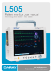
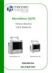
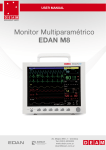
Download M8000VET /M9000VET Veterinary Monitor User`s Manual
Transcript
M8000VET /M9000VET Veterinary Monitor User’s Manual Guangdong Biolight Meditech Co., Ltd. Address: Innovation First Road, Technology Innovation Coast, Jinding, Zhuhai, P.R.CHINA Tel: +86-756-3399900 Fax: +86-756-3399989 http://www.blt.com.cn J/M9000vet-028-2008A1 Preface Thank you for using M8000VET/M9000VET veterinary monitor. In order to enable you to skillfully operate Monitor as soon as possible, we provide this user’s manual with delivery. When you install and use this instrument for the first time, it is imperative that you read carefully all the information that accompanies this instrument. Based on the need to improve the performance and reliability of the parts and the whole instrument, we sometimes will make some amendments to the instrument (including the hardware and software). As a result, there might be cases of discrepancies between the manual and the actual situation of products. When such discrepancies occur, we will try our best to amend or add materials. Your comments and suggestions are welcome. Our liaison-way: Address: Innovation First Road, Technology Innovation Coast, Jinding, Zhuhai, P.R.CHINA Tel: +86-756-3399900 Fax: +86-756-3399989 Post code: 519085 Toll-free: +86-800-830-1016 Statement This manual contains exclusive information protected by copyright laws and we reserve its copyright. Without written approval of manufacturer no parts of this manual shall be photocopied, Xeroxed or translated into other languages. The contents and version contained in this manual are subject to amendments without notification. The version number of this manual: MSP.A1 I Liabilities of the Manufacturer Only under the following circumstances will manufacturer be responsible for the safety, reliability and performance of the instrument. All the installation, expansion, readjustment, renovation or repairs are conducted by the personnel certified by manufacturer. The electrical safety status at the installation site of the instrument conforms to the national standards. The instrument is used in accordance with the operation procedures. Copyright reserved © 2008 Guangdong Biolight Meditech Co., Ltd. II CONTENTS CHAPTER 1 GENERAL INTRODUCTION ............................................................... 1-1 1.1 INTENDED USE .......................................................................................................... 1-1 1.2 ABOUT THIS MANUAL .............................................................................................. 1-1 1.3 BRIEF INTRODUCTION TO THE MONITOR ............................................................... 1-3 1.4 APPEARANCE AND STRUCTURE OF THE MONITOR ................................................. 1-4 1.5 SOCKETS .................................................................................................................. 1-5 1.6 FUNCTION BUTTONS AND TRIM KNOB ON THE FRONT PANEL ............................... 1-7 1.7 SPECIFICATIONS AND PERFORMANCE CRITERIA OF THE MONITOR ...................... 1-9 CHAPTER 2 IMPORTANT SAFETY NOTES ............................................................ 2-1 2.1 GENERAL SAFETY .................................................................................................... 2-1 2.2 SOME IMPORTANT NOTES FOR SAFETY.................................................................... 2-3 2.3 CLASSIFICATIONS .................................................................................................... 2-5 2.4 SAFE OPERATING AND HANDLING CONDITIONS ..................................................... 2-5 CHAPTER 3 PREPARATIONS BEFORE THE USE OF THE MONITOR ............ 3-1 3.1 UNPACKING THE CASE ............................................................................................. 3-1 3.2 CONNECTING TO POWER ......................................................................................... 3-1 3.3 CONNECTING TO THE CENTRAL MONITOR SYSTEM .............................................. 3-3 3.4 POWER ON THE MONITOR ....................................................................................... 3-4 3.5 CONNECTING TO VARIOUS KINDS OF SENSORS....................................................... 3-4 3.6 PREPARATION OF RECORDER .................................................................................. 3-4 CHAPTER 4 OPERATION INSTRUCTIONS FOR THE MONITOR .................... 4-1 4.1 SCREEN MODE .......................................................................................................... 4-1 4.2 MAIN MENU .............................................................................................................. 4-5 4.3 SCREEN DISPLAY .................................................................................................... 4-27 CHAPTER 5 PARAMETERS MEASUREMENT....................................................... 5-1 5.1 MEASUREMENT OF ECG/HR .................................................................................. 5-1 5.2 MEASUREMENT OF RESP........................................................................................ 5-9 5.3 MEASUREMENT OF SPO2/PULSE ........................................................................... 5-11 5.4 MEASUREMENT OF TEMP .................................................................................... 5-15 III 5.5 MEASUREMENT OF NIBP ...................................................................................... 5-17 5.6 MEASUREMENT OF IBP ......................................................................................... 5-26 5.7 MEASUREMENT OF CO2 (SIDESTREAM, CPT) ...................................................... 5-32 5.8 MEASUREMENT OF CO2 (MAINSTREAM, IRMA) ................................................. 5-37 5.9 MEASUREMENT OF CO2 (MICROSTREAM, LOFLO)............................................. 5-41 5.10 MEASUREMENT OF CO2 (MAINSTREAM, CAPNOSTAT5)............................... 5-48 5.11 MEASUREMENT OF AG (IRMA).......................................................................... 5-51 CHAPTER 6 ALARM..................................................................................................... 6-1 6.1 ALARM PRIORITY .................................................................................................... 6-1 6.2 ALARM MODES ........................................................................................................ 6-1 6.3 ALARM SETUP .......................................................................................................... 6-2 6.4 ALARM CAUSE ......................................................................................................... 6-4 6.5 SILENCE/SUSPENSION .............................................................................................. 6-5 6.6 PARAMETER ALARM ................................................................................................ 6-6 6.7 WHEN AN ALARM OCCURS ...................................................................................... 6-6 6.8 ALARM DESCRIPTION AND PROMPT ....................................................................... 6-6 CHAPTER 7 RECORDING........................................................................................... 7-1 CHAPTER 8 THE MAINTENANCE AND CLEANING ........................................... 8-1 8.1 SYSTEM CHECK ....................................................................................................... 8-1 8.2 BATTERY MAINTENANCE ........................................................................................ 8-2 8.3 GENERAL CLEANING ............................................................................................... 8-3 8.4 CLEANING AGENTS .................................................................................................. 8-4 8.5 DISINFECTION .......................................................................................................... 8-4 CHAPTER 9 ACCESSORIES AND ORDERING INFORMATION......................... 9-1 APPENDIX A DEFAULT SYSTEM SETUP ................................................................... 1 A.1 SYSTEM ...................................................................................................................... 1 A.2 ALARM LIMIT ............................................................................................................ 4 APPENDIX B GUIDANCE AND MANUFACTURE’S DECLARATION OF EMC .. 6 IV M8000Vet/M9000Vet veterinary monitor user’s manual Chapter 1 General Introduction 1.1 Intended use This veterinary monitor is intended to be used in special procedure labs and other areas of a veterinary hospital or clinic where veterinary monitoring systems are needed. The monitoring parameters include 3-lead or 5-lead electrocardiography (ECG), respiration (Resp), non-invasive blood pressure (NIBP), invasive blood pressure (IBP), pulse oximetry (SpO2), temperature (Temp), end tidal carbon dioxide (EtCO2) and anesthetic gas (AG). 1.2 About this Manual This user’s manual consists of the following chapters: Chapter 1 gives an introduction to the content and the specific signs of this manual, the main features and appearance of the monitor, the basic operations of various buttons, the meanings of the signs on the monitor, specifications and performance criteria of the monitor, the ambient requirements for the working and storage of the monitor. Chapter 2 gives important safety notes. Please do read this chapter before using the monitor! Chapter 3 gives an introduction to the preparatory steps before using the monitor. Chapter 4 provides general operation instruction for the monitor, including illustrations of the screen display, normal selection for soft button on screen, details for entry of veterinary patient data and trend maps, also. Chapter 5 gives details of specific parameter measurement, preparatory steps, cables or probes connection, setup of parameters, maintenance and cleaning of equipments and sensors. Chapter 6 gives detailed description of system alarm, including level and mode of alarm, default setting and changing procedure of alarm parameters, prompt of specific alarms, and the general operation to carry out when an alarm occurs. Chapter 7 gives detailed description of record function. 1-1 M8000Vet/M9000Vet veterinary monitor user’s manual Chapter 8 gives general maintenance and cleaning methods of the monitor and its parts. Signs in this manual: 0 Warning: Means it must be strictly followed so as to prevent the operator or the veterinary patient from being harmed. Caution: Means it must be followed so as not to damage the instrument. ) Note: Important information or indications regarding the operation or use. ) Note: This manual introduced the product that with full configuration. Some functions of the product you bought may not be provided. 1-2 M8000Vet/M9000Vet veterinary monitor user’s manual 1.3 Brief Introduction to the Monitor The Monitor has features as follows: Multiple measuring functions include 3-lead, 7-lead ECG/HR, RESP, dual TEMP, SpO2/Pulse, NIBP, dual IBP, EtCO2 and AG are optional. Complete built-in module design ensures stable and reliable performance Unique all-lead ECG on-one-screen display, which can facilitate the diagnosis and analysis of cardiac disease Can store the trend data for 120 - 168 hours and has the function of displaying trend data and trend graphs Function of alarm event reviewing, can store 1000 - 1800 pieces of alarm events Function of NIBP measurement reviewing, can store 750 – 1000 pieces of NIBP measurement data Function of reviewing 10 - 30 minutes one important lead’s EGC waveform Built-in recorder is optional and it supports real-time recording, trigger printout by alarm Parameter display with big character Optional function of Calculator of drug concentration Optional function of Display of oxyCRG Function of Display of short trend 12.1″or 10.4″authentic color high brightness TFT LCD monitor Portable design, stylish and convenient Rechargeable maintenance-free battery, can continue working when AC power is off Can be connected with the central unit to realize centralized monitoring Is resistant to high-frequency electrotome and is protected against defibrillation effects 1-3 M8000Vet/M9000Vet veterinary monitor user’s manual 1.4 Appearance and Structure of the Monitor Alarm light Various kinds of sockets (See Fig. 1-5-1) Recorder Function button zone Trim Knob (See Fig.1-6-1) (See Fig. 1-6-1) Fig. 1-4-1 The appearance of M8000VET veterinary monitor Alarm light Various kinds of sockets (See Fig. 1-5-1) Recorder Trim Knob (See Fig.1-6-1) Function button zone (See Fig. 1-6-1) Fig. 1-4-2 The appearance of M9000VET veterinary monitor Caution: The AC input socket at the back panel of the monitor can be connected with 100-240V AC power by electrical wires supplied with this instrument. 1-4 M8000Vet/M9000Vet veterinary monitor user’s manual 1.5 Sockets EtCO2 IBP ECG CO2/AG SpO2 NIBP Receptacle for dehydration flask TEMP Fig. 1-5-1 Various sockets on the side panel NETWORK FUSE T1.6A FUSE Electric AC socket Network connector AC 100V-240V 9Pin D type socket Equipotentiality Terminal Fig. 1-5-2 Various sockets on the back panel ) Note: The 9 Pin D type socket (RS-232) is only used for maintenance and upgrading of the monitor by manufacturer. ) Note: The Network Connector is a standard RJ45 socket and being used for connection with the central monitoring system provided by manufacturer. 1-5 M8000Vet/M9000Vet veterinary monitor user’s manual 0 Warning: The sensor cable sockets on Monitor can only be connected with the sensor cables supplied with this instrument and no other cables shall be used. Notes on the signs on the monitor Signs Notes on the signs Defibrillator-proof type CF equipment (Refer to IEC 60601-2-27) The unit displaying this symbol contains a F-Type isolated (floating) applied part providing a high degree of protection against shock, and is defibrillator-proof. Defibrillator-proof type BF equipment (Refer to IEC 60601-1:1995) The unit displaying this symbol contains a F-Type isolated (floating) applied part providing a high degree of protection against shock, and is defibrillator-proof. Attention! Please refer to the document supplied with this instrument (this manual)! Non-ionizing radiation Dangerous voltage Equipotentiality Alternating current (AC) Symbol for the marking of electrical and electronics devices according to Directive 2002/96/EC. The device, accessories and the packaging have to be disposed of waste correctly at the end of the usage. Please follow local ordinances or regulations for disposal. ECG Short for “Electrocardiogram” RESP Short for “Respiration” SpO2 Short for “Pulse Oxygen Saturation” TEMP IBP NIBP Short for “Temperature” Short for “Invasive Blood Pressure” Short for “Non-invasive Blood Pressure” 1-6 M8000Vet/M9000Vet veterinary monitor user’s manual Short for “End tidal carbon dioxide” EtCO2 Short for “Anesthetic gas” AG 1.6 Function Buttons and Trim Knob on the Front Panel AC/BAT CHARGE TREND MAIN FREEZE SUSPEND NIBP /RECORD /SILENCE /STAT MENU The Trim Knob is used for: Turn left or turn right to move the cursor. Press down to perform an operation, such as open the menu dialog or selects one option. Fig. 1-6-1 Function Buttons and Trim Knob on the Front Panel 1.6.1 The Signs and Operation Instructions Within the Function Button Zone Signs AC/BAT CHARGE MAIN TREND Notes on the signs Operation instructions of function buttons Indicating light of AC/DC When the monitor is connected to the AC power, this indicating light is green (it is unrelated to the ON/OFF state of the monitor). When the monitor is not connected to AC power and the battery is used as the power source, this indicating light is orange. Indicating light of CHARGE When the monitor is connected to the AC power of charge, this indicating light is turn-on. When the monitor is full of charge, this indicating light is turnoff. Power button Press this button once and the monitor starts up. Repress this button, then the monitor is switched off. Return to Main Screen Press this button once to exit the present menu and return to main screen. Trend Review Press this button once to see the Trend Graph and the Trend Table 1-7 M8000Vet/M9000Vet veterinary monitor user’s manual FREEZE /RECORD Switching type button Freeze (or defreeze) the waveforms /Record the real-time waveforms Press this button in 2 seconds to freeze waveform, press again to defreeze waveform. Press this button over 2 seconds can start real-time recording. In case the real-time recording is underway, pressing this button will terminate real-time recording. SUSPEND /SILENCE Switching type button Suspend the sounding of Alarm /Close the sounding of Alarm Press this button in 2 seconds to make the monitor alarm paused or cancel the pause. Press this button over 2 seconds can silence the monitor’s audio system or cancel the silence. NIBP /STAT Switching type button Begin (or Stop) the measurement of NIBP /Begin the STAT Press this button in 2 seconds to start or stop the NIBP measurement. Press this button over 2 seconds to make NIBP module working at STAT measurement mode and perform continuous NIBP measurement within 5 minutes. MENU Menu Press this button to display menu option. 1.6.2 Basic Operations Turn the Trim Knob to select the item or soft button on the screen Press MAIN button to return to main screen Press the Trim Knob to confirm selection Perform the operation Fig. 1-6-2 Flow chart of basic operations ) Note: The system menu is located at the left bottom corner. By operating the Trim Knob in the above flow chart, select the options or make them spring out, and for detailed item selection, please refer to Chapter 4. 1-8 M8000Vet/M9000Vet veterinary monitor user’s manual 1.7 Specifications and Performance Criteria of the Monitor 1.7.1 Classifications Refer to chapter 2.3. 1.7.2 Specifications Size and Weight Size 318 mm×264 mm×152 mm Weight 4.5 kg Power supply Power Voltage AC 100-240V 50/60Hz Power Input ≤85VA Fuse T1.6AL/250V, Φ5×20 (mm) Safety class Category I Display LCD Size M9000Vet: 12.1″ M8000Vet: 10.4″ Type Color TFT-LCD Resolution 800×600 pixels or higher Indicators Alarm LED 1 (Yellow/Red) AC Power LED 1 (Green/Orange) Battery Charge LED 1 (Yellow) System output Network Ethernet RF Wireless LAN 433MHz, 10mW (optional) Battery Type Rechargeable Lead acid cell, 12V/2.0AH Charge time ≤10 hours (2 batteries for 20 hours) ≥60 minutes (2 batteries for 120 minutes) Operating time under the New and fully charged battery at 25℃ ambient normal use and full charge temperature and NIBP work on AUTO mode for 20 minutes interval. 1-9 M8000Vet/M9000Vet veterinary monitor user’s manual Operating time after the first ≥5 minutes alarm if low battery Battery (option) Type Charge time Rechargeable Lithium ion battery 11.1V/4.0AH 11.1V/2.0AH ≤6 hours ≤6 hours ≥240 minutes ≥120 minutes Operating time under the New and fully charged battery at 25℃ ambient normal use and full charge temperature and NIBP work on AUTO mode for 20 minutes interval. Operating time after the first ≥10 minutes alarm if low battery Environment Ambient Temperature Operating temperature: 0~+40℃ Transportation and storage temperature: –20~+55℃ Relative humidity Working ≤85% Transportation and storage ≤93% Atmospheric pressure Working 860~1060 hPa Transportation and storage 500~1060 hPa ECG Lead Mode Lead selection 1. 2. 1. 2. 5-leads ECG input 3-leads ECG input I, II, III, aVR, aVL, aVF, VI, II, III Gain AUTO, 0.25x, 0.5x, 1.0x, 2.0x, 4.0x Input impedance Electrode offset potential ≥5.0 MΩ MON ≥105dB OPS ≥105dB MON 0.5~40Hz OPS 1~25Hz ±500mV d.c. Leakage Current <10 uA ECG signal range ±6.0 mV Baseline recovery <5s after Defibrillation. (MON or OPS mode) Pacemaker pulses No rejection of pulses with amplitudes of ±2mV ~ ±700 mV and durations of 0.5 ~ 2.0 ms. CMRR Frequency response 1-10 M8000Vet/M9000Vet veterinary monitor user’s manual Insulation Breakdown Voltage 4000VAC 50/60Hz Indication of electrode separation Every electrode (exclusive of RL) Sweep speed 12.5 mm/s, 25 mm/s, 50 mm/s Measurement Range 10~350 bpm Refreshing time Per 4 pulses Resolution 1 bpm Accuracy ±1% or ±1 bpm, whichever is greater Sensitivity ≥0.2mVP-P Alarm range 0~350 bpm, continuously adjustable between upper limit and lower limit Alarm indication Sound and light alarming Time to Alarm for Tachycardia Average 4s Tall T-Wave Rejection Capability 0-1mV T-Wave amplitude HR HR change from 80 to 120 bpm: Response Time of Heart Rate Range: 6 to 10s Meter to Change in Heart HR change from 80 to 40 bpm: Rate Range: 6 to 10s NIBP Way of measurement Range of measurement Automatic oscillometry SYS 30~270 mmHg DIA 10~220 mmHg MEAN 20~235 mmHg Cuff pressure range 0~280 mmHg Resolution 1 mmHg Pressure Accuracy Static Clinical ±2% or ±3 mmHg, whichever is greater ±5 mmHg average error ≤8 mmHg standard deviation Unit mmHg, kPa Pulse rate range 40 ~ 240 bpm Inflation time for cuff Less than 40s. (standard cuff) 1-11 M8000Vet/M9000Vet veterinary monitor user’s manual Total cycle time Overpressure Protection Horse Dog Cat Intervals for AUTO measurement time Range of alarm Alarm indication Measurement Mode 20 to 45s typical (dependent on heart rate and motion artifact) Hardware and software double protections 315±10 mmHg 265±10 mmHg 255±10 mmHg 1,2,3,4,5,10,15,20,30,45,60,90 minutes 2,4,8 hours SYS 0~300 mmHg, continuously adjustable between upper limit and lower limit DIA 0~300 mmHg, continuously adjustable between upper limit and lower limit MEAN 0~300 mmHg, continuously adjustable between upper limit and lower limit Sound and light alarming Horse Manual, Auto and STAT Dog Manual, Auto and STAT Cat Manual, Auto SpO2 BLT-SpO2 Measurement Range 0~100% Resolution 1% Accuracy At 70~100%, ±2% At 0~69%, unspecified Data update period <13s Alarm User-selectable upper and lower SpO2 limits PR Measurement Range 25~250 bpm Resolution 1 bpm Accuracy ±1% or ±1 bpm, whichever is greater Data update period <13s Alarm User-selectable upper and lower pulse rate limits Nellcor-SpO2 ( option) Measurement Range 0~100% 1-12 M8000Vet/M9000Vet veterinary monitor user’s manual Resolution 1% Accuracy At 70~100%, ±2 digits At 0~69%, unspecified Perfusion Range 0.03% ~ 20% Data update period Average 7s Alarm PR User-selectable upper and lower SpO2 limits Measurement Range 20~250 bpm Resolution 1 bpm Accuracy ±3 digits Data update period Average 7s Alarm User-selectable upper and lower pulse rate limits TEMP Measurement Range 0.0~50.0℃ Accuracy ±0.1℃ Resolution 0.1℃ Unit Celsius (℃), Fahrenheit (℉) Refreshing time 1s Self check Every 10 minutes Accuracy At 45.1℃~50.0℃, ±0.2℃ (exclusive of probe) At 25.0℃~45.0℃, ±0.1℃ (exclusive of probe) At 0.0℃~24.9℃, ±0.2℃ (exclusive of probe) Channel 2 Connecting cable Compatible with YSI-400 Range of alarm 0.0~50.0℃, continuously adjustable between upper limit and lower limit Alarm indication Sound and light alarming RESP Method Impedance variation between RA-LL (R-F) Measuring impedance range 0.2 ~3Ω Excitation frequency 64.8 kHz Excitation current ≤300μA at 64.8 kHz 1-13 M8000Vet/M9000Vet veterinary monitor user’s manual Base line impedance range 500~4000Ω (50~120 kHz exciting frequency) Measurement Range 0~150 rpm Resolution 1 rpm Accuracy ±2 rpm Gain x1,x2,x4 Sweep speed 6.25 mm/s, 12.5 mm/s, 25 mm/s Delay of Apnea Alarm Off, 20s, 40s, 60s Alarm indication Sound and light indication Measurement Range -50 ~ +300 mmHg Resolution 1 mmHg Unit mmHg, kPa Accuracy Static Dynamic ± 2mmHg or 2% of the reading, whichever is greater (exclusive of transducer) ± 4mmHg or 4% of the reading, whichever is greater (inclusion of transducer) ± 4mmHg or 4% of the reading, whichever is greater Channel 2 Sensitivity of transducer 5uV/V/mmHg, 2% Impedance of transducer 300~3000Ω Bandwidth d.c. ~ 15Hz IBP Arterial Pressure (ART) Pulmonary Artery Pressure Transducer sites (PA) Left Atrium Pressure (LAP) Right Atrium Pressure (RAP) Central Venous Pressure (CVP) Intracranial Pressure (ICP) ART 0~200mmHg PA 0~300 mmHg CVP Selection of measurement -10~20 mmHg range LAP -50~300 mmHg RAP AUTO ICP 1-14 M8000Vet/M9000Vet veterinary monitor user’s manual (Among them, the AUTO switches automatically at an interval of 10 mmHg so as to ensure the waveform is at the state most suitable for observation) Alarm indication Sound and light indication EtCO2 (Sidestream,CPT) Measure method Infrared spectrum Measure mode Sidestream Measurement Range 0.0~13.1% (0~99.6 mmHg) Resolution 1 mmHg Unit %, mmHg, kPa Accuracy At <5 % CO2,±0.3% (±2.0 mmHg) At ≥5 % CO2, < ±10 % of reading Range of respiration rate 3~150 rpm measurement Offset calibration: auto, manual Calibration Gain calibration 0.0~13.1 % (0~99.6mmHg), continuously adjustable Range of alarm between upper limit and lower limit Alarm indication Sound and light indication EtCO2 (Mainstream,IRMA) Measure method Infrared spectrum Measure mode Mainstream Measurement Range 0.0~13.1% (0~99.6 mmHg) Resolution 1 mmHg Unit %, mmHg, kPa Accuracy ±0.5 % (±4.0 mmHg) or <±10 % of reading, which is greater Rise time (at 10 L/min) ≤90 ms Total system response time <1s Range of respiration rate 0~150 rpm measurement RR Accuracy ±1 rpm Range of alarm 0.0~13.1 % (0~99.6mmHg), continuously adjustable between upper limit and lower limit 1-15 M8000Vet/M9000Vet veterinary monitor user’s manual Alarm indication Sound and light indication EtCO2 (Microstream,LoFlo) Measure method Infrared spectrum Measure mode Microstream Warm up time Capnogram displayed in less than 20 s, At an ambient temperature of 25℃, full specifications within 2 minutes. CO2Measurement Range 0 ~ 19.7%(0 ~ 150 mmHg) CO2 Resolution 1mmHg CO2 Stability Short-Term Drift: Drift over four hours≤0.8mmHg. Long-Term Drift: Accuracy specification will be maintained over a 120 hours period. unit %, mmHg, kPa CO2 Accuracy (at 760 mmHg, ambient temperature of 25°C) 0 ~ 40 mmHg, ±2 mmHg 41 ~ 70 mmHg, ±5% of reading 71 ~100 mmHg, ±8% of reading 101 ~ 150 mmHg, ±10% of reading Above 80 breath per minute ± 12% of reading Gas temperature at 25℃. CO2 response time <3s (includes transport time and rise time) Respiration Rate Range 2~150 rpm Respiration Rate Accuracy ±1 rpm Sample Flow Rate 50 ml/min ±10 ml/min Alarm indication Sound and light indication EtCO2 (Mainstream,CAPNOSTAT5) Measure method Infrared spectrum Measure mode Mainstream Warm up time Capnogram displayed in less than 15 s, At an ambient temperature of 25℃, full specifications within 2 minutes. CO2Measurement Range 0 ~ 19.7%(0 ~ 150 mmHg) CO2 Resolution 1mmHg CO2 Accuracy 0 ~ 40 mmHg, ±2 mmHg 41 ~ 70 mmHg, ±5% of reading 71 ~100 mmHg, ±8% of reading 101 ~ 150 mmHg, ±10% of reading 1-16 M8000Vet/M9000Vet veterinary monitor user’s manual Temperature at 35℃. CO2 Stability Short-Term Drift: Drift over four hours≤0.8 mmHg. Long-Term Drift: Accuracy specification will be maintained over a 120 hours period. Rise time <60ms unit %, mmHg, kPa Respiration Rate Range 0~150 rpm Respiration Rate Accuracy ±1 rpm Alarm indication Sound and light indication AG (IRMA) Measure method Infrared spectrum Measure mode Mainstream Fi and Et values CO2,N2O,O2,agent (HAL, ISO, ENF, SEV, DES) Resolution 1mmHg Unit %, mmHg Calibration Warm-up time Rise time (at 10 L/min) Total system response time Room air calibration performed automatically when changing airway adapter (<5s) Concentrations reported in less than 10s, full accuracy within 1 min CO2 ≤ 90 ms O2 ≤ 300 ms N2O ≤ 300 ms Hal, Iso, Enf, Sev, Des ≤ 300 ms <1s Measurement range of AG: Gas Measurement range CO2 0-10 % N2O 0-100 % O2 10-100 % HAL, ISO, ENF 0-5% SEV 0-8% DES 0-18% 1-17 Accuracy ± 0.5% or ± 10% of reading, whichever is greater ± 2% or ± 10% of reading, whichever is greater ±3 % ± 0.15% or ± 10% of reading, whichever is greater ± 0.15% or ± 10% of reading, whichever is greater ± 0.15% or ± 10% of reading, whichever is greater M8000Vet/M9000Vet veterinary monitor user’s manual Respiration rate range 0~150 rpm Respiration rate accuracy ±1 rpm Alarm indication Sound and light indication Recorder (Option) Method Thermal dot array Paper width 50 mm Record width 40 mm Paper Speed 12.5 mm/s ,25 mm/s ,50 mm/s Traces Maximum 3 tracks Alarm Level Low, medium and high Indication Auditory and visual Setup Default and custom Silence All alarms can be silenced Volume 45~85 dB measured at 1 meter 1-18 M8000Vet/M9000Vet veterinary monitor user’s manual Chapter 2 Important Safety Notes 0 Warning: The monitor is intended for VETERINARY USE ONLY. Do not use on human patients. 0 Warning: Only trained doctors and nurses can use the device. 0 Warning: The monitor is neither a therapeutic instrument nor a device that can be used at home. 2.1 General Safety 1. Safety precautions for safe installation The input socket of monitor can be connected to the electrical wires and common electrical wire can be used. Only the power supply type of AC 100-240V 50/60Hz specified by monitor can be used. Connect the electrical wire to a properly grounded socket. Avoid putting the socket used for it in the same loop of such devices as the air conditioners, which regularly switch between ON and OFF. Avoid putting the monitor in the locations where it easily shakes or wobbles. Enough space shall be left around the monitor so as to guarantee normal ventilation. Make sure the ambient temperature and humidity are stable and avoid the occurrence of condensation in the work process of the monitor. 0 Warning: Never install the monitor in an environment where flammable anesthetic gas is present. 2. Monitor conforms to the safety requirements of IEC 60601-1:1995. This monitor is protected against defibrillation effects. 3. Notes on signs related to safety 2-1 M8000Vet/M9000Vet veterinary monitor user’s manual Defibrillator-proof type CF equipment (refer to IEC 60601-2-27) The unit displaying this symbol contains a F-Type isolated (floating) applied part providing a high degree of protection against shock, and is defibrillator-proof. The type CF applied parts provide a higher degree of protection against electric shock than that provided by type BF applied parts. Attention! Please refer to the documents accompanying this monitor (this manual)! Defibrillator-proof type BF equipment (IEC 60601-1:1995) The unit displaying this symbol contains a F-Type isolated (floating) applied part providing a high degree of protection against shock, and is defibrillator-proof. 4. When a defibrillator is applied on a patient, the monitor may have transient disorders in the display of waveforms. If the electrodes are used and placed properly, the display of the monitor will be restored within 10 seconds. During defibrillation, please note to remove the electrode of chest lead and move the electrode of limb lead to the side of the limb. The electrode of the defibrillator should not come into direct contact with the monitoring electrodes. Please ensure the monitor is reliably grounded and the electrodes used repeatedly should be kept clean. 0 Warning: When conducting defibrillation, do not come into contact with the patient, the bed and the monitor. Otherwise serious injury or death could be resulted in. 5. To guarantee the safe operation of the monitor, Monitor is provided with various replaceable parts, accessories and consuming materials (such as sensors and their cables, electrode pads). Please use the products provided or designated by the manufacturer. 6. Monitor only guarantees its safety and accuracy under the condition that it is connected to the devices provided or designated by manufacturer. If the monitor is connected to other undesignated electrical equipment or devices, safety hazards may occur for causes such as the cumulating of the leakage current. 7. To guarantee the normal and safe operation of the monitor, a preventive check and maintenance should be conducted for the monitor and its parts every 6-12 months 2-2 M8000Vet/M9000Vet veterinary monitor user’s manual (including performance check and safety check) to verify the instrument can work in a safe and proper condition and it is safe to the medical personnel and the patient and has met the accuracy required by clinical use. Caution: The monitor does not contain any parts for self-repair by users. The repair of the instrument must be conducted by the technical personnel been authorized by manufacturer. 2.2 Some important notes for safety PATIENT NUMBER The monitor can only be applied to one patient at one time. INTERFERENCE Do not use cellular phone in the vicinity of this equipment. High level of electromagnetic radiation emitted from such devices may result in strong interference with the monitor performance. ACCIDENTAL SPILLS To avoid electric shock or device malfunction, liquids must not be allowed to enter the device. If liquids have entered the device, take it out of service and have it checked by a service technician before it is used again. ACCURACY If the accuracy of any value displayed on the monitor or printed on a printout paper is questionable, determine the patient’s vital signs by alternative means. Verify that all equipment is working correctly. ALARMS Do not rely exclusively on the audible alarm system for patient monitoring. Adjustment of alarm volume to a low level or off during patient monitoring may result in a hazard to the patient. Remember that the most reliable method of patient monitoring combines close personal surveillance and correct operation of monitoring equipment. The functions of the alarm system for monitoring the patient must be verified at regular intervals. BEFORE USE Before putting the system into operation, please visually inspect all connecting cables for signs of damage. Damaged cables and connectors must be replaced immediately. Before using the system, the operator must verify that it is in correct working order and operating condition. Periodically, and whenever the integrity of the product is in doubt, test all functions. CABLES Route all cables away from patient’s throat to avoid possible strangulation. 2-3 M8000Vet/M9000Vet veterinary monitor user’s manual TO CLEAR PATIENT DATA When monitoring a new patient, you must clear all previous patient data from the system. To accomplish this, shut down the device, then turn on it. Selecting 〈New patient〉in〈main setup〉menu can also clear the previous patient data . DISPOSAL OF PACKAGE Dispose of the packaging materials, please observe the applicable waste control regulations and keeping it out of children’s reach. EXPLOSION HAZARD Do not use this equipment in the presence of flammable anesthetics, vapors or liquids. LEAKAGE CURRENT TEST When interfacing with other equipment, a test for leakage current must be performed by qualified biomedical engineering personnel before using with patients. BATTERY POWER The device is equipped with a battery pack. The battery discharges even when the device is not in use. Store the device with a fully charged battery and take out the battery, so that the service life of the battery will not be shortened. DISPOSAL OF ACCESSORIES AND DEVICE Disposable devices are intended for single use only. They should not be reused as performance could degrade or contamination could occur. The service life of this monitor is five years. At the end of its service life, the product described in this manual, as well as its accessories, must be disposed of in compliance with the guidelines regulating the disposal of such products. If you have questions concerning disposal of products, please contact manufacturer or its representatives. EMC Magnetic and electrical fields are capable of interfering with the proper performance of the device. For this reason, make sure that all external devices operated in the vicinity of the monitor comply with the relevant EMC requirements. X-ray equipment or MRI devices are a possible source of interference as they may emit higher levels of electromagnetic radiation. Also, keep cellular phones or other telecommunication equipment away from the monitor. INSTRUCTION FOR USE For continuous safe use of this equipment, it is necessary that listed instructions were followed. However, instructions listed in this manual in no way can supersede established medical practices concerning patient care. LOSS OF DATA Should the monitor at any time temporarily lose patient data, close patient observation or alternative monitoring devices should be used until monitor function is restored. If the monitor does not automatically resume operation within 60 seconds, restart the monitor using the power on/off switch. Once monitoring is restored, you should verify correct monitoring state and alarm function. 2-4 M8000Vet/M9000Vet veterinary monitor user’s manual 2.3 Classifications The Monitor is classified, according to IEC 60601-1:1995 as: Type of protection against electric shock: I Degree of protection against electric shock: BF: EtCO2, AG CF: ECG, RESP, TEMP, IBP, NIBP, SpO2 Degree of protection against harmful ingress of water: Degree of safety of application in the presence of a flammable anesthetic-mixture with air or with oxygen or nitrous oxide: Mode of operation: Ordinary Equipment (enclosed equipment without protection against ingress of water) Not suitable Continuous operation I: Class I equipment BF: Type BF applied part CF: Type CF applied part Not suitable: Equipment is not suitable for use in the presence of flammable anesthetic mixture with air or with oxygen or nitrous oxide. 2.4 Safe Operating and Handling Conditions Method(s) of sterilization or disinfection recommended by the manufacturer: Electromagnetic interference Electro surgical interference damage Diathermy instruments influence Defibrillation shocks Auxiliary outputs Sterilization: not applicable Disinfection: See “The Maintenance and Cleaning of the System->General Cleaning” No cellular telephone nearby No damage Displayed values and prints may be disturbed or erroneous during diathermy. The monitor specifications fulfill the requirements of IEC 60601-1, IEC 60601-2-27, IEC 60601-2-49, IEC 60601-2-34. The system must fulfill the requirements of standard IEC 60601-1-1. 2-5 M8000Vet/M9000Vet veterinary monitor user’s manual Chapter 3 Preparations Before the Use of the Monitor 3.1 Unpacking the Case Unpack the packaging case Open the packaging case, accessories include: electrical wire, various patient sensors and user’s manual (this manual), warranty card, certificate and particular paper and the lower foam case contains the monitor. Remove the monitor and accessories Caution: Please place the monitor on level and stable supporting plane, not on the places that can easily shock or wake. Enough room should be left around the monitor so as to guarantee normal ventilation. Keep all the packaging materials for future use in transportation or storage. Check the monitor and accessories Check the monitor and its accessories one by one in accordance with the particular paper. Check to see if the parts have any mechanical damages. In case of problems, please contact us or our agent. 3.2 Connecting to Power 3.2.1 AC Power Confirm the rated AC current is: AC 100-240V 50/60Hz Use the electrical wires provided along with the instrument, put its output end plug (round headed) into the AC current socket on the back of the monitor, and the plug of input end into a grounded socket of the mains (It must be a special socket of the hospital), connect the monitor through the earth one of electrical wires. When the indicating light above the power switch on the panel of the monitor is green, it means the AC power is on. And when the monitor is not connected to AC power and the DC battery is used as the power source, the indicating light is orange. 3-1 M8000Vet/M9000Vet veterinary monitor user’s manual 0 Warning: The monitor must be connected to a properly installed power outlet with protective earth contacts only. If the installation does not provide for a protective earth conductor, disconnect the monitor from the power line and operate it on battery power. ) Note: The equipment has no mains switch. The equipment is switched completely only by disconnecting the power supply from the wall socket. The wall socket has to be easily accessible. ) Note: For measurements in or near the heart we recommend connecting the monitor to the potential equalization system. Use the green and yellow potential equalization cable and connect it to the pin labeled with the symbol. 3.2.2 Battery Power The monitor has a battery pack to provide power to the monitor whenever AC power is interrupted. The battery is generally referred to as the “battery”. You must charge the battery before using it. There is no external charger. The battery is charged when the monitor is connected to AC power. To assure a fully charged battery that is ready for use, we recommend that the monitor be plugged into AC power whenever it is not in use. Depending on usage, you can get at least 120 minutes of battery power on pair of new, fully-charged battery on the monitor. NIBP and SpO2 monitoring and the usage of the recorder will drain battery power faster than other parameters. ) Note: When the monitor is connected to AC power, the battery is in a state of being recharged. When it is unable to be connected to the AC power, the battery can be used to supply power, and at this time it is unnecessary to use the electrical wires, and the instrument can be switched on directly. ) Note: A “Battery Low” message displaying at the technical alarm information area of screen and an audible system alarm indicate approximate 5 minutes of battery life remaining. You should connect the monitor to an AC power source when the message is displayed. 3-2 M8000Vet/M9000Vet veterinary monitor user’s manual ) Note: This monitor contains a rechargeable battery. The average life span of this type of battery is approximately three years. When replacement becomes necessary, contacting a qualified service representative to perform the replacement. ) Disposal Note: Should this product become damaged beyond repair, or for some reason its service life is considered to be at an end, please observe all local, state, and federal regulations that relate to the disposal of products that contain lead, batteries, plastics, etc. Install Battery The battery storage is located at the bottom of the monitor, following the steps to install a battery. 1、Open the battery gate according to the direction marked on the monitor. 2、Turn the baffle up clockwise. 3、Push the battery into the gate with the electrode point to the bottom of the monitor. 4、After pushing the battery inside the storage withdraw, the baffle turn back to the middle position. 5、 Close the gate. Uninstall battery 1、 Open the battery gate according to the direction marked on the monitor. 2、Turn the baffle up clockwise. 3、Take out the battery. Then close the gate. 3.3 Connecting to the Central Monitor System 0 Warning: Accessory equipment connected to the analog and digital interface must be certified according to the respective IEC standards (e.g. IEC 60950 for data processing equipment and IEC 60601-1:1995 for medical equipment). Furthermore all configurations shall comply with the valid version of the system standard IEC 60601-1-1. Everybody who connects additional equipment to the signal input part or signal output part configures a medical system, and is therefore responsible that the system complies with the requirements of the valid version of the system standard IEC 60601-1-1. If in doubt, consult the technical service department or your local representative. 3-3 M8000Vet/M9000Vet veterinary monitor user’s manual If the user intends to connect the monitor to the central monitoring system, plug its connecting electrical cable into the Network Connector at the back of the monitor. ) Note: This monitor can only be connected to the central monitoring system provided by manufacturer, do not attempt to connect this monitor to other central monitoring system. 3.4 Power on the Monitor Press the power switch on the front panel of the monitor. About 30 seconds after the monitor is switched on, after passing the self-examination of the system, the monitor enters the monitoring screen. 0 Warning: In case the monitor is found to be working abnormally or indication of errors appears, please do not use this monitor for monitoring and should contact the after-sale service center as soon as possible. 3.5 Connecting to Various Kinds of Sensors Connect sensor cables to the relevant sockets on the monitor and put sensors on the monitored locations on the body of the patient. Refer to the relevant content of Chapter 5 for details. 0 Warning: For safety reasons, all connectors for patient cables and sensor leads (with the exception of temperature) are designed to prevent inadvertent disconnection, should someone pull on the leads. Do not route cables in a way that they may present a stumbling hazard. Do not install the monitor in a location where it may drop to the patient. All consoles and brackets used must have a raised edge at the front. 3.6 Preparation of Recorder If the monitor you use has been provided with a recorder, before starting of monitoring please check if the recorder has had recording thermal paper installed. The thermal side (that is the smoother side) should face upwards and a small section should be pulled out onto the outlet of the paper (on the right panel of the monitor). 3-4 M8000Vet/M9000Vet veterinary monitor user’s manual If record paper has been used up, following the steps to install recording paper. 1. Push down the switch to open recorder. 2. Install the paper with the thermal side upwards. 3. Close the recorder with a section of paper outside of the storage. For detailed operation information, refer to Fig. 3-6-1 Fig. 3-6-1 Install Recording Paper 3-5 M8000Vet/M9000Vet veterinary monitor user’s manual Chapter 4 Operation Instructions for the Monitor ) Note: In each menu, press〈Previous〉to return to the previous menu and press the〈Main〉button to return to main screen. In all the dialogue windows, there is help info to indicate the current operation. ) Note: The monitor configuration is consist of standard and non-standard parameter configuration, and their operation methods are basically the same. The standard parameters include 5-lead ECG, RESP, SpO2, Single TEMP and NIBP, and the optional parameters include 2-channel TEMP, 2-channel IBP, EtCO2 and AG. ) Note: The monitor applies to large animals, medium-size animals and small animals. The patient types include Horse, Dog and Cat. When monitoring a cat or small animal, set to cat; when monitoring dogs or medium-size animals, set to dog; when monitoring horses or large animals, set to horse. 4.1 Screen mode In the <Select Screen> of the <Main Setup>menu, 7 kinds of different screen display modes can be selected, namely: Standard, NIBP Review, Big Numerics, Short Trend, 7 leads, oxyCRG, Other Bed. They are respectively showed as follow: 1) Standard The ECG waveform of one lead is displayed on the uppermost region above the 4-1 M8000Vet/M9000Vet veterinary monitor user’s manual waveforms (this lead is called key monitoring lead and is set by the <ECG1> option in <ECG>), and the waveforms below are displayed differently according to different configurations. 2) NIBP Review The recent groups of NIBP measurement results are displayed below the waveforms and the measurement records can be browsed by turning the trim knob. 3) Big Numerics The main parameters are displayed in big font, e.g. HR, SpO2, NIBP, RESP and EtCO2. 4-2 M8000Vet/M9000Vet veterinary monitor user’s manual 4) Short Trend The short trend diagram relevant to the parameters is displayed on the upper-left corner of the waveform. 5) 7-Leads The ECG waveforms of 7-lead are displayed in the waveform display zone, they are I, II, III, aVR, aVL, aVF, and V- respectively. 4-3 M8000Vet/M9000Vet veterinary monitor user’s manual 6) OxyCRG The trend diagrams of HR, SpO2 and RESP within 8 minutes are displayed under the waveforms. 7) Other Bed The info for other beds is showed below the waveforms, including one waveform and parts of parameters. Among them, through <Bed NO>, the number of online machine can be selected and through <Bed wave> the waveform display of other beds can be selected. Press <Run> to initiate monitoring of other beds, and press <Stop> to terminate the present monitoring of other beds. Switching from monitoring of other beds screen to other screens will automatically terminate the present monitoring of other beds. 4-4 M8000Vet/M9000Vet veterinary monitor user’s manual 4.2 Main menu Select Screen Such eight display modes as Standard, NIBP Review, Big Numerics, Short Trend, 7 leads, oxyCRG and Other Bed can be selected. And the display mode varies according to different manufacturer configurations. Monitor Setup Click and open the dialog of monitor configuration. Conduct some configurations of the monitor. Trend Review Click and open the dialog of trend browse. Browse trend tables or trend diagrams. Alarm Review Click and open the dialog of alarm event review. Browse alarm events. Alarm Setup Click and open the dialog of alarm configuration. Conduct configuration of alarm parameters. New Patient Terminate the monitoring of the current patient and initiate the monitoring of a new patient. Pressing the option will delete the monitoring data of the current patient and patient Info and initiate the monitoring of a new patient. Patient info Click and open the dialog of patient info. It provides the input and browse of patient info. Drug Dose Calc Click and open the dialog of drug concentration. Open the calculation tool of drug concentration and it provides the calculation and printing of drug calculation and titration tables. Caution: After initiating the monitoring of a new patient, the data of historical patients will be completely eliminated. 4-5 M8000Vet/M9000Vet veterinary monitor user’s manual 4.2.1 Monitor Setup Beep volume Set the volume of BEEP and options are Off, 1, 2 and 3. After one selection is made, a testing beep will be produced. Alarm volume Set the alarm volume and options are Off, 1, 2 and 3. After one selection is made, a testing beep will be produced. Wave Setup Click and open the dialog of waveform configuration. Conduct the customization of screen waveforms and relevant waveform displays can be selected according to needs. Select Module Click and open the dialog of module configuration. Some of the modules not in current use can be switched off, and after switching-off, the relevant parameters and waveforms will not be displayed and no alarm will be made. Trend storage Click and open the dialog of configuration of trend storage. It provides the configuration function on the mode of trend storage and several modes of trend storage can be defined. Short Trend Click and open the dialog of short trend diagram. Some scales and time of short trend diagram can be defined. System Setup Click and open the dialog of system configuration. Conduct the configuration and maintenance of systems. System info Click and open the dialog of system info. Some info of the system will be displayed, such as version info. Waveform Setup 4-6 M8000Vet/M9000Vet veterinary monitor user’s manual Waveform 1 Select the waveform displayed in the first line, and according to the lead types, different ECG waveforms can be selected (Note: The lead must be the ECG waveform, and cannot be switched off). At 3-Leads mode, it is the key monitoring lead and it is defaulted as Lead II. Waveform 2 Select the waveform displayed in the second line, and options are Off, Cascade and random waveform. When selecting <Cascade>, waveform 2 is the cascade of waveform 1. Waveform 3 Select the waveform displayed in the third line. Select Off close the wave display or select certain waveform to display. Waveform 4 Select the waveform displayed in the fourth line. Select Off close the wave display or select certain waveform to display. Waveform 5 Select the waveform displayed in the fifth line. Select Off close the wave display or select certain waveform to display. Waveform 6 Select the waveform displayed in the sixth line. Select Off close the wave display or select certain waveform to display. Waveform 7 Select the waveform displayed in the seventh line. Select Off close the wave display or select certain waveform to display. Select Module 4-7 M8000Vet/M9000Vet veterinary monitor user’s manual Enable/Disable the display of SpO2 module. After switching-off, the SpO2 parameters and relevant alarm will not be displayed and the current SpO2 waveform will be automatically switched off. After it is open, the SpO2 waveform will also be opened. NIBP module Please refer to SpO2 module instruction RESP module Enable/Disable the display of RESP module. After switching-off, the RESP parameters and relevant alarm will no be displayed and the current RESP waveform will be automatically switched off. After it is open, if there is no CO2 module, the RESP waveform will be opened automatically. CO2 module Enable/Disable the display of CO2 module. After switching-off, the CO2 parameters and relevant alarm will no be displayed and the current CO2 waveform will be automatically switched off. After it is open, the CO2 waveform will be automatically open, if there is an RESP waveforms, the RESP waveform will be switched off. GAS module Please refer to SpO2 module instruction TEMP module Click and open the dialog of TEMP module setup. SpO2 module 4-8 M8000Vet/M9000Vet veterinary monitor user’s manual TEMP 1 module TEMP 2 module Enable/Disable the display of TEMP 1 module Enable/Disable the display of TEMP 2 module IBP module Click and open the dialog of IBP module setup IBP1 module Enable/Disable the display of IBP1 module. After switching-off, no IBP1 parameters and relevant alarm will be displayed and the current IBP1 waveform will be automatically switched off. After it is open, the IBP1 waveform will also be opened. IBP2 module Please refer to IBP1 module instruction Trend Storage Setup 4-9 M8000Vet/M9000Vet veterinary monitor user’s manual Interval time Select the cycle intervals of trend storage and options are Off, 1min, 2min, 3min, 4min, 5min, 10min, 15min, 20min, 25min and 30min. NIBP storage Enable/Disable the switch of NIBP storage. When it is enabled, it indicates after NIBP measurement completed, a record will be stored. Alarm storage Enable/Disable the switch of alarm storage. When it is enabled, it indicates if there is a high alarm of physiological parameters a record will be stored. Warn storage Enable/Disable the switch of warning storage. When it is enabled, it indicates if there is a medium alarm of physiological parameters a record will be stored. Short trend Setup 4-10 M8000Vet/M9000Vet veterinary monitor user’s manual Time scale Select the time interval of short trend diagram. Options are 5min, 10min, 15min, 20min, 30min, 1h and 2h. HR scale Select the scale of heart rate for short trend diagram. Options are 0~160/min and 0~300/min. SpO2 scale Select the scale of SpO2 for short trend diagram. Options are 40~100%, 60~100% and 80~100%. RESP scale Select the scale of respiration rate for short trend diagram. Options are 0~8/min, 0~24/min, 0~50/min and 0~100/min. ST scale Select the scale of ST-segment for short trend diagram. Options are -2~+2mm, -5~+5mm and -9~+9mm. IBP1 scale Select the scale of IBP1 for short trend diagram. Options are 0~300mmHg, 0~150mmHg, 0~200mmHg, 0~100mmHg, -20~50mmHg and -50~300mmHg. IBP2 scale Select the scale of IBP2 for short trend diagram. Options are 0~300mmHg, 0~150mmHg, 0~200mmHg, 0~100mmHg, -20~50mmHg and -50~300mmHg. EtCO2 scale Select the scale of EtCO2 for short trend diagram. Options are 0~30mmHg, 0~60mmHg and 0~100mmHg. System Setup Language The categories of languages can be selected. To change the language, it is necessary to restart the monitor. Recorder Setup Click and open the dialog of recorder configuration. Time Setup Click and open the dialog of time configuration. After the time of the system has been configured, please restart the monitor. Mode Config Click and open the dialog of mode configuration. 4-11 M8000Vet/M9000Vet veterinary monitor user’s manual Alarm level Click and open the dialog of alarm level configuration. Machine Setup Click and open the dialog of machine maintenance. Enter the interface of machine maintenance and it is necessary to enter the password (password is 125689) Recorder Setup Record Wave1 Select the waveform recording in the first line. Select certain waveform to record. It cannot be switched off. Record Wave2 Select the waveform recording in the second line. Select Off close the wave display or select certain waveform to record. Record Wave3 Select the waveform recording in the third line. Select Off close the wave display or select certain waveform to display. Record Time Select the time duration of the waveform for each recording. Options are 8s, 12s and 16s. Record interval Select the time interval for cycle recording. Options are Off, 1min, 2min, 3min, 4min, 5min, 10min, 15min, 20min, 25min and 30min. Record Grid Enable/Disable recording of the grids when the recorder is producing waveforms. Alarm Record Enable/Disable the alarm recording at the high level of physiological alarm. Warn Record Enable/Disable the warn recording at the medium level of physiological alarm. Delay Time Delayed recordings start documenting on the recorder strip from a preset time before the recording is started. This interval is called the “Delay Time” and can be set to Real time, 4s or 8s. 4-12 M8000Vet/M9000Vet veterinary monitor user’s manual Time Setup The user can configure system time. The user is advised to set system time before implementing monitoring. If the configuration is to be conducted during the process of monitoring, the user is advised to switch off the monitor after exiting the current window and then restart it. The time for the revision takes effect after the current window is exited. Mode Setup Default Config Select the default configuration defined by the manufacturer and options are Cancel, Horse, Dog and Cat. When monitoring a cat or small animal, set to cat, when monitoring dogs or medium-size animals, set to dog, when monitoring horses or large animals, set to horse. 4-13 M8000Vet/M9000Vet veterinary monitor user’s manual User Config Select the mode of user saving. Select the previous custom configuration, select〈Cancel〉to abort it. Save Config Save the current configuration info as custom configuration, enter the name of the user custom configuration, select〈OK〉to save the current mode and select 〈Cancel〉to cancel saving. Delete Config Delete the previous data of custom configuration, select the custom configuration that needs to be deleted; press the selected mode to delete the mode, and press〈Cancel〉to cancel deleting. Caution: The mode name cannot be black when saving current configuration, otherwise, the custom configuration will not be save. Alarm level Setup Alarm levels of all the parameters can be configured. Press <Set Alarm level > option, the cursor will move to the region of configuring alarm levels. If the alarm level of a certain parameter is to be configured, first move the cursor to the alarm level of that parameter, press the option and then select the alarm level, Options are low, med and high. Machine Setup 4-14 M8000Vet/M9000Vet veterinary monitor user’s manual Maintenance Click and open the dialog of system maintenance Factory Manufacturer maintenance is not an operation option for users and it must be operated by the technical and maintenance personnel authorized by manufacturer. CO2 Gain Cal Conduct gain calibration on the sidestream CO2 module. This function is only valid on sidestream CO2 and when the sampling pump has been started. CO2 Cal Mode Open or close the CO2 calibration mode. When conducting calibration on sidestream CO2, set the CO2 cal mode to ON. HUM Select the frequency of the AC power supply and options are 50Hz and 60Hz. It is mainly configured according to the frequency of local power supply. Gas zero Conduct zero calibration on mainstream CO2 module or anesthesia gas module. Press this button, the following dialog will pop up. Select〈OK〉to conduct zero-calibration operation. If〈Cancel〉is selected, the zero-calibration will not be implemented. ) Note: The zero-calibration of Gas is only valid on the mainstream CO2 module and AG module of IRMA Company. 4-15 M8000Vet/M9000Vet veterinary monitor user’s manual System Maintenance Trend Setup Click and open the dialog of trend display configuration. Conduct configurations of trend diagrams and trend tables. Color Click and open the dialog of color configuration and configure colors of parameters and waveforms. Network Setup Click and open the dialog of network configuration. Conduct network configurations. Over-press Initiate NIBP over-pressure test Manometer Initiates NIBP manometer test. NIBP reset Reset NIBP module. Demo Switch on or switch off demonstration function Recorder cali. Conduct speed calibration of the recorder. This operation must be conducted when the recorder is changed. Trend Setup The user can define various trend display info according to needs or use the display configuration for default trend. 4-16 M8000Vet/M9000Vet veterinary monitor user’s manual Trend Graph1 Configuration of trend diagram. There are a total of three pages of trend diagrams and on each page trend diagram can be configured for six regions, and options are Off, HR, SpO2, NIBP, PR, Resp, CO2, T1, T2, AA, N2O, O2, P1, P2, ST, HR+SpO2, SpO2+PR, Resp+CO2, PR+CO2, T1+T2, P1+P2, AA+CO2 ,N2O+O2. It is possible to have self-configurations on the contents of the trend diagrams and at least one page of trend diagrams shall be configured. 4-17 M8000Vet/M9000Vet veterinary monitor user’s manual Trend Table Configuration of trend tables There are a total of three pages of trend tables and on each page trend table can be configured for six regions, and options are HR, SpO2, NIBP (S/D), NIBP (M), IBP1 (S/D), IBP1 (M), IBP2 (S/D), IBP2 (M), Resp, PR, T1, T2, CO2, AA, N2O, O2 ,ST. It is possible to have self-configurations on the contents of the trend tables and at least one page of trend tables shall be configured. Color Setup Enter the interface of color configuration, the colors of various parameters and waveforms can be configured. 4-18 M8000Vet/M9000Vet veterinary monitor user’s manual Network Setup In the interface of network configuration, such items as IP address, Net mask, Gateway, Machine number can be configured. The configuration is mainly necessary when the monitor connecting to the Central Unit. System info Version It displays the version number of software. Module SN It displays the product serial number of module. Serial Number It displays the serial number of the machine. 4-19 M8000Vet/M9000Vet veterinary monitor user’s manual 4.2.2 Trend Review Trend Graph Trend Table Page Press this option and turn the trim knob to conduct the paging operation. Press it again to restore the initial status. If more than one page of trend diagrams or trend tables are configured, then the paging is switched between the trend diagrams or trend tables between different pages. Cursor Press this option, turn the trim knob and move the cursor in the trend diagrams or trend tables. Press it again to restore the initial status. It is possible to move the cursor in the trend diagrams and trend tables. In the trend tables, it is possible to browse the trend records by moving the cursor, and if it moves to the left side or the right side of trend diagram , continue moving can roll the trend diagram by 1/4 screen to the left or right. 4-20 M8000Vet/M9000Vet veterinary monitor user’s manual Record Press this option to record the trend tables of the current page, but the trend diagram does not support recording. Scale Press this option and the time intervals for one page of trend diagrams can be selected. Options are 1h, 2h, 4h, 6h, 8h, 10h, 12h, 24h, 48h and 72h. Graph Press this option to switch to the display of trend diagram. Table Press this option to switch to the display of trend tables 4.2.3 Alarm Review <</>> Select this button, turn the trim knob to roll the records back and forth. 1/1 Select this button, turn the trim knob to turn the pages back and forth. Record Print the currently selected alarm events through the recorder; and if no recorder is configured, this option is invalid. Exit Exit the dialog of alarm review 4-21 M8000Vet/M9000Vet veterinary monitor user’s manual 4.2.4 Alarm Setup Common Alarm Click and open the dialog of common parameters alarm. It can setup the alarm limits of common parameters. IBP Alarm Click and open the dialog of IBP alarm. It can setup the alarm limits of IBP. 4-22 M8000Vet/M9000Vet veterinary monitor user’s manual GAS Alarm Click and open the dialog of GAS alarm. It can setup the alarm limits of the GAS module. Alarm Record Click and open the dialog of alarm recording. Configure whether the alarm records of various modules are recorded. Only when the switch for alarm recording of the module and the switch for alarm record in the record setup have been switched on, the physiological alarm in the relevant modules will trigger the alarm recording. 4-23 M8000Vet/M9000Vet veterinary monitor user’s manual Alarm volume Configure the volume of alarm and options are off,1,2 and 3. Once a level is selected, a testing beep will be produced. ) Note: In each dialog of alarm configuration, press the button〈Adjust Alarm〉and the cursor moves to the adjustment region of alarm limits. Press the button〈Enable All〉and all the alarms will be opened. If the user desires to adjust the alarm parameter of a certain parameter, first move the cursor onto the label of that parameter, and then press the trim knob to move the cursor up and down to select the parameter to be adjusted for revision. 4.2.5 Patient info 4-24 M8000Vet/M9000Vet veterinary monitor user’s manual Case No. The case number of patients (It can be configured according to the actual status of the hospital and a maximum of 10 letters can be entered), press〈Del〉to delete and〈Clear〉to clear; enter〈OK〉to confirm. Name Patient name (It can be selected among A-Z and 0-9 and a maximum of 10 letters can be entered) enter〈OK〉to confirm. Height Body height of patient (Turn the trim knob with an increment or decrement of 1 cm) Weight Body weight of patient (Turn the trim knob with an increment or decrement of 1 kg) Sex Gender of patient (male or female) Age Age of patient (Turn the trim knob with an increment or decrement of 1 year) Room No. Number of patient’s room. Patient’s room number can be displayed in the central unit. Bed No. Number of patient’s bed. Patient’s bed number can be displayed in the central unit. 4.2.6 Drug Dose Calc This calculation of drug concentration is mainly aimed at facilitating the work of physicians. It conducts concentration calculation on some commonly used drugs. A content of titration table can be output through recorder. In the system, the following categories of drugs can be calculated: AMINOPHYLLINE, DOBUTAMINE, DOPAMINE, EPINEPHRINE, HEPARIN, ISUPREL, LIDOCAINE, NIPRIDE, NITROGLYCERIN, and PITOCIN. In addition, it provides DRUG_A, DRUG_B, DRUG_C, DRUG_D and DRUG_E to displace any other drugs flexibly. The following formulas are used for the calculation of drug dosage: 4-25 M8000Vet/M9000Vet veterinary monitor user’s manual Drug concentration equal to total amount of drug divided by liquid volume Liquid velocity equal to drug dosage divided by drug concentration Duration time equal to total amount of drug divided by drug dosage Drug dosage equal to velocity of IV drip multiply drug concentration In the window of drug calculation, the operator should first select the name of the drug to be calculated, confirm the patient weight and then enter other known values. Drug name Move the cursor to〈Drug name〉, press the trim knob, then turn the trim knob to select drug, and only one kind of drug can be selected for calculation at one time. DRUG_A, DRUG_B, DRUG_C, DRUG_D and DRUG_E are only codes for drugs rather than their real names. The units for these five kinds of drugs are fixed and the operator can select the appropriate units according to the habits of the drugs. The rules of the units are as follow: DRUG_A, DRUG_B, DRUG_C are fixed at the serial units of gram (g), milligram (mg) and microgram (mcg). DRUG_D is fixed at the serial units of unit, k unit and m unit. DRUG_E is fixed at the unit of mEq. Weight The operator should enter the patient weight first, and as independent info the weight is only used in the function of the calculation of drug concentration. Turn the trim knob to move the cursor to the positions of the various calculation items in the calculation formula respectively, turn the trim knob, and select calculation value, then press the trim knob and confirm the selected calculation value. When the calculation value is selected, the value of the calculated item will be displayed at relevant locations. There are range limits for the value adoption of each calculation Item, if the calculation results exceed the range, “---”will be displayed. Regarding this function of drug calculation, the values for other individual items can only be entered after the weight and drug name have been entered. In the system, the values that are given initially are only a group of random initial values and the operator shall not take this value as the calculation standard and a group of values appropriate to the patient must be reentered according to the physicians’ comments. Each kind of drugs has a fixed unit or unit series and the operator must select the appropriate units according to the physicians' comments. In the unit series of the same unit, the addition of the units will be automatically adjusted in accordance with the current entered value. When the expressed range that can be expressed by this unit is exceeded, the system will display “---”. When the operator has entered the value of a certain item, the system will give a prompt in the menu so as to remind the operator to verify the correctness of the entered value. 4-26 M8000Vet/M9000Vet veterinary monitor user’s manual Only by ensuring the correctness of the entered values, the calculated values can be reliable and safe. In case of neonatal, drip velocity and volume per drip are invalid. The values in the table may not be related to the patient monitored on this bed. Therefore the weight of this menu and the weight in the patient info are two different values. The values in this menu item are not affected by the values in the patient info. Titration table Select〈Titration〉in the menu of drug calculation to enter the interface of titration table. In the titration table, turn the trim knob to〈Base〉, then press the trim knob to select the desired item. Options are Dose, Trans speed and Drop speed. After selecting, press the trim knob to confirm the selection. Move the cursor to〈Step〉and press the trim knob to select the step size; the selectable range is 1-10. Move the cursor to〈Dose Type〉and press the trim knob to select the dosage unit. Move the cursor to〈Page Up /Down〉, press the trim knob, and then turn the trim knob to browse the previous page and next page. Move the cursor to〈Record〉, press the trim knob to give the output of the data of the titration table on the currently displayed interface. Move the cursor to〈Exit〉, press the trim knob to return to the window of drug calculation. 4.3 Screen display 4-27 M8000Vet/M9000Vet veterinary monitor user’s manual This Monitor adopts color LCD screen with high brightness, which can display parameters, waveforms, system status and other prompt info. The main screen is mainly divided into three regions, they are respectively: Display zone of system info and alarm prompt info (the uppermost part) Waveform display zone (left, and It shall vary according to different screen types) Parameter display zone (right and lowest part) 4.3.1 System status The system time and status of battery capacity are displayed on the upper right corner. System time Battery capacity Notes on battery capacities: Battery capacity is full Battery capacity is half-full Battery capacity is exhausted Only when the monitor is powered by battery and is recharging the battery, the icon for battery capacity is displayed. If AC power in current use and the battery capacity is full, the icon will not be displayed. ) Note: When the battery capacity is exhausted, the system produces an alarm sound, prompting the user to plug in the AC power for recharging; if it is not recharged in time, the monitor will be automatically switched off due to insufficient capacity more than 5 minutes. Caution: When the energy level of the battery is exhausted, plug in the AC power to recharge, and then the battery indication may quickly return to “Full battery level”; the AC plug should be kept plugged in for more than 10 hours so as to ensure the full capacity of the battery. 4-28 M8000Vet/M9000Vet veterinary monitor user’s manual 4.3.2 Info display region The upper region of the screen is the info display region, which is used to display the status of alarm sound, alarm suspension countdown and alarm info. Status of alarm sound The alarm sound is in “Off” status, and if a new alarm is generated, the “Off” status of alarm sound will be automatically cancelled. Pause the alarm, and if a new alarm is generated, the “Pause” status of alarm sound will be automatically cancelled. Alarm indicating zone Physiological Technical alarm parameter alarm Alarm levels Red base color is high alarm Yellow base color is medium and low alarm The order displayed by the physiological parameter alarm is displayed from left to right in turn according to the alarm levels. Parameter alarm The value of that parameter displayed on the upper part of the screen will flash to indicating the alarm of that parameter. 4-29 M8000Vet/M9000Vet veterinary monitor user’s manual Chapter 5 Parameters Measurement 5.1 Measurement of ECG/HR 5.1.1 Principles of Measuring Before the mechanical contraction, the heart will firstly produce electrization and biological current, which will be conducted to body surface through tissue and humors; the current will present difference in potential in different locations of the body, forming potential difference ECG, also known as body surface ECG or regular ECG, is obtained by recording this changing potential difference to form a dynamic curve. Monitor measures the changes in the body surface potentials caused by the heart of the patient, observes the cardioelectric activities, records the cardioelectric waveforms and calculates the HR through the multiple electrodes connected to ECG cable. 5.1.2 Precautions during ECG Monitoring 0 Warning: Before connecting the ECG cables to the monitor, please check if the lead wires and cables have been worn out or cracked. If so, they should be replaced. 0 Warning: It is imperative to only use the ECG cables provided with the instrument by manufacturer. 0 Warning: The equipment is capable of displaying the ECG signal in the presence of pacemaker pulses without rejecting pacemaker pulses. 0 Warning: To avoid burning, when the electrotome operation is performed, the electrodes should be placed near the middle between ESU grounding pad and electrotome and the electrotome should be applied as far as possible from all other electrodes, a distance of at least 15 cm/6 in is recommended. 0 Warning: When the electrotome operation is performed, the ECG leads should be intertwisted as much as possible. The main unit of the instrument should be placed at a distance from the operation table. Power wires and the ECG lead cables should be partitioned and should not be in parallel. 0 Warning: The monitor is protected against defibrillation effect. When applying defibrillator to the patient, the monitor will experience transient disorderly waveforms. If the electrodes are used and placed correctly, the display of the monitor will be restored within 5 seconds. During defibrillation, the chest leads such as V1~V6 should be removed and such limb electrodes as RA, LA, RL, LL should be moved to the side of the limbs. 5-1 M8000Vet/M9000Vet veterinary monitor user’s manual 0 Warning: All the electrodes and conducting part shall not be into contact with any other conductors including the ground. For the sake of patient safety, all the leads on the ECG cables must be attached to the patient. 0 Warning: When conducting defibrillation, it is imperative to only use the electrodes recommended by manufacturer. 0 Warning: Do not come into contact with the patient, bed and the monitor during defibrillation. Warning: The monitor cannot be directly applied to heart and cannot be used for the measurement of endocardio ECG. 0 ) Note: When several parts of equipment are interconnected, the total leakage current is limited to the safety range according to standards IEC 60601-2-27. 5.1.3 Preparatory Steps before the Measurement of ECG/HR 1) Plug the ECG cable into the ECG socket of the monitor. 2) Place the electrodes onto the body of the patient and connect them to the relevant lead wires of the ECG cables, and at this moment ECG waveforms will appear on the screen. 3) Set the parameters relevant to ECG monitoring. 5.1.4 Connecting the ECG Cables to the Monitor Monitor is provided with three different ECG cables relevant to 3-Lead ECG module, 5-Lead ECG module: 3-lead ECG cable RA LA CH1 3-leads ECG monitoring LL 5-lead ECG cable R RL A CH1 7-leads ECG monitoring C(C4) LA LL Fig. 5-1-1 Connect the ECG cable to the monitor 1) 3-lead ECG cable Including three limb leads: RA, LL, and LA. Relevant ECG socket ECG1 (It can only be connected to this socket and can not 5-2 M8000Vet/M9000Vet veterinary monitor user’s manual be connected to ECG2). Realize 3-lead ECG monitoring. 2) 5-lead ECG cable Including four limb leads: RA, RL, LL, LA and one chest-lead C (C4). Relevant ECG socket ECG1 (It can only be connected to this socket and can not be connected to ECG2). Realize 7-lead ECG monitoring. 5.1.5 Connecting the ECG Electrodes to the Patient 1) Connection steps Lead contact Sites where leads are attached to the body must be properly prepared to optimize contact. Dogs and cats have enough electrolyte material on their skin and hair so that merely moistening lead sites with 70% isopropyl alcohol is appropriate. This will usually be sufficient for ECG monitoring for a short time, 30 to 60 minutes, depending upon the relative humidity. For monitoring during longer periods, an electrode paste should be used. It is best to first wet the hair at the lead attachment site with alcohol; then place paste on the moistened hair and skin. It is important that the paste be in direct contact with skin. For patients with dense undercoat, rub paste with fingers to assure that it has made contact with skin. Crocodile clips are supplied with this monitor and they must open wide enough to firmly but gently grasp the skin. Connect the cable leads to the electrodes. ) Note: For patients who tremble a lot or patients with especially weak ECG signals, it might be difficult to extract the ECG signals, and it is even more difficult to conduct HR calculation. For severely burnt patients, it may be impossible to stick the electrodes on and it may be necessary to use the special pin-shape electrodes. In case of bad signals, care should be taken to place the electrodes on the soft portions of the muscle. ) Note: Check the irritation caused by each electrode to the skin, and in case of any inflammations or allergies, the electrodes should be replaced and the user should relocate the electrodes every 24 hours or at a shorter interval. 5-3 M8000Vet/M9000Vet veterinary monitor user’s manual ) Note: When the amplifier is saturated or overloaded, the input signal is medical meaningless, then the equipment gives an indication on the screen. 2) Location for electrode placement Fig. 5-1-2 Indicative map of the placement of ECG electrodes The following table shows the lead name to identify each lead wire and its associated color of AHA and IEC standards. AHA Label AHA Color IEC Label IEC Color RA White R Red Right foreleg. LA Black L Yellow Left foreleg. RL Green N Black Right hind leg. LL Red F Green Left hind leg. V Brown C White 4th Intercostal Space (left) Location When conducting 3-leads ECG monitoring, use 3-lead ECG cable. The three limb-leads of RA, LA and LL as shown in Fig. 5-1-2, will be placed on the relevant locations. This connection can establish the lead of I, II, III. When conducting 7-leads ECG monitoring, use 5-lead ECG cable. The four limb-leads of RA, LA, RL and LL as shown in Fig. 5-1-2, will be placed on the relevant locations. This connection can establish the lead of I, II, III, aVR, aVL, aVF; according to actual needs, chest lead V can be placed on any of the locations between V1~V6, respectively making one lead of V1~V6 established. 5-4 M8000Vet/M9000Vet veterinary monitor user’s manual 5.1.6 Setup of ECG/HR parameters ECG1 Select the first lead ECG waveform, and this lead is the key monitoring lead. ECG2 Select the second lead ECG waveform. ECG3 Select the third lead ECG waveform. ECG gain Select the gain item of ECG waveform,and options are AUTO, 0.25x, 0.5x, 1.0x, 2.0x and 4.0x. HR source Select HR source item, and options are AUTO, ECG and PLETH. Beep Volume Select the volume of BEEP, and options are Off, 1,2 and 3. Once an option is selected, a testing beep will be produced. Alarm setup Click and open the dialog of alarm setup. ECG setup Click and open the dialog of ECG setup. ECG replay Click and open the dialog of ECG replay. • Alarm setup 5-5 M8000Vet/M9000Vet veterinary monitor user’s manual ECG alarm Click and open the dialog of HR alarm Adjust alarm Select this option to enter the configuration of alarm limits and configure the limits by turning the trim knob to select the high limits and low limits, and exit by selecting〈EXIT〉. The upper part is the high limit and the lower part is the low limit. The configuration range of high limit is 0~350 bpm continuously adjustable, not lower than low limit and the configuration range of low limit is 0~350 bpm continuously adjustable, not higher than the high limit. HR alarm Select <ON> to enable HR over limit alarm; select <OFF> to disable HR over limit alarm. • ECG Setup Lead Type Select the lead type of ECG input, and options are 5 leads, 3 leads, Auto. Scan speed Select the scanning speed of ECG waveforms and options are 12.5mm/s, 5-6 M8000Vet/M9000Vet veterinary monitor user’s manual 25mm/s and 50mm/s. The output speed of the recorder remains the same as the scanning speed of the ECG lead. MODE Select monitoring mode, and options are User, Diagnosis, Monitor and Operation. Resp Lead Select the calculation methods of RESP lead, and options are RA-LL, RA-LA, RL-LA and RL-LL. DRIFT Select the modes of drift filtrations, and options are Off, Drift 1 and Drift 2. EMG Select myoelectric filtration, and options are Off, 25Hz and 40Hz. HUM Select hum frequency filtration, and options are Off and on. Specific frequencies (50HZ, 60HZ) are configured in〈Machine Setup〉and they must be configured according to the frequency of local power supply. Display PR Select to simultaneity display pulse rate. If simultaneity display of PR is selected, PR will be simultaneity displayed at the lower left corner of the ECG parameter display region. • ECG replay <</>> Select this button and it is possible to roll the waveform block by turning the trim knob back and forth, with 5 seconds each block. 1/1 Select this button, and it is possible to turn the pages back and forth, and the number before “/” shows the current page and the number following “/” shows total page numbers. Record Print the enlarged waveform in current selection through the recorder. Exit Exit the dialog of ECG replay. 5-7 M8000Vet/M9000Vet veterinary monitor user’s manual The states of the filter under various modes of ECG Filter Drift filter HUM filter EMG filter UNFI OFF OFF OFF OPS Drift 2 ON 25Hz MON Drift 1 ON 40Hz USER Optional Optional Optional ECG mode ) Note:Under the mode of UNFI, OPS and MON, the state of the filter cannot be regulated. Only under the state of USER can the state be regulated. Caution: When “3 Lead” is selected as <Lead Type>, ECG is in 3-lead input mode, and only Lead I, II or III can be measured. Caution: When “5 Lead” is selected as <Lead Type>, ECG is in 5-lead input mode, and Lead I, II, III, aVR, aVL and aVF and one chest lead can be measured at the same time 5.1.7 Maintenance and Cleaning If there is any sign that the ECG cable may be damaged or deteriorated, replace it with a new one instead of continuing its application on the patient. To avoid extended damage to the equipment, disinfection is only recommended when stipulated as necessary in the hospital maintenance schedule, disinfection facilities should be cleaned first. Cleaning: Use a piece of clean cloth moistened in water or mild soap solution to clean the ECG cable. Disinfection: Use a piece of clean cloth to wipe the surface of the cable with a 10% bleach solution or 2% Cidex®, clean with clear water and wipe it dry. 5-8 M8000Vet/M9000Vet veterinary monitor user’s manual 5.2 Measurement of RESP 5.2.1 Principles of Measuring Monitor measures RESP with the method of impedance. When a patient exhales and inhales, changes will take place in the size and shape of the thoracic cavity, causing consequent changes in the impedance between the two electrodes installed at the patient’s chest. Based on the cycle of impedance changes, the respiration rate can be calculated. 5.2.2 Preparatory Steps of the Measurement of RESP 1) Plug the 5-lead ECG cable into the ECG socket of the monitor. 2) Place the various pads of the electrodes onto the body of patient and connect them to the relevant lead cables. At this moment, the screen will show RESP waves and the RESP rate will be calculated. 3) Set the parameters relevant to RESP monitoring. 5.2.3 Connect the ECG Cable with Patient and the Monitor To measure RESP parameters, it is unnecessary to use other cables and it is only necessary to use the two RA and LL leads in the 5-lead ECG cable. So please refer to Fig. 5-1-1 to plug the 5-lead ECG cable into the CH1 ECG socket and refer to Fig. 5-1-2 to place the RA and LL leads onto the body of patient. 0 Warning: For the sake of safety, all the leads on the 5-lead ECG cable must be connected to the body of patient. Caution: In order to get the best RESP waveforms, when selecting leadⅡfor measuring RESP, it is advised to place RA and LL electrodes cornerways. Caution: For reducing the influence of rhythmic blood flow on RESP electrode pickup impedance changes, avoid the liver area and ventricles of heart in the line between RA and LL electrodes. This is particularly important for small animals. Caution: The measurement of RESP is not applicable for patient with excessive motion, otherwise it may cause the mistake of RESP alarm. 5-9 M8000Vet/M9000Vet veterinary monitor user’s manual 5.2.4 Setup of RESP parameters Scan speed Select the scanning speed of RESP waveform, and options are 6.25mm/s, 12.5mm/s and 25mm/s. Resp gain RESP source Select the waveform gain, and options are 1x, 2x and 4x. When the system is configured with CO2 module, RESP source can be selected as AUTO, ECG and EtCO2.Only when the monitor that user has bought has CO2 module, EtCO2 of RESP source is valid, otherwise the RESP source is defaulted as ECG. Apnea alarm Suffocation alarm occurs when the time of zero RESP rate has reached this time scale, the alarm will be set off. Options are Off, 20s, 40s and 60s. RESP alarm Click and open the dialog of RESP alarm configuration. 5-10 M8000Vet/M9000Vet veterinary monitor user’s manual Adjust alarm Select this option to enter the configuration of alarm limits; conduct the configurations by turning the trim knob to select high or low limits and exit by selecting 〈EXIT〉. The upper part is the high limit and the lower one is the low limit. RESP The configuration range of alarm high limit is 0 ~ 150rpm continuously adjustable, no lower than the low limit; The configuration range of alarm low limit is 0~150rpm continuously adjustable, no higher than the high limit. RESP alarm Select <ON> to enable RESP over limit alarm; select <OFF> to disable RESP over limit alarm. 5.2.5 Maintenance and Cleaning No special operation demanded. Please refer to chapter 5.1.7. 5.3 Measurement of SpO2/Pulse 5.3.1 Principles of Measuring The measurement of degree of blood oxygen saturation (also known as pulse oxygen saturation, usually shortened as SpO2) adopts the principles of light spectra and volume tracing. The LED emits lights with two specific bandwidths, which are selectively absorbed by hemoferrum and desoxyhemoglobin. The optical receptor measures the changes in the light intensity after the light passes the capillary network and estimates the ratio of hemoferrum and the total hemoglobin. Degree of pulse oxygen saturation %= hemoferrum ×100% hemoferrum + desoxyhemoglobin Abnormal hemoglobin, carboxyhemoglobin, oxidative hemoglobin are not directly measured, for they are not the affecting factors in the measurement of SpO2 The sensor measurement wavelengths are nominally 660nm for the Red LED and 940nm for infrared LED. Monitor adopts FFT filter and signal correlation techniques to deal with SpO2 module’s pulse waveform signals. Before the measurement of SpO2, the noise produced in the false trace is smoothed so as to the eliminate disturbance in the measurement of saturation. In case of weak blood pulse, the noise produced by some confinements of electrical properties is greatly reduced. The monitor is designed for measurement and recording of functional saturation. 5.3.2 Preparatory Steps before the Measurement of SpO2/Pulse 1) Plug the SpO2 sensor cable into the SpO2 socket of the monitor. 2) Select a sensor and clip that is appropriate for the patient. 3) Clean the sensor and sensor clip separately before and after each use. 4) Put the sensor on the tongue or ear of animal. The preferred sensor application site for canine, feline and equine animals is on the tongue, with the sensor’s optical components 5-11 M8000Vet/M9000Vet veterinary monitor user’s manual positioned on the center of the tongue. Alternatively, the sensor and clip may be applied to the animal’s lip, toe, ear, prepuce, or vulva. 5) Set up the parameters relevant to SpO2 and pulse monitoring. Caution: In case it is necessary to add a clip to fix the sensor, the cable instead of the sensor itself should be clipped. Please note that the cable of sensor should not be pulled with force. ) Note: Frequent movements of the sensor may result in errors in the readings of the monitor. ) Note: When using SpO2 sensor, care should be taken to shield external light sources, such as light of thermo therapy or ultraviolet heating light, otherwise the measurements may be disturbed. Under such conditions as shock, hypothermia, anemia or the use of blood vessel-activating drugs, and with the existence of such substances as carboxyhemoglobin, methemoglobin, methylene blue the result of the SpO2 measurement will be possibly not accurate. ) Note: SpO2 waveform is not proportional to the pulse volume. 0 Warning: In case NIBP and SpO2 are measured at the same time, please do not place the SpO2 sensor and the NIBP cuff on the same end of the limb, for the measurement of NIBP will block blood flow, affecting the measurement of SpO2. 0 Warning: Do not use the sterile supplied SpO2 sensors if the packing or the sensor is damaged and return them to the vendor. 0 Warning: Prolonged use or the patient’s condition may require changing the sensor site periodically. Change the sensor site and check skin integrity, circulatory status, and correct alignment at least every 4 hours. 5.3.3 Setup of SpO2/Pulse parameters 5-12 M8000Vet/M9000Vet veterinary monitor user’s manual Beep Volume Select the BEEP volume and options are Off, 1, 2 and 3. Once an option is selected, a testing beep will be produced. HR source Select the option of HR source, and options are AUTO, ECG and PLETH. When selecting AUTO, the HR source is ECG with the priority; and if there is no current ECG, the system automatically derives HR from SpO2. Scan speed Select the scanning speed of the ECG waveform, and options are 12.5mm/s, 25mm/s and 50mm/s. Alarm Setup Click and open the dialog of SpO2 alarm configuration. Adjust alarm Select this option to enter the configuration of alarm limits; conduct the configurations by turning the trim knob to select high and low limits and exit by selecting 〈EXIT〉. The upper part is the high limit and the lower one is the low limit; the range of SpO2 alarm high limit is 50~100%continuously adjustable, no lower than the low limit, 5-13 M8000Vet/M9000Vet veterinary monitor user’s manual the range of SpO2 alarm low limit is 50~100% continuously adjustable, no higher than the high limit. The range of PR alarm high limit is 0~300 bpm continuously adjustable, no lower than the low limit, The range of PR alarm low limit is 0~300 bpm continuously adjustable, no higher than the high limit. SpO2 alarm Select <ON> to enable SpO2 over limit alarm; select <OFF> to disable SpO2 over limit alarm. PR alarm Select <ON> to enable PR over limit alarm; select <OFF> to disable PR over limit alarm. 5.3.4 Maintenance and Cleaning 0 Warning: Do not subject the sensor to autoclaving. Do not immerse the sensor into any liquid. Do not use any sensor or cable that may be damaged or deteriorated. ) Note: When disposing the disposable SpO2 probe or useless SpO2 probe, please observe all local, state, and federal regulations that relate to the disposal of this products or similar products. For reusable SpO2 sensor Please unplug the sensor from the monitor before cleaning or disinfection. Clean or disinfect the sensor before attaching to a new patient. Cleaning: Use a piece of clean cloth moistened in water or mild soap solution to clean the sensor and patient contact surfaces. Disinfection: Use a piece of clean cloth to wipe the sensor and patient contact surfaces with a 10% bleach solution or 70% isopropyl alcohol, clean with clear water and wipe it dry. 5.3.5 Signal strength prompt The signal strength prompt is used to indicate if the SpO2 signal strength measured is adequacy. Prompt Weak Signal * ** *** Description The invalidation weak signal The low intensity signal The medium intensity signal The high intensity signal 5-14 M8000Vet/M9000Vet veterinary monitor user’s manual 5.4 Measurement of TEMP 5.4.1 Brief Introduction to Measurement of TEMP Monitor measures TEMP with TEMP sensors. The TEMP module of monitor uses TEMP cable compatible with YSI-400 series sensors. The minimum time to get accurate temperature measuring value is 3 minutes. The monitor has two ports for body TEMP measurement, and can measure the temperature of two channels at the same time. 5.4.2 Preparatory Steps of the Measurement of TEMP 1) Plug the TEMP cables into the TEMP sockets of the monitor. 2) Place the TEMP sensors on body of patient and the screen will show the value of TEMP measurement. 3) Set the parameters relevant to TEMP. 5.4.3 Connecting Patient and Monitor Refer to Fig.1-5-1 and plug the TEMP cable into the sockets marked with TEMP (either of TEMP1 and TEMP2), and then stick the TEMP sensor securely onto the body of patient. Caution: The TEMP sensor and cables should be handled with care. When not in use, the sensor and the cable should be rounded into loose ring shape. 5.4.4 Setup of TEMP Parameters Unit Select the unit of TEMP, and options are ℃ and ℉. T1 label Select the labeling name for TEMP 1, and options are T1, Eso, Naso, Tymp, Rect, Blad and Skin. T2 label Select the labeling name for TEMP 2,and options are T2, Eso, Naso, Tymp, Rect, Blad and Skin. 5-15 M8000Vet/M9000Vet veterinary monitor user’s manual Alarm Setup Click and open the dialog of configuration for TEMP alarm. Adjust alarm Select this option to enter the configuration of alarm limits; conduct the configurations by turning the trim knob to select high or low limits and exit by selecting 〈EXIT〉. The upper part is the high limit and the lower one is the low limit. TEMP1 alarm high limit, its configuration range is 0~50℃ continuously adjustable, no lower than the low limit; the configuration range of TEMP1 alarm low limit is 0~50℃ continuously adjustable, no higher than the high limit. TEMP2 alarm high limit, its configuration range is 0~50℃ continuously adjustable, no lower than the low limit; the configuration range of TEMP2 alarm low limit is 0~50℃ continuously adjustable, no higher than the high limit. T1 alarm Select <ON> to enable T1 over limit alarm; select <OFF> to disable T1 over limit alarm. T2 alarm Select <ON> to enable T2 over limit alarm; select <OFF> to disable T2 over limit alarm. 5.4.5 Maintenance and Cleaning For reusable temp probes: 1. The temp probe should not be heated above 100℃. It should only be subjected briefly to temperatures between 80℃ and 100℃. 2. Only detergents containing no alcohol can be used for disinfection. 3. The rectal probes should be used, if possible, in conjunction with a protective rubber cover. Cleaning: Use a piece of clean cloth moistened in water or mild soap solution to clean the probe. Disinfection: 5-16 M8000Vet/M9000Vet veterinary monitor user’s manual Use a piece of clean cloth to wipe the surface of the cable with 70% isopropyl alcohol, a 10% bleach solution or 2% Cidex®, clean with clear water and wipe it dry. 0 Warning: Disposable TEMP probes must not be re-sterilized or reused. ) Note: For protecting environment, the disposable TEMP probe must be recycled or disposed of properly. ) Disposal Note: Should the TEMP probe become damaged beyond repair, or for some reason its useful life is considered to be at an end, please observe all local, state, and federal regulations that relate to the disposal of this products or similar products. 0 Warning: The calibration of temperature measurement is necessary for every two years (or as frequently as dictated by your Hospital Procedures Policy). When you need calibrate the temperature, contact the manufacture please. ) Note: The self-test of the temperature measurement is performed automatically once every 10 minutes during the monitoring. The test procedure lasts about one second and does not affect the normal measurement of the temperature monitoring. ) Note: If Temperature to be measured beyond probe’s measuring range, over measuring range alarm will display on the screen. Check out if probe is on the corresponding patient body site, or change it to other site on the patient. ) Note: If “TEMP self-check error” display on the screen, it is possibly that something is wrong with the temperature capture circuit, the operator should stop using the monitor and contact with the company. 5.5 Measurement of NIBP 5.5.1 Brief Introduction to Measurement of NIBP The monitor automatically conducts measurement of NIBP with the method of shockwave. The method of shockwave indirectly estimates the systolic and diastolic pressures within the blood vessels by measuring the change of the pressure within blood pressure cuff along with the volume of the arteries and calculates the average pressure. 5-17 M8000Vet/M9000Vet veterinary monitor user’s manual The measurement time of BP on a calm patient is less than 40s, and when each measurement ends, the cuff automatically deflates to zero. The monitor applies to large animals, medium-size animals and small animals. The monitor measures the blood pressure during the time of deflation. Monitor automatically conducts the second and third inflation measurements in case during the first inflation it is unable to measure the value of BP, and gives out the information for measurement failures. The longest cuff pressure maintaining duration is 120 seconds, and when the time is exceeded, the air will be deflated automatically. The monitor has been designed with hardware protection circuit regarding overpressure, errors of microprocessors, and the occurrence of power failure. 5.5.2 Preparatory Steps of Measurement of NIBP 1) Plug the air hose of cuff into the NIBP socket of the monitor and tighten it clockwise to ensure secure contact of the plug and the socket (Please note that the plug should be loosened by turning counterclockwise first before unplugging). 2) Place the cuff on the veterinary patient. Place the patient on a padded surface or chair to provide comfort. Shivering will inhibit the monitor from making a determination. Cuff placement for a cat A cat may be left in its owner’s lap to keep it calm. Measurements are best done in an area of the hospital away from noise and bright lights. The animal may be held so that the front limbs are free for cuff placement. In conscious patients, the tail may be the most appropriate location for placement of the cuff. Cats may be most comfortable in sternal recumbency making the tail a more preferable site. For the median artery on the foreleg, place the cuff around the forelimb, between the elbow and carpus. It is not necessary to center the cuff over the artery which is on the medial side of the leg because of the fully encircling bladder design. Hair need not be clipped except when heavily matted. In cats less than five pounds when measurements are difficult to obtain, place the cuff around the leg above the elbow to obtain measurements from the brachial artery. Measurements from the coccygeal artery may be used by placing the cuff around the base of the tail but not in anesthetized patients. As shown in Fig.5-5-1. 5-18 M8000Vet/M9000Vet veterinary monitor user’s manual Fig.5-5-1 Cat cuff placement Cuff placement for a dog For measurements in dogs, it is preferable to use the right lateral, sternal or dorsal recumbent positions. That is not a problem in anesthetized patients, but it may be difficult to get large dogs to cooperate for proper positioning. If the dog is in a sitting position, place the front paw on the operator’s knee and take measurements from the metacarpus. Sites for cuff placement are the metacarpus, metatarsus and anterior tibial. In anesthetized patients, most surgeries are done on the posterior part of the body so the metacarpal area of the forelimb is most convenient. In situations where this is not possible, the cuff should be wrapped around the metatarsus just proximal to the tarsal pad or around the hind leg just distal to the hock. The tail site should not be used for cuff placement during anesthesia. It is not necessary to center the cuff over the artery because of the fully encircling bladder design. If the hair over the artery site is too thick or matted for good contact, it should be clipped. As shown in Fig.5-5-2. Fig.5-5-2 Dog cuff placement Large animals A large animal such as a horse should be in a stock, standing still, or lying down. For horses and cows, the cuff can be wrapped around the base of the tail using the coccygeal artery on the ventral surface. 3) Set the parameters and modes relevant to NIBP. 5-19 M8000Vet/M9000Vet veterinary monitor user’s manual ) Note: Make sure that the air conduit connecting the blood pressure cuff and the monitor is neither blocked nor tangled, and avoid compression or restriction of air conduit. 5.5.3 Connecting to Patient and the Monitor Refer to Fig. 1-5-1 to plug the connector of air hose on cuff into the socket marked with NIBP and wrap the cuff onto the limb of patient. Make sure the mark of Φ on the cuff is placed on the femoral artery of the limb and the air hose should be below the cuff so as to ensure the air hose is not snarled after coming out of the cuff. The white line on the cuff should be within the range of “ ”, otherwise it will be necessary to replace it with a more suitable cuff (smaller or larger one). The cuff should be placed on the same plane with the heart so as to prevent the errors in readings caused by the effects of hydrostatics of the blood column between the heart and the cuff. If the position of the cuff is higher than the plane of heart, the measured BP readings tend to be smaller; in case the position of the cuff is lower than the plane of the heart, the measured BP readings tend to be higher. ) Note: The accuracy of measurement of BP depends on the suitability of the cuff. Select the size of the cuff according to the size of the limb of patient. The width of the cuff should be 40% of the circumference of the limb or 2/3 of the length of the limb. 0 Warning: You must not perform NIBP measurements on patients with sickle-cell disease or under any condition that the skin is damaged or expecting to be damaged. For a thrombasthemia patient, it is important to determine whether measurement of the blood pressure shall be done automatically. The determination should be based on the clinical evaluation. Prolonged non-invasive blood pressure measurements in Auto mode be associated with purport, ischemia and neuropathy in the limb wearing the cuff. When monitoring a patient, examine the extremities of the limb frequently for normal color, warmth and sensitivity. If any abnormality is observed, stop the blood pressure measurements. 5-20 M8000Vet/M9000Vet veterinary monitor user’s manual 5.5.4 Setup of NIBP Parameters Auto Time Configure the cycle intervals of BP measurement and options are 1min, 2min, 3min, 4min, 5min, 10min, 15min, 30min, 60min, 90min, 2Hour, 4Hour and 8Hour. During measurements, It cannot be altered. Configure the measurement mode of NIBP and options are Manual, Auto and Mode STAT. If STAT mode is configured, after measurement, the system will be automatically configured as the previous measurement mode. If STAT is selected, the rapid measurement will be initiated once it is confirmed. Object Objects of measurements shall be configured, and options are horse, dog and cat. When monitoring a cat or small animal, set the object to cat, when monitoring dogs or medium-size animals, set to dog, when monitoring horses or large animals, set to horse. The selection of objects of measurements during the measuring process will terminate the ongoing measurement. Init_Inflate Select the inflation pressure. You can change the cuff inflation pressure before any measurement. If you change the pressure, the monitor will use the new value for the next NIBP measurement. Unit Select the unit for the NIBP measurement, and options are kPa and mmHg. Leakage Air Leakage test NIBP Alarm Click and open the dialog of alarm configuration of NIBP. 5-21 M8000Vet/M9000Vet veterinary monitor user’s manual Adjust alarm Select this option to enter the configuration of alarm limits; conduct the configurations by turning the trim knob to select high or low limits and exit by selecting 〈EXIT〉. The upper part is the high limit and the lower one is the low limit. Sys alarm high limit, its configuration range is 0~300mmHg continuously adjustable, no lower than the low limit; the configuration range of Sys alarm low limit is 0~ 300mmHg continuously adjustable, no higher than the high limit. Dia alarm high limit, its configuration range is 0~300mmHg continuously adjustable, no lower than the low limit; the configuration range of Dia alarm low limit is 0~ 300mmHg continuously adjustable, no higher than the high limit. Mean alarm high limit, its configuration range is 0~300mmHg continuously adjustable, no lower than the low limit; the configuration range of Mean alarm low limit is 0~ 300mmHg continuously adjustable, no higher than the high limit. Sys alarm Select <ON> to enable Sys over limit alarm; select <OFF> to disable Sys over limit alarm. Dia alarm Select <ON> to enable Dia over limit alarm; select <OFF> to disable Dia over limit alarm. Mean alarm Select <ON> to enable Mean over limit alarm; select <OFF> to disable Mean over limit alarm. 5.5.5 Precautions during Measurement When using the STAT measurement or AUTO measurement, if the time duration is relatively long, care must be taken to check such abnormalities as purple spots, coldness and numbness at the limb end. If there are such phenomena, the cuff should be relocated or the measurement of NIBP should be halted. The presence of factors that change the properties of the cardiovascular dynamics of 5-22 M8000Vet/M9000Vet veterinary monitor user’s manual patient will adversely affect the measurement value of the monitor, and shock and hypothermia will also affect the accuracy of the measurement. When the built-in main artery balloon pump is applied on the patient, the measurement value of NIBP will be affected. For the limb that is on an intravenous drip or in a catheter insertion, or if the patient is connected to the heart-lung machine, or the patient is experiencing shiver or convulsions, the measurement of NIBP cannot be conducted. When errors occur in the measurement of NIBP, the error codes will appear in the parameter display zone of the NIBP, and for the cause of the errors, please refer to chapter 6.8.6. 5.5.6 Blood pressure reference values The blood pressure values for cats are not breed-specific. However, the most sensitive way to detect changes in feline blood pressure is also by comparing individual blood pressure readings taken over time. Normal feline blood pressure: 124/84. The normal values for dogs are breed-specific. Those for Golden Retrievers, Labradors and giant breeds tend to be lower than the overall average, and those for greyhounds and in general racing hounds tend to be higher. The table that follows lists the normal values for common dog breeds using oscillometric blood pressure monitors. Average canine blood pressure: 133/75. Breed Systolic(mmHg) Labrador Retriever 118 ± 17 Golden Retriever 122 ± 14 Great Pyrenees 120 ± 16 Yorkshire Terrier 121 ± 12 West Highland 126 ± 6 Border Collie 131 ± 14 King Charles Spaniel 131 ± 16 German Shepherd 132 ± 13 Terrier 136 ± 16 Bullterrier 134 ± 12 Chihuahua 134 ± 9 Miniature Breeds 136 ± 13 Pomeranian 136 ± 12 Beagle 140 ± 15 Dachshound 142 ± 10 Saluki 143 ± 16 Greyhound 149 ± 20 Pointer 145 ± 17 5-23 Diastolic(mmHg) 66 ± 13 70 ± 11 66 ± 6 69 ± 13 83 ± 7 75 ± 12 72 ± 14 75 ± 10 76 ± 12 77 ± 17 84 ± 12 74 ± 17 76 ± 13 79 ± 13 85 ± 15 88 ± 10 87 ± 16 83 ± 15 Pulse Rate(bpm) 99 ± 19 95 ± 15 95 ± 15 120 ± 14 112 ± 13 101 ± 21 124 ± 24 108 ± 23 104 ± 16 122 ± 6 109 ± 12 117 ± 13 131 ± 14 104 ± 16 98 ± 17 98 ± 22 114 ± 28 102 ± 14 M8000Vet/M9000Vet veterinary monitor user’s manual 5.5.7 Periodic Check Calibration 0 Warning: The calibration of the NIBP measurement is necessary for every two years (of as frequently as dictated by your Hospital Procedures Policy). The performance should be checked according to the following details. Procedure of the Pressure Transducer Calibration: 1) Replace the cuff of the device with a rigid metal vessel with a capacity of 500 ml ±5%. 2) Connect a calibrated reference manometer with an error less than 0.8 mmHg and a ball pump by means of a T-piece connector and hoses to the pneumatic system. 3) Access the NIBP menu. 4) Turn the trim knob to the〈Manometer〉option and press. Then the NIBP module has started performing calibration. 5) Inflate the pneumatic system to 0, 50 and 200 mmHg by ball pump separately. The difference between the indicated pressure of the reference manometer and the indicated pressure of the monitor will not exceed 3 mmHg. Otherwise, please contact our customer service. 6) Press the〈NIBP/STAT〉button on front panel can stop the calibration. Monitor NIBP Hose Reference Manometer Ball Pump Metal Vessel Fig. 5-5-3 Diagram of NIBP calibration Air Leakage check Procedure of the air leakage test: 1) Connect the cuff securely with the socket for NIBP air hole. 2) Wrap the cuff around the cylinder of an appropriate size. 3) Access the NIBP setup window. 4) Select the〈Air Leakage〉option and press. Then the prompt “Air Leakage test” 5-24 M8000Vet/M9000Vet veterinary monitor user’s manual will appear on the NIBP parameter area indicating that the system has started performing Air Leakage test. 5) The system will automatically inflate the pneumatic system to about 180mmHg. 6) After 20 seconds or so, the system will automatically open the deflating valve, which marks the completion of an air leakage test. 7) If no error information displays on NIBP parameter area, it indicates that the airway is in good situation and no air leaks exist. However if the prompt “AIR SYSTEM LEAK” appears in the place, it indicates that the airway may have air leaks. In this case, the user should check for loose connection. After confirming secure connections, the user should re-perform the air leakage test. If the failure prompt still appears, please contact the manufacturer for repair. 8)Press the〈NIBP/STAT〉button on front panel can also stop the test. Cylinder Metal Vessel Monitor NIBP Fig. 5-5-4 Hose Cuff Diagram of air leakage check 5.5.8 Maintenance and Cleaning 0 Warning: Do not squeeze the rubber hose on the cuff. Do not allow liquid to enter the connector socked at the front of the monitor. Do not wipe the inner part of the connector socked when cleaning the monitor. 0 Warning: If liquid is inadvertently splashed on the equipment or its accessories, or may enter the conduit or inside the monitor, contact local customer service center. 0 Warning: Disposable blood pressure cuff must not be re-sterilized or reused. ) Disposal Note: Should the blood pressure cuff become damaged beyond repair, or for some reason its useful life is considered to be at an end, please observe all local, state, and federal regulations that relate to the disposal of this products or similar products. For Reusable Blood Pressure Cuff Cleaning: 1. Please clean the cuff termly. 5-25 M8000Vet/M9000Vet veterinary monitor user’s manual 2. Take down the cuff from the connector, take out the bladder from the cover of the cuff. 3. Use a piece of clean cloth moistened in water or mild soap solution to clean the bladder and the tube. 4. Clean the cover of the cuff with the mild soap solution. 5. Dry the cover and the bladder, then take the bladder into the cover to use again. 0 Warning: Clean the bladder frequently, will cause the bladder scathed, except the necessary, do not clean the bladder. Do not dry the bladder and cover with high temperature. If need the high level disinfecting, please selecting the disposable cuff. 5.6 Measurement of IBP 5.6.1 Brief Introduction to Measurement of IBP The method of IBP measurement is direct measuring the BP of artery or veins on the pressure sensor mainly through liquid coupling so as to obtain the pressure curve of the continuous BP. The IBP parameters of Monitor can select Arterial Pressure (ART), Pulmonary Artery pressure (PA), Left Atrium Pressure (LAP), Right Atrium Pressure (RAP), Central Venous Pressure (CVP), Intracranial Pressure (ICP). Monitor has two measurement channels for IBP, and the IBP of two channels can be measured at the same time. 5.6.2 Preparatory Steps for Measurement of IBP 1) Plug the cable of IBP into the IBP socket (either IBP1 or IBP2), and connecting cable to the pressure transducer. Fill the pressure transducer and extension tube with saline water mixed with heparin. Press the flexible valve to expel the saline water from the air outlet to expel air bubbles, and then reset it to zero. ) Note: The method of touching test is to touch slightly the surface with finger. Waveforms should appear on the screen of the main unit. The blue ball cover should be put on the surface immediately when the energy converter is not used. ) Note: Anytime the user applies a new transducer, it should be verified or periodically verified according to the hospital operating rules. 5-26 M8000Vet/M9000Vet veterinary monitor user’s manual 0 Warning: Disposable pressure transducer should not be reused. And it must be used before expired data. Do read the expired data on the IBP accessory package bag. 0 Warning: When the monitor is used with HF surgical equipment, the transducer and the cables must be avoided conductive connection to the HF equipment to protect against burns to the patient. The specified transducer is designed to protect against the effects of a discharge of a cardiac defibrillator. When the patient is in the defibrillation, the waveform of IBP maybe distorted temporarily. After the defibrillation, the monitoring will go on normally, the operation mode and the user configuration are not affected. 0 Warning: The operator should avoid contact with the conductive parts of the appurtenance when being connected or applied. 2) Plug the cable of IBP into the IBP socket on the right panel of the monitor. Connect the extension tube of the transducer and blood vessel with the artery needles and secure them, then make sure three-way valve 1 and three-way valve 2 are in a state of ON. At this moment, BP waveforms should appear on the screen of the monitor. 3) Set up parameters and modes relevant to IBP. 5.6.3 Setup of IBP Parameters IBP Label Select the names of IBP labels. Options are IBP1, IBP2, ART, CVP, PA, RAP, ICP and LAP. Unit Select the units of IBP, and options are mmHg, kPa and cmH2O. 5-27 M8000Vet/M9000Vet veterinary monitor user’s manual Scan speed Select the scanning speed of IBP waveforms, and options are 12.5mm/s, 25mm/s and 50mm/s. Wave scales Select the scale of IBP waveforms and options are AUTO, 0~200mmHg, 0~300mmHg, -10~20mmHg and -50~300mmHg. Display Select the format of IBP display, and options are S/D (M), S/D, Mean and M (S/D). IBP Zero Conduct zero-calibration on IBP. IBP Alarm Click and open the dialog of IBP alarm limit configuration. Adjust alarm Select this option to enter the configuration of alarm limits; conduct the configurations by turning the trim knob to select high and low limits and exit by selecting 〈EXIT〉. The upper part is the high limit and the lower one is the low limit. Sys alarm high limit, its configuration range is -50~300mmHg continuously adjustable, no lower than the low limit; the configuration range of Sys alarm low limit is -50~ 300mmHg continuously adjustable, no higher than the high limit. Dia alarm high limit, its configuration range is -50~300mmHg continuously adjustable, no lower than the low limit; the configuration range of Dia alarm low limit is -50~ 300mmHg continuously adjustable, no higher than the high limit. Mean alarm high limit, its configuration range is -50 ~ 300mmHg continuously adjustable, no lower than the low limit; the configuration range of Mean alarm low limit is -50~300mmHg continuously adjustable, no higher than the high limit. Sys alarm Select <ON> to enable Sys over limit alarm; select <OFF> to disable Sys over limit alarm. Dia alarm Select <ON> to enable Dia over limit alarm; select <OFF> to disable Dia 5-28 M8000Vet/M9000Vet veterinary monitor user’s manual over limit alarm. Mean alarm Select <ON> to enable Mean over limit alarm; select <OFF> to disable Mean over limit alarm. 5.6.4 Calibration of Zero-point Start the unit and preheat it for 3 minutes. If it is in a stable state, turn off three-way valve 2 and turn on three-way valve 1, and then select option in <IBP Zero> of <IBP Setup>, then it can be seen on the screen that the scanning baseline has returned to zero baseline. ) Note: In the course of zeroing, should turn off the three-way valve near artery needle, don't connect artery needle with patient and make sure there is no air inside the whole tube. 5.6.5 Connecting to Patient As shown in Fig.5-6-1 Fig.5-6-1 Schematic diagram for installation of IBP sensor ) Note: The pressure measuring side of the transducer should be on the same plane as the heart of the patient in the process of zero-setting and measurement and the user should make sure there is no air inside the whole tube in order to assure the correctness of the measured results. If air is found in tube or in pressure transducer, they must be rinsed by physiological salt solution. 5-29 M8000Vet/M9000Vet veterinary monitor user’s manual 0 Warning: If liquid (not the liquid which used to douche the tubes and pressure transducers) spills on equipment or accessories, especially when the liquid is likely to enter the equipment or transducer, contacting with the maintenance department of the hospital immediately. 5.6.6 Setup of Range The setup of IBP module range can provide you with the best waveforms and the best measurement results. Based on different contents of measurement, there are two ranges for selection, and each group has 5 options: ·Arterial Pressure (ART): AUTO, 0-50mmHg, 50-150mmHg, 100-240mmHg, 0-300mmHg ·Pulmonary Artery pressure (PA), Left Atrium Pressure (LAP) ,Right Atrium Pressure (RAP) ,Central Venous Pressure (CVP), Intracranial Pressure (ICP) AUTO, 0-20mmHg, 0-30mmHg, 0-50mmHg, 0-80mmHg ) Note: AUTO will adjust the scale on which the pressure waveform is displayed on the screen automatically for the best observation status. 5.6.7 IBP Transducer Zero and Calibration IBP Transducer Zero 0 Warning: It is the responsibility of the user to ensure that a zero procedure has recently been done on the transducer, otherwise there will be no recent, valid zero value for the instrument to use, which may result in inaccurate measurement results. Procedure of the IBP Transducer Zero: 1) Turn off patient stopcock before you start the zero procedure. 2) The transducer must be vented to atmospheric pressure before the zero procedure. 3) The transducer should be placed at the same height level with the heart, approximately mid-axially line. 4) Access the Set IBP menu. 5) Turn the dial to pick the Zero1 item (Pick the Zero2 item when zeroing channel 2 IBP) and press will start zero the transducer. 6) Wait 3 seconds for the Zeroing procedure end and the pressure value that is displayed on screen will approximately return to zero. Caution: Zero procedure should be performed before starting the monitoring and at least once a day and whenever after each disconnect-and-connect of the cable. IBP Calibration Caution: ¾ Mercury calibration should be performed by the biomedical engineering department 5-30 M8000Vet/M9000Vet veterinary monitor user’s manual either whenever a new transducer is used, or as frequently as dictated by your Hospital Procedures Policy. ¾ The purpose of the calibration is to ensure that the system gives you accurate measurements. ¾ Before starting a mercury calibration, a zero procedure must be performed. ¾ If you need to perform this procedure yourself you will need the following pieces of equipment: z Standard sphygmomanometer z 3-way stopcock z Tubing approximately 25 cm long The Calibration Procedure: 0 Warning: You must never perform this procedure while patient is being monitored. 1) 2) 3) 4) Close the stopcock that was open to atmospheric pressure for the zero calibration. Attach the tubing to the sphygmomanometer. Ensure that connection that would lead to patient is off. Connect the 3-way connector to the 3-way stopcock that is not connected to the patient catheter. 5) Open the port of the 3-way stopcock to the sphygmomanometer. 6) Inflate to make the mercury bar rise to 0, 50 and 200 mmHg separately. The difference between the indicated pressure of the sphygmomanometer and the indicated pressure of the monitor will not exceed ±4% or ±4 mmHg, whichever is greater. Otherwise, please contact the manufacturer. 7) After calibration, disassemble the blood pressure tubing and the attached 3-way valve. 5.6.8 Maintenance and Cleaning 0 Warning: The disposable transducers or domes must not be re-sterilized or re-used. ) Note: For protecting environment, the disposable transducers or domes must be recycled or disposable of properly. ) Disposal Note: When disposing the disposable transducers or domes and tubing, please observe all local, state, and federal regulations that relate to the disposal of this products or similar products. 5-31 M8000Vet/M9000Vet veterinary monitor user’s manual 5.7 Measurement of CO2 (Sidestream, CPT) Use the CO2 measurement to monitor the patient’s respiratory status and to control patient ventilation. The measurement principle is primarily based on the fact that CO2 molecules can absorb special infrared light, where the intensity of infrared light passing the respiratory gas is measured with a photo detector. As some of the infrared light is absorbed by the CO2 molecules, the amount of light passing the gas probe depends on the concentration of the measured CO2. 5.7.1 Brief Introduction to Measurement of Sidestream EtCO2 z According to the Fig. 5-7-1, snap slantways dehydration flask on the receptacle fixed on the side panel of monitor. It will click into place when properly seated. Receptacle fixed on the side panel of monitor Dehydration flask z Fig.5-7-1 The installation sketch map of dehydration flask According to the Fig. 5-7-2, nip slantways the dehydration flask and disconnect from the receptacle fixed on the side panel of monitor. It will be remove the dehydration flask. Receptacle fixed on the side panel of monitor 捏住此处 Nip here Dehydration flask Nip here 捏住此处 Fig.5-7-2 Remove sketch map of dehydration flask 5-32 M8000Vet/M9000Vet veterinary monitor user’s manual z According to the Fig. 5-7-3, one end of the sampling tube has been connected with screw thread interface of the dehydration flask, and the other end of the sampling tube has been connected with the screw thread interface tube (Φ10mm ) of the patient Anaesthesia machine or Ventilator(If not the type screw thread interface tube, please connect the requirement type tube) the sampling tube’s port can also been fixed on the naris of patient with adhesive plaster. This end connected with this Sampling tube This end connected with the mask of patient z z z z Fig. 5-7-3 Connected with dehydration flask Select <CO2 Setup> button in Main Screen, then select the <Start> and press this button to start sampling pump, begin measuring EtCO2. Pay attention to the water level of dehydration flask. If the highest water level reaches, Please replace the dehydration flask in time to prevent the module from soaking by water. When air is getting across the sampling tube, a period of time will cost. So, a delay time will appear from starting measure to showing waveform in the screen and measuring result. Please keep the sampling tube clean, and prevent the tube from clogging by dust. ) Note : Dehydration flasks and sampling tubes are disposable, please use products provided or designated by manufacturer. 5-33 M8000Vet/M9000Vet veterinary monitor user’s manual 5.7.2 Setup of CO2 parameters Scan speed Select the scanning speed of RESP waveforms, and options are 6.25mm/s, 12.5mm/s and 25mm/s. RESP source Select RESP source. And options are AUTO, ECG and EtCO2. Unit Select the unit for CO2, and options are mmHg, % and kPa. Resp Gain Select the gain of RESP waveform, and options are 1x, 2x and 4x. Alarm setup Click and open the dialog of CO2 alarm. Start Press this button to start the sampling pump to initiate the measurement of CO2 (only valid on sidestream CO2). Stop Press this button to switch off the sampling pump and terminate the measurement of CO2 (only valid on sidestream CO2). Offset cal Select the mode of drift calibration. Options are Cancel, Automatic and Manual. During the common measurements, please remain the default configuration as Automatic. Only when it is necessary to conduct gain calibration should this option be configured as Manual (only valid on sidestream CO2 which sampling pump has been started). Back to Main Return to main screen. 5-34 M8000Vet/M9000Vet veterinary monitor user’s manual Adjust alarm Select this option to enter the configuration of alarm limits; conduct the configurations by turning the trim knob to select high or low limits and exit by selecting 〈EXIT〉. The upper part is the high limit and the lower one is the low limit. EtCO2 alarm high limit, its configuration range is 0.0~13.1% (0~ 99.6mmHg) continuously adjustable, no lower than the low limit; the configuration range of EtCO2 alarm low limit is 0.0~13.1% (0~99.6mmHg) continuously adjustable, no higher than the high limit. FiCO2 alarm high limit, its configuration range is 0.0 ~ 13.1% (0 ~ 99.6mmHg) continuously adjustable, no lower than the low limit; the configuration range of FiCO2 alarm low limit is 0.0~13.1% (0~99.6mmHg) continuously adjustable, no higher than the high limit. RESP alarm high limit, its configuration range is 0~150 rpm continuously adjustable, no lower than the low limit; the configuration range of RESP alarm low limit is 0~ 150rpm continuously adjustable, no higher than the high limit. EtCO2 alarm Select <ON> to enable EtCO2 over limit alarm; select <OFF> to disable EtCO2 over limit alarm. FiCO2 alarm Select <ON> to enable FiCO2 over limit alarm; select <OFF> to disable FiCO2 over limit alarm. RESP alarm Select <ON> to enable RESP over limit alarm; select <OFF> to disable RESP over limit alarm. Apnea alarm when the time of zero RESP rate has reached this time scale, the alarm will be set off. Options are Off, 20s, 40s and 60s. ) Note: EtCO2 alarm cannot be switched off. 5-35 M8000Vet/M9000Vet veterinary monitor user’s manual Caution: When the monitor is powered on, the pump in the CO2 module is set off as default configuration. Since long-time running of sampling pump could shorten the life of CO2 module, please start sampling pump manually, and stop the sampling pump after monitoring has been finished. 5.7.3 Gain Calibration Please carry out gain calibration and manual offset calibration, when the following conditions happened: 1. The module has been used for between half a year and one year. 2. The precision of EtCO2 reading has been doubted by clinical physician. 3. After the latest calibration, atmospheric pressure or height above sea level varies evidently. The apparatus has already been calibrated before leaving factory. User can directly apply it to measuring in normal conditions, to the exclusion of the previous conditions. Gain calibration and manual offset calibration must be carried out if the previous conditions happened. The following procedures must be observed: 1. In parameter setup, please adjust <VIEW TYPE> of the CO2 module to InsCO2; When the monitor has been run for 30 minutes, one end of the sampling tube has been connected with the module, the other end has been exposed in the undefiled atmosphere, please adjust <OFFSET CAL> to MANU(Manual)in the CO2 setup dialog, and press the button of <OFFSET CAL> to start offset calibration. Please connect the adjusting device according to Fig.5-7-4. While the standard gas of pressure CO2 5.0%(38.0mmHg)getting across the sampling tube, observe pressure measuring apparatus carefully to ensure that the pressure of standard gas is one standard atmosphere (the range of error is ±5%). Then press the <GAIN CAL>of CO2 parameter setup dialog box, a password input box will emerge. Please input the password to start gain calibration. About five seconds later, the reading having calibrated will be shown in the screen. This end Connects with pressure measurement apparatus. Standard CO2 gas Monitor Sampling tube Three way tap Fig.5-7-4 Gain calibration sketch map 5-36 M8000Vet/M9000Vet veterinary monitor user’s manual Warning: The standard gas of which the pressure of CO2 is 5.0%(38.0mmHg) must be used during gain calibration. Otherwise, measurement values will not be accurate. ) Note: User may only calibrate the device under the instruction of the technical personnel authorized by company. Moreover, wrong calibrating procedure may result in false reading. 5.8 Measurement of CO2 (Mainstream, IRMA) ) Note: You can only use PHASEIN IRMA mainstream EtCO2 probe provided by the manufacturer to perform EtCO2 monitoring on the monitor. 5.8.1 Preparatory Steps for Measurement of mainstream EtCO2 1)Plug the IRMA connector into the CO2 socket on the left side panel of the monitor. 2)Snap the IRMA sensor head on top of the IRMA airway adapter. It will click into place when properly seated. 3)A green LED indicates that the IRMA sensor is ready for use. 4)Connect IRMA/airway adapter 15 mm male connector to the breathing circuit Y-piece. 5-37 M8000Vet/M9000Vet veterinary monitor user’s manual 5 ) Connect the IRMA/airway adapter 15 mm female connector to the patient’s endotracheal tube. 5.8.2 Pre-use check Perform the tightness check of the patient circuit with the IRMA sensor head snapped on the IRMA airway adapter. Check that the connections have been made correctly by verifying an actual CO2 waveform on the monitor display. 5.8.3 Room Air calibration Room air calibration of the oxygen sensor will be performed automatically at regular intervals whenever the IRMA sensor head is disconnected from the IRMA airway adapter. If the IRMA sensor is kept in operation for a long time period without being disconnected from the airway adapter, or if the operating temperature for the oxygen sensor changes significantly, the IRMA sensor will indicate that a new room air calibration is required and a message will appear on the monitor. 5.8.4 Sensor Alarms Indicate Description of the status LED situated on the IRMA sensor head: Steady green light System OK Steady red light Sensor error Blinking red light Check adapter 5-38 M8000Vet/M9000Vet veterinary monitor user’s manual 5.8.5 Setup of CO2 parameters Scan speed Select the scanning speed of RESP waveforms, and options are 6.25mm/s, 12.5mm/s and 25mm/s. RESP source Select RESP source. Options are AUTO, ECG and EtCO2. Unit Select the unit for CO2, Options are mmHg, % and kPa. Resp Gain Select the gain of RESP waveform, and options are 1x, 2x and 4x. Alarm Setup Click and open the dialog of CO2 alarm. Back to Main Return to main screen. 5.8.6 Precautions during Measurement 1. Plug IRMA probe into the CO2 socket on the left side panel of the monitor, then connect IRMA airway adapter to the breathing circuit Y-piece. After the monitor is powered up, and it functions normally with the CO2 module indicator light turns green. The IRMA CO2 sensor is ready for use and there is no need to start the sample pump. 2. Do not place the IRMA airway adapter between the ET tube and an elbow, as this may allow patient secretions to block the adapter windows. 3. To keep secretions from pooling on the windows, position the IRMA airway adapter 5-39 M8000Vet/M9000Vet veterinary monitor user’s manual with its windows in a vertical position and not in a horizontal position. Measuring window 4. To prevent “rain-out” and moisture,from draining into the IRMA airway adapter, do not place the airway adapter in a gravity dependent position. 5. Do not use the IRMA airway adapter with nebulized medications as this may affect the light transmission of the airway adapter windows. 6. Never sterilize or immerse the IRMA sensor in liquid. 7. Do not apply tension to the sensor cable. 8. If error occurs in IRMA sensor, the indicate light will keep in red, and blink in red means the sensor is check the airway adapter. 9. Use a piece of clean cloth and alcohol for IRMA CO2 cleaning. 5.8.7 Maintenance and Cleaning 5.8.7.1 Zero reference calibration Gas readings should be verified with a reference instrument at regular intervals. A zero reference calibration of the IR measurement should be performed whenever an offset in gas readings is discovered or if “GAS CONC. OUT OF RANGE” alarms appear when measuring room air. Zero Reference calibration is performed by snapping a new IRMA airway adapter onto the IRMA sensor, without connecting the airway adapter to the patient circuit, and then using the <host instrument> to transmit a calibration command to the IRMA sensor. Special care should be taken to avoid breathing into the adapter during the zero reference calibration procedure. The presence of ambient air (21% O2 and 0% CO2) in the IRMA airway adapter is of crucial importance for a successful zero reference calibration. Always perform a pre-use check after performing zero reference calibration. 5.8.7.2 Cleaning Cleaning: Use a piece of clean cloth moistened in water or mild soap solution to clean the sensor. Disinfection: Use a piece of clean cloth to wipe the surface of the sensor with a 70% ethanol or 70% isopropyl alcohol. 5-40 M8000Vet/M9000Vet veterinary monitor user’s manual 5.9 Measurement of CO2 (Microstream, LoFlo) Use the CO2 measurement to monitor the patient’s respiratory status and to control patient ventilation. 5.9.1 Preparing to Measure CO2 1. Attaching the LoFlo Module Cable To attach the LoFlo module cable, plug the cable into the CO2 socket on the left side panel of the monitor by matching the key on the cable to the key on the connector. (Fig.5-9-1) Fig.5-9-1 Caution: To remove the module cable from the monitor, grasp the collar surrounding the cable and pull up. 2. Attaching the Sample Cell Follow these steps: 1) Insert the LoFlo sample cell into the LoFlo sample cell receptacle .A “click” will be heard when the sample cell is properly inserted. (Fig.5-9-2, Fig.5-9-3) Fig.5-9-2 Fig.5-9-3 5-41 M8000Vet/M9000Vet veterinary monitor user’s manual ) Note: Inserting the sample cell into the receptacle automatically starts the sampling pump. Removal of the sample cell turns the sample pump off. To remove the sample cell from the sample cell receptacle, press down on the locking tab and pull the sample cell from the sample cell receptacle. 2) Ensure that the LoFlo module exhaust tube vents gases away from the module environment. 3) Wait for the CO2 module to warm up. The monitor will display the Sensor Warm Up message for approximately one minute while the module warms up to operating temperature. The message disappears when the module is ready for use. ) Note: Warm up time varies with ambient temperature of the module. 5.9.2 Setup of CO2 parameters Scan speed Select the scanning speed of RESP waveforms, and options are 6.25mm/s, 12.5mm/s and 25mm/s. RESP source Select RESP source. And options are AUTO, ECG and EtCO2. Unit Select the unit for CO2, and options are mmHg, % and kPa. Resp Gain Select the gain of RESP waveform from ECG, and options are 1x, 2x and 4x. Alarm setup Click and open the dialog of CO2 alarm. CO2 setup Click and open the dialog of CO2 setup. Back to Main Return to main screen. 5-42 M8000Vet/M9000Vet veterinary monitor user’s manual Adjust alarm Select this option to enter the configuration of alarm limits; conduct the configurations by turning the trim knob to select high or low limits and exit by selecting 〈EXIT〉. The upper part is the high limit and the lower one is the low limit. EtCO2 alarm high limit, its configuration range is 0.0 ~ 13.1%(0 ~ 99.6mmHg) continuously adjustable, no lower than the low limit; the configuration range of EtCO2 alarm low limit is 0.0~13.1% (0~99.6mmHg) continuously adjustable, no higher than the high limit. FiCO2 alarm high limit, its configuration range is 0.0 ~ 13.1%(0 ~ 99.6mmHg) continuously adjustable, no lower than the low limit; the configuration range of FiCO2 alarm low limit is 0.0~13.1% (0~99.6mmHg) continuously adjustable, no higher than the high limit. RESP alarm high limit, its configuration range is 0~150 rpm continuously adjustable, no lower than the low limit; the configuration range of RESP alarm low limit is 0~ 150rpm continuously adjustable, no higher than the high limit. EtCO2 alarm Select <ON> to enable EtCO2 over limit alarm; select <OFF> to disable EtCO2 over limit alarm. FiCO2 alarm Select <ON> to enable FiCO2 over limit alarm; select <OFF> to disable FiCO2 over limit alarm. RESP alarm Select <ON> to enable RESP over limit alarm; select <OFF> to disable RESP over limit alarm. Apnea alarm when the time of zero RESP rate has reached this time scale, the alarm will be set off. Options are Off, 10s, 20s, 40s and 60s. 5-43 M8000Vet/M9000Vet veterinary monitor user’s manual ) Note: EtCO2 alarm cannot be switched off. Gas Temp Select the temperature of gas.(Turn the trim knob with an increment or decrement of 1℃) Barometric Select the Atmospheric pressure. (Turn the trim knob with an increment or decrement of 1mmHg) EtCO2 Period Select the response time of EtCO2, the options are 1 breath, 10s and 20s. Zero Gas Select the gas type of zeroing, the options are Air and N2. Compensation Select the concentration of oxygen. (Turn the trim knob with an increment or decrement of 1%) Balance gas Select the balance gas type, the options are Air, N20 and Helium. Anesthetic Select the concentration of balance gas. (Turn the trim knob with an increment or decrement of 0.1%) Zero Press the button to start zeroing. It is only valid when the system detects that the module can be zeroed. 5.9.3 Zero Zeroing allows the LoFlo module or CAPNOSTAT 5 sensor to adjust to the optical characteristics, in order to obtain accurate readings. While zeroing is recommended the first time a LoFlo module or CAPNOSTAT 5 sensor is connected to the unit, it is only absolutely necessary when the message Zero Required is displayed. 5-44 M8000Vet/M9000Vet veterinary monitor user’s manual 0 Warning: Always ensure that the sample cell is properly connected to the LoFlo module before zeroing. Always ensure that the CAPNOSTAT5 sensor is properly connected to the airway adapter before zeroing. Follow these steps: 1) Ensure that the nasal cannula or airway adapter is not connected to the patient or close to any source of CO2 (including the patient's, your own, exhaled breath and ventilator exhaust valves). 1) Press the〈Zero〉option in〈CO2 Setup〉menu. The unit zeroes the module and displays the Zero In Progress message for approximately 15-20 seconds. The message disappears upon completion of the zeroing. ) Note: Do not attempt zeroing for 20 seconds after removing the adapter or cannula from the patient’s airway. This time allows any CO2 remaining in the adapter or cannula to dissipate before zeroing. Do not attempt to zero the module while the adapter or cannula is in the patient’s airway. Do not attempt zeroing if the temperature is not stable. Zeroing with CO2 in the adapter or cannula can lead to inaccurate measurements or other error conditions. If you attempt zeroing while CO2 remains in the adapter or cannula, the time required to zero the module may be increased. 5.9.4 Applying Microstream airway adapter or cannula For intubated patients requiring an airway adapter: Install the airway adapter at the proximal end of the circuit between the elbow and the ventilator Y section. (Fig.5-9-4) Fig.5-9-4 For intubated patients with an integrated airway adapter in the breathing circuit: Connect the male connector on the straight sample line to the female port on the airway adapter. (Fig.5-9-5) 5-45 M8000Vet/M9000Vet veterinary monitor user’s manual Fig.5-9-5 For non-intubated patients: Place the nasal cannula onto the patient. (Fig.5-9-6) Fig.5-9-6 For nasal or oral-nasal cannulas with oxygen delivery, place the cannula on the patient as shown then attach the oxygen supply tubing to the oxygen delivery system and set the prescribed oxygen flow. Warning: Always connect the airway adapter to the sensor before inserting the airway adapter into the breathing circuit. In reverse, always remove the airway adapter from the breathing circuit before removing the sensor. 0 Caution: Always disconnect the cannula, airway adapter or sample line from the sensor when not in use. 5.9.5 Removing Exhaust Gases from the System 0 Warning: When using the microstream CO2 measurement on patients who are receiving or have recently received anesthetics, connect the outlet to a scavenging system, or to the anesthesia machine/ventilator, to avoid exposing medical staff to anesthetics. Use an exhaust tube to remove the sample gas to a scavenging system. Attach it to the microstream sensor at the outlet connector. 5.9.6 Safety considerations 0 Warning: Do not use in the presence of flammable anesthetics or other flammable gasses. Use of the LoFlo Module in such environment may present an explosion hazard. Electrical Shock Hazard: Always disconnect the LoFlo Module before cleaning. Do not use if it appears to have been damaged. Refer servicing to qualified 5-46 M8000Vet/M9000Vet veterinary monitor user’s manual service personnel. Do not position the sensor cables or tubing in any manner that may cause entanglement or strangulation. Reuse, disassembly, cleaning, disinfecting or sterilizing the single patient use cannula kits and on-airway adapters may compromise functionality and system performance leading to a user or patient hazard. Performance is not guaranteed if an item labeled as single patient use is reused. Inspect the microstream on- airway adapters, microstream sampling kits and CO2 airway adapters for damage prior to use. Do not use the microstream onairway adapters, microstream sampling kits and CO2 airway adapters if they appear to be damaged or broken. Replace the microstream on- airway adapters, microstream sampling kits and CO2 airway adapters if excessive secretions are observed. Monitor the CO2 waveform (Capnogram). If you see changes or abnormal appearance check the airway adapters and the sampling line. Replace it if needed. Do not operate the LoFlo Module when it is wet or has exterior condensation. Do not apply excessive tension to any cable. Do not use device on patients that can not tolerate the withdrawal of 50 ml/min±10 ml/min from the airway or patients that can not tolerate the added dead space to the airway. Do not connect the exhaust tube to the ventilator circuit. Caution: Use only accessories provided by manufacturer. Do not sterilize or immerse the LoFlo Module in liquids. Do not clean the LoFlo Module and accessories except as directed in this manual. Remove the LoFlo sampling kit sample cell from the receptacle when not in use. Do not stick appendage into sample receptacle. Always insert sample cell before inserting the on-airway adapter into the ventilated circuit Always remove the on-airway adapter from the ventilated circuit before removing the sample cell. ) Note: This product and its accessories are latex free. After the life cycle of the LoFlo Module and its accessories have been met, disposal should be accomplished following national and local requirements. 5-47 M8000Vet/M9000Vet veterinary monitor user’s manual Nitrous oxide, elevated levels of oxygen and helium can influence the CO2 measurement. Please setup gas compensation according to actual state. Barometric pressure compensation is required to meet the stated accuracy of the LoFlo Module. 5.10 Measurement of CO2 (Mainstream, CAPNOSTAT5) 5.10.1 Preparing to Measure CO2 1. Attaching the CAPNOSTAT 5 sensor cable To attach the CAPNOSTAT 5 sensor cable, plug the cable into CO2 socket on the left side panel of the monitor by matching the key on the cable to the key on the connector (Fig.5-9-1). Caution: To remove the sensor cable from the monitor, grasp the collar surrounding the cable and pull up. 2. Selecting a mainstream airway adapter Select an airway adapter based on the patient's size, ET tube diameter and monitoring situation. 3. Attaching the airway adapter to the CAPNOSTAT 5 sensor Before attaching the airway adapter to the CAPNOSTAT 5 sensor, verify that the airway adapter windows are clean and dry. Clean or replace the adapter if necessary. Follow these steps: 1) Align the arrow on the bottom of the airway adapter with the arrow on the bottom of the sensor. 2) Press the sensor and airway adapter together until they click. 3) Wait for the airway adapter and sensor to warm up. The monitor will display the Sensor Warm Up message for approximately one minute while the sensor and adapter warm to operating temperature. The message disappears when the sensor is ready for use. ) Note: Warm up time varies with ambient temperature of the module. 4. Zero Please refer to chapter 5.9.3 5. Attaching the airway adapter to the airway circuit After zeroing, attach the airway adapter to the airway circuit as follow. (Fig.5-10-1) 5-48 M8000Vet/M9000Vet veterinary monitor user’s manual Fig.5-10-1 6. Ensure the airway air-proof and ready to measure 5.10.2 Setup of CO2 parameter Please refer to chapter 5.9.2 5.10.3 Zero Please refer to chapter 5.9.3 5.10.4 Safety considerations 0 Warning: Do not use in the presence of flammable anesthetics or other flammable gasses. Use of the CAPNOSTAT5 sensor in such environment may present an explosion hazard. Electrical Shock Hazard: Always disconnect the CAPNOSTAT5 sensor before cleaning. Do not use if it appears to have been damaged. Refer servicing to qualified service personnel. Do not position the sensor cables or tubing in any manner that may cause entanglement or strangulation. Reuse, disassembly, cleaning, disinfecting or sterilizing the single patient use CO2 airway adapters may compromise functionality and system performance leading to a user or patient hazard. Performance is not guaranteed if an item labeled as single patient use is reused. Inspect the CO2 airway adapters for damage prior to use. Do not use the CO2 airway adapters if they appear to be damaged or broken. Replace the CO2 airway adapters if excessive secretions are observed. If the CO2 waveform (Capnogram) appears abnormal, inspect the CO2 airway adapters and replace if needed. 5-49 M8000Vet/M9000Vet veterinary monitor user’s manual Monitor the CO2 waveform (Capnogram) for elevated baseline. Elevated baseline can be caused by sensor or patient problems. Periodically check the CAPNOSTAT5 sensor and tubing for excessive moisture or secretion buildup. Do not operate the CAPNOSTAT5 sensor when it is wet or has exterior condensation. Caution: Use only accessories provided by manufacturer. Do not sterilize or immerse the CAPNOSTAT5 sensor in liquids. Do not clean the CAPNOSTAT5 sensor and accessories except as directed in this manual. It is recommended that the CO2 sensor be removed from the circuit whenever an aerosolized medication is delivered. This is due to the increased viscosity of the medications which may contaminate the sensor windows, causing the sensor to fail prematurely. Do not apply excessive tension to the CAPNOSTAT5 sensor cable. ) Note: This product and its accessories are latex free. After the life cycle of the CAPNOSTAT5 sensor and its accessories have been met, disposal should be accomplished following national and local requirements. Nitrous oxide, elevated levels of oxygen and helium can influence the CO2 measurement. Please setup gas compensation according to actual state. Barometric pressure compensation is required to meet the stated accuracy of the CAPNOSTAT5 sensor. Do not place the combined CO2 sensor between the ET tube and the elbow, as this may allow patient secretions to block the adapter windows. Position the combined CO2 sensor with its windows in a vertical and not a horizontal position: this helps keep patient secretions from pooling on the windows. 5.10.5 Maintenance and Cleaning For CAPNOSTAT 5 Sensor and LoFlo Module The outside of the module or sensor may be cleaned and disinfected by wiping with 70% isopropyl alcohol, a 10% bleach solution, or mild soap. After cleaning, wipe with a clean, water-dampened cloth to rinse. Dry before use. For Reusable Airway Adapters Reusable airway adapters may be cleaned by rinsing in a warm soapy solution, followed by soaking in a liquid disinfectant such as 70% isopropyl alcohol, a 10% bleach solution, 5-50 M8000Vet/M9000Vet veterinary monitor user’s manual Cidex® or System 1® (refer to the disinfectant manufacturer's instructions for use). Adapters should then be rinsed with sterile water and dried. Reusable airway adapters may also be pasteurized or autoclaved. Autoclave at 121℃ (250℉) for 20 minutes, unwrapped. Before reusing the adapter, ensure the windows are dry and residue-free, and that the adapter has not been damaged during handling or by the cleaning process. 5.11 Measurement of AG (IRMA) AG module is used to measure respiratory and anesthetic gases of a patient during anesthesia. The measuring principle is that anesthetic gas can absorb infrared light. Gases that can be measured by AG module are able to absorb infrared light. Besides, each gas has its own absorption characteristic. First the gas is driven into a sample cell. Then the optic infrared filter selects the infrared light with special wavelength to penetrate this gas. For a given volume, the higher is the gas concentration, the more infrared light is absorbed. We may measure the quantity of the infrared light that have penetrated the gas and then calculate the gas concentration via specialized formula. If you desire to measure multiple gases, you should install various infrared filters in the AG module MAC is defined as the minimum alveolar concentration at steady-state that prevents reaction to a standard surgical stimulus (skin incision) in 50% of patients at 1 atmosphere (i.e. sea level). ) Note: The AG measurement of monitor can only uses IRMA mainstream probe provided by the manufacturer. 5.11.1 Preparatory Steps for Measurement of AG 1) See 5.8.1 for preparatory steps 2) A green LED indicates that the IRMA sensor is ready for use. A blue LED indicates that may measurement of AG. 3) Always position the IRMA sensor with the O2 cell pointing upwards. And the O2 cell can be taken out by whirling it. 5-51 M8000Vet/M9000Vet veterinary monitor user’s manual 5.11.2 Pre-use check 1、Before connecting the IRMA airway adapter to the breathing circuit, verify the O2 calibration by checking that the O2 reading on the monitor is correct (21%). See 5.8.3 for instructions on how to perform room air calibration. 2、Verify that there has not been any accumulation of gas between the IRMA sensor head and the BLUEYE windows by checking that the CO2 and Agent readings on the monitor are correct before connecting a patient to the breathing circuit. 3、 Perform the tightness check of the patient circuit with the IRMA sensor head snapped on the IRMA airway adapter. 4、 Check that the connections have been made correctly by verifying an actual gas waveform on the monitor display. 5.11.3 Room Air Calibration Room air calibration of the oxygen sensor will be performed automatically at regular intervals whenever the IRMA sensor head is disconnected from the IRMA airway adapter. If the IRMA sensor is kept in operation for a long time period without being disconnected from the airway adapter, or if the operating temperature for the oxygen sensor changes significantly, the IRMA sensor will indicate that a new room air calibration is required and a message will appear on the monitor. Use the following procedure to perform a room air calibration of the oxygen sensor: 1. Disconnect the IRMA sensor from the airway adapter. 2. Wait until the LED starts blinking with red light. 3. Snap the IRMA sensor back on the airway adapter. 4. Check that the LED turns green. 5. Check that the O2 reading on the monitor is 21%. 5.11.4 Sensor Alarms Indicate Description of the status LED situated on the IRMA sensor head: 5-52 M8000Vet/M9000Vet veterinary monitor user’s manual Steady green light System OK Steady blue light Anesthetic agent present Steady red light Sensor error Blinking red light Check adapter 5.11.5 Setup of AG parameter AA type Select the types of anesthetic gas, and options are AA, HAL, ENF, ISO, SEV and DES. After the monitor is turn on, if no AA types are configured, there will be a technical alarm prompting the configuration of AA and need to designate a kind of anesthetic gas. Considering safety, the configuration will not be saved after the monitor is switched off. Alarm Setup Click and open the dialog of anesthetic gas. AA alarm Click and open the dialog of AA alarm. 5-53 M8000Vet/M9000Vet veterinary monitor user’s manual Adjust alarm Select this option to enter the configuration of alarm limits; conduct the configurations by turning the trim knob to select high and low limits and exit by selecting 〈EXIT〉.The upper part is the high limit and the lower one is the low limit. EtAA alarm high limit, its configuration range is 0.0~30.0% continuously adjustable, no lower than the low limit; the configuration range of EtAA alarm low limit is 0.0~ 30.0% continuously adjustable, no higher than the high limit. FiAA alarm high limit, its configuration range is 0.0~30.0% continuously adjustable, no lower than the low limit; the configuration range of FiAA alarm low limit is 0.0~ 30.0% continuously adjustable, no higher than the high limit. EtAA alarm Select <ON> to enable EtAA over limit alarm; select <OFF> to disable EtAA over limit alarm. FiAA alarm Select <ON> to enable FiAA over limit alarm; select <OFF> to disable FiAA over limit alarm. O2 alarm Click and open the dialog of O2 alarm. 5-54 M8000Vet/M9000Vet veterinary monitor user’s manual Adjust alarm Select this option to enter the configuration of alarm limits. conduct the configurations by turning the trim knob to select high and low limits and exit by selecting 〈EXIT〉. The upper part is the high limit and the low one is the low limit. FiO2 alarm high limit, its configuration range is 18~100% continuously adjustable, no lower than the low limit; the configuration range of FiO2 alarm low limit is 18~100% continuously adjustable, no higher than the high limit. FiO2 alarm cannot be switched off, and when lower than 18% it will trigger high alarm. EtO2 alarm high limit, its configuration range is 10~100% continuously adjustable, no lower than the low limit; the configuration range of EtO2 alarm low limit is 10~100% continuously adjustable, no higher than the high limit. FiO2 alarm Select <ON> to enable FiO2 over limit alarm; select <OFF> to disable FiO2 over limit alarm. EtO2 alarm Select <ON> to enable EtO2 over limit alarm; select <OFF> to disable EtO2 over limit alarm. ) Note: FiO2 alarm cannot be switched off. N2O alarm Click and open the dialog of N2O alarm. 5-55 M8000Vet/M9000Vet veterinary monitor user’s manual Adjust alarm Select this option to enter the configuration of alarm limits; conduct the configurations by turning the trim knob to select high and low limits and exit by selecting 〈EXIT〉. The upper part is the high limit and the lower one is the low limit. FiN2O alarm high limit, its configuration range is 0~82% continuously adjustable, no lower than the low limit; the configuration range of FiN2O alarm low limit is 0~82% continuously adjustable, no higher than the high limit. EtN2O alarm high limit, its configuration range is 0~100% continuously adjustable, no lower than the low limit; the configuration range of EtN2O alarm low limit is 0~100% continuously adjustable, no higher than the high limit. FiN2O alarm Select <ON> to enable FiN2O over limit alarm; select <OFF> to disable FiN2O over limit alarm. EtN2O alarm Select <ON> to enable EtN2O over limit alarm; select <OFF> to disable EtN2O over limit alarm. 5.11.6 Precautions during Measurement 1. See 5.8.6 2. The lifetime of the IRMA oxygen sensor cell is up to six months since its leaving factory. If it cannot work normally or the parameter cannot be accurate measured due to exceeding time limit, please timely change the oxygen sensor cell. 3. If the IRMA airway adapter is detached from the sensor, or low voltage of oxygen sensor cell, or there is something wrong with the sensor, the prompting message may pop up on one of above conditions. 5-56 M8000Vet/M9000Vet veterinary monitor user’s manual 5.11.7 Maintenance and Cleaning 5.11.7.1 Oxygen sensor replacement Replace the oxygen sensor every four months, when indicated by the monitor or whenever the oxygen readings are questionable. 5.11.7.2 Zero reference calibration Gas readings should be verified with a reference instrument at regular intervals. A zero reference calibration of the IR measurement should be performed whenever an offset in gas readings is discovered or if “GAS CONC. OUT OF RANGE” alarms appear when measuring room air. Zero Reference calibration is performed by snapping a new IRMA airway adapter onto the IRMA probe, without connecting the airway adapter to the patient circuit, and then using the <host instrument> to transmit a calibration command to the IRMA probe. Allow the IRMA probe to warm up for at least 15 minutes after power on, and 2 minutes after changing airway adapter, before transmitting the calibration command. Zero Reference calibration is performed by snapping a new IRMA airway adapter onto the IRMA sensor, without connecting the airway adapter to the patient circuit, and then using the <host instrument> to transmit a calibration command to the IRMA sensor. Special care should be taken to avoid breathing into the adapter during the zero reference calibration procedure. The presence of ambient air (21% O2 and 0% CO2) in the IRMA airway adapter is of crucial importance for a successful zero reference calibration. Always perform a pre-use check after performing zero reference calibration. Warning: Incorrect probe zero calibration will result in false gas readings. 5.11.7.3 Cleaning Cleaning: Use a piece of clean cloth moistened in water or mild soap solution to clean the sensor. Disinfection: Use a piece of clean cloth to wipe the surface of the sensor with a 70% ethanol or 70% isopropyl alcohol. 5-57 M8000Vet/M9000Vet veterinary monitor user’s manual Chapter 6 Alarm This chapter gives general information about the alarm and corresponding remedies. ) Note: The equipment generates all the auditory and visual alarms through speaker, LED and screen. 6.1 Alarm Priority There are two kinds of alarms, defined as physiological alarm and technical alarm. Physiological alarms refer to those alarms triggered by patient’s physiological situation which could be considered dangerous to his or her life, such as SpO2 exceeding alarm limit (parameter alarms). Technical alarms refer to system failure, which can make certain monitoring process technically impossible or make monitoring result unbelievable. General alarm belongs to those situations that cannot be categorized into these two cases but still need to pay some attention. Each alarm, either technical or physiological, has it’s own priority. Alarms in the monitor are divided into three priorities, that is: high priority, medium priority and low priority. High priority alarm indicates the patient’s life is in danger. It is the most serious alarm. Medium priority alarm means serious warning. Low priority alarm is a general warning. Only alarm priority of parameters exceeding limits alarm can be modified by the user, the other alarm priorities of physiological and technical alarms are preset by the system and they can not be changed by the user. 6.2 Alarm Modes When alarm occurs, the monitor may raise the user’s attention in two ways, which are auditory prompt, visual prompt and description. Visual prompt is given by alarm indicating lamp and screen of the monitor, auditory prompt is given by speaker in the device. Physiological alarm information is displayed in the Physiological Alarm area. Most of technical alarm information is displayed in the Technical Alarm area. Technical alarms related to NIBP measurement are displayed in the NIBP parameter area. The Physiological Alarm area is on the upmost right part of the screen. The Technical Alarm area is to the left side of the Physiological Alarm area. The alarm sound and visual display comply with clause 201.3.2 of the standard IEC 601-1-8. 6-1 M8000Vet/M9000Vet veterinary monitor user’s manual ) Note: The concrete presentation of each alarm prompt is related to the alarm priority. Alarm Sound The high/medium/low-level alarms are indicated by the system in following different audio ways: Alarm level High Medium Low Audio prompt Mode is “DO-DO-DO------DO-DO, DO-DO-DO------DO-DO”, which is triggered once every 10 seconds. Mode is “DO-DO-DO”, which is triggered once every 25 seconds. Mode is “DO-”, which is triggered once every 25 seconds. Alarm Lamp light The high/medium/low-level alarms are indicated by the system in following different visual ways: Alarm level Visual prompt High Alarm indicating lamp flashes in red with 2 Hz. Medium Alarm indicating lamp flashes in yellow with 0.5 Hz. Low Alarm indicating lamp lights on in yellow. Screen Display Physiological alarm: The parameter, which triggers the alarm, splashes in the frequency of 2Hz on the screen. The physiological alarm area on the screen displays alarm message, and red indicates high priority alarm, yellow indicates medium or low priority alarm. When Technical alarm or General alarm occurs, the Technical alarm area displays alarm message, red indicates high priority alarm, yellow indicates medium or low priority alarm, cyan indicates general message. ) Note: When alarms of different priorities occur at the same time, the monitor prompts the one of the highest priority. 6.3 Alarm Setup Set Alarm volume Step 1: Select <Alarm Volume> item in Menu: <MENU> Æ <Alarm Setup> Æ < Alarm Volume >. Step 2: Set < Alarm Volume > item to <Off>, <1>, <2>, <3>. Set alarm limits of physiological parameters 6-2 M8000Vet/M9000Vet veterinary monitor user’s manual The alarm limit of each physiological parameter can be set in its menu, and they are continuous in alarm range. For example: ECG alarm setup: Step 1: Select Menu <ECG> Step 2: Configure the following parameters related to ECG alarm, <HR LO> and <HR HI>. Please refer to above operation for Methods of Alarm setup of the other parameters It is important to set physiological alarm limits properly. The monitor can’t give medicinal alarm prompt in clinical application with improper setting of physiological alarm limit. The physiological alarm occurs when the measurement exceeds the set parameter limits. Please refer to above operation for Methods of alarm setup of the other parameters. ECG Alarm configuration Alarm levels configuration 6-3 M8000Vet/M9000Vet veterinary monitor user’s manual Alarm recording configuration Alarm indication of physiological parameters Audio: when alarm occurs, the system generates alarm sound to raise the user’s attention (audio alarm can be disabled). Visual: The parameter flashes on the display area of the screen and alarm LED lights. 0 Warning:The lower limit and the upper limit of parameter must be set based on clinical practices and general clinical experiences. ) Note:When parameter alarm level is off, alarm will be disabled, even if the measurement results exceed the limits. Alarm indicating lamp in the front of the monitor will alarm at the highest level, if different levels alarms coexist. 6.4 Alarm Cause Alarm of the monitor includes: 1. Physiological Alarm 2. Technical Alarm 3. General Prompt Physiological Alarm When the measuring value has exceeded the set parameter limit and its <ALM LEV> is not <OFF>, the monitor alarms. The monitor wouldn’t alarm with absence of either of the two conditions. 6-4 M8000Vet/M9000Vet veterinary monitor user’s manual Technical Alarm Once system fault occurs, the monitor will alarm immediately and trigger corresponding operations, such as stop displaying values and waveforms, erase the last screen to avoid misleading. The screen displays more than one fault message by alterative. General Prompt Sometimes there are alarms similar to Technical Alarms but can be considered as normally. The condition, which triggers this kind of alarm wouldn’t bring danger to the patient. 6.5 Silence/Suspension SILENCE Press the < SUSPEND/SILENCE > button on the front panel for more than 2 seconds can shut off all sounds until the < SUSPEND/SILENCE> button is pressed again. When the system is in SILENCE status, any newly generated alarm will cancel the SILENCE status and make the system back to normal status. When in the SILENCE status, the icon the screen. will be displayed in the left upper of SUSPENSION Press the < SUSPEND/SILENCE> button on the front panel for less than 2 seconds can close all audio and visual prompt and description about all the physiological alarms and to make the system enter ALARM PAUSE status. The rest seconds for alarm pause is displayed in the Physiological Alarm area. And the symbol is displayed in the System Prompt area. The time for Alarm Suspension is 2 minutes. When in the PAUSE status, press the < SUSPEND/ SILENCE> button again to restore the normal alarm status. Besides, during PAUSE status, newly occurring technical alarm will cancel the PAUSE status and the system will come back to the normal alarm status. The symbol disappears, too. ) Note: Whether an alarm will be reset depends on the status of the alarm cause. But by pressing <SUSPEND/SILENCE> button can permanently shut off audio sound of Lead Off or Sensor Off alarms. 6-5 M8000Vet/M9000Vet veterinary monitor user’s manual 6.6 Parameter Alarm The setup for parameter alarm is in their menus. In the menu for a specific parameter, you can check and set the alarm limit, alarm status. The setup is isolated from each other. When a parameter alarm is off, a symbol “ ” displays near the parameter. If the alarms are turned off individually, they must be turned on individually. For the parameters whose alarm switch is set to ON, the alarm will be triggered when at least one of them exceeds alarm limit. The following actions take place: 1. Alarm message displays on the screen as described in alarm mode; 2. The monitor beeps in its corresponding alarm class and volume; 3. If alarm recording is on, the recorder starts alarm recording at set interval. 6.7 When an Alarm Occurs ) Note: When an alarm occurs, you should always check the patient’s condition first. Check the alarm message appeared on the screen. It is needed to identify the alarm and act appropriately, according to the cause of the alarm. 1. Check the patient’s condition. 2. Identify which parameter is alarming or which kind of alarm it is. 3. Identify the cause of the alarm. 4. Silence the alarm, if necessary. 5. When cause of alarm has been over, check that the alarm is working properly. You will find the alarm messages for the individual parameter in their appropriate parameter chapters of this manual. 6.8 Alarm Description and Prompt 6.8.1 ECG Alarm Physiological Alarm: Message Cause Alarm Level HR too high HR measuring value is above the upper alarm limit User-selectable HR too low HR measuring value is below the lower alarm limit User-selectable 6-6 M8000Vet/M9000Vet veterinary monitor user’s manual Technical Alarm: Message Cause Alarm Level ECG RA LA LL V- LEAD OFF ECG electrode fall off the skin or ECG cables fall off the monitor Low ECG electrode polarized ECG electrode polarized Low ECG communication error ECG measurement failure or communication failure Low HR alarm error Alarm failure Low 6.8.2 RESP Alarm Physiological Alarm: Message Cause Alarm Level RR too high RR measuring value is above the upper alarm limit User-Selectable RR too low RR measuring value is below the lower alarm limit User-Selectable RESP Apnea No signal for breath in specific interval User-Selectable Technical Alarm: Message Cause RR alarm error Alarm failure Alarm Level Low 6.8.3 SpO2 Alarm Physiological Alarm: Message Cause Alarm Level SpO2 too high SpO2 measuring value is above the upper alarm limit SpO2 too low SpO2 measuring value is below the lower alarm limit PR too high PR measuring value is above the upper alarm limit User-Selectable PR too low PR measuring value is below the lower alarm limit User-Selectable Medium ,High User-Selectable Medium ,High User-Selectable Technical Alarm: Message Cause Alarm Level SpO2 sensor off SpO2 sensor may be disconnected from the patient or the monitor Low SpO2 communication error SpO2 measurement failure or communication error Low SpO2 alarm error Alarm failure Low PR alarm error Alarm failure Low 6-7 M8000Vet/M9000Vet veterinary monitor user’s manual SpO2 sensor failure SpO2 sensor failure Low Prompt: Message Cause Alarm Level Search pulse SpO2 module is searching for pulse SpO2 search too long Search pulse too long No alarm High 6.8.4 TEMP Alarm Physiological Alarm: Message Cause Alarm Level TEMP1 too high TEMP1 measuring value is above upper alarm limit User-Selectable TEMP1 too low TEMP1 measuring value is below lower alarm limit User-Selectable TEMP2 too high TEMP2 measuring value is above upper alarm limit User-Selectable TEMP2 too low TEMP2 measuring value is below lower alarm limit User-Selectable Technical Alarm: Message Cause Alarm Level TEMP1 sensor off TEMP1 sensor may be disconnected from user or monitor Low TEMP2 sensor off TEMP2 sensor may be disconnected from user or monitor Low TMEP communication error TEMP measurement error or communication error Low TMEP1 alarm error Alarm failure Low TEMP2 alarm error Alarm failure Low T1 over measuring range TEMP1 over measuring range Low T1 below measuring range TEMP1 below measuring range Low T2 over measuring range TEMP2 over measuring range Low T2 below measuring range TEMP2 below measuring range Low TEMP Self checking error TEMP calibration failure Low 6.8.5 IBP Alarm Physiological Alarm: Message Cause Alarm Level IBP SYS1 too high SYS measuring value of channel 1 is above 6-8 User-Selectable M8000Vet/M9000Vet veterinary monitor user’s manual upper alarm limit IBP SYS1 too low SYS measuring value of channel 1 is below lower alarm limit User-Selectable IBP DIA1 too high DIA measuring value of channel 1 is above upper alarm limit User-Selectable IBP DIA1 too low DIA measuring value of channel 1 is below lower alarm limit User-Selectable IBP MAP1 too high MAP measuring value of channel 1 is above upper alarm limit User-Selectable IBP MAP1 too low MAP measuring value of channel 1 is below lower alarm limit User-Selectable IBP SYS2 too high SYS measuring value of channel 2 is above upper alarm limit User-Selectable IBP SYS2 too low SYS measuring value of channel 2 is below lower alarm limit User-Selectable IBP DIA2 too high DIA measuring value of channel 2 is above upper alarm limit User-Selectable IBP DIA2 too low DIA measuring value of channel 2 is below lower alarm limit User-Selectable IBP MAP2 too high MAP measuring value of channel 2 is above upper alarm limit User-Selectable IBP MAP2 too low MAP measuring value of channel 2 is below lower alarm limit User-Selectable Technical Alarm Message Cause Alarm Level IBP1 sensor off IBP cable of channel 1 falls off from monitor Low IBP2 sensor off IBP cable of channel 2 falls off from monitor Low IBP communication error IBP communication error Low IBP1 alarm error Alarm failure Low IBP2 alarm error Alarm failure Low Prompt: Message Cause Alarm Level IBP1 Checking IBP1 zero calibration is in progress. IBP1 Errlose IBP1 zero calibration failed for IBP1 cable falls off. IBP1 Errtimeout IBP1 zero calibration failed for time is out. IBP1 Check OK IBP1 zero calibration success. IBP2 Checking IBP2 zero calibration is in progress. IBP2 Errlose IBP2 zero calibration failed for IBP2 cable falls off. IBP2 Errtimeout IBP2 zero calibration failed for time is out. IBP2 Check OK IBP2 zero calibration success. 6-9 No alarm M8000Vet/M9000Vet veterinary monitor user’s manual 6.8.6 NIBP Alarm Physiological Alarm: Message Cause Alarm Level NIBP SYS too high NIBP SYS measuring value is above upper alarm limit User-Selectable NIBP SYS too low NIBP SYS measuring value is below lower alarm limit User-Selectable NIBP DIA too high NIBP DIA measuring value is above upper alarm limit User-Selectable NIBP DIA too low NIBP DIA measuring value is below lower alarm limit User-Selectable NIBP MAP too high NIBP MAP measuring value is above upper alarm limit User-Selectable NIBP MAP too low NIBP MAP measuring value is below lower alarm limit User-Selectable Technical Alarm 1 (display in description area): Message Cause Alarm Level NIBP communication error NIBP measurement failure or communication failure Low NIBP SYS alarm error Alarm failure Low NIBP DIA alarm error Alarm failure Low NIBP MAP alarm error Alarm failure Low Technical Alarm 2 (display in description area below NIBP mean arterial pressure value): Message Cause SELF-TEST FAILED Transducer or other hardware failure. LOOSE CUFF AIR LEAK AIR PRESSURE ERROR a. Cuff is completely unwrapped. b. The cuff is not connected. c. Horse cuff used in cat mode. Air leak in pneumatics, hose, or cuff. Unable to maintain stable cuff pressure, e.g. kinked hose. a. WEAK SIGNAL RANGE EXCEEDED Alarm Level Very weak patient signal due to a loosely wrapped cuff. b. The pulse of patient is too weak. Measurement range exceeds module specification. Low Low Low Low Low a. EXCESSIVE MOTION Too many retries due to interference of motion artifact. b. Signal is too noisy during measurement, e.g. patient has severe tremor. c. Irregular pulse rate, e.g. arrhythmia. Low 6-10 Low M8000Vet/M9000Vet veterinary monitor user’s manual OVERPRESSURE SENSED Cuff pressure exceeds the specified upper safety limit. Could be due to rapid squeezing or bumping of cuff. Low SIGNAL SATURATED Large motion artifact that saturates the BP amplifier’s amplitude handing capability. Low AIR SYSTEM LEAK Module reports Air Leakage failure while in the Pneumatic Test mode. Low SYSTEM FAILURE Module occurs abnormal processor event. Low TIME OUT CUFF TYPE ERR Measurement took more than 120 seconds in horse, 105 seconds in cat mode. Cat cuff used in Horse mode. Low Low Prompt (display in description area below NIBP mean arterial pressure value): Message Cause Alarm Level NIBP Resetting NIBP measurement module is resetting Over Press Testing NIBP is testing Over-Pressure Manometer Testing NIBP is testing Manometer Pneumatic Testing NIBP is testing Pneumatic No alarm 6.8.7 System Alarm and Prompt Technical Alarm Message Cause Alarm Level Battery failure Battery failure or no battery Low Battery low Voltage of battery is too low Medium Key error Keyboard error Low Recorder error No paper in the recorder when recording or the recorder door is open or recorder is absent Low Prompt Message Cause Alarm Level Recording... Recorder is in printing operation No alarm 6.8.8 CO2Alarm (CPT module, IRMA module) Physiological Alarm: Message Cause Alarm Level EtCO2 too high EtCO2 measuring value is above upper alarm limit EtCO2 too low EtCO2 measuring value is below lower alarm limit 6-11 User-Selectable M8000Vet/M9000Vet veterinary monitor user’s manual Technical Alarm: Message Cause Alarm Level CO2 sensor off CO2 sensor off patient or off the monitor Low CO2 communication error CO2 module failure or communication failure Low CO2 alarm error Co2 alarm function failure Low Check airway adapter CO2 airway adapter disconnected with CO2 sensor Low CO2 measurement Over range CO2 measurement Over range, need verify zero Medium CO2 sensor error CO2 sensor error Medium 6.8.9 CO2Alarm (LoFlo module, CAPNOSTAT5 module) Physiological Alarm: Message Cause Alarm Level EtCO2 Hi EtCO2 measuring value is above upper alarm User-Selectable EtCO2 Lo EtCO2 measuring value is below lower alarm User-Selectable FiCO2 Hi FiCO2 measuring value is above upper alarm User-Selectable FiCO2 Lo FiCO2 measuring value is below lower alarm User-Selectable Apnea No breath detected in the set period User-Selectable Message Cause Alarm Level Sensor Over Temp Sensor over temperature. High Sensor Faulty Sensor error High Check Sampling Line Sampling line blockage or damage; Low Technical Alarm: Sampling line is kinked or pinched; Exhaust tube is blocked. Zero Required Negative CO2 detected; the module needs to High be zeroed. CO2 Out of Range The calculated CO2 value is out of range. Low Check adapter The adapter is removed from the module. Low Sensor no initialized Sensor or module is not initialized Low Message Cause Alarm Level Zero in Progress Zeroing is in progress. No Alarm Prompt: 6-12 M8000Vet/M9000Vet veterinary monitor user’s manual Sensor Warm Up Module is warming up. No Alarm 6.8.10 AG alarm and promotion Physiological alarm: Message Cause Alarm Level EtAA too high EtAA is above upper alarm limit EtAA too low EtAA is below lower alarm limit FiAA too high FiAA is above upper alarm limit FiAA too low FiAA is below lower alarm limit EtN2O too high EtN2O is above upper alarm limit EtN2O too low EtN2O is below lower alarm limit FiN2O too high FiN2O is above upper alarm limit FiN2O too low FiN2O is below lower alarm limit EtO2 too high EtO2 is above upper alarm limit EtO2 too low EtO2 is below lower alarm limit FiO2 too high FiO2 is above upper alarm limit FiO2 too low FiO2 is below lower alarm limit User-Selectable User-Selectable User-Selectable User-Selectable User-Selectable User-Selectable Technical Alarm: Message Cause Alarm Level GAS communication error GAS module failure or communication error Medium Check Airway Adapter Airway adaptor of GAS module disconnected with sensor Medium Replace O2 sensor Oxygen sensor disconnected with module Medium O2 sensor low Weak oxygen sensor signal Medium GAS sensor error GAS sensor error GAS CONC. Out of Range Measurement of GAS module over range Room Air Calibration Required Measurement of oxygen density is not correct. 6-13 Low Medium High M8000Vet/M9000Vet veterinary monitor user’s manual Chapter 7 Recording Monitor carries out the recording function by the built-in recorder. Alarm recording Monitor provides the function of alarm trigger recording. To make alarm recording available, Please keep <Alarm Record >of <Recorder setup> of <System setup> in <Monitor setup> menu is ON, and adjust alarm level of alarm parameter to non-close. If any monitoring parameter exceeds the limit and <Alarm Record> is ON, recorder will print all monitoring parameter values in the alarm time. Moreover, if monitor alarms continuously, recorder will print every two minutes. Auto recording Monitor has the function of Auto recording. To make Auto recording available, user can adjust <Record Interval> of <Recorder Setup> of <System Setup> in <Monitor Setup> to a necessary interval time. All monitoring parameter values and waveforms will be recorded automatically according to the determined period. Real-Time recording Monitor has the function of real time recording. If <FREEZE/RECORD> key in the front panel has been pressed over 2 seconds, the waveform and data of cardiac electro and SpO2 can be recorded in real time. If <FREEZE/RECORD> pressed again, real time recording will end. The lead ECG waveform (determined by <Record Wave> in <Recorder Setup>) will be monitoring in emphasis, when ECG waveforms are being recorded. ) Note: During real time recording, three waveforms can be recorded at the same time. Users can configure the waveforms according to need. Please refer to chapter 4.2.1. Measurement parameter values of individual module have been recorded on the top of waveforms. 7-1 M8000Vet/M9000Vet veterinary monitor user’s manual Chapter 8 The Maintenance and Cleaning 8.1 System Check An effective maintenance schedule should be established for your monitoring equipment and reusable supplies. This should include inspection as well as general clearing on a regular basis. The maintenance schedule must comply with the policies of your institution’s infection control unit and/or biomedical department. Check with your biomedical department to be sure preventive maintenance and calibration has been done. The User Maintenance Instruction contains detailed information. Before using the monitor, check the equipment following these guidelines: Check the equipment for obvious mechanical damage. Check all the outer cables, inserted modules and accessories for fraying or other damage. Qualified service personnel should repair or replace damaged or deteriorated cables. Check all the functions relevant to patient monitoring, make sure that the monitor is in good condition. If you find any damage on the monitor, stop using the monitor on patient, and contact the biomedical engineer of the hospital or Manufacturer’s Customer Service immediately. ) Note: Refer to the User Maintenance Instruction for more comprehensive checkout procedures. The overall check of the monitor, including the safety check, should be performed only by qualified personnel once every 6 to 12 month, and whenever the monitor is fixed up. ¾ Inspect the safety relevant labels for legibility. ¾ Verify that the device functions properly as described in the instructions for use. ¾ Test the protection earth resistance according IEC 60601-1:1995, Limit 0.1ohm. ¾ Test the earth leakage current according IEC 60601-1:1995, Limit: NC 500uA, SFC 1000uA. ¾ Test the patient leakage current according IEC 60601-1:1995, Limit: 100uA(BF), 10uA(CF). ¾ Test the patient leakage current under single fault condition with mains voltage on the applied part according IEC 60601-1:1995, Limit: 5mA(BF), 50uA(CF). The leakage current should never exceed the limit. The data should be recorded in an 8-1 M8000Vet/M9000Vet veterinary monitor user’s manual equipment log. If the device is not functioning properly or fails any of the above tests, the device has to be repaired. The synchronism of the defibrillator should be checked by in the frequency described in the hospital regulations. At least every 3 months, it should be checked by the biomedical engineer of the hospital or qualified service technician. All the checks that need to open the monitor should be performed by qualified service technician. The safety and maintenance check can be conducted by persons from the manufacturer. You can obtain the material about the customer service contract from the local office. The circuit diagrams, parts lists and calibration instructions of the patient monitor can be provided by the manufacturer. 0 Warning: If the hospital or agency that is responding to using the monitor does not follow a satisfactory maintenance schedule, the monitor may become invalid, and the human health may be endangered. ) Note: To ensure maximum battery life, please ensure that the battery is fully charged when you are keeping the device in storage for an extended period of time, and then take out the battery. 0 Warning: Refer the battery replacement only to manufacturer’s service technician. 8.2 Battery Maintenance A rechargeable and maintenance-free battery is designed for Patient Monitor, which enables continuous working when AC power off. Special maintenance is not necessary in the normal situation. Please pay attention to the followings in using for more durable usage and a better capability. Operate the patient monitor in the environment according to the instruction. Use AC power for the patient monitor when available. Recharge the battery sooner when it is off. The volume of battery will not be charged to what it should be, when the battery has not been charged for a long time. If the monitor is not used for long time, the AC power should be plugged in until the 8-2 M8000Vet/M9000Vet veterinary monitor user’s manual battery is fully recharged, then take out the battery, so that the service life of the battery will not be shortened. Avoid exposed and sun shine. Avoid infrared and ultraviolet radiation. Avoid moist, dust and erosion from acid gas. For Lithium ion battery: A lithium ion battery needs at least two conditioning cycles when it is put into use for the first time. A battery conditioning cycle is one complete, uninterrupted charge of the battery, followed by a complete, uninterrupted discharge of the battery. A lithium ion battery should be conditioned regularly to maintain its useful life. Condition a battery once when it is used or stored for two months, or when its run time becomes noticeably shorter. To condition a lithium ion battery, follow this procedure: 1. Disconnect the monitor from the patient and stop all monitoring and measuring procedures. 2. Place the lithium ion battery in need of conditioning into battery compartment of the monitor. 3. Connect the monitor to the AC mains. Allow the battery to be charged uninterruptedly for above 6 hours. 4. Remove the AC mains and allow the monitor to run from the battery until it shuts off. 5. Reconnect the monitor to the AC mains. Allow the battery to be charged uninterruptedly for above 6 hours. Now the battery is conditioned and the monitor can be returned to service. 8.3 General Cleaning 0 Warning: Before cleaning the monitor or the sensors, make sure that the equipment is switched off and disconnected from the power line. The Patient Monitor must be kept dust-free. Regular cleaning of the monitor shell and the screen is strongly recommended. Use only non-caustic detergents such as soap and water to clean the monitor shell. Please pay special attention to the following items: 1. Avoid using ammonia-based or acetone-based cleaners such as acetone. 2. Most cleaning agents must be diluted before use. Follow the manufacturer’s directions carefully to avoid damaging the monitor. 3. Don’t use the grinding material, such as steel wool etc. 8-3 M8000Vet/M9000Vet veterinary monitor user’s manual 4. Don’t let the cleaning agent enter into the chassis of the system. 5. Don’t leave the cleaning agents at any part of the equipment. 8.4 Cleaning Agents Examples of disinfectants that can be used on the instrument casing are listed below: Diluted soap solution Diluted Ammonia Water Diluted Sodium Hypochlorite (Bleaching agent). ) Note: The diluted sodium hypochlorite from 500ppm (1:100 diluted bleaching agent) to 5000ppm (1:10 bleaching agents) is very effective. The concentration of the diluted sodium hypochlorite depends on how many organisms (blood, mucus) on the surface of the chassis to be cleaned. Hydrogen Peroxide 3% Alcohol 70% Isopropanol 70% The surface of patient monitor can be cleaned with hospital-grade ethanol and dried in air or with crisp and clean cloth. The manufacturer has no responsibility for the effectiveness of controlling infectious disease using these chemical agents. Please contact infectious disease experts in you hospital for details. 8.5 Disinfection To avoid extended damage to the equipment, disinfection is only recommended when stipulated as necessary in the Hospital Maintenance Schedule. Disinfection facilities should be cleaned first. Appropriate disinfection materials for ECG leads, SpO2 sensor, blood pressure cuff, TEMP probe, CO2 sensor and AG sensor are introduced in the corresponding chapters respectively. 0 Warning: Do not use EtO gas or formaldehyde to disinfect the monitor. 8-4 M8000Vet/M9000Vet veterinary monitor user’s manual Chapter 9 Accessories and Ordering Information This chapter lists the recommendation accessories used in this device. 0 Warning: The accessories listed below are specified to be used in this device. The device will be possibly damaged or lead some harm if any other accessories are used. Accessories List 1. ECG Accessory ECG Electrode Description PN Electrode with snap clips Binding Electrode (4.0 mm aperture) 5-lead ECG cable(6pin, snap, IEC) BLT-EABF 3-lead ECG cable(6pin, snap, IEC) BLT-EAAF 5-lead ECG cable(6pin, snap, AHA) BLT-EABA 3-lead ECG cable(6pin, snap, AHA) BLT-EAAA 5-lead ECG cable(6pin, 4mm inserted, IEC) BLT-EABK 3-lead ECG cable(6pin, 4mm inserted, IEC) BLT-EAAK 5-lead ECG cable(6pin, 4mm inserted, AHA) BLT-EABI 3-lead ECG cable(6pin, 4mm inserted, AHA) BLT-EAAI Accessory Description PN SpO2 sensor Ear Clip sensor (5Pin) A0212-SE125PU ECG CABLE 2. SpO2 Tongue clip 3. NIBP Accessory NIBP CUFF (Disposable) Patient Type Limb Girth (cm) Cuff Size (cm) Small 3.3-5.6 1.6 Small 4.2-7.1 3.2 Medium 5-10.5 4 Medium 6.9-10.7 5 Medium 8.9-15 6 Medium 12.4-16.8 7.5 9-1 M8000Vet/M9000Vet veterinary monitor user’s manual NIBP CUFF (Disposable) NIBP CUFF (Reusable) Large 20-27 11 Super large 25.3-34.3 14 Super large 32.1-43.4 17 Medium 9-16 5 Large 13-20 8 Large 20-28 11 Super large 25-35 14.4 4. TEMP Accessory Temperature Probe Description 90044 YSI 400 Series 5. IBP Accessory IBP Transducer Description Deltran® II (DPT-248) 6. CO2 Accessory Description Water trap EtCO2 (Sidestream, CPT) Sample line 3-way stopcock IRMA CO2 sensor EtCO2 (Mainstream, IRMA) Extension cable Airway adapter EtCO2 (Microstream, LoFlo) EtCO2 (MainStream, CAPNOSTAT5) LoFlo Module Sample line CAPNOSTAT5 CO2 Sensor Airway Adapter 7. AG Accessory AG (IRMA) Description IRMA OR sensor IRMA OR+ sensor 9-2 M8000Vet/M9000Vet veterinary monitor user’s manual IRMA AX sensor AG (IRMA) Extension cable Airway adapter 9-3 M8000Vet/M9000Vet veterinary monitor user’s manual Appendix A Default System Setup There are three options of default system setup:HORSE, DOG and CAT. A.1 System 1. Standard Configuration 1) Trend Graph Configuration Region Parameter Region 1 Region 2 Region 3 Region 4 Region 5 Region 6 HR SpO2 PR NIBP Resp T1+T2 2)Trend Table Configuration Page 1 Region Parameter Region 1 Region 2 Region 3 Region 4 Region 5 Region 6 HR SpO2 PR NIBP(S/D) NIBP(M) Resp Page 2 Region Parameter Region 1 Region 2 Region 3 HR T1 T2 Standard Configuration + dual IBP 2. 1)Trend Graph Configuration Page 1 Region Parameter Region 1 Region 2 Region 3 Region 4 Region 5 HR SpO2 P1 P2 Resp Page 2 Region Parameter Region 1 Region 2 Region 3 Region 4 PR NIBP T1+T2 NIBP 2)Trend Table Configuration 1 M8000Vet/M9000Vet veterinary monitor user’s manual Page 1 Region Parameter Region 1 Region 2 Region 3 Region 4 Region 5 HR SpO2 P1 P2 Resp Page 2 Region Parameter Region 1 Region 2 Region 3 Region 4 Region 5 PR NIBP(S/D) NIBP(M) T1 T2 Standard Configuration + dual IBP + EtCO2 3. 1)Trend Graph Configuration Page 1 Region Parameter Region 1 Region 2 Region 3 Region 4 Region 5 HR SpO2 P1 P2 CO2 Page 2 Region Parameter Region 1 Region 2 Region 3 Region 4 PR NIBP Resp T1+T2 2)Trend Table Configuration Page 1 Region Parameter Region 1 Region 2 Region 3 Region 4 Region 5 HR SpO2 P1(S/D) P2(M) CO2 Page 2 Region Parameter Region 1 Region 2 Region 3 Region 4 Region 5 Region 6 PR NIBP(S/D) NIBP(M) Resp T1 T2 2 M8000Vet/M9000Vet veterinary monitor user’s manual 4. Standard Configuration + dual IBP + EtCO2+AG 1)Trend Graph Configuration Page 1 Region Parameter Region 1 Region 2 Region 3 Region 4 Region 5 HR SpO2 P1 P2 CO2 Page 2 Region Parameter Region 1 Region 2 Region 3 Region 4 Region 5 Region 6 PR NIBP Resp O2+N2O AA T1+T2 2)Trend Table Configuration Page 1 Region Parameter Region 1 Region 2 Region 3 Region 4 Region 5 HR SpO2 P1(S/D) P2(M) CO2 Page 2 Region Parameter Region 1 Region 2 Region 3 Region 4 Region 5 Region 6 PR NIBP(S/D) NIBP(M) Resp T1 T2 Page 3 Region Parameter Region 1 Region 2 Region 3 Region 4 CO2 N2O AA O2 3 M8000Vet/M9000Vet veterinary monitor user’s manual A.2 Alarm Limit 1. Setup of parameters alarm limit for HORSE Parameter 2. Low limit High limit HR (bpm) 30 50 SpO2 (%) 90 100 PR (bpm) 30 50 RR (rpm) 5 35 T1 (℃) 37.5 38.6 T2 (℃) 37.5 38.6 NIBP SYS(mmHg) 80 130 NIBP DIA (mmHg) 20 70 NIBP MEAN (mmHg) 60 90 IBP1 SYS (mmHg) 80 130 IBP1 DIA (mmHg) 20 70 IBP1 MEAN (mmHg) 60 90 IBP2 SYS (mmHg) 80 130 IBP2 DIA (mmHg) 20 70 IBP2 MEAN (mmHg) 60 90 EtCO2 (%) 2.7 8.1 FiCO2 (%) 0 1.1 EtAA (%) 0.0 3.0 FiAA (%) 0.0 5.0 EtN20 (%) 0 82 FiN20 (%) 0 82 EtO2 (%) 10 100 FiO2 (%) 18 100 Setup of parameters alarm limit for DOG Parameter Low limit High limit HR (bpm) 70 160 SpO2 (%) 90 100 PR (bpm) 70 160 RR (rpm) 8 40 T1 (℃) 38.1 39.2 T2 (℃) 38.1 39.2 70 180 NIBP SYS (mmHg) 4 M8000Vet/M9000Vet veterinary monitor user’s manual 3. NIBP DIA (mmHg) 35 90 NIBP MEAN (mmHg) 60 125 IBP1 SYS (mmHg) 70 160 IBP1 DIA (mmHg) 35 90 IBP1 MEAN (mmHg) 60 125 IBP2 SYS (mmHg) 70 160 IBP2 DIA (mmHg) 35 90 IBP2 MEAN (mmHg) 60 125 EtCO2 (%) 2.7 8.1 FiCO2 (%) 0 1.1 EtAA (%) 0.0 3.0 FiAA (%) 0.0 5.0 EtN20 (%) 0 82 FiN20 (%) 0 82 EtO2 (%) 10 100 FiO2 (%) 18 100 Setup of parameters alarm limit for CAT Parameter Low limit High limit HR (bpm) 90 200 SpO2 (%) 90 100 PR (bpm) 90 200 RR (rpm) 8 40 T1 (℃) 38.1 39.2 T2 (℃) 38.1 39.2 NIBP SYS(mmHg) 90 200 NIBP DIA (mmHg) 40 105 NIBP MEAN (mmHg) 60 110 IBP1 SYS (mmHg) 90 200 IBP1 DIA (mmHg) 40 105 IBP1 MEAN (mmHg) 60 110 IBP2 SYS (mmHg) 90 200 IBP2 DIA (mmHg) 40 105 IBP2 MEAN (mmHg) 60 110 EtCO2 (%) 2.7 8.1 FiCO2 (%) 0 1.1 EtAA (%) 0.0 3.0 5 M8000Vet/M9000Vet veterinary monitor user’s manual FiAA (%) 0.0 5.0 EtN20 (%) 0 82 FiN20 (%) 0 82 EtO2 (%) 10 100 FiO2 (%) 18 100 6 M8000Vet/M9000Vet veterinary monitor user’s manual Appendix B Guidance and Manufacture’s Declaration of EMC Guidance and manufacture’s declaration – electromagnetic emissionsfor all EQUIPMENT and SYSTEMS Guidance and manufacture’s declaration – electromagnetic emission The Multi-parameter Monitor is intended for use in the electromagnetic environment specified below. The customer of the user of the Multi-parameter Monitor should assure that it is used in such and environment. Emission test Compliance Electromagnetic environment – guidance RF emissions Group 1 The Multi-parameter Monitor uses RF energy only CISPR 11 for its internal function. Therefore, its RF emissions are very low and are not likely to cause any interference in nearby electronic equipment. The Multi-parameter Monitor is suitable for use in all RF emission Class A establishments other than domestic and those CISPR 11 directly connected to the public low-voltage power Harmonic emissions Class A supply network that supplies buildings used for IEC 61000-3-2 domestic purposes. Voltage fluctuations/ flicker emissions Complies IEC 61000-3-3 Guidance and manufacture’s declaration – electromagnetic immunity – for all EQUIPMENT and SYSTEMS Guidance and manufacture’s declaration – electromagnetic immunity The Multi-parameter Monitor is intended for use in the electromagnetic environment specified below. The customer or the user of Low Frequency Therapeutic Device should assure that it is used in such an environment. Electromagnetic environment Immunity test IEC 60601 test level Compliance level guidance Electrostatic discharge Floors should be wood, concrete or ±6 kV contact ±6 kV contact (ESD) ceramic tile. If floor are covered with ±8 kV air ±8 kV air IEC 61000-4-2 synthetic material, the relative humidity should be at least 30%. Electrical fast ±2 kV for power supply ±2 k V for power supply Mains power quality should be that of transient/burst a typical commercial or hospital lines lines IEC 61000-4-4 environment. ±1 kV for input/output ±1 kV for input/output lines lines Surge Mains power quality should be that of ±1 kV differential mode ±1 kV differential mode IEC 61000-4-5 a typical commercial or hospital ±2 kV common mode ±2 kV common mode environment. Voltage dips, short <5% UT <5% UT Mains power quality should be that of interruptions and (>95% dip in UT) (>95% dip in UT) a typical commercial or hospital voltage variations on for 0.5 cycle for 0.5 cycle environment. power supply input 40% UT 40% UT lines (60% dip in UT) (60% dip in UT) IEC 61000-4-11 for 5 cycles for 5 cycles 70% UT 70% UT (30% dip in UT) (30% dip in UT) for 25 cycles for 25 cycles <5% UT <5% UT (>95% dip in UT) (>95% dip in UT) for 5 sec for 5 sec Power frequency 3A/m 3A/m Power frequency magnetic fields (50Hz) magnetic field should be at levels characteristic of a IEC 61000-4-8 typical location in a typical commercial or hospital environment. NOTE UT is the a.c. mains voltage prior to application of the test level. 6 M8000Vet/M9000Vet veterinary monitor user’s manual Guidance and manufacture’s declaration – electromagnetic immunity – for EQUIPMENT and SYSTEMS that are not LIFE-SUPPORTING Guidance and manufacture’s declaration – electromagnetic immunity The Multi-parameter Monitor is intended for use in the electromagnetic environment specified below. The customer or the user of Low Frequency Therapeutic Device should assure that it is used in such an environment. Immunity test IEC 60601 test level Compliance level Electromagnetic environment - guidance Portable and mobile RF communications equipment should be used no closer to any part of the patient monitor, including cables, than the recommended separation distance calculated from the equation applicable to the frequency of the transmitter. Recommended separation distance Conducted RF IEC 61000-4-6 Radiated RF IEC 61000-4-3 3 Vrms 150 kHz to 80 MHz 3 V/m 80 MHz to 2.5 GHz ⎡ 3.5 ⎤ d =⎢ ⎥ P ⎣ V1 ⎦ 1 Vrms ⎡ 3.5 ⎤ d =⎢ ⎥ P ⎣ E1 ⎦ 3 V/m 80 MHz to 800 MHz ⎡7⎤ d =⎢ ⎥ P ⎣ E1 ⎦ 800 MHz to 2.5 GHz Where P is the maximum output power rating of the transmitter in watts (W) according to the transmitter manufacturer and d is the recommended separation distance in metres (m). Field strengths from fixed RF transmitters, as determined by an electromagnetic site survey,a should be less than the compliance level in each frequency range.b Interference may occur in the vicinity of equipment marked with the following symbol: NOTE 1 At 80 MHz and 800 MHz, the higher frequency range applies. NOTE 2 These guidelines may not apply in all situations. Electromagnetic propagation is affected by absorption and reflection from structures, objects and people. a Field strengths from fixed transmitters, such as base stations for radio (cellular/cordless) telephones and land mobile radios, amateur radio, AM and FM radio broadcast and TV broadcast cannot be predicted theoretically with accuracy. To assess the electromagnetic environment due to fixed RF transmitters, an electromagnetic site survey should be considered. If the measured field strength in the location in which the Low Frequency Therapeutic Device is used exceeds the applicable RF compliance level above, the Low Frequency Therapeutic Device should be observed to verify normal operation. If abnormal performance is observed, additional measures may be necessary, such as reorienting or relocating the Low Frequency Therapeutic Device. b Over the frequency range 150 kHz to 80 MHz, field strengths should be less than 3 V/m. 7 M8000Vet/M9000Vet veterinary monitor user’s manual Recommended separation distances between portable and mobile RF communications equipment and the EQUIPMENT or SYSTEM – for EQUIPMENT or SYSTEM that are not LIFE-SUPPORTING Recommended separation distances between portable and mobile RF communications equipment and the Low Frequency Therapeutic Device The Multi-parameter Monitor is intended for use in an electromagnetic environment in which radiated RF disturbances are controlled. The customer or the user of the Low Frequency Therapeutic Device can help prevent electromagnetic interference by maintaining a minimum distance between portable and mobile RF communications equipment (transmitters) and the Low Frequency Therapeutic Device as recommended below, according to the maximum output power of the communications equipment. Separation distance according to frequency of transmitter Rated maximum output (m) power of transmitter 80 MHz to 800 MHz 800 MHz to 2.5 GHz 150 kHz to 80 MHz (W) ⎡ 3.5 ⎤ d =⎢ ⎥ P ⎣ V1 ⎦ ⎡ 3.5 ⎤ d =⎢ ⎥ P ⎣ E1 ⎦ ⎡7⎤ d =⎢ ⎥ P ⎣ E1 ⎦ 0.01 0.35 0.12 0.23 0.1 1.1 0.38 0.73 1 3.5 1.2 2.3 10 11 3.8 7.3 100 35 12 23 For transmitters rated at a maximum output power not listed above, the recommended separation distance d in metres (m) can be estimated using the equation applicable to the frequency of the transmitter, where P is the maximum output power rating of the transmitter in watts (W) according to the transmitter manufacturer. NOTE 1 At 80 MHz and 800 MHz, the separation distance for the higher frequency range applies. NOTE 2 These guidelines may not apply in all situations. Electromagnetic propagation is affected by absorption and reflection from structures, objects and people. 8


