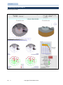
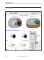
1
Manufactured by Metrovision under ISO9001:2008 ISO13485: 2003 certified quality system J USER'S MANUAL METROVISION - 4 rue des Platanes 59840 PERENCHIES FRANCE Tel: +33 3 20 17 19 50 [email protected] Version 03/02/2014 Fax: +33 3 20 17 19 51 http://www.metrovision.com © 2014 Metrovision GENERAL CONTENT GENERAL CONTENT ..................................................................................................................................................... 3 GENERAL INTRODUCTION ........................................................................................................................................... 5 EXAMINATION BASICS................................................................................................................................................. 7 CLINICAL INTEREST OF THE VISUAL FIELD EXAM ............................................................................................................. 8 APPLICATION TO VISUAL FUNCTION ASSESSMENTS (HANDICAPS, ..) .............................................................................................. 10 STATIC PERIMETRY ........................................................................................................................................................ 10 KINETIC PERIMETRY ...................................................................................................................................................... 15 MIXED PERIMETRY ............................................................................................................................................................. 15 MOTION PERIMETRY .......................................................................................................................................................... 16 BLUE/YELLOW PERIMETRY ................................................................................................................................................... 16 REPRESENTATION OF THE VISUAL FIELD................................................................................................................................... 17 WHICH EXAMS TO CHOOSE ....................................................................................................................................... 19 STAT PERIMETRY ........................................................................................................................................................... 21 FAST PERIMETRY ........................................................................................................................................................... 22 OTHER TESTS ................................................................................................................................................................. 23 BLUE/YELLOW PERIMETRY............................................................................................................................................ 24 MOTION PERIMETRY ..................................................................................................................................................... 25 KINETIC AND MIXED PERIMETRIES ................................................................................................................................ 26 REALIZATION OF AN EXAM........................................................................................................................................ 27 PATIENT'S INSTALLATION ..................................................................................................................................................... 28 REALIZATION OF STATIC PERIMETRY EXAMS ............................................................................................................................. 30 QUALITY CONTROL DURING THE EXAMINATION ........................................................................................................................ 31 REALIZATION OF KINETIC PERIMETRY EXAMS ............................................................................................................................ 32 EXPLOITATION OF RESULTS ....................................................................................................................................... 33 ACCESS TO RESULTS ...................................................................................................................................................... 34 QUALITY CONTROL........................................................................................................................................................ 34 DISPLAY OPTIONS ......................................................................................................................................................... 35 VISUALIZATION MODES ................................................................................................................................................ 36 ANALYSES ...................................................................................................................................................................... 37 GLOBAL ANALYSIS ......................................................................................................................................................... 37 FOLLOW-UP ANALYSIS......................................................................................................................................................... 39 ANALYSIS OF NEUROLOGICAL VISUAL FIELDS ................................................................................................... 41 3 / 94 Copyright © 2014 Metrovision Visual field ANALYSIS OF THE INCAPACITY INDEX ...................................................................................................................................... 42 VISION SIMULATION ........................................................................................................................................................... 42 SUPERIMPOSITION WITH THE EYE FUNDUS .............................................................................................................................. 43 ANALYSIS OF ZONES............................................................................................................................................................ 44 PATIENT'S INFORMATION .................................................................................................................................................... 45 STORING THE RESULTS ........................................................................................................................................................ 45 PRINTING THE RESULTS ....................................................................................................................................................... 45 TECHNICAL SPECIFICATIONS ...................................................................................................................................... 47 MONCV1 SPECIFICATIONS ................................................................................................................................................... 48 MONCV3 AND MONPACK SPECIFICATIONS ............................................................................................................................. 48 EQUIVALENCES OF GLOBAL INDICES ....................................................................................................................................... 48 CLINICAL EXAMPLES .................................................................................................................................................. 49 NORMAL VISUAL FIELD ........................................................................................................................................................ 50 EARLY GLAUCOMA (STAGE 1) ............................................................................................................................................... 52 ESTABLISHED GLAUCOMA (STAGE 2) ...................................................................................................................................... 54 ESTABLISHED GLAUCOMA (STADE 3) ...................................................................................................................................... 56 ADVANCED GLAUCOMA (STADE 4) ........................................................................................................................................ 58 ADVANCED GLAUCOMA - EVOLUTION FOLLOW UP.................................................................................................................... 60 ADVANCED GLAUCOMA (STADE 5) ........................................................................................................................................ 62 RETINITIS PIGMENTOSA ....................................................................................................................................................... 64 ATROPHIC MACULAR DEGENERATION ..................................................................................................................................... 66 HYDROXYCHLOROQUINE INTOXICATION .................................................................................................................................. 68 VENOUS OCCLUSION........................................................................................................................................................... 70 TOXOPLASMOSIS SCAR ........................................................................................................................................................ 72 TILTED DISK ...................................................................................................................................................................... 74 SEVERE MYOPIA................................................................................................................................................................. 76 PAPILLARY OEDEMA ........................................................................................................................................................... 78 LEBER OPTIC NEUROPATHY AT AN EARLY STAGE ........................................................................................................................ 80 COMPRESSIVE OPTIC NEUROPATHY ........................................................................................................................................ 82 PITUITARY ADENOMA ......................................................................................................................................................... 84 KERATOCONUS AND GLAUCOMA ........................................................................................................................................... 86 EDITION DE PROCEDURES ......................................................................................................................................... 89 BIBLIOGRAPHY .......................................................................................................................................................... 93 4 / 94 Copyright © 2014 Metrovision General introduction GENERAL INTRODUCTION This document describes the use of the Visual Field application available on the Vision Monitor system. Before reading this document, you should be familiarized with the general information related to the hardware and software of the Vision Monitor. This information is available in the following documents: DOCUMENT CONTENT The Vision Monitor – Introduction and general operation Recommendations for the installation of Vision Monitor Systems Installation of MonPackONE system Installation of MonCvONE system Installation of MonCv3 system 5 / 94 General functions of the Vision Monitor software Recommendations for the installation of equipment Installation and safety of the equipment Copyright © 2014 Metrovision Visual field 6 / 94 Copyright © 2014 Metrovision Examination basics EXAMINATION BASICS The visual field examination provides very useful information for the detection, diagnosis, follow-up of patients. However, time limitations of everyday clinical practice require an optimization of examination procedures. This document will first recall the basics of modern perimetry before presenting the different approaches available on the Vision Monitor system. 7 / 94 Copyright © 2014 Metrovision Visual field CLINICAL INTEREST OF THE VISUAL FIELD EXAM The island of vision The visual field is usually represented as an "island of vision" that corresponds to the domain of perception of light for a steady eye. In a normal subject, sensitivity to light is best at the fovea. It decreases regularly as a function of eccentricity until the absolute limits that are defined by the morphology of the head: 60 degrees on the nasal side, 95 degrees on the temporal side, 60 degrees superior, 70 degrees en inferior. There is one accident within these limits: the blind spot that is located 15 degrees away from fixation, on the temporal side. Detection, analysis and follow up of pathological alterations The interest of the visual field exam results from the anatomic organization of the visual system, particularly from the pathway of nerve fibers and from the vascularization. Paracentral scotoma in glaucoma Nerve fiber bundles and blood vessels of the retina - in the retina, nerve fibers follow well defined courses. A damage of the optic nerve head results in an alteration of the visual field following the corresponding nerve fibers, either under the form of a paracentral scotoma or an arcuate scotoma (Bjerrum). 8 / 94 Copyright © 2014 Metrovision Arcuate scotoma in glaucoma Principe des examens - concerning the vascularization of the retina and optic nerve head, an impairment results in a local alteration in correspondence with the visual field. - in visual pathways, quadranopsia = impairment of a visual field quadrant hemianopsia.= impairment of a visual field hemi field (right or left) homonymous defect = visual field defect found in the same location of the visual field of both eyes; it is characteristic of post chiasmatic lesions. The localization of damages located between the retina and the occipital cortex can be determined from the type of visual field alteration: Visual field, visual aptitudes and quality of life The visual field is a determining factor for the quality of life. The peripheral visual field allows detecting and avoiding obstacles when moving around or going down the stairs. The macular visual field is essential for reading, the recognition of faces and many other tasks of everyday life. Role of the central and pericentral visual fields in reading. Reading involves a sequence of eye fixations. During each fixation, a word is identified thanks to its image projected over the fovea (zone with green circle) while the text that follows is analyzed with the right pericentral field (zone with blue dotted line) to determine the position of the next fixation. In the case of "fast" reading, the number of fixations is reduced. They are made directly near the center of words and omitting "small" words such as articles. 9 / 94 Image of a text projected on the retina during reading As a consequence, it is important to be able to determine the repercussions of a visual field defect on the visual aptitudes and on the quality of life. Copyright © 2014 Metrovision Visual field Application to driving For Group 1 (ordinary motor cycles, private cars): the horizontal binocular visual field must not be less than 120° and must extend on, at least, 50° to the right and left, and 20° up and down. There must be no defect within a radius of 20° with regard to the central axis. As proposed by Esterman, there is a higher density of test points in the central field, along the horizontal memeridian and within the lower hemi-filed. The incapacity index is equal to the addition all unseen points. For Group 2 (vehicles over 3500 kg, professional vehicles, …): The horizontal binocular visual field must not be less than 160° and must extend of at least 70° to the left and right and 30° up and down. There must be no defect within a radius of 30° with regard to the central axis. Application to visual function assessments (handicaps, ..) The binocular visual field is estimated with test III/4 without dissociation of both eyes. Tests points are distributed according to the functional significance of the different visual field areas. STATIC PERIMETRY Several techniques are available for measuring the visual field. This paragraph presents the various techniques proposed on Metrovision's devices: static perimetry, kinetic perimetry, mixed perimetry and motion perimetry. Their applications, advantages and drawbacks are also described. Static perimetry consists in determining the smallest luminance level detected among a series of fixed dots. Inside the visual field limits, the sensitivity to lights depends on numerous factors: background luminance, stimulus luminance, size, color and the duration of the test. These parameters have been standardized to achieve reproducible measurements of light sensitivity. So, the Vision Monitor parameters are identical to those of the GOLDMANN perimeter: an ambient luminance of 10 cd/m2 (low photopic level that reduces the time for light adaptation, standardized test sizes (III = 26 arc minutes, V = 103 arc minutes), a white color. 10 / 94 Copyright © 2014 Metrovision Principe des examens Maps of measuring points Most perimetry devices use a map of points arranged according to a regular grid pattern for example every 6 degrees. The corresponding exams procedures are available on Metrovision devices under the names of STAT30, STAT24 and STAT10. Deficit of the inter-papillo-macular fibers in optic neuritis. This deficit is easily revealed with the FAST24 procedure but frequently not detected with a procedure such as STAT24 because of the absence of test points in the corresponding zone More efficient detection and follow-up due to the increase of test points density in "sensitive" zones Other maps are also proposed on Metrovsion devices with the following FAST examination procedures: FAST30, FAST24 and FAST12. Results obtained from a patient with glaucoma: procedure FAST30 shows clearly that the deficit is connected to the blind spot, which is a sign of the glaucoma severity whereas procedure STAT30 does not show this connection. These procedures use a layout of test points corresponding to the most frequent alterations of the retina and optic nerve fibers, hence the name FAST (Fiber Adapted Static Testing). Better evaluation of functional repercussions of alterations. For example, the presence of tests at 2 and 5 degrees eccentricity from the fovea in procedures FAST30 and FAST24 allows a best evaluation of the impairment gravity. These procedures have several advantages: 11 / 94 Reduction of diagnosis errors risk which may result from the lack of tests in some strategical zones These two results have been obtained on the same patient. The FAST30 procedure shows that the deficit is getting as close as 2 degrees from the fovea. This may explain an alteration of reading capacities. With the STAT30 “classic” procedure, the test closest to the fovea is at an eccentricity of 4.5 degrees and the graphic representation may be misleading. Copyright © 2014 Metrovision Visual field Strategies for the measurement of sensitivity thresholds Definition of sensitivity thresholds The curve below represents the response frequency of the patient according to the test luminance. If the luminance of the test is low, it is normally not seen except in the case of patient errors called false positive. If the test is very luminous, it is normally seen except in case of patient errors called false negative. When the luminance increases, the frequency of responses gradually increases. The sensitivity threshold corresponds to the luminance level for which 50 percent of responses are obtained. Sensitivity thresholds in a normal patient Many measurement strategies of sensitivity threshold have been developed to meet the needs of the standard clinic. Their aim is to detect and quantify the visual field alterations, these alterations or deficits being defined with regard to a population of "normal" subjects. The sensitivity threshold is denominated according to a scale graduated in decibels (dB). The highest sensitivity level (31 dB) corresponds to the lowest test luminance (0.2 cd/m²) and the lowest sensitivity (0 dB) to the highest test luminance (318 cd/m²). The scale is logarithmic: a 1 dB reduction in sensitivity corresponds to a test luminance multiplied by 1.25 a 3 dB reduction in sensitivity corresponds to a test luminance multiplied by 2 (1.25 x 1.25 x 1.25) ; a 10 dB reduction in sensitivity corresponds to a test luminance multiplied by 10 (1.25 x 1.25 … repeated 10 times) The deficit in a point of the visual field is the difference between the threshold value measured on the patient and this threshold average value in normal subjects. This deficit is significant only if its value is out of the normal limits, for example if less than 5% of normal subjects have a deficit superior or equal to the obtained value Therefore, it is important to know precisely the normal values of sensitivity thresholds as well as their interindividual variations. It is also usefull to control the different factors affecting the results so as to obtain the best possible discrimination. The graph below shows the normal limits of sensitivity thresholds along the horizontal section of the visual field (horizontal meridian). 12 / 94 Copyright © 2014 Metrovision Principe des examens If this global component is subtracted from the deficit, the interindividual variations of the normal population are significantly reduced as well as the variations in pathological subjects. Normal limits of sensitivity thresholds along the horizontal meridian In the normal population, there are important interindividual variations of approximatly 10 dB for the central field and increasing beyond 15 degrees of eccentricity. These variations are due to many factors: age (thresholds decrease beyond 45 years of age), the patient's response criteria (some patients want to be sure to see the test before answering), the level of tiredness, the ocular media transparency,... The age is an easily identifiable data and taking it into account allows reducing in a noticeable way the interindividual variations. The graph below shows these variations after taking into account age for a point of the central visual field. From these considerations, it follows that 2 different types of analysis can be made: an « analysis of deficits » in which the patient’s results are compared to the normal population of the same age and an « analysis of corrected deficits » in which the global component has been subtracted from the deficits. Normal limits of the sensitivity threshold as a function of age age for a point of the central visual field Supraliminal strategies Many measurement strategies have been developed to meet the needs of the current clinic. The supraliminal strategy is the fastest: tests are presented at a luminance level well above the normal population threshold. The test is recorded as normal if the patient answers and abnormal if he doesn't. Therefore, only one presentation is enough for each tested point. The supraliminal level (that is to say at how many dB above the normal threshold the presentation is done) is the object of a compromise. If it is too low, many abnormal points will be detected by mistake. If it is too high, the exam will only detect important alterations. In practice, a supraliminal level of 10 dB is used for a "rough" screening (research of severe deficits, patients who are not very cooperative,...). A supraliminal level of 6 dB is often used for the fast screening of deficits in the periphery (beyond 30 degrees) and 4 dB for the central field. Staircase strategies The "staircase" strategy consists in a first phase of rapid search with a 4 dB step by step progression starting from "not seen" to "seen" (1), (2), (3). These variations are brought down to 4 dB for young subjects but remain relatively high (8 dB) for subjects beyond 60 years of age. An important part of these variations affects the whole visual field. So, the deficit in one point includes a global component affecting the entire visual field which is not specific of a pathological process as well as a local component. 13 / 94 When a response is obtained, the progression is reversed and the step size decreased to 2 dB, until no response is obtained (4), (5). For strategy "4-2", the threshold value is estimated as the average of the test Copyright © 2014 Metrovision Visual field luminance when a change of progression is made (in this example, points (3) and (5). . With strategy "4-2-2", testing is continued until 3 changes of progression are made and with strategy "42-2-2", 4 changes of progression. This last strategy is used to determine the value of temporal fluctuations from the spread of luminance of points that correspond to a change of progression (in this example: (3), (5), (7) and (8)...). Speedy strategies Speedy strategies optimize the examination time to make it compatible with clinical constraints. The visual field is evaluated in several phases, allowing a significant reduction of the examination duration. phase 1 : determination of the global component of the deficit On the Vision Monitor, the determination of the global component of the deficit is performed at the beginning of the exam. This allows the early detection of technical errors such as patient misunderstanding, wrong optical correction, having a global influence on thresholds. The determination is performed by measuring the thresholds in 5 points located in the macular area. In the case of an abnormally low value, a message is displayed to inform the operator. He may then check an eventual technical error and, in case of an error, restart the measurement after correcting the cause. phase 2 : seed points It consists in a pre-evaluation of the visual field performed in 16 points judiciously chosen. Each of 14 / 94 these points is first tested to detect a deficit. If there is no response for the 2 first presentations, a 3rd presentation is made at maximum luminance. If no response is obtained, it is concluded that there is an absolute deficit. If a response is obtained, then additional presentations are made to determine the relative deficit. phase 3 : neighbor points The results obtained with seed points are used for the evaluation of the other points of the visual field. These points are initially tested at the same relative level than the nearest seed point. If no response is obtained, the luminance of the test is increased until the patient does respond. If a response is obtained, luminance is decreased until the patient no longer responds. Comparison of the static perimetry strategies A compromise must be found between the goal of the examination (detection, analysis and quantification of specific deficits) and clinical constraints such as the duration of the examination, the patient's fatigue, his or her response capabilities, his or her cooperation, etc... The duration of the examination depends on the response time of the patient and also on the volume and complexity of deficits. The speedy strategy is generally about 3 times faster than the staircase strategy. Speedy strategies are theoretically less precise because they perform a simplified measurement so as to reduce the exam duration. However, if the patient's decrease of reliability with the exam duration, is taken ointo account, the accuracy obtained is generally comparable. To sum up, staircase strategies are essentially recommended in case of a precise determination of thresholds in a reduced number of points (foveolar threshold,...). Copyright © 2014 Metrovision Principe des examens KINETIC PERIMETRY Kinetic perimetry principles Comparison perimetries In kinetic perimetry tests are presented with a constant luminance so as to determine the limits (or isopters) of zones which are seen at this level of luminance. Several isopters can be obtained by using different test luminances, tests with low luminance being seen, in normal subjects, only in the central part of the visual field. of static and kinetic Kinetic perimetry is much faster than static perimetry in the case of severe deficits in the periphery. In the previous example, only one kinetic test allows a quick estimation of the alteration whereas a great number of presentations are necessary in static perimetry to obtain the same result. The same tests can be used to define the limits of zones having a deficit, for example the blind spot. Isopters obtained with kinetic perimetry in a normal subject In the case of central field deficit, kinetic perimetry can hardly evaluate the depth of deficits. So the evaluation of the central field is rather better performed with static perimetry. Mixed perimetry Mixed perimetry combines the evaluation of the peripheral field with kinetic perimetry (an isopter) and the evalution of central field with static perimetry. It presents several specific characteristics: a fast evaluation of the whole visual field the determination of central deficits peripheral extent a significant saving of time in the case of severe deficit because zones of the central field found to present an absolute deficit with kinetic parimetry are only controlled with static. 15 / 94 Copyright © 2014 Metrovision Visual field Example of mixed perimetry in a glaucoma patient: the peripheral isopter determines quickly the presence of an extended absolute deficit and shows the extension of the central deficit to the periphery ("breakthrough"), which is a sign of severity. Note: Some pathologies result in different responses for static and kinetic stimulations (stato-kinetic dissociations CHARLIER & al, 1989). In those cases, it can be usefull to perform fully static and fully kinetic exams. Motion perimetry Motion perimetry uses movement as a stimulus. A grating of vertical bars is displayed on the stimulator screen. The local visual stimulus is obtained with the movement of one of the bars. It moves horizontally for a short moment and thereafter returns to its initial position. A measurement of the sensitivity threshold is obtained by changing the amplitude of the movement. The patient task which is to press a push button when he perceives a movement of the stimulus is not really different from the task of static perimetry. This technique has several advantages: Motion stimulation has increased sensitivity for the detection of early deficits in glaucoma (WU, 1998, WESTCOTT, 1999, BRUSINI & al, 2009). It is specifically affected by alterations of the magnocellular pathway (DUARTE & al, 2013) The results obtained are displayed and analyzed in the same way as classic static perimetry. Read the chapter "Clinical examples" for examples of clinical results. Motion stimulation is much less sensitive than classic perimetry to optical problems such as refractive errors or diffusion of light at the level of cornea or crystalline lens. Blue/Yellow perimetry Blue/Yellow perimetry aims at specifically testing the "S" cones system with blue stimulations while inhibiting other type of photoreceptors responses (rods and cones "M" and "L") with a yellow background. The potential interest is an early detection of patients with glaucoma, the "S" cones system being particularly sensitive to early alterations because of the "S" cones low density (SAMPLE & WEINREB, 1990). However, 16 / 94 many recent studies show that the results obtained with this exam present significant variations in the long term (HUTCHING & al, 2001) and do not seem to detect glaucoma earlier than classic perimetry (VAN DEN SCHOOT & al, 2010). Copyright © 2014 Metrovision Principe des examens Representation of the visual field How to find marks in the visual field A parallel is the set of locations with the same eccentricity. Geometric references are needed to identify the position of a deficit within the visual field. The macular visual field covers the central 20 degrees (eccentricity with respect to fixation less than 10 degrees). This region is the most sensitive to luminance contrast and to small spatial details. The central visual field covers the 40 central degrees (up to 20 degrees of eccentricity). The peripheral visual field covers the region over 20 degrees of eccentricity. Meridians provide another important reference. The horizontal meridian makes the separation between the superior and inferior visual fields; the vertical meridian between the nasal and temporal visual fields. Each meridian is characterized by its inclination angle with respect to the horizontal meridian. Each position within the visual field can easily be identified by its meridian and its parallel. Isopters and sensitivity profiles Traditional representations of the visual field are largely inspired from the techniques used by topographers: sensitivity profiles: they are similar to topographic cuts and represent cuts of the visual field along a given meridian. maps of isopters (Goldmann representation): isopters are similar to the level curves of topographers and connect points with equal sensitivity, 17 / 94 Copyright © 2014 Metrovision Visual field Sensitivity maps The Vision Monitor proposes a unique representation that combines the advantages of different representation modes by displaying on the same graph: the sensitivity value of each tested point, the map of sensitivity levels: the computer determines a "smooth" surface that fits the tested points. “pseudo isopters”: the map of sensivity levels is made with "true" colors which are very easily distinguished and their separations are similar to isopters curves. This type of representation makes easier the understanding of the deficits localization and of their topography. 18 / 94 The representation of the same result in 3 dimensions (3D map) makes the interpretation of the visual field topography easier. Copyright © 2014 Metrovision Which exams to choose WHICH EXAMS TO CHOOSE This chapter describes the various exam protocols available with the Vision Monitor. 19 / 94 Copyright © 2014 Metrovision Visual field The table below summarizes the various protocols available according to the device configuration: MonCvONE MonCv3, MonPackONE BASIC MonCv3, MonPackONE PRO Static perimetry adapted to fibers density FAST30, FAST24, FAST12, FAST-fovea FAST30, FAST24, FAST12, FAST-fovea FAST30, FAST24, FAST12, FAST-fovea Conventional static perimetry STAT30, STAT24, STAT12, STAT-fovea STAT30, STAT24, STAT12, STAT-fovea STAT30, STAT24, STAT12, STAT-fovea Test of driving aptitude Group 1 Visual aptitudes G1 Visual aptitudes G1 Visual aptitudes G1 Test of driving aptitude Group 2 Visual aptitudes G2 Visual aptitudes G2 Visual aptitudes G2 Expertise Incapacity index Incapacity index Incapacity index Low vision Low vision status Low vision status Low vision status Peripheral static test FAST-60 FAST-60 FAST-60 Blue / Yellow perimetry BY-30, BY-24, BY-12, BY-fovea Motion perimetry Motion-30, Motion-24, Motion-12 Kinetic perimetry 3 kinetic isopters + 1 isopter for the blind spot Mixed perimetry Mix-30, Mix-24, Mix-12 Note: Other protocols can be performed on request or thanks to ther program for the edition of procedures proposed with the EXPERT version (see chapter "Edition of procedures"). 20 / 94 Copyright © 2014 Metrovision Which exams to choose STAT PERIMETRY STAT perimetry corresponds to conventional automated perimetry. It makes use of a grid of testing points with a regular spacing of 6 degrees for procedures STAT-30 and STAT-24 and 2 degrees for STAT-10. Procedures Maps Clinical applications STAT-30 STAT perimetry up to 30 degrees of eccentricity. evaluation of deficits in glaucoma follow-up of glaucoma follow-up of chloroquine treatments, evaluation of maculopathies with a visual acuity better than 0.2. 9 points in staircase strategy and the rest in fast strategy STAT-24 STAT perimetry up to 24 degrees of eccentricity. 9 points in staircase strategy and the rest in fast strategy STAT-10 STAT perimetry covering the macular zone up to 10 degrees of eccentricity. Staircase strategy. STAT-fovea Measure of the foveolar threshold. Staircase strategy. 21 / 94 Copyright © 2014 Metrovision can be realized as a complement of previous tests. Visual field FAST PERIMETRY Procedures Maps Clinical applications FAST-30 FAST perimetry with 94 points over the central visual field. follow-up of all pathologies affecting the central visual field (glaucoma, optic neuritis, vascular diseases, macular and papillary oedema), evaluation of deficits within the macular, papillary and centro-caecal areas of the visual field. FAST-24 glaucoma follow-up. FAST-12 FAST perimetry over the macular area up to 12 degrees of eccentricity. follow-up of chloroquine treatments, evaluation of maculopathies with a visual acuity better than 0.2. FAST perimetry with 79 points over the central visual field. FAST-fovea Measure of the foveolar threshold with staircase strategy 22 / 94 Copyright © 2014 Metrovision can be realized as a complement of previous tests Which exams to choose OTHER TESTS Procedures Maps Clinical applications Visual aptitudes G1 Binocular exam designed to evaluate the ability to drive. evaluation of driving aptitudes group G1 evaluation of driving aptitudes group G2 evaluation of visual incapacity (Esterman score) evaluation of patients with a visual acuity below 0.2. evaluation of periphery in neuroophthalmic diseases. Can be used as a complement of previous FAST-30 or STAT-30 tests. NOTE: this test must be realized without optical correction to avoid masking the peripheral fied with spectacle frames. Supraliminal strategy Visual aptitudes G2 Binocular exam designed to evaluate the ability to drive. Supraliminal strategy Assessment Binocular exam designed to evaluate the visual incapacity. Supraliminal strategy Low vision status Visual field performed with a size V test. Supraliminal strategy Fast-60 44 points between 30 and 60 degrees of eccentricity. Supraliminal strategy 23 / 94 Copyright © 2014 Metrovision Visual field BLUE/YELLOW PERIMETRY (available only on models MonCv3, MonPack – PRO version) Procedures Maps Clinical applications BY-30 Blue over yellow perimetry over the central field. evaluation of deficits of central field. detection and follow up of glaucoma in young patients evaluation of maculopathies with a visual acuity better than 0.2. evaluation of deficits in young patients with glaucoma. Fast strategy. BY-24 Blue over yellow perimetry over the central field. Fast strategy BY-12 Blue over yellow perimetry over the macular area Fast strategy BY-fovea Measure of the foveolar threshold (can be realized as a complement of previous tests). Staircase strategy 24 / 94 Copyright © 2014 Metrovision Which exams to choose MOTION PERIMETRY (only available on models MonCV3 and MonPack, PRO version) Procedures Maps Clinical applications Motion-30 Motion perimetry covering the central field with 92points. fast detection of visual field alterations detection of glaucoma detection of alterations of the macula Fast strategy Motion-24 Motion perimetry covering the central field with 78points. Fast strategy Motion-12 Motion perimetry covering the macular area Fast strategy 25 / 94 Copyright © 2014 Metrovision Visual field KINETIC AND MIXED PERIMETRIES (only available on models MonCV1) Procedures Clinical applications Kinetic Complete evaluation of the visual field with a peripheral isopter, an intermediate isopter and a central isopter together with the evaluation of the blind spot limits. Mix-30 Combines the evaluation of the peripheral visual field with kinetic perimetry to the evaluation of the central field with FAST perimetry (94 points). evaluation of neuro-ophthalmic disorders. first exam of a new patient: global evaluation including a kinetic evaluation of the periphery, a static evaluation of the central field and, as an option, the foveolar threshold, follow-up of optic neuritis and glaucoma patients at an advanced stage, unexplained loss of visual acuity, neuro-ophthalmology patients, aphakia, pseudophakia and important ametropias. Mix-24 Combines the evaluation of the peripheral visual field with kinetic perimetry to the evaluation of the central field with FAST perimetry (79 points). first exam of a new patient: global evaluation including a kinetic evaluation of the periphery, a static evaluation of the central field and, as an option, the foveolar threshold, follow-up of optic neuritis and glaucoma patients at an advanced stage unexplained loss of visual acuity, neuro-ophthalmology patients, aphakia, pseudophakia and important ametropias. retinal pathologies (diabetes, retinitis pigmentosa, retinal detachment, venous occlusions,...) Mix-12 Combines with evaluation of the peripheral visual field with kinetic perimetry to the evaluation of the macular area with FAST perimetry. 26 / 94 evaluation of the macula with a rapid screening of the periphery. Copyright © 2014 Metrovision Realization of an exam REALIZATION OF AN EXAM 27 / 94 Copyright © 2014 Metrovision Visual field Patient's installation Once the visual field program (for white/white perimetry exams) or "PRO" visual field program (for Blue/Yellow and Motion perimetry) is started, the control window shows the list of examination protocols available on the instrument. At first, click on to access to the identification of the patient, his date of birth and examined eye Then click on the icon which corresponds to the selected examination protocol Note: if there are more than 16 different protocols available on your equipment, use the lift bar on the right side of the window to display the next protocols. Adjust the height of the seat and of the electric table (if available) to achieve the best possible comfort for the patient. Place an occluder over the non tested eye... It is very important to enter the date of birth following the requested format so that the program can calculate the age of the patient and determine the visual field reference map. Place the optical correction for near vision (33 cm). The Enter the sphere and cylinder for distance optimal refractive correction takes into account the vision and the program automatically fills the correction for distance vision (sphere and cylinder) with « Correction » with the value to be used during an addition depending on the age of the patient. the exam. Age (years) < 30 30-39 40-44 45-49 50-55 > 55 Addition to the fare vision correction 0 +0.50 +1.00 +1.50 +2.25 +3.00 The correction used during the exam can be computed automatically by clicking on button during the identification of the patient. 28 / 94 Notes Visual field exams performed in automated static perimetry use size III stimulations which are far less sensitive to refractive errors than stimuli with smaller sizes (0, I and II). Copyright © 2014 Metrovision Realization of an exam SLOAN (Vision Research, 1961) has demonstrated that a small refractive error does not alter significantly the sensitivity thresholds with size III stimuli. The correction for astigmatism is taken into account by adding half the value of the cylinder correction to the spherical correction. When the pupils are dilated with a mydriatic agent that paralyses the accommodation, refractive correction does no longer depend on age. In that case, select the option “dilated pupils”. If the examination protocol starts with the evaluation of the periphery, do not put the refractive correction during the evaluation of the periphery but only when the program starts the assessment of the central visual field (the program will pause at the appropriate time). The correction value given by the program corresponds to the nearest correction available in the set of large field lenses provided by Metrovision. However, it is possible to obtain the exact spherical correction putting in the configuration file the following lines: [REFRACTION] metrovision=false Installation on MonCvONE . After the identification of the patient and tested eye, the head rest moves automatically to position the eye at the center of the cupola. Use the navigation button 1 to place the eye in the center of the rectangle of the video monitor. Notes For monocular exams, the fixation point is positionned 15 degrees to the left for the right eye and 15 degrees to the rigth for the left eye. For binocular exams, the fixation ids located at the center. Installation on MonCv3 and MonPackONE Adjust the vertical position of the chin rest with command button 1 so that the examined eye is at the level of the eye marks 2. 29 / 94 On the video control, the tested eye should be within the rectangular control area. Copyright © 2014 Metrovision Visual field Realization of static perimetry exams Explanation to the patient The pupil diameter can also be measured by pressing Static tests are presented at maximum luminance in 4 different positions. button on the right side of the video window. Explain to the patient that he/she must fixate constantly the fixation target and press the button every time he/she perceives a small light anywhere in the periphery. If the fixation point is not well seen, increase its luminance by clicking on . The program automatically proceeds to the next phase when 3 patient’s responses are recorded or after the operator presses button . Measurement of the response time A series of tests is presented to allow the measurement of the patient's response time and adjust the time between 2 test presentations. This duration is displayed on the button and may be changed by clicking on that same button anytime during the examination. Fixation control / Measurement of the pupil diameter (option) If the option automated fixation control is available, initialize the fixation control by pressing button "INIT CtFIX". The program takes several measurements of the eye gaze orientation which will be used during the exam to detect fixation errors. A message is displayed in the video window to indicate the result of the initialization. If the initialization is successful, the responses of the patients will be validated only when fixation is correct. If the initialization is not successful, repeat the same command or use command "INIT CtMOV" which detects eye movements during the exam. 30 / 94 Identification of the limits of the blind spot The program continues with the determination of the blind spot position and limits. 16 positions situated at the presumed place of the blind spot are successively tested until obtaining two consecutive no-answers. Then the program determines the blind spot horizontal and vertical limits. Copyright © 2014 Metrovision Realization of an exam Determination of the individual correction of the reference map The reference map of the Vision Monitor is taking into account the patient's age. However, there are several other factors that may affect globally the threshold values: the opacity of ocular media, the pupil size, refraction, the patient's response criterion, etc... These factors result in a deficit affecting the whole central visual field. In order to determine this global component of the deficit, the program measures the sensitivity threshold in 5 points of the macular zone. In that case, you should search for the eventual cause of abnormality: the poor understanding or poor cooperation of the patient, the reduced transparency of the ocular media or very small pupil size, an incorrect optical correction, a central deficit of the visual field covering the position of the 5 test points. After having corrected the cause of error, repeat the measurement by clicking on . If the new measurement is in agreement with the previous one, click on to start the examination. If several trials have not been successful in achieving a reproducible result or if the visual field presents a central defect, click on to enter "0" from the keyboard. (it corresponds to the patient's age). If these measures are within the normal limits, the program will continue the exam. If it's not, the program will display a message indicating that the value is abnormal. Thereafter, click on to start the examination. It can be useful to impose a higher value than the one found by the program when dealing with a tired or poorly cooperating patient. Quality control during the examination The quality bar located below the visual field result provides information allowing a rapid assessment of the quality of the on-going exam. Items are displayed in green when the results quality is satisfactory and red when a problem occurs. The number of tests that have been validated with respect to the total number of tests that have been programmed. 31 / 94 Fixation quality is indicated by the number of fixations errors (1 in this example) relative to the total number of controls (19 in this example). Attention quality is regularly tested with “false” tests that are not seen by the patient. Responses to these tests are counted as attention losses (2 in this example) relative to the number of controls (22 in this example). Cases when the patient responds abnormally fast after the presentation of the stimulus are also counted as losses of attention. Copyright © 2014 Metrovision Visual field This item indicates the type of equipment used to perform the examination. In the present example, the stimulator was a cupola (c) with test sizes equivalent to Goldmann III. The color of the display indicates whether the stimulator was calibrated properly (GREEN color) or not (RED color). . Realization of kinetic perimetry exams Peripheral isopter Frequently, the first responses of the patient are not satisfactory because he or she has not properly understood the examination's principle. In that case, you may, after a few tests restart the isopter by clicking on button . In this mode, you can retest all the measurements of the isopter by clicking on one of the buttons , ... which are displayed nearby the different measurements. When you have completed all control measurements, If tests are not perceived because their luminance is too low, you may restart the isopter with a higher luminance (i.e. a lower sensitivity level) by clicking on . button Once the program has completed the evaluation of the isopter, it enters the mode "CONTROL MEASUREMENTS". click on button Smart ways: if you want to switch to the manual mode before the completion of the isopter, after clicking on button , click on to stop the examination, you can modify the velocity of the stimulus by clicking on button . . Control measurements (manual mode) The entire evaluation of the isopter is normally performed automatically by the program. However, in some circumstances, the user may want to perform additional controls about the validity of responses. For that purpose, click on button during the examination of the isopter. The program indicates that this command has been memorized by displaying a mark on the same button: . 32 / 94 . Copyright © 2014 Metrovision Realization of an exam EXPLOITATION OF RESULTS 33 / 94 Copyright © 2014 Metrovision Visual field ACCESS TO RESULTS The access to results is not allowed during the progress of exams as it may lead to interactions between the data in process of acquisition and previous exam data. However, it is possible to read the results from a second PC linked to the "exam" PC through the computer network, even during the realization of exams. This list appears like a book whose pages can be turned To access to exams results, click on button situated in the Vision Monitor The access to results menu is displayed on the screen: by clicking on buttons and .. To visualize one result of this list, just click on its icon. The bar located below the visual field result provides information allowing a rapid assessment of the quality of the exam. Items that are displayed in GREEN color indicate satisfying quality, those in RED color indicate an inadequate quality. To display an exam result, simply indicate the name of the patient in the Selection criteria and then click on button to get the results in alphabetical order or in chronological order. Then the program displays the list of results corresponding to the selection criteria. QUALITY CONTROL The number of validated measures should be equal to the number of measures initially programmed (101 in 34 / 94 the example hereby). This number is different only when the examination was not entirely completed. Copyright © 2014 Metrovision Exploitation of results The reliability of the patient’s fixation is indicated by the number of fixation losses (0 in this example) relative to the number of controls (13). The individual correction of the reference map is also an important parameter for the interpretation of results. Its value is displayed in red color if it does not fit with the age of the patient. The quality of the patient's attention is assessed regularly throughout the exam by the presentation of tests that cannot be seen by the patient. Responses to these tests are counted as "attention errors" (2 in this example) with respect to the number of controls (14). Cases when the patient's response occurs within an abnormally short delay after the stimulus presentation are also counted as attention errors. The last item provides the identification of the stimulation used for the examination. In the present example, the stimulus is equivalent to the size III Goldmann tests. This item is displayed in green color if the apparatus was correctly calibrated and in red color otherwise. DISPLAY OPTIONS The display options allow selecting the display that is most suited to your needs. The mixed display realizes a dilation of the central part of the visual field and a compression of the periphery. Graduations are therefore more densely packed in the periphery. The linear scale is constant over the entire visual field. Graduations are the same for the central and the peripheral visual field. Mixed The following example shows the same examination results with the different display options that are proposed. Linear scale x1 35 / 94 Copyright © 2014 Metrovision Visual field Linear scale x2 Linear scale x4 Linear scale x3 Linear scale x5 VISUALIZATION MODES Click on button to access the menu of the various visualization modes. The following example shows the same result with the different visualization modes which are available. 2D sensitivity map 36 / 94 Copyright © 2014 Metrovision Exploitation of results Smart way: Easy way: use the keyboard arrows "right" and "left" to change the angle of the meridian. The field orientation is indicated by the icon situated on the upper right of the image. 2D deficit map Sensitivity profile The profile is realized by cutting the visual field "mountain" along a meridian. Abscissas: the eccentricity of measures. Ordinates: the sensitivities of measures in dB. 3D sensitivity map ANALYSES Click on the button to obtain the menu of the various available analysis. Easy way: You can also use keyboard shortkeys: « T » for global analysis, « E » evolution follow up, « I » patient's information. GLOBAL ANALYSIS The global analysis provides a complete status of the visual field exam. These 3 windows can be displayed one by one by clicking on the Vision Monitor control bar. This analysis is displayed on the as 3 separate windows which are printed on a single page. 37 / 94 Copyright © 2014 Metrovision Visual field The map of deficit probability indicates the probability of deficits with respect to age-matched normal values. The map of corrected deficit probability indicates the probability of deficits with respect to normal values referenced to the individual base level of the patient. The 2D sensitivity map includes the values of the threshold measurements superimposed upon the grey scale map. This map highlights the visual field topography. The global analysis determines several global indices that characterize the visual field: The same data is represented in 3D to make easier the understanding of the topography. Deficit values are obtained by subtracting measures performed on the patient from normal age matched values. Corrected deficit values are obtained by subtracting measures performed on the patient from "normal" values referenced to the individual base level of the patient. 38 / 94 The average deficit is defined as the average visual loss that is computed over the whole set of measuring points with respect to the "normal" data from age-matched controls. WARNING ! An alteration is characterized by a deficit with a positive sign. Do not confuse with the sensitivity threshold deviation which has a negative sign in case of alteration. Copyright © 2014 Metrovision Exploitation of results The corrected average deficit provides the average sensitivity loss of the patient with respect to his or her own "normal" map (corrected by his own base level). The deficit variance characterizes the dispersion of deficits. Spatial fluctuation characterizes local threshold variations: it is the average of sensitivity difference between each measurement point and its neighbors. Temporal fluctuation characterizes short term threshold variations. It is calculated only for those points measured with a "staircase" strategy. example hereby). This histogram shows the number of tested points with a deficit larger than a given value. The area in green color represents the "normal" limits for an age-matched population. Note: The area in orange color that characterizes the patient takes into account the uncertainty of each measurement. The "true" curve of the patient should be in between the upper and lower limits of the orange area. The average response time provides an evaluation of the quality of patient's responses. The global analysis also determines a histogram of the distribution of deficits (area in orange color in the Follow-up analysis This analysis allows the evaluation of the evolution of the visual field over several consecutive exams. The analysis is performed by displaying the last examination result and then selecting the analysis of the visual field evolution. The program automatically searches on the hard disk all the exams performed with the same patient's name, with the same tested eye and the same birth date. The program displays: 39 / 94 Easy way: These different windows can be displayed one by one by clicking on the Vision Monitor control bar. the last exam performed the list of exams sorted in chronologic order, the evolution of global indices, the map with the velocity of sensitivity variation over the last exams. Copyright © 2014 Metrovision Visual field List of exams Map of rates of evolution The program automatically determines the rate of evolution of sensitivity threshold in each point of the visual field: The program displays the list of exams selected in chronological order. A checkbox is associated to each exam of the list. This checkbox can be used to invalidater / unvalidate the results taken into account in the analysis. zones for which a loss of sensitivity is observed are displayed in red color, zones for which sensitivity thresholds are improving are displayed in green color, if the evolution rate is statistically significant, the value of the rate of evolution is displayed in dB per unit of time, if the evolution is not statistically significant, the program displays a question mark. Notes The analysis is, by default, performed on the last 3 visual fields (or 2 if only 2 results are available). Evolution of global indices This graph represents the evolution in time of each of the global indices: the average deficit, the corrected average deficit, the spatial fluctuation, the temporal fluctuation, the average response time. The number of exams used for the analysis can be modified by pressing the right and left arrows of the keyboard after having selected the window with the map of evolution. Exams can also be invalidated by deselecting them in the list of examinations. Note The statistical validity criterion is based on the Student law with a confidence interval of 95 percent. 40 / 94 Copyright © 2014 Metrovision Exploitation of results Analysis of neurological visual fields The analysis of neurological visual fields program: displays both eyes visual fields, compares the deficits of visual field quadrants, compares the deficits of right and left hemi fields, compares the deficits of upper and lower hemi fields, compare the deficit of both eyes visual fields (congruence) For results of static exams The average deficit is determined for each quadrant and hemi field. Congruence is computed as the geometric mean of congruent deficits of both eyes: For example, Csg = SQUARE ROOT (DsgRE. DsgLE) with: Csg = congruence of the upper left quadrant, DsgRE = average deficit of the upper left quadrant of right eye. The value of congruence varies between 0% (no correspondence between the visual fields of both eyes) to 100% (perfect correspondence). Example of visual field in a pituary adenoma Congruent deficits have the same localization for both eyes, which is likely to indicate of post chiasmatic lesion. The analysis covers the results of both eyes, if they exist. DsgLE = average deficit of the upper left quadrant of left eye. Results are summarized in the following table, with 3 columns that correspond to the left eye, right eye and congruence and 3 lines that correspond to the quadrant analysis, right-left hemi field analysis and upper-lower hemi field analysis. For this, the program automatically searches on the data base the result of the exam performed on the other eye, with the same patient’s name and date of birth. The analysis is performed separately for results obtained with static or kinetic perimetry. For results of kinetic perimetry The program determines the surface area of each isopter in degrees 2. Congruence is determined as the geometrical mean of congruent surface areas of each eye: for example, Csg = SQUARE ROOT (SsgRE x SsgLE) with: 41 / 94 Copyright © 2014 Metrovision Visual field Csg = congruence of the left upper quadrant SsgRE surface area of the left upper quadrant of the right eye SsgLE surface area of the left upper quadrant of the left eye Results are summarized in the following table, with 3 columns that correspond to the left eye, right eye and congruence and 3 lines that correspond to the quadrant analysis, right-left hemi field analysis and upper-lower hemi field analysis. Analysis of the incapacity index This analysis provides an automated determination of the incapacity index from the visual field exam. In the example hereby, the incapacity index is of 46%. Vision simulation The purpose of this analysis is to simulate the influence of the patient’s alteration of vision. It allows explaining to the patient or his/her relatives the possible benefits from a treatment or a surgery or the risks involved in situations such as driving a car. It is only a simulation and is provided only as such. It does not take into account parameters such as eye movements or completion phenomena. The program proposes a choice of 8 photographs that can be selected with a single mouse click. 42 / 94 The program displays 2 windows. The first window shows the image as it is seen by a normal subject (zones which are not tested by the visual field procedure no not appear). The second window shows a simulation of what is seen by the patient, calculated from the result of his/her exam. Copyright © 2014 Metrovision Exploitation of results The printout of the vision simulation presents on one page the result from the visual field exam, the image as seen by a normal object and the simulation of the image seen by the patient. Superimposition with the eye fundus This analysis allows the superposition of visual field map on the image of the patient's eye fundus or OCT image. The superposition method is that proposed by Pr BEK (BEK, 1990). When the analysis is started, a new window entitled "SUPERIMPOSE IMAGE OF EYE FUNDUS" is displayed with a map of the values of measured points and isopters (equal response lines). Note: This map is reversed "upside – down" with respect to the visual field map so that in can be superimposed over the image of the eye fundus. Note: Use the button in the control bar of the Vision Monitor program to import a photograph in the results database. The photograph can be imported either through a computer network or from a storage media such as a USB key, a CDROM, etc… The image of the eye fundus should then appear in the superposition window. The next step is to acquire the digital image of the eye fundus. Click on the button and a menu will be displayed to allow the selection of the image. 43 / 94 The next step is to define the position of the fovea and the papilla that are used as references for a precise superposition of the visual field map. Copyright © 2014 Metrovision Visual field In order to better identify these features, the red component of the image can be eliminated by clicking on button . Corrections: - the position of the eye fundus image can be finely adjusted with the arrow keys of the keyboard "up", "down", "right" and "left", - the magnification of the eye fundus image can be finely adjusted by pressing simultaneously the "SHIFT" key and the arrow keys of the keyboard "up", "down", "right" and "left", - the image can also be rotated by pressing the keys "page up" and "page down". Axis: Click on button and then click on the corresponding point of the image. Axis and parallels (every 5 degrees of eccentricity) can be added with a simple click on button Perform the same operation with button and the corresponding point of the image. The program automatically adjusts the image and displays the result of the superposition. An additional click on button allows the restoration of the red component of the image. Printing the results, exporting the results to other applications: To print the final result, click on. To save the result image click on button or on bouton copy in the clipboard. Analysis of zones First step: click on the different test points to be selected for each zone. Click on button to validate and move to the next zone. When the 5 zones have been defined, the program proposes to save their definition as a file for future use. Click on button previous definition of zones 44 / 94 . to recover the Copyright © 2014 Metrovision to make a Exploitation of results When the selection of zones is completed, the program displays an histogram with the average deficit for each of the selected zones as well as the gray density map where the selected zones can be identified from the color of the tested points. The result can be printed by clicking on “PRINT” then “print analysis of zones” Patient's information This command gives access to the patient's information. It can be useful for adding comments before recording or printing the result. Storing the results Click on button to store the results on the hard disk. The reference number of the record appears on top of the screen. The crossed sign on the button indicates that the result has been stored. Printing the results The print command allows to print the visual field result and the different analysis. Easy way: You can also print the results and the different analysis directly from the results' menu. 45 / 94 Copyright © 2014 Metrovision Visual field 46 / 94 Copyright © 2014 Metrovision Technical specifications TECHNICAL SPECIFICATIONS 47 / 94 Copyright © 2014 Metrovision Visual field MonCvONE specifications Radius (cm) Background luminance (cd/m2) Limits of the tested visual field (degrees) Size of tests 30 10 U=60 N=70 T=100 D=70 Equivalent Goldmann I, II, III, IV, V Duration of stimulus (msec) 300 MonCv3 and MonPackONE specifications Eye - Screen distance (cm) Background luminance (cd/m2) Limits of visual field (degrees) (avec déplacement du point de fixation) 30 10 U=60 N=60 T=75 D=60 Size of tests Equivalent Goldmann I, II, III, IV, V Duration of stimulus (msec) 300 Equivalences of global indices VISION MONITOR Average deficit Corrected average deficit Variance of deficits Spatial fluctuation Temporal fluctuation Average response time 48 / 94 OCTOPUS HUMPHREY + MD = mean defect - MD = mean deviation Not available - PD = pattern deviation (average) LV = loss variance CLV = corrected loss variance PSD = pattern standard deviation (= variance square root) CPSD = corrected pattern standard deviation SF = short term fluctuation SF = short term fluctuation Not available Not available Copyright © 2014 Metrovision Clinical examples CLINICAL EXAMPLES Acknowledgements to: Dr Defoort-Dhellemmes (Lille), Pr Hache (Lille), Dr Zanlonghi (Nantes). 49 / 94 Copyright © 2014 Metrovision Visual field Normal visual field Reference normal001 50 / 94 Copyright © 2014 Metrovision Clinical examples 43 years old emmetropic patient, Left eye examined with FAST30 procedure and a +1.25 optical correction. The quality of the exam is excellent: no attention and fixation losses. These indicators are all green. The fovea has a normal threshold and the blind spot is well localized. No diffuse alteration: the average deficit and the corrected average deficit are identical. Absence of localized deficit: it is highlighted by the topography and the corrected average deficit which is nil. 51 / 94 Copyright © 2014 Metrovision Visual field Early glaucoma (stage 1) Reference glaucoma003 52 / 94 Copyright © 2014 Metrovision Clinical examples 50 years old patient with -3D myopia Examination of the right eye performed with the FAST24 procedure and without optical correction. The eye fundus shows a localized loss of fibers. Quite good quality exam: Only one loss of fixation and two losses of attention on 12 controls. The foveolar threshold is normal and the size of the blind spot is within normal limits (defined by a vertical ellipse on the plot) Alterations of the visual field: No diffuse alteration. Relative fascicular alteration in the upper field in relation with the alteration of the eye fundus. The scotoma does not approach less than 5 degrees from the fovea and it is not linked to the papilla. Global indices are not yet altered in a significant way. 53 / 94 Copyright © 2014 Metrovision Visual field Established glaucoma (stage 2) Reference glaucoma006 54 / 94 Copyright © 2014 Metrovision Clinical examples 55 years old patient with -6 D myopia. Right eye examined with the FAST24 procedure and a -3D optical correction. The quality of the exam is good: Almost no fixation and attention losses. The foveolar threshold is normal. Large blind spot related to the myopia. Analysis of visual field alterations: No diffuse alteration. Wide upper deficit linked to the blind spot and approaching at 2 degrees from the fovea. The visual field is free from any deficit. Global indices are significantly altered. 55 / 94 Copyright © 2014 Metrovision Visual field Established glaucoma (stade 3) Reference glaucoma013 56 / 94 Copyright © 2014 Metrovision Clinical examples 58 years old patient with -3D myopia. Left eye examined with the FAST24 procedure without optical correction. Good quality exam: Low number of fixation and attention losses. Normal foveolar threshold. Analysis of deficits: Absolute deficit approaching at 5 degrees from the fixation point and linked to the blind spot. Nasal step. These deficits are in correspondence with the eye fundus alteration (neuroretinal rim at 3h and 5h). 57 / 94 Copyright © 2014 Metrovision Visual field Advanced glaucoma (stade 4) Reference glaucoma007 58 / 94 Copyright © 2014 Metrovision Clinical examples 77 years old emmetropic patient. Left eye examined with the FAST24 procedure and a +3D optical correction. Good quality exam: Low number of fixation and attention losses. Normal foveolar threshold. Analysis of deficits: No diffuse alteration. Absolute annular deficit in the lower visual field area and relative in the upper area, linked to the blind spot. Nasal step. The deficit approaches less than 5 degrees from the fixation point. 59 / 94 Copyright © 2014 Metrovision Visual field Advanced glaucoma - evolution follow up 60 / 94 Copyright © 2014 Metrovision Clinical examples 69 years old patient followed for 24 years for glaucoma. The follow up of the visual field evolution has been realized with the last 16 exams. The evolution of global indices shows an increase of the average deficit and of the corrected average deficit (after subtracting the global component). The evolution velocity map has been calculated with the last 4 exams and shows zones deteriorating over time in red color. In that case it shows that the progression is done over the pericentral points, which is characteristic of glaucoma and eliminates the possibility of a visual field evolution due to cataract or ARMD. 61 / 94 Copyright © 2014 Metrovision Visual field Advanced glaucoma (stade 5) Reference glaucoma003 62 / 94 Copyright © 2014 Metrovision Clinical examples 50 years old patient with -2D myopia Left eye examined with the MIX24 procedure without optical correction. Good quality exam: Only one fixation loss on 19 tests and no attention loss. The flattening on the upper nasal is not due to the optical correction (it also appears on the peripheral isopter which is realized without optical correction). According to the orthoptist, it is not due either to a ptosis. Analysis of the visual field alterations: No diffuse deficit. The foveolar threshold is slightly altered. Ring deficit largely absolute. A 10 degrees diameter central island of vision remains with a peripheral crescent on the temporal side. 63 / 94 Copyright © 2014 Metrovision Visual field Retinitis pigmentosa Reference rp001 64 / 94 Copyright © 2014 Metrovision Clinical examples 21 years old emmetropic patient. Right eye examined with the MIX24 procedure without optical correction. Good quality exam: There are no attention or fixation losses. The foveolar threshold is normal. Analysis of the visual field alterations: No diffuse alteration. Alteration of the pericentral static field with a preservation of the peripheral isopter realized in kinetic perimetry. 65 / 94 Copyright © 2014 Metrovision Visual field Atrophic macular degeneration Reference mac001 66 / 94 Copyright © 2014 Metrovision Clinical examples 79 years old emmetropic patient. Right eye examined with the FAST30 procedure and a +3D optical correction. Good quality exam: There are no attention losses. The numerous fixation losses (10 on 17 controls) can be explained by the presence of a central scotoma and the fixation on the inferior edge of this scotoma. The presence of a well delimited blind spot shows a good stability of the fixation. Analysis of the visual field deficits: There is no diffuse deficit. Purely central deficit with steep slopes indicating a process without fast evolution. 67 / 94 Copyright © 2014 Metrovision Visual field Hydroxychloroquine intoxication Reference aps001 68 / 94 Copyright © 2014 Metrovision Clinical examples 57 years old emmetropic patient. Left eye tested with the FAST24 procedure and a +3D optical correction. Good quality exam: There are no fixation and attention losses. Analysis of the visual field alterations: No diffuse deficit. Relative perifoveolar alteration (between 5 and 7 degrees of eccentricity). Global indices remain normal. 69 / 94 Copyright © 2014 Metrovision Visual field Venous occlusion Reference venousocclu001 70 / 94 Copyright © 2014 Metrovision Clinical examples 58 years old emmetropic patient. Right eye examined with the MIX24 procedure and a +3D optical correction. Good quality exam: There is no fixation loss and only 2 attention losses over a total of 19 controls. Analysis of the visual field alterations: No diffuse deficit. The alteration of the visual field is in correspondence with the bleedings visible on the eye fundus. The foveolar threshold is altered (20 dB sensitivity) because of the presence of a macular oedema. 71 / 94 Copyright © 2014 Metrovision Visual field Toxoplasmosis scar Reference toxo001 72 / 94 Copyright © 2014 Metrovision Clinical examples 58 years old emmetropic patient. Left eye examined with the FAST24 procedure and a +3D optical correction. Good quality exam: No fixation or attention losses. Analysis of the visual field deficits: No diffuse deficit. Absolute deficit approaching at 2 degrees from the fixation point. The perfect superimposition of the eye fundus alteration and the visual field deficit (below) shows the absence of other pathologies. 73 / 94 Copyright © 2014 Metrovision Visual field Tilted disk Reference tdisk001 74 / 94 Copyright © 2014 Metrovision Clinical examples 54 years old emmetropic patient. Right eye examined with the FAST24 procedure and a +3D optical correction. Good reliability of the exam: No attention or fixation losses. The foveolar threshold is within the normal limits. Analysis of the visual field deficits: Absence of diffuse deficit. Important relative deficit of the temporal hemifield. The fact that it is relative is in favor of an alteration which is not of a neurological origin. 75 / 94 Copyright © 2014 Metrovision Visual field Severe myopia Reference myopia001 76 / 94 Copyright © 2014 Metrovision Clinical examples 50 years old patient, high myopia (-14D) Right eye examined with the procedure MIX24 and a -12D optical correction. Excellent quality of exam: No fixation loss and only one attention lost for 20 controls. Analysis of the visual field deficits: Absence of diffuse deficit. The atrophy visible on the inferior eye fundus corresponds to a relative scotoma on the upper visual field. There is no sign of glaucoma because the visual field deficit does exactly superimpose over the eye fundus alteration. 77 / 94 Copyright © 2014 Metrovision Visual field Papillary oedema Reference papillaryoed001 78 / 94 Copyright © 2014 Metrovision Clinical examples 13 years old emmetropic patient. Right eye examined with the FAST24 procedure and without optical correction. Good quality exam: Low number of attention and fixation losses. Analysis of the visual field deficits: No diffuse deficit. Local deficit approaching at 2 degrees from the fixation point. 79 / 94 Copyright © 2014 Metrovision Visual field Leber optic neuropathy at an early stage Reference opticneuro001 80 / 94 Copyright © 2014 Metrovision Clinical examples 30 years old emmetropic patient. Reduction of VA on both eyes 1 month ago. Right eye examined with the MIX24 procedure without optical correction. Exam of average quality 20% attention and fixation losses Analysis of visual field alterations No diffuse deficit. Centro-caecal scotoma with alteration of the foveolar threshold. The eye fundus does not show any optical atrophy. Very discrete papillary hyperemia at the limit of significance. Visual evoked potentials 60’ pattern reversal: delayed P100 (125 ms) 15’ pattern reversal: reduced amplitude and normal implicit time Source : ZANLONGHI X. Electrophysiologie visuelle, examens fonctionnels et neuropathie optique génétique. Réflexions ophtalmologiques. 2010,15, 132, 20-27 81 / 94 Copyright © 2014 Metrovision Visual field Compressive optic neuropathy Reference opticneuro002 82 / 94 Copyright © 2014 Metrovision Clinical examples 44 years old emmetropic patient. Complains for the last few months about a partial loss of words with his right eye when reading. His visual acuity is 20/20 OD and OS. Right eye examined with the MIX24 procedure. Good quality exam: No fixation or attention losses. Analysis of the visual field deficits: Diffuse alteration of 4 dB, centro-cecal scotoma, inferior fascicular deficit On the eye fundus, global papillary palor in the lower part Source : ZANLONGHI X. Electrophysiologie visuelle, examens fonctionnels et neuropathie optique génétique. Réflexions ophtalmologiques. 2010,15, 132, 20-27. 83 / 94 Copyright © 2014 Metrovision Visual field Pituitary adenoma Reference piturayad001 84 / 94 Copyright © 2014 Metrovision Clinical examples 34 years old emmetropic patient. Exam realized with the FAST24 procedure, without optical correction. The neurological analysis allows the direct comparison of the visual fields of both eyes. It reveals the bi-temporal deficit. 85 / 94 Copyright © 2014 Metrovision Visual field Keratoconus and glaucoma Reference kera001 86 / 94 Copyright © 2014 Metrovision Clinical examples 55 years old patient Examination of the left eyerealized with the MOTION-24 procedure (motion perimetry). The exam could not be realized with classical contrast perimetry due to the presence of a keratoconus. Average quality exam: 3 fixation losses for a total of 15 controls. Analysis of visual field deficits: Severe absolute arcuate scotoma connecting with the blind spot and approaching at 2 degrees from the fixation point. 87 / 94 Copyright © 2014 Metrovision Visual field 88 / 94 Copyright © 2014 Metrovision Edition de procédures EDITION DE PROCEDURES This program allows permanent modifications of the examination tests. The use of the editing program requires advance training. The use of this program without appropriate training is not recommended as it may produce alterations of examination procedures and erroneous results. 89 / 94 Copyright © 2014 Metrovision Visual field 1- in the main menu, click on yellow icon for access to preferences a small window opens with the message “Enter password for procedures” 2- enter the password and click on the button “OK” this will validate the access to procedures 3- click on the icon for Visual Field exams 4- click on the button “edition of procedures” 5- the menu for editing procedures opens: what we will do next is read a test procedure, modify it and then save it with a new name click on the button “read procedure” to read 6- the menu of procedures is organized in pages you first get the list of pages which are available click on the button corresponding to the page of the menu you want to select. 7- now click on the icon of the procedure that you want to work on 8- you get a new page with the list of parameters of that procedure If you change one parameter of the stimulation, you will see the result on the stimulation unit. When you have made the changes which are requested, click on the button “VALIDATION” on the bottom right of the page. You can also use the button “COPY” to make a copy of the page in the clipboard or the button “PRINT” to print the same page. The « EXPORT » button allows the exportation of the table of stimulation parameters as a file named table.txt. This file defines the position (X,Y) of the different test points. It can be modified with a text editor or a spreadsheet application. Once it has been modified, it can be re-imported with button « IMPORT ». 90 / 94 Copyright © 2014 Metrovision Edition de procédures 9- Now you have to select the page of the menu where you will save the new procedure. Click on the corresponding button. 10- The program lists the tests available in that page and opens a small window to allow you to enter the name of the test. If you want to create a new test, enter a new name (limited to 8 characters, capital letters, without ‘_’ character). After entering the name, click on the OK button. 11- After saving the new procedure, the program returns to the editing menu. From there you can repeat the procedure to create/modify another procedure or you can go back to the examination menu by clicking on “EXAM MENU” . 91 / 94 Copyright © 2014 Metrovision Visual field 92 / 94 Copyright © 2014 Metrovision BibliographY BIBLIOGRAPHY BEK T. Accurate superimposition of visual field data onto fundus photographs, Acta Ophthalmol. 1990,68,11-18. BEDWELL C.H. Visual fields. A basis for efficient investigation. Butterworths. 1982. BRUSINI P. & al. Motion perimetry in glaucoma. Communication at SOI, 2009. CHARLIER J., DEFOORT S., ROULAND J.F., HACHE J.C. Comparison of automated kinetic and static visual fields in neuro-ophthalmology patients. PERIMETRY Update 1988/1989. HEIJL A. editor. KUGLER and GHEDINI publisher. 1989,3-8. CHARLIER J., MOUSSU, HACHE J.C. Optimization of computer assisted perimetry. Doc. Ophthal. Proc. Series, 1983, 35, 359-364. CHARLIER J., SACHY J., VERNIER F., HACHE J.C. Dynamic representation of the visual field. Doc. Ophthalmol. Proc. Series., 1987, 49, 263-270. DRANCE S.M., ANDERSON D. Automatic perimetry in glaucoma. A practical guide. Grune and Stratton Inc. 1984. DUARTE I.C., CUNHA G., CASTELHANO J., SALES F., REIS A., CASTELO-BRANCO M. Developmental dissociation of visual dorsal stream parvo and magnocellular representations and the functional impact of negative retinotopic BOLD responses. Brain and cognition. 2013, 83, 72-79. DUBOIS-POULSENS A. Le champ visuel Masson et Cie, Paris, 1952. GROCHOWICKI M., VIGHETTO A., BERQUET S., KHALFALLAH Y., SASSOLAS G. Pituitary adenomas: automatic static perimetry and Goldmann perimetry. A comparative study of 345 visual field charts. British J Ophthalmol. 1991,75,219-221. GROCHOWICKI M., VIGHETTO A., BERQUET S., KHALFALLAH Y., SASSOLAS G. Pituitary adenomas: assessment of modern means of neuro-ophthalmological subjective investigation. Neuro-ophthalmology. 1991,2,1,19-24. GROCHOWICKI M., VIGHETTO A., PISSAVIN C. Pseudotumor cerebri. Longitudinal study using contrast sensitivity and automated static perimetry. Neuro Ophthalmology. 1990, 10,2,97-108. HUTCHING N., HOSKING S.L., WILD J.M., FLANAGAN J.G. Long-term fluctuation in short-wavelength automated perimetry in glaucoma suspects and glaucoma patients. Invest. Ophthalmol. Vis. Sci. , 2011, 42, 10, 2332-2337 ISO 12866 Norme internationale sur les Instruments ophtalmiques, Périmètres, 2009. SAMPLE P.A., WEINREB R.N. Color perimetry for assessment of primary open-angle glaucoma. Ophthalmol. Vis Sci. 1990, 31, 9, 1869-1875. Invest. SLOAN L.L. Area and luminance of test object as variables in examination of the visual field by projection perimetry. Vision Research, 1961, 1,1-2, 121-138. TROBE J.D., GLASER J.S. The visual fields manual. A practical guide to testing and interpretation. Triad Publishing Co 1983. 93 / 94 Copyright © 2014 Metrovision Visual field VAN DER SCHOOT J., REUS N.J., COLEN T.P., LEMIJ H.G. The ability of short-wavelength perimetry to predict conversion to glaucoma. Ophthalmology. 2010, 117, 1, 30-34. VERNIER F., CHARLIER J., NGUYEN D. Application of CRT technology to the clinical evaluation of visual functions. Optical Engineering, 1988, 27, 123-128. VERRIEST G., VAN CASTEELE J. Le champ visuel clinique Acta belgica de arte medicinali et pharmaceutica militari 18 : 35 - 205, 1972. VIGHETTO A., GROCHOWICKI M., AIMARD G. Altitudinal hemianopia in multiple sclerosis. Neuro-ophthalmology. 1991,11,1,25-27. WALSH T.J. Visual fields. Examination and interpretation. Ophthalmology Monographs. American Academy of Ophthalmology. 1990. WESTCOTT M.C., FITZKE F.W., CRABB D.P., HITCHING R.A. Characteristics of frequency-of-seeing curves for a motion stimulus in glaucoma eyes, glaucoma suspect eyes, and normal eyes Original Vision Research, 1999, 39, 3, 631-639. WORKING GROUP 39 First interprofessional standards for visual field testing. Adv. Ophthalmol. 1980,40,173-224. WU J., COFFREY M., REIDY A., WORMALD R. Impaired motion sensitivity as a predictor of subsequent field loss in glaucoma suspects: the Roscommon Glaucoma Study. British J Ophthalmol; 1998, 82, 534-537. 94 / 94 Copyright © 2014 Metrovision